Embed Size (px)
Citation preview
© 2010 Carmona-Ribeiro, publisher and licensee Dove Medical Press Ltd. This is an Open Access article which permits unrestricted noncommercial use, provided the original work is properly cited.
International Journal of Nanomedicine 2010:5 249–259
International Journal of Nanomedicine
249
R e v I e w
Dovepressopen access to scientific and medical research
Open Access Full Text Article
submit your manuscript | www.dovepress.com
Dovepress
Biomimetic nanoparticles: preparation, characterization and biomedical applications
Ana Maria Carmona-Ribeiro
Biocolloids Lab, Departamento de Bioquímica, Instituto de Química, Universidade de São Paulo, São Paulo, Brazil
Correspondence: Ana Maria Carmona-Ribeiro Biocolloids Lab, Departamento de Bioquímica, Instituto de Química, Universidade de São Paulo, Caixa Postal 26077, 05513-970 São Paulo SP, Brazil Tel +55 11 3091 2164 Fax +55 11 3815 5579 email [email protected]
Abstract: Mimicking nature is a powerful approach for developing novel lipid-based devices
for drug and vaccine delivery. In this review, biomimetic assemblies based on natural or
synthetic lipids by themselves or associated to silica, latex or drug particles will be discussed. In
water, self-assembly of lipid molecules into supramolecular structures is fairly well understood.
However, their self-assembly on a solid surface or at an interface remains poorly understood. In
certain cases, hydrophobic drug granules can be dispersed in aqueous solution via lipid adsorption
surrounding the drug particles as nanocapsules. In other instances, hydrophobic drug molecules
attach as monomers to borders of lipid bilayer fragments providing drug formulations that are
effective in vivo at low drug-to-lipid-molar ratio. Cationic biomimetic particles offer suitable
interfacial environment for adsorption, presentation and targeting of biomolecules in vivo.
Thereby antigens can effectively be presented by tailored biomimetic particles for development
of vaccines over a range of defined and controllable particle sizes. Biomolecular recognition
between receptor and ligand can be reconstituted by means of receptor immobilization into
supported lipidic bilayers allowing isolation and characterization of signal transduction steps.
Keywords: cationic lipid, phospholipids, bilayer fragments, vesicles, silica, polymeric particles,
antigens, novel cationic immunoadjuvants, drugs
IntroductionParticles are important in pharmaceutical and biomedical research since their size
scale can be similar to that of proteins or DNA. They can mimic structural and func-
tional aspects of viruses, bacteria and other biological assemblies and are currently
being used in: imaging;1 biosensing;2 gene and drug delivery;3 and vaccines.4,5 On the
other hand, cationic lipids can be combined with negatively charged biomolecules or
biological structures. Silica,6 latex,7 or hydrophobic drug particles8 have been coated
with cationic lipids and characterized by means of adsorption isotherms, mean particle
size from dynamic light scattering, surface potential analysis and colloidal stability.
Properties of the intervening medium such as pH and ionic strength were system-
atically varied for achieving optimal lipid bilayer deposition and colloid stability.6,9
Polystyrene sulfate (PSS) nanoparticles covered with a dioctadecyldimethylammonium
bromide (DODAB) bilayer10 were used for adsorption of serum proteins,11 cholera
toxin,11 Taenia crassiceps antigens,12,13 recombinant proteins,13,14 polysaccharides15–17
and giant DNA.18 Similarly, lipid assemblies such as liposomes, vesicles, solid lipid
nanoparticles and lipidic bilayer fragments (BF) or disks are as versatile as particles.
Alec Bangham produced the first liposomes in 1965 in Cambridge UK.19 The liposome
“membrane” model prepared from phospholipids has lead to much of our present
International Journal of Nanomedicine 2010:5250
Carmona-Ribeiro Dovepress
submit your manuscript | www.dovepress.com
Dovepress
knowledge on membrane properties. In addition, a variety
of bilayer structures formed by dialkyldimethylammonium
halides20 and other synthetic amphiphiles21–24 were introduced
to mimic membrane properties furnishing unique opportuni-
ties to investigate structure-function relationships. Since the
major requirement to form a supramolecular assembly of the
bilayer type was an approximately cylindrical amphiphilic
molecule with a geometric parameter between 0.5 and 1.0,25
not only natural phospholipids were prone to form bilayers.
Structural and functional aspects of biological membranes
were also copied in a variety of biomimetic systems. Bilayers
were the preferential supramolecular assembly for several
synthetic amphiphiles as dialkyldimethylammonium bromide
or chloride,20 sodium dihexadecylphosphate22,26,27 and many
other molecules.28,29 However, lipid BF or disks remained as
a much less explored bilayer assembly. Lately, our group has
been developing novel formulations for hydrophobic drugs or
vaccines based on these structures. Amphotericin B solubi-
lized at the rim of DODAB BF provided a novel formulation
with excellent activity against systemic candidiasis in mice
at low drug dose30,31 with low nephrotoxicity.32 BF have also
been useful to cover particles such as silica, latex or insoluble
drug particles with lipids.33,34 This mini-review aims at an
overview on preparation, characterization and biomedical
applications for lipid-based biomimetic particles.
Lipid BF or disksUpon probe sonication of aqueous egg yolk lecithin disper-
sions, the high kinetic energy given by the sonicator to the
lipid particles, caused their breaking down on colliding
with each other.35 The collisions completely disrupted the
multilamellar particles into short-lived bilayer fragments,
which then reaggregated to form single-shelled vesicles of
roughly uniform size.35 From this early study up to now, discs
have been reported for a variety of amphiphiles and lipids in
aqueous dispersions over a broad range of experimental con-
ditions.10,36 They were found from self-assembly of lipids and
proteins,37–39 lipids and micelle-forming compounds40,41 lipids
and hydrophilic polymers such as polyethylene-glycol lipids
(PEGylated lipids)42,43 or charged lipids dispersed by probe
sonication.27,44–46 The major repulsive interactions preventing
fusion of lipid BF and discs in dispersion were electrostatic,
steric and/or eletrosteric. In particular, probe sonication of the
synthetic and cationic lipid dioctadecyldimethylammonium
bromide (DODAB) can yield disrupted vesicles: the bilayer
fragments, BF, or disks.10 The existence of BF in aqueous
dispersions of sodium dihexadecylphosphate, or dioctadecy-
ldimethylammonium bromide or chloride obtained by probe
sonication has been supported by the following evidences:
(i) osmotic non-responsiveness of the dispersion indicative
of absence of inner vesicle compartment;47 (ii) transmission
electron microscopy (TEM) micrographs with electronic
staining;27 (iii) cryo-TEM micrographs;45 (iv) fluid and solid
state coexistence and complex formation with oppositely
charged surfactant;48 (v) solubilization of hydrophobic drugs
at the borders of DODAB bilayer fragments, which does not
occur for DODAB closed bilayer vesicles.49,50 They differ
from the closed vesicles by providing hydrophobic borders
at their edges that are absent in closed bilayer systems such
as vesicles or liposomes. Under conditions of low ionic
strength, due to electrostatic repulsion, the charged BF
remain colloidally stable in aqueous dispersions. These
BF are not micelles; due to its cylindrical molecular shape
DODAB or sodium dihexadecylphosphate (DHP) molecules
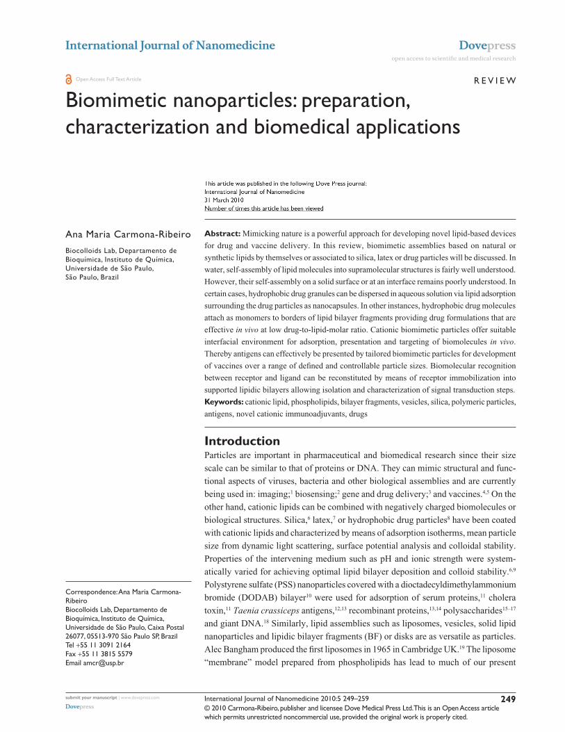
self-assemble as bilayers.51,52 Figure 1 shows BF from
different lipids.
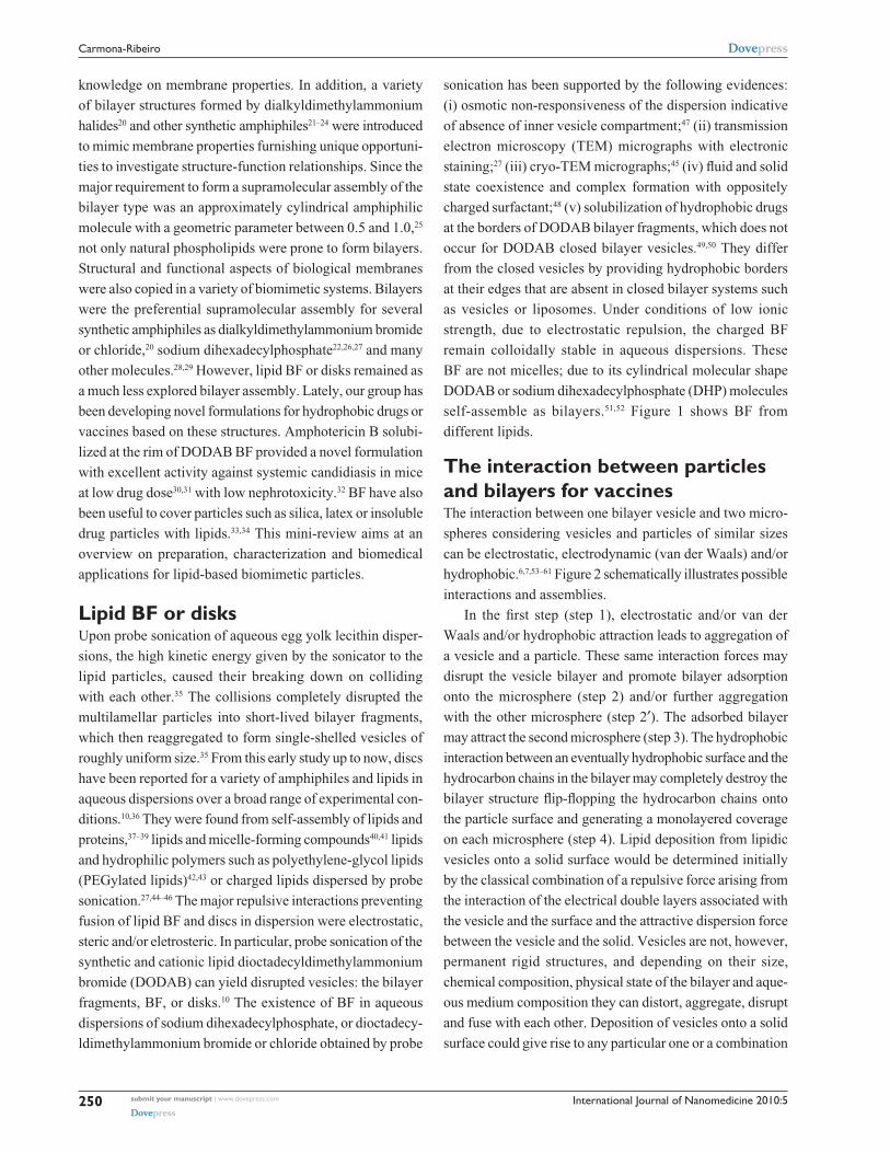
The interaction between particles and bilayers for vaccinesThe interaction between one bilayer vesicle and two micro-
spheres considering vesicles and particles of similar sizes
can be electrostatic, electrodynamic (van der Waals) and/or
hydrophobic.6,7,53–61 Figure 2 schematically illustrates possible
interactions and assemblies.
In the first step (step 1), electrostatic and/or van der
Waals and/or hydrophobic attraction leads to aggregation of
a vesicle and a particle. These same interaction forces may
disrupt the vesicle bilayer and promote bilayer adsorption
onto the microsphere (step 2) and/or further aggregation
with the other microsphere (step 2′). The adsorbed bilayer
may attract the second microsphere (step 3). The hydrophobic
interaction between an eventually hydrophobic surface and the
hydrocarbon chains in the bilayer may completely destroy the
bilayer structure flip-flopping the hydrocarbon chains onto
the particle surface and generating a monolayered coverage
on each microsphere (step 4). Lipid deposition from lipidic
vesicles onto a solid surface would be determined initially
by the classical combination of a repulsive force arising from
the interaction of the electrical double layers associated with
the vesicle and the surface and the attractive dispersion force
between the vesicle and the solid. Vesicles are not, however,
permanent rigid structures, and depending on their size,
chemical composition, physical state of the bilayer and aque-
ous medium composition they can distort, aggregate, disrupt
and fuse with each other. Deposition of vesicles onto a solid
surface could give rise to any particular one or a combination
International Journal of Nanomedicine 2010:5 251
Biomedical applications of biomimetic nanoparticlesDovepress
submit your manuscript | www.dovepress.com
Dovepress
of these processes. Unilamellar phosphatidylcholine vesicles
were reported to break open and adhere to a mica surface to
form a bilayer coating.62
Phospholipid monolayers with lipid haptens inserted
were supported by hydrophobic glass and useful for specific
adherence of macrophages and cell surface recognition stud-
ies, but did not serve as hosts for transmembrane proteins.63
Dipalmitoylphosphatidylcholine (DPPC) and phospha-
tidylinositol (PI) from vesicles adsorbed onto negatively
charged ballotini (hydrophobic) glass beads as a monolayer
with their head groups uppermost.64
The easiest method for preparing high quality phospho-
lipid bilayers on a flat hydrophilic surface was the direct
fusion of small unilamellar vesicles.65 This method stemmed
from making unilamellar membranes on glass coverslips for
spectroscopic studies.65 Phospholipid fusion at the hydro-
philic surface such as freshly cleaved mica could be induced
at elevated temperatures for those lipids of higher transition
temperature with traces of divalent cations such as Ca2+.
The other method for preparing supported membranes of
biological interest was the controlled transfer of monolay-
ers to the surface using the Langmuir. trough. Using this
method the content in each leaflet was easily controlled.66
The main advantages of the vesicle fusion method seemed
to be simplicity and the most natural lateral pressure in the
bilayer in comparison to the lateral pressures obtained with
the Langmuir trough. However, the content in each leaflet
could not be controlled using fusion. Palmitoyloleoylphos-
phatidylcholine (POPC) vesicles without major protruding
molecular moieties spread on a glass surface and formed
A B
DC
Figure 1 A) Lipid BF of dioctadecyldimethylammonium bromide (DODAB)45 or B) sodium dihexadecylphosphate (DHP)27 or C) DSPC/cholesterol/PeG-DSPe(5000) mixtures at 12 mol% PeG-DSPe(5000)42 or D) DSPC: cholesterol: ceramide-PeG5000 carrying bacteriorhodopsin.43
Notes: with exception of micrograph in B) which was obtained by TeM after negatively staining the sample, all micrographs were obtained by cryo-TeM. In C), disks were observed edge-on (arrow) or face-on (arrow head). Bars denote 100 nm. Copyright 1995 and 1991 American Chemical Society; 2005 and 2007 elsevier. Adapted with permission from Carmona-Ribeiro AM, Castuma Ce, Sesso A, Schreier S. Bilayer structure and stability in dihexadecyl phosphate dispersions. J Phys Chem. 1991;95:5361–5366. Johansson e, engvall C, Arfvidsson M, Lundahl P, edwards K. Development and initial evaluation of PeG-stabilized bilayer disks as novel model membranes. Biophys Chem. 2005;113:183–192. Johansson e, Lundquist A, Zuo S, edwards K. Nanosized bilayer disks: attractive model membranes for drug partition studies. Biochim Biophys Acta. 2007;1768:1518–1525. Andersson M, Hammarstrom L, Edwards K. Effect of bilayer phase transitions on vesicle structure, and its influence on the kinetics of viologen reduc-tion. J Phys Chem. 1995;99(39):14531–14538.Abbreviations: DHP, sodium dihexadecylphosphate; DODAB, dioctadecyldimethylammonium bromide; DSPe, distearoylphosphatidylethanolamine; PeG, polyethyleneglycol; TeM, transmission electron microscopy.
monolayer – covered particles
bilayer – covered particle
particles
1
2’
4
aggregate
2 3
Figure 2 The interaction between one bilayer vesicle and two particles. 6,7, 53–61 Copy-right 1999 elsevier. Adapted with permission from Carmona-Ribeiro AM, Lessa MM. Interactions between bilayer vesicles and latex. Colloids Surf A. 1999;153:355–361.
International Journal of Nanomedicine 2010:5252
Carmona-Ribeiro Dovepress
submit your manuscript | www.dovepress.com
Dovepress
a supported planar bilayer.67 In contrast, Escherichia coli
(E. coli) lipid vesicles adsorbed as entire vesicles to the
surface forming a supported vesicle layer on glass.67 The
difference in behavior upon deposition on glass was due to
chemical structure of E. coli lipids. Lipopolysaccharides
from E. coli are bulky and have strongly hydrated polar
heads. Their vesicles simply adhered and formed a supported
vesicle layer on glass.67 Fusion in-between vesicles attached
to the surface was prevented by steric repulsion.67 For DPPC
and DSPC bilayers on hydrophilic silicon/water interface,
single and double bilayers have been prepared and character-
ized via neutron reflectivity.68 This technique investigated the
structure, hydration and roughness of the layers and allowed
to determine the distance between two deposited bilayers.68
The outermost bilayer was highly hydrated and floated at 2 to
3 nm above the first one.68 Adhesion of a DODAB vesicle
layer onto the rough and highly hydrated surface of cells
was electrostatically driven. Cationic vesicles at low ionic
strength surrounded the bacterial cell as a vesicle layer.69
Absence of DODAB vesicle disruption upon interaction
with the bacteria was depicted from absence of [14C]-sucrose
leakage from large vesicles in experiments where this
marker was used to label the inner water compartment of the
vesicles.70 Given the quaternary ammonium moiety of the
DODAB molecule, its antimicrobial effect was systemati-
cally evaluated and its differential cytotoxicity established
as illustrated in Table 1.
In spite of its dose dependent-toxicity, DODAB induced
delayed-type hypersensibility (DTH), a marker for cell-
mediated immune responses. This interesting property
allowed DODAB to find many uses as an efficient immunoad-
juvant mainly for veterinary uses but also in humans in a few
instances.13,74–79 Supramolecular assemblies of DODAB BF
by themselves or after interaction with supporting particles
were recently combined with three different model antigens
in separate and tested as immunoadjuvants.13 DODAB-based
immunoadjuvants carrying antigens at reduced DODAB
dose (0.01–0.1 mM) induced superior DTH responses in
mice in comparison to alum. Thus, the cationic immunoad-
juvant was either reduced to a single-component, nanosized
system – DODAB BF – or was a dispersion of cationic
particles with controllable nature and size as obtained after
covering silica or polystyrene sulfate latex (PSS) with a
cationic DODAB bilayer. DODAB BF interacted with pro-
teins both via the hydrophobic effect and the electrostatic
attraction at low ionic strength. DODAB based adjuvants
exhibited good colloid stability while complexed with the
antigens, complete absence of toxicity in mice (ie, local or
general reactions) and a remarkable induction of Th1 immune
response at reduced doses of cationic and toxic DODAB lipid.
DODAB vesicle disruption by probe sonication at low ionic
strength (0.1–5.0 mM monovalent salt) produced DODAB
BF which remained electrostatically stabilized in dispersion
by the electrostatic repulsion in between fragments. DODAB
BF also interacted with oppositely charged particles such
as silica or polystyrene sulfate (PSS) latex to produce the
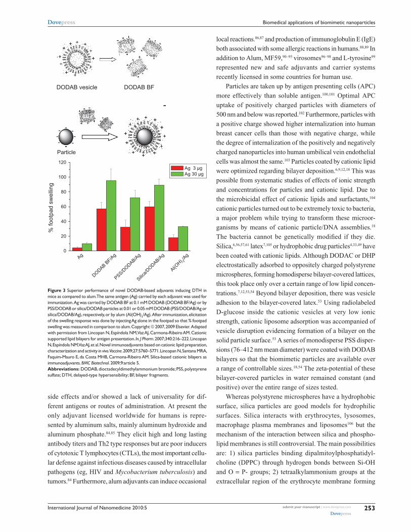
cationic particulates. Figure 3 schematically introduced the
novel cationic immunoadjuvants based on reduced DODAB
doses and their compared DTH response.12–14
The final DODAB concentration required to cover all
particles with a bilayer can be easily calculated from total
surface area for particles and bilayers and from the mean
molecular area for the lipid at the air-water interface.6,7,11,56
Sizing, zeta-potentials and polydispersity index for the novel
cationic adjuvants as compared to the similarly cationic
Al(OH)3 evidenced a superior colloid stability in contrast to
the one exhibited by alum.13,14 Table 2 illustrates the physical
properties of DODAB BF, PSS/DODAB and silica/DODAB
used in Figure 3 for antigen presentation.
Chemical, biological (eg, engineered viruses and bacteria),
polymeric or liposomal adjuvants have been developed and
tested for vaccines.4,5,80–83 However, most of them induced
Table 1 Differential cytotoxicity of DODAB against some eukaryotic* and prokaryotic cells**
Cell type Viable cells/mL [DODAB]50%survival/mM Ref.
Normal Balb-c 3T3 mouse fibroblasts* 104 1.000 71
SV40-transformed SVT2 mouse fibroblasts* 104 1.000 71
C. albicans* 2 × 10⁶ 0.010 72
E. coli** 2 × 10⁷ 0.028 70, 73
S. typhimurium** 2 × 10⁷ 0.010 73
P. aeruginosa** 3 × 10⁷ 0.005 73
S. aureus** 3 × 10⁷ 0.006 73
Abbreviation: DODAB, dioctadecyldimethylammonium bromide.
International Journal of Nanomedicine 2010:5 253
Biomedical applications of biomimetic nanoparticlesDovepress
submit your manuscript | www.dovepress.com
Dovepress
local reactions.86,87 and production of immunoglobulin E (IgE)
both associated with some allergic reactions in humans.88,89 In
addition to Alum, MF59,90–95 virosomes96–98 and L-tyrosine99
represented new and safe adjuvants and carrier systems
recently licensed in some countries for human use.
Particles are taken up by antigen presenting cells (APC)
more effectively than soluble antigen.100,101 Optimal APC
uptake of positively charged particles with diameters of
500 nm and below was reported.102 Furthermore, particles with
a positive charge showed higher internalization into human
breast cancer cells than those with negative charge, while
the degree of internalization of the positively and negatively
charged nanoparticles into human umbilical vein endothelial
cells was almost the same.103 Particles coated by cationic lipid
were optimized regarding bilayer deposition.6,9,12,18 This was
possible from systematic studies of effects of ionic strength
and concentrations for particles and cationic lipid. Due to
the microbicidal effect of cationic lipids and surfactants,104
cationic particles turned out to be extremely toxic to bacteria,
a major problem while trying to transform these microor-
ganisms by means of cationic particle/DNA assemblies.18
The bacteria cannot be genetically modified if they die.
Silica,6,56,57,61 latex7,105 or hydrophobic drug particles4,33,49 have
been coated with cationic lipids. Although DODAC or DHP
electrostatically adsorbed to oppositely charged polystyrene
microspheres, forming homodisperse bilayer-covered lattices,
this took place only over a certain range of low lipid concen-
trations.7,12,53,54 Beyond bilayer deposition, there was vesicle
adhesion to the bilayer-covered latex.53 Using radiolabeled
D-glucose inside the cationic vesicles at very low ionic
strength, cationic liposome adsorption was accompanied of
vesicle disruption evidencing formation of a bilayer on the
solid particle surface.53 A series of monodisperse PSS disper-
sions (76–412 nm mean diameter) were coated with DODAB
bilayers so that the biomimetic particles are available over
a range of controllable sizes.18,54 The zeta-potential of these
bilayer-covered particles in water remained constant (and
positive) over the entire range of sizes tested.
Whereas polystyrene microspheres have a hydrophobic
surface, silica particles are good models for hydrophilic
surfaces. Silica interacts with erythrocytes, lysosomes,
macrophage plasma membranes and liposomes106 but the
mechanism of the interaction between silica and phospho-
lipid membranes is still controversial. The main possibilities
are: 1) silica particles binding dipalmitoylphosphatidyl-
choline (DPPC) through hydrogen bonds between Si-OH
and O = P- groups; 2) tetraalkylammonium groups at the
extracellular region of the erythrocyte membrane forming
DODAB vesicle
+ +++
++
++
+ ++
+ +++
++
++
+ ++
+
0
20
40
60
80
100
120
Silica/
DODAB/Ag
PSS/DODAB/A
g
DODAB BF/A
gAg
Al(OH) 3
/Ag
% fo
otpa
d sw
ellin
g
Ag 3 µg Ag 30 µg
DODAB BF
Particle
Figure 3 Superior performance of novel DODAB-based adjuvants inducing DTH in mice as compared to alum. The same antigen (Ag) carried by each adjuvant was used for immunization. Ag was carried by DODAB BF at 0.1 mM DODAB (DODAB BF/Ag) or by PSS/DODAB or silica/DODAB particles at 0.01 or 0.05 mM DODAB (PSS/DODAB/Ag or silica/DODAB/Ag), respectively, or by alum (Al(OH)3/Ag). After immunization, elicitation of the swelling response was done by injecting Ag alone in the footpad so that % footpad swelling was measured in comparison to alum. Copyright 2007, 2009 elsevier. Adapted with permission from Lincopan N, espíndola NM, vaz AJ, Carmona-Ribeiro AM. Cationic supported lipid bilayers for antigen presentation. In. J Pharm. 2007;340:216–222. Lincopan N, espíndola NM, vaz AJ, et al. Novel immunoadjuvants based on cationic lipid: preparation, characterization and activity in vivo. Vaccine. 2009;27:5760–5771. Lincopan N, Santana MRA, Faquim-Mauro e, da Costa MHB, Carmona-Ribeiro AM. Silica-based cationic bilayers as immunoadjuvants. BMC Biotechnol. 2009;9:article 5.Abbreviations: DODAB, dioctadecyldimethylammonium bromide; PSS, polystyrene sulfate; DTH, delayed-type hypersensibility; BF, bilayer fragments.
side effects and/or showed a lack of universality for dif-
ferent antigens or routes of administration. At present the
only adjuvant licensed worldwide for humans is repre-
sented by aluminum salts, mainly aluminum hydroxide and
aluminum phosphate.84,85 They elicit high and long lasting
antibody titers and Th2 type responses but are poor inducers
of cytotoxic T lymphocytes (CTLs), the most important cellu-
lar defense against infectious diseases caused by intracellular
pathogens (eg, HIV and Mycobacterium tuberculosis) and
tumors.84 Furthermore, alum adjuvants can induce occasional
International Journal of Nanomedicine 2010:5254
Carmona-Ribeiro Dovepress
submit your manuscript | www.dovepress.com
Dovepress
ion pairs with dissociated silanol on the silica particle and
generating hemolytic effects observed for silica. Adsorption
isotherms of 4 different bilayers on hydrophilic silica over a
range of experimental conditions helped to clarify this issue.56
The separate use of synthetic charged membranes with phos-
phate or tetraalkylammonium groups as polar heads such as
are DODAB and DHP bilayer vesicles, to obtain adsorption
isotherms on silica established the relative importance of
phosphate or tetraalkylammonium on the mechanism of
phospholipid deposition onto hydrophilic silica particles.
Formation of ion pairs between the quaternary ammonium in
the choline moiety of the phospholipid and the deprotonated
silanol drove vesicle adhesion to the particle but vesicle rup-
ture and bilayer deposition was determined by the cooperative
occurrence of several hydrogen bridges between silanol and
the phosphate moiety on the phospholipid.56 There was a low
affinity between neutral phospholipids and the silica surface
and a high affinity for the cationic amphiphile over a range
of pH values.57 Tris-hydroxymethylaminomethane (Tris)
used as a buffer increased the affinity between PC and silica
at pH 7.4 due to Tris adsorption on silica with an increase
in the surface density of hydroxyls on the surface available
to hydrogen bridging with phosphate phospholipid groups.
Bilayer deposition, however, was unambiguously confirmed
by the three techniques only for the interaction DPPC
vesicles/silica over 1 hours at 65°C and for the interaction
DODAB vesicles/silica over the all range of experimental
conditions tested.57 A simple spectrophotometric method for
identifying entire bilayer deposition onto solid particles was
developed from incorporation of the optical probe merocya-
nine 540 onto the outer bilayer vesicle surface. Upon bilayer
deposition on the particle, sandwiching the marker between
bilayer and solid particle reduced light absorption. Thereby
reduction of light absorption by merocyanine was quanti-
tatively related to bilayer deposition.57 For the interaction
between cationic DODAB/DPPC and anionic PI/DPPC ves-
icles with zinc citrate dispersions the majority of the adsorp-
tion was in the form of intact liposomes.107 When liposomes
interacted with hydrophilic solid surfaces bearing ionizable
groups such as citrate or silanol, the pH affected the extent
of adsorption.107 For anionic liposomes, adsorption decreased
with pH. For cationic liposomes, adsorption increased with
pH.107 The fusion and spreading of phospholipid bilayers on
negatively charged glass surfaces was dependent on pH and
ionic strength.108 Membrane fusion of negatively charged
membranes was favored by low pH and high ionic strength
whereas membrane fusion of positively charged membranes
onto the surface occurred under all conditions tested.108
The interaction between particles and bilayers for drug deliveryThe particle concept encompasses a broad variety of par-
ticulates: lipid particles (eg, a bilayer fragment); polymeric;
mineral or metallic particles; bacterial cells; viruses; mam-
malian cells with several organelles and particles of insoluble,
hydrophobic drugs. The lipid covered-latexes were useful
as hosts for receptors,60,109,110 as coatings reducing protein
adsorption on the particles110 and in chromatography.111–113 The
potential of hybrid particle-lipid systems in diagnostics and
therapeutics has also been realized.8,114–116 In drug formulation,
lipid nanoparticles of the anticancer drug chlorambucil were
prepared by ultrasonication, using stearic acid as the core lipid
and DODAB as surface modifier.117 The presence of DODAB
on the lipid nanoparticles resulted in greater accumulation
of the drug in tumors.117 For the encapsulation of cisplatin,
bilayer-coating circumvented the limited solubility of cisplatin
Table 2 Physical properties of the novel cationic immunoadjuvants in a1 mM NaCl (pH 6.3) or b5 mM TrisHCl (pH 7.4) at 5 × 10⁹ PSS particles/ml or 0.1 mg/ml silica or 0.1 mg/ml Al(OH)3. Copyright 2009 elsevier. Adapted with permission from Lincopan N, espíndola NM, vaz AJ, et al. Novel immunoadjuvants based on cationic lipid: preparation, characterization and activity in vivo. Vaccine. 2009;27:5760–5771.
Dispersion DODAB/mM Mean diameter/nm Zeta-potential/mV Polydispersity index
DODAB BFa 1.0 73 ± 1 42 ± 2 0.240 ± 0.01
PSSa – 236 ± 4 -61 ± 1 0.130 ± 0.03
PSS/DODABa 0.01 246 ± 3 44 ± 3 0.085 ± 0.03
Al(OH)3a – 458 ± 3 28 ± 3 0.191 ± 0.04
DODAB BFb 1.0 74 ± 1 33 ± 5 0.270 ± 0.04
Silicab – 294 ± 2 -47 ± 2 0.180 ± 0.02
Silica/DODABb 0.05 373 ± 7 33 ± 2 0.260 ± 0.02
Al(OH)3b – 700 ± 20 14 ± 2 0.317 ± 0.01
Abbreviations: DODAB, dioctadecyldimethylammonium bromide; PSS, polystyrene sulfate; DTH, delayed-type hypersensibility; BF, bilayer fragments.
International Journal of Nanomedicine 2010:5 255
Biomedical applications of biomimetic nanoparticlesDovepress
submit your manuscript | www.dovepress.com
Dovepress
in water and produced cisplatin nanocapsules, bean-shaped
nanoprecipitates of cisplatin coated by a lipid bilayer.118 The
nanocapsules represented a novel lipid formulation of cisplatin
characterized by a very high cisplatin-to-lipid ratio and cyto-
toxicity against tumor cells in vitro as compared to the free
drug. The formation of the nanocapsules critically depended
on the presence of negatively charged phospholipids and posi-
tively charged aqua-species of cisplatin.118,119 The effect of PEG
on the stability of the cisplatin nanocapsules was studied by
incorporating PEG conjugated to phosphatidylethanolamine
(DSPE-PEG2000).120 Cisplatin release from the nanocapsules
depended on the temperature, the surrounding medium, and
the lipid composition of the bilayer coat. Sterically stabilized
cisplatin nanocapsules containing 6 mol % DSPE-PEG served
as the starting formulation for in vivo studies addressing the
anti-tumor efficacy of cisplatin nanocapsules in tumor-bearing
mice; there was a requirement of anionic phospholipid for suc-
cessful nanoencapsulation of the cationic aqua-cisplatin.120
Miconazole or amphotericin B were formulated in
DODAB or DHP BF.34,49 Some of these formulations required
low drug-to-lipid molar ratio due to limited drug loading
capacity of the BF at their rims. For example, BF loading
capacity for monomeric amphotericin B was 0.1 mM ampho-
tericin B at 2 mM DODAB meaning that one drug molecule
required 20 molecules of cationic lipid to become soluble in
the BF nanostructure. At and above this 1:20 drug-to-lipid
molar ratio, all solubilization sites at the rim of the BF were
occupied. Therefore, further addition of drug resulted in
appearance of aggregated amphotericin B in the dispersion
as easily monitored by systematic determination of size dis-
tribution by means of photon correlation spectroscopy.
In order to formulate hydrophobic drugs with the
DODAB lipid at high drug-to-lipid molar ratios, we took
advantage of the “sticky” property of chaotropic dihydrogen
phosphate anion which converted miconazole or ampho-
tericin B drug particles into negatively charged particles.
Thereafter, anionic drug particles could be coated by the
DODAB cationic lipid.34,49 These formulations were tested
against C. neoformans and Candida and were very effective
due to DODAB activity against fungi. The cationic lipid alone
exhibited minimal fungicidal concentrations (MFC) equal to
2 and 2 to 250 mg/L against C. neoformans and Candida,
respectively. In combination, over the first hour, fungicidal
activity was due to DODAB with lipid capsules retarding
drug action. At 48 hours and 104 cfu/mL, MFC (mg/L) against
Candida albicans was reduced from 4 to 1 amphotericin B
(at 2 DODAB), and from 8 to 1 miconazole (at 1 DODAB).
Calculations of synergism indexes showed synergistic action
of both antimicrobial drugs: the cationic DODAB lipid and
the microbicidal drug34,49 so that the DODAB/miconazole
(MCZ) formulations should be further tested regarding thera-
peutic activity in vivo. Table 3 illustrated the efficacy of MCZ
in DODAB or DHP BF despite the low dose of drug.
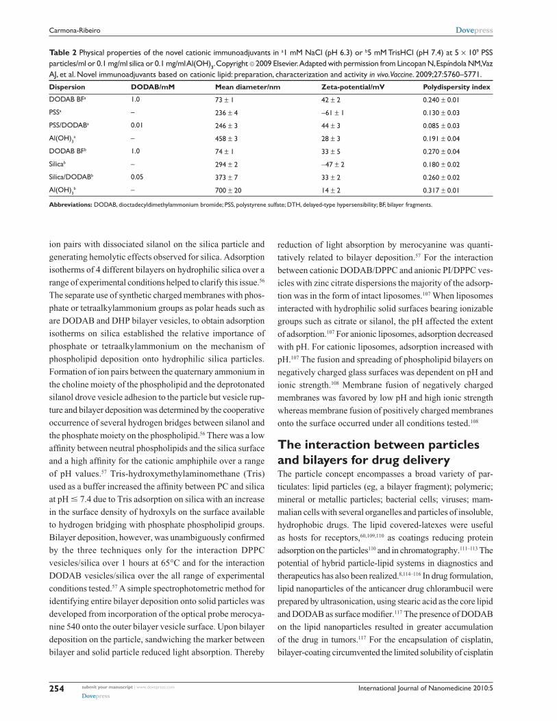
In summary, two major strategies were developed to formu-
late hydrophobic drugs with bilayer fragments. These strategies
can be better visualized in Figure 4. In Figure 4 (a), coalescence
of BF around the drug granule would encapsulate drug par-
ticles at high drug-to-lipid molar ratios.33,34,49,50 In Figure 4 (b),
monomolecular drug solubilization would be achieved at low
drug-to-lipid molar ratios.30,31,33 Therefore, these discoidal,
charged BF dispersed the hydrophobic drug particles in water
both at low and high drug-to-lipid molar ratios.
PEG decorated lipid bilayers are widely used in drug
delivery.121 In these hybrid polymer/lipid systems, there is a
transition from a dispersed lamellar phase (liposomes) to a
micellar phase mediated by the formation of small discoidal
micelles. The onset of disk formation took place at low
PEG-lipid concentrations (5 mol%) and the size of the
disks decreased as more PEG-lipid was added to the lipid
mixture.122 Stable dispersions dominated by flat bilayer disks
could be prepared from a carefully optimized mixture of
1,2-distearoyl-sn-glycero-3-phosphocholine (DSPC), choles-
terol, and 1,2-distearoyl-sn-glycero-3-phosphoethanolamine-
N-[methoxy(polyethyleneglycol)-5000][PEG-DSPE(5000)].
By varying the content of PEG-DSPE (5000), the disks
diameter varied from about 15 to 60 nm. Disks compared
favorably to uni- and multilamellar liposomes for hydrophilic
drug partitioning employing immobilized disks in glass
capillaries.42 The major repulsive interactions preventing
fusion of these BF were steric. They provided larger areas
Table 3 Minimal fungicidal concentration (MFC) for Zoltec® (fluconazol), miconazole (MCZ), MCZ/DODAB BF or MCZ/DHP BF against Candida albicans. Copyright 2006 elsevier. Adapted with permission from vieira DB, Pacheco LF, Carmona-Ribeiro AM. Assembly of a model hydrophobic drug into cationic bilayer frag-ments. J Colloid Interface Sci. 2006;293:240–247.
Dispersion MFC
µM mg/mL
Zoltec® 6.5 2
DODAB 4000 250
MCZ 8 4
MCZ and DODAB 2 1
MCZ and DHP 4 2
Abbreviations: DODAB, dioctadecyldimethylammonium bromide; PSS, polystyrene sulfate; DTH, delayed-type hypersensibility; BF, bilayer fragments.
International Journal of Nanomedicine 2010:5256
Carmona-Ribeiro Dovepress
submit your manuscript | www.dovepress.com
Dovepress
than vesicles or liposomes for hydrophilic drug partitioning
and from this point of view were considered as an attractive
and sometimes superior alternative to liposomes.43
Reconstitution of receptor-ligand recognition in artificial
biomimetic particles is a very promising but hitherto unex-
plored area for research. The major advantage of these par-
ticles is the possibility of complete quantification of binding
from simple analytical methods and techniques such as cen-
trifugation for separation between free and bound receptors or
free and bound ligands. Aiming at the production of bilayer
covered silica particles, the interaction between silica par-
ticles and lipid vesicles or BF has been systematically studied
by our group since 1997.56–61 As a result optimal coverage of
silica particles with a PC bilayer was recently achieved.59,60
At pH 6.3, limiting PC adsorption indicative of one-bilayer
deposition on each silica particle was obtained at and above
10 mM NaCl. Increasing ionic strength provided increasing
attractive van der Waals attraction between vesicle and
particle so that vesicles ruptured upon contact with particles
and covered them with one bilayer.59 Keeping ionic strength
at 10 mM NaCl, the effect of increasing pH was decreasing
affinity between PC and silica.59 These experiments revealed
an important role for hydrogen bonding between silanol on
silica and phosphate on PC driving bilayer deposition. Bilayer
deposition improved colloid stability of silica as shown from
absence of particulate sedimentation.59
Silica-based biomimetic particles successfully immobi-
lized proteins or DNA13 or allowed isolation and reconstitu-
tion of receptor-ligand specific interaction.60
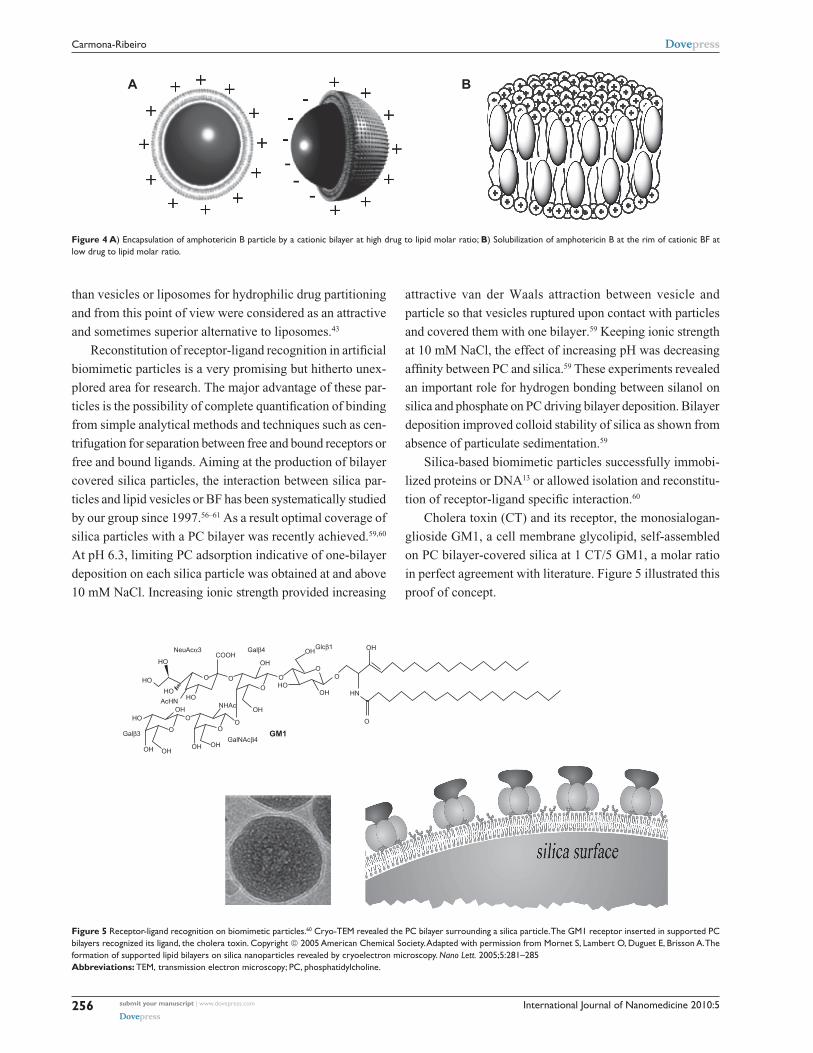
Cholera toxin (CT) and its receptor, the monosialogan-
glioside GM1, a cell membrane glycolipid, self-assembled
on PC bilayer-covered silica at 1 CT/5 GM1, a molar ratio
in perfect agreement with literature. Figure 5 illustrated this
proof of concept.
OHOH OH
OH
OH
OH
OH
OH OH
OH
HO
HOHO
HOHO
HO
AcHN
O
NeuAcα3
Galβ3
Galβ4 Glcβ1
GalNAcβ4
O
OO
O
OOOO
O
O
HN
NHAc
COOH
GM1
Figure 5 Receptor-ligand recognition on biomimetic particles.60 Cryo-TeM revealed the PC bilayer surrounding a silica particle. The GM1 receptor inserted in supported PC bilayers recognized its ligand, the cholera toxin. Copyright 2005 American Chemical Society. Adapted with permission from Mornet S, Lambert O, Duguet e, Brisson A. The formation of supported lipid bilayers on silica nanoparticles revealed by cryoelectron microscopy. Nano Lett. 2005;5:281–285Abbreviations: TeM, transmission electron microscopy; PC, phosphatidylcholine.
A B
Figure 4 A) encapsulation of amphotericin B particle by a cationic bilayer at high drug to lipid molar ratio; B) Solubilization of amphotericin B at the rim of cationic BF at low drug to lipid molar ratio.
International Journal of Nanomedicine 2010:5 257
Biomedical applications of biomimetic nanoparticlesDovepress
submit your manuscript | www.dovepress.com
Dovepress
ConclusionsThe intermolecular forces between lipids and particles have
to be understood in depth before optimal biomimetic particles
based on lipids can be obtained. Over the years our system-
atic studies on such interactions produced some examples
of optimized bilayer deposition on silica or latex particles.
Furthermore, charged BF solubilized hydrophobic drugs
and complexed with antigens yielding novel supramolecular
assemblies for drug and vaccine delivery either by providing
hydrophobic edges for drug or antigen complexation
or by covering silica, drug or polymeric particles with
minute amounts of lipid. Low doses of cationic lipid in
the formulations avoided their well described toxicity. Our
contribution to the field may be summarized in a thorough
description of the differential cytotoxicity of the DODAB
cationic lipid, and the invention of novel charged carriers to
formulate drugs or vaccines at reduced charged lipid dose.
Encapsulation of drugs, reconstitution of biomolecular
recognition, antigen presentation and antimicrobial therapy
were some examples of biomedical applications for biomi-
metic particles that were provided by this mini-review.
AcknowledgmentsFinancial support from FAPESP and CNPq is gratefully
acknowledged.
References 1. El-Sayed IH, Huang X, El-Sayed MA. Surface plasmon resonance
scattering and absorption of anti-EGFR antibody conjugated gold nanoparticles in cancer diagnostics: applications in oral cancer. Nano Lett. 2005;5:829–834.
2. Medintz AR, Clapp JS, Melinger JR, Deschamps H, Mattoussi A. Reagentless Biosensing assembly based on quantum dot-donor Förster resonance energy transfer. Adv Mater. 2005;17:2450–2455.
3. Carmona-Ribeiro AM. Bilayer-forming synthetic lipids: drugs or carriers? Curr Med Chem. 2003;10:2425–2446.
4. O’Hagan DT, Singh M, Ulmer JB. Microparticles for the delivery of DNA vaccines. Immunol Rev. 2004;199:191–200.
5. Caputo A, Sparnacci K, Ensoli B, Tondelli L. Functional Polymeric nano/microparticles for surface adsorption and delivery of protein and DNA vaccines. Curr Drug Delivery. 2008;5:230–242.
6. Moura SP, Carmona-Ribeiro AM. Cationic bilayer fragments on silica at low ionic strength: competitive adsorption and colloid stability. Langmuir. 2003;19:6664–6667.
7. Carmona-Ribeiro AM, Midmore BR. Synthetic bilayer adsorption onto polystyrene microspheres. Langmuir. 1992;8:801–806.
8. Carmona-Ribeiro AM. Biomimetic particles in drug and vaccine delivery. J Liposome Res. 2007;17:165–172.
9. Pereira EMA, Vieira DB, Carmona-Ribeiro AM. Cationic bilayers on polymeric particles: effect of low NaCl concentration on surface coverage. J Phys Chem B. 2004;108:11490–11495.
10. Carmona-Ribeiro AM. Lipid bilayer fragments and disks in drug deliv-ery. Curr Med Chem. 2006;13:1359–1370.
11. Lincopan N. Carmona-Ribeiro AM. Protein assembly onto cationic supported bilayers. J Nanosci Nanotechnol. 2009;9:3578–3586.
12. Lincopan N, Espíndola NM, Vaz AJ, Carmona-Ribeiro AM. Cat-ionic supported lipid bilayers for antigen presentation. In. J Pharm. 2007;340:216–222.
13. Lincopan N, Espíndola NM, Vaz AJ, et al. Novel immunoadjuvants based on cationic lipid: preparation, characterization and activity in vivo. Vaccine. 2009;27:5760–5771.
14. Lincopan N, Santana MRA, Faquim-Mauro E, da Costa MHB, Carmona-Ribeiro AM. Silica-based cationic bilayers as immunoadjuvants. BMC Biotechnol. 2009;9:article 5.
15. Vieira DB, Lincopan N, Mamizuka EM, Petri DFS, Carmona-Ribeiro AM. Competitive adsorption of cationic bilayers and chitosan on latex: optimal biocidal action. Langmuir. 2003;19:924–932.
16. Correia FM, Petri DFS, Carmona-Ribeiro AM. Colloid stability of lipid/polyelectrolyte decorated latex. Langmuir. 2004;20:9535–9540.
17. Araujo FP, Petri DFS, Carmona-Ribeiro AM. Colloid stability of sodium dihexadecyl phosphate/poly(diallyldimethylammonium chloride) deco-rated latex. Langmuir. 2005;21:9495–9501.
18. Rosa H, Petri DFS, Carmona-Ribeiro AM. Interactions between bac-teriophage DNA and cationic biomimetic particles. J Phys Chem B. 2008;112:16422–16430.
19. Bangham AD. editor. Liposome Letters. London: Academic Press; 1983. p. 1–405.
20. Kunitake T, Okahata Y, Tamaki K, Kumamaru F, Takayanagi M. Forma-tion of the bilayer membrane from a series of quaternary ammonium salts. Chem Lett. 1977;6:(4):387–390.
21. Hargreaves WR, Deamer DW. Liposomes from ionic, single-chain amphiphiles. Biochemistry. 1978;17(18):3759–3768.
22. Mortara RA, Quina FH. Chaimovich H. Formation of closed vesicles from a simple phosphate diester. Preparation and some properties of vesicles of dihexadecyl phosphate. Biochem Biophys Res Commun. 1978;81:1080–1086.
23. Czarniecki MF, Breslow R. Photochemical probes for model membrane structures. J Am Chem Soc. 1979;101:3675–3676.
24. Suedholter EJR, Engberts JBFN, Hoekstra DJ. Vesicle formation by two novel synthetic amphiphiles carrying micropolarity reporter head groups. J Am Chem Soc. 1980;102:2467–2469.
25. Israelachvili JN, Mitchell DJ, Ninham BW. Theory of self-assembly of lipid bilayers and vesicles. Biochim Biophys Acta. 1977;470:185–201.
26. Carmona-Ribeiro AM, Yoshida LS, Sesso A, Chaimovich H. Perme-abilities and stabilities of large dihexadecylphosphate and dioctadecyldi-methylammonium chloride vesicles. J Colloid Interface Sci. 1984;100: 433–443.
27. Carmona-Ribeiro AM, Castuma CE, Sesso A, Schreier S. Bilayer structure and stability in dihexadecyl phosphate dispersions. J Phys Chem. 1991;95:5361–5366.
28. Fuhrhop JH, Fritsch D. Bolaamphiphiles form ultrathin, porous and unsymmetric monolayer lipid membranes. Acc Chem Res. 1986;19: 130–137.
29. Segota S, Tezak D. Spontaneous formation of vesicles. Adv Colloid Interface Sci. 2006;121:51–75.
30. Vieira DB, Carmona-Ribeiro AM. Synthetic bilayer fragments for solubilization of amphotericin B. J Colloid Interface Sci. 2001;244: 427–431.
31. Lincopan N, Mamizuka EM, Carmona-Ribeiro AM. In vivo activity of a novel amphotericin B formulation with synthetic cationic bilayer fragments. J Antimicrob Chemother. 2003;52:412–418.
32. Lincopan N, Mamizuka EM, Carmona-Ribeiro AM. Low nephrotoxicity of an effective amphotericin B formulation with cationic bilayer frag-ments. J Antimicrob Chemother. 2005;55:727–734.
33. Pacheco LF, Carmona-Ribeiro AM. Effects of synthetic lipids on solubilization and colloid stability of hydrophobic drugs. J Colloid Interface Sci. 2003;258:146–154.
34. Lincopan N, Carmona-Ribeiro AM. Lipid-covered drug particles: com-bined action of dioctadecyldimethylammonium bromide and amphoteri-cin B or miconazole. J Antimicrob Chemother. 2006;58: 66–75.
35. Finer EG, Flook AG, Hauser H. Mechanism of sonication of aqueous egg yolk lecithin dispersions and nature of the resultant particles. Biochim Biophys Acta. 1972;260:49–58.
36. Nath A, Atkins WM, Sligar SG. Applications of phospholipid bilayer nanodiscs in the study of membranes and membrane proteins. Biochemistry. 2007;46:2059–2069.
International Journal of Nanomedicine 2010:5258
Carmona-Ribeiro Dovepress
submit your manuscript | www.dovepress.com
Dovepress
37. Bayburt TH, Sligar SG. Single-molecule height measurements on microsomal cytochrome P450 in nanometer-scale phospholipid bilayer disks. Proc Natl Acad Sci U S A. 2002;99:6725–6730.
38. Lyukmanova EN, Shenkarev ZO, Paramonov AS, et al. Lipid-protein nanoscale bilayers: a versatile medium for NMR investigations of membrane proteins and membrane-active peptides. J Am Chem Soc. 2008;130:2140–2141.
39. Hazelbauer GL, Falke JJ, Parkinson JS. Bacterial chemoreceptors: high-performance signaling in networked arrays. 2008;33:9–19.
40. Lawaczeck R, Kainosho M, Chan SI. The formation and annealing of structural defects in lipid bilayer vesicles. Biochim Biophys Acta. 1976; 443:313–330.
41. Almgren M. Mixed micelles and other structures in the solubilization of bilayer lipid membranes by surfactants. Biochim Biophys Acta. 2000;1508:146–163.
42. Johansson E, Engvall C, Arfvidsson M, Lundahl P, Edwards K. Devel-opment and initial evaluation of PEG-stabilized bilayer disks as novel model membranes. Biophys Chem. 2005;113:183–192.
43. Johansson E, Lundquist A, Zuo S, Edwards K. Nanosized bilayer disks: attractive model membranes for drug partition studies. Biochim Biophys Acta. 2007;1768:1518–1525.
44. Pansu RB, Arrio B, Roncin J, Faure J. Vesicles versus membrane frag-ments in DODAC suspensions. J Phys Chem. 1990;94:796–801.
45. Andersson M, Hammarstrom L, Edwards K. Effect of bilayer phase transitions on vesicle structure, and its influence on the kinetics of viologen reduction. J Phys Chem. 1995;99(39):14531–14538.
46. Meyer HW, Richter W, Rettig W, Stumpf M. Bilayer fragments and bilayered micelles (bicelles) of dimyristoylphosphatidylglycerol (DMPG) are induced by storage in distilled water at 4°C. Colloids Surf A: Physicochem Eng Aspects. 2001;183–185:495–504.
47. Carmona-Ribeiro AM, Chaimovich H. Preparation and characteriza-tion of large dioctadecyldimethylammonium chloride liposomes and comparison with small sonicated vesicles. Biochim Biophys Acta. 1983;733:172–179.
48. Cocquyt J, Olsson U, Olofsson G, van der Meeren P. Temperature quenched DODAB dispersions: fluid and solid state coexistence and complex formation with oppositely charged surfactant. Langmuir. 2004;20:3906–3912.
49. Vieira DB, Pacheco LF, Carmona-Ribeiro AM. Assembly of a model hydrophobic drug into cationic bilayer fragments. J Colloid Interface Sci. 2006;293:240–247.
50. Vieira DB, Carmona-Ribeiro AM. Cationic nanoparticles for delivery of amphotericin B: preparation, characterization and activity in vitro. J Nanobiotechnol. 2008;6:article 6.
51. Carmona-Ribeiro AM. Synthetic amphiphile vesicles. Chem Soc Rev. 1992;21:209–214.
52. Israelachvili JN. Intermolecular and surface forces. 2nd ed. London; Academic Press Limited: 1992.
53. Tsuruta LR, Lessa MM, Carmona-Ribeiro AM. Interactions between dioctadecyldimethylammonium chloride or bromide bilayers in water. Langmuir. 1995;11:2938–2943.
54. Tsuruta LR. Lessa MM. Carmona-Ribeiro AM. Effect of particle size on colloid stability of bilayer-covered polystyrene microspheres. J Colloid Interface Sci. 1995;175:470–475.
55. Tsuruta LR, Carmona-Ribeiro AM. Counterion effects on colloid stabil-ity of cationic vesicles and bilayer-covered polystyrene microspheres. J Phys Chem. 1996;100:7130–7134.
56. Rapuano R, Carmona-Ribeiro AM. Physical adsorption of bilayer membranes on silica. J Colloid Interface Sci. 1997;193:104–111.
57. Rapuano R, Carmona-Ribeiro AM. Supported bilayers on silica. J Colloid Interface Sci. 2000;226:299–307.
58. Carmona-Ribeiro AM, Lessa MM. Interactions between bilayer vesicles and latex. Colloids Surf A. 1999;153:355–361.
59. Moura SP, Carmona-Ribeiro AM. Biomimetic particles: optimization of phospholipid bilayer coverage on silica and colloid stabilization. Langmuir. 2005;21:10160–10164.
60. Moura SP, Carmona-Ribeiro AM. Biomimetic particles for isolation and reconstitution of receptor function. Cell Biochem Biophys. 2006;44: 446–452.
61. Moura SP, Carmona-Ribeiro AM. Adsorption behavior of DODAB/DPPC vesicles on silica. J Colloid Interface Sci. 2007;313:519–526.
62. Horn RG. Direct measurement of the force between two lipid bilay-ers and observation of their fusion. Biochim Biophys Acta. 1984;778: 224–228.
63. Lin LC, Weis RM, McConnell HM. Induction of helical liposomes by Ca2+-mediated intermembrane binding. Nature. 1982;296:164–165.
64. Jackson S, Reboiras MD, Lyle IG, Jones MN. Adsorption of phospholipid vesicles on solid surfaces. Faraday Discuss Chem Soc. 1986;81: 291–301.
65. Brian AA, McConnell HM. Allogeneic stimulation of cytotoxic T cells by supported planar membranes. Proc Natl Acad Sci U S A. 1984;81: 6159–6163.
66. Tamm LK, McConnell HM. Supported phospholipid bilayers. Biophys J. 1985;47:105–113.
67. Nollert P, Kiefer H, Jaehnig F. Lipid vesicle adsorption versus formation of planar bilayers on solid surfaces. Biophys J. 1995;69:1447–1455.
68. Charitat T, Bellet-Amalric E, Fragneto G, Graner F. Adsorbed and free lipid bilayers at the solid-liquid interface. Eur Phys J B. 1999;8: 583–593.
69. Tápias GN, Sicchierolli SM, Mamizuka EM, Carmona-Ribeiro AM. Interactions between cationic vesicles and Escherichia coli. Langmuir. 1994;10:3461–3465.
70. Martins LMS, Mamizuka EM, Carmona-Ribeiro AM. Cationic vesicles as bactericides. Langmuir. 1997;13:5583–5587.
71. Carmona-Ribeiro AM, Ortis F, Schumacher RI, Armelin MCS. Interactions between cationic vesicles and cultured mammalian cells. Langmuir. 1997;13:2215–2218.
72. Campanhã MTN, Mamizuka EM, Carmona-Ribeiro AM. Interactions between cationic vesicles and Candida albicans. J Phys Chem B. 2001; 105:8230–8236.
73. Campanhã MTN, Mamizuka EM, Carmona-Ribeiro AM. Interactions between cationic liposomes and bacteria: the physical-chemistry of the bactericidal action. J Lipid Res. 1999;40:1495–1500.
74. Gall D. The adjuvant activity of aliphatic nitrogenous bases. Immunology. 1966;11:369–386.
75. Dailey MO, Hunter RL. The role of lipid in the induction of hapten-specific delayed hypersensitivity and contact sensitivity. J Immunol. 1974;112:1526–1534.
76. Hilgers LA, Snippe H, DDA as an immunological adjuvant. Res Immunol. 1992;143:494–503.
77. Tsuruta LR, Quintilio W, Costa MHB, Carmona-Ribeiro AM. Interac-tions between cationic liposomes and an antigenic protein: the physical chemistry of the immunoadjuvant action. 1997;38:2003–2011.
78. Klinguer-Hamour C, Libon C, Plotnicky-Gilquin H, et al. DDA adjuvant induces a mixed Th1/Th2 immune response when associated with BBG2Na, a respiratory syncytial virus potential vaccine. Vaccine. 2002;20:2743–2751.
79. Korsholm KS, Agger EM, Foged C, et al. The adjuvant mechanism of cationic dimethyldioctadecylammonium liposomes. Immunology. 2007;121:216–226.
80. Gregoriadis G, McCormack B, Obrenovic M, Saffie R, Zadi B, Perrie Y. Vaccine entrapment in liposomes. Methods. 1999;19:156–162.
81. Perrie Y, Mohammed AR, Kirby DJ, McNeil SE, Bramwell VW. Vaccine adjuvant systems: enhancing the efficacy of sub-unit protein antigens. Int J Pharm. 2008;364:272–280.
82. O’Hagan DT, Singh M. Microparticles as vaccine adjuvants and delivery systems. Expert Rev Vaccines. 2003;2:269–283.
83. Xiang SD, Scholzen A, Minigo G, et al. Pathogen recognition and development of particulate vaccines: Does size matter? Methods. 2006;40:1–9.
84. Gupta R. Aluminum compounds as vaccine adjuvants. Adv Drug Delivery Rev. 1998;32:155–172.
International Journal of Nanomedicine 2010:5
International Journal of Nanomedicine
Publish your work in this journal
Submit your manuscript here: http://www.dovepress.com/international-journal-of-nanomedicine-journal
The International Journal of Nanomedicine is an international, peer-reviewed journal focusing on the application of nanotechnology in diagnostics, therapeutics, and drug delivery systems throughout the biomedical field. This journal is indexed on PubMed Central, MedLine, CAS, SciSearch®, Current Contents®/Clinical Medicine,
Journal Citation Reports/Science Edition, EMBase, Scopus and the Elsevier Bibliographic databases. The manuscript management system is completely online and includes a very quick and fair peer-review system, which is all easy to use. Visit http://www.dovepress.com/ testimonials.php to read real quotes from published authors.
259
Biomedical applications of biomimetic nanoparticlesDovepress
submit your manuscript | www.dovepress.com
Dovepress
Dovepress
85. Jefferson T, Rudin M, Di Pietrantonj C. Adverse events after immuni-sation with aluminium-containing DTP vaccines: systematic review of the evidence. Lancet Infectious Diseases. 2004;4:84–90.
86. Clements C, Griffiths E, Clements C, Griffiths E. The global impact of vaccines containing aluminium adjuvants. Vaccine. 2002;20:S24–S33.
87. Trollfors B, Bergfors E, Inerot A. Vaccine related itching nodules and hypersensitivity to aluminium. Vaccine. 2005;23:975–976.
88. Lindblad E. Aluminium adjuvants-in retrospect and prospect. Vaccine. 2004;22:3658–3668.
89. Gupta R, Siber G. Adjuvants for human vaccines – current status, problems and future prospects. Vaccine. 1995;13:1263–1276.
90. Singh M, Ugozzoli M, Kazzaz J, et al. A preliminary evaluation of alternative adjuvants to alum using a range of established and new generation vaccine antigens. Vaccine. 2006;24:1680–1686.
91. Singh M, O’Hagan D. Advances in vaccine adjuvants. Nature Biotechnology. 1999;17:1075–1081.
92. Ott G, Barchfeld G, Chernoff D, Radhakrishnan R, van Hoogevest P, Van Nest G. MF59. Design and evaluation of a safe and potent adjuvant for human vaccines. Pharm Biotechnol. 1995;6:277–296.
93. Traquina P, Morandi M, Contorni M, Van Nest G. MF59 adjuvant enhances the antibody response to recombinant hepatitis B surface antigen vaccine in primates. J Infect Dis. 1996;174:1168–1175.
94. Granoff D, McHugh Y, Raff H, Mokatrin A, Van Nest G. MF59 adjuvant enhances antibody responses of infant baboons immunized with Haemophilus influenzae type b and Neisseria meningitis group C oligosaccharide-CRM197 conjugate vaccine. Infect Immun. 1997;65: 1710–1715.
95. Podda A, Del Giudice G. MF59-adjuvanted vaccines: increased immunogenicity with an optimal safety profile. Expert Rev Vaccines. 2003; 2:197–203.
96. Cusi M. Applications of influenza virosomes as a delivery system. Hum Vaccin. 2006;2:1–7.
97. Glück R, Burri K, Metcalfe I. Adjuvant and antigen delivery properties of virosomes. Curr Drug Deliv. 2005;2:395–400.
98. Huckriede A, Bungener L, Stegmann T, et al. The virosome concept for influenza vaccines. Vaccine. 2005;23:S26-S38.
99. Baldrick P, Richardson D, Wheeler A. Review of L-tyrosine confirm-ing its safe human use as an adjuvant. J Appl Toxicol. 2002;22:333–344.
100. Kovacsovics-Bankowski M, Clark K, Benacerraf B, Rock KL. Efficient major histocompatibility complex class I presentation of exogenous antigen upon phagocytosis by macrophages. Proc Natl Acad Sci U S A. 1993;90:4942–4946.
101. Vidard L, Kovacsovics-Bankowski M, Kraeft SK, Chen LB. Benacerraf B, Rock KL. Analysis of MHC class II presentation of par-ticulate antigens of B lymphocytes. J Immunol. 1996;156:2809–2818.
102. Foged C, Brodin B, Frokjaer S, Sundblad A. Particle size and surface charge affect particle uptake by human dendritic cells in an in vitro model. Int J Pharm. 2005;298:315–322.
103. Osaka T, Nakanishi T, Shanmugam S, Takahama S, Zhang H. Effect of surface charge of magnetite nanoparticles on their internalization into breast cancer and umbilical vein endothelial cells. Colloids and Surf B: Biointerfaces. 2009;71(2):325–330.
104. Carmona-Ribeiro AM, Vieira DB, Lincopan N. Cationic surfactants and lipids as anti-infective agents. Anti-Infective Agents in Medicinal Chemistry. 2006;5:33–51.
105. Carmona-Ribeiro AM. Bilayer vesicles and liposomes as interface agents. Chem Soc Rev. 2001;30:241–247.
106. Nash T, Allison AC, Harington JS. Physico-chemical properties of silica in relation to its toxicity. Nature. 1966;210:259–261.
107. Catuogno C, Jones MN. The interaction of cationic and anionic vesicles with zinc citrate dispersions. Colloids Surf A. 2000;163:165–176.
108. Cremer PS, Boxer SG. Formation and spreading of lipid bilayers on planar glass supports. J Phys Chem. 1999;103:2554–2559.
109. Sicchierolli SM, Carmona-Ribeiro AM. Incorporation of the cholera toxin receptor in phospholipid-covered polystyrene microspheres. Colloids and Surf B: Biointerfaces. 1995;5:57–64.
110. Sicchierolli SM, Carmona-Ribeiro AM. Biomolecular recognition at phospholipid-covered polystyrene microspheres. J Phys Chem. 1996;100:16771–16775.
111. Hautala JT, Linden MV, Wiedmer SK, et al. Simple coating of capillaries with anionic liposomes in capillary electrophoresis. J Chromatogr A. 2003;1004:81–90.
112. Haratake M, Hidaka S, Ono M, Nakayama M. Preparation of an ion-exchangeable polymer bead wrapped with bilayer membrane structures for high performance liquid chromatography. Anal Chim Acta. 2007;589:76–83.
113. Gulcev MD, Lucy CA. Factors affecting the behavior and effectiveness of phospholipid bilayer coatings for capillary electrophoretic separa-tions of basic proteins. Anal Chem. 2008;80:1806–1812.
114. Al-Jamal WT, Kostarelos K. Liposome-nanoparticle hybrids for multimodal diagnostic and therapeutic applications. Nanomedicine. 2007;2:85–98.
115. Singh R, Tian B, Kostarelos K. Artificial envelopment of nonenvel-oped viruses: enhancing adenovirus tumor targeting in vivo. FASEB J. 2008;22:3389–3402.
116. Soo PL, Dunne M, Liu J, Allen C. Nano-sized advanced delivery systems as parenteral formulation strategies for hydrophobic anti-cancer drugs. In: Biotechnology: Pharmaceutical Aspects, de Villiers MM. Aramwit P. Kwon GS, editors. Heidelberg: Springer; 2009. p. 349–383.
117. Sharma P, Ganta S, Denny WA, Garg S. Formulation and pharmacoki-netics of lipid nanoparticles of a chemically sensitive nitrogen mustard derivative: Chlorambucil. Int J Pharm. 2009;367:187–194.
118. Burger KN, Staffhorst RW, de Vijlder HC, et al. Nanocapsules: lipid-coated aggregates of cisplatin with high cytotoxicity. Nature Medicine. 2002;8:81–84.
119. Chupin V, de Kroon AIP, de Kruijff B. Molecular architecture of nanocapsules, bilayer-enclosed solid particles of cisplatin. J Am Chem Soc. 2004;126:13816–13821.
120. Velinova MJ, Staffhorst RW, Mulder WJ, et al. Preparation and stabil-ity of lipid-coated nanocapsules of cisplatin: anionic phospholipid specificity. Biochim Biophys Acta. 2004;1663:135–142.
121. Lasic DD. Sterically stabilized vesicles. Angew Chem Int Ed Engl. 1994;33(17):1685–1683.
122. Johnsson M, Edwards K. Liposomes, disks, and spherical micelles: aggregate structure in mixtures of gel phase phosphatidylcholines and poly(ethylene glycol)-phospholipids. Biophys J. 2003;85:3839–3847.
123. Mornet S, Lambert O, Duguet E, Brisson A. The formation of sup-ported lipid bilayers on silica nanoparticles revealed by cryoelectron microscopy. Nano Lett. 2005;5:281–285.