Embed Size (px)
Citation preview
Chapter One Introduction
١
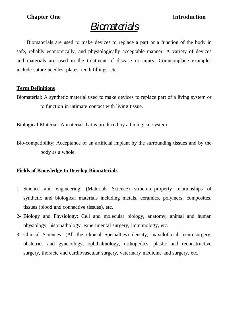
Biomaterials Biomaterials are used to make devices to replace a part or a function of the body in
safe, reliably economically, and physiologically acceptable manner. A variety of devices
and materials are used in the treatment of disease or injury. Commonplace examples
include suture needles, plates, teeth fillings, etc.
Term Definitions
Biomaterial: A synthetic material used to make devices to replace part of a living system or
to function in intimate contact with living tissue.
Biological Material: A material that is produced by a biological system.
Bio-compatibility: Acceptance of an artificial implant by the surrounding tissues and by the
body as a whole.
Fields of Knowledge to Develop Biomaterials
1- Science and engineering: (Materials Science) structure-property relationships of
synthetic and biological materials including metals, ceramics, polymers, composites,
tissues (blood and connective tissues), etc.
2- Biology and Physiology: Cell and molecular biology, anatomy, animal and human
physiology, histopathology, experimental surgery, immunology, etc.
3- Clinical Sciences: (All the clinical Specialties) density, maxillofacial, neurosurgery,
obstetrics and gynecology, ophthalmology, orthopedics, plastic and reconstructive
surgery, thoracic and cardiovascular surgery, veterinary medicine and surgery, etc.
Chapter One Introduction
٢
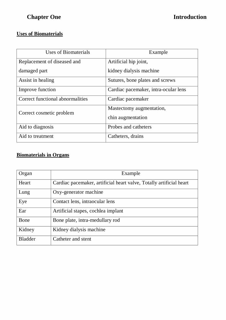
Uses of Biomaterials
Uses of Biomaterials Example
Replacement of diseased and
damaged part
Artificial hip joint,
kidney dialysis machine
Assist in healing Sutures, bone plates and screws
Improve function Cardiac pacemaker, intra-ocular lens
Correct functional abnormalities Cardiac pacemaker
Correct cosmetic problem Mastectomy augmentation,
chin augmentation
Aid to diagnosis Probes and catheters
Aid to treatment Catheters, drains
Biomaterials in Organs
Organ Example
Heart Cardiac pacemaker, artificial heart valve, Totally artificial heart
Lung Oxy-generator machine
Eye Contact lens, intraocular lens
Ear Artificial stapes, cochlea implant
Bone Bone plate, intra-medullary rod
Kidney Kidney dialysis machine
Bladder Catheter and stent
Chapter One Introduction
٣
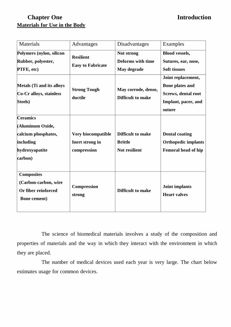
Materials for Use in the Body
Materials Advantages Disadvantages Examples
Polymers (nylon, silicon
Rubber, polyester,
PTFE, etc)
Resilient
Easy to Fabricate
Not strong
Deforms with time
May degrade
Blood vessels,
Sutures, ear, nose,
Soft tissues
Metals (Ti and its alloys
Co-Cr alloys, stainless
Steels)
Strong Tough
ductile
May corrode, dense,
Difficult to make
Joint replacement,
Bone plates and
Screws, dental root
Implant, pacer, and
suture
Ceramics
(Aluminum Oxide,
calcium phosphates,
including
hydroxyapatite
carbon)
Very biocompatible
Inert strong in
compression
Difficult to make
Brittle
Not resilient
Dental coating
Orthopedic implants
Femoral head of hip
Composites
(Carbon-carbon, wire
Or fiber reinforced
Bone cement)
Compression
strong Difficult to make
Joint implants
Heart valves
The science of biomedical materials involves a study of the composition and
properties of materials and the way in which they interact with the environment in which
they are placed.
The number of medical devices used each year is very large. The chart below
estimates usage for common devices.

Chapter One Introduction
٤
Device Usage Estimate
Contact lens 75,000,000
Hip and knee prostheses 1,000,000
Catheter 300,000,000
Heart valve 200,000
Vascular graft 400,000
Breast implant 300,000
Dental implant 500,000
Pace maker 200,000
Renal dialyzer 25,000,000
Cardiovascular 2,000,000
Intraocular lens 7,000,000
Left ventricular assist devices 100,000
Selection of Biomedical Materials
The process of material selection should ideally be for a logical sequence involving:
1- Analysis of the problem;
2- Consideration of requirement;
3- Consideration of available material and their properties leading to:
4- Choice of material.
The choice of a specific biomedical material is now determined by consideration of the
following:
1- A proper specification of the desired function for the material;
2- An accurate characterization of the environment in which it must function, and
the effects that environment will have on the properties of the material;
3- A delineation of the length of time the material must function;
4- A clear understanding of what is meant by safe for human use.
Chapter One Introduction
٥
Materials Evaluation
As the number of available materials increases, it becomes more and more important
to be protected from unsuitable products or materials, which haven't been thoroughly
evaluated.
Most manufacturers of materials operate an extensive quality assurance program and
materials are thoroughly tested before being released to the general practitioner.
1- Standard Specifications: Many standard specification tests of both national and
international standards organizations (ISO) are now available, which effectively
maintain quality levels. Such specifications normally give details for:
(a) the testing of certain products,
(b) the method of calculating the results
(c) the minimum permissible result, which is acceptable.
2- Laboratory Evaluation: Laboratory tests, some of which are used in standard
specification, can be used to indicate the suitability of certain materials. It is
important that methods used to evaluate materials in laboratory give results, which
can be correlated with clinical experience.
3- Clinical Trials: Although laboratory tests can provide many important and useful
data on materials, the ultimate test is the controlled clinical trial and verdict of
practitioners after a period of use in general practice. Many materials produce good
results in the laboratory, only to be found lacking when subjected to clinical use.
The majority of manufacturers carry out extensive clinical trials of new materials,
normally in cooperation with a university or hospital department, prior to releasing
a product for use by general practitioners.
The most common classes of materials used as biomedical materials are
polymers, metals, and ceramics. These three classes are used singly and in combination to
form most of the implantation devices available today.
Chapter One Introduction
٦
1- Polymers
There are a large number of polymeric materials that have been used as implants or
part of implant systems. The polymeric systems include acrylics, polyamides, polyesters,
polyethylene, polysiloxanes, polyurethane, and a number of reprocessed biological
materials.
Some of the applications include the use of membranes of ethylene-vinyl-acetate
(EVA) copolymer for controlled release and the use of poly-glycolic acid for use as a
resorbable suture material. Some other typical biomedical polymeric materials applications
include: artificial heart, kidney, liver, pancreas, bladder, bone cement, catheters, contact
lenses, cornea and eye-lens replacements, external and internal ear repairs, heart valves,
cardiac assist devices, implantable pumps, joint replacements, pacemaker, encapsulations,
soft-tissue replacement, artificial blood vessels, artificial skin, and sutures.
As bioengineers search for designs of ever increasing capabilities to meet the needs of
medical practice, polymeric materials alone and in combination with metals and ceramics
are becoming increasingly incorporated into devices used in the body.
2- Metals
The metallic systems most frequently used in the body are:
(a) Iron-base alloys of the 316L stainless steel
(b) Titanium and titanium-base alloys, such as
(i)Ti-6% Al-4%V, and commercially pure ≥ 98.9%
(ii) Ti-Ni (55% Ni and 45% Ti)
(c) Cobalt base alloys of four types
(i) Cr (27-30%), Mo (5-7%), Ni (2-5%)
(ii) Cr (19-21%), Ni (9-11%), W (14-16%)
(iii) Cr (18-22%), Fe (4-6%), Ni (15-25%), W (3-4%)
(iv)Cr (19-20%), Mo (9-10%), Ni (33-37%)
Chapter One Introduction
٧
The most commonly used implant metals are the 316L stainless steels, Ti-6%-4%V,
and Cobalt base alloys of type "i" and "ii". Other metal systems being investigated include
Cobalt-base alloys of type "iii" and "iv", and Niobium and shape memory alloys, of which
(Ti 45% - 55%Ni) is receiving most attention. Further details of metallic biomedical
materials will be given later.
3- Composite Materials
Composite materials have been extensively used in dentistry and prosthesis designers
are now incorporating these materials into other applications. Typically, a matrix of
ultrahigh-molecular-weight polyethylene (UHMWPE) is reinforced with carbon fibers.
These carbon fibers are made by pyrolizing acrylic fibers to obtain oriented graphitic
structure of high tensile strength and high modulus of elasticity. The carbon fibers are 6-
15µm in diameter, and they are randomly oriented in the matrix. In order for the high
modulus property of the reinforcing fibers to strengthen the matrix, a sufficient interfacial
bond between the fiber and matrix must be achieved during the manufacturing process.
This fiber reinforced composite can then be used to make a variety of implants such as
intra-medullary rods and artificial joints. Since the mechanical properties of these
composites with the proportion of carbon fibers in the composites, it is possible to modify
the material design flexibility to suit the ultimate design of prostheses.
Composites have unique properties and are usually stronger than any of the single
materials from which they are made. Workers in this field have taken advantages of this
fact and applied it to some difficult problems where tissue in-growth is necessary.
Examples:
Deposited Al2O3 onto carbon;
Carbon / PTFE;
Al2O3 / PTFE;
PLA-coated Carbon fibers.

Chapter One Introduction
٨
4 – Ceramics
The most frequently used ceramic implant materials include aluminum oxides, calcium
phosphates, and apatites and graphite. Glasses have also been developed for medical
applications. The use of ceramics was motivated by:
(i) their inertness in the body,
(ii) their formability into a variety of shapes and porosities,
(iii) their high compressive strength, and
(iv) some cases their excellent wear characteristics.
Selected applications of ceramics include:
(a) hip prostheses,
(b) artificial knees,
(c) bone grafts,
(d) a variety of tissues in growth related applications in
(d.1) orthopedics
(d.2) dentistry, and
(d.3) heart valves.
Applications of ceramics are in some cases limited by their generally poor mechanical
properties: (a) in tension; (b) load bearing, implant devices that are to be subjected to
significant tensile stresses must be designed and manufactured with great care if ceramics
are to be safely used.
5 – Biodegradable Materials
Another class of materials that is receiving increased attention is biodegradable
materials. Generally, when a material degrades in the body its properties change from their
original values leading to altered and less desirable performance. It is possible, however, to
design into an implant's performance the controlled degradation of a material, such that
natural tissue replaces the prosthesis and its function.
Chapter One Introduction
٩
Examples include: Suture material that hold a wound together but resorb in the
body as the wound heals and gains strength. Another application of these materials occurs
when they are used to encourage natural tissue to grow. Certain wound dressings and
ceramic bone augmentation materials encourage tissue to grow into them by providing a
"scaffold". The scaffold material may or may not resorb over a period of time but in each
case, natural tissue has grown into the space, then by restoring natural function. One final
application of biodegradable materials is in drug therapy, where it is possible to chemically
bond certain drugs to the biodegradable material, when these materials are placed within
the body the drug is released as the material degrades, thereby providing a localized,
sustained release of drugs over a predictable period of time.
Success and Failure are seen with Biomaterials and Medical Devices
Most biomaterials and medical devices perform satisfactorily, improving the quality of
life for the recipient or saving lives. Still, man-made constructs are never perfect.
Manufactured devices have a failure rate. Also, all humans differ in genetics, gender, body
chemistries, living environment, and physical activity. Furthermore, physicians also differ
in their "talent" for implanting devices.
The other side to the medical device success story is that there are problems,
compromises and complications that occur with medical devices.
Central issues for the biomaterials scientist, manufacturer, patient, physician, and
attorney are:
1- what represents good design;
2- Who should be responsible when devices perform with an inappropriate host
response;
3- What is the cost/risk or cost/benefit ratio for the implant or therapy?
These five characteristics of biomaterial science-multidisciplinary, multi-material,
need driven, substantial market, and risk-benefit, color the field of biomaterials.
Chapter One Introduction
١٠
What Subjects are Important to Biomaterials Science?
1- Toxicology
A biomaterial should not be toxic, unless it is specifically engineered for
such requirements (for example a "smart" bomb" drug delivery system that targets
cancer cells and destroy them). Toxicology for biomaterials deals with the
substances that migrate out of the biomaterials. It is reasonable to say that a
biomaterial should not give off anything from its mass unless it is specifically
designed to do so.
2- Biocompatibility
It is the ability of a material to perform with an appropriate host response
in a specific application. "Appropriate host response" includes lack of blood
clotting, resistance of bacterial colonization and normal heating. The operational
definition of biocompatible "the patient is alive so it must be biocompatible".
3-Functional Tissue Structure and Pathobiology
Biomaterials incorporated into medical devices are implanted into tissues
and organs. Therefore, the key principles governing the structure of normal and
abnormal cells, tissues or organs, the technique by which the structure and
function of normal and abnormal tissues are studied, and the fundamental
mechanisms of disease processes are critical considerations to workers in the
field.
4- Healing
Special processes are invoked when a material or device heals in the
body. Injury to tissue will stimulate the well-defined inflammatory reaction
sequence that leads to healing. When a foreign body is present in the wound site,
the reaction sequence is referred to as the "foreign body reaction". This reaction
will differ in intensity and duration depending upon the anatomical site involved.
Chapter One Introduction
١١
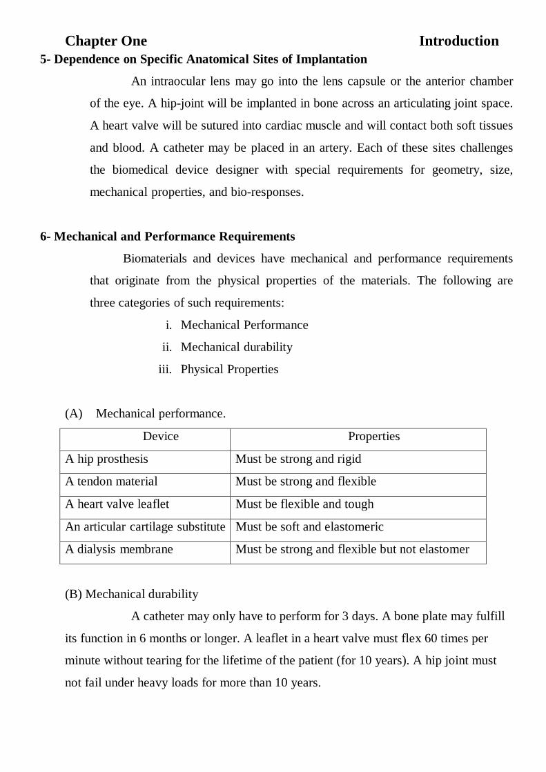
5- Dependence on Specific Anatomical Sites of Implantation
An intraocular lens may go into the lens capsule or the anterior chamber
of the eye. A hip-joint will be implanted in bone across an articulating joint space.
A heart valve will be sutured into cardiac muscle and will contact both soft tissues
and blood. A catheter may be placed in an artery. Each of these sites challenges
the biomedical device designer with special requirements for geometry, size,
mechanical properties, and bio-responses.
6- Mechanical and Performance Requirements
Biomaterials and devices have mechanical and performance requirements
that originate from the physical properties of the materials. The following are
three categories of such requirements:
i. Mechanical Performance
ii. Mechanical durability
iii. Physical Properties
(A) Mechanical performance.
Device Properties
A hip prosthesis Must be strong and rigid
A tendon material Must be strong and flexible
A heart valve leaflet Must be flexible and tough
An articular cartilage substitute Must be soft and elastomeric
A dialysis membrane Must be strong and flexible but not elastomer
(B) Mechanical durability
A catheter may only have to perform for 3 days. A bone plate may fulfill
its function in 6 months or longer. A leaflet in a heart valve must flex 60 times per
minute without tearing for the lifetime of the patient (for 10 years). A hip joint must
not fail under heavy loads for more than 10 years.
Chapter One Introduction
١٢
(C) The physical properties
The dialysis membrane has a specified permeability. The articular cup of
the hip joint has high lubricity. The intraocular lens has clarity and refraction
requirements.
7- Industrial Involvement
A significant basic research effort is now under way to understand how
biomaterials function and how to optimize them. At the same time, companies are
producing implants for use in humans and appropriate to the mission of the company,
earning profits on the sale of medical devices.
Industry deals well with technologies, such as packaging, sterilization,
storage, distribution and quality control, and analysis. These subjects are specialized
technologies, often ignored by academic researchers.
8- Ethics
A wide range of ethical considerations impact biomaterials. Like most
ethical questions, an absolute answer may be difficult to come by. Some articles have
addressed ethical questions in biomaterials and debated the important points.
9- Regulation
The patient demands safe medical devices. To prevent inadequately tested
devices and materials from coming on the market, and to screen out individuals clearly
unqualified to produce biomaterials. The International Standards Organization (ISO)
has introduced international standards for world community.

CHAPTER TWO PROPERTIES OF BIOMATERIALS
١
1- Physical Properties
(i) Mechanical Properties of Biomaterials
The tensile test is a common testing procedure used to provide data for
characterization of biomaterials. The discussion below focuses on special
considerations needed fro tensile testing biological soft tissue (e.g. ligament and
tendon) compared to traditional engineering materials (e.g. aluminum and steel).
(a) Traditional Engineering Materials
Common assumptions used for testing and analysis of traditional materials are
that they are homogeneous, exhibit small deformations and are linearly elastic.
Young's modulus (E), the slope of the elastic portion of stress-strain curve, is a
quantity often used to assess a material stiffness. The linear elastic assumption
makes the determination of "E" relatively straight-forward as it can be assessed
anywhere along the initial linear portion of the curve.
Stress-strain curve of traditional engineering materials
CHAPTER TWO PROPERTIES OF BIOMATERIALS
٢
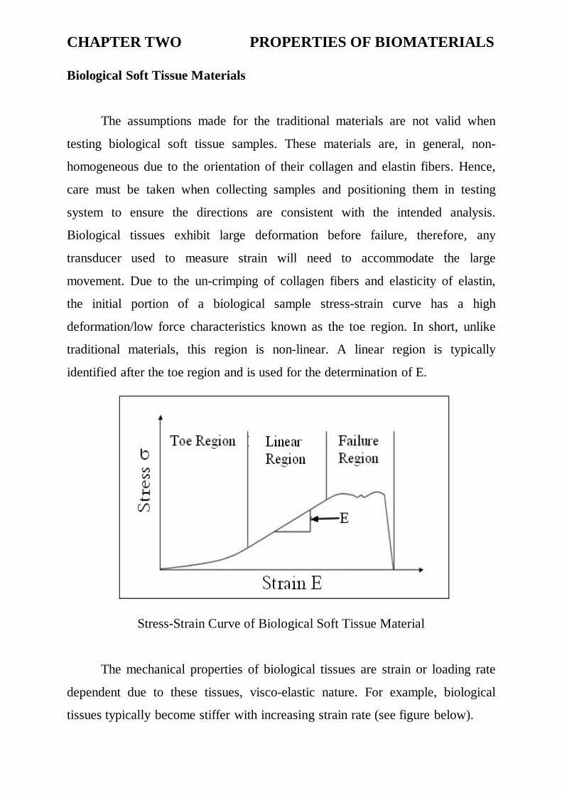
Biological Soft Tissue Materials
The assumptions made for the traditional materials are not valid when
testing biological soft tissue samples. These materials are, in general, non-
homogeneous due to the orientation of their collagen and elastin fibers. Hence,
care must be taken when collecting samples and positioning them in testing
system to ensure the directions are consistent with the intended analysis.
Biological tissues exhibit large deformation before failure, therefore, any
transducer used to measure strain will need to accommodate the large
movement. Due to the un-crimping of collagen fibers and elasticity of elastin,
the initial portion of a biological sample stress-strain curve has a high
deformation/low force characteristics known as the toe region. In short, unlike
traditional materials, this region is non-linear. A linear region is typically
identified after the toe region and is used for the determination of E.
Stress-Strain Curve of Biological Soft Tissue Material
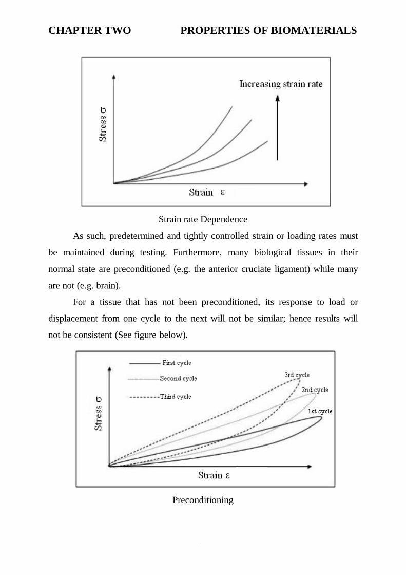
The mechanical properties of biological tissues are strain or loading rate
dependent due to these tissues, visco-elastic nature. For example, biological
tissues typically become stiffer with increasing strain rate (see figure below).
CHAPTER TWO PROPERTIES OF BIOMATERIALS
٣
Strain rate Dependence
As such, predetermined and tightly controlled strain or loading rates must
be maintained during testing. Furthermore, many biological tissues in their
normal state are preconditioned (e.g. the anterior cruciate ligament) while many
are not (e.g. brain).
For a tissue that has not been preconditioned, its response to load or
displacement from one cycle to the next will not be similar; hence results will
not be consistent (See figure below).
Preconditioning
CHAPTER TWO PROPERTIES OF BIOMATERIALS
٤
Depending on the type of tissue being tested or response of interest (e.g.
sudden impact or fatigue failure), preconditioning as part of the testing protocol
may or may not be necessary.
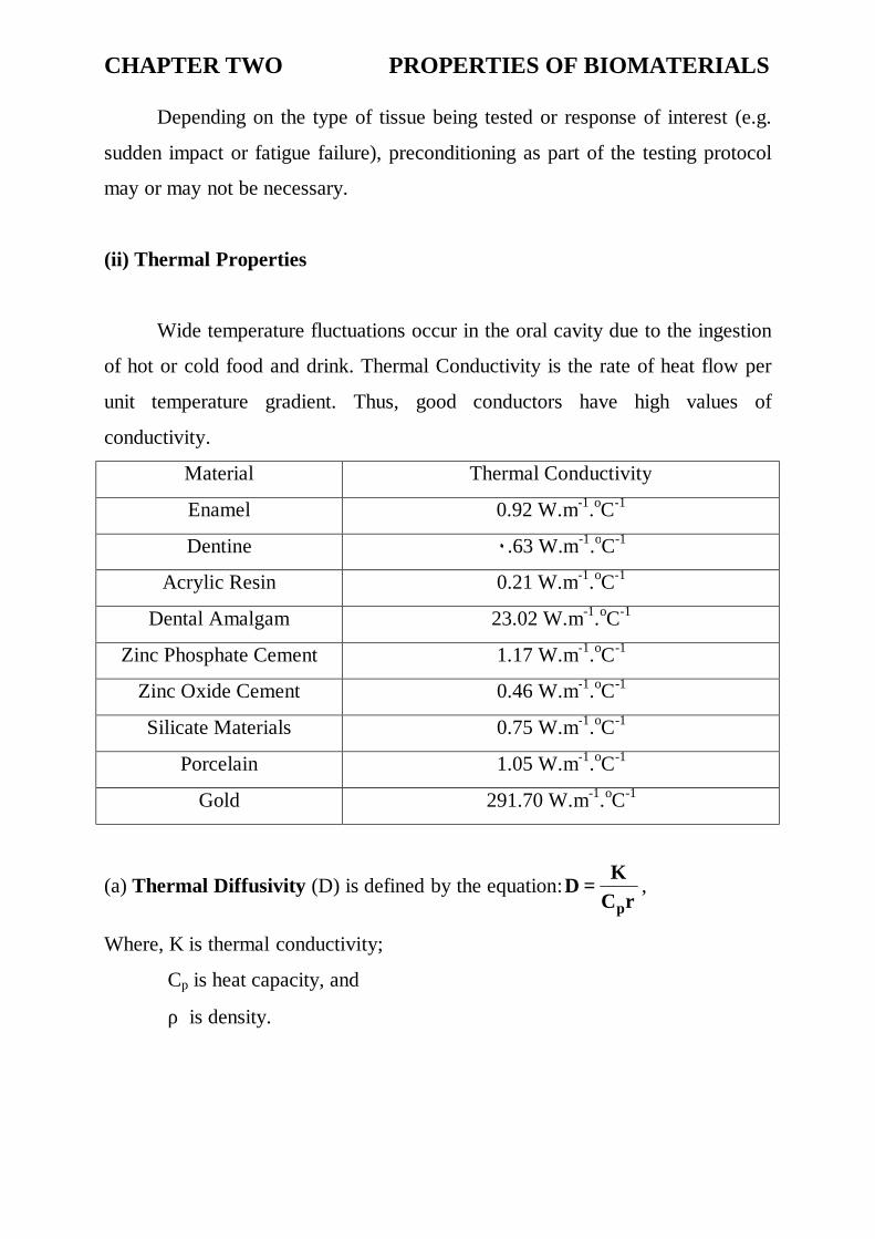
(ii) Thermal Properties
Wide temperature fluctuations occur in the oral cavity due to the ingestion
of hot or cold food and drink. Thermal Conductivity is the rate of heat flow per
unit temperature gradient. Thus, good conductors have high values of
conductivity.
Material Thermal Conductivity
Enamel 0.92 W.m-1.oC-1
Dentine .63 W.m-1.oC-1٠
Acrylic Resin W.m-1.oC-1 0.21
Dental Amalgam W.m-1.oC-1 23.02
Zinc Phosphate Cement 1.17 W.m-1.oC-1
Zinc Oxide Cement 0.46 W.m-1.oC-1
Silicate Materials 0.75 W.m-1.oC-1
Porcelain 1.05 W.m-1.oC-1
Gold 291.70 W.m-1.oC-1
(a) Thermal Diffusivity (D) is defined by the equation:ρ
=pC
KD ,
Where, K is thermal conductivity;
Cp is heat capacity, and
ρ is density.
CHAPTER TWO PROPERTIES OF BIOMATERIALS
٥
Measurements of thermal diffusivity are often made by embedding a
thermocouple in a specimen of material and plunging the specimen into hot or
cold liquid. If the temperature, recorded by the thermocouple, rapidly reaches
that of the liquid, this indicates a high value of diffusivity. A slow response
indicates a lower value of diffusivity is preferred. In many circumstances, a low
value of diffusivity is preferred. There are occasions on which a high value is
beneficial. For example, a denture base material, ideally, should have a high
value of thermal diffusivity in order that the patient retains a satisfactory
response to hot and cold stimuli in the mouth.
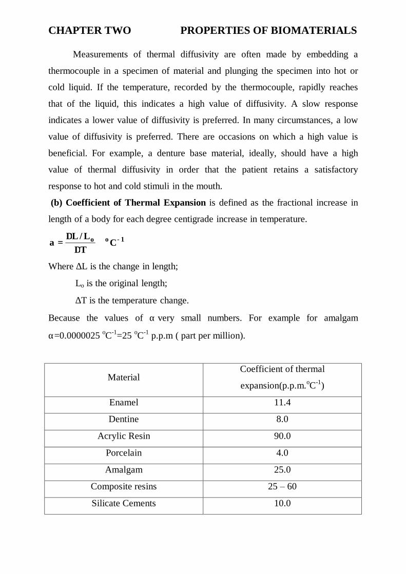
(b) Coefficient of Thermal Expansion is defined as the fractional increase in
length of a body for each degree centigrade increase in temperature.
TL/L o
∆∆
=α 1o C−
Where ∆L is the change in length;
Lo is the original length;
∆T is the temperature change.
Because the values of α very small numbers. For example for amalgam
α=0.0000025 oC-1=25 oC-1 p.p.m ( part per million).
Material Coefficient of thermal
expansion(p.p.m.oC-1)
Enamel 11.4
Dentine 8.0
Acrylic Resin 90.0
Porcelain 4.0
Amalgam 25.0
Composite resins 25 – 60
Silicate Cements 10.0

CHAPTER TWO PROPERTIES OF BIOMATERIALS
٦
This property is particularly important for filling materials. For filling
materials, the most ideal combination of properties would be low value of
diffusivity combined with (α) similar to that for tooth substance.
(2) Chemical Properties
One of the main factors, which determine the durability of a material, is
its chemical stability. Material should not dissolve, erode or corrode, nor should
they leach important constituents into oral fluids.
(i) Solubility and Erosion
The solubility of a material is a measurement of the extent to which it will
dissolve in a given fluid, for example, water or saliva. Erosion is a process
which combines the chemical process of dissolution with a mild mechanical
action.
These properties are particularly important for all restorative materials
since a high solubility or poor resistance to erosion will severely limit the
effective lifetime of the restoration.
The pH of oral fluids may vary from pH4 to pH8.5, representing a range
from mildly acidic to mildly alkaline. Highly acidic soft drinks and the use of
chalk-containing tooth-pastes extend this range from a lower end of pH2 up to
pH11. It is possible for a material to be stable at near neutral pH7 values but to
erode rapidly at extremes of either acidity or alkalinity.
Standard tests of solubility often involve the storage of disc specimens of
materials in water for a period of time, the results being quoted as the percentage
weight loss of the disc. Such methods, however, often give misleading results.
CHAPTER TWO PROPERTIES OF BIOMATERIALS
٧
When comparing silicate and phosphate cements, for example, silicate materials
appear more soluble in simple laboratory test, but in practice they are more
durable than the phosphates.
(ii) Leaching of Constituents
Many materials, when placed in an aqueous environment, absorb water by
a diffusion process. Constituents of the material may be lost into the oral fluids
by a diffusion process commonly referred to as leaching.
Some soft acrylic polymers, used for cushioning, the fitting surfaces of
dentures rely on the absence of relatively large quantities of plasticizer in the
acrylic resin for their softness. The slow leaching of plasticizer causes the resin
to become hard and, therefore, ineffective as a cushion.
Occasionally, leaching is used to the benefit of the patient. For example,
in some cements containing calcium hydroxide, slow leaching causes an alkaline
environment in the base of deep cavities. This has the dual benefit of being
antibacterial and of encouraging secondary dentine formation.
(iii) Corrosion
It is a term which specifically characterizes the chemical reactivity of
metals and alloys. Metals and alloys are good electrical conductors and many
corrosion processes involve the setting up of an electrolytic cell as a first stage
in the process.
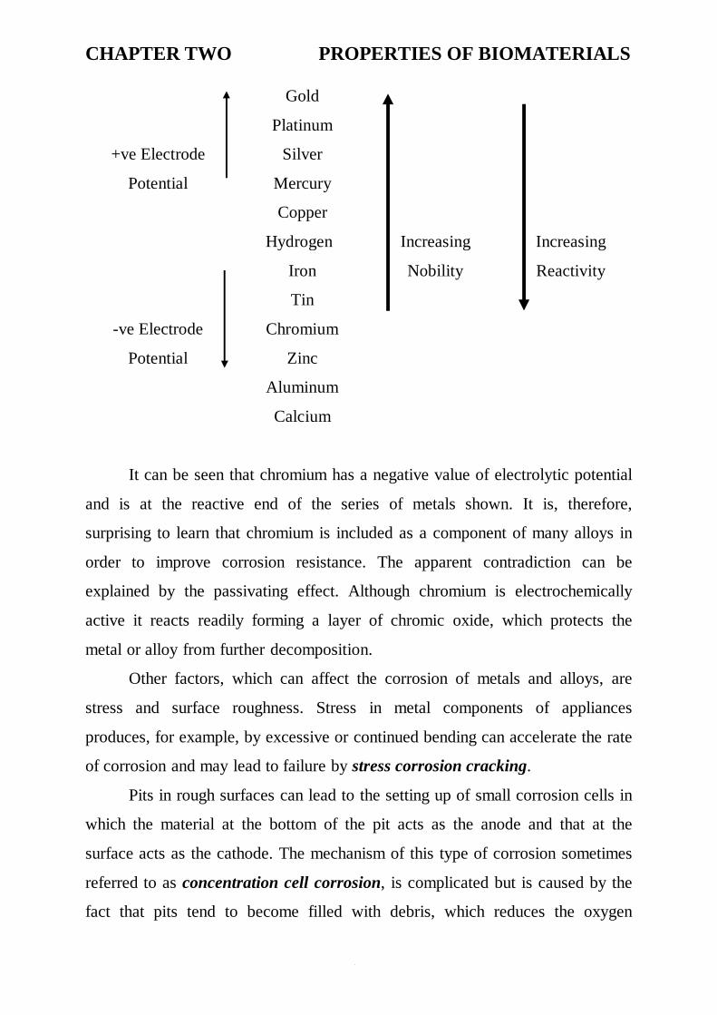
The tendency of a metal to corrode can be predicted from its electrode
potential. It can be seen from the figure below, that materials with large negative
electrode potential values are more reactive whilst those with large positive
values are far less reactive and are often referred to as noble metals.
CHAPTER TWO PROPERTIES OF BIOMATERIALS
٨
+ve Electrode
Potential
Gold
Platinum
Silver
Mercury
Copper
Increasing
Nobility
Increasing
Reactivity
Hydrogen
-ve Electrode
Potential
Iron
Tin
Chromium
Zinc
Aluminum
Calcium
It can be seen that chromium has a negative value of electrolytic potential
and is at the reactive end of the series of metals shown. It is, therefore,
surprising to learn that chromium is included as a component of many alloys in
order to improve corrosion resistance. The apparent contradiction can be
explained by the passivating effect. Although chromium is electrochemically
active it reacts readily forming a layer of chromic oxide, which protects the
metal or alloy from further decomposition.
Other factors, which can affect the corrosion of metals and alloys, are
stress and surface roughness. Stress in metal components of appliances
produces, for example, by excessive or continued bending can accelerate the rate
of corrosion and may lead to failure by stress corrosion cracking.
Pits in rough surfaces can lead to the setting up of small corrosion cells in
which the material at the bottom of the pit acts as the anode and that at the
surface acts as the cathode. The mechanism of this type of corrosion sometimes
referred to as concentration cell corrosion, is complicated but is caused by the
fact that pits tend to become filled with debris, which reduces the oxygen

CHAPTER TWO PROPERTIES OF BIOMATERIALS
٩
concentration in the base of the pit compared with the surface. In order to reduce
corrosion by this new mechanism, metals and alloys used in the mouth should be
polished to remove surface irregularities.
Ideally, a material placed into a patient's mouth should be non-toxic, non-
irritant, have no carcinogenic or allergic potential and if used as a filling
material, should be harmless to the pulp.
CHAPTER THREE BIOCERAMICS
١
(I) Bio-ceramics
Ceramics are used for the repair and restoration of diseased or damaged parts
of the musculo-skeletal system.
Bio-ceramics may be:
1- Bioinert like Alumina (Al2O3), Zirconia (ZrO2);
2- Resorbable like tri-calcium phosphate (TCP);
3- Bioactive like Hydroxyapatite, bioactive glasses, and glass-ceramics;
4- Porous for tissue in-growth (hydroxyapatite-coated metals, alumina) of the jaw
bone.
Applications include: Replacement for hips, knees, teeth, tendons and
ligaments, and repair for periodontal disease, maxillofacial reconstruction,
augmentation and stabilization, spinal fusion and bone fillers after tumor surgery.
Carbon coatings are thrombo-resistant and are used for prosthetic heart valves.
(II) Types of Bio-ceramics – Tissue Attachment
The mechanism of tissue attachment is directly related to the type of tissue
response at the implant interface. No material implanted in living tissues is inert; all
materials elicit a response from living tissues.
Four types of response allow different means of achieving attachment of
prostheses to the musculo-skeletal system.
The types of implant-tissue response are:
CHAPTER THREE BIOCERAMICS
٢
a- If the material is toxic, the surrounding tissue dies;
b- the material is nontoxic and biologically inactive (nearly inert), a fibrous tissue
of variable thickness forms;
c- If the material is nontoxic and biologically active (bioactive), an interfacial
bond forms;
d- If the material is nontoxic and dissolves, the surrounding tissue replaces it.
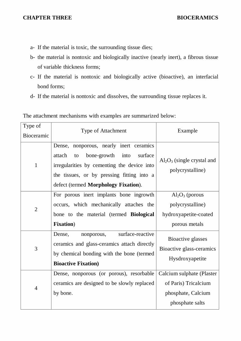
The attachment mechanisms with examples are summarized below:
Type of
Bioceramic Type of Attachment Example
1
Dense, nonporous, nearly inert ceramics
attach to bone-growth into surface
irregularities by cementing the device into
the tissues, or by pressing fitting into a
defect (termed Morphology Fixation).
Al2O3 (single crystal and
polycrystalline)
2
For porous inert implants bone ingrowth
occurs, which mechanically attaches the
bone to the material (termed Biological
Fixation)
Al2O3 (porous
polycrystalline)
hydroxyapetite-coated
porous metals
3
Dense, nonporous, surface-reactive
ceramics and glass-ceramics attach directly
by chemical bonding with the bone (termed
Bioactive Fixation)
Bioactive glasses
Bioactive glass-ceramics
Hysdroxyapetite
4
Dense, nonporous (or porous), resorbable
ceramics are designed to be slowly replaced
by bone.
Calcium sulphate (Plaster
of Paris) Tricalcium
phosphate, Calcium
phosphate salts
CHAPTER THREE BIOCERAMICS
٣
(III) Nearly Inert Crystalline Bioceramics
Inert refers to materials that are essentially stable with little or no tissue
reactivity when implanted within the living organism.
When a biomaterial is nearly inert (type1) and the interface is not chemically or
biologically bonded, there is relative movement and progressive development of a
non-adherent fibrous capsule in both soft and hard tissues. Movement at the
biomaterial-tissue interface eventually leads to deterioration in function of the
implant or the tissue at the interface or both.
Bone at an interface with type1, nearly inert, implant is very often structurally
weak because of disease, localized death of bone, or stress shielding when higher
elastic modules of the implant prevents the bone from being loaded properly. Most
notable among the nearly inert ceramics are alumina and special forms of carbon and
silicon.
High density high purity (>99.5%) alumina (α-Al2O3) was the first bioceramic
widely used clinically. It is used in load-bearing hip prostheses and dental implants,
because of its combination of excellent corrosion resistance, good biocompatibility,
and high wear resistance, and high strength.
Although some dental implants are single-crystal sapphire, most alumina
devices are very-fine-grained polycrystalline α-Al2O3. A very small amount of
magnesia (<0.5%) is used as an aid to sintering and to limit grain growth during
sintering.
Strength, fatigue resistance, and fracture toughness of polycrystalline α-Al2O3
are a function of grain size and percentage of sintering aid, i.e. purity. Alumina with
an average grain size of <4µm and >99.7% purity exhibits good flexural strength and
excellent compressive strength.

CHAPTER THREE BIOCERAMICS
٤
Low wear rates have led to wide-spread use of alumina non-cemented cups,
press fitted into the acetabulum (socket) of the hip. The cups are stabilized by bone
growth into grooves or round pegs. The mating femoral ball surface is also of
alumina, which is bonded to a metallic stem. Though long-term results in general
have been excellent, it is essential that the age of the patient, nature of the disease of
the joint, and bioceramics of the repair be considered carefully before any prosthesis
is used.
The primary use of alumina is for the ball of the hip joint, with the socket
component being made of ultrahigh molecular weight polyethylene (PE). Other
clinical applications of alumina implants include knee prostheses, bone screws, jaw
bone reconstruction, segmental bone replacement, and blade, screws or post-type
dental implants.
(IV) Porous Ceramics
The concept behind nearly inert, micro-porous bioceramics (type2) is the
ingrowths of tissue into pores on the surface or throughout the implant. The increased
interfacial area between the implant and the tissues result in an increased inertial
resistance to movement of the device in the tissue. The interface is established by the
living tissue in the pores. This method of attachment is often termed biological
fixation. It is capable of withstanding more complex stress states than type1 implants,
which achieve only morphological fixation.
The limitation associated with type2 porous implants is that, for tissue to
remain viable and healthy, it is necessary for the pores to be greater than 100 to
150µm in diameter. The large interfacial area required for the porosity is due to the
need to provide a blood supply to the ingrown connective tissue. Vascular tissue does
not appear in pores, which measure less than 100µm. If micro-movement occurs at
the interface of a porous implant, tissue is damaged, the blood supply may be cut off,
tissue dies, inflammation ensues and the interfacial stability can be destroyed.
CHAPTER THREE BIOCERAMICS
٥
The potential advantage offered by a porous ceramic is the inertness combined
with the mechanical stability of the highly convoluted interface developed when bone
grows into the pores of the ceramic. Mechanical requirements of prostheses,
however, severely restrict the use of low strength porous ceramics to low-load or
non-load bearing applications. Studies show that, when load bearing is not a primary
requirement, nearly inert porous ceramics can provide a functional implant.
When pore sizes exceed 100µm, bone will grow within the interconnecting
pore channels near the surface and maintain its blood supply and long-term health. In
this manner, the implant serves as a structural bridge and model or scaffold for bone
formation. The microstructures of certain marine corals make an almost ideal casting
material for obtaining structures with highly controlled pore sizes.
Several types of coral are promising, with pore-size range s of 40-160µm and
200-1000µm. After the coral shape is machined, it is fired to drive off CO2 from the
limestone, forming a porous structure of calcia, or transformed directly into
hydroxyapatite ceramic.
Porous ceramic surfaces can also be prepared by mixing soluble metal or salt
particles into the surface. The pore size and structure are determined by the size and
shape of the soluble particles that are subsequently removed with a suitable etchant.
The porous surface layer produced by this technique is an integral part of the
underlying dense ceramic phase. Materials, such as alumina, may also be made
porous by using a suitable foaming agent that evolves gases during heating.
Porous materials are weaker than the equivalent bulk form. As the porosity
increases, the strength of the material decreases rapidly. Much surface area is also
exposed, so that the effects of the environment on decreasing the strength become
much more important than for dense nonporous materials. ρ−σ=σ c
oe
Where σ is strength;
σο is strength at zero porosity (Nonporous);
CHAPTER THREE BIOCERAMICS
٦
c is a constant;
and ρ is porosity.
(V) Bioactive Glasses and Glass-Ceramics
Another approach to the solution of the problems of interfacial attachment is
the use of bioactive materials (type3). The concept of bioactive materials is
intermediate between resorbable and bioinert.
Certain compositions of glasses, ceramics, glass-ceramics, and composites
have been shown to bond to bone. These materials are also called bioactive ceramics.
Some, even more specialized compositions of bioactive glasses, will bond to soft
tissues as well as bone. A common characteristic of such bioactive materials is a
modification of the surface that occurs upon implantation. The surface forms a
biologically active hydroxycarbonate apatite (HCA) layer, which provides the
bonding interface with tissues. The HCA phase that forms on bioactive implants has
the same chemical structure as the mineral phase in bone, and is therefore responsible
for interfacial bonding.
The bonding results in an interface that resists substantial mechanical forces.
Bonding to bone was first demonstrated for a range of bioactive glasses, which
contained specific amounts of SiO2, CaO, and P2O5. These glasses contained less than
60 mol% SiO2, high contents of Na2O, and CaO, and had a high CaO/ P2O5 ratio.
Such a composition produced a surface that was highly reactive when exposed to an
aqueous medium.
Many bioactive silica glasses are based upon the formula (called 45S5), which
signifies 45wt% SiO2, S as the network former, and a 5 to 1 molar ratio of Ca to P (in
form of CaO and P2O5 do not bond to bone. 45S5 glass implants have been used
successfully for replacement of ear bones and maintenance of the jaw bone for
denture wearers for up to eight years, with nearly 90% retention ratio.
CHAPTER THREE BIOCERAMICS
٧
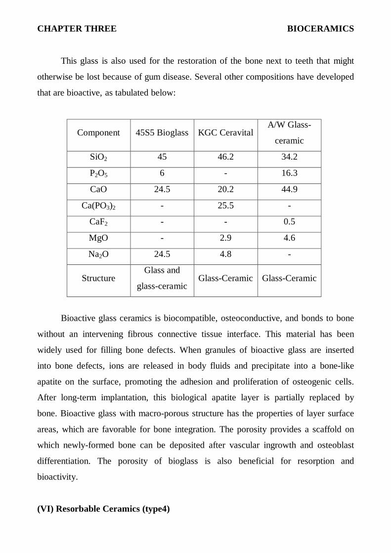
This glass is also used for the restoration of the bone next to teeth that might
otherwise be lost because of gum disease. Several other compositions have developed
that are bioactive, as tabulated below:
Bioactive glass ceramics is biocompatible, osteoconductive, and bonds to bone
without an intervening fibrous connective tissue interface. This material has been
widely used for filling bone defects. When granules of bioactive glass are inserted
into bone defects, ions are released in body fluids and precipitate into a bone-like
apatite on the surface, promoting the adhesion and proliferation of osteogenic cells.
After long-term implantation, this biological apatite layer is partially replaced by
bone. Bioactive glass with macro-porous structure has the properties of layer surface
areas, which are favorable for bone integration. The porosity provides a scaffold on
which newly-formed bone can be deposited after vascular ingrowth and osteoblast
differentiation. The porosity of bioglass is also beneficial for resorption and
bioactivity.
(VI) Resorbable Ceramics (type4)
Component 45S5 Bioglass KGC Ceravital A/W Glass-
ceramic
SiO2 45 46.2 34.2
P2O5 6 - 16.3
CaO 24.5 20.2 44.9
Ca(PO3)2 - 25.5 -
CaF2 - - 0.5
MgO - 2.9 4.6
Na2O 24.5 4.8 -
Structure Glass and
glass-ceramic Glass-Ceramic Glass-Ceramic
CHAPTER THREE BIOCERAMICS
٨
They are designed to degrade gradually over a period of time and be replaced
by the natural host tissue. This leads to a very thin or nonexistent interfacial
thickness. This is the optimal solution to the problem of biomaterials if the
requirements of strength and short-term performance can be met. Natural tissues can
repair themselves and are gradually replaced throughout life by a continual turnover
of cell population. As we grow older, the replacement of cells and tissues is slower
and less efficient, which is why parts "wear out", unfortunately sum faster than
others. Thus resorbable biomaterials are based on the same principles of repair which
have evolved over millions of years.
One of the unique advantages of the resorbable ceramic is that its initial pore-
size can be small, thereby possessing high mechanical strength compared to the
strength of more porous substances. As the ceramic dissolves, it becomes more and
more porous allowing the ingrowth of more supporting tissue to occur. As a result,
mechanical integrity is maintained and stress concentrations minimized.
Complications in development of resorbable bio-ceramics are:
(1) Maintenance of strength and the stability of the interface during the
degradation period and replacement by the natural host tissue.
(2) Matching resorption rates to the repair to the repair rate of body tissues which
themselves vary enormously. Some dissolve too rapidly and some too slowly.
Because large quantities of material may be replaced, it is also essential that a
resorbable biomaterial consist only of metabolically acceptable substances.
This criterion imposes considerable limitations on the compositional design of
resorbable biomaterials.
Resorbable bioceramics have been used too treat maxillofacial defects, for
obliterating periodontal pockets, as artificial tendons and as composite bone plates.
CHAPTER THREE BIOCERAMICS
٩
Calcium Phosphate Ceramics
Different phases of calcium phosphate ceramics are used depending upon
whether a resorbable or bioactive material is desired. These include dicalcium
phosphate (CaHPO4) and hydroxyapatite Ca10 (PO4)6(OH)2 [HA].
Applications include dental implants, skin treatments, gum treatment, jawbone
reconstruction, orthopedics, facial surgery, ear, nose and throat repair, and spinal
surgery.
The mechanical behavior of calcium phosphate ceramics strongly influences
their application as implants. Tensile and compressive strength and fatigue resistance
depend on the total volume of porosity. Because HA implant have low reliability
under tensile load, such calcium phosphate bioceramics can only be used as powders,
or as small, unloaded implants with reinforcing metal posts, coatings on metal
implants, low-bonded porous implants where bone growth acts as a reinforcing phase
and as the bioactive phase in a composite.
Calcium phosphate (CaP) biomaterials are available in various physical forms
(particles or blocks; dense or porous). One of their main characteristics is their
porosity. The ideal pore size for bioceramic is similar to that of spongy bone.
Macroprosity (pore size >50µm) is intentionally introduced into the material by
adding volatile substances or porogens (naphthalene, sugar, hydrogen peroxide,
polymer beads, fibers, etc) before sintering at high temperatures.
Microporosity is formed when the volatile materials are released.
Microporosity is related to pore size <10µm. Microporosity is the result of the
sintering process, where the sintering temperature and time are critical parameters. It
has been demonstrated that microporosity allows body fluid circulation whereas
CHAPTER THREE BIOCERAMICS
١٠
macroporosity provides a scaffold for bone colonization. Average pore size diameter
of 560µm is reported as the ideal macropore size for bone ingrowth compared to a
smaller size (300µm). The main difference between the different commercially
available BCP (biophasic calcium phosphate) are the microporosities, which are
dependent on sintering process.
Composites and Coatings
One of the primary restrictions on clinical use of bioceramics is the uncertain
lifetime under the complex stress states, slow crack growth, and cyclic fatigue that
arise in many clinical applications. Two solutions to these limitations are the use of
bioactive ceramics as coatings or in composites. Much of the rapid growth in the field
of bioactive ceramics is due to development of various composite and coating
systems.
Composites have been composed of plastic, carbon, glass, or ceramic matrices
reinforced with various types of fibers, including carbon, SiC, stainless steel, HA,
phosphate glass, and ZrO2. In most cases the goal is to increase flexural strength and
strain to failure and decrease elastic modulus. The strongest composite achieved to
date is A/W glass-ceramic containing a dispersion of tetragonal Zirconia, which has a
bend strength of 700MPa, and fracture toughness of 4MPam1/2.
CHAPTER THREE BIOCERAMICS
١١
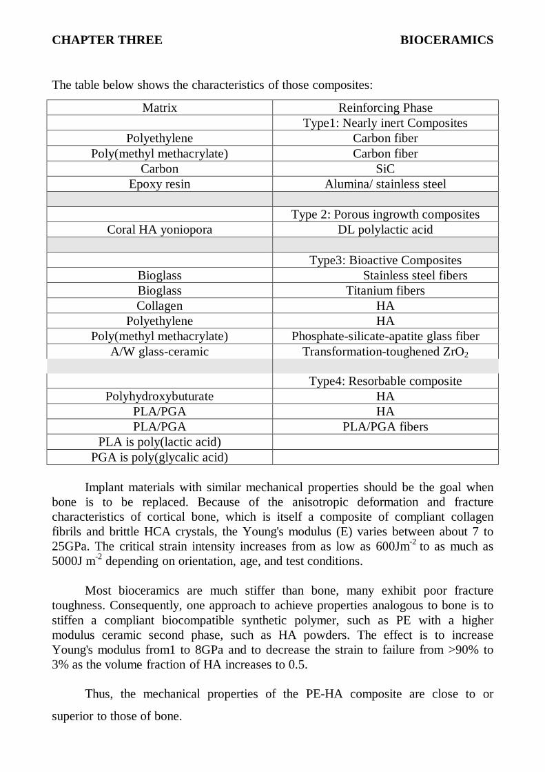
The table below shows the characteristics of those composites:
Matrix Reinforcing Phase Type1: Nearly inert Composites
Polyethylene Carbon fiber Poly(methyl methacrylate) Carbon fiber
Carbon SiC Epoxy resin Alumina/ stainless steel
Type 2: Porous ingrowth composites
Coral HA yoniopora DL polylactic acid
Type3: Bioactive Composites Bioglass Stainless steel fibers Bioglass Titanium fibers Collagen HA
Polyethylene HA Poly(methyl methacrylate) Phosphate-silicate-apatite glass fiber
A/W glass-ceramic Transformation-toughened ZrO2 Type4: Resorbable composite
Polyhydroxybuturate HA PLA/PGA HA PLA/PGA PLA/PGA fibers
PLA is poly(lactic acid) PGA is poly(glycalic acid)
Implant materials with similar mechanical properties should be the goal when bone is to be replaced. Because of the anisotropic deformation and fracture characteristics of cortical bone, which is itself a composite of compliant collagen fibrils and brittle HCA crystals, the Young's modulus (E) varies between about 7 to 25GPa. The critical strain intensity increases from as low as 600Jm-2 to as much as 5000J m-2 depending on orientation, age, and test conditions. Most bioceramics are much stiffer than bone, many exhibit poor fracture toughness. Consequently, one approach to achieve properties analogous to bone is to stiffen a compliant biocompatible synthetic polymer, such as PE with a higher modulus ceramic second phase, such as HA powders. The effect is to increase Young's modulus from1 to 8GPa and to decrease the strain to failure from >90% to 3% as the volume fraction of HA increases to 0.5.
Thus, the mechanical properties of the PE-HA composite are close to or
superior to those of bone.

CHAPTER THREE BIOCERAMICS
١٢
Another promising approach toward achieving high toughness, ductility and
Young's modulus matching that of bone was developed. This composite uses sintered
316 stainless steel of 50-, 100-, 200-µm or Titanium fibers, which provide an
interconnected fibrous matrix which then impregnated with molten 45S5 bioglass.
After the composite is cooled and annealed, very strong and tough material results,
with metal to glass volume ratio between46 to
64 .
Stress enhancement of up to 340MPa is obtained in bending with substantial
ductility of up to 10% elongation, which bends 90o without fracturing.
Coatings
A biometric coating, which has reached a significant level of clinical
application, is the use of HA as a coating on porous metal surfaces for fixation of
orthopedic prostheses. This approach combines biological and bioactive fixation.
Though a wide range of methods have been used to apply the coating, plasma spray
coating is usually preferred. The table below lists the bioceramic coatings:
Substrate Coating 316L stainless steel Pyrolytic carbon 316L stainless steel 45S5 bioglass 316L stainless steel αAl2O3-HA-TiN 316L stainless steel HA
Co-Cr alloy 45S5 bioglass Co-Cr alloy HA
Ti-6Al-4V alloy 45S5 bioglass Ti-6Al-4V alloy HA Ti-6Al-4V alloy Al2O3
Ti-6Al-4V alloy HA/ABS glass[ABS is alkali borosilicate glass]

CHAPTER THREE BIOCERAMICS
١٣
1- Carbon
The medical use of pyrolytic carbon coatings on metal substrates were used in
heart surgery. The first time the low-temperature isotropic (LTI) carbon coatings
were used in humans was a prosthetic heart valve.
Almost all commonly used prosthetic heart valves today have LTI carbon coatings
for the orifice and/or occluder because of their excellent resistance to blood clot
formation and long fatigue life. More than 600,000 lives have been prolonged
through the use of these bioceramic-in-heart valves.
Three types of Carbon are used in biomedical devices:
1- Low temperature Isotropic (LTI);
2- Ultra low Temperature Isotropic (ULTI);
3- Glassy Carbons.
The LIT, ULTI and glassy carbon are sub-crystalline forms and represent a
lower degree of crystal perfection. There is no order between the layers such as there
is in graphite; therefore the crystal structure of these carbons is two-dimensional.
Such a structure, called turbostratic, has densities between 1400-2100 kg.m-3.
High density LTI carbons are the strongest bulk forms of carbon and their
strength can further be increased by adding silicon.
ULTI carbon can also be produced with high densities and strength, but it is
available only as a thin coating (0.1 to 1µm) of pure carbon.
Glassy carbon is inherently a low density material and, as such, is weak. Its
strength cannot be increased through processing.
CHAPTER THREE BIOCERAMICS
١٤
The turbostratic carbon materials have extremely good wear resistance, some
of which can be attributed to their toughness, i.e. their capacity to sustain large local
elastic strains under concentrated or point loading without galling or incurring surface
damage.
Another unique characteristic of the turbostratic carbons is that they do not
fatigue. The ultimate strength of turbostratic carbon, as opposed to metals, does not
degrade with cyclical loading.
Carbon surfaces are not only thromboresistant, but also appear to be
compatible with the cellular elements of blood'; they do not influence plasma proteins
or alter the activity of plasma enzymes. One of the proposed explanations for the
blood compatibility of these materials is that they absorb blood proteins on their
surface without altering them.
2- Hydroxyapatite (HA)
A second bioceramic coating which has reached a significant level of clinical
applications is the use of HA as a coating on porous metal surfaces for fixation of
orthopedic prostheses.
Resorption or biodegradation of calcium phosphate ceramic is caused by:
(i) Physiochemical dissolution, which depends on the solubility product of the
material and local pH of its environment;
(ii) Physical disintegration into small particles due to preferential chemical
attack of grain boundaries;
(iii) Biological factors, such as phagocytosis, which causes a decrease in local
pH.
CHAPTER THREE BIOCERAMICS
١٥
The rate of biodegradation increases as:
(a) Surface area increases;
(b) Crystallinity increases;
(c) Crystal perfection decreases;
(d) Crystal and grain size decrease;
(e) Ionic substitutions of ++− 2223 Sr ,Mg ,CO in HA take place.
Factors which result in a decreasing rate of biodegradation include:
(1) F-substitution in HA;
(2) Mg2+ substitution in β-TCP [β −Ca3(PO4)2 , β-tricalcium phosphate];
(3) Decreasing β-TCP/HA ratios in biphasic calcium phosphate.
Because of these variables, it is necessary to control the microstructure and
phase state of resorbable calcium phosphate bioceramic in addition to achieving
precise compositional control to produce a given rate of resorption in the body.
Natural Composites
Natural occurring composites are within us all. On the macroscale, soft and
hard tissues are formed from a complex structural array of organic fibers and matrix.
Soft tissues are formed from elastic (elastin) and non-elastic fibers (collagen)
with a cellular matrix between the fibers. Biological structures, such as tendon,
linking muscles to bone, are low in elastin, thus allowing muscle movement to be
translated to the bone. However, ligaments linking bone to bone are high in elastin
allowing movement between bones but retaining sufficient support to stop joints
dislocating.
CHAPTER THREE BIOCERAMICS
١٦
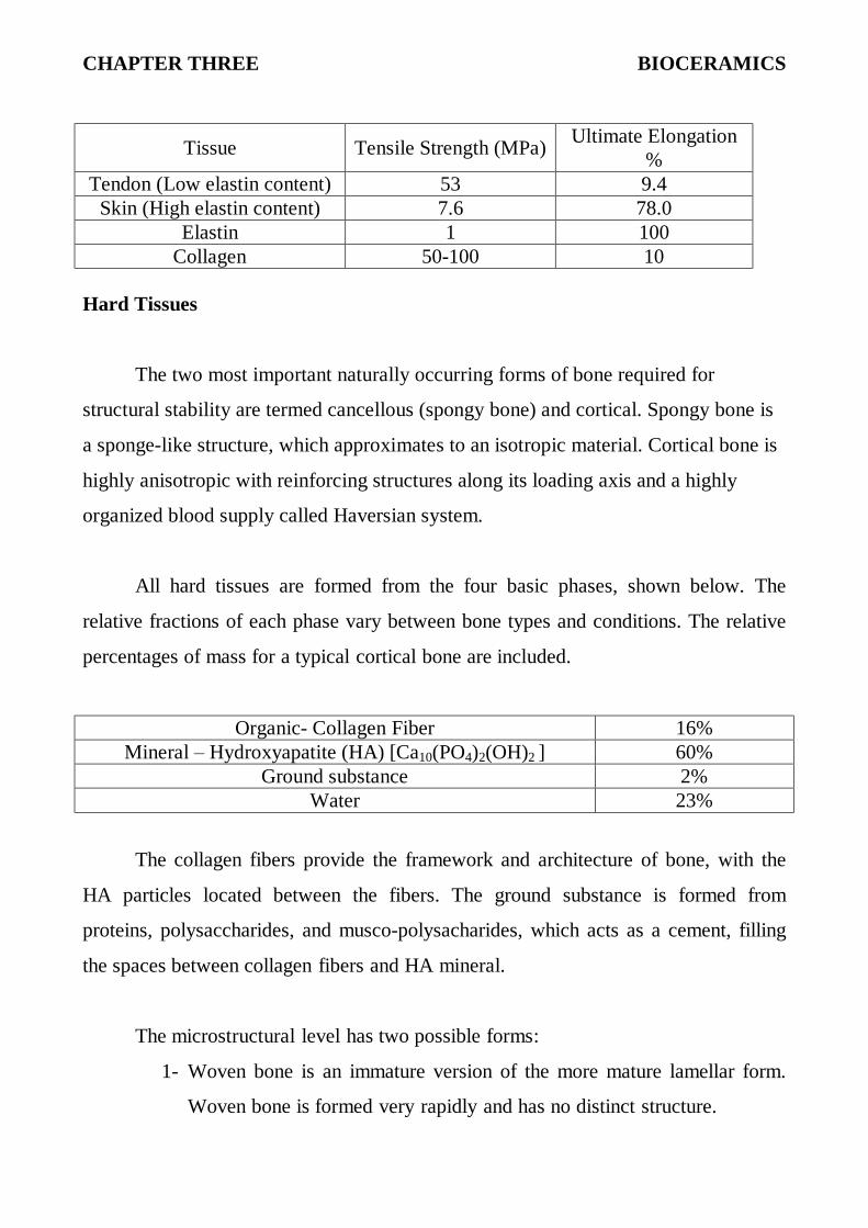
Tissue Tensile Strength (MPa) Ultimate Elongation %
Tendon (Low elastin content) 53 9.4 Skin (High elastin content) 7.6 78.0
Elastin 1 100 Collagen 50-100 10
Hard Tissues
The two most important naturally occurring forms of bone required for
structural stability are termed cancellous (spongy bone) and cortical. Spongy bone is
a sponge-like structure, which approximates to an isotropic material. Cortical bone is
highly anisotropic with reinforcing structures along its loading axis and a highly
organized blood supply called Haversian system.
All hard tissues are formed from the four basic phases, shown below. The
relative fractions of each phase vary between bone types and conditions. The relative
percentages of mass for a typical cortical bone are included.
Organic- Collagen Fiber 16% Mineral – Hydroxyapatite (HA) [Ca10(PO4)2(OH)2 ] 60%
Ground substance 2% Water 23%
The collagen fibers provide the framework and architecture of bone, with the
HA particles located between the fibers. The ground substance is formed from
proteins, polysaccharides, and musco-polysacharides, which acts as a cement, filling
the spaces between collagen fibers and HA mineral.
The microstructural level has two possible forms:
1- Woven bone is an immature version of the more mature lamellar form.
Woven bone is formed very rapidly and has no distinct structure.

CHAPTER THREE BIOCERAMICS
١٧
2- Lamellar bone is formed into concentric rings called Osteons with
central blood supply or Haversian systems. Each osteon is formed from
4-20 rings, with each ring being 4-7 mm thick and having a different
fiber orientation. The arrangement of different fiber orientations in each
layer gives the osteon the appearance of successive light and dark layers.
In the centre of the rings, there is a Haversian canal which contains the
blood supply. Whilst the outer layer is a cement layer formed from
ground substance, it is less mineralized than the rest of the bone and has
no collagen fibers. Consequently, the cement line is a site of weakness.
Synthetic Bone Grafting Materials
These materials must be:
1- Biocompatible with host tissues, i.e.
a- non-toxic;
b- non-allergic;
c- non-carcinogenic;
d- non-inflammatory
2- Able to stimulate bone induction; 3- Resorbable following replacement by bone; 4- Radio-opaque; 5- Capable of withstanding sterilization 6- Inexpensive and stable to variation of temperature and humidity; 7- It has sufficient porosity to allow bone conduction and growth.

CHAPTER FOUR Polymer as Biomaterial
١
Polymer as Biomaterial Polymers have assumed an important role in medical applications. In most
of these applications, polymers have little or no competition from other types of
materials. Their unique properties are:
1- Flexibility;
2- Resistance to biochemical attack;
3- Good biocompatibility;
4- Light weight;
5- Available in a wide variety of compositions with adequate physical and
mechanical properties;
6- Can be easily manufactured into products with the desired shape.
Applications in biomedical field as:
1- Tissue engineering;
2- Implantation of medical devices and artificial organs due to its inert
nature;
3- Prostheses;
4- Dentistry;
5- Bone repair;
6- Drug delivery and targeting into sites of inflammation or tumors;
7- Plastic tubing for intra-venous infusion;
8- Bags for the transport of blood plasma;
9- Catheter.
A few of the major classes of polymer are listed below:
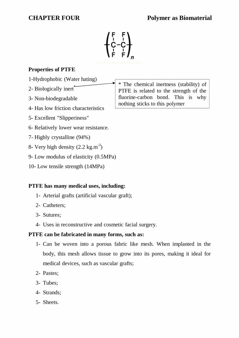
(1) (PTFE) Polytetrafluoroethylene is a fluorocarbon–based polymer.
Commercially, the material is best known as Teflon. It is made by free-radical
polymerization of tetrafluoroethylene and has a carbon backbone chain, where
each carbon has two fluorine atoms attached to it.
CHAPTER FOUR Polymer as Biomaterial
٢
Properties of PTFE
1-Hydrophobic (Water hating)
2- Biologically inert*
3- Non-biodegradable
4- Has low friction characteristics
5- Excellent "Slipperiness"
6- Relatively lower wear resistance.
7- Highly crystalline (94%)
8- Very high density (2.2 kg.m-3)
9- Low modulus of elasticity (0.5MPa)
10- Low tensile strength (14MPa)
PTFE has many medical uses, including:
1- Arterial grafts (artificial vascular graft);
2- Catheters;
3- Sutures;
4- Uses in reconstructive and cosmetic facial surgery.
PTFE can be fabricated in many forms, such as:
1- Can be woven into a porous fabric like mesh. When implanted in the
body, this mesh allows tissue to grow into its pores, making it ideal for
medical devices, such as vascular grafts;
2- Pastes;
3- Tubes;
4- Strands;
5- Sheets.
* The chemical inertness (stability) of PTFE is related to the strength of the fluorine-carbon bond. This is why nothing sticks to this polymer
CHAPTER FOUR Polymer as Biomaterial
٣
Disadvantages of PTFE
PTFE has relatively low wear resistance. Under compression or in
solutions where rubbing or abrasion can occur, it can produce wear particles.
These can result in a chronic inflammatory reaction, an undesirable outcome.
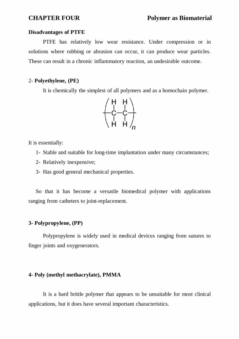
2- Polyethylene, (PE)
It is chemically the simplest of all polymers and as a homochain polymer.
It is essentially:
1- Stable and suitable for long-time implantation under many circumstances;
2- Relatively inexpensive;
3- Has good general mechanical properties.
So that it has become a versatile biomedical polymer with applications
ranging from catheters to joint-replacement.
3- Polypropylene, (PP) Polypropylene is widely used in medical devices ranging from sutures to
finger joints and oxygenerators.
4- Poly (methyl methacrylate), PMMA
It is a hard brittle polymer that appears to be unsuitable for most clinical
applications, but it does have several important characteristics.
CHAPTER FOUR Polymer as Biomaterial
٤
(a) It can be prepared under ambient conditions so that it can be
manipulated in the operating theater or dental clinic, explaining its
use in dentures and bone cement.
(b) The relative success of many joint prostheses is dependent on the
performance of the PMMA cement, which is prepared intra-
operatively by mixing powdered polymer with monomeric
methylmethacrylate, which forms a dough that can be placed in
the bone, where it then sets.
The disadvantages of PMMA
(a) The exotherm of polymerization;
(b) The toxicity of the volatile methylmethacrylate;
(c) The poor fracture toughness.
(But no better material has been developed to date)
5- Polyesters
6-Polyurathanes
Denture Base Resins Although individual denture bases may be formed from metals or metal
alloys, most denture bases are fabricated using common polymers. Such
polymers are chose based on:
(a) Availability;
(b) Dimensional stability;
(c) Handling characteristics;
(d) Color;
(e) Compatibility with oral tissues.
CHAPTER FOUR Polymer as Biomaterial
٥
General Techniques
Several processing techniques are available for the fabrication of denture
bases. Each technique is available for the fabrication of an accurate impression
of the edentulous arch. Using this impression, a dental cast is generated. In turn,
a resin recorded base is fabricated on the cast. Wax is added to the record base
and the teeth are positioned in the wax.
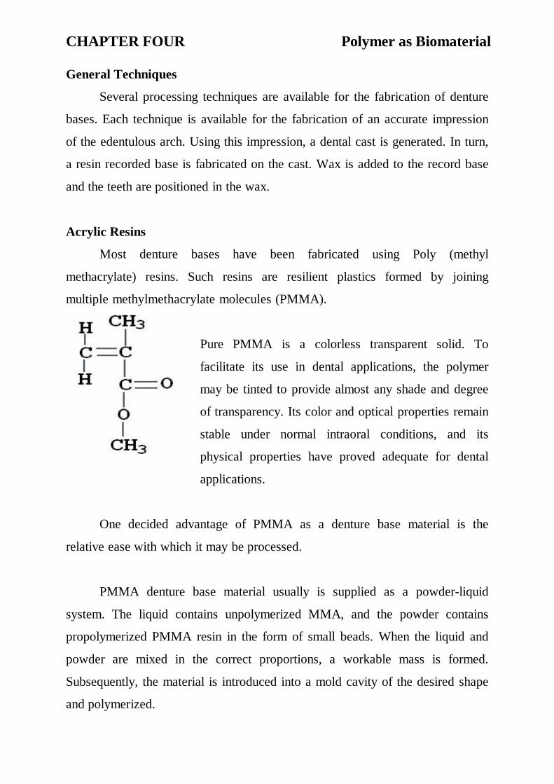
Acrylic Resins
Most denture bases have been fabricated using Poly (methyl
methacrylate) resins. Such resins are resilient plastics formed by joining
multiple methylmethacrylate molecules (PMMA).
Pure PMMA is a colorless transparent solid. To
facilitate its use in dental applications, the polymer
may be tinted to provide almost any shade and degree
of transparency. Its color and optical properties remain
stable under normal intraoral conditions, and its
physical properties have proved adequate for dental
applications.
One decided advantage of PMMA as a denture base material is the
relative ease with which it may be processed.
PMMA denture base material usually is supplied as a powder-liquid
system. The liquid contains unpolymerized MMA, and the powder contains
propolymerized PMMA resin in the form of small beads. When the liquid and
powder are mixed in the correct proportions, a workable mass is formed.
Subsequently, the material is introduced into a mold cavity of the desired shape
and polymerized.

CHAPTER FOUR Polymer as Biomaterial
٦
Properties of Denture Base Resin
When, methyl methacrylate monomer is polymerized, to form Poly
(methyl methacrylate), the density of the mass changes from 0.94 to
1.19gm/cm3. This change in density results in a volumetric shrinkage of 7%.
Based on projected volumetric shrinkage of 7%, an acrylic resin denture base
should exhibit a linear shrinkage of approximately 2%.
PMMA absorbs relatively small amounts of water when placed in an
aqueous environment. Nevertheless, this water exerts significant effect on the
mechanical and dimensional properties of the polymer.
PMMA exhibits a water sorption value of 0.69mg/cm2. Although this
amount of water may seem inconsequential, it exerts significant effect on the
dimensions of polymerized denture base. Laboratory trials indicate a linear
expansion caused by water absorption is approximately equal to the thermal
shrinkage encountered as a result of the polymerization process. Hence these
processes almost offset one another.
Although denture base resins are soluble in a variety of solvents and a
small amount of monomer may be leached, they are virtually insoluble in the
fluids commonly encountered in the oral cavity.
The strength of an individual denture base resin is dependent on several
factors. These factors include:
(a) Composition of the resin;
(b) Processing technique;
(c) Conditions presented by the oral environment.
CHAPTER FOUR Polymer as Biomaterial
٧
Because of the resilient nature of denture base resins, some elastic
deformation that is recoverable deformation also occurs. Clinically, this means
that load application produces stresses within a resin and a change in the overall
shape of the denture base. When the load is released, stresses within the resin are
relaxed and the denture base returns to its original shape. Nevertheless, the
existence of plastic deformation prevents complete recovery. Therefore, some
permanent deformation remains.
Perhaps the most important determinant of overall resin strength is the
degree of polymerization exhibited by the material. As the degree of
polymerization increases the strength of the resin also increases.
Resin Teeth for Prosthodentic Applications
PMMA resins used in the fabrication of prosthetic teeth are similar to
those used in denture base construction. Nevertheless, the degree of cross-
linking within prosthetic teeth is somewhat greater than that within polymerized
denture bases. This increase is achieved by elevating the amount of cross-linking
agent in the denture base liquid, that is, the monomer. The resultant polymer
displays enhanced stability and improved clinical properties.
Despite the current emphasis on resin teeth, prosthetic teeth also may be
fabricated using dental porcelain. Hence a comparison of resin and porcelain
teeth is provided that:
(a) Resin teeth display greater fracture toughness than porcelain teeth. As a
result, resin teeth are less likely to chip or fracture on impact, such as
when a denture is dropped;
CHAPTER FOUR Polymer as Biomaterial
٨
(b) Resin teeth are easier to adjust and display greater resistance to thermal
shock;
(c) Porcelain teeth display better dimensional stability and increasing wear
resistance;
(d) Porcelain teeth, especially when contacting surfaces have been
roughened often cause significant wear of opposing enamel and gold
surfaces. As a result, porcelain teeth should not oppose such surfaces,
and if they are used, they should be polished periodically to reduce such
abrasive damage;
(e) As a final note, resin teeth are capable of chemical bonding with
commonly used denture base resins. Porcelain teeth do not form
chemical bonds with denture resins and must be retained by other means,
such as mechanical undercuts and silanization.
Materials in Maxillofacial Prosthetic
Despite improvements in surgical and restorative techniques, the materials
used in maxillofacial prosthetics are far from ideal. An ideal material should be
inexpensive, biocompatible, strong, and stable. In addition, the material should
be skin-like in color and texture. Maxillofacial materials must exhibit resistance
to tearing and should be able to withstand moderate thermal and chemical
challenges. Currently, no material fulfills all of these requirements. A brief
description of maxillofacial materials is included in the following paragraphs:
Latexes
Latexes are soft, inexpensive materials that may be used to create lifelike
prostheses. Unfortunately, these materials are weak, degenerate rapidly, exhibit
color instability and can cause allergic reactions.

CHAPTER FOUR Polymer as Biomaterial
٩
A recently developed synthetic latex is a tripolymer of butylacrylate,
methyl methacrylate, and methyl metharylamide. This material is nearly
transparent, but has limited applications.
Vinyl Plastisols
They are plasticized vinyl resin sometimes are used in maxillofacial
applications. Plastsols are thick liquids comprising small vinyl particles
dispersed in a plasticizer. Colorants are added to these materials to match
individual skin tones. Unfortunately, vinyl plastisols harden with age because
plasticizer loss. Ultraviolet light also has an adverse effect on these materials.
For these reasons, the use of vinyl is limited.
Silicone Rubbers
Both heat-vulcanizing and room temperature vulcanizing silicones are in
use today and both exhibit advantages and disadvantages.
Room temperature vulcanizing silicones are supplied as single- paste
systems. These silicones are not as strong as the heat-vulcanized silicones and
generally are monochromatic.
Heat-vulcanizing silicone is supplied as a semi-solid material that requires
milling, packing under pressure, and 30-minute heat treatment application cycle
at 180oC. Heat vulcanizing silicone displays better strength and color than room
temperature vulcanizing silicone.
Polyurethane polymers
Polyurethane is the most recent of the materials used in maxillofacial
prosthetics. Fabrication of a polyurethane prosthesis requires accurate
proportioning of three materials. The material is placed in a stone or metal mold
and allowed to polymerize at room temperature. Although a polyurethane
CHAPTER FOUR Polymer as Biomaterial
١٠
prosthesis has a natural feel and appearance, it is susceptible to rapid
deterioration.
The loss of natural teeth, through disease or trauma, has for many years
been compensated by the provision of artificial teeth in the form of bridges and
dentures. These essentially provide an aesthetic replacement of crown of the
tooth but do nothing to replace the root and its attachment to the bone of the jaw.
Natural Polymers
Natural polymers, or polymers, derived from living creatures, are of great
interest in the biomaterials field. In the area of tissue-engineering, for example,
scientists and engineers look for scaffold on which one may successfully grow
cells to replace damaged tissue.
Typically, it is desirable for these scaffolds to be:
(1) Biodegradable;
(2) Non-toxic/ non-inflammatory;
(3) Mechanically similar to the tissue to be replaced;
(4) Highly porous;
(5) Encouraging of cell attachments and growth;
(6) Easy and cheap to manufacture;
(7) Capable of attachment with other molecules ( to potentially
increase scaffold interaction with normal tissue)
Normal polymers often easily fulfill these expectations, as they are
naturally engineered to work well within the living beings from which they
come. Three examples of natural polymers that have been previously studied for
use as biomaterials are: collagen, chitosan, and alginate.
CHAPTER FOUR Polymer as Biomaterial
١١
Collagen is the most widely found protein in mammals (25% of our protein
mass) and is the major provider of strength to tissue. A typical collagen
molecule consists of three interwined protein chains that form a helical structure
similar to a typical staircase). These molecules polymerize together to form
collagen fibers of varying length, thickness and interweaving pattern (some
collagen molecules will form ropelike structures, while others will form meshes
or networks). There are actually at least 15 different types of collagen, differing
in their structure, function, location, and other characteristics. The predominant
form used in biomedical applications, however, is type I collagen, which is a
"rope-forming" collagen and can be found almost everywhere in the body,
including skin and bone.
Collagen can be resorbed into the body, is non-toxic produces only a
minimal immune response, and is excellent for attachment and biological
interaction with cell. Collagen may also be processed into a variety of formats,
including porous sponges, gels and sheets, and can be cross-linked with
chemicals to make it stronger or to alter its degradation rate. The number of
biomedical applications in which collagen has been utilized is too high to count
here, it not only explored for use in various types of surgery, cosmetics, and
drug delivery, but in bio-prosthetic implants and tissue-engineering of multiple
organs as well. Cells grown in collagen often come close to behaving as they do
within the body, which is why collagen is so promising when one is trying to
duplicate natural tissue function and healing.
However, some disadvantages to using collagen as a cell substrate do
exist. Depending on how it is processed, collagen can potentially cause
alteration of cell behavior (e.g. changes in growth or movement), have
inappropriate mechanical properties, or undergo contraction (shrinkage).
Because cells interact so easily with collagen, cells can actually pull and
reorganize collagen fibers, causing scaffolds to lose their shape if they are not
CHAPTER FOUR Polymer as Biomaterial
١٢
properly stabilized by cross-linking or mixing with another less "vulnerable
material".
Fortunately, collagen can be easily combined with other biological or
synthetic materials, to improve its mechanical properties or change the way cells
behave when grown upon it.
Chitosan
It is derived from chitin, a type of polysaccharide (sugar) that is present in
the hard exoskeletons of shellfish like shrimp and crab. Chitin has sparked
interest in the tissue-engineering field due to several desirable properties:
1- Minimal foreign body reaction;
2- Mild processing conditions (synthetic polymers often need to be
dissolved in harsh chemicals; chitosan will dissolve in water based on
pH);
3- Controllable mechanical/biodegradation properties (such as scaffold
porosity);
4- Availability of chemical side groups for attachment to other molecules.
Chitosan has already been investigated for use in the engineering of
cartilage, nerve and liver tissues. Chitosan has also been studied for use in
wound healing and drug delivery. Current difficulties with using chitosan as a
polymer scaffold in tissue-engineering, however, include low strength and
inconsistent behavior with seeded cells. Fortunately, chitosan may be easily
combined with other materials in order to increase its strength and cell-
attachment potential. Mixtures with synthetic polymers such as poly (vinyl
alcohol) and poly (ethylene glycol) or natural polymers such as collagen have
already been produced.

CHAPTER FOUR Polymer as Biomaterial
١٣
Alginate
It is a polysaccharide derived from brown seaweed. Like chitosan,
alginate can be processed easily in water and has been found to be fairly non-
toxic and non-inflammatory enough, so that it has been approved in some
countries for wound dressing and for use in food products. Alginate is
biodegradable, has controllable porosity, and may be linked to other biologically
active molecules. Interestingly, encapsulation of certain cell types into alginate
beads may actually enhance cell survival and growth. In addition, alginate has
been explored for use in liver, nerve, heart, and cartilage tissue-engineering.
Unfortunately, some drawbacks of alginate include mechanical weakness and
poor cell adhesion. Again, to overcome these limitations, the strength and cell
behavior of alginate have been enhanced by mixing with other materials,
including the natural polymers agarose and chitosan.
Chapter Five Metals and Alloys
١
Metals and Alloys Metals are used as biomaterial due to their excellent electrical and thermal
conductivity and mechanical properties. Since some electrons are independent in
metals, they can quickly transfer an electric charge and thermal energy. The
mobile free electrons as the binding force to hold the positive metal ions
together. This attraction is strong, as evidenced by the closely-packed atomic
arrangement resulting in high specific gravity and high melting points of most
metals. Since the metallic bond id essentially non-directional, the position of the
metal ions can be altered without destroying the crystal structure, resulting in a
plastically deformable solid.
Some metals are used as passive substitutes for hard tissue replacement
such as:
1- Total hip;
2- Knee joints;
3- For fracture healing aids as bone plates and screws;
4- Spinal fixation devices;
5- Dental implants, because of their excellent mechanical properties, and
corrosion resistance;
6- Vascular stents;
7- Catheter guide wires.
Stainless Steels
Stainless steel was first used successfully as an important material in the
surgical field.
I- Type 302 stainless steel was introduced, which is stronger and more
resistant to corrosion than the vanadium steel;
Chapter Five Metals and Alloys
٢
II- Type 316 stainless steel was introduced, which contains a small
percentage of molybdenum (18-8sMo) to improve the corrosion
resistance in chloride solution (salt water);
III- Type 316L stainless steel. The carbon content was reduced from 0.08
to a maximum amount of 0.03% for better corrosion resistance to
chloride solution.
The inclusion of molybdenum enhances resistance to pitting corrosion in salt
water. Even the 316L stainless steels may corrode in the body under certain
circumstances in highly stressed and oxygen depleted region, such as the
contacts under the screws of the bone fracture plate. Thus, these stainless steels
are suitable to use only in temporary implant devices, such as fracture plates,
screws, and hip nails.
CoCr Alloys
There are basically two types of cobalt-chromium alloys:
1- The CoCrMo alloy [ Cr (27-30%), Mo (5-7%), Ni (2.5%)] has been used
for many decades in dentistry, and in making artificial joints;
2- The CoNiCrMo alloy [Cr (19-21%), Ni (33-37%), and Mo (9-11%)] has
been used for making the stems of prostheses for heavily loaded joints,
such as knee and hip.
The ASTM lists four types of CoCr alloys, which are recommended for
surgical implant applications:
1) CoCrMo alloy [Cr (29-30%), Mo (5-7%), Ni (2.5%)];
2) CoCrWNi alloy [Cr (19-21%), W (14-16%), Ni (9-11%)];
3) CoNiCrMo alloy [Ni (33-37%), Cr (19-21%), Mo (9-11%)];
4) CoNiCrMoWFe alloy [Ni (15-25%), Cr (18-22%), Mo (3-4%), W (3-4%),
Fe (4-6%)].
Chapter Five Metals and Alloys
٣
The two basic elements of the CoCr alloys form a solid solution of up to
65% Co. The molybdenum is added to produce finer grains, which results in
higher strengths after casting. The chromium enhances corrosion resistance, as
well as solid solution strengthening of the alloy.
The CoNiCrMo alloy contains approximately 35% Co and Ni each. The
alloy is highly corrosion resistant to seawater (containing chloride ions) under
stress.
Titanium and its Alloys Titanium and its alloys are getting great attention in both medical and
dental fields because of:
(a) Excellent biocompatibility;
(b) Light weight;
(c) Excellent balance of mechanical properties;
(d) Excellent corrosion resistance.
They are commonly used for implant devices replacing failed hard tissue,
for example, (1) artificial hip joints, (2) artificial knee joint, (3) bone plate,
(4) dental implants, (5) dental products, such as crowns, bridges and dentures,
and (6) used to fix soft tissue, such as blood vessels.
In the elemental form, titanium has a high melting point (1668oC) and
possesses a hexagonal closely packed structure (hcp) α up to a temperature of
882.5oC. Titanium transforms into a body centered cubic structure (bcc) β above
this temperature.
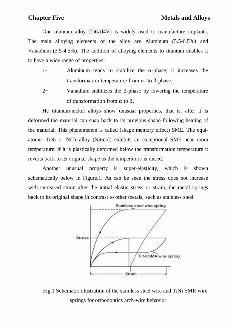
Chapter Five Metals and Alloys
٤
One titanium alloy (Ti6Al4V) is widely used to manufacture implants.
The main alloying elements of the alloy are Aluminum (5.5-6.5%) and
Vanadium (3.5-4.5%). The addition of alloying elements to titanium enables it
to have a wide range of properties:
1- Aluminum tends to stabilize the α-phase; it increases the
transformation temperature from α- to β-phase.
2− Vanadium stabilizes the β-phase by lowering the temperature
of transformation from α to β.
He titanium-nickel alloys show unusual properties, that is, after it is
deformed the material can snap back to its previous shape following heating of
the material. This phenomenon is called (shape memory effect) SME. The equi-
atomic TiNi or NiTi alloy (Nitinol) exhibits an exceptional SME near room
temperature: if it is plastically deformed below the transformation temperature it
reverts back to its original shape as the temperature is raised.
Another unusual property is super-elasticity, which is shown
schematically below in Figure.1. As can be seen the stress does not increase
with increased strain after the initial elastic stress or strain, the metal springs
back to its original shape in contrast to other metals, such as stainless steel.
Fig.1 Schematic illustration of the stainless steel wire and TiNi SMR wire
springs for orthodontics arch-wire behavior
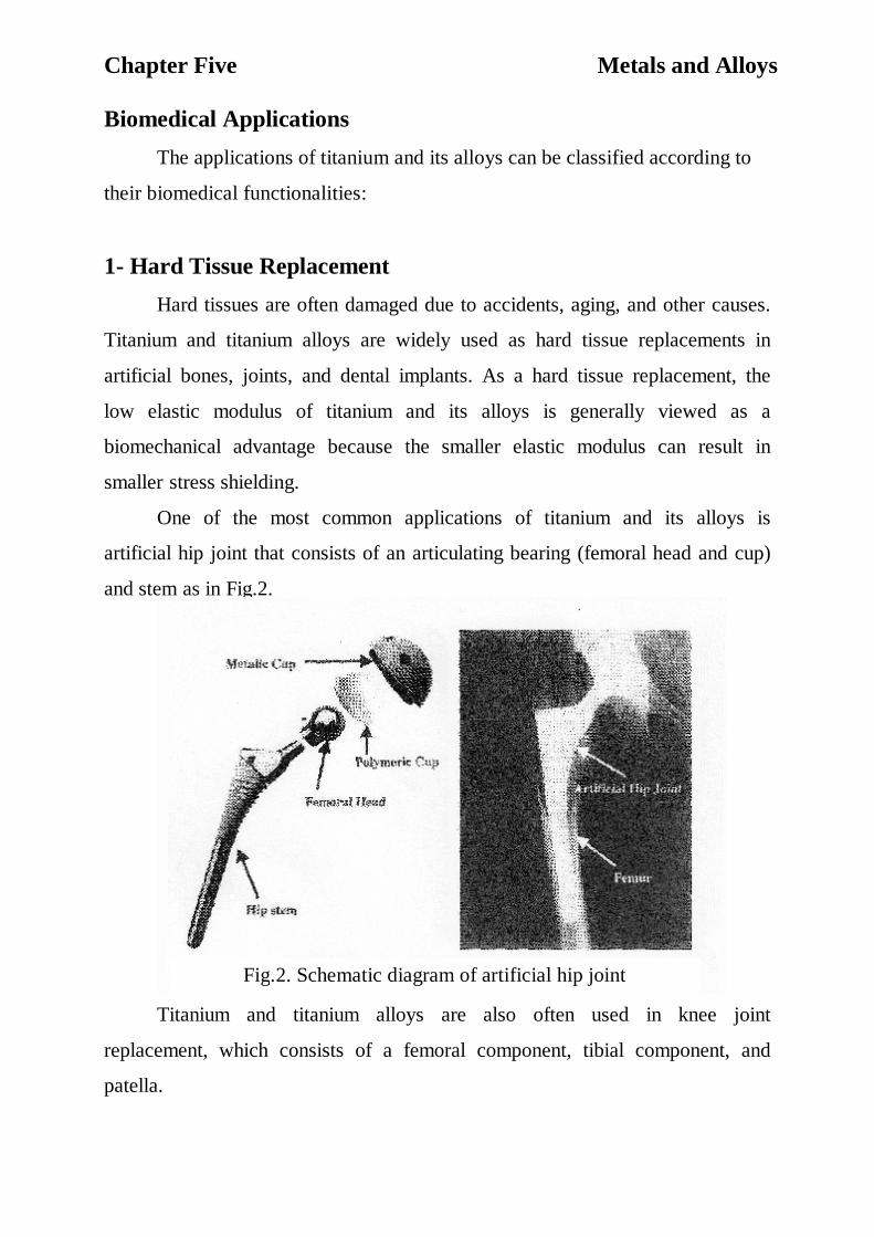
Chapter Five Metals and Alloys
٥
Biomedical Applications
The applications of titanium and its alloys can be classified according to
their biomedical functionalities:
1- Hard Tissue Replacement Hard tissues are often damaged due to accidents, aging, and other causes.
Titanium and titanium alloys are widely used as hard tissue replacements in
artificial bones, joints, and dental implants. As a hard tissue replacement, the
low elastic modulus of titanium and its alloys is generally viewed as a
biomechanical advantage because the smaller elastic modulus can result in
smaller stress shielding.
One of the most common applications of titanium and its alloys is
artificial hip joint that consists of an articulating bearing (femoral head and cup)
and stem as in Fig.2.
Titanium and titanium alloys are also often used in knee joint
replacement, which consists of a femoral component, tibial component, and
patella.
Fig.2. Schematic diagram of artificial hip joint
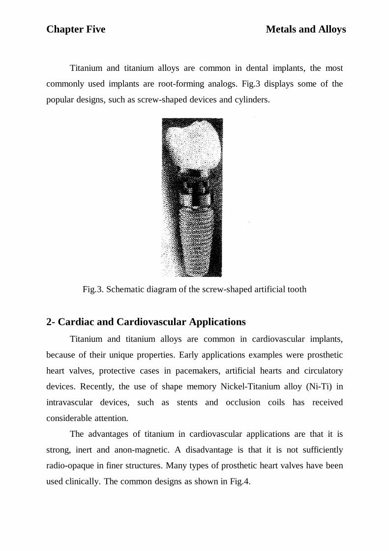
Chapter Five Metals and Alloys
٦
Titanium and titanium alloys are common in dental implants, the most
commonly used implants are root-forming analogs. Fig.3 displays some of the
popular designs, such as screw-shaped devices and cylinders.
Fig.3. Schematic diagram of the screw-shaped artificial tooth
2- Cardiac and Cardiovascular Applications
Titanium and titanium alloys are common in cardiovascular implants,
because of their unique properties. Early applications examples were prosthetic
heart valves, protective cases in pacemakers, artificial hearts and circulatory
devices. Recently, the use of shape memory Nickel-Titanium alloy (Ni-Ti) in
intravascular devices, such as stents and occlusion coils has received
considerable attention.
The advantages of titanium in cardiovascular applications are that it is
strong, inert and anon-magnetic. A disadvantage is that it is not sufficiently
radio-opaque in finer structures. Many types of prosthetic heart valves have been
used clinically. The common designs as shown in Fig.4.
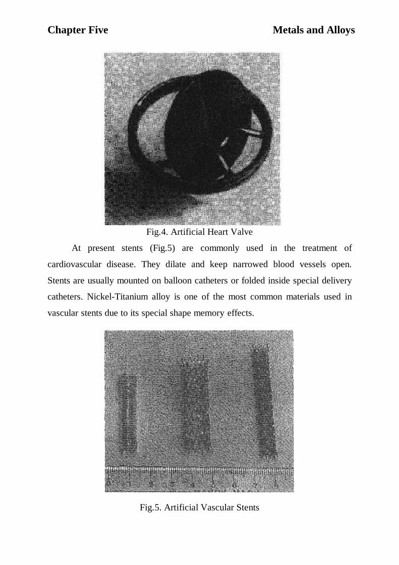
Chapter Five Metals and Alloys
٧
Fig.4. Artificial Heart Valve
At present stents (Fig.5) are commonly used in the treatment of
cardiovascular disease. They dilate and keep narrowed blood vessels open.
Stents are usually mounted on balloon catheters or folded inside special delivery
catheters. Nickel-Titanium alloy is one of the most common materials used in
vascular stents due to its special shape memory effects.
Fig.5. Artificial Vascular Stents
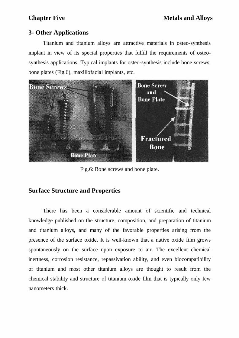
Chapter Five Metals and Alloys
٨
3- Other Applications Titanium and titanium alloys are attractive materials in osteo-synthesis
implant in view of its special properties that fulfill the requirements of osteo-
synthesis applications. Typical implants for osteo-synthesis include bone screws,
bone plates (Fig.6), maxillofacial implants, etc.
Fig.6: Bone screws and bone plate.
Surface Structure and Properties
There has been a considerable amount of scientific and technical
knowledge published on the structure, composition, and preparation of titanium
and titanium alloys, and many of the favorable properties arising from the
presence of the surface oxide. It is well-known that a native oxide film grows
spontaneously on the surface upon exposure to air. The excellent chemical
inertness, corrosion resistance, repassivation ability, and even biocompatibility
of titanium and most other titanium alloys are thought to result from the
chemical stability and structure of titanium oxide film that is typically only few
nanometers thick.
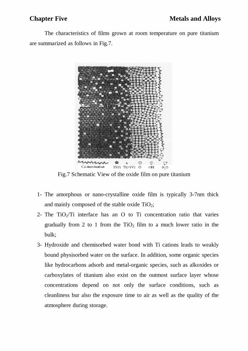
Chapter Five Metals and Alloys
٩
The characteristics of films grown at room temperature on pure titanium
are summarized as follows in Fig.7.
Fig.7 Schematic View of the oxide film on pure titanium
1- The amorphous or nano-crystalline oxide film is typically 3-7nm thick
and mainly composed of the stable oxide TiO2;
2- The TiO2/Ti interface has an O to Ti concentration ratio that varies
gradually from 2 to 1 from the TiO2 film to a much lower ratio in the
bulk;
3- Hydroxide and chemisorbed water bond with Ti cations leads to weakly
bound physisorbed water on the surface. In addition, some organic species
like hydrocarbons adsorb and metal-organic species, such as alkoxides or
carboxylates of titanium also exist on the outmost surface layer whose
concentrations depend on not only the surface conditions, such as
cleanliness bur also the exposure time to air as well as the quality of the
atmosphere during storage.
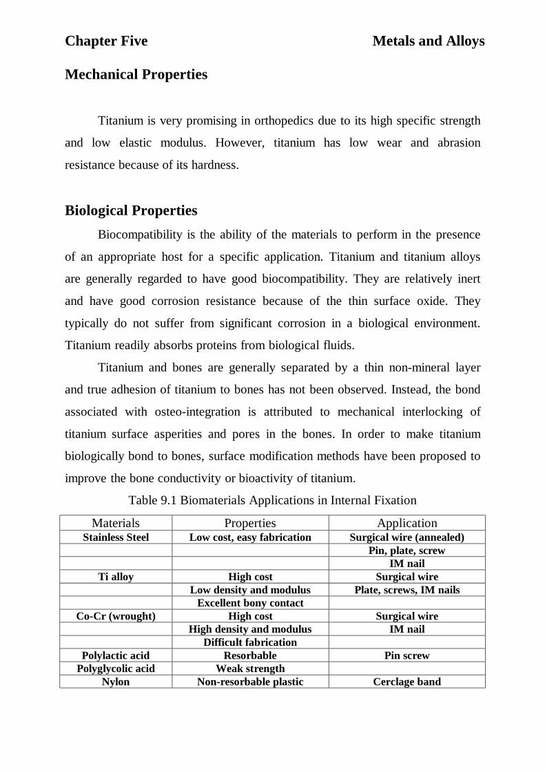
Chapter Five Metals and Alloys
١٠
Mechanical Properties
Titanium is very promising in orthopedics due to its high specific strength
and low elastic modulus. However, titanium has low wear and abrasion
resistance because of its hardness.
Biological Properties
Biocompatibility is the ability of the materials to perform in the presence
of an appropriate host for a specific application. Titanium and titanium alloys
are generally regarded to have good biocompatibility. They are relatively inert
and have good corrosion resistance because of the thin surface oxide. They
typically do not suffer from significant corrosion in a biological environment.
Titanium readily absorbs proteins from biological fluids.
Titanium and bones are generally separated by a thin non-mineral layer
and true adhesion of titanium to bones has not been observed. Instead, the bond
associated with osteo-integration is attributed to mechanical interlocking of
titanium surface asperities and pores in the bones. In order to make titanium
biologically bond to bones, surface modification methods have been proposed to
improve the bone conductivity or bioactivity of titanium.
Table 9.1 Biomaterials Applications in Internal Fixation
Materials Properties Application Stainless Steel Low cost, easy fabrication Surgical wire (annealed)
Pin, plate, screw IM nail
Ti alloy High cost Surgical wire Low density and modulus Plate, screws, IM nails Excellent bony contact
Co-Cr (wrought) High cost Surgical wire High density and modulus IM nail Difficult fabrication
Polylactic acid Resorbable Pin screw Polyglycolic acid Weak strength
Nylon Non-resorbable plastic Cerclage band

Chapter Five Metals and Alloys
١١
Table 9.3 Biomaterials fir Total Joint Replacements
Materials Properties Application Co-Cr alloy Stem, head (ball) Heavy, hard, stiff
(casted or wrought) Cup, porous coating High wear resistance Metal backing
Ti alloy Stem porous coating Low stiffness Metal backing Low wear resistance
Pure titanium Porous coating Excellent osseousintegration Tantulum Porous structure Excellent osseousintegration
Good mechanical strength Alumina Ball, cup Hard, brittle
High wear resistance Zirconia Ball Heavy and high toughness
High wear resistance UHMWPE Cup Low friction, wear debris
Low creep resistance PMMA Bone cement fixation Brittle, weak in tension
Low fatigue strength
Table 9.4 Types of Total Joint Replacements Joint Types Hip Bull and Socket
Knee Hinged, semi-constrained, surface replacement Uni-compartment or bio-compartment
Shoulder Bull and Socket Ankle Surface replacement Elbow Hinged, unconstrained, surface replacement Wrist Ball and socket, space filter Finger Hinged, space filter
Dental Materials Dental amalgam is an alloy made of liquid mercury and other solid
materials particulate alloys made of silver, tin, copper, etc. The solid alloy is
mixed with (liquid) mercury in a mechanical vibrating mixer and the resulting
material is packed into the prepared cavity. One of the solid alloys is composed
of at least 65% silver, and not more than 29% tin, 6% copper, 2% zinc, and 3%
mercury. The final composition of dental amalgams typically contains 45% to
55% mercury, 35% to 45% silver, and about 15% tin after fully set in about one
day.
Chapter Five Metals and Alloys
١٢
Gold and gold alloys are useful metals in dentistry as a result of their
durability, stability, and corrosion resistance. Gold alloys are introduced by two
methods: casting and malleting. Gold alloys are used for cast restorations, since
they have mechanical properties which are superior to those of pure gold. The
pure gold is relatively soft, so this type of restoration is limited to areas not
subjected to much stress.
Other Metals
Tantalum has been subjected to animal implant studies and has been
shown very biocompatible. Due to its poor mechanical properties and its high
density (16.6 gm/cm3) it is restricted to few applications such as wire sutures for
plastic surgeons and neurosurgeons, and a radioisotope for bladder tumors.
Surface modifications of metal alloys such as coatings by plasma spray, physical or chemical vapor deposition, ion implantation, and fluidized bed deposition have been used in industry. Coating implants with tissue compatible material such as hydroxyapatite, oxide ceramics, bio-glass, and pyrolytic carbon are typical applications in implants. Such efforts have been largely ineffective if the implants are subjected to a large loading. The main problem is in the delaminating of the coating or eventual wear of the coating. The added cost of coating or ion implanting hinders the use of such techniques unless the technique shows unequivocal superiority compared to the non-treated implants. Defining Terms
In vitro: In glass, as in a test tube. An in vitro test is one done in the laboratory,
usually involving isolated tissues, organs, or cells.
In vivo: A test performed in a living body or organism.
Passivation: Production of corrosion resistance by a surface layer of reaction
products (normally oxide layer which is impervious to gas and
water)
Passivity: Resistance to corrosion by a surface layer of reaction products.
Pitting: A form of localized corrosion, in which, pits form on the metal surface.
Chapter Six Hard Tissue Replacements
١
Hard Tissue Replacements
Bone Repair and Joint Implants A large number of devices are available for the repair of the bone tissue. 1- Bone Repair
The principal functions of the skeleton are to provide a frame to support
the organ-systems, and to determine the direction and range of body movements.
Bone provides an anchoring point for the most of skeletal muscles and
ligaments.
Bone is the only tissue able to undergo spontaneous regeneration and to
remodel its micro- and macro-structure. This is accomplished through a delicate
balance between an osteogenic (bone forming) and osteoclastic (bone removing)
process.
Nature provides different types of mechanisms to repair fracture in order
to be able to cope with different mechanical environments about a fracture. With
external fracture fixation, the bone fragments are held in alignment by pins
placed through the skin onto the skeleton, structurally supported by external
bars. With internal fracture fixation, the base fragments are held by wires,
screws, plates, and/or intra-medullary devices.
All the internal fixation devices should meet the general requirements of
biomaterials, that is, biocompatibility, sufficient strength, corrosion resistance,
and a suitable mechanical environment for fracture healing. From this
perspective, stainless steel, cobalt-chrome alloys, and titanium alloys are most
suitable for internal fixation. Most internal fixation devices persist in the body
after the fracture has healed, often causing discomfort and requiring removal.
Recently, biodegradable polymers, for example, polylactic acid (PLA) and
polyglycolic acid (PGA) have been used to treat minimally loaded fractures,
thereby eliminating the need for a second surgery for implant removal.
Chapter Six Hard Tissue Replacements
٢
Wires
Surgical wires are used to reattach fragments of bone, like the greater
trochanter, which is often detached during total hip replacement. They are also
used to provide additional stability in long-oblique or spiral fractures of long
bones which have already been stabilized by other means.
Twisting and knotting is unavoidable when fastening wires to bone,
however, by doing so, the strength of the wire can be reduced by 25% or more
due to stress concentration. This can be overcome by using a thicker wire, since
its strength increases directly proportional to its diameter (Fig 9.1).
Pins
Pins are widely used primarily to hold fragments of bones together
provisionally or permanently and to guide large screws during insertion. To
facilitate implantation, the pins have different tip designs which have been
optimized for different types of bone. The trochar tip is the most efficient in
cutting hence; it is often used for cortical bone.
The holding power of the pin comes from elastic deformation surrounding
bone. In order to increase the holding power to bone, threaded pins are used.
Most pins are made of 316L stainless steel; however, recently, biodegradable
pins made of polylactic or polyglycolic acid have been employed for the
treatment of minimally loaded fractures. (Fig.9.2)
Screws
Screws are the most widely used devices for fixation of bone fragments.
There are two types of bone screws:
1- Cortical bone screws, which have small threads, and
2- Cancellous screws which have large threads to get more thread-to-bone
contact. They may have either V or buttress threads.
Chapter Six Hard Tissue Replacements
٣
The holding power of screws can be affected by the size of the pilot
drill-hole, the depth of screw engagement, the outside diameter of the screw, and
quality of the bone. (Fig.9.3)
Plates
Plates are available in a wide variety of shapes and are intended to
facilitate fixation of bone fragments. They range from the very rigid, intended to
produce primary bone healing, to the relatively flexible, intended to facilitate
physiological loading of bone.
The rigidity and strength of a plate in bending depend on the cross-
sectional shape and material of which it is made. The effect of the material on
the rigidity of the plate is defined by the elastic modulus of the material for
bending and by the shear modulus for twisting. Thus, given the same
dimensions, a titanium alloy plate will be less rigid than a stainless steel plate,
since the elastic modulus of each alloy is 110 and 200GPa respectively. (Fig.9.5)
Joint Replacement
The design of an implant for joint replacement should be based on the
kinematics and dynamic load transfer characteristics of the joint. The material
properties shape, and methods used for fixation of the implant to the patient
determines the load transfer characteristics. This is one of the most important
elements that determine long-term survival of the implant, since bone responds
to changes in load transfer with a remodeling process. Overloading the implant-
bone interface or shielding it from load transfer may result in bone resorption
and subsequent loosening of the implant. The articulating surfaces of the joint
should function with minimum friction and produce the least amount of wear
products. The implant should be securely fixed to the body as early as possible
(ideally immediately after implantation); however, removal of the implant
should not require destruction of a large amount of surrounding tissues.
Chapter Six Hard Tissue Replacements
٤
Implant Fixation Method
Fixation of implants with polymethyl-methacrylate (PMMA, bone
cement) provides immediate stability, allowing patients to bear all of their
weight on the extremity at once. In contrast, implants which depend on bone
ingrowth require the patient to wait about 12 weeks to bear full weight. Being a
viscoelastic polymer, it has the ability to function as a shock absorber. It allows
loads to be transmitted uniformly between the implant and bone, reducing
localized high-content stress.
The problems with bone-cement interface may arise from intrinsic factors,
such as the properties of the PMMA and bone, as well as extrinsic factors such
as the cementing techniques.
Porous Ingrowth Fixation
Bone ingrowth can occur with inert implants which provide pores larger
than 25µm in diameter, which is the size required to accommodate an osteon.
For the best ingrowth in clinical practice, pore size range should be 100 to
350 µm and pores should be interconnected with each other with similar size of
opening.
Commercially, pure titanium, titanium alloys, tantalum, and calcium
hydroxyapatite (HA) are currently used in porous coating materials. With pure
titanium, three different types of porosity can be achieved:
1- Plasma spray coating;
2- Sintering of wire mesh;
3- Sintering of beads on an implant surface.
Thermal processing of the porous coating may weaken the underlying metal
(implant). Additional problems may result from flaking of the porous coating
materials, since loosened metal particles may cause severe wear when they
migrate into the articulation.
Chapter Six Hard Tissue Replacements
٥
Recently, a cellular, structural biomaterial comprised of 15 to 25%
tantalum has been developed. The average pore size is about 550µm, and the
pores are fully interconnected.
Total Joint Replacement
1- Hip Joint Replacement
The prosthesis for total hip replacement consists of a femoral component
and an acetabular component. (Fig. ) The femoral stem is divided into head,
neck, and shaft. The femoral stem is made of Ti alloy or CoCr alloy and is fixed
into a reamed medullary canal by cementation or press fitting. The femoral head
is made of CoCr alloy, alumina or zirconia. The acetabular component is
generally made of ultra-high molecular-weight polyethylene (UHMWPE). The
hip joint is a ball-and-socket joint which derives its stability from cony ity of the
implants, pelvic, and capsule.
2- Knee Joint Replacements
The prosthesis for total knee joint replacement consists of femoral, tibial,
and patellar components. Compared to the hip joint, the knee has a more
complicated geometry and movement biomechanics, and it is not intrinsically
stable. In a normal knee, the center of movement is controlled by the geometry
of the ligaments.
Total knee replacement can be implanted with or without cement, the
latter relying on porous coating for fixation. The femoral components are
typically made of CoCr alloy and the monolithic tibial components are made of
UHMWPE.
In modular component, the tibial polyethylene component is assembled
onto a titanium alloy tibial tra . The patellar component is made of UHMWPE,
and titanium alloy back is added to components designed for uncemented use.
Chapter Six Hard Tissue Replacements
٦
3- Ankle Joint Replacement
Total ankle replacements have not met with as much success as total hip and
knee replacements, and typically loosen within a few years of service. This is
mainly due to the high load transfer demand over the relatively small ankle
surface area and need to replace three articulating surfaces (tibial, talar, and
fibular). The materials used to construct ankle joints are usually CoCr alloy and
UHMWPE.
4- Shoulder Joint Replacements
The prostheses for total shoulder replacement consist of humeral and
glenoid components. Like the femoral stem, the humeral component can be
divided into head, neck, and shaft. The shoulder has the largest range of motion
in the body, which results from a shallow ball and socket joint, which allows a
combination of rotation and sliding motions between the joint surfaces.
5- Elbow Joint Replacements
The elbow joint is a hinge-type joint allowing mostly flexion and
extension, but having a polycentric motion. The elbow joint implants are hinged,
semi-constrained, or unconstrained. These implants, like those of the ankle, have
a high failure rate and are not used commonly. The high loosening rate is the
result of high rotational movements, limited bone stock for fixation, and
minimal ligamentous support.
6- Finger Joint Replacements Finger joint replacements are divided into three types:
1- Hinge 2- Polycentric 3- Space-filler
The most widely used are the space-filler type. These are of high performance silicone rubber (poly dimethylsiloxane) and are stabilized with a passive fixation method.

Chapter Six Hard Tissue Replacements
٧
Chapter Seven Composite Biomaterials
١
Composite Biomaterials Biomaterials are solids which contain two or more distinct constituent
materials or phases, on a scale larger than the atomic. The tem "composite" is
usually reserved for those materials in which the distinct phases are separated on
a scale larger than the atomic, and in which properties such as the elastic
modulus are significantly altered in comparison with those of a homogeneous
material. Accordingly, reinforced plastics such as fiberglass as well as natural
materials, such as bone are viewed as composite materials. Natural composites
often exhibit hierarchical structures in which particulate, porous, and fibrous
structural features are seen in different micro-scales.
Composite materials offer a variety of advantages in comparison with
homogeneous materials. Those include the ability for the scientist or engineer to
exercise considerable control over material properties. This is the potential for
stiff, strong, light-weight materials as well as for highly resilient and compliant
materials. Some applications of composites in biomaterial applications are:
(1) Dental filling composites;
(2) Reinforced methyl methacrylate bone cement and ultra-high molecular
weight polyethylene;
(3) Orthopedic implants with porous surfaces.
Structure
The properties of composite materials depend very much upon structure.
Composites differ from homogeneous materials in that considerable control can
be exerted over the larger scale structure, and hence over the desired properties.
In particular, the properties of a composite material depend upon the shape of
the heterogeneities, upon the volume fraction occupied by them, and upon the
interface among the constituents. The shape of the heterogeneities in a
composite material is classified as follows:
Chapter Seven Composite Biomaterials
٢
a- The particle, with no long dimension;
b- The fiber, with one long dimension;
c- The platelet or lamina, with two long dimensions.
Bonds on Properties
Mechanical properties in many composite materials depend on structure
in a complex way, however for some structures; the prediction of properties is
relatively simple. The simplest composite structures are the idealized Voigt and
Reuss models, shown in Fig.2. The dark and light areas in these diagrams
represent the two constituent materials in the composite. In contrast to most
composite structures, it is easy to calculate the stiffness of materials with the
Voigt and Reuss structures, since in the Voigt structure the strain is the same in
both constituents, in the Reuss structure the stress is the same. The Young's
modulus, E, of the Voigt composite is given by:
]V1[EVEE imii −+=
Where Ei is the Voigt modulus of the inclusions,
Vi is the volume fraction of the inclusions,
Em is the Young's modulus of the matrix.
The Reuss stiffness R, is less than that of the Voigt model
m
1i
i
iE
V1EVE
−−+=
The Voigt and Reuss models provide upper and lower bounds
respectively upon the stiffness of a composite of arbitrary phase geometry. For
composite materials which are isotropic, the more complex relations of Hashin
and Shtrikman provide tighter bounds upon the moduli both the Young's and
shear moduli must be known for each constituent to calculate these bounds.

Chapter Seven Composite Biomaterials
٣
An isotopic material has the same material properties in any direction.
Anisotropic composites offer superior strength and stiffness in comparison with
isotropic ones.
Particulate Composites
It is often convenient to stiffen or harden a material, commonly a
polymer, by the incorporation of particulate inclusions. The shape of the
particles is important. In isotropic systems, stiff composite, followed by fibers
and the last effective geometry for stiff inclusions is the spherical particles.
Particle reinforcement has been used to improve the properties of bone
cement. For example, inclusion of bone particles in PMMA cement somewhat
improves the stiffness and improves the fatigue life considerably. Moreover, the
bone particles at the interface with the patient's bone are ultimately resorbed and
are replaced by ingrown new bone tissue.
Rubber, used in catheters, rubber gloves, etc is usually reinforced with
very fine particles of silica (SiO2) to make the rubber stronger and tougher.
Teeth with decayed regions have traditionally been restored with metals
such as silver amalgam. Metallic restoration is not considered desirable for
interior teeth for cosmetic reasons. Acrylic resins and silicate cements had been
used for anterior teeth, but their poor material properties led to short service life
and clinical failures. Dental composite resins have virtually replaced these
materials and are very commonly used to restore posterior teeth as well as
anterior teeth.
Chapter Seven Composite Biomaterials
٤
The dental composite resins consist of a polymer matrix and stiff
inorganic inclusions. The inorganic inclusions confer a relatively high stiffness
and high wear resistance on the material. Available dental composite resins use
quartz, barium glass and colloidal silica as fillers. Fillers have particle size from
0.04 to 13µm, and concentrations from 33 to 78% by weight.
In restoring a cavity, the dentist mixes several constituents, then places
them in the prepared cavity to polymerize. For this procedure to be successful
the viscosity of the mixed paste must be sufficiently low and the polymerization
must be controllable.
Dental composites have a Young's modulus in the range 10 to 16GPa, and
the compressive strength from 170 to 260 MPa. These composites are still
considerably less stiff than dental enamel, which contains about 99% mineral.
The thermal expansion of dental composites, as with other dental
materials exceeds that of tooth structure. Moreover, there is a contraction during
polymerization to 1.2 to 1.6%. These effects are thought to contribute to leakage
saliva, bacteria, etc. at the interface margins. Such leakage in some cases can
cause further decay of the tooth. All the dental composites exhibit creep.
Fibrous Composites
Fibers incorporated in a polymer matrix increase the stiffness, fatigue life,
and other properties. Fibers are mechanically more effective in achieving a stiff,
strong composite than particles. Materials can be prepared in fiber form with
very few defects which concentrate stress. Fibers such as graphite are stiff
(Young's modulus is 200–800GPa) and strong (the tensile strength is 2.7–
5.5GPa). Composites made from them can be as strong as steel but much lighter.