Embed Size (px)
Citation preview
Biomaterial surface energy-driven ligand assemblystrongly regulates stem cell mechanosensitivityand fate on very soft substratesTojo Razafiarisona,b, Claude N. Holensteina,b, Tino Staubera,b, Milan Jovica,b, Edward Vertudesa,b, Marko Loparicc,Maciej Kaweckid, Laetitia Bernardd, Unai Silvana,b,1, and Jess G. Snedekera,b,1,2
aDepartment of Orthopedics, Balgrist University Hospital, University of Zurich, 8008 Zurich, Switzerland; bInstitute for Biomechanics, ETH Zurich, 8008Zurich, Switzerland; cBiozentrum and Swiss Nanoscience Institute, University of Basel, 4056 Basel, Switzerland; and dLaboratory for Nanoscale MaterialsScience, Empa Dübendorf, 8600 Dübendorf, Switzerland
Edited by Kristi S. Anseth, University of Colorado, Boulder, CO, and approved March 21, 2018 (received for review March 22, 2017)
Although mechanisms of cell–material interaction and cellularmechanotransduction are increasingly understood, the mechanicalinsensitivity of mesenchymal cells to certain soft amorphous bio-material substrates has remained largely unexplained. We revealthat surface energy-driven supramolecular ligand assembly canregulate mesenchymal stem cell (MSC) sensing of substrate me-chanical compliance and subsequent cell fate. Human MSCs werecultured on collagen-coated hydrophobic polydimethylsiloxane(PDMS) and hydrophilic polyethylene-oxide-PDMS (PEO-PDMS) ofa range of stiffnesses. Although cell contractility was similarly di-minished on soft substrates of both types, cell spreading and os-teogenic differentiation occurred only on soft PDMS and nothydrophilic PEO-PDMS (elastic modulus <1 kPa). Substrate surfaceenergy yields distinct ligand topologies with accordingly distinctprofiles of recruited transmembrane cell receptors and related fo-cal adhesion signaling. These differences did not differentially reg-ulate Rho-associated kinase activity, but nonetheless regulatedboth cell spreading and downstream differentiation.
mechanobiology | stem cell | PDMS | surface energy | ligand assembly
Studies of stem cell behavior on soft biomaterials have typi-cally employed 2D platforms of synthetic polymers coated
with monomeric protein ligands (1). Such studies (2–4) havedemonstrated that modulating the stiffness of porous gels candirect stem cell fate. However, experimental outcomes on amor-phous biomaterial substrates vary widely. Beyond the inherentbiological variability of eukaryotic cell culture systems, existingmodels of cell–biomaterial interaction fail to coherently ex-plain divergence of experimental results. For instance, it hasuntil now not been understood why mesenchymal stem cells(MSCs) readily attach and spread on soft elastomeric silicone(3–5), while they tend to not spread on similarly soft substratessuch as polyacrylamide that have been coated with similar ex-tracellular matrix ligands (3, 4, 6).Although cell responses on synthetic hydrogels and elastomers
are regularly compared, these materials present very distinctchemical and physical features (7). Among these features, onecharacteristic that has been widely ignored is the inherent dif-ference in surface energy of these material classes. In the field ofbiomedical implant design, surface energy has long been recog-nized to control protein adhesion and downstream cellular re-action (8, 9). The property of biomaterial surface energy can beviewed as the physical work done by intermolecular forces actingto increase phase surface area. As such, surface energy dependson the charge and polarity of the outermost functional groups ofthe biomaterial. Surface energy can be increased by the presenceof polar functional groups, with higher energy substrates havingmore polar groups yielding a more hydrophilic surface (10, 11).Monomeric type I collagen is a widely used model extracellularmatrix ligand comprising both polar and apolar amino acid res-idues (Fig. 1). We have shown previously that biomaterial
surface energy plays a dominant role in determining whichgroups are exposed after deposition, with a downstream influ-ence on the supramolecular organization of adsorbed collagenlayers (12). In this earlier work, we demonstrated that surfaceenergy on stiff (2.15–2.40 MPa), atomically flat substrates steerscell–material interactions and promotes osteogenic MSC differ-entiation by regulating the topography of the adsorbed ECMprotein layer presented to the cells. To achieve these insights, wedesigned a polydimethylsiloxane (PDMS)-based platform inwhich stiffness and surface energy can be independently tailoredin a straightforward manner by addition of surfactant in smallquantities. The chief technical challenge was to rigorously con-trol for, and prevent, potential confounding effects of any divergentchemical, physical, and mechanical properties at the cell–materialinterface.We now adapt this tunable biomaterial system to achieve very
soft substrates (elastic modulus <1 kPa) to isolate and investigatethe role of surface energy on human MSC mechanosensitivity tosubstrate stiffness. We specifically focused on collagen I as amodel ligand that supports osteogenic, tenogenic, or adipogenicdifferentiation in a substrate stiffness-dependent manner (6). Wefound that surface energy indeed regulates cell adhesion anddifferentiation on soft and hard substrates, with hydrophobicsurfaces suppressing cell mechanosensitivity to bulk materialstiffness. We reveal how surface energy directs ligand topography
Significance
Cell instructive biomaterial cues are a major topic of interest inboth basic and applied research. In this work, we clarify howsurface energy of soft biomaterials can dramatically affect mes-enchymal stem cell receptor recruitment and downstream signal-ing related to cell fate. We elucidate how surface protein self-assembly and the resulting surface topology can act to steermechanotransduction and related biological response of attachedcells. These findings fill a critical gap in our basic understanding ofcell–biomaterial interaction and highlight soft biomaterial surfaceenergy as a dominant design factor that should not be neglected.
Author contributions: T.R., U.S., and J.G.S. designed research; T.R., C.N.H., T.S., M.J., E.V.,M.L., M.K., L.B., and U.S. performed research; C.N.H., M.L., M.K., and L.B. contributed newreagents/analytic tools; T.R., C.N.H., M.J., E.V., M.L., M.K., L.B., U.S., and J.G.S. analyzeddata; and T.R., C.N.H., U.S., and J.G.S. wrote the paper.
The authors declare no conflict of interest.
This article is a PNAS Direct Submission.
This open access article is distributed under Creative Commons Attribution-NonCommercial-NoDerivatives License 4.0 (CC BY-NC-ND).1U.S. and J.G.S. contributed equally to this work.2To whom correspondence should be addressed. Email: [email protected].
This article contains supporting information online at www.pnas.org/lookup/suppl/doi:10.1073/pnas.1704543115/-/DCSupplemental.
Published online April 16, 2018.
www.pnas.org/cgi/doi/10.1073/pnas.1704543115 PNAS | May 1, 2018 | vol. 115 | no. 18 | 4631–4636
APP
LIED
BIOLO
GICAL
SCIENCE
S
Dow
nloa
ded
by g
uest
on
Mar
ch 5
, 202
0
to alternately promote or inhibit cell mechanosensitivity andresponse to soft amorphous substrates. This understanding likelyresolves conflicting mechanistic theories on how MSCs sense andreact to soft biomaterial substrates (3, 4, 13–15). More broadly,the present study demonstrates biomaterial surface energy as acrucial consideration in soft biomaterial design, and one thatcannot be neglected in study of stem cell–biomaterial interactionand cell fate.
ResultsTo isolate biological effects of surface-driven ligand assembly, wedeveloped a PDMS-based platform that allowed stiffness andsurface energy to be varied independently. Polar (hydrophilic)PDMS surfaces were obtained by adding 0.2% PDMS-b-PEOsurfactant with a neutral and polar polyether to the standardPDMS (12). We established mechanically equivalant PDMS andPEO-PDMS substrates over a wide stiffness range (elasticmoduli from over 2,000 to less than 1 kPA), generated byadjusting base-to-catalyst mixing ratios. As expected (3), softerelastomers yielded more viscous material responses (SI Appen-dix, Fig. S1). Microscale mechanical surface homogeneity wasdemonstrated using subcellular-sized indenter tips (10 μm ra-dius) (SI Appendix, Fig. S2). In contrast to previous reports ofhigh microscale elastic moduli of soft PDMS (4), we measuredan elastic modulus of less than 1 kPa for 80:1 PDMS at 10% s−1
strain rates (SI Appendix, Fig. S2B), consistent with our previousinvestigations on stiff elastomeric substrates (12). Surface treat-ment with a heterobifunctional protein cross-linker (sulfo-SANPAH) did not alter surface mechanics (SI Appendix, Fig.S2B). Collagen coating only slightly increased nanoscale surfacestiffness (SI Appendix, Table S1). A colorimetric micro bicin-choninic acid (microBCA) assay verified effectively equivalentadsorption of collagen on both substrate classes over the fullstiffness range. The quantification experiments revealed an ap-proximate protein load of 10 ng/mm2, representing an average of2 × 104 collagen type I molecules per square micrometer and167.3 × 104 molecules per square micrometer in the case of thesynthetic peptide (SI Appendix, Fig. S3A). This results in a the-oretical average distance between molecules of 3.54 nm and0.39 nm, respectively. Although collagen and GFOGER formquaternary structures that increase the actual intermoleculardistance, this remains below the ligand spacing previously shownto affect cell spreading (16). Nevertheless, an effect of differentligand densities at the nanoscale on nucleation and maturationof focal adhesions, and potentially on differentiation, cannot becompletely excluded (17).
Consistent with our previously reported observations (12),atomic force microscopy (AFM) imaging analysis of collagen-coated PDMS and PEO-PDMS of different stiffness indicated aclear difference in ligand layer topography between polar andapolar surfaces. On PDMS, all collagen-coated surfaces appearedrough with an average roughness (Ra) between 3.12 and 4.10 nmwith more prominent aggregates on stiffer substrates (SI Appendix,Fig. S4). On the other hand, PEO-PDMS surfaces presented asmoother ligand layer with an average roughness between 0.63 and1.01 nm (SI Appendix, Fig. S4). Thus, collagen conformation andsupramolecular organization atop these material surfaces dependson competitive interplay of collagen–substrate and collagen–col-lagen interactions (Fig. 1). On hydrophobic PDMS, molecules arecovalently bound to the surface but do not lie flat. Instead,immobilized monomers adopt a folded conformation that mayinteract with further collagen molecules that are still in suspen-sion. These intermolecular collagen interactions typically result inthe formation of multilayer molecular aggregates. Such aggrega-tion suggests a higher affinity for collagen–collagen interactionthan for collagen–surface interaction. On the contrary, collagenmolecules seeded onto hydrophilic PEO-PDMS lie flat within arelatively smooth collagen layer. This tendency of collagen to lie inmonolayer on the more polar PEO-PDMS indicates that colla-gen–surface affinity dominates the interactions involved in colla-gen deposition on this hydrophilic material (18, 19). While thekinetics of collagen and GFOGER attachment to the two materialclasses is similar (SI Appendix, Fig. S3 E and F) there was ap-proximately threefold increased density of sulfo-SANPAH cross-linker on the hydrophobic PDMS substrates used in these exper-iments (SI Appendix, Fig. S5). Importantly, this suggests thathigher amounts of covalent surface cross-linker cannot overcomethe effects of material surface energy in driving biologically rele-vant differences in ligand supramolecular assembly.To test whether surface energy affects cell adhesion on elas-
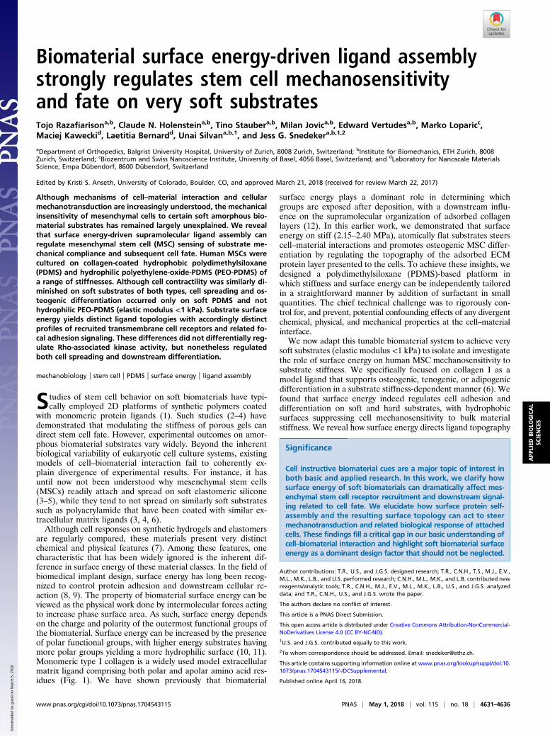
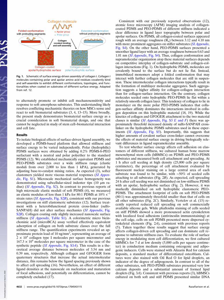
tomers of different stiffness, we cultured human bone marrowmesenchymal stem cells (hBMSCs) on PDMS and PEO-PDMSsubstrates and measured both cell attachment and spreading. At1 h after cell seeding at high density (25,000 cells per squarecentimeter), the percentage of cell attachment evaluated byfluorescent nuclear staining on a large central area of eachsubstrate was found to be similar, with ∼50% of seeded cellsattaching to all substrates (Fig. 2B). As expected, cell spreading24 h after cell seeding was not diminished on low-stiffness PDMSwith an apolar, hydrophobic surface (Fig. 2). However, it wasmarkedly diminished on soft hydrophilic elastomeric PEO-PDMS. The attachment footprint of cells on soft PEO-PDMS(80:1) was approximately threefold smaller than that of cells onall other substrates (Fig. 2C). Similarly, Vertelov et al. (15) re-cently reported reduced cell spreading on soft commerciallyavailable silicone gels. While phalloidin staining of cells seededon stiff PDMS showed a more pronounced actin cytoskeletonwith localized focal adhesions (antivinculin immunostaining) atthe cell edge, cells on soft PDMS presented more dispersed cy-toskeletal elements (Fig. 2A), consistent with previous reports(5). Taken together these results suggest that surface energyaffects collagen-driven cell spreading and can dominate cell re-sponse to substrate stiffness cues. To evaluate the role of surfaceenergy in modulating stem cell differentiation, we first culturedhBMSCs for 7 d at low density (5,000 cells per square centime-ter) in coinduction medium containing osteogenic and adipo-genic inducers. Cells were stained with Alizarin Red for calciumdeposits, a standard marker of differentiated osteoblasts. Cul-tures were also stained with Oil Red O for lipid droplets, anindicator of the degree of adipogenesis. In contrast to all of theother substrates, soft PEO-PDMS substrates presented very lowcalcium deposits and a substantial amount of formed lipiddroplets (Fig. 3A). Consistent with previous reports (3), hBMSCscultured on both soft and stiff PDMS presented similar DNA
Fig. 1. Schematic of surface energy-driven assembly of collagen I. Collagen Imolecules containing polar and apolar amino acid residues covalently bindand self-assemble to exhibit different conformations, topologies, and func-tionalities when coated on substrates of different surface energy. Adaptedfrom ref. 12.
4632 | www.pnas.org/cgi/doi/10.1073/pnas.1704543115 Razafiarison et al.
Dow
nloa
ded
by g
uest
on
Mar
ch 5
, 202
0
amounts and alkaline phosphatase (ALP) activity (Fig. 3 B andC); however, the amount of DNA on soft PEO-PDMS substrateswas significantly lower compared with the other substrates, sug-gesting reduced proliferation (20) (Fig. 3B). Quantification ofALP activity revealed a threefold lower expression of this osteo-genic marker on soft PEO-PDMS compared with the stiff material(Fig. 3C).We further investigated the hBMSC differentiation in basal
growth medium for 14 d at low seeding density (5,000 cells persquare centimeter) by staining for ALP and calcium deposition.Previous studies (4, 12) have reported a tendency for differen-tiation toward osteogenic lineages when stem cells are culturedon PDMS substrates independently of their stiffness. Consistentwith this observation, hBMSCs cultured on all tested substrates,except for soft PEO-PDMS, exhibited a positive staining for ALPand a high calcium deposit (Fig. 3D). Collectively, our datasuggest that surface energy is a key factor in stem cell differen-tiation as driven by substrate stiffness.A unique traction force microscopy (TFM) approach was
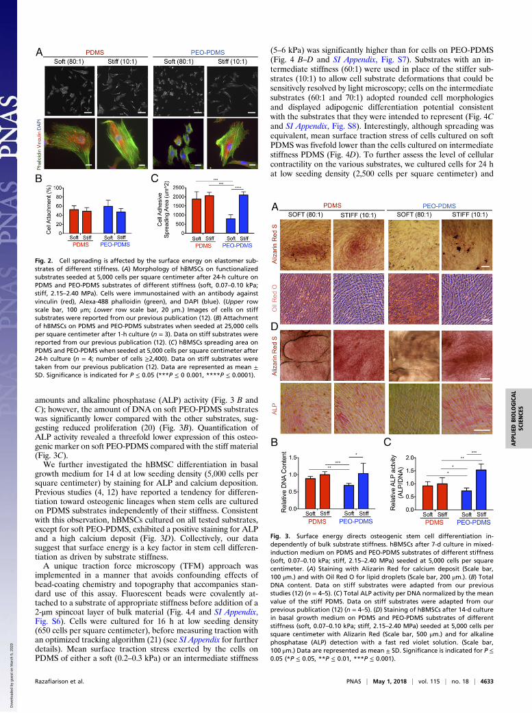
implemented in a manner that avoids confounding effects ofbead-coating chemistry and topography that accompanies stan-dard use of this assay. Fluorescent beads were covalently at-tached to a substrate of appropriate stiffness before addition of a2-μm spincoat layer of bulk material (Fig. 4A and SI Appendix,Fig. S6). Cells were cultured for 16 h at low seeding density(650 cells per square centimeter), before measuring traction withan optimized tracking algorithm (21) (see SI Appendix for furtherdetails). Mean surface traction stress exerted by the cells onPDMS of either a soft (0.2–0.3 kPa) or an intermediate stiffness
(5–6 kPa) was significantly higher than for cells on PEO-PDMS(Fig. 4 B–D and SI Appendix, Fig. S7). Substrates with an in-termediate stiffness (60:1) were used in place of the stiffer sub-strates (10:1) to allow cell substrate deformations that could besensitively resolved by light microscopy; cells on the intermediatesubstrates (60:1 and 70:1) adopted rounded cell morphologiesand displayed adipogenic differentiation potential consistentwith the substrates that they were intended to represent (Fig. 4Cand SI Appendix, Fig. S8). Interestingly, although spreading wasequivalent, mean surface traction stress of cells cultured on softPDMS was fivefold lower than the cells cultured on intermediatestiffness PDMS (Fig. 4D). To further assess the level of cellularcontractility on the various substrates, we cultured cells for 24 hat low seeding density (2,500 cells per square centimeter) and
Fig. 2. Cell spreading is affected by the surface energy on elastomer sub-strates of different stiffness. (A) Morphology of hBMSCs on functionalizedsubstrates seeded at 5,000 cells per square centimeter after 24-h culture onPDMS and PEO-PDMS substrates of different stiffness (soft, 0.07–0.10 kPa;stiff, 2.15–2.40 MPa). Cells were immunostained with an antibody againstvinculin (red), Alexa-488 phalloidin (green), and DAPI (blue). (Upper rowscale bar, 100 μm; Lower row scale bar, 20 μm.) Images of cells on stiffsubstrates were reported from our previous publication (12). (B) Attachmentof hBMSCs on PDMS and PEO-PDMS substrates when seeded at 25,000 cellsper square centimeter after 1-h culture (n = 3). Data on stiff substrates werereported from our previous publication (12). (C) hBMSCs spreading area onPDMS and PEO-PDMS when seeded at 5,000 cells per square centimeter after24-h culture (n = 4; number of cells ≥2,400). Data on stiff substrates weretaken from our previous publication (12). Data are represented as mean ±SD. Significance is indicated for P ≤ 0.05 (***P ≤ 0 0.001, ****P ≤ 0.0001).
Fig. 3. Surface energy directs osteogenic stem cell differentiation in-dependently of bulk substrate stiffness. hBMSCs after 7-d culture in mixed-induction medium on PDMS and PEO-PDMS substrates of different stiffness(soft, 0.07–0.10 kPa; stiff, 2.15–2.40 MPa) seeded at 5,000 cells per squarecentimeter. (A) Staining with Alizarin Red for calcium deposit (Scale bar,100 μm.) and with Oil Red O for lipid droplets (Scale bar, 200 μm.). (B) TotalDNA content. Data on stiff substrates were adapted from our previousstudies (12) (n = 4–5). (C) Total ALP activity per DNA normalized by the meanvalue of the stiff PDMS. Data on stiff substrates were adapted from ourprevious publication (12) (n = 4–5). (D) Staining of hBMSCs after 14-d culturein basal growth medium on PDMS and PEO-PDMS substrates of differentstiffness (soft, 0.07–0.10 kPa; stiff, 2.15–2.40 MPa) seeded at 5,000 cells persquare centimeter with Alizarin Red (Scale bar, 500 μm.) and for alkalinephosphatase (ALP) detection with a fast red violet solution. (Scale bar,100 μm.) Data are represented as mean ± SD. Significance is indicated for P ≤0.05 (*P ≤ 0.05, **P ≤ 0.01, ***P ≤ 0.001).
Razafiarison et al. PNAS | May 1, 2018 | vol. 115 | no. 18 | 4633
APP
LIED
BIOLO
GICAL
SCIENCE
S
Dow
nloa
ded
by g
uest
on
Mar
ch 5
, 202
0
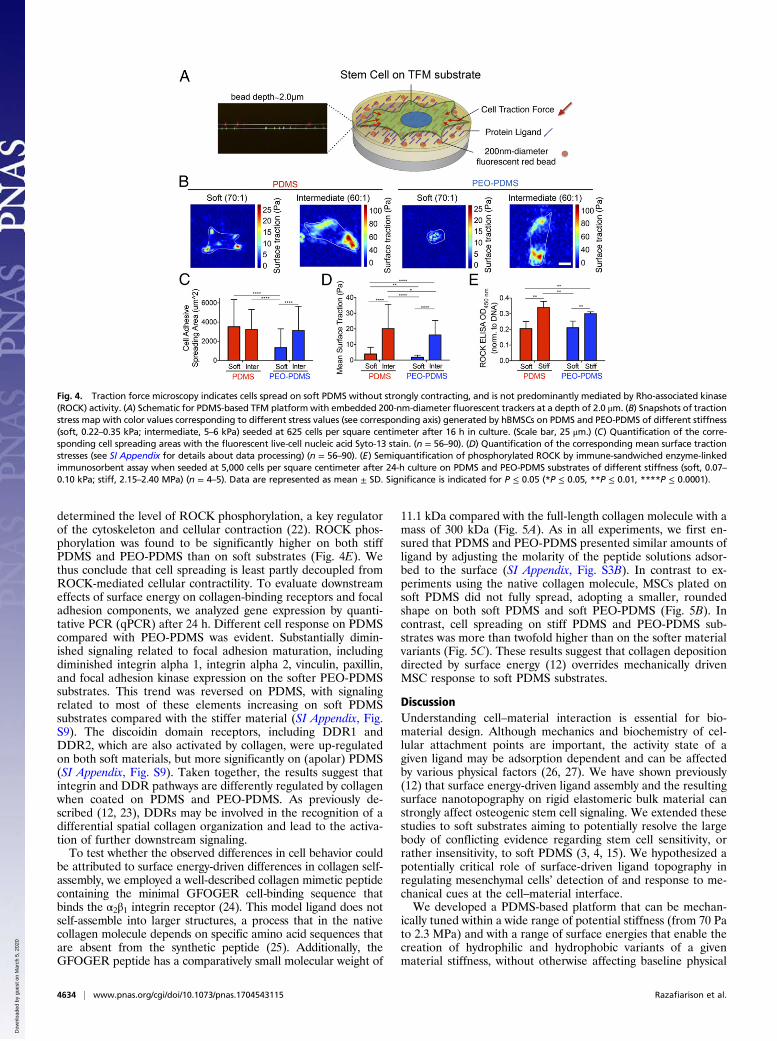
determined the level of ROCK phosphorylation, a key regulatorof the cytoskeleton and cellular contraction (22). ROCK phos-phorylation was found to be significantly higher on both stiffPDMS and PEO-PDMS than on soft substrates (Fig. 4E). Wethus conclude that cell spreading is least partly decoupled fromROCK-mediated cellular contractility. To evaluate downstreameffects of surface energy on collagen-binding receptors and focaladhesion components, we analyzed gene expression by quanti-tative PCR (qPCR) after 24 h. Different cell response on PDMScompared with PEO-PDMS was evident. Substantially dimin-ished signaling related to focal adhesion maturation, includingdiminished integrin alpha 1, integrin alpha 2, vinculin, paxillin,and focal adhesion kinase expression on the softer PEO-PDMSsubstrates. This trend was reversed on PDMS, with signalingrelated to most of these elements increasing on soft PDMSsubstrates compared with the stiffer material (SI Appendix, Fig.S9). The discoidin domain receptors, including DDR1 andDDR2, which are also activated by collagen, were up-regulatedon both soft materials, but more significantly on (apolar) PDMS(SI Appendix, Fig. S9). Taken together, the results suggest thatintegrin and DDR pathways are differently regulated by collagenwhen coated on PDMS and PEO-PDMS. As previously de-scribed (12, 23), DDRs may be involved in the recognition of adifferential spatial collagen organization and lead to the activa-tion of further downstream signaling.To test whether the observed differences in cell behavior could
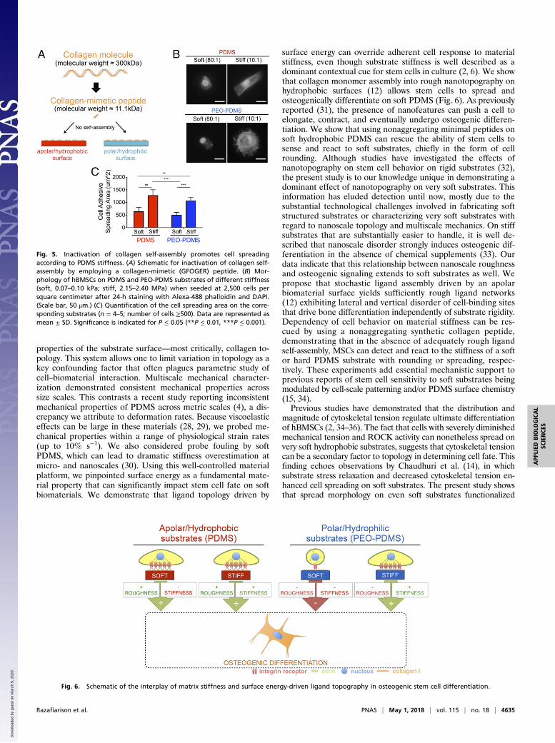
be attributed to surface energy-driven differences in collagen self-assembly, we employed a well-described collagen mimetic peptidecontaining the minimal GFOGER cell-binding sequence thatbinds the α2β1 integrin receptor (24). This model ligand does notself-assemble into larger structures, a process that in the nativecollagen molecule depends on specific amino acid sequences thatare absent from the synthetic peptide (25). Additionally, theGFOGER peptide has a comparatively small molecular weight of
11.1 kDa compared with the full-length collagen molecule with amass of 300 kDa (Fig. 5A). As in all experiments, we first en-sured that PDMS and PEO-PDMS presented similar amounts ofligand by adjusting the molarity of the peptide solutions adsor-bed to the surface (SI Appendix, Fig. S3B). In contrast to ex-periments using the native collagen molecule, MSCs plated onsoft PDMS did not fully spread, adopting a smaller, roundedshape on both soft PDMS and soft PEO-PDMS (Fig. 5B). Incontrast, cell spreading on stiff PDMS and PEO-PDMS sub-strates was more than twofold higher than on the softer materialvariants (Fig. 5C). These results suggest that collagen depositiondirected by surface energy (12) overrides mechanically drivenMSC response to soft PDMS substrates.
DiscussionUnderstanding cell–material interaction is essential for bio-material design. Although mechanics and biochemistry of cel-lular attachment points are important, the activity state of agiven ligand may be adsorption dependent and can be affectedby various physical factors (26, 27). We have shown previously(12) that surface energy-driven ligand assembly and the resultingsurface nanotopography on rigid elastomeric bulk material canstrongly affect osteogenic stem cell signaling. We extended thesestudies to soft substrates aiming to potentially resolve the largebody of conflicting evidence regarding stem cell sensitivity, orrather insensitivity, to soft PDMS (3, 4, 15). We hypothesized apotentially critical role of surface-driven ligand topography inregulating mesenchymal cells’ detection of and response to me-chanical cues at the cell–material interface.We developed a PDMS-based platform that can be mechan-
ically tuned within a wide range of potential stiffness (from 70 Pato 2.3 MPa) and with a range of surface energies that enable thecreation of hydrophilic and hydrophobic variants of a givenmaterial stiffness, without otherwise affecting baseline physical
Fig. 4. Traction force microscopy indicates cells spread on soft PDMS without strongly contracting, and is not predominantly mediated by Rho-associated kinase(ROCK) activity. (A) Schematic for PDMS-based TFM platformwith embedded 200-nm-diameter fluorescent trackers at a depth of 2.0 μm. (B) Snapshots of tractionstress map with color values corresponding to different stress values (see corresponding axis) generated by hBMSCs on PDMS and PEO-PDMS of different stiffness(soft, 0.22–0.35 kPa; intermediate, 5–6 kPa) seeded at 625 cells per square centimeter after 16 h in culture. (Scale bar, 25 μm.) (C) Quantification of the corre-sponding cell spreading areas with the fluorescent live-cell nucleic acid Syto-13 stain. (n = 56–90). (D) Quantification of the corresponding mean surface tractionstresses (see SI Appendix for details about data processing) (n = 56–90). (E) Semiquantification of phosphorylated ROCK by immune-sandwiched enzyme-linkedimmunosorbent assay when seeded at 5,000 cells per square centimeter after 24-h culture on PDMS and PEO-PDMS substrates of different stiffness (soft, 0.07–0.10 kPa; stiff, 2.15–2.40 MPa) (n = 4–5). Data are represented as mean ± SD. Significance is indicated for P ≤ 0.05 (*P ≤ 0.05, **P ≤ 0.01, ****P ≤ 0.0001).
4634 | www.pnas.org/cgi/doi/10.1073/pnas.1704543115 Razafiarison et al.
Dow
nloa
ded
by g
uest
on
Mar
ch 5
, 202
0
properties of the substrate surface—most critically, collagen to-pology. This system allows one to limit variation in topology as akey confounding factor that often plagues parametric study ofcell–biomaterial interaction. Multiscale mechanical character-ization demonstrated consistent mechanical properties acrosssize scales. This contrasts a recent study reporting inconsistentmechanical properties of PDMS across metric scales (4), a dis-crepancy we attribute to deformation rates. Because viscoelasticeffects can be large in these materials (28, 29), we probed me-chanical properties within a range of physiological strain rates(up to 10% s−1). We also considered probe fouling by softPDMS, which can lead to dramatic stiffness overestimation atmicro- and nanoscales (30). Using this well-controlled materialplatform, we pinpointed surface energy as a fundamental mate-rial property that can significantly impact stem cell fate on softbiomaterials. We demonstrate that ligand topology driven by
surface energy can override adherent cell response to materialstiffness, even though substrate stiffness is well described as adominant contextual cue for stem cells in culture (2, 6). We showthat collagen monomer assembly into rough nanotopography onhydrophobic surfaces (12) allows stem cells to spread andosteogenically differentiate on soft PDMS (Fig. 6). As previouslyreported (31), the presence of nanofeatures can push a cell toelongate, contract, and eventually undergo osteogenic differen-tiation. We show that using nonaggregating minimal peptides onsoft hydrophobic PDMS can rescue the ability of stem cells tosense and react to soft substrates, chiefly in the form of cellrounding. Although studies have investigated the effects ofnanotopography on stem cell behavior on rigid substrates (32),the present study is to our knowledge unique in demonstrating adominant effect of nanotopography on very soft substrates. Thisinformation has eluded detection until now, mostly due to thesubstantial technological challenges involved in fabricating softstructured substrates or characterizing very soft substrates withregard to nanoscale topology and multiscale mechanics. On stiffsubstrates that are substantially easier to handle, it is well de-scribed that nanoscale disorder strongly induces osteogenic dif-ferentiation in the absence of chemical supplements (33). Ourdata indicate that this relationship between nanoscale roughnessand osteogenic signaling extends to soft substrates as well. Wepropose that stochastic ligand assembly driven by an apolarbiomaterial surface yields sufficiently rough ligand networks(12) exhibiting lateral and vertical disorder of cell-binding sitesthat drive bone differentiation independently of substrate rigidity.Dependency of cell behavior on material stiffness can be res-cued by using a nonaggregating synthetic collagen peptide,demonstrating that in the absence of adequately rough ligandself-assembly, MSCs can detect and react to the stiffness of a softor hard PDMS substrate with rounding or spreading, respec-tively. These experiments add essential mechanistic support toprevious reports of stem cell sensitivity to soft substrates beingmodulated by cell-scale patterning and/or PDMS surface chemistry(15, 34).Previous studies have demonstrated that the distribution and
magnitude of cytoskeletal tension regulate ultimate differentiationof hBMSCs (2, 34–36). The fact that cells with severely diminishedmechanical tension and ROCK activity can nonetheless spread onvery soft hydrophobic substrates, suggests that cytoskeletal tensioncan be a secondary factor to topology in determining cell fate. Thisfinding echoes observations by Chaudhuri et al. (14), in whichsubstrate stress relaxation and decreased cytoskeletal tension en-hanced cell spreading on soft substrates. The present study showsthat spread morphology on even soft substrates functionalized
Fig. 5. Inactivation of collagen self-assembly promotes cell spreadingaccording to PDMS stiffness. (A) Schematic for inactivation of collagen self-assembly by employing a collagen-mimetic (GFOGER) peptide. (B) Mor-phology of hBMSCs on PDMS and PEO-PDMS substrates of different stiffness(soft, 0.07–0.10 kPa; stiff, 2.15–2.40 MPa) when seeded at 2,500 cells persquare centimeter after 24-h staining with Alexa-488 phalloidin and DAPI.(Scale bar, 50 μm.) (C) Quantification of the cell spreading area on the corre-sponding substrates (n = 4–5; number of cells ≥500). Data are represented asmean ± SD. Significance is indicated for P ≤ 0.05 (**P ≤ 0.01, ***P ≤ 0.001).
Fig. 6. Schematic of the interplay of matrix stiffness and surface energy-driven ligand topography in osteogenic stem cell differentiation.
Razafiarison et al. PNAS | May 1, 2018 | vol. 115 | no. 18 | 4635
APP
LIED
BIOLO
GICAL
SCIENCE
S
Dow
nloa
ded
by g
uest
on
Mar
ch 5
, 202
0
with a suitable extracellular matrix ligand is sufficient to directMSCs to an osteogenic fate. Conversely, previous studies havedemonstrated that stiff substrates which confine cell spreading onmicropatterned surfaces can promote adipogenic differentiation(35, 36). Thus, spread cell morphology, rather than the develop-ment of cytoskeletal contractility per se, seems to be a necessarycondition to determine cell fate in the 2D culture conditions thatform the majority basis of our understanding on stem cellmechanosensitivity. This conclusion contrasts with recent experi-ments with 3D platforms that reported cell fate to be independentof cell morphology but rather more traction dependent (37, 38).Still, 3D culture systems have their own disadvantages, includingrelative lack of control over hydrostatic stresses and osmotic gra-dients, and further work is required to determine how ROCK-mediated contractility and spread cell morphology potentially in-teract to regulate cell signaling or act as a central checkpoint indetermining cell fate.Collectively these results are striking, with potentially critical
implications for research employing type I collagen as a “stan-dard” 2D cell culture reagent. We demonstrate that stochasticcollagen self-assembly translates to large potential for experi-mental variability, and/or systematically biased biological out-comes. Other ECM ligands, such as fibronectin, can behave quitedifferently from collagen (e.g., SI Appendix, Fig. S10), and workis required to characterize which combinations of biomaterials,interface chemistry, extracellular protein milieu, and cells yieldreliably emergent system behavior.In conclusion, we have demonstrated that surface energy-
driven ligand self-assembly (12) can steer a cell to very different
fates on soft substrates. Controlling for surface energy enablesstem cells to spread and differentiate according to PDMS stiff-ness. These findings fill an important gap in our collective un-derstanding (3, 4), explaining why stem cells spread and undergoosteogenic differentiation on soft apolar silicone when coatedwith collagen compared with rounding on soft polar substratessuch as polyacrylamide. Although thoroughly described in thefield of rigid biomaterials used in implants (8, 9), effects ofsurface energy on very soft substrates are difficult to control, andas such have been widely ignored in opinion-leading papers onstem cell–matrix interaction (3, 4). We suggest that surface en-ergy is nonetheless a major biomaterial design factor that mustbe considered when designing cell-instructive biomaterials.
MethodsTunable Surface Energy PDMS Substrate Preparation. The 12- to 25-mm-diameter glass coverslips were cleaned with milli-Q H2O and ethanol (12). ThePDMS base was mixed first with or without 0.2% (wt/wttotal) the surfactantpolydimethylsiloxane-b-ethylene oxide. The PDMS cross-linker was thenmixed in different ratios ranging from 80:1 to 10:1 base:cross-linker, spreadon the glass coverslips.
Additional Methods. The preparation of the various biocompatible surfaces,and the methods used, are described in SI Appendix, SI Materials andMethods.
ACKNOWLEDGMENTS. The authors thank Barbara Niederöst for technicalcontribution to this work. This work was generously supported by the SwissNational Science Foundation (Grants 138221 and 205321).
1. MacQueen L, Sun Y, Simmons CA (2013) Mesenchymal stem cell mechanobiology andemerging experimental platforms. J R Soc Interface 10:20130179.
2. Engler AJ, Sen S, Sweeney HL, Discher DE (2006) Matrix elasticity directs stem celllineage specification. Cell 126:677–689.
3. Trappmann B, et al. (2012) Extracellular-matrix tethering regulates stem-cell fate. NatMater 11:642–649.
4. Wen JH, et al. (2014) Interplay of matrix stiffness and protein tethering in stem celldifferentiation. Nat Mater 13:979–987.
5. Prager-Khoutorsky M, et al. (2011) Fibroblast polarization is a matrix-rigidity-dependent process controlled by focal adhesion mechanosensing. Nat Cell Biol 13:1457–1465.
6. Sharma RI, Snedeker JG (2010) Biochemical and biomechanical gradients for directedbone marrow stromal cell differentiation toward tendon and bone. Biomaterials 31:7695–7704.
7. Higuchi A, Ling Q-D, Chang Y, Hsu S-T, Umezawa A (2013) Physical cues of bioma-terials guide stem cell differentiation fate. Chem Rev 113:3297–3328.
8. Liu X, et al. (2007) Influence of substratum surface chemistry/energy and topographyon the human fetal osteoblastic cell line hFOB 1.19: Phenotypic and genotypic re-sponses observed in vitro. Biomaterials 28:4535–4550.
9. Mager MD, LaPointe V, Stevens MM (2011) Exploring and exploiting chemistry at thecell surface. Nat Chem 3:582–589.
10. Ramos SC, et al. (2011) Influence of polar groups on the wetting properties of ver-tically aligned multiwalled carbon nanotube surfaces. Theor Chem Acc 130:1061–1069.
11. de Gennes PG (1985) Wetting: Statics and dynamics. Rev Mod Phys 57:827–863.12. Razafiarison T, Silván U, Meier D, Snedeker JG (2016) Surface-driven collagen self-
assembly affects early osteogenic stem cell signaling. Adv Healthc Mater 5:1481–1492.13. Li J, Han D, Zhao Y-P (2014) Kinetic behaviour of the cells touching substrate: The
interfacial stiffness guides cell spreading. Sci Rep 4:3910.14. Chaudhuri O, et al. (2015) Substrate stress relaxation regulates cell spreading. Nat
Commun 6:6364.15. Vertelov G, et al. (2016) Rigidity of silicone substrates controls cell spreading and stem
cell differentiation. Sci Rep 6:33411.16. Cavalcanti-Adam EA, et al. (2007) Cell spreading and focal adhesion dynamics are
regulated by spacing of integrin ligands. Biophys J 92:2964–2974.17. Kilian KA, Mrksich M (2012) Directing stem cell fate by controlling the affinity and
density of ligand-receptor interactions at the biomaterials interface. Angew Chem IntEd Engl 51:4891–4895.
18. Dupont-Gillain CC, Jacquemart I, Rouxhet PG (2005) Influence of the aggregationstate in solution on the supramolecular organization of adsorbed type I collagenlayers. Colloids Surf B Biointerfaces 43:179–186.
19. Narayanan B, Gilmer GH, Tao J, De Yoreo JJ, Ciobanu CV (2014) Self-assembly ofcollagen on flat surfaces: The interplay of collagen-collagen and collagen-substrateinteractions. Langmuir 30:1343–1350.
20. Mih JD, et al. (2011) A multiwell platform for studying stiffness-dependent cell bi-ology. PLoS One 6:e19929.
21. Holenstein CN, Silvan U, Snedeker JG (2017) High-resolution traction force microscopyon small focal adhesions: Improved accuracy through optimal marker distribution andoptical flow tracking. Sci Rep 7:41633.
22. Riveline D, et al. (2001) Focal contacts as mechanosensors: Externally applied localmechanical force induces growth of focal contacts by an mDia1-dependent andROCK-independent mechanism. J Cell Biol 153:1175–1186.
23. Lund AW, Stegemann JP, Plopper GE (2009) Mesenchymal stem cells sense three di-mensional type I collagen through discoidin domain receptor 1. Open Stem Cell J 1:40–53.
24. Wojtowicz AM, et al. (2010) Coating of biomaterial scaffolds with the collagen-mimetic peptide GFOGER for bone defect repair. Biomaterials 31:2574–2582.
25. Prockop DJ, Fertala A (1998) Inhibition of the self-assembly of collagen I into fibrilswith synthetic peptides. Demonstration that assembly is driven by specific bindingsites on the monomers. J Biol Chem 273:15598–15604.
26. Arima Y, Iwata H (2007) Effect of wettability and surface functional groups on proteinadsorption and cell adhesion using well-defined mixed self-assembled monolayers.Biomaterials 28:3074–3082.
27. Schaap-Oziemlak AM, Kühn PT, van Kooten TG, van Rijn P (2014) Biomaterial–stemcell interactions and their impact on stem cell response. RSC Adv 4:53307–53320.
28. Lakes RS (2004) Viscoelastic measurement techniques. Rev Sci Instrum 75:797–810.29. Nordin M, Frankel VH (2013) Basic Biomechanics of the Musculoskeletal System
(Lippincott Williams & Wilkins, Baltimore).30. Meli F, Küng A (2007) AFM investigation on surface damage caused by mechanical
probing with small ruby spheres. Meas Sci Technol 18:496–502.31. Oh S, et al. (2009) Stem cell fate dictated solely by altered nanotube dimension. Proc
Natl Acad Sci USA 106:2130–2135.32. Dalby MJ, Gadegaard N, Oreffo ROC (2014) Harnessing nanotopography and integrin-
matrix interactions to influence stem cell fate. Nat Mater 13:558–569.33. Dalby MJ, et al. (2007) The control of human mesenchymal cell differentiation using
nanoscale symmetry and disorder. Nat Mater 6:997–1003.34. Fu J, et al. (2010) Mechanical regulation of cell function with geometrically modu-
lated elastomeric substrates. Nat Methods 7:733–736.35. Kilian KA, Bugarija B, Lahn BT, Mrksich M (2010) Geometric cues for directing the
differentiation of mesenchymal stem cells. Proc Natl Acad Sci USA 107:4872–4877.36. McBeath R, Pirone DM, Nelson CM, Bhadriraju K, Chen CS (2004) Cell shape, cyto-
skeletal tension, and RhoA regulate stem cell lineage commitment. Dev Cell 6:483–495.
37. Huebsch N, et al. (2010) Harnessing traction-mediated manipulation of the cell/matrixinterface to control stem-cell fate. Nat Mater 9:518–526.
38. Khetan S, et al. (2013) Degradation-mediated cellular traction directs stem cell fate incovalently crosslinked three-dimensional hydrogels. Nat Mater 12:458–465.
4636 | www.pnas.org/cgi/doi/10.1073/pnas.1704543115 Razafiarison et al.
Dow
nloa
ded
by g
uest
on
Mar
ch 5
, 202
0