Embed Size (px)
Citation preview
Preprint. The final version will be published in Studies in History and Philosophy of the Biological and Biomedical Sciences.
1
Biology meets Physics:
Reductionism and Multi-scale Modeling of Morphogenesis
Sara Green1 & Robert Batterman2
Abstract
A common reductionist assumption is that macro-scale behaviors can be described "bottom-up" if only sufficient details about lower-scale processes are available. The view that an "ideal" or "fundamental" physics would be sufficient to explain all macro-scale phenomena has been met with criticism from philosophers of biology. Specifically, scholars have pointed to the impossibility of deducing biological explanations from physical ones, and to the irreducible nature of distinctively biological processes such as gene regulation and evolution. This paper takes a step back in asking whether bottom-up modeling is feasible even when modeling simple physical systems across scales. By comparing examples of multi-scale modeling in physics and biology, we argue that the “tyranny of scales” problem presents a challenge to reductive explanations in both physics and biology. The problem refers to the scale-dependency of physical and biological behaviors that forces researchers to combine different models relying on different scale-specific mathematical strategies and boundary conditions. Analyzing the ways in which different models are combined in multi-scale modeling also has implications for the relation between physics and biology. Contrary to the assumption that physical science approaches provide reductive explanations in biology, we exemplify how inputs from physics often reveal the importance of macro-scale models and explanations. We illustrate this through an examination of the role of biomechanics modeling in developmental biology. In such contexts, the relation between models at different scales and from different disciplines is neither reductive nor completely autonomous, but interdependent.
1 Department of Science Education, University of Copenhagen, Øster Voldgade 3, 1350 Copenhagen, DK. Email: [email protected]. 2 Department of Philosophy, University of Pittsburgh, 1028-A Cathedral of Learning, Pittsburgh, PA 15260.

Preprint. The final version will be published in Studies in History and Philosophy of the Biological and Biomedical Sciences.
2
1. Introduction An important reductionist assumption is that multi-scale systems can be described “bottom-up”, if
only sufficient details about the states of the components are available. Historically, this assumption
has been debated in philosophical discussions about whether biology is reducible to physics. The
positivist ideal of a unity of science pictured the relations between scientific disciplines in a “layer-
cake” hierarchy where theories from respective disciplines target a specific level or scale of
phenomena (Oppenheim & Putnam, 1958).3 Physics was considered the most fundamental “model
discipline” targeting the lowest organizational level, and progressive reduction was considered an
important aspect of scientific development (see also Hütteman & Love, 2016).
The view that an ideal or fundamental physics would be sufficient to explain all macro-scale
phenomena has been met with criticism from philosophers of biology. Scholars have stressed the
irreducibility of biological features, such as gene regulation or evolution, and argued that biological
explanations are irreducible to physical laws and principles (e.g., Bechtel & Richardson, 1993;
Bertalanffy, 1969; Burian et al., 1996; Dupré, 1993; Machamer et al., 2000; Mayr, 1988; 2004; Winther,
2009). These contributions have offered important insights to distinctive features of living systems
and biological research. However, an important question that is rarely addressed is whether the ideal
of progressive reduction of higher-level explanations is supported in physics, i.e., in the discipline that
was taken as a model for the reductionist ideal. We argue that lessons from multi-scale modeling offer
resistance to reductionism which cross-cut discussions in philosophy of biology and philosophy of
physics.
We focus on what Mayr (1988) calls explanatory reduction, which involves explaining phenomena at
higher scales in terms of processes at lower scales or levels of organization (e.g., molecules or genes).4
Other important aspects of reductive explanations are that they typically analyze biological parts in
isolation from their original context and give explanatory priority only to factors internal to the system
(Kaiser, 2015). In recent discussions on explanatory reduction it is debated whether the constitution
of macroscale systems by microscale components allows the researcher to explain the system only
with reference to properties of the lower scale constituents (Brigandt & Love, 2012). For instance,
although the composition of polypeptides is reducible to a sequence of amino acids, it has been argued
that it is not possible to explain protein folding from physical laws and knowledge about amino acids
alone (Love & Hütteman, 2011). The prospect of reductive explanations in biology and physics is,
however, an ongoing issue of debate.
3 We use the term ”level” when referring more explicitly to part-whole relations in a hierarchical description or a functional
system (demarcated by boundaries such as the cell membrane), but we prefer the term ”scale” when referring to spatial scaling because biological ”levels” are often not straightforwardly distinguished (see also Noble, 2012). For a more detailed discussion of biological levels and part-whole relations, see (Kaiser, 2015). 4 Explanatory reduction is distinguished from Constitutive reduction and Theory reduction. Constitutive reduction (also called
ontological reduction or (token) physicalism) refers to the acceptance that biological systems are nothing but physical-chemical systems. Theory reduction considers the possibilities of reducing (in a logical sense) higher-level theories in special sciences to more fundamental ones (cf., Rosenberg & Arp, 2010; Sarkar, 1998; Schaffner, 1993; Winther, 2009). More recently, philosophers of biology have discussed this kind of reduction in terms of explanatory relevance, e.g., whether the explanatory power in biology is constituted by physico-chemical principles or biological mechanisms (Machamer et al., 2000; Weber, 2008). A separate kind of reductionism, methodological reductionism, considers heuristic strategies that simplify the problem space for scientific analysis (Brigandt & Love, 2012; Bechtel & Richardson, 1993; Green, 2015).
Preprint. The final version will be published in Studies in History and Philosophy of the Biological and Biomedical Sciences.
3
This paper sheds further light on the debate on reductionism by clarifying how lessons from multi-
scale modeling in both physics and biology offer resistance to the idea that multi-scale systems can be
modelled and explained “bottom-up”. Secondly, unlike what one might expect from physical science
approaches, we argue that work within biomechanics brings attention to the problems of
understanding biological processes and parts in isolation from their original context in cells or tissue
structures (Kaiser, 2015). Thus, rather than enforcing reductionism, physical science approaches can
help reveal the limitations for reducing explanations in developmental biology to genetics.
Accordingly, we argue that the role of physical science approaches in biology with respect to
reductionism should be revisited.
Our aim is to bring attention to the tyranny of scales problem that has so far mainly been discussed in
the context of physics (Batterman, 2012; Oden, 2006; see however Lesne, 2013).5 The problem refers
to the scale-dependency of physical behaviors that presents a hard challenge for modeling and
explaining multi-scale systems. No single mathematical model can account for behaviors at all spatial
and temporal scales, and the modeler must therefore combine different mathematical models relying
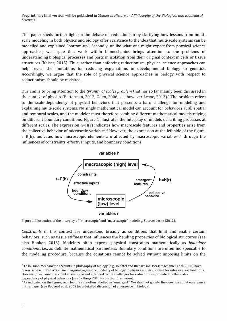
on different boundary conditions. Figure 1 illustrates the interplay of models describing processes at
different scales. The expression h=H(r) indicates how macroscale features and properties arise from
the collective behavior of microscale variables.6 However, the expression at the left side of the figure,
r=R(h), indicates how microscopic elements are affected by macroscopic variables h through the
influences of constraints, effective inputs, and boundary conditions.
Figure 1. Illustration of the interplay of “microscopic” and “macroscopic” modeling. Source: Lesne (2013).
Constraints in this context are understood broadly as conditions that limit and enable certain
behaviors, such as tissue stiffness that influences the bending properties of biological structures (see
also Hooker, 2013). Modelers often express physical constraints mathematically as boundary
conditions, i.e., as definite mathematical parameters. Boundary conditions are often indispensable to
the modeling procedure, because the equations cannot be solved without imposing limits on the
5 To be sure, mechanistic accounts in philosophy of biology (e.g., Bechtel and Richardson 1993; Machamer et al. 2000) have
taken issue with reductionism in arguing against reducibility of biology to physics and in allowing for interlevel explanations. However, mechanistic accounts have so far not attended to the challenges for reductionism provided by the scale-dependency of physical behaviors (see Skillings 2015 for further discussion). 6 As indicated on the figure, such features are often labelled as “emergent”. We shall not go into the question about emergence
in this paper (see Boogerd et al. 2005 for a detailed discussion of emergence in biology).

Preprint. The final version will be published in Studies in History and Philosophy of the Biological and Biomedical Sciences.
4
domain of the model. In this paper we describe how the scale-dependent behavior of physical and
biological systems forces researchers to combine different experimental and representational
strategies targeting specific scales.7 We illustrate this with examples from both physics and biology.
Since we are drawing on contemporary cases of multi-scale modeling, the reductionist may object that
all we point to are practical limitations of current science. That is, one may object that it in principle
should be possible to explain macro-scale systems with reference only to lower scale molecular
details. The specific methodologies will indeed develop and change over time. However, it should be
noted that the need to combine different approaches arises due to the fundamental challenge that
concepts used to characterize systems and their behaviors can change as one changes scale: They are
multi-valued across scales (Wilson, 2012). For example, not only does the concept of “surface” change
as one moves toward the nanoscale (where there is typically more “surface” than bulk material) but so
do the kinds of concepts we need to characterize the dominant behaviors at the different scales
(Bursten, 2015; see Section 2).8 Against the background of this complexity, we find appeals to in
principle derivations empty without suggestions of how to make such inferences (see also Batterman,
2016). Rather than logical possibilities for explanatory reduction, this paper is concerned with the
challenges faced in scientific practice. In our view, explanatory reduction fails if macroscale models,
measurements, and concepts are indispensable for explanations of multi-scale systems. Showing that
these are indeed indispensable is the main aim of this paper.9
Considering the challenges for bottom-up approaches in physics also calls for us to revisit the relation
between physics and biology. Our analysis does not imply or support any sharp distinction between
contributions from biology and physics. Both disciplines are highly diverse and often intertwined in
interdisciplinary fields.10 A weaker distinction of disciplinary inputs is, however, useful for revisiting
the relation between physical science approaches in biology and reductionism. As mentioned above,
physics has often been pictured as a discipline solely targeting the most fundamental “lower levels”,
with a preference for simple deterministic models. Interestingly, appeals to physical science
approaches in the examined cases of multi-scale modeling in developmental biology do not support
this view. Rather, they show that macro-scale features (i.e., those at cell and tissue level) are
indispensable and irreducible to lower-scale explanations. Moreover, we propose that the requirement
of macroscale parameters (e.g., tissue stiffness) as boundary conditions for models at lower scales
(Figure 1) provides a concrete instantiation of top-down effects (Section 4.1). We highlight how recent
7 This paper focuses on the adequacy of explanatory reduction through a demonstration of the requirement of multiple
models. As an anonymous reviewer pointed out, it is possible to agree with the inadequacy of explanatory reduction but argue that a single higher-level model is adequate. Our account would offer resistance also to a monistic anti-reductionist view of this kind but we do not develop such an argument in the paper. 8 Already Galileo (Discorsi, 1638) pointed to the importance of scale when considering the disproportional relation between
the minimal thickness of bone structures and animal size. Biologists investigating morphological constraints on animal form have similarly stressed that macroscale physics does not apply to microorganisms. At this scale, gravity is a weaker force whereas surface properties and Brownian motion are central parts of the analysis (see e.g., Purcell, 1977; Vogel, 2009). Similarly, the models most useful to model molecular behavior are rarely the most useful for modeling tissues. See Section 2 for further clarification. 9 We provide a clarification of what is meant by indispensability at the end of Section 3, derived from case examples.
10 The intertwinement of physics and biology is explicit in biophysics that comprises a range of important research areas such
as membrane physics, biomechanics, as well as research on the energetics of protein folding, molecular motors, and mechanosensors (Dunn and Price, 2015; Morange, 2011). Many of these developments also benefit from engineering approaches, and there are important differences between physics and engineering. But because the models we examine are developed in physics, we shall not in this paper distinguish between physics and bioengineering.

Preprint. The final version will be published in Studies in History and Philosophy of the Biological and Biomedical Sciences.
5
insights to biomechanical aspects of morphogenesis challenge deeply entrenched presuppositions
about the explanatory priority of lower scales, e.g., of the special priority of the molecular or genetic
level in developmental biology (cf. Rosenberg, 1997). These challenges are not specifically directed at
developmental biology, and we shall comment more briefly on how similar insights can be derived
from studies of multi-scale cardiac modeling and cancer research.
We shall proceed as follows. Section 2 introduces the tyranny of scales problem in physical and
biological contexts. Section 3 examines the application of physical science approaches in
developmental biology and highlights the importance of tissue-scale mechanics for embryo
development. Section 4 elaborates on the specific challenges posed for explanatory reduction in the
context of multi-scale modeling in biology in light of Sections 2 and 3. We describe the relation
between models at different scales (and from physics and biology) as non-reductive and
interdependent. Section 5 offers concluding remarks.
2. The tyranny of scales in physics and biology
One of the hardest problems in modeling the behaviors of physical systems is to deal with structures
that exist across different spatial scales. Generally, relying on a single mathematical model to describe
the behavior of a physical system at all scales is not possible, because dynamical and material
properties are scale-dependent (Wilson, 2012). Even successful modeling of a relatively simple multi-
scale system such as a steel beam requires different models.11 At atomic scales, steel has a regular
lattice structure but at higher scales it exhibits elastic behavior that is well-described by the Navier-
Cauchy elasticity equations (see Figure 2). These equations model the material as a continuum and
completely ignore atomic structure. Additionally, and very importantly, at intermediate (meso) scales,
steel presents a host of other structures such as lamellar inclusions of pearlite, cracks, grain
boundaries, etc. To fully understand the behavior of bending steel requires that one bridges across
these widely separated scales, i.e., that one can combine models at different scales that inform each
other. The problem is hard because “the principal physics governing events often changes with scale,
so that the models themselves must change in structure as the ramifications of events pass from one
scale to another” (Oden, 2006, p. 2930).
11
The steel beam is actually incredibly hard to model and is far from simple. Simple should here be understood in comparison to biological systems.

Preprint. The final version will be published in Studies in History and Philosophy of the Biological and Biomedical Sciences.
6
Figure 2. Macro and microstructures of steel. Source: Batterman (2012).
Modeling in biology must also confront the tyranny of scales. Like the steel example, some aspects of
biological structures require continuum models whereas others have to take into account the
structural diversity of and stochastic relations between the discrete interacting cells and molecules
(Lesne, 2013). In biology, researchers face the additional challenge that different integrated processes
operate also on different time-scales, from milliseconds to hours, days or even years (Davidson et al.,
2009). As Newman et al. (2011, p. 313) clarify, developmental systems display both discrete and
continuous aspects, depending on the specific spatial and temporal scale of the specific developmental
processes. These aspects make the modeling strategy “inescapably hybrid, mathematically and
computationally” (Newman et al., 2011, p. 313).
Continuum models treat discrete and diverse entities that exist in finite numbers as a continuous
variable. These models are used to model macroscopic behaviors that are relatively independent of
smaller-scale properties and local dynamics of the system components (Batterman, 2016). This
situation can be seen as analogous to how many applications of the ideal gas law are independent of
information about the specific dynamic trajectories of individual molecules of the gas because
microscopic fluctuations average out. In biology, such fluctuations can also be buffered by regulatory
circuits, yielding robust functions despite perturbations at lower levels (Lesne, 2013). For instance,
when modeling cell motion at tissue scales or at that of the whole embryo, developmental biologists
often rely on coupled partial differential equations that ignore the stochastic properties of interactions
between individual molecules and cells. Similar to mean-field approaches in physics, they study the

Preprint. The final version will be published in Studies in History and Philosophy of the Biological and Biomedical Sciences.
7
collective dynamics of the population of cells rather than the individual components (Lesne, 2013).
These modeling choices are not just motivated by considerations about tractability but also reflect the
problem that some features cannot be modeled at all scales. Just like one cannot attribute features like
temperature and pressure to an individual gas molecule, so are some features of cell populations not
possible to derive from models of isolated cells. That is, macro-scale phenomena require alternative
modeling frameworks. Tissue-scale models of cell movement typically rely on reaction-diffusion
equations (modeling e.g. chemotaxis responses or the mechanical influence of the ECM) or integro-
differential equations that model cells as flows, akin to processes in fluid dynamics. This can be done
via Navier-Stokes equations, or via mass action laws describing chemical kinetics (Hatzikirou &
Deutsch, 2011).12
The most appropriate model for capturing biological phenomena also depends on temporal scaling.
Shawky and Davidson (2015, p. 154) clarify how engineering models of embryonic tissues “range from
elastic solid-like to viscous liquid-like, depending on the time-scale of measurement”.13 When
modeling complex phenomena like a developing embryo or the human heart, researchers must
combine deterministic and stochastic models to couple processes across spatial and temporal scales.
At tissue-scales the relevant measurements span over longer times, allowing one to ignore fluctuations
in biomolecular species. This often gives more accurate predictions. In contrast, at the molecular scale
the dynamics is “dominated by random and short time fluctuations” in the concentrations of ions or
proteins (Qu et al., 2011, p. 22). Combining different kinds of models in large-scale simulations
spanning multiple spatial and temporal scales is far from trivial because the models often make
different predictions about what will happen with the same system over time (Qu et al., 2011). But
modeling the whole system using only one modeling framework is not possible because different
aspects of the system dominate the behavior at characteristic scales.
One consequence of the “multi-valuedness” of multi-scale systems is that different details must be
ignored by models operating at different spatial and temporal scales. Just as any useful model in
science should ignore the degrees of freedom irrelevant for the specific modeling task, “a multiscale
model should not intend to keep track of all details at all scales but only of the relevant details,
whatever their scales” (Lesne, 2013, p. 17). As we shall clarify in the following section, boundary
conditions play a crucial role for the purpose of representing physical and biological constraints and
for combining models that account for complementary aspects of the system.
2.1. Boundary conditions
We begin again with a simple example from physics and then move to the biological context. An
example where boundary conditions play an essential role is in the modeling of the harmonic
structure of a violin string. One can determine the modes of the standing wave of a vibrating string by
solving a wave equation. To solve the partial differential equation requires the imposition of
mathematical boundary conditions that fix the endpoints of the string (Batterman, 2012; Wilson,
12
These models can be extremely complex and it is often necessary to discretize the continuum equations using numerical strategies such as Finite Element Methods (see Section 3.1). 13
Consider how water in a swimming pool can seem to be very solid on the short time scale in which a diver belly flops. On the other hand, as one wades in, the water seems quite un-solid like.

Preprint. The final version will be published in Studies in History and Philosophy of the Biological and Biomedical Sciences.
8
2012). Thus, it requires that the string at the bridge and nut of the violin remains absolutely stationary
as the string vibrates. Unfortunately, while essential for the determination of the modes, if such strict
conditions on the string’s endpoints were actually imposed on the violin, it would make it impossible
for the vibrations in the string to be amplified by the sound box of the violin and we would be unable
to hear the instrument. Modeling the behavior of the vibrations that get amplified via the sound box
requires that one completely shift scales and focuses on the molecular and sub-molecular interactions
between the string and the bridge. Here the equations are of a completely different mathematical type.
This is the realm of molecular dynamics governed by ordinary differential equations. The lesson here
is that sometimes (quite often in fact) one needs to impose boundary conditions in order to efface
physical details that will not allow one to model the behavior of interest at a given scale.14 As
mentioned, this is also the case for multi-scale modeling in biology where researchers combine
discrete and continuous models, depending on the spatial scale of the biological phenomenon.
For modeling processes at the cell and tissue level, such as formation of the vertebrate limb,
continuum models work well and are justified by the quantity of the elements modeled, the robustness
of the regulatory dynamics, and the scale for which the phenomenon can be observed (Newman et al.,
2011). As in the steel and violin examples, homogenizing heterogeneous entities as a continuum is a
necessary requirement for the modeling procedure. For instance, solving a set of partial differential
equations for morphological deformations involved in limb development requires that cells or cell
layers be treated as fixed in space and time (Newman et al. 2011, pp. 320-221). Ignoring the
microscopic processes of cell-cell interactions and subcellular mechanisms can, however, be
problematic in contexts where a small number of elements have a large impact on a system. In such
cases, upper-scale models must be combined with lower-scale discrete models (e.g., Langevin
equations) that capture individual (microscopic) details such as kinetic rates of particular proteins.
These models, in turn, often require that macroscale properties, including mechanical properties and
dynamics of environmental inputs, are fixed or ignored as boundary conditions. A hard challenge in
multi-scale modeling is therefore to connect discrete to continuous models to bridge the gap between
modeling frameworks targeting different spatial scales.
The role of boundary conditions in modeling of biological multi-scale systems can be further clarified
through concrete examples from developmental biology. In such examples, boundary conditions are
used to represent biomechanical constraints on morphogenic movements of epithelial sheets. The
establishment of tissue boundaries and geometrical structures during morphogenesis is mediated and
stabilized by interconnected adhesions between cells and the extracellular matrix (ECM). Adhesions
fix cells and cell populations in structures with varying degrees of freedom for bending and motility
(Davidson et al. 2009). The proteins involved in adhesion serve both mechanical and signaling roles
through force-transmission and mechano-transduction, i.e., conversion of a mechanical stimulus into
chemical cues that influence biochemical pathways (see also Section 3.2). One focus area in
developmental biology examines the ability of cells to undergo changes in shape that impose apical-
basal asymmetries and yield bending as displayed in Figure 3a and 3b.
14 Mark Wilson (2012) calls this effacement “physics avoidance.”

Preprint. The final version will be published in Studies in History and Philosophy of the Biological and Biomedical Sciences.
9
Figure 3. Mechanics of epithelial sheets (from Davidson 2012, p. 83).
The densely packed structure of epithelial tissues can change shape in response to unbalanced
mechanical stresses, and understanding the production and propagation of mechanical forces is
therefore important for understanding development (Shawky & Davidson, 2015). Mechanical
modeling in this context relates physical forces applied to an object (stress) to the resulting changes in
the shape of the object (strain).15 Stiffness describes the bending properties of the material, including
the ability of the material to resist deformation.16 The degree of strain a material exhibits when a
defined stress is applied is expressed in the material’s elastic modulus. Determining the stiffness of the
epithelium and surrounding tissues (Figure 3d) involves finding values for material parameters, such
as Young’s Modulus and various tensor fields. Importantly, defining such parameters for the viscous
materials of biological tissues involves multi-scale analysis, because the characteristic of macroscale
physical forces acting on the integrated effects of the dynamics of cell populations are practically
invisible at the molecular scale (Davidson et al., 2009). Tractable models of various epithelial
movements, however, require ignoring extreme lower-scale (molecular or genetic) details.
In the modeling of the violin’s harmonic (continuum-scale) structure, the purpose of the modeling
strategies is to crush lower-scale detail into boundary conditions or mechanical constraints. In biology
similar strategies are employed. Here is biophysicist Lance Davidson:
15
The concepts are here used in the technical sense of mechanics where stress is force per cross-sectional area of a material, and strain refers to the amount of deformation. 16
In the biological context, the stiffness, or elastic response of a material to an applied force, depends not only on the material properties of the body, but also on its geometrical properties, and how it is held in place by the surrounding materials such as the ECM and protein structures connecting the cells.

Preprint. The final version will be published in Studies in History and Philosophy of the Biological and Biomedical Sciences.
10
Mechanical boundary conditions allow engineers and physicists to simplify problems of
mechanics by abbreviating complex structures with simpler structures that have limited
degrees of motion… [B]oundary conditions are mathematical statements that can
indicate restrictions on movement or rotation in any direction. Thus, biomechanical
analyses of embryos do not necessarily need to recreate the entire embryo but rather
simulate parts whose movement or margins are restricted by explicit boundary
conditions. (Davidson, 2012, p. 83)
As in the violin case, boundary conditions allow researchers to simplify problems by effacing lower
scale details, but this effacement is not just a matter of mathematical convenience. Rather, it is
required for the localization and identification behaviors at a given scale. Ideally, from a reductionist
perspective, one would like to be able to determine the values for the material parameters at a more
fundamental scale and model the system “bottom-up” in one coherent model capturing the “sum of the
parts”. But it is virtually impossible to do this even for physical modeling of biomechanical properties
of adhesion because not all relevant processes can be measured and modeled in the same way. Shawky
and Davidson (2015) review a number of different experimental techniques to measure mechanical
properties of relevance for understanding adhesion and argue that multi-scale analysis is unavoidable.
Tissue scale measurements, e.g. of bulk mechanical responses, can account for biomechanical
properties of the whole system but retain many uncontrollable variables and cannot account for
feedback in cell-signaling and molecular pathways in response to stress. Measurements at the
molecular and cell scale, in turn, can account for finer-grained interactions between cells and
molecules, but these techniques require that individual cells or molecules be removed from their
native environment. Accordingly, experiments and models targeting the molecular scale cannot
account for the constraints imposed by the system as a whole on the degree of freedom of microscale
processes.
Thus, the possibility of bottom-up modeling is blocked by the need for boundary conditions imposed
at higher scales. These limitations to the reductionist approach are also stressed by investigators in
the Cardiac Physiome project, initiated by Denis Noble and Jim Bassingthwaighte, as follows:
[C]omplex systems like the heart are inevitably multiscalar, composed of elements of a
diverse nature, constructed spatially in a hierarchical fashion. This requires linking
together different types of modeling at the various levels. It is neither possible nor
explanatory to attempt to model at the organ and system levels in the same way as at the
molecular level and cellular level… [I]f we did not include the constraints that the cell as
a whole exerts on the behavior of its molecules [we would be lost in a mountain of data].
(Bassingthwaighte, et. al., 2009, p. 597.)
The reliance on boundary conditions in multi-scale modeling highlights the importance of system-level
constraints and how some details can be irrelevant for modeling a specific process at a characteristic
scale (Batterman, 2012; 2016). To understand how the system functions as a whole, different models
must be combined through careful attention to the boundary conditions imposed for each description.
The combination of models at different scales is particularly challenging in the context of
developmental biology because the cytoskeleton and adhesions are not just coupled mechanically
across scales, but are also involved in complex intra and intercellular signaling pathways. In the

Preprint. The final version will be published in Studies in History and Philosophy of the Biological and Biomedical Sciences.
11
following, we shall further clarify the interdependency through an examination of insights from
mechanical modeling in developmental biology.
3. Tissue mechanics in embryo development
Much research in developmental biology for the past decades has focused on gene regulatory
networks and cell signaling pathways (Peter and Davidson, 2015; Rosenberg 1997). But researchers
are increasingly realizing that many developmental processes can only be studied through multi-scale
modeling (Brodland et al., 1994, Davidson 2012, Newman et al., 2011; Wyczalkowski et al., 2012). In
the following we examine in further detail how mechanical force production and propagation of stress
and strain contribute to the shaping of the early embryo. We start with a case that illustrates how
mechanical modeling brings insight to the importance of macro-physical properties of cells and ECM
layers for developmental processes.
3.1 Mechanical modeling of gastrulation
We begin by examining the use of mechanical modeling in research on the gastrulation process, i.e. the
period in embryogenesis where morphological complexity and cell patterns are established from
simple multicellular systems. Gastrulation involves radical spatial transformations where the three
germ layers (ectoderm, mesoderm and endoderm) are established and take up specific topological
positions through highly coordinated cell movements. The different germ layers later give rise to
different tissue types. Sea urchin embryos have for many years been used as a model organism to
study gastrulation because of their simple organization, optical transparency, and lately also because
of discovered commonalities between sea urchin genomes and that of vertebrates (Rast et al., 2006).
The first steps in sea urchin development involve radial cleavage resulting in the formation of a hollow
sphere called a blastula. Sea urchin gastrulation is traditionally divided into two phases called primary
and secondary invagination. During primary invagination, a flattened epithelial sheet called the vegetal
plate thickens, bends inwards and gives rise to a gut rudiment (archenteron) that elongates over a
couple of hours. In the second step, the tip of the invaginating area reaches the inner surface of the
apical plate (opposite the base of the organism) and crosses the blastocoel (Figure 4).
Figure 4. Illustration of sea urchin development. See text for clarification.

Preprint. The final version will be published in Studies in History and Philosophy of the Biological and Biomedical Sciences.
12
In response to controversies about the mechanisms of primary invagination, Davidson and colleagues
developed a mechanical model representing the relations between five candidate mechanisms and
biomechanical constraints (Davidson et al. 1995). The mechanical model represents the biomechanical
properties of the elastic structures in the embryo that resist and direct forces of invagination.
Examples include the stiffness of the ECM, as well as cytoskeletal and extracellular fibers. The
researchers used finite element mechanical models to simulate the spatio-temporal process of primary
invagination as proposed by five different mechanisms (Figure 5). Finite element methods are
commonly used in engineering and biomechanical analysis to find approximate solutions for partial
differential equations representing spatial deformations of complex structures. They divide complex
physical structures (cells, cell layers, proteins etc.) into finite element subunits that represent a block
of material. In this study, the simulation consisted of a 3D representation of the mesenchyme blastula
where the geometry of the finite elements was based on data from imaging measures (transmission
electron microscopy) of living embryos. The blastula was modeled as a system of three cell layers with
associated values for mechanical and morphological parameters such as thickness, elasticity and strain
of the elements based on experiments and estimations.
Figure 5A illustrates the inner cell layer, the apical lamina and hyaline layer of the blastocoel. The cell
functions involved in gastrulation (adhesion and mechanotransduction) are mediated by a fibrous
meshwork of proteins (e.g., contractile protusions). Figures 5B-5F show the alternative mechanisms
proposed for primary invagination. Since our main focus will be on the role of the mechanical model,
we will just briefly summarize characteristics of the proposed mechanisms in the figure text.
Figure 5. Simulation of the five mechanisms for primary invagination via the finite element method (source:
Davidson et al., 1995). A) Illustration of the inner cell layer, the apical lamina and hyaline layer. B) The apical
contraction hypothesis: Invagination results from apical constriction of cells in the vegetal plate. C) The cell
tractor hypothesis: Invagination follows a directed movement of cells towards the center of the plate while

Preprint. The final version will be published in Studies in History and Philosophy of the Biological and Biomedical Sciences.
13
tractoring on contracting protrusions on the outer layer of the blastula. D) The apical contractile ring hypothesis:
Circumferential contraction of an apical protein cable encircling the vegetal plate, causing inward bending of the
vegetal plate. D) The apicobasal contraction hypothesis: Contraction in the cytoskeleton generates a compressive
force that causes the vegetal epithelium cells to buckle inward. E) The gel swelling hypothesis: A glycosylated
protein is secreted regionally into the apical lamina, leading the cells to swell. The swelling of the apical lamina
cells, but not the hyaline layer, creates inward bending. G) Representation of the deformation of the embryo.
The simulations based on the mechanical model showed that each mechanism can only operate within
a specific range of physical properties of the epithelial sheet, related to the relative stiffness of the cell
layers and ECM. For each mechanism, physical constraints define the range of possible parameter
values for the relative elastic moduli of the cells in the late mesenchyme blastula (cell layer, apical
lamina and hyaline layer). This allows the model to make testable predictions for mechanical
properties associated with the cell shape changes for each mechanism. Figure 6 shows the material
parameter space allowed for efficient invagination (greater than 12 μm) by the five mechanisms. For
instance, the apical constriction mechanism (5B) can only work if the relative stiffness between the
apical lamina and cell layer is more than 13 to 1, and the hyaline layer less than 5 times as stiff as the
cell layer (20 Pa). In contrast, the gel swelling mechanism (5E) entails that the hyaline layer is more
than 60 times as stiff as the cell layer.
Figure 6. Material parameter space. See text for details. Source: Davidson et al. (1995).
The mechanical model was subsequently used to design experimental procedures to measure relevant
mechanical properties of the elastic modulus of the cellular and extracellular matrix. Davidson et al.
(1999) conducted a compression test of the blastula wall of sea urchin embryos to measure the
stiffness of the wall over time. The test showed that the apical constriction mechanism and the apical
ring contraction mechanism are physically implausible because the stiffness of the blastula wall is
much lower than the apical ECM.
Physical modeling can thus reveal important insights to the possible parameter space for robust
geometric pattern production as well as insights concerning the sensitivity of developmental

Preprint. The final version will be published in Studies in History and Philosophy of the Biological and Biomedical Sciences.
14
processes to biomechanical factors. As mentioned in Section 2.1, because the physical forces act on the
integrated effects of cell populations, biomechanical modeling of the viscous materials requires macro-
scale measurements and models (Davidson et al., 2009; Davidson, 2012). One possible objection is that
the examined macroscale properties are merely background conditions for biological explanations.
The following section responds to this objection by drawing on recent investigations of how
mechanical feedback from the cellular environment influences gene expression, cell differentiation
and cell movement.
3.2. The explanatory power of macroscale biomechanics
The example just described shows that developmental processes are sensitive to physical properties of
cell layers and extracellular matrices. We consider the above example to be a search for an explanatory
model that includes both force generating mechanisms and mechanical constraints. In the words of
Davidson et al. (1995, p. 2005): “Any explanation of how primary invagination works must incorporate
both the passive mechanical properties of the embryo as well as the force-generating mechanisms
within the epithelial sheet driving invagination”. Note here that the different aspects are distinguished
by the conceptualization of “passive” (external) physical properties and “active” (cell-driven)
molecular mechanisms. While these terms nicely capture the difference between cell-autonomous
“programmed” mechanisms and trajectories towards physically constrained states, the terminology
may lead to an underestimation of the explanatory relevance of the latter.17 Many developmental
biologists, including Davidson himself (personal communication), are therefore concerned with the
way that this terminology may downplay the explanatory power of physical aspects of development
(see also Davidson et al., 2009; Love, 2015). The concern is that physical aspects are mainly taken to
describe but not explain.
This issue is relevant also to debates about explanatory reduction. The reductionist may claim that the
higher-level phenomena picked out by mechanical studies, although useful for the analysis, do no
genuine explanatory work since the difference-making factors captured by biological explanations are
all encoded in the gene regulatory network. 18 Such arguments are dependent on further assumptions
about what Hütteman and Love (2011) call intrinsicality, i.e., about the way in which a phenomenon or
system is individuated and other aspects regarded as background conditions. For instance, attempts to
explain cell functions in terms of molecular mechanisms rely on the cell membrane as a boundary
between internal and external (background) conditions. Since background conditions are often taken
to play a minor explanatory role (if any), an important question is whether such individuation criteria
are justified, or rather reflect local and perhaps idiosyncratic explanatory norms and methodologies.
17
Davidson et al. (2009) ponder about whether one can distinguish “active” from “passive” properties in practice as these may not be easily defined or distinguished. The terms do, however, point to an interesting difference in how biological analysis draws on functional concepts that are not as apparent in physics. Discussions about the implications of functional language in biology are, however, beyond the scope of this paper. 18
For example, Rosenberg (1997) questions the explanatory autonomy of macroscale models and explanations in molecular developmental biology. While acknowledging that factors such as the maternal cellular structure play a causal role in embryogenesis, he highlights that explanations provided by developmental molecular biologists do not include all conditions that would be causally sufficient for the development of the embryo. For further discussion of this issue, see (Kaiser, 2015, Chapter 6).

Preprint. The final version will be published in Studies in History and Philosophy of the Biological and Biomedical Sciences.
15
The explanatory priority of molecular models and explanations may be partly justified through the
successful empirical demonstration of genetic difference-making. Appealing to genetic difference-
making is, however, insufficient in the context of developmental biology because manipulations of
genetic causes typically treat environmental and biomechanical factors as fixed (Brigandt & Love,
2012; Robert, 2004). In Section 2 we argued that measurement and modeling of biophysical
parameters at different scales require that different aspects of the system are fixed as boundary
conditions. Similarly, Davidson argues that it is not possible to simultaneously measure and
manipulate genetic or molecular pathways and physical forces in a similar way to determine their
relative influence on the bending behavior and movement of cells:
The model systems where molecular and cellular manipulations are simplest are some of
the most challenging to measure absolute forces or material properties. By contrast, the
model systems where tissue-scale forces and material properties can be directly
measured are the most challenging to manipulate genetically. (Davidson, 2012, p. 85)
In other words, because of the tyranny of scales and the complexity of biological systems, modeling of
morphogenesis requires either developing models for the tissue scale processes, where many details
on genetic and molecular force factors must be ignored, or on molecular scales where tissue level
parameters must be held constant as boundary conditions. Accordingly, appealing to genetic
difference-making is insufficient to dismiss the explanatory relevance of macroscale features.
What would it take to settle this issue in practice? Miller and Davidson (2013) describe the greatest
challenge for studies of biomechanics as the difficulty of studying physical forces in the same way as
genetic difference-making is studied. In the latter case, individual genes or regulatory circuits can
sometimes be ‘knocked out’ to study their effects, but it is typically not possible to knock out a physical
force. However, new experimental techniques afford an examination of physical difference making in
biology (Wyczalkowski et al., 2012; Love, forthcoming). Imaging tools such as video or traction force
microscopy, confocal time-lapse microscopy, and fluorescent techniques now allow for quantitative
measurement of geometry changes and gradient velocities of moving cells (Brodland et al., 2010;
Davidson, 2012). Isolation and detection of force-generating effects in tissues can also be conducted
via laser microdissection or microsurgery (Miller & Davidson, 2013). For instance, a portion of the
epithelial layer can be cut with a laser to isolate effects of force transformation on other cells from this
layer.19 Additionally, physical models are increasingly supplemented with advanced computer
simulations for studying of trajectories of changes in cell and tissue-shapes (Wyczalkowski et al.,
2012). Importantly, experimental designs utilizing these new technologies have revealed that treating
cell and tissue mechanics as non-explanatory background conditions is misleading because mechanical
cues can directly influence cell differentiation and gene expression through force transmission
(Hutson & Ma, 2008; Levayer & Lecuit, 2012; Vogel & Sheetz, 2006; Wozniak & Chen, 2009). We
mention just a few examples below.
Many cells respond to mechanical signals from the microenvironment where strain for instance can be
transmitted via ECM fibers or sensed by stretch-sensitive channels (Miller and Davidson, 2013). By
culturing human mesenchymal stem cells on elastic substrates with controllable stiffness, Engler et al.
19 An alternative strategy is to fix the tissue on silicone membranes or other deformable substrates for mechanical manipulation (Miller and Davidson, 2013).

Preprint. The final version will be published in Studies in History and Philosophy of the Biological and Biomedical Sciences.
16
(2006) were able to direct cells towards osteogenic, neurogenic and myogenic fates with decreasing
stiffness. Similarly, in vivo studies of fruit fly and amphibian development show how high levels of
mechanical strain can trigger cell differentiation (Beloussov & Grabovsky, 2006; Beloussov et al., 2006;
Brodland et al., 1994). In the developing Drosophila embryo, gene expression is initiated by
mechanical deformation (Farge, 2003), and in vitro studies of human mesenchymal stem cells suggest
that certain transcription factors are sensitive to changes in mechanical forces such as stiffness of the
growth substrate (Fu et al., 2010). These experiments suggest that biomechanical properties of
macroscale structures, such as mechano-transduction through stretching, contraction and
compression of tissues, can serve as effective inputs on lower-scale processes and produce measurable
effects on gene expression and signaling pathways (see also Desprat et al., 2008; Fernandez-Gonzalez
et al., 2009; Pouille et al., 2009). The dependency of the fate and identity of cells on the macro- and
microenvironment, e.g., on the tissue boundaries and stiffness of the substrate, confounds the
assumption that initial and boundary conditions are merely background conditions in models and
explanations.
Boundary conditions identified through biomechanical modeling, as described in the work of Davidson
et al. (2015), can also help address questions about how physical constraints influence biological
variation, in extant organisms as well as in future evolutionary transitions. Figure 6 shows how the
different mechanisms of gastrulation are constrained by physical factors defining a space of possible
parameter values for the relative stiffness of the cell layer, apical lamina and hyaline layers.
Importantly, the material parameter space illustrated in Figure 6 does not allow for gradual
transitions between these mechanisms; “For example, gradual steps along a trajectory through elastic
property space do not allow the gel swelling mechanism to change to the apical constriction
mechanism because neither mechanism can generate a sufficiently invaginated gastrula with
intermediate elastic moduli” (Davidson et al. 1995, p. 2016). Thus, defining the boundaries of causal
possibilities through biomechanical modeling can provide insights to how the mechanical design of
embryos constrains possible evolutionary transitions between different developmental mechanisms.
The examples presented also allow us to specify what we mean by indispensability concerning macro-
scale features and boundary conditions. Modeling of and experimentation on physical factors, as
exemplified in this section, show how biomechanics make a necessary difference to developmental
outcomes. Biomechanical modeling in this context stresses that macro-scale parameters such as tissue
stiffness are causally and explanatorily indispensable. Attention to boundary conditions helps specify
the aspects that make macro-scale features requisite. As we have shown in Sections 2 and 3, imposing
limits on the domain of a model or an experiment by holding some properties fixed is often required
for solving equations or for intervening on a complex system. The reliance on such strategies reveals
interesting aspects of the complexity and scale-dependency of multi-scale systems. Boundary
conditions inform about what features of the system are ignored or fixed when investigating processes
at characteristic scales, and the requirement to combine models with different boundary conditions
reveals the contexts for which such assumptions are no longer feasible. Moreover, boundary
conditions are used to represent the constraints imposed by meso- and macroscale structures on the
behavior of processes at lower scales. In the following, we elaborate further on how attention to the
role of boundary conditions in biomechanical modeling can help specify the functional role of higher-
level constraints as top-down effects.

Preprint. The final version will be published in Studies in History and Philosophy of the Biological and Biomedical Sciences.
17
4. Understanding living systems across scales
So far, we have argued that the appeal to an ideal physics or molecular biology to provide reductive
explanations of upper-scale properties to features at lower scales must confront several challenges.
Even when modeling relatively simple physical systems across scales, bottom-up modeling is not
feasible (Section 2). Moreover, work on biomechanics in developmental biology underscores the
importance of macroscale structures and constraints, rather than appealing to explanations at lower
levels (Section 3).
The increasing application of physical science approaches in developmental biology neither supports
explanatory reductionism nor diminishes the importance of biological models. Rather, multi-scale
modeling projects reveal the requirement to combine different types of models. In the context of multi-
scale modeling in nano-science, Bursten (2015) has described the relation between models
synthesized in an explanation as a non-reductive model interaction. This notion captures how the
models intersect at the point where the boundary conditions employed by one model can no longer be
ignored (see also Wilson, 2012). How information from various sources is more specifically integrated
in multiscale modeling in biology, and whether an explanatory synthesis can be reached like in
examples from physics and nanoscience, will be important topics for future research. As a first step,
our aim in this paper is primarily to demonstrate the need for multi-scale modeling strategies and to
argue that the relations between the models are non-reductive.
As mentioned in Section 2, continuum models used in developmental biology abstract from and distort
many aspects of the microscale properties of discrete cells. However, as illustrated through studies of
cell adhesion, it is typically not possible to account for the behavior that dominates at higher scales
through measurements and models at the molecular scales (Davidson, 2012). As in the examples with
steel and the violin, modeling tissue biomechanics requires treating some microscale details as
boundary conditions. Attention to boundary conditions can help clarify which details are considered
explanatorily irrelevant for specific modeling tasks. To reach a full explanation the researcher must
employ models that capture different aspects. Recall, for instance, the importance of the details at the
boundaries for hearing the violin or how the production of specific morphogens can impact cell
differentiation (Section 2). In the latter case, the modeler must incorporate results from microscale
models that can account for kinetic details of specific molecular processes (Newman et al., 2011). Such
models, in contrast, treat many biomechanical properties as fixed and do not account for how the
system as a whole changes over time. Accordingly, modelers must find ways to employ different
models so as to bridge between different processes at different temporal and spatial scales. Moreover,
in the context of multi-scale modeling in biology, gaps must also be bridged between physical and
biological models.
How is this gap bridged in practice? As mentioned in Section 2, cell adhesion plays an important role in
force-transmission and mechano-transduction. Accordingly, Wyczalkowski et al. (2012, p. 132) stress
that: “Biomechanical forces are the bridge that connects genetic and molecular-level to tissue-level
deformations that shape the developing embryo”. The connection is established via models of meso-
scale structures. Noble (2012) refers to the modeling approach as a ‘middle-out’ approach. In the
context of modeling of steel, a middle-out approach involves treating crack, voids, and pearlitic
inclusions as structures that dominate at scales intermediate between the continuum and the atomic.

Preprint. The final version will be published in Studies in History and Philosophy of the Biological and Biomedical Sciences.
18
Some biological modelers have similarly stressed the importance of studying structures at levels
intermediate between genes/molecules and tissue. The most important intermediate structure, quite
naturally, is that of the cell (or a block of cells with similar properties) and their relationships to the
extra cellular matrix. Cell-centered modeling thus establishes a connection to tissue-scale
deformations while allowing for a kind of “black-boxing" of lower scale genetic and chemical features
by treating these as boundary conditions or constraints on cell behaviors. Merks and Glazier argue
that:
[T]he cell provides a natural level of abstraction for mathematical and computational modeling of
development. Treating cells phenomenologically immediately reduces the interactions of roughly
105-106 gene products to 10 or so behaviors: cells can move, divide, die, differentiate, change
shape, exert forces, secrete and absorb chemicals and electrical charges, and change their
distribution of surface properties. (Merks and Glazier, 2005, p. 117)
Furthermore, as noted above, it would be a mistake to focus only tissue behaviors:
Ignoring cells is dangerous. Macroscopic models, which treat tissues as continuous substances
with bulk mechanical properties… reproduce many biological phenomena but fail when structure
develops or functions at the cell scale. Although continuum models are computationally efficient
for describing non-cellular materials like bone, extracellular matrix (ECM), fluids and diffusing
chemicals, many cell-centered models reproduce experimental observations missing from
continuum models. (Merks and Glazier, 2005, p. 118)
One must therefore ask what is the appropriate scale or level to begin to address developmental
modeling. In practice, modelers often start at the cellular scale and “feed forward” (or “up”) to the level
of tissue and ultimately to the level of the organ or organism. To bridge between stochastic models at
the molecular scale and continuum models at tissue scale researchers for instance rely on lattice-gas
automaton models that mimic cell movement via simple migration and interaction rules between cells
(see Figure 7; Hatzikirou & Deutsch, 2011). Another example of cell-based modeling is the Subcellular
Element Model (Newman et al. 2011). These models include dynamics at the smaller scale in a coarse-
grained manner, but they are also dependent on parameters that set boundary conditions for
environmental influences. Depending on the stability of the environment and tissue deformations, cell
models can efface environmental dynamics as a static boundary condition or incorporate a dynamic
environment (e.g. by drawing on a matrix describing a vector field).

Preprint. The final version will be published in Studies in History and Philosophy of the Biological and Biomedical Sciences.
19
Figure 7. Models used at different temporal and spatial scales in developmental biology. Figure drawn with
inspiration from (Hatzikirou and Deutsch, 2011; Lesne, 2013; Newman et al. 2011).
As mentioned above, connecting the models does not involve a process of reduction of one model to a
more fundamental one. The models combined in multi-scale modeling projects like the ones examined
are explanatorily independent but epistemologically interdependent (Potochnik 2009, p. 19). They are
explanatorily independent in the sense that processes at different scales must be modelled by
different, and often conflicting, theoretical frameworks. The combination of models forms a pluralistic
mosaic of different strategies rather than a reductive explanation (see Figure 7). However, when
models are combined in complex multi-scale simulations, they are not completely autonomous. They
are epistemologically interdependent in the sense that the success of the patchwork of explanatory
models, and also the solution of specific mathematical models, often depend on sources of information
that are represented by another model. Feedback between models in terms of model inputs, e.g., as
boundary conditions and inputs to meso-scale models, imposes a self-consistent scheme on modeling
across scales.
4.1. Boundary conditions and top-down effects
The identification of boundary conditions at higher scales can also feed into models at lower scales and
give a concrete interpretation of top-down effects. To make sense of the feedback across different
scales, modelers typically divide up the system into complex processes at different scales as pictured
on Figure 8 (see also Noble, 2012). Gene expression patterns influence the availability and frequency
of proteins (such as cell adhesion molecules) that again influence cell differentiation, cell-cell
interactions, intermolecular force production, surface tension variation, and resulting phase
separations that give rise to tissue deformations. Similarly, molecular signaling affects the intra- and
extracellular force production that can alter cell-shape changes and lead to cell movement, cross-scale
signaling and ultimately to morphological changes. However, the view that the molecular scale has
explanatory priority is challenged by how force propagation in tissues and cells is continuously fed

Preprint. The final version will be published in Studies in History and Philosophy of the Biological and Biomedical Sciences.
20
back into the molecular level, and how biomechanical factors regulate levels of gene expression
through biomechanical constraining relations and initial conditions defined by the microenvironment
(Section 3.2).
Figure 8. Interplay and feedback between processes at different scales, e.g. between signaling at the cellular sale and mechanical property changes at tissue scale. Source: Miller & Davidson (2013).
Influences going from the scale of tissues to the molecular level are often described through
metaphysically challenging concepts such as ‘top-down’ or ‘downward’ causation, and the feedback
relations are sometimes described as ‘reciprocal’, ‘circular’ or ‘mutual’ causation (Lesne, 2013). It is
not our aim in this paper to engage in a philosophical discussion about whether such top-down effects
are causal or constitutive (see e.g., Craver & Bechtel, 2007; Kaiser, 2015). Rather, we aim to clarify how
attention to the role of boundary conditions in multi-scale modeling allows for a more concrete
interpretation of top-down effects.
Section 3 highlighted how cell differentiation and cell motility not only depend upon gene regulation
and cell signaling but also on the material properties of the cells and tissues. Boundary conditions in
this context represent the constraints on the bending behavior and deformations given by the physical
properties of the biological structures and the environmental context of the cell. An additional example
is how developmental mechanisms in fish and frog embryos are dependent on the ability of the
notochord ‘backbone’ (that later provides attachment for skeletal muscles) to spatially straighten the
embryo. This phenomenon cannot be explained solely at the molecular level. Davidson et al. (2009, p.
2017) stress that “[t]he capacity of the notochord to resist bending as it extends the embryo comes
from the structure of the whole notochord. Measurements at the level of the individual collagen fiber
or fluid-filled cell that make up the structure would not reveal the mechanical properties of the whole
notochord”. Biomechanical approaches to development thus stress the importance of spatial
organization and the influence of system-level constraints on the behavior of processes at lower scales.
These are often represented as boundary conditions for lower-scale models.

Preprint. The final version will be published in Studies in History and Philosophy of the Biological and Biomedical Sciences.
21
Macroscale parameters are often needed to account for higher-level constraints that are ignored in
studies of micro-scales processes. With the example of studies of adhesion across scales (Section 2),
we highlighted that measurements at the molecular or cell scale cannot account for mechanical
properties at the tissue scale because the analysis removes molecules and cells from the context of the
system as a whole. Tissue-scale parameters are therefore needed as boundary conditions that
constrain the dynamical modeling of microscale processes. Boundary conditions play a similar role in
multi-scale cardiac modeling. Noble highlights that phenomena like cardiac rhythms and fibrillations
cannot be understood or modeled at the level of proteins and DNA alone because these phenomena
only exist because of the (productive and limiting) constraints of cell and tissue structures. He
expresses the notion of downward causation mathematically as “the influences of initial and boundary
conditions on the solutions of the differential equations used to represent the lower level processes”
(Noble 2012, p. 55). The heart rhythm cannot be modelled bottom-up from ionic current models
because solving the equations requires boundary conditions (e.g., cell voltage) provided by action
potential models.20 Similarly, if the attempt is to develop simulations of the whole heart, molecular and
cell-scale models need boundary conditions from propagation models (partial differential equations)
that incorporate data from optimal mapping of tissue structures (Carusi et al., 2012).
Multi-scale modeling of biological systems thus highlights how researchers need to take into account
the ways in which tissue structures productively constrain the degrees of freedom of lower-scale
processes such as gene expression, cell differentiation, cell proliferation, and cell movement. This
aspect has recently also been emphasized as relevant for cancer research, where an increasing group
of scholars argues that tumor development should be understood as an abnormal developmental
process enabled by alteration of tissue constraints (Nelson & Bissel, 2006; Shawky & Davidson, 2015).
Reporting on simulation results from modeling of chick embryos, Newman et al. (2011, p. 162) for
instance observe that: “Repeated growth and division will lead to the formation of either epithelial
sheets or spherical tumor-like cell clusters, depending on the boundary conditions”. Some cancer
researchers have recently argued that since stiffness of the tissue and of the ECM has been shown to
influence tumor development, macroscale biomechanical factors are relevant not only for
understanding cancer but also for designing treatments (see also Paszek et al., 2005; Bizzari & Cucina,
2014). Biological research is still at an early stage of investigating the details and significance of such
interactions, but it is a research area in rapid development with important philosophical and scientific
implications.
5. Concluding remarks
It seems intuitive that if a biological system is composed of nothing but physical components it should
be possible to derive macroscale properties “bottom-up” from molecular descriptions. From this
picture, macroscale properties seem to follow from molecular descriptions, in an explanatory as well
as an ontological sense. The simple steel example (Section 2) offers resistance to this view because the
dynamics of the system at macroscale that are relevant to engineers cannot be derived from the
structural models of the atomic lattice structures. This conclusion may be surprising given that physics
20
The parameters required for identifying such constraints are not possible to measure and model at the molecular scale. Isolation procedures that use the voltage-clamp technique to study individual ion channels neglect the context of the cell as a whole, and micro-scale models must be calibrated via microelectrode measurements of the cell voltage (a cell parameter).

Preprint. The final version will be published in Studies in History and Philosophy of the Biological and Biomedical Sciences.
22
is often associated with reductionism in the context of biology. Yet, examples from multi-scale
modeling in both physics and biology show that modelers in both domains must confront the tyranny
of scales problem. There is no single approach that can account for all relevant aspects of multi-scale
systems.
We have illustrated how the scale-dependency of physical and biological behaviors forces modelers to
combine different mathematical models relying on different boundary conditions. By examining the
role of biomechanics in multi-scale modeling of morphogenesis in developmental biology, we have
described the relation between models at different scales and from different disciplines as
explanatorily independent and epistemologically interdependent (Potochnik, 2009, p. 19). The models
are explanatorily independent in the sense that they describe different processes at characteristic
scales while drawing on different (and often conflicting) theoretical frameworks. At the same time, the
models are interdependent in the sense that the success of one model depends on sources of
information that are not explicitly represented but covered via other models or sources of information.
Multi-scale modeling can therefore shed light on how processes at different scales are interconnected.
The examples we have discussed also shed new light on the role of physical science approaches in
biology. The role of biomechanical modeling in developmental is very different from the expectation
that physical science approaches target a lower and more fundamental level than biological models
and explanations. The examples show that morphological changes in the developing embryo are not
only regulated genetically (bottom-up) but also by the capacity of the tissue as a whole to generate
force and maintain tissue stiffness. In this context, the input from physics does not support
explanatory reduction of higher to lower scales. Rather, the examples provide insight to how
macroscale parameters influence or set boundaries for causal processes at lower scales.
Inputs from physics may also help identify conditions that make some system-level processes
relatively independent of molecular details. Intuitively, the explanatory power of models would
increase as the number of molecular details increase. But this intuition is often misguided. In many
instances, macroscale behaviors are not dependent on specific atomic or molecular details. Accounts
arguing for the explanatory priority of lower-scale descriptions fail to explain why macro-scale
explanation have explanatory autonomy, i.e. why models and explanations are successful despite
ignoring many microscale details (cf., Batterman, 2016). Moreover, the examples outlined in this paper
suggest that many developmental processes, including gene regulation and cell differentiation, are
directly influenced by meso-and macro-scale biomechanical parameters. Failing to account for
environmental and systemic constraints on lower-scale processes often result in a failure to
understand the functionality of the system (Noble, 2012). The requirement of boundary conditions to
represent such top-down influences may thus provide a concrete interpretation of top-down effects.
Taken together, these aspects provide resistance to the view that macroscale properties are
dispensable for explaining multi-scale biological systems.

Preprint. The final version will be published in Studies in History and Philosophy of the Biological and Biomedical Sciences.
23
Acknowledgements
We would like to thank William Bechtel, Fridolin Gross, Maria Serban, Nicholaos Jones, and two anonymous
reviewers for very useful comments to an earlier version of this paper. We also thank Alan Love for bringing the
work of Lance Davidson to our attention. R. B.’s work was supported by a grant from the John Templeton
Foundation.
References
Bassingthwaighte, J., Hunter, P., & Noble, D. (2009). The Cardiac Physiome: Perspectives for the future. Experimental Physiology, 94(5), 597-605.
Batterman, R. W. (2012). The tyranny of scales. In R. W. Batterman (Ed.), Oxford Handbook of Philosophy of Physics (pp. 255-286). Oxford: Oxford University Press.
Batterman, R. W. (2016). Autonomy of Theories. An Exlanatory Problem. Nous. DOI:10.1111/nous.12191
Bechtel, W., & Richardson, R. C. (1993). Discovering complexity: Decomposition and localization as strategies in scientific research. Princeton, NJ: Princeton University Press.
Beloussov, L. V., & Grabovsky, V. I. (2006). Morphomechanics: Goals, basic experiments and models. International Journal of Developmental Biology, 50, 81-92.
Beloussov, L. V., Luchinskaya, N. N., Ermakov, A. S., & Glagoleva, N. S. (2006). Gastrulation in amphibian embryos, regarded as a succession of biomechanical feedback events. International Journal of Developmental Biology, 50(2/3), 113-122.
Bertalanffy, L. v. (1969). General Systems Theory. Foundations, Development, Applications. New York: George: Braziller. Bizzarri, M., & Cucina, A. (2014). Tumor and the microenvironment: a chance to reframe the paradigm of
carcinogenesis? BioMed Research International, Article 934038. Boogerd, F. C., Bruggeman, F., Richardson, R., Achim, S., & Westerhoff, H. V. (2005). Emergence and its place in
nature: a case study of biochemical networks. Synthese, 145, 131–164.
Brigandt, I., & Love, A. (2012). Reductionism in biology. Stanford Encyclopaedia of Philosophy, http://stanford.library.sydney.edu.au/entries/reduction-biology/
Brodland, G. W., Gordon, R., Scott, M. J., Björklund, N. K., Luchka, K. B., Martin, C. C., . . . Shu, D. (1994). Furrowing surface contraction wave coincident with primary neural induction in amphibian embryos. Journal of Morphology, 219(2), 131-142.
Brodland, G. W., Conte, V., Cranston, P. G., . . ., Miodownik, M. (2010). Video force microscopy reveals the mechanics of ventral furrow invagination in drosophila. Proceedings of the National Academy of Sciences of the United States of America, 107(51), 22111-22116.
Burian, R., Richardson, R. C. & Van der Steen, W. J. (1996). Against Generality: Meaning in Genetics and Philosophy.Studies in History and Philosophy of Science, 27(1), 1-29. Bursten, J. (2015). Surfaces, Scales and Synthesis: Reasoning at the Nanoscale. Dissertation, University of Pittsburgh.

Preprint. The final version will be published in Studies in History and Philosophy of the Biological and Biomedical Sciences.
24
Carusi, A., Burrage, K., & Rodríguez, B. (2012). Bridging experiments, models and simulations: An integrative approach to validation in computational cardiac electrophysiology. American Journal of Physiology - Heart and Circulatory Physiology, 303(2), H144-H155.
Craver, C. F., & Bechtel, W. (2007). Top-down causation without top-down causes. Biology & Philosophy, 22(4), 547-563.
Davidson, L. A. (2012). Epithelial machines that shape the embryo. Trends in Cell Biology, 22(2), 82-87.
Davidson, L. A., von Dassow, M., & Zhou, J. (2009). Multi-scale mechanics from molecules to morphogenesis. The International Journal of Biochemistry & Cell Biology, 41(11), 2147-2162.
Davidson, L. A., Koehl, M. A., Keller, R., & Oster, G. F. (1995). How do sea urchins invaginate? Using biomechanics to distinguish between mechanisms of primary invagination. Development, 121(7), 2005-2018.
Davidson, L. A., Oster, G. F., Keller, R. E., & Koehl, M. A. R. (1999). Measurements of mechanical properties of the blastula wall reveal which hypothesized mechanisms of primary invagination are physically plausible in the sea urchin strongylocentrotus purpuratus. Developmental Biology, 209(2), 221-238.
Desprat, N., Supatto, W., Pouille, P., Beaurepaire, E., & Farge, E. (2008). Tissue deformation modulates twist expression to determine anterior midgut differentiation in drosophila embryos. Developmental Cell, 15(3), 470-477.
Dunn, A., & Price, A. (2015). Energetics and forces in living cells. Physics Today, 68(2), 27-32.
Dupré, J. (1993). The disorder of things: Metaphysical foundations of the disunity of science. Cambridge, Mass: Harvard University Press.
Engler, A. J., Sen, S., Sweeney, H. L., & Discher, D. E. (2006). Matrix elasticity directs stem cell lineage specification. Cell, 126(4), 677-689.
Farge, E. (2003). Mechanical induction of twist in the drosophila foregut/stomodeal primordium. Current Biology, 13(16), 1365-1377.
Fernandez-Gonzalez, R., de Matos Simoes, S., Röper, J., Eaton, S., & Zallen, J. A. (2009). Myosin II dynamics are regulated by tension in intercalating cells. Developmental Cell, 17(5), 736-743.
Fu, J., Wang, Y., Yang, M. T., Desai, R. A., Yu, X., Liu, Z., & Chen, C. S. (2010). Mechanical regulation of cell function with geometrically modulated elastomeric substrates. Nature Methods, 7(9), 733-736.
Green, S. (2015). Can biological complexity be reverse engineered? Studies in History and Philosophy of Biological and Biomedical Sciences, 53, 73-83.
Hatzikirou, H. & Deutsch, A. (2011). Cellular automata as microscopic models of cell migration in heterogeneous environmens, in: Schatten, G. P., Schnell, S., Maini, P., Newman, S. A., & Newman, T. (Eds.). Multiscale modeling of developmental systems (pp. 311-340). San Diego: Academic Press.
Hooker, C. (2013). On the import of constraints in complex dynamical systems. Foundations of Science, 18(4), 757-780.
Hutson, M. S., & Ma, X. (2008). Mechanical aspects of developmental biology: Perspectives on growth and form in the (post)-genomic age. Physical Biology, 5(1), 015001.

Preprint. The final version will be published in Studies in History and Philosophy of the Biological and Biomedical Sciences.
25
Hüttemann, A. & Love, A. C. (2011). Aspects of reductive explanation in biological science: intrinsicality, fundamentality, and temporality. The British Journal for the Philosophy of Science, axr006.
Hüttemann, A. & Love, A. C. (2016). Reduction. In P. Humphreys (Ed.) The Oxford Handbook of Philosophy of Science (pp. 460-483). New York, NY: Oxford University Press.
Kaiser, M. I. (2015). Reductive explanation in the biological sciences. Springer International Publishing Switzerland.
Lesne, A. (2013). Multiscale analysis of biological systems. Acta Biotheoretica, 61(1), 3-19.
Levayer, R., & Lecuit, T. (2012). Biomechanical regulation of contractility: Spatial control and dynamics. Trends in Cell Biology, 22(2), 61-81.
Love, A. (forthcoming). Combining genetic and physical causation in developmental explanations. In C. K. Waters, & J. Woodward (Eds.), Causal reasoning in biology. Minneapolis: University of Minnesota Press.
Love, A. (2015). Developmental biology. Stanford Encyclopedia of Philosophy. http://plato.stanford.edu/entries/biology-developmental/
Love, A.C. and A. Hüttemann (2011). Comparing part-whole explanations in biology and physics, in D. Dieks, W.J. Gonzalez, S. Hartmann, T. Uebel, and M. Weber (eds.), Explanation, prediction, and confirmation. Berlin: Springer, 183–202.
Machamer, P., Darden, L., & Craver, C. (2000). Thinking about mechanisms. Philosophy of Science, 67(1), 1-25.
Mayr, E. (1988). Toward a new philosophy of biology: Observations of an evolutionist. Cambridge, MA: Harvard University Press. Mayr, E. (2004). What makes biology unique? Considerations on the autonomy of a scientifi c discipline. New York: Cambridge University Press.
Merks, R. M., & Glazier, J. A. (2005). A cell-centered approach to developmental biology. Physica A: Statistical Mechanics and its Applications, 352(1), 113-130.
Miller, C. J., & Davidson, L. A. (2013). The interplay between cell signalling and mechanics in developmental processes. Nature Reviews Genetics, 14(10), 733-744.
Morange, M. (2011). Recent opportunities for an increasing role for physical explanations in biology. Studies in History and Philosophy of Biological and Biomedical Research, 42, 139-144.
Nelson, C. M., & Bissell, M. J. (2006). Of extracellular matrix, scaffolds, and signaling: tissue architecture regulates development, homeostasis, and cancer. Annual review of cell and developmental biology, 22, 287.
Newman, S. A., Christley, S., Glimm, T., Hentschell, H. G. E., Kazmierczak, B., Zhang, Y.-T., Zhu, J. & Alber, M. (2011). Multiscale Models for Vertebrate Limb Development, In: Schatten, G. P., Schnell, S., Maini, P., Newman, S. A., & Newman, T. (Eds.) Multiscale modeling of developmental systems (pp. 311-340). San Diego: Academic Press.
Noble, D. (2012). A theory of biological relativity: No privileged level of causation. Interface Focus, 2(1), 55-64.
Oden, J. T. (2006). Finite elements of nonlinear continua. New York: Dover Publications.

Preprint. The final version will be published in Studies in History and Philosophy of the Biological and Biomedical Sciences.
26
Oppenheim, P., & H. Putnam (1958). The unity of science as a working hypothesis, in H. Feigl, M. Scriven and G. Maxwell (eds.), Concepts, theories, and the mind-body problem (pp. 3-36). Minneapolis: University of Minnesota Press.
Paszek, M. J., Zahir, N., Johnson, K. R., Lakins, J. N., Rozenberg, G. I., Gefen, A., ... & Hammer, D. A. (2005). Tensional homeostasis and the malignant phenotype. Cancer Cell, 8(3), 241-254.
Peter, I.S., Davidson, E.H., (2015). Genomic Control Process, Development and Evolution. Oxford: Academic Press, Elsevier.
Potochnik, A. (2009). Explanatory independence and epistemic interdependence: A case study of the optimality approach. British Society for the Philosophy of Science, 61, 213-233.
Pouille, P. A., Ahmadi, P., Brunet, A. C., & Farge, E. (2009). Mechanical signals trigger myosin II redistribution and mesoderm invagination in drosophila embryos. Science Signaling, 2(66), ra16.
Purcell, E. M. (1977). Life at low Reynolds number. American Journal of Physics, 45(1), 3-11.
Qu, Z., Garfinkel, A., Weiss, J. N., & Nivala, M. (2011). Multi-scale modeling in biology: How to bridge the gaps between scales? Progress in Biophysics and Molecular Biology, 107(1), 21-31.
Rast, J. P., Smith, L. C., Loza-Coll, M., Hibino, T., & Litman, G. W. (2006). Genomic insights into the immune system of the sea urchin. Science (New York, N.Y.), 314(5801), 952-956.
Robert, J.S. (2004), Embryology, epigenesis, and evolution: taking development seriously. New York: Cambridge University Press.
Rosenberg, A., & Arp, R. (2010). Philosophy of biology: An anthology. Chichester, U.K.: Wiley-Blackwell.
Rosenberg, A. (1997). Reductionism redux: computing the embryo. Biology and Philosophy, 12(4), 445-470.
Sarkar, S. (1998). Genetics and reductionism. Cambridge: Cambridge University Press.
Schaffner, K. (1993). Theory structure, reduction, and disciplinary integration in biology. Biology and Philosophy 8(3), 319-347.
Shawky, J. H., & Davidson, L. A. (2015). Tissue mechanics and adhesion during embryo development. Developmental Biology, 401(1), 152-164.
Skillings, D. J. (2015). Mechanistic Explanation of Biological Processes. Philosophy of Science, 82(5), 1139-1151.
Vogel, S. (2009). Glimpses of creatures in their physical worlds. Princeton, NJ: Princeton University Press.
Vogel, V., & Sheetz, M. (2006). Local force and geometry sensing regulate cell functions. Nature Reviews Molecular Cell Biology, 7(4), 265-275.
Weber, M. (2008). Causes without mechanisms: experimental regularities, physical laws, and neuroscientific explanation. Philosophy of Science 75, 995-1007.
Wilson, M. (2012). What is classical mechanics anyway? In R. Batterman (Ed.), Oxford Handbook of Philsophy of Physics (pp. 43-106). Oxford: Oxford University Press.

Preprint. The final version will be published in Studies in History and Philosophy of the Biological and Biomedical Sciences.
27
Winther, R. G. (2009). Schaffner’s model of theory reduction: Critique and reconstruction. Philosophy of Science, 76(2), 119-142.
Wozniak, M. A., & Chen, C. S. (2009). Mechanotransduction in development: A growing role for contractility. Nature Reviews Molecular Cell Biology, 10(1), 34-43.
Wyczalkowski, M. A., Chen, Z., Filas, B. A., Varner, V. D., & Taber, L. A. (2012). Computational models for mechanics of morphogenesis. Birth Defects Research Part C: Embryo Today: Reviews, 96(2), 132-152.