Embed Size (px)
Citation preview
WCJC Biology 2402 Rev 082110 PAGE 1
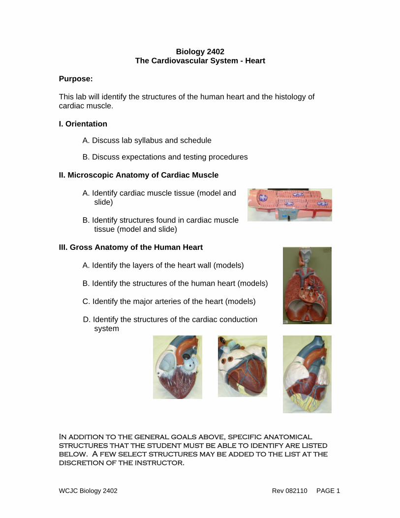
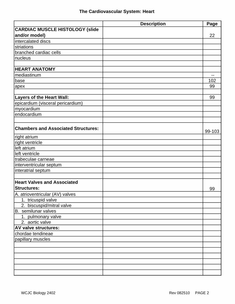
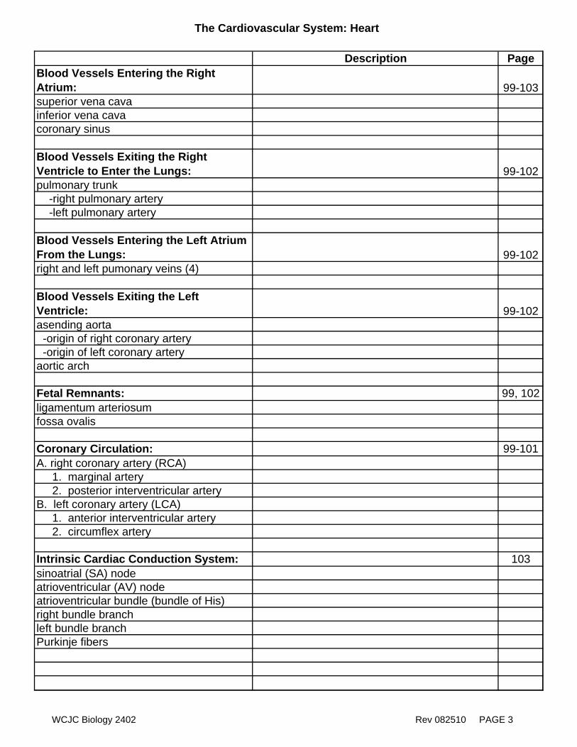
Biology 2402 The Cardiovascular System - Heart
Purpose: This lab will identify the structures of the human heart and the histology of cardiac muscle. I. Orientation
A. Discuss lab syllabus and schedule B. Discuss expectations and testing procedures
II. Microscopic Anatomy of Cardiac Muscle
A. Identify cardiac muscle tissue (model and slide)
B. Identify structures found in cardiac muscle
tissue (model and slide) III. Gross Anatomy of the Human Heart A. Identify the layers of the heart wall (models) B. Identify the structures of the human heart (models) C. Identify the major arteries of the heart (models) D. Identify the structures of the cardiac conduction
system In addition to the general goals above, specific anatomical structures that the student must be able to identify are listed below. A few select structures may be added to the list at the discretion of the instructor.
The Cardiovascular System: Heart
Description PageCARDIAC MUSCLE HISTOLOGY (slide and/or model) 22intercalated discsstriationsbranched cardiac cellsnucleus
HEART ANATOMYmediastinum --base 102apex 99
Layers of the Heart Wall: 99epicardium (visceral pericardium) myocardiumendocardium
Chambers and Associated Structures: 99-103right atriumright ventricleleft atriumleft ventricletrabeculae carneaeinterventricular septuminteratrial septum
Heart Valves and Associated Structures: 99A. atrioventricular (AV) valves 1. tricuspid valve 2. biscuspid/mitral valveB. semilunar valves 1. pulmonary valve 2. aortic valveAV valve structures:chordae tendineaepapillary muscles
WCJC Biology 2402 Rev 082510 PAGE 2
The Cardiovascular System: Heart
Description PageBlood Vessels Entering the Right Atrium: 99-103superior vena cavainferior vena cavacoronary sinus
Blood Vessels Exiting the Right Ventricle to Enter the Lungs: 99-102pulmonary trunk -right pulmonary artery -left pulmonary artery
Blood Vessels Entering the Left Atrium From the Lungs: 99-102right and left pumonary veins (4)
Blood Vessels Exiting the Left Ventricle: 99-102asending aorta -origin of right coronary artery -origin of left coronary arteryaortic arch
Fetal Remnants: 99, 102ligamentum arteriosumfossa ovalis
Coronary Circulation: 99-101A. right coronary artery (RCA) 1. marginal artery 2. posterior interventricular arteryB. left coronary artery (LCA) 1. anterior interventricular artery 2. circumflex artery
Intrinsic Cardiac Conduction System: 103sinoatrial (SA) nodeatrioventricular (AV) nodeatrioventricular bundle (bundle of His)right bundle branchleft bundle branchPurkinje fibers
WCJC Biology 2402 Rev 082510 PAGE 3
WCJC Biology 2402 Rev 082110 PAGE 4
Biology 2402 Cardiovascular Physiology:
ECG, Heart Sounds, Pulse, and Blood Pressure Purpose: This lab will cover many aspects of cardiovascular physiology including: 1) collection and analysis of an ECG, 2) identification of heart sounds, 3) identifying pulse points, and 4) measurement of blood pressure
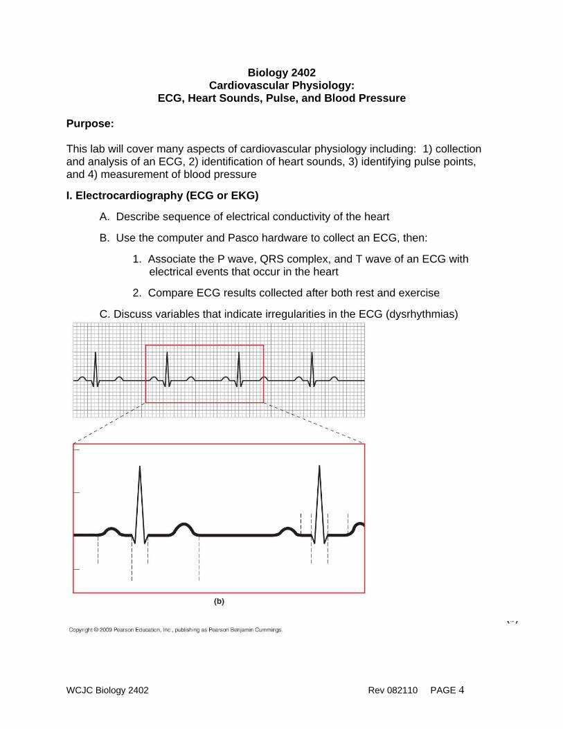
I. Electrocardiography (ECG or EKG)
A. Describe sequence of electrical conductivity of the heart B. Use the computer and Pasco hardware to collect an ECG, then:
1. Associate the P wave, QRS complex, and T wave of an ECG with electrical events that occur in the heart
2. Compare ECG results collected after both rest and exercise
C. Discuss variables that indicate irregularities in the ECG (dysrhythmias)
WCJC Biology 2402 Rev 082110 PAGE 5
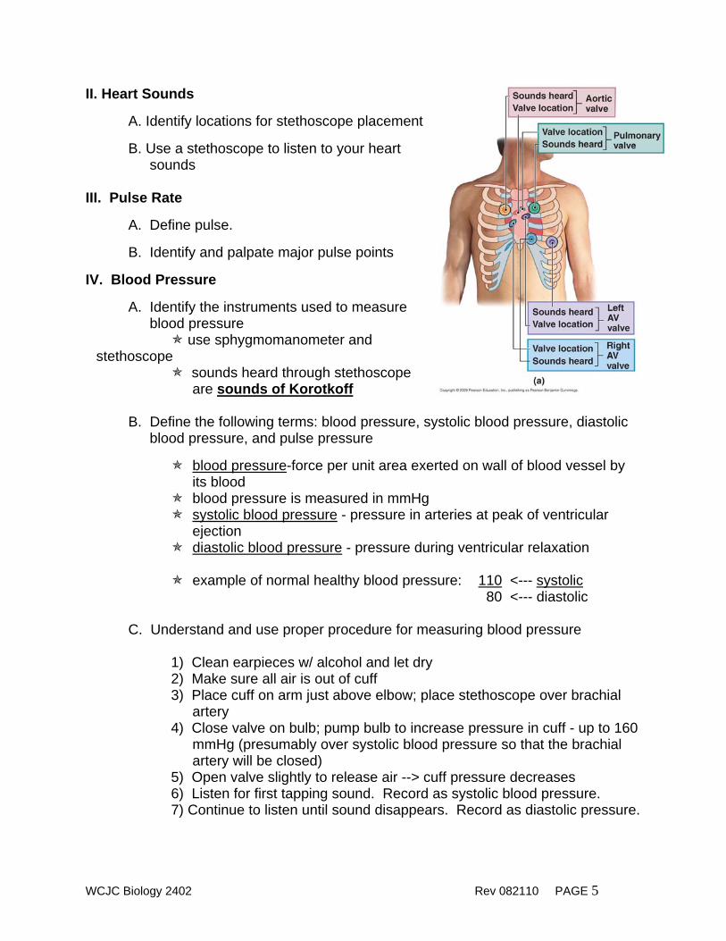
II. Heart Sounds A. Identify locations for stethoscope placement B. Use a stethoscope to listen to your heart
sounds III. Pulse Rate
A. Define pulse.
B. Identify and palpate major pulse points
IV. Blood Pressure
A. Identify the instruments used to measure blood pressure
use sphygmomanometer and stethoscope
sounds heard through stethoscope are sounds of Korotkoff
B. Define the following terms: blood pressure, systolic blood pressure, diastolic
blood pressure, and pulse pressure
blood pressure-force per unit area exerted on wall of blood vessel by its blood
blood pressure is measured in mmHg systolic blood pressure - pressure in arteries at peak of ventricular
ejection diastolic blood pressure - pressure during ventricular relaxation
example of normal healthy blood pressure: 110 <--- systolic
80 <--- diastolic C. Understand and use proper procedure for measuring blood pressure
1) Clean earpieces w/ alcohol and let dry 2) Make sure all air is out of cuff 3) Place cuff on arm just above elbow; place stethoscope over brachial
artery 4) Close valve on bulb; pump bulb to increase pressure in cuff - up to 160
mmHg (presumably over systolic blood pressure so that the brachial artery will be closed)
5) Open valve slightly to release air --> cuff pressure decreases 6) Listen for first tapping sound. Record as systolic blood pressure. 7) Continue to listen until sound disappears. Record as diastolic pressure.
WCJC Biology 2402 Rev 082110 PAGE 6
Biology 2402 The Cardiovascular System - Blood Vessels
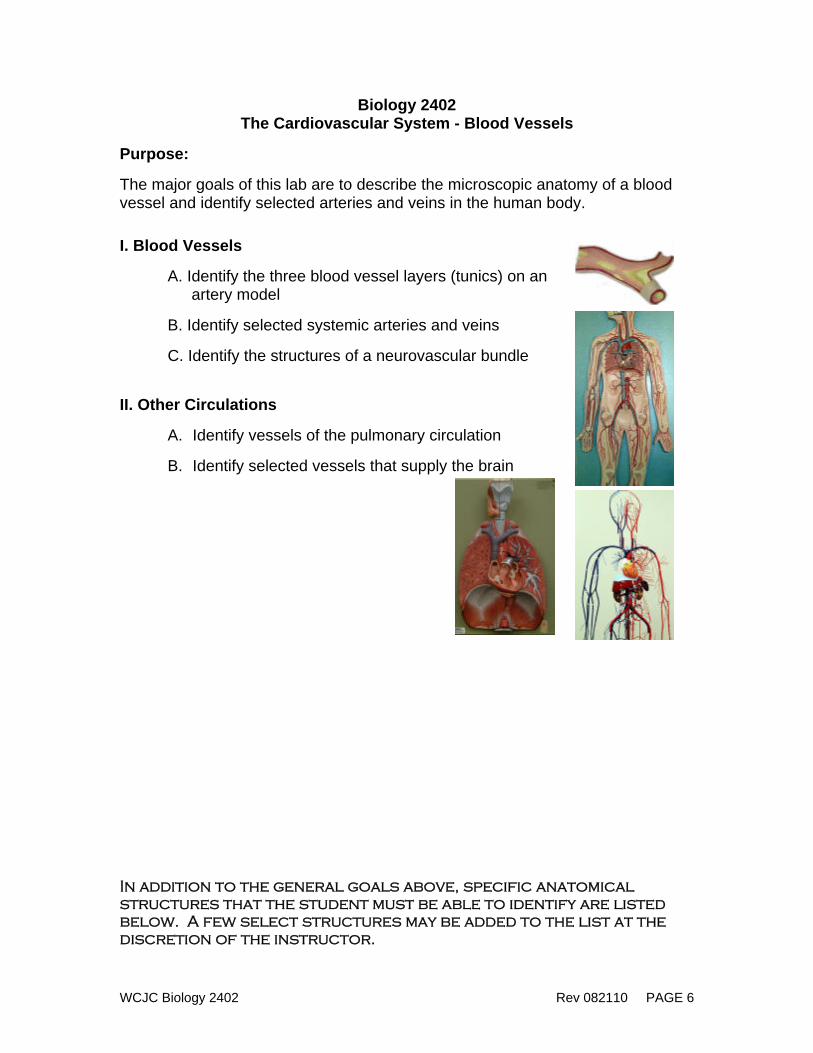
Purpose: The major goals of this lab are to describe the microscopic anatomy of a blood vessel and identify selected arteries and veins in the human body. I. Blood Vessels
A. Identify the three blood vessel layers (tunics) on an artery model
B. Identify selected systemic arteries and veins C. Identify the structures of a neurovascular bundle II. Other Circulations
A. Identify vessels of the pulmonary circulation
B. Identify selected vessels that supply the brain In addition to the general goals above, specific anatomical structures that the student must be able to identify are listed below. A few select structures may be added to the list at the discretion of the instructor.
The Cardiovascular System: Blood Vessels
Description PageBLOOD VESSEL HISTOLOGY 111tunica intima/internatunica mediatunica externa/adventitia)
neurovascular bundle -nerve -artery -vein
MAJOR SYSTEMIC ARTERIESI. Branches of the Aorta: 99, 113, 114 A. ascending aorta B. aortic arch 1. brachiocephalic trunk 2. left common carotid artery 3. left subclavian artery C. thoracic aorta D. abdominal aortaintercostal arteries
II. Arteries to the Head and Neckcommon carotid artery A. internal carotid artery B. external carotid arteryvertebral artery
III. Artery Supply to Upper Limbs: 113subclavian arteryaxillary arterybrachial arteryradial arteryulnar arterypalmar archesdigital arteriesIV. Arteries of the Abdomen 114-115celiac trunk A. left gastric artery B. common hepatic artery C. splenic arterysuperior mesenteric arteryrenal arterygonadal artery: -ovarian artery (in female) -testicular artery (in male)inferior mesenteric artery
WCJC Biology 2402 Rev 082210 PAGE 7
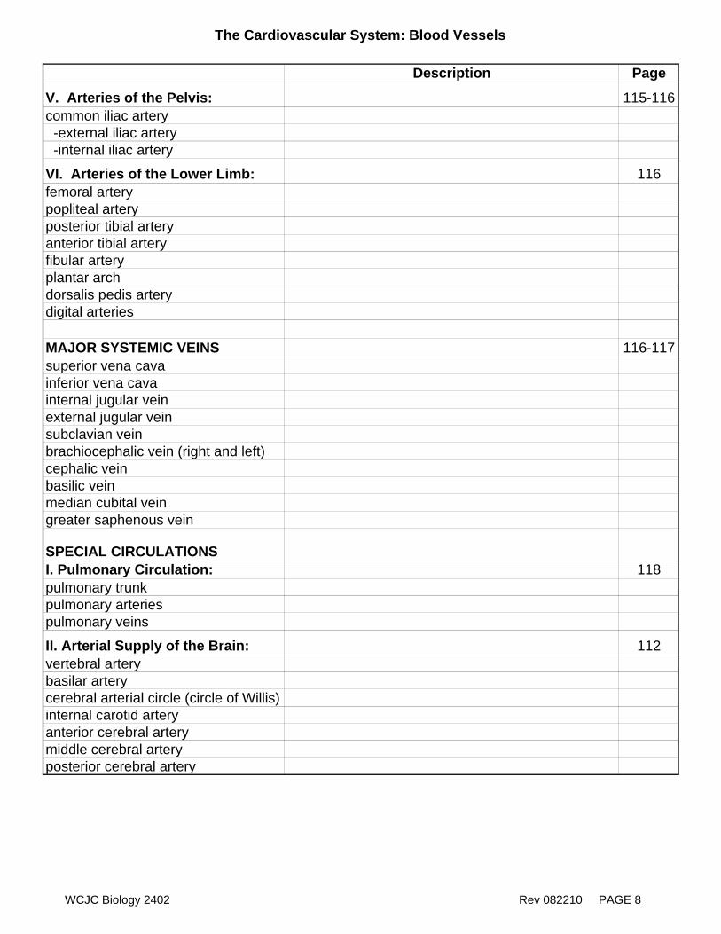
The Cardiovascular System: Blood Vessels
Description Page
V. Arteries of the Pelvis: 115-116common iliac artery -external iliac artery -internal iliac artery
VI. Arteries of the Lower Limb: 116femoral arterypopliteal arteryposterior tibial arteryanterior tibial arteryfibular arteryplantar archdorsalis pedis arterydigital arteries
MAJOR SYSTEMIC VEINS 116-117superior vena cavainferior vena cavainternal jugular veinexternal jugular veinsubclavian veinbrachiocephalic vein (right and left)cephalic veinbasilic veinmedian cubital veingreater saphenous vein
SPECIAL CIRCULATIONSI. Pulmonary Circulation: 118pulmonary trunkpulmonary arteriespulmonary veins
II. Arterial Supply of the Brain: 112vertebral arterybasilar arterycerebral arterial circle (circle of Willis)internal carotid arteryanterior cerebral arterymiddle cerebral arteryposterior cerebral artery
WCJC Biology 2402 Rev 082210 PAGE 8
WCJC Biology 2402 Rev 082110 PAGE 9
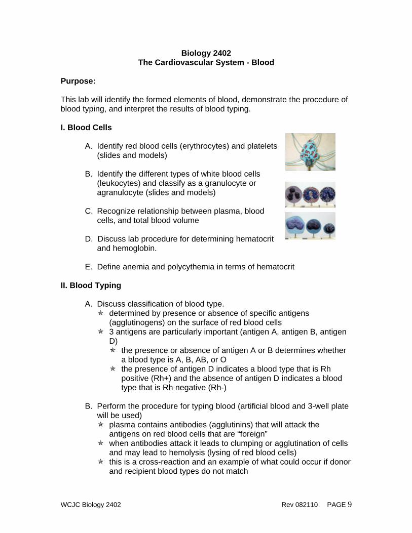
Biology 2402 The Cardiovascular System - Blood
Purpose: This lab will identify the formed elements of blood, demonstrate the procedure of blood typing, and interpret the results of blood typing. I. Blood Cells
A. Identify red blood cells (erythrocytes) and platelets (slides and models)
B. Identify the different types of white blood cells
(leukocytes) and classify as a granulocyte or agranulocyte (slides and models)
C. Recognize relationship between plasma, blood
cells, and total blood volume D. Discuss lab procedure for determining hematocrit
and hemoglobin. E. Define anemia and polycythemia in terms of hematocrit
II. Blood Typing
A. Discuss classification of blood type. determined by presence or absence of specific antigens
(agglutinogens) on the surface of red blood cells 3 antigens are particularly important (antigen A, antigen B, antigen
D) the presence or absence of antigen A or B determines whether
a blood type is A, B, AB, or O the presence of antigen D indicates a blood type that is Rh
positive (Rh+) and the absence of antigen D indicates a blood type that is Rh negative (Rh-)
B. Perform the procedure for typing blood (artificial blood and 3-well plate
will be used) plasma contains antibodies (agglutinins) that will attack the
antigens on red blood cells that are “foreign” when antibodies attack it leads to clumping or agglutination of cells
and may lead to hemolysis (lysing of red blood cells) this is a cross-reaction and an example of what could occur if donor
and recipient blood types do not match
WCJC Biology 2402 Rev 082110 PAGE 10
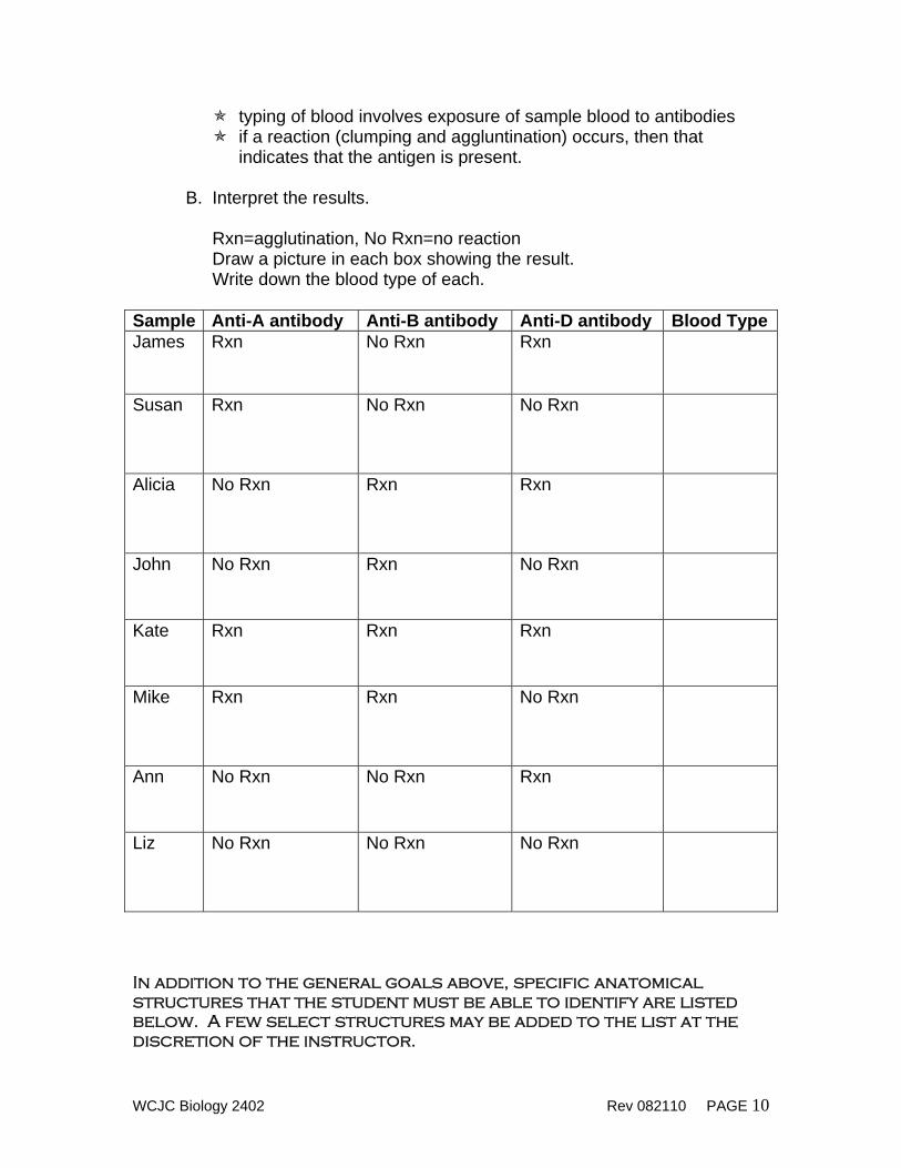
typing of blood involves exposure of sample blood to antibodies if a reaction (clumping and aggluntination) occurs, then that
indicates that the antigen is present. B. Interpret the results.
Rxn=agglutination, No Rxn=no reaction Draw a picture in each box showing the result. Write down the blood type of each.
Sample Anti-A antibody Anti-B antibody Anti-D antibody Blood TypeJames Rxn No Rxn Rxn
Susan Rxn No Rxn No Rxn
Alicia No Rxn Rxn Rxn
John No Rxn Rxn No Rxn
Kate Rxn Rxn Rxn
Mike Rxn Rxn No Rxn
Ann No Rxn No Rxn Rxn
Liz No Rxn No Rxn No Rxn
In addition to the general goals above, specific anatomical structures that the student must be able to identify are listed below. A few select structures may be added to the list at the discretion of the instructor.
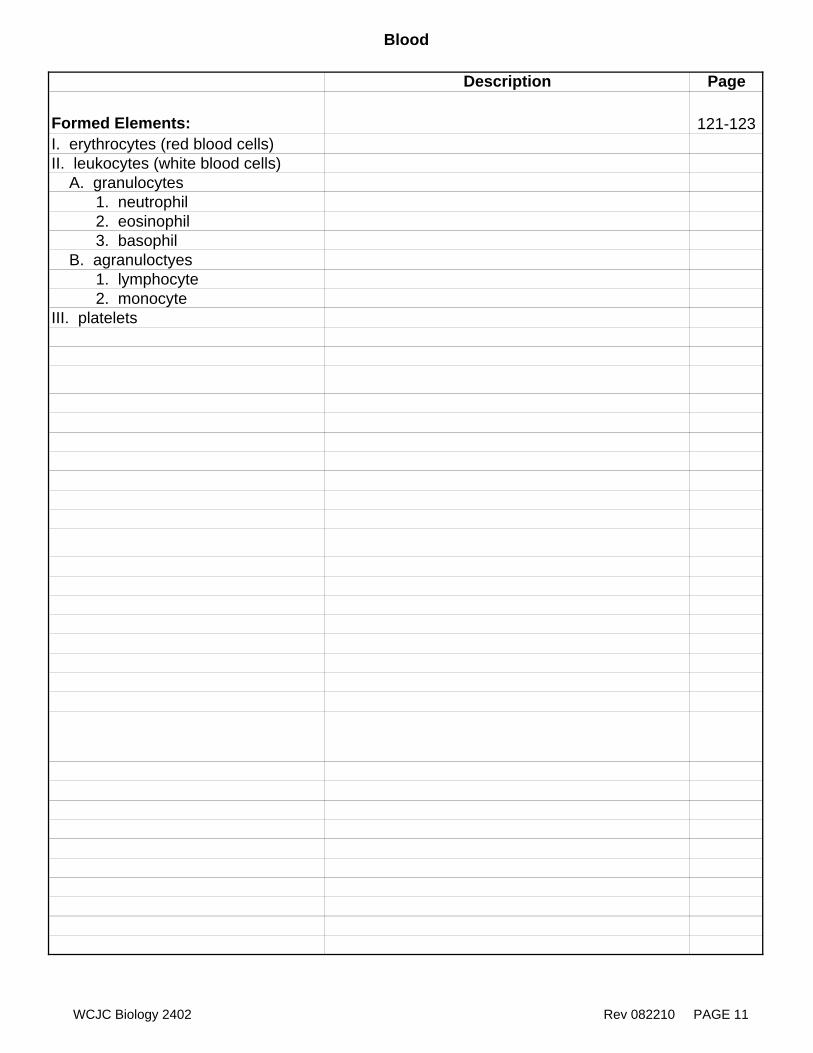
Blood
Description Page
Formed Elements: 121-123I. erythrocytes (red blood cells)II. leukocytes (white blood cells) A. granulocytes 1. neutrophil 2. eosinophil 3. basophil B. agranuloctyes 1. lymphocyte 2. monocyteIII. platelets
WCJC Biology 2402 Rev 082210 PAGE 11
WCJC Biology 2402 Rev 082110 PAGE 12
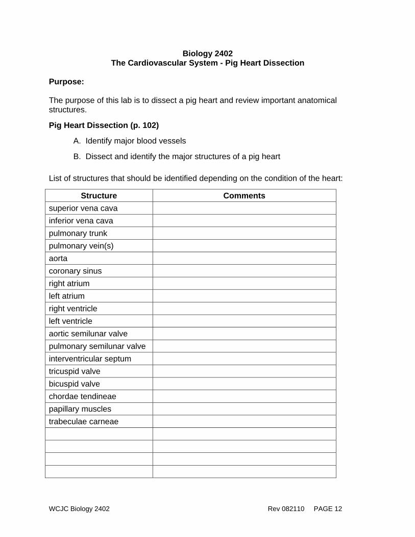
Biology 2402 The Cardiovascular System - Pig Heart Dissection
Purpose: The purpose of this lab is to dissect a pig heart and review important anatomical structures.
Pig Heart Dissection (p. 102)
A. Identify major blood vessels
B. Dissect and identify the major structures of a pig heart
List of structures that should be identified depending on the condition of the heart:
Structure Comments superior vena cava inferior vena cava pulmonary trunk pulmonary vein(s) aorta coronary sinus right atrium left atrium right ventricle left ventricle aortic semilunar valve pulmonary semilunar valve interventricular septum tricuspid valve bicuspid valve chordae tendineae papillary muscles trabeculae carneae

WCJC Biology 2402 Rev 082110 PAGE 13
Biology 2402 The Respiratory System
Purpose: This lab will identify the structures of the respiratory system and measure pulmonary function using spirometery. I. Anatomy of the Respiratory System
A. Identify the respiratory structures of the head, throat and thoracic cavity (models)
B. Observe the histology of a healthy lung and a diseased lung (slides)
II. Spirometry
A. Define tidal volume (TV), expiratory reserve volume (ERV), inspiratory reserve volume (IRV), vital capacity (VC).
B. Recognize normal values of variables of pulmonary function tests C. Measure pulmonary volumes and capacities using a wet spirometer
Step Volume/Capacity Trial 1 (ml) Trial 2 (ml) Trial 3 (ml) Average (ml) 1 Tidal volume
2 Expiratory reserve volume
3 Vital capacity (VC=TV+ERV+IRV)
4 Inspiratory reserve volume
CALCULATE: IRV=VC-(TV+ERV)
5 % normal vital capacity
CALCULATE: Your VC x 100%= Predicted VC
In addition to the general goals above, specific anatomical structures that the student must be able to identify are listed below. A few select structures may be added to the list at the discretion of the instructor.
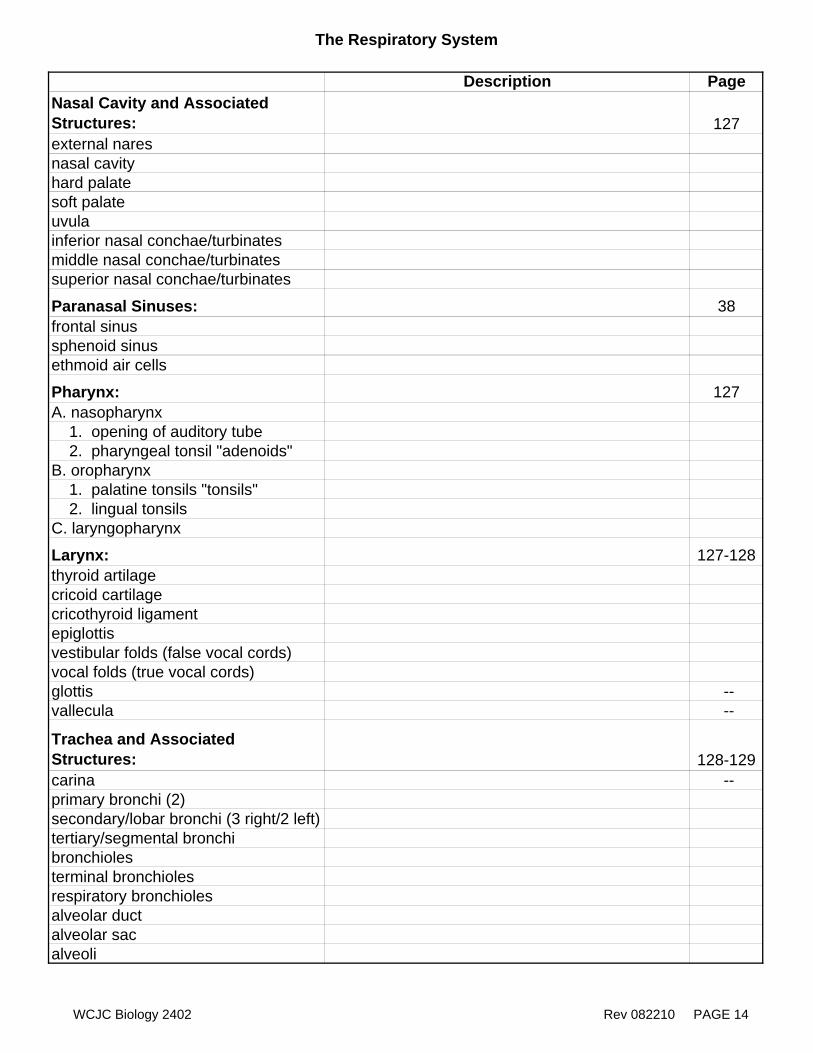
The Respiratory System
Description PageNasal Cavity and Associated Structures: 127external naresnasal cavityhard palatesoft palateuvulainferior nasal conchae/turbinatesmiddle nasal conchae/turbinatessuperior nasal conchae/turbinates
Paranasal Sinuses: 38frontal sinussphenoid sinusethmoid air cells
Pharynx: 127A. nasopharynx 1. opening of auditory tube 2. pharyngeal tonsil "adenoids"B. oropharynx 1. palatine tonsils "tonsils" 2. lingual tonsilsC. laryngopharynx
Larynx: 127-128thyroid artilagecricoid cartilagecricothyroid ligamentepiglottisvestibular folds (false vocal cords)vocal folds (true vocal cords)glottis --vallecula --
Trachea and Associated Structures: 128-129carina --primary bronchi (2)secondary/lobar bronchi (3 right/2 left)tertiary/segmental bronchibronchiolesterminal bronchiolesrespiratory bronchiolesalveolar ductalveolar sacalveoli
WCJC Biology 2402 Rev 082210 PAGE 14
The Respiratory System
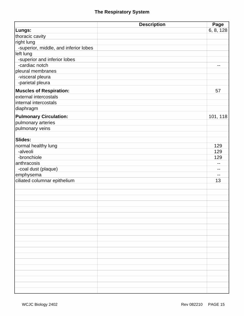
Description PageLungs: 6, 8, 128thoracic cavityright lung -superior, middle, and inferior lobesleft lung -superior and inferior lobes -cardiac notch --pleural membranes -visceral pleura -parietal pleura
Muscles of Respiration: 57external intercostalsinternal intercostalsdiaphragm
Pulmonary Circulation: 101, 118pulmonary arteriespulmonary veins
Slides:normal healthy lung 129 -alveoli 129 -bronchiole 129anthracosis -- -coal dust (plaque) --emphysema --ciliated columnar epithelium 13
WCJC Biology 2402 Rev 082210 PAGE 15
WCJC Biology 2402 Rev 082110 PAGE 16
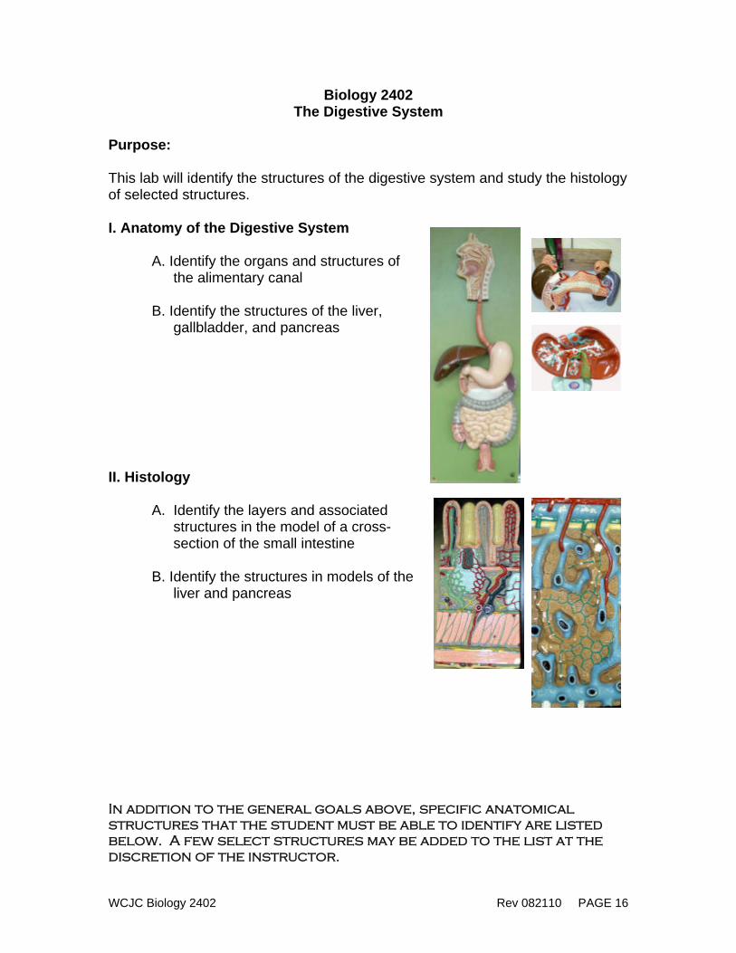
Biology 2402 The Digestive System
Purpose: This lab will identify the structures of the digestive system and study the histology of selected structures. I. Anatomy of the Digestive System
A. Identify the organs and structures of the alimentary canal
B. Identify the structures of the liver,
gallbladder, and pancreas II. Histology
A. Identify the layers and associated structures in the model of a cross-section of the small intestine
B. Identify the structures in models of the
liver and pancreas In addition to the general goals above, specific anatomical structures that the student must be able to identify are listed below. A few select structures may be added to the list at the discretion of the instructor.
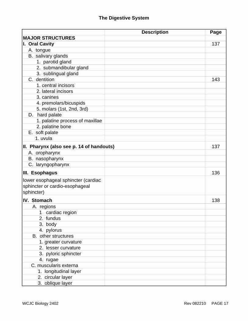
The Digestive System
Description PageMAJOR STRUCTURESI. Oral Cavity 137 A. tongue B. salivary glands 1. parotid gland 2. submandibular gland 3. sublingual gland C. dentition 143 1. central incisors 2. lateral incisors 3. canines 4. premolars/bicuspids 5. molars (1st, 2nd, 3rd) D. hard palate 1. palatine process of maxillae 2. palatine bone E. soft palate 1. uvula
II. Pharynx (also see p. 14 of handouts) 137 A. oropharynx B. nasopharynx C. laryngopharynx
III. Esophagus 136lower esophageal sphincter (cardiac sphincter or cardio-esophageal sphincter)
IV. Stomach 138 A. regions 1 cardiac region 2. fundus 3. body 4. pylorus B. other structures 1. greater curvature 2. lesser curvature 3. pyloric sphincter 4. rugae C. muscularis externa 1. longitudinal layer 2. circular layer 3. oblique layer
WCJC Biology 2402 Rev 082210 PAGE 17
The Digestive System
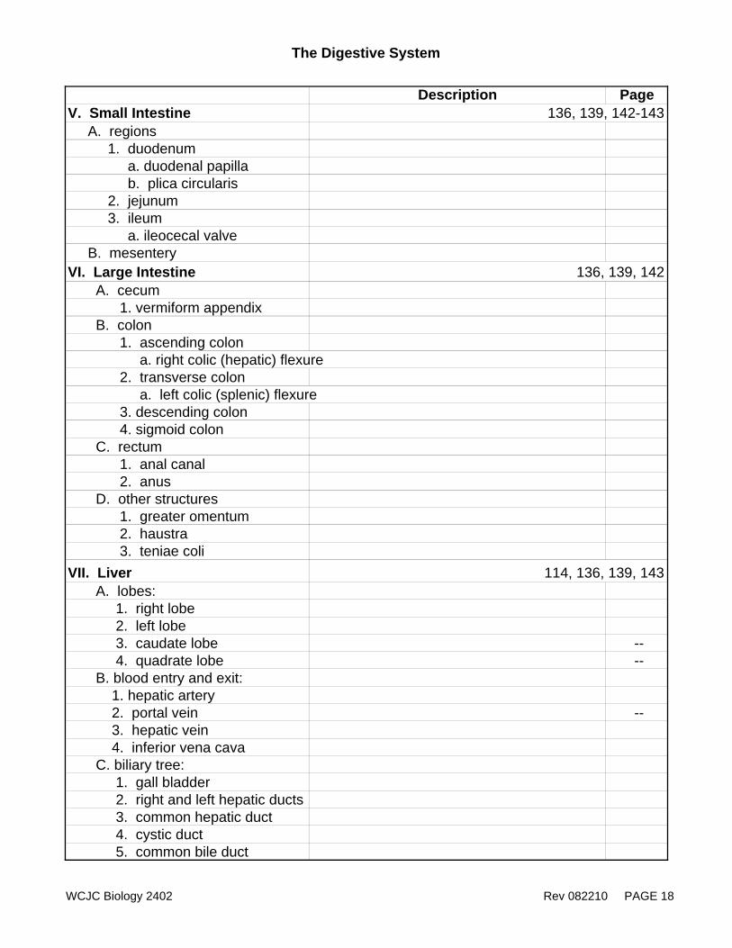
Description PageV. Small Intestine 136, 139, 142-143 A. regions 1. duodenum a. duodenal papilla b. plica circularis 2. jejunum 3. ileum a. ileocecal valve B. mesenteryVI. Large Intestine 136, 139, 142 A. cecum 1. vermiform appendix B. colon 1. ascending colon a. right colic (hepatic) flexure 2. transverse colon a. left colic (splenic) flexure 3. descending colon 4. sigmoid colon C. rectum 1. anal canal 2. anus D. other structures 1. greater omentum 2. haustra 3. teniae coliVII. Liver 114, 136, 139, 143 A. lobes: 1. right lobe 2. left lobe 3. caudate lobe -- 4. quadrate lobe -- B. blood entry and exit: 1. hepatic artery 2. portal vein -- 3. hepatic vein 4. inferior vena cava C. biliary tree: 1. gall bladder 2. right and left hepatic ducts 3. common hepatic duct 4. cystic duct 5. common bile duct
WCJC Biology 2402 Rev 082210 PAGE 18
The Digestive System
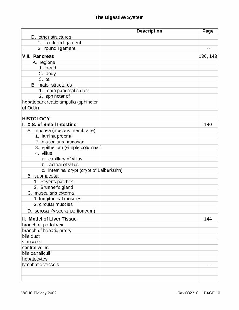
Description Page D. other structures 1. falciform ligament 2. round ligament --
VIII. Pancreas 136, 143 A. regions 1. head 2. body 3. tail B. major structures 1. main pancreatic duct 2. sphincter of hepatopancreatic ampulla (sphincter of Oddi)
HISTOLOGYI. X.S. of Small Intestine 140 A. mucosa (mucous membrane) 1. lamina propria 2. muscularis mucosae 3. epithelium (simple columnar) 4. villus a. capillary of villus b. lacteal of villus c. Intestinal crypt (crypt of Leiberkuhn) B. submucosa 1. Peyer's patches 2. Brunner's gland C. muscularis externa 1. longitudinal muscles 2. circular muscles D. serosa (visceral peritoneum)
II. Model of Liver Tissue 144branch of portal veinbranch of hepatic arterybile ductsinusoidscentral veinsbile canaliculihepatocyteslymphatic vessels --
WCJC Biology 2402 Rev 082210 PAGE 19
The Digestive System
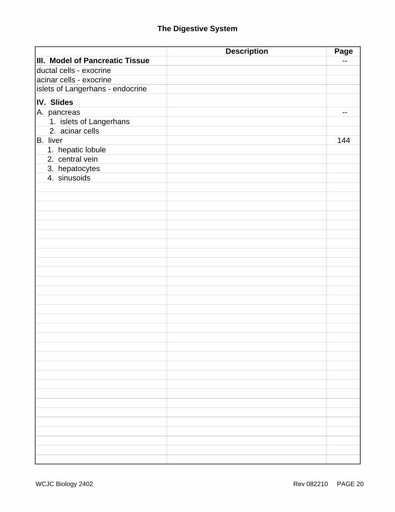
Description PageIII. Model of Pancreatic Tissue --ductal cells - exocrineacinar cells - exocrineislets of Langerhans - endocrine
IV. SlidesA. pancreas -- 1. islets of Langerhans 2. acinar cellsB. liver 144 1. hepatic lobule 2. central vein 3. hepatocytes 4. sinusoids
WCJC Biology 2402 Rev 082210 PAGE 20
WCJC Biology 2402 Rev 082110 PAGE 21
Biology 2402 The Urinary System
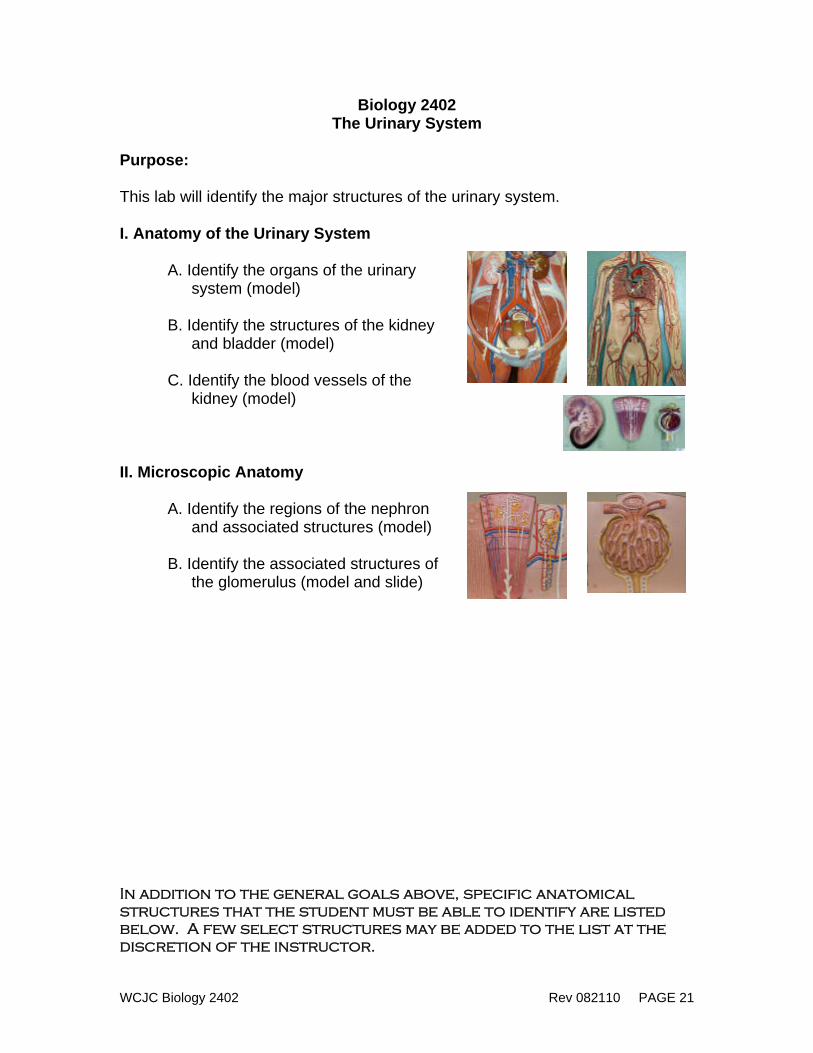
Purpose: This lab will identify the major structures of the urinary system. I. Anatomy of the Urinary System
A. Identify the organs of the urinary system (model)
B. Identify the structures of the kidney
and bladder (model) C. Identify the blood vessels of the
kidney (model) II. Microscopic Anatomy
A. Identify the regions of the nephron and associated structures (model)
B. Identify the associated structures of
the glomerulus (model and slide) In addition to the general goals above, specific anatomical structures that the student must be able to identify are listed below. A few select structures may be added to the list at the discretion of the instructor.
The Urinary System
Structure Description Page
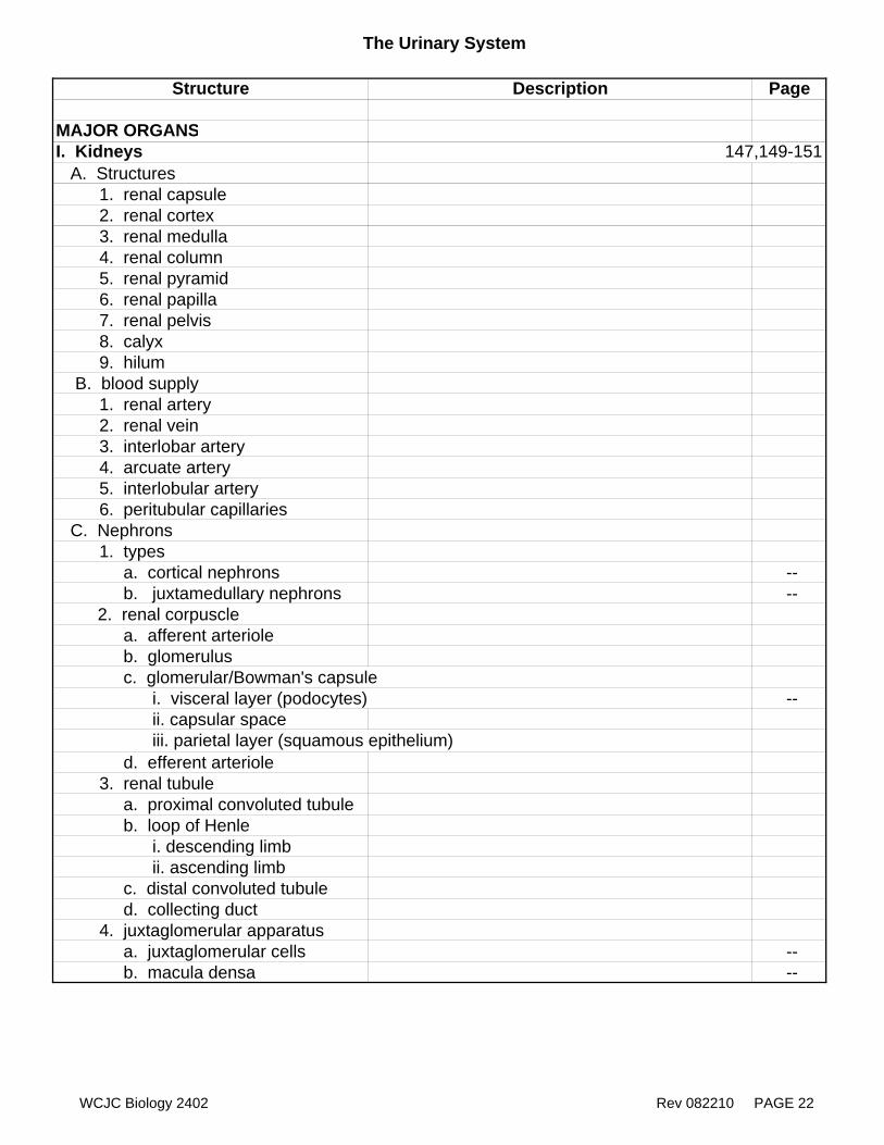
MAJOR ORGANSI. Kidneys 147,149-151 A. Structures 1. renal capsule 2. renal cortex 3. renal medulla 4. renal column 5. renal pyramid 6. renal papilla 7. renal pelvis 8. calyx 9. hilum B. blood supply 1. renal artery 2. renal vein 3. interlobar artery 4. arcuate artery 5. interlobular artery 6. peritubular capillaries C. Nephrons 1. types a. cortical nephrons -- b. juxtamedullary nephrons -- 2. renal corpuscle a. afferent arteriole b. glomerulus c. glomerular/Bowman's capsule i. visceral layer (podocytes) -- ii. capsular space iii. parietal layer (squamous epithelium) d. efferent arteriole 3. renal tubule a. proximal convoluted tubule b. loop of Henle i. descending limb ii. ascending limb c. distal convoluted tubule d. collecting duct 4. juxtaglomerular apparatus a. juxtaglomerular cells -- b. macula densa --
WCJC Biology 2402 Rev 082210 PAGE 22
The Urinary System
Description Page
II. Ureters 147-148
III. Urinary bladder 147-148detrusor musclerugaeurinary trigone
IV. Urethra 148
V. Adrenal glands 147
SLIDE OF KIDNEY 12, 151glomerulusBowman's capsule (simple squamous epithelium)capsular spacerenal tubule (cuboidal epithelium)
WCJC Biology 2402 Rev 082210 PAGE 23
WCJC Biology 2402 Rev 082110 PAGE 24
Biology 2402 The Reproductive System
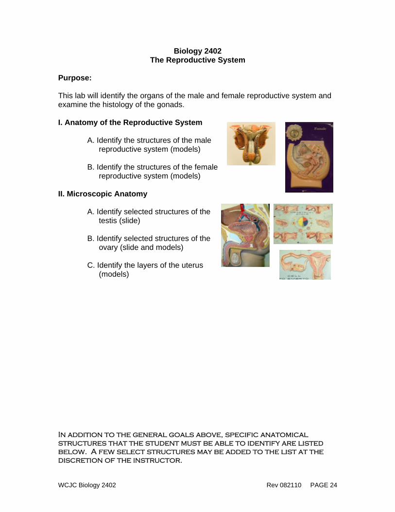
Purpose: This lab will identify the organs of the male and female reproductive system and examine the histology of the gonads. I. Anatomy of the Reproductive System
A. Identify the structures of the male
reproductive system (models) B. Identify the structures of the female
reproductive system (models)
II. Microscopic Anatomy A. Identify selected structures of the
testis (slide) B. Identify selected structures of the
ovary (slide and models) C. Identify the layers of the uterus
(models)
In addition to the general goals above, specific anatomical structures that the student must be able to identify are listed below. A few select structures may be added to the list at the discretion of the instructor.
The Reproductive System
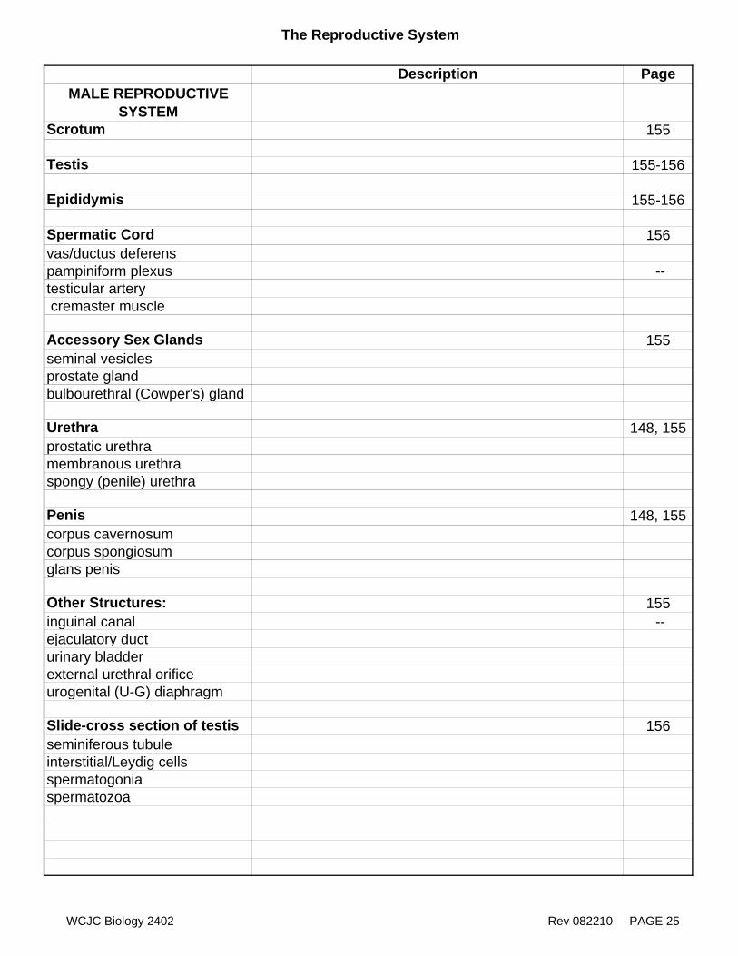
Description PageMALE REPRODUCTIVE
SYSTEMScrotum 155
Testis 155-156
Epididymis 155-156
Spermatic Cord 156vas/ductus deferens pampiniform plexus --testicular artery cremaster muscle
Accessory Sex Glands 155seminal vesiclesprostate glandbulbourethral (Cowper's) gland
Urethra 148, 155prostatic urethramembranous urethraspongy (penile) urethra
Penis 148, 155corpus cavernosumcorpus spongiosumglans penis
Other Structures: 155inguinal canal --ejaculatory ducturinary bladder external urethral orificeurogenital (U-G) diaphragm
Slide-cross section of testis 156seminiferous tubuleinterstitial/Leydig cellsspermatogoniaspermatozoa
WCJC Biology 2402 Rev 082210 PAGE 25
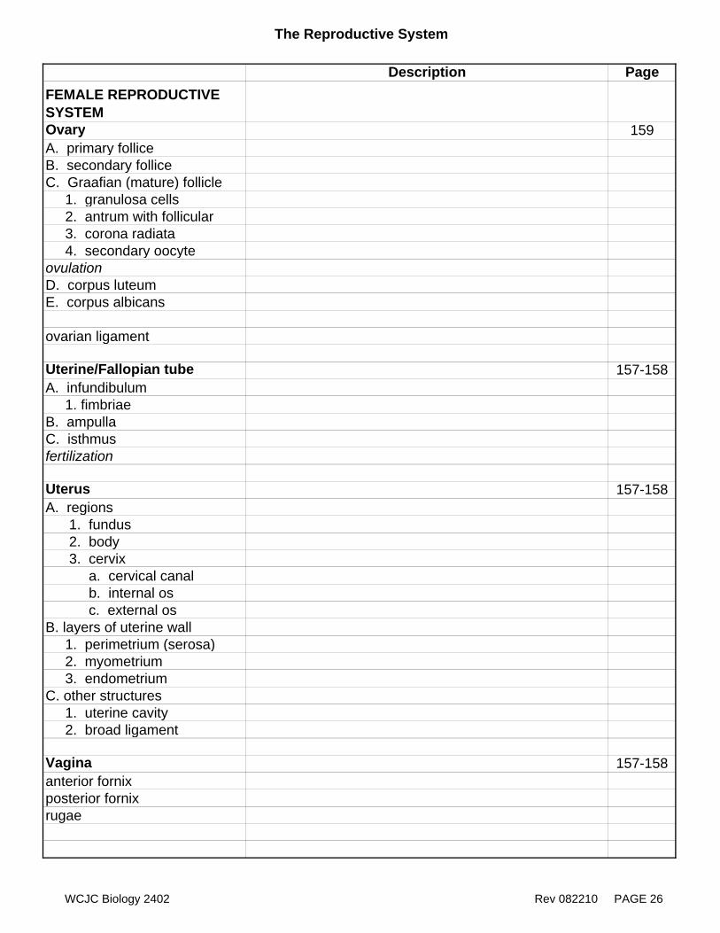
The Reproductive System
Description PageFEMALE REPRODUCTIVE SYSTEMOvary 159A. primary folliceB. secondary folliceC. Graafian (mature) follicle 1. granulosa cells 2. antrum with follicular 3. corona radiata 4. secondary oocyteovulationD. corpus luteumE. corpus albicans
ovarian ligament
Uterine/Fallopian tube 157-158A. infundibulum 1. fimbriaeB. ampullaC. isthmusfertilization
Uterus 157-158A. regions 1. fundus 2. body 3. cervix a. cervical canal b. internal os c. external osB. layers of uterine wall 1. perimetrium (serosa) 2. myometrium 3. endometriumC. other structures 1. uterine cavity 2. broad ligament
Vagina 157-158anterior fornixposterior fornixrugae
WCJC Biology 2402 Rev 082210 PAGE 26
The Reproductive System
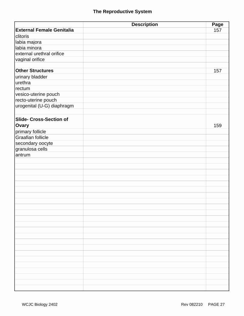
Description PageExternal Female Genitalia 157clitorislabia majoralabia minoraexternal urethral orificevaginal orifice
Other Structures 157urinary bladder urethra rectumvesico-uterine pouchrecto-uterine pouchurogenital (U-G) diaphragm
Slide- Cross-Section of Ovary 159primary follicleGraafian folliclesecondary oocytegranulosa cellsantrum
WCJC Biology 2402 Rev 082210 PAGE 27