Embed Size (px)
Citation preview
Biology 218, practise Exam 2, 2011
1
Figure 3 The long-range effect of Sqt does not depend on the induction of the endogenous cyc or sqt genes. a, Design and predictions for the experiments shown in b-e. b-e, Single-cell injection of 4 pg of sqt RNA plus lineage tracers biotin-dextran and rhodamine-dextran into wild-type (b, d, n = 18/18) and cyc;sqt double mutant embryos (c, e, n = 12/12) at the 128±256-cell stage. b, c, Focus is on the expression domain of ntl in marginal cells; cyc;sqt double mutants were distinguished from sibling embryos (b) by the lack of ntl expression on the dorsal side (c). d, e, High-magni®cation views of the embryos in b and c; the lineage tracer biotin-dextran was detected with the ABC kit from Vectastain (red).
The zebrafish Nodal signal Squint functions as a morphogen, by Yu Chen & Alexander F. Schier.
Figure 3, reproduced above, demonstrates that “The long-range effect of Sqt does not depend on the induction of the endogenous cyc or sqt genes.” Discuss this experiment and how the authors can draw this conclusion. Genetic and developmental basis of evolutionary pelvic reduction in threespine sticklebacks Michael D. Shapiro1*, Melissa E. Marks1*, Catherine L. Peichel2*, Benjamin K. Blackman1, Kirsten S. Nereng1, Bjarni Jo´nsson3, Dolph Schluter4 & David M. Kingsley1 Hindlimb loss has evolved repeatedly in many different animals by means of molecular mechanisms that are still unknown. To determine the number and type of genetic changes underlying pelvic reduction in natural populations, we carried out genetic crosses between threespine stickleback fish with complete or missing pelvic structures. Genome‐wide linkage mapping shows that pelvic reduction is controlled by one major and four minor chromosome regions. Pitx1 maps to the major chromosome region controlling most of the variation in pelvic size. Pelvic‐reduced fish show the same left–right asymmetry seen in Pitx1 knockout mice, but do not show changes in Pitx1 protein sequence. Instead, pelvic‐reduced sticklebacks show site‐specific regulatory changes in Pitx1 expression, with reduced or absent expression in pelvic and caudal fin precursors. Regulatory mutations in

Biology 218, practise Exam 2, 2011
2
major developmental control genes may provide a mechanism for generating rapid skeletal changes in natural populations, while preserving the essential roles of these genes in other
processes. The left figure from the Shapiro paper shows expression of pitx1 in marine versus fresh-water sticklebacks. The paper was on pelvic reduction (abstract above). Propose a
hypothesis to explain why there is more pitx1 expression in the caudal fin of the marine form.
When the AER is removed early, only proximal limb structures develop, while when the AER is removed later, more distal limb structures develop. How does the progress zone model explain this observation?
What is an alternative model to explain the stage-dependent effects on limb patterning of AER removal?
Explain the differences between in situ hybridization and immunohistochemistry. Be clear on what type of molecule is being detected, and what provides the specificity for each technique
In many vertebrates, there is no dorsal-ventral gradient of BMP4 protein during gastrulation, however, there is a gradient of BMP4 activity. How might this be accomplished?

Biology 218, practise Exam 2, 2011
3
What do you conclude about somite patterning from the above experiments?
Pieces of the notochord, when grafted into the anterior of the limb bud, will cause digit duplications. Propose a hypothesis to explain this result.
13. The following molecules play important roles in specifying cell fate. For each protein, write whether it is a secreted protein, a membrane protein, a cytoplasmic component of the signaling pathway, or a transcription factor (1 pt each).
Sonic Hedgehog MyoD Ephrin HoxC6 Chordin
For ONE of the above proteins in any tissue or cell type, briefly describe how this molecule functions to specify cell fate (2 pts). I am more interested in the embryonic process that this molecule is involved in than the molecular basis for the molecule’s action.
Put the following terms in the appropriate boxes in this cross section of a chick embryo:
Notochord (4pts) sclerotome (2 pts) dermomyotome (2 pts) spinal cord (2 pts) lateral plate

Biology 218, practise Exam 2, 2011
4
Note: you will not fill in all boxes, and you may use either the immature (left) or more developed (right) side of the drawing.
Fill in the blanks. All answers must come from the list of words and phrases; most of the above words will not be used, and some may be used more than once. Some questions may have more than one possible correct answer. For questions 1-6, choose from the following secreted signals and transcription factors: BMP4 chordin, emx1 epidermal growth
factor (EGF) fibroblast growth factor (fgf)
goosecoid noggin pax2.1
siamois sfrp sonic hedgehog (shh)
tlc
vegT wnt7a hoxC6 ß-catenin Fill in 4 boxes, putting gene products on the appropriate side of the Xenopus gastrula: chordin, noggin, siamois, BMP4 (not all boxes will be filled).
5. ________________________
6. ________________________
Biology 218, practise Exam 2, 2011
5
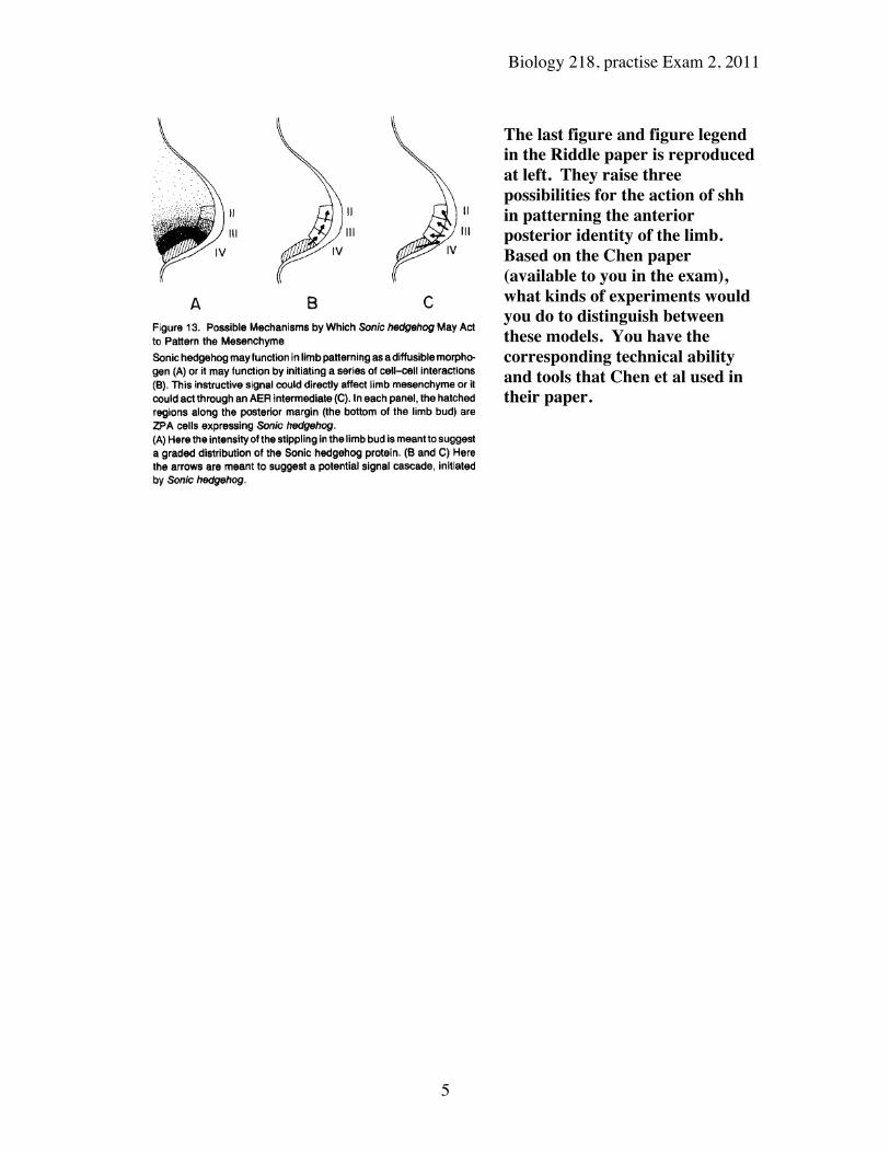
The last figure and figure legend in the Riddle paper is reproduced at left. They raise three possibilities for the action of shh in patterning the anterior posterior identity of the limb. Based on the Chen paper (available to you in the exam), what kinds of experiments would you do to distinguish between these models. You have the corresponding technical ability and tools that Chen et al used in their paper.

Biology 218, practise Exam 2, 2011
6
Fig. 5. Expression of Tbx5 (A-C) and Tbx4 (D-F) in three types of Dasokus (the extra limbs developed after experimental manipulation). As judged from their positions, (A,D) are wing-type, (B,E) are intermediate-type and (C,F) are leg-type. w, wing bud; d, Dasoku limb bud; le, leg bud. Anterior is to the top. (A) Tbx5 is expressed in the wing and Dasoku limb buds. The arrowhead indicates the posterior limit of Tbx5 expression in the flank. (B,C) Tbx5 is expressed in the cephalic region of the Dasoku limb buds. The
arrowheads indicate Tbx5 expression in the flank. (D,E) Tbx4 is expressed in the leg bud and caudal region of the Dasoku limb buds. Tbx4 is not expressed in the flank between the Dasoku and the leg bud (arrow in D). (F) Ventral view of an embryo with a Dasoku on the right side. Tbx4 is expressed in the leg and Dasoku limb buds. Tbx4 expression appears weak in the cephalic margin of the Dasoku limb bud (arrowhead). The broken line indicates the expanded domain of Tbx4 expression on the right side, compared with the left. How were the ectopic limbs induced? How does this experiment support the idea that tbx5 and tbx4 confer limb identity?
Weaver et al. concluded from this figure that maternal behavior (High versus Low) led to epigenetic effects. What were the epigenetic effects demonstrated in this figures? Do the dark bands in panel a represent protein, mRNA, or DNA? How do the bands demonstrate that there were epigenetic effects? Describe the importance of the B-actin bands?