Embed Size (px)
Citation preview

83
BIOLOGICAL OSTEOSYNTHESIS: MINIMINAL INVAZIVE PLATE OSTHEOSYNTHESIS VS. CASTING/SPLINTING
Adelina PROTEASA, Roxana DASCĂLU, Larisa SCHUSZLER, Cornel IGNA
Faculty of Veterinary Medicine, Banat University of Agricultural Sciences and Veterinary Medicine
“King Michael I of Romania” from Timisoara, 119 Aradului Street, Phone:+40256277213, Fax:+40256277118, Timisoara, Romania, [email protected]
[email protected], [email protected], [email protected]
Corresponding author email: [email protected] Abstract In Romania, in the orthopedic veterinary practice, splinting/casting was considered an acceptable treatment of some types of fractures, namely, stable, closed reductible fractures.The greatest advantage of this type of asset is the biological osteosynthesis, favored by indirect reduction, preservation of the blood supply of all fragments, including small ones, which easily can turn into bone sequesters when regional vasculature is impaired. External fixation provided by casts and splints has several significant advantages compared with internal fixation methods: no need for implants, low postoperative infection rate, minimal disruptions of the fracture hematoma and the low cost of the procedure. Complications that can occur, usually due to improper selection of cases, inaccurate application technique and / or poor postoperative management can be minimized by using the minimally invasive plate osteosynthesis, maintaining the pros of bone healing. Key words: cast/splint, MIPO, biological osteosynthesis
INTRODUCTION The biological method of fracture treatment limits the effect of rigid stability and highlights the rapid healing due to conservation of soft tissues adjacent to the fracture site (Palmer, 1999). When preoperative radiographs show the impossibility of perfect anatomical reduction, priority changes from an absolute bone reconstruction to an acceptable spatial alignment and blood resource preservation (Aron et al., 1995; Hulse, 1997; Johnson et al., 1998; Palmer and Aron, 1996). In this paper, we intend to present a statistical report of orthopedic cases in our clinic, , to mention the main methods used in recent years, with advantages and disadvantages, to highlight the latest trends in the field of veterinary orthopedics and put in balance two methods of biological osteosynthesis: casting/splinting (more in this paper) and MIPO –Minimally Invasive plate Ostheosyntesis. Having completed all the above objectives, we want to highlight the importance of veterinary
orthopedics and the need to improve treatment techniques. MATERIALS AND METHODS To achieve our aim, records from 2007 to 2011 in our clinic were considered for the study and statistically analyzed using basic methods and indices, registering the types of treatment, and complications during monitoring. RESULTS AND DISCUSSIONS Between 2007 and 2011, we received for consultation and/or treatment 5987 patients, of which 20% aimed the orthopedic field. 9% were diagnosed with fractures, 6% with fractures of the long bones of the appendicular skeleton (humerus, radius and ulna, femur, tibia and fibula). Basically, the average was 1197 ± 248 clinical cases per year, of which 72 ± 14 were diagnosed with fractures of long bones (Tabel 1).
Scientific Works. Series C. Veterinary Medicine. Vol. LXI (1)ISSN 2065-1295; ISSN 2343-9394 (CD-ROM); ISSN 2067-3663 (Online); ISSN-L 2065-1295
84
Tabel 1. Summary of records taken into study
Year Total Orthopedics Fractures Long bones fractures
2007 902 192 82 56
2008 1418 252 129 72
2009 968 240 102 67
2010 1423 277 121 94
2011 1276 240 115 70
Total 5987 1201 549 359
Mean 1197 240 110 72
StDev 248 31 18 14
Of the 359 cases representing fractures of long bones of the appendicular skeleton, 58% received a biological osteosynthesis method (mostly splinting/ casting, also external fixators, interlocking nails inserted percutaneously) and the remaining 42% - open reduction internal fixation method (Table 2).
Tabel 2. Summary of categories of treatment
Year BOS1 ORIF2 TOTAL
2007 41 15 56
2008 36 36 72
2009 41 26 67
2010 53 41 94
2011 37 33 70
Total 208 151 359
External cooptation provided by casting has several significant advantages compared with internal fixation methods: no need for implants, postoperative infection rate is minimal, no disruptions in fracture outbreak and low cost of procedure (Oakley, 1999; Tomlinson, 1991). Complications that can occur, usually due to improper selection of cases, incorrect application technique and/or poor postoperative management, include: delayed union, malunion, nonunion, joint laxity/ankylosis, dermatitis, soft tissue swelling and pressure sores (the term sometimes downplay the severity of injuries - "pressure ulcers") (Tomlinson, 1991; Oakley, 1999; Weinstein and Ralphs, 2004; Campbell, 2006). Indications of external fixation by casting is limited to soft tissue injuries - minor pinpoint wounds, fractures that occurred within 8 hours
1 BOS – Biological Osteosynthesis 2 ORIF – Open Reduction Internal Fixation
and stable fractures of the distal radius and ulna and the extremities of the fore- and hind limbs (Piermattei et al., 2006). Casts and splints are contraindicated in the treatment of distal diaphyseal fractures of mini and toy breeds because of the high incidence of nonunions and also, in the treatment of giant dogs breeds fractures of as unique method of fixations (Toombs, 2005). Application of casts/splints requires closed reduction of fracture, under fluoroscopy. Closed reduction is typically obtained and maintained by applying traction and contertraction movements, ideally, with minimal soft tissue trauma (Piermattei et al., 2006). Open reduction method aimes especially fractures located distal to the elbow and stifle, where soft tissues are not an impediment to assess the degree of reduction by palpation. casts have the greatest applicability on these sections. Indirect reduction has a higher success rate in smaller animals and in those with long limbs compared to large breeds, to chondrodystrophic or those with very well highlighted muscular mass (Piermattei et al., 2006). Indirect reduction should be performed as soon as the state of the animal allows a safe general anesthesia because any delay increases muscle spasm and thus the difficulty of reduction. It is not recommended to wait for decreasing of the swelling as this will happen only after the local circulation will normalize. Primary, the contracture is originated by the muscles is likely to answer to phisical traction, general anesthesia and muscle relaxants. After 2-3 days, inflammation and proliferative changes produce a permanent contracture and difficult to overcome (Piermattei et al., 2006). Applying a cast/splint is often seen as a minor procedure. However, if the fracture is not properly aligned, if the bandage is not applied properly or if the postoperative care is not appropriate, major complications may occur with severe implications on the functionality of the limb (angular deformities) or even amputation (Altizer, 2004). The most frequent complications of casting/splinting are the pressure sores due to technical deficiencies or to loose enforcement. Casts that ends on the proximal phalanx region
85
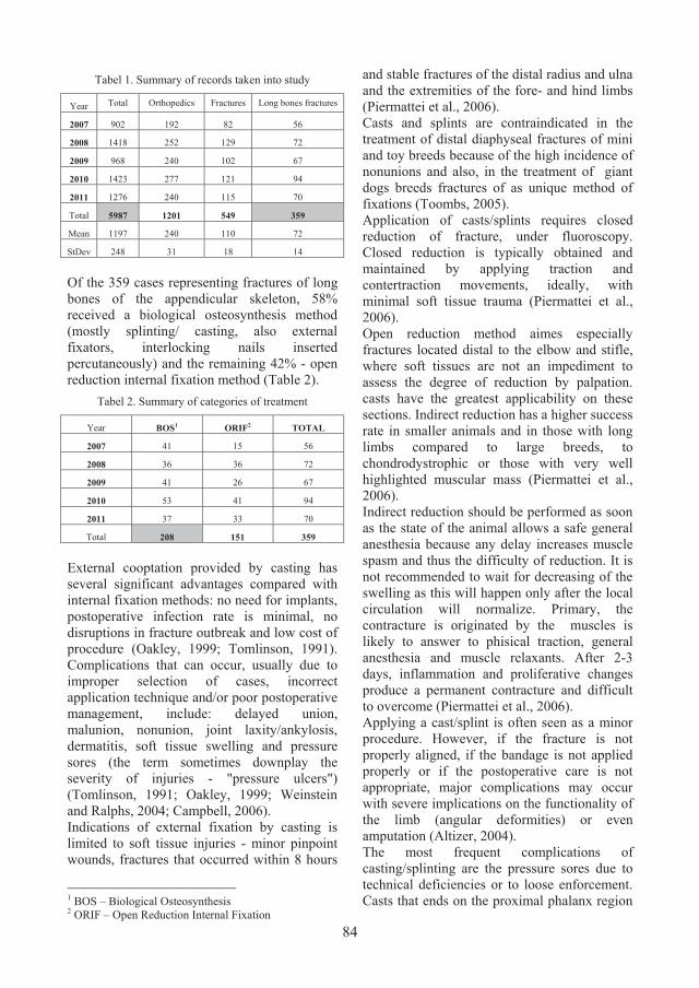
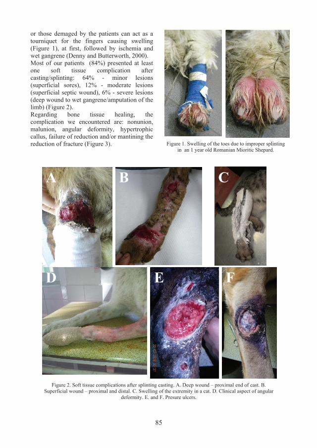
or those demaged by the patients can act as a tourniquet for the fingers causing swelling (Figure 1), at first, followed by ischemia and wet gangrene (Denny and Butterworth, 2000). Most of our patients (84%) presented at least one soft tissue complication after casting/splinting: 64% - minor lesions (superficial sores), 12% - moderate lesions (superficial septic wound), 6% - severe lesions (deep wound to wet gangrene/amputation of the limb) (Figure 2). Regarding bone tissue healing, the complication we encountered are: nonunion, malunion, angular deformity, hypertrophic callus, failure of reduction and/or mantining the reduction of fracture (Figure 3).
Figure 1. Swelling of the toes due to improper splinting in an 1 year old Romanian Mioritic Shepard.
Figure 2. Soft tissue complications after splinting casting. A. Deep wound – proximal end of cast. B. Superficial wound – proximal and distal. C. Swelling of the extremity in a cat. D. Clinical aspect of angular
deformity. E. and F. Presure ulcers.
A B C
D E F

86
A.
B.
C.
D.
E.
F.
Figura 3. Complications of casting. A. Malunion with angular deformity. B. Failure of fracture reduction (predisposed to la defect nonunion). C. Opening of the fracture site under the cast due to lack of stability D.
Hyperthrophic nonunion. E. Failure of fracture reduction – improper case selection; nonunine. F. Vicious callus after improper fracture reduction
A 2011 published research regarding the complication of casting/splinting showed that 60% of patients showed minor lesions – erythema, swelling, sores, without any sign of infections, 20 % presented moderate lesions as superficial septic wound requiring specific
treatment and 20 % of patients had a severe alteration of general state, fever, lameness, skin necrosis, gangrene (Meeson et al., 2011). During the past three decades, internal fixation has become increasingly popular for fracture management and limb reconstruction.
87
As a result, during their training, orthopaedic surgeons receive less formal instruction in the art of extremity immobilization and cast application and removal (Halanski and Noonan, 2008). In this regard, we bring in discussion a method of internal fixation which retains all the advantages of casting/splinting but also of rigid internal fixation, overcoming most difficulties, namely, the minimally invasive plate osteosynthesis. CONCLUSIONS 20% of cases presented in our clinic required an orthopedic treatment, 9% being diagnosed with fractures. Most (84%) patients that underwent a casting/splinting procedure suffered a soft tissue complications even minor. Regarding bone tissue healing, the complication we encountered are: nonunion, malunion, angular deformity, hyperthrophic callus, failure of reduction and/or mantining the reduction of fracture. MIPO retains all the advantages of casting/splinting but also of rigid internal fixation, overcoming most dezadvanteges. REFERENCES Altizer, L., 2004. Casting for immobilization.
Orthopedic Nursing. 23(2):136-141. Aron, D.N., Palmer, R.H., Johnson, A.L., 1995.
Biologic strategies and a balanced concept for repair of highly comminuted long bone fractures. Comped Contin Educ Pract Vet, 17:35.
Campbell, B.G., 2006. Dressings, bandages and splints for wound management in dogs and cats. Vet Clin North Am Small Anim Pract, 36:759-791.
Denny, R.H., Butterworth, J.S., 2000. A guide to canine and feline orthopaedic surgery. Fourth Edition, Blackwell Publishing – Blackwell Science.
Halanski, M., Noonan, K.J., 2008. Cast and splint immobilization: complications. J Am Acad Orthop Surg. 2008 Jan;16(1):30-40.
Hulse, D., Hyman, W., Nori, M., Slater, M., 1997. Reduction in plate strain by addition of an intramedullary pin. Ver Surg, 26:451.
Johnson, A.L., Smith, C.W., Schaeffer, D.J., 1998. Fragment recontruction and bone plate fixation versus bridging plate fixation for treating highly comminuted femoral fractures in dogs: 35 cases (1987-1997), JAVMA, 213:1157.
Meeson, R.L., Davidson, C., Arthurs, G.I., 2011. Soft-tissue injuries associated with cast apllication for
distal limb orthopaedic conditions. Vet Comp Orthop Traumatol, 2:126-131.
Oakley, R.E., 1999. External Coaptation, Vet Clin North Am Small Anim Pract, 29:1083-1095.
Palmer, R.H., 1999. Fracture management and bone healing. Biological osteosynthesis. Vet Clin North Am Small Anim Pract, 1172-1185.
Palmer, R.H., 1999. Fracture management and bone healing. Biological osteosynthesis. Vet Clin North Am Small Anim Pract, 1172-1185.
Palmer, R.H., Aron, D.N., 1996. Biology versus biomechanics. Proceedings of the Sixth Annual American College of Veterinary Surgeons Symposium, San Francisco, SUA, p. 171.
Piermattei, D., Flo, G., DeCamp, C. 2006. Handbook of small animals orthopedics and fracture repair, Fourth Edition, Saunders Elsevier, St. Louis, SUA.
Tomlinson, J., 1991. Complications of fractures repaired with casts and splints. Vet Clin North Am Small Anim Pract, 21:735-744.
Toombs, J.P., 2005. Fractures of the radius, In Johnson, L.A., Houlton, E.F.J., Vannini, R., AO Principles of fracture management in the dog and cat, AO Publishing, Clavadelerstasse, Elveția, pp.236-259.
Weinstein, J., Ralphs, C.S., 2004. External Coaptation. Clin Tech Small Anim Pract, 19:98-104.