Embed Size (px)
Citation preview
BIOL 3380 Combined Lab Report
Name: Younghee Kwon
Circle your lab section:
T–AM
W-AM R-AM F-AM
T–PM W-PM R-PM F-PM
Instructor: Dr. Scott Rippel
Graduate TA: Nymisha
Date: November 9, 2014
Partner: Mauricio
The Expression and Purification of His6 – tagged Recombinant Green Fluorescence Protein
(rGFP) from E. coli by Ni+2
-Agarose Affinity Chromatography
Abstract:
The purpose of these series of experiment was to express and purify a His6 – tagged
recombinant form of GFP (rGFP) from the E.Coli strain BL21(DE3)<pLysS>< pRSETA-GFPUV
> using the Ni+2
agarose affinity chromatography technology. The GFPUV was induced and
expressed, and rGFP crude extract was then purified using the Ni+2
agarose affinity
chromatography. The protein concentration, purity, and presence of rGFP in column fractions
were analyzed via Bradford Assay, SDS-PAGE/Coomassie Blue analysis, and Western Blot
respectively. The elution fraction E3 had the highest activity of 74198 RFUs, and the Bradford
Assay estimated the total protein amount in E3 fraction to be 171.25 ug. Its specific activity was
calculated to be 1150356.6 RFUs/mg. SDS-PAGE estimated the purity of rGFP in E3 fraction to
be around 90%. Based upon the estimated purity of the E3 fraction, the estimated total yield of
rGFP was calculated to be 154.125 ug. Estimated molecular weight of purified rGFP knowing
average molecular weight of amino acid was calculated to be 33.5kDa while the relative
molecular weight of rGFP based on the extrapolation line on the standard curve estimated to be
30.33kDa. Western Blot analysis confirmed the presence of rGFP using specific antibodies.
Introduction:
In 1962, Osamu Shimomura successfully isolated a bioluminescent protein that gave
off blue light in a glowing jellyfish, Aequorea Victoria. Further studies revealed the existence of
green fluorescent protein (GFP) which explained a jellyfish glowing green instead of blue. GFP
absorbed protein’s blue light and re-emitted green light. GFP’s ability to process blue light to
green was found to be integral to its structure, chromophore. In 1988, Martin Chalfie
successfully introduced the gene for GFP into small worm known as Caenorhabditis elegans,
and the expression of GFP was reported. Tracing the localization of specific proteins in living
organism was possible by fusing gene for GFP with the genes for other proteins. Roger Tsein
extended the color palette beyond green and the range of available tags which emitted light at
slightly different wavelengths (Nobel Laureates, 2008).
Shimomura explained that GFP contained a special structure called chromophore which
was the chemical group that was responsible for absorbing and emitting light. The chromophore
was protected inside of an 11 stranded beta-barrel and was able to withstand changes in chemical
reagents, temperature, and pH. GFP was able to stay extremely stable in such harsh conditions
which made the application of GFP more extensive. Wild type GFP contained open reading
frame which coded for a 238 amino acid protein, and its relative molecular weight was to be
around 27kDa. Instead of a wild type GFP, mutant form of GFP (GFPUV) which contained
additional amino acid called a His6 at its n-terminus was used in experiment due to its
optimization for higher bacterial expression and maximal fluorescence when excited by UV light.
It has its excitation wavelength at 395nm and emission wavelength at 510nm (Rippel, 2014).
A recombinant protein with a string of six histidine residues fused on its N-terminus
was used for affinity purification. Six consecutive histidines were used since they proved to be
more efficient than two histidines in binding to the column. Expressed His6 – tagged recombinant
form of GFP (rGFP) could be purified by Ni+2
-Agarose Affinity Chromatography since the string
of negatively charged histidine residues can be specifically bound to immobilized metal ions
which were positively charged nickel. The tight interaction between His6 tagged and Ni2+
made
the proteins of interest remained intact while other contaminants in crude extract were washed
away from the column. Imidazole was then run through the column to elute His6 tagged proteins
off a Ni2+
column. Imidazole had a very similar structure to the His6 tag and acted as a
competitor to displace the His6 tagged proteins (Rippel, 2014).
The purpose of these series of experiments was to express and purify rGFP in E.coli
using Ni+2
-Agarose Affinity Chromatography and consequently conduct further analyses to
estimate concentration, purity, relative molecular weight of rGFP and lastly to confirm the
presence of rGFP.
Materials and Methods:
Expressing rGFP in E.coli:
Grow bacterial culture. Incubate 10ml of liquid LB growth media. Set it out to grow at 37oC
overnight with vigorous shaking until it turns cloudy, indication of saturation. 500 ml of liquid

LB growth media [100ug/ml Amp;25ug/ml Cam in 1 liter baffled flask, pre-warmed-30 degrees
Celsius] is inoculated with enough of the saturated overnight culture so it reaches an OD600 of
0.1. Again, this 500mL culture is left to grow overnight at 37C with vigorous shaking in order
to increase the OD600 to 0.5. When OD600 is raised up to 0.5 which equals time zero, transfer
1ml of the culture into 1.5ml centrifuge tube and centrifuge to obtain pellet while discarding the
supernatant. The centrifuge tube containing bacterial pellet is labeled “G0” and stored at -20
degrees Celsius. The culture is induced with 1Mm of IPTG and continued to grow. After 3 hours
post induction, 1ml of the culture is pelleted into a different 1.5mL centrifuge tube, and the
bacterial pellet is labeled “G3”. Additionally, collect 15ml of “G0” and pellet the centrifuge tube.
Label the remaining pellet “G3-15ml.” Both “G3” and “G3-15ml” are stored at -20C.
Preparation of rGFP crude extract:
Add 500ul of breaking buffer (10mM Tris, pH 8.0; 150mM NaCl) twice to the “G3-15ml” frozen
bacterial pellet. After the addition of breaking buffer, immediately thaw out/broke out the pellet
by pipetting breaking buffer up and down. Transfer this homogeneous solution into a 1.5ml
centrifuge tube and vortex for 5 minutes. Place it in 37oC water bath for 10 minutes. Observe and
record the fluorescence seen in the pellet and supernatant samples by using hand held UV light in
the dark room. Decant the supernatant to a new centrifuge tube without using the pipetman and
label it GCE.
Preparing a Ni+2 –agarose column:
Place a small amount of glass wool into a 3ml plastic syringe and place it to a ring stand. Pipet
breaking buffer into the syringe so the breaking buffer is overflowing. Eliminate any air bubbles
in syringe column by adding additional breaking buffer to the column and allowing several drops
to flow out. With at least 500ul of breaking buffer on top of the glass wool, pipet 1ml of 50%
slurry of Ni+2 agarose into the column and open the luer-lock to “gravity pack” the agarose
matrix in the column. Pre-equilibrate the column (adds 10-fold more buffer than the bed-volume
of the column) to wash out any ethanol left behind in the column.
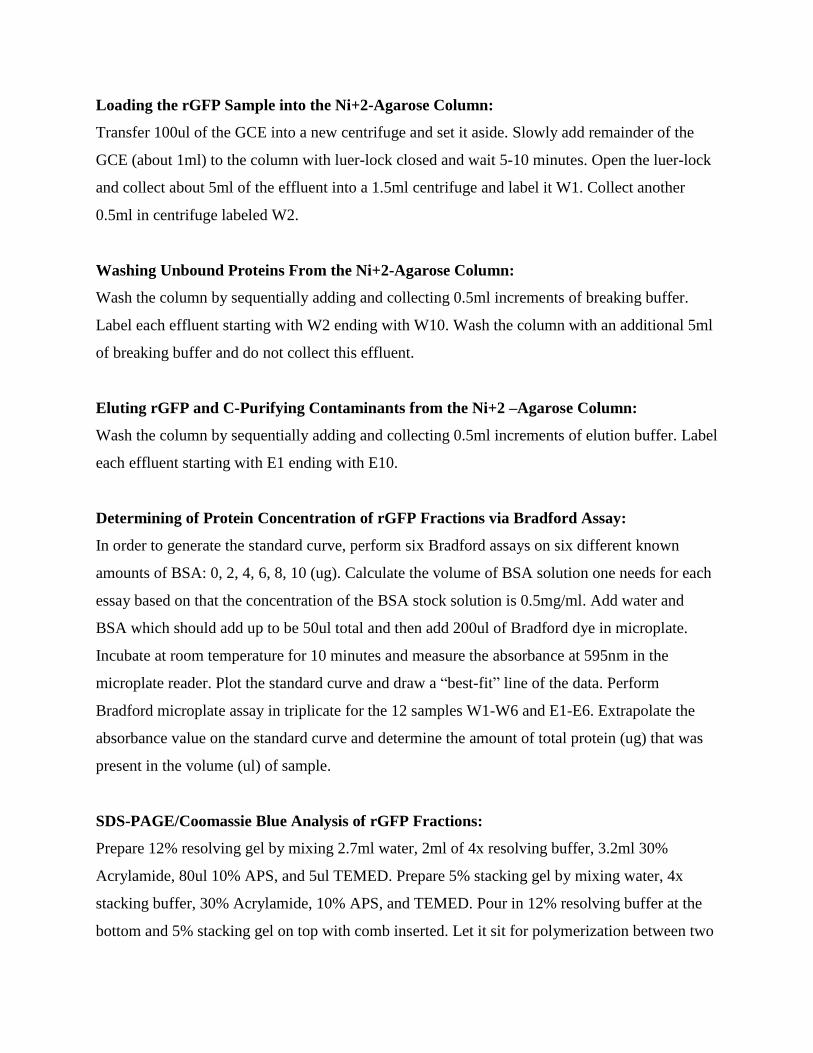
Loading the rGFP Sample into the Ni+2-Agarose Column:
Transfer 100ul of the GCE into a new centrifuge and set it aside. Slowly add remainder of the
GCE (about 1ml) to the column with luer-lock closed and wait 5-10 minutes. Open the luer-lock
and collect about 5ml of the effluent into a 1.5ml centrifuge and label it W1. Collect another
0.5ml in centrifuge labeled W2.
Washing Unbound Proteins From the Ni+2-Agarose Column:
Wash the column by sequentially adding and collecting 0.5ml increments of breaking buffer.
Label each effluent starting with W2 ending with W10. Wash the column with an additional 5ml
of breaking buffer and do not collect this effluent.
Eluting rGFP and C-Purifying Contaminants from the Ni+2 –Agarose Column:
Wash the column by sequentially adding and collecting 0.5ml increments of elution buffer. Label
each effluent starting with E1 ending with E10.
Determining of Protein Concentration of rGFP Fractions via Bradford Assay:
In order to generate the standard curve, perform six Bradford assays on six different known
amounts of BSA: 0, 2, 4, 6, 8, 10 (ug). Calculate the volume of BSA solution one needs for each
essay based on that the concentration of the BSA stock solution is 0.5mg/ml. Add water and
BSA which should add up to be 50ul total and then add 200ul of Bradford dye in microplate.
Incubate at room temperature for 10 minutes and measure the absorbance at 595nm in the
microplate reader. Plot the standard curve and draw a “best-fit” line of the data. Perform
Bradford microplate assay in triplicate for the 12 samples W1-W6 and E1-E6. Extrapolate the
absorbance value on the standard curve and determine the amount of total protein (ug) that was
present in the volume (ul) of sample.
SDS-PAGE/Coomassie Blue Analysis of rGFP Fractions:
Prepare 12% resolving gel by mixing 2.7ml water, 2ml of 4x resolving buffer, 3.2ml 30%
Acrylamide, 80ul 10% APS, and 5ul TEMED. Prepare 5% stacking gel by mixing water, 4x
stacking buffer, 30% Acrylamide, 10% APS, and TEMED. Pour in 12% resolving buffer at the
bottom and 5% stacking gel on top with comb inserted. Let it sit for polymerization between two
glass plates to take place. Once polymerized, place the gel into electrophoresis tank. Remove the
comb and fill up the tank with 500ml of 1;10 diluted electrophoresis buffer. Prepare samples of
G0, G3, GCE, W2, W3, E2, and E3 based upon the given table, vortex them for 1 minute, boil
them for 2 minutes, vortex them again for 1 minute and centrifuge them at the maximum speed
for about 30 seconds. Set up electrophoresis tank, load samples, and operate the power supply at
200 volts for 45 minutes. Remove gel and proceed to staining and de-staining processes.
SDS-PAGE/Western Blot Transfer of rGFP Fractions:
Add 2-ME (beta- mercaptoethanol) to the 1.5ml sample tubes (G0, G3, GCE, W2, W4, E2, E3),
vortex for 1 minutes, boil for 2 minutes, vortex for 1 minute, spin for 30 seconds, and then load
the samples into another gel which was run at 200 volts for 45 minutes. The gel was then placed
in transfer cassette where 3 sheets of filter paper, nitrocellulose membrane, 12% resolving gel,
another 3 sheets of filter paper were placed respectively and left to incubate.
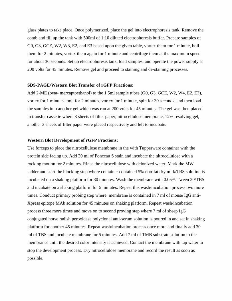
Western Blot Development of rGFP Fractions:
Use forceps to place the nitrocellulose membrane in the with Tupperware container with the
protein side facing up. Add 20 ml of Ponceau S stain and incubate the nitrocellulose with a
rocking motion for 2 minutes. Rinse the nitrocellulose with deionized water. Mark the MW
ladder and start the blocking step where container contained 5% non-fat dry milk/TBS solution is
incubated on a shaking platform for 30 minutes. Wash the membrane with 0.05% Tween 20/TBS
and incubate on a shaking platform for 5 minutes. Repeat this wash/incubation process two more
times. Conduct primary probing step where membrane is contained in 7 ml of mouse IgG anti-
Xpress epitope MAb solution for 45 minutes on shaking platform. Repeat wash/incubation
process three more times and move on to second proving step where 7 ml of sheep IgG
conjugated horse radish peroxidase polyclonal anti-serum solution is poured in and sat in shaking
platform for another 45 minutes. Repeat wash/incubation process once more and finally add 30
ml of TBS and incubate membrane for 5 minutes. Add 7 ml of TMB substrate solution to the
membranes until the desired color intensity is achieved. Contact the membrane with tap water to
stop the development process. Dry nitrocellulose membrane and record the result as soon as
possible.
Results:
The growth of the bacterial expression system for purifying rGFP started with inoculating
liquid LB media with a single bacterial colony of strain G (BL21<DE3>pLysS, pRSETA-
GFPUV ). At time equaled zero (𝑂𝐷600 ~0.5), a sample was designated as G0 which also implied
that no IPTG induction had taken place. G0 culture was then induced with IPTG and labeled as
G3 after 3 hours of post induction. Induction of IPTG inhibited the Lac repressor to be bound
onto the lac promoter and abled the production of the excess of T7 RNA polymerase.
Consequently, they were bound to the T7 promoters and triggered the transcription and
translation of the protein of interest (rGFP).
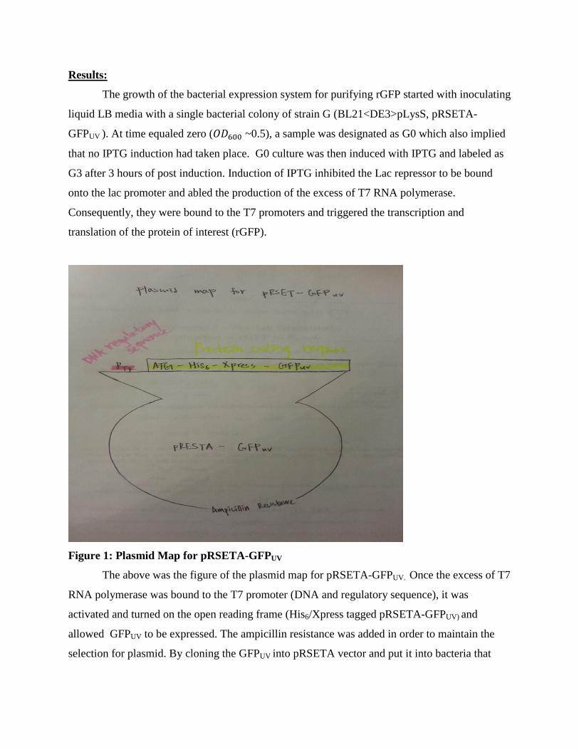
Figure 1: Plasmid Map for pRSETA-GFPUV
The above was the figure of the plasmid map for pRSETA-GFPUV. Once the excess of T7
RNA polymerase was bound to the T7 promoter (DNA and regulatory sequence), it was
activated and turned on the open reading frame (His6/Xpress tagged pRSETA-GFPUV) and
allowed GFPUV to be expressed. The ampicillin resistance was added in order to maintain the
selection for plasmid. By cloning the GFPUV into pRSETA vector and put it into bacteria that

could process transcription and translation, the expression of GFPUV was under the regulated
system. It was now under inducible promoter.
Figure 2: Schematic Diagram of rGFP
Based on the rGFP cloning procedure and GFP structure descried in the lecture, a
schematic diagram figure of rGFP could be constructed as above. Important domain/regions,
chromophore location, and amino acid distances were specified in the schematic diagram as well.
The His6 tag which was a string of six histidine residues fused on near the 5’ N-terminus was
transcribed in the “CAT” sequences. The Xpress Epitope Tag started with the “GAT” and ended
with the “AAG” codon. The GFPUV DNA sequence was located about 238 amino acids toward
the 3’ C-terminus end. The chromophore which was responsible for the fluorescence of GFPuv
was located at around the 65-67 amino acids.
3’ C Terminus
279 1 5 11 39 104-106 277
His6 tag
24 32
Xpress Epitope Tag
GFPUV
chromophore
GFPUV
5’ N terminus
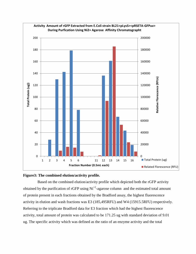
Figure3: The combined elution/activity profile.
Based on the combined elution/activity profile which depicted both the rGFP activity
obtained by the purification of rGFP using Ni+2
-agarose column and the estimated total amount
of protein present in each fractions obtained by the Bradford assay, the highest fluorescence
activity in elution and wash fractions was E3 (185,495RFU) and W4 (15915.5RFU) respectively.
Referring to the triplicate Bradford data for E3 fraction which had the highest fluorescence
activity, total amount of protein was calculated to be 171.25 ug with standard deviation of 9.01
ug. The specific activity which was defined as the ratio of an enzyme activity and the total
0
20000
40000
60000
80000
100000
120000
140000
160000
180000
200000
0
20
40
60
80
100
120
140
160
180
200
1 2 3 4 5 6 11 12 13 14 15 16R
ela
tive
Flo
resc
en
ce (
RFU
s)
Tota
l Pro
tein
(u
g))
Fraction Number (0.5mL each)
Activity Amount of rGFP Extracted from E.Coli strain BL21<pLysS><pRSETA-GFPuv> During Purfication Using Ni2+ Agarose Affinity Chromatographt
Total Protein (ug)
Related Florescence (RFU)
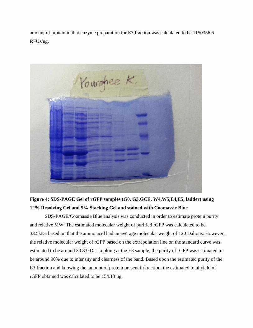
amount of protein in that enzyme preparation for E3 fraction was calculated to be 1150356.6
RFUs/ug.
Figure 4: SDS-PAGE Gel of rGFP samples (G0, G3,GCE, W4,W5,E4,E5, ladder) using
12% Resolving Gel and 5% Stacking Gel and stained with Coomassie Blue
SDS-PAGE/Coomassie Blue analysis was conducted in order to estimate protein purity
and relative MW. The estimated molecular weight of purified rGFP was calculated to be
33.5kDa based on that the amino acid had an average molecular weight of 120 Daltons. However,
the relative molecular weight of rGFP based on the extrapolation line on the standard curve was
estimated to be around 30.33kDa. Looking at the E3 sample, the purity of rGFP was estimated to
be around 90% due to intensity and clearness of the band. Based upon the estimated purity of the
E3 fraction and knowing the amount of protein present in fraction, the estimated total yield of
rGFP obtained was calculated to be 154.13 ug.

Figure 5: Western Blot
The Western Blot above corresponded with the findings of the SDS-PAGE/Coomassie
blue analysis, and the bands presented at around 35kDa validate the presence of rGFP since its
molecular weight was close to the relative molecular weight found in the previous lab
(~30.33kDa). The first column did not show any bands since it represented G0.
Conclusions/Discussion:
The purpose of these series of experiments was successfully accomplished based on the
results obtained from several methods of procedures. High percentage of purity was obtained,
and qualitative monitoring of GFP activity was consistent with quantitative monitoring of GFP
activity. W4 and E3 had the strongest fluorescence, and W4 (6366.2 RFUs) and E3(74198 RFUs)
also had the highest number of activity compared to the other washes and elutions in fluorescent
microplate reader date.
Due to several unique characteristics of GFP learned throughout the series of experiments
such as easy and immediate real-time detection, no cofactor required for fluorescence, and
possibility of dual labeling/detection, GFP or recombinant form of GFP (rGFP) could be used as
valuable and easily accessible resource in variety fields of research areas (Scott, 2014). The GFP
expression could be used to monitor gene expression and protein localization. Specifically,
scientists could further utilize GFP to monitor specific target proteins. GFP could be used to
pinpoint the specific location of proteins that might have possible implication for the growth of
tumors, viruses, and so on. Being able to track down harmful target proteins, scientists could
isolate the gene and conduct further researches on that specific gene of interest and possibly
contribute to fixing the occurring problems.
Furthermore, BL21 was so unique in a way that one could locate any gene of interest in
the place GFPUV, and the gen would be expressed. Thus, follow-on experiments could also be
conducted on how different types of recombinant GFP reacted to the variable conditions such as
internal body temperature to figure out one particular GFP variant which adhered to the
procedures better than others under certain circumstances.
References:
Novel Laureates “for the discovery and development of the green fluorescent protein, GFP.” The
Nobel Prize in Chemistry 2008 (2008). Web.
Rippel, Scott, and Elizabeth Pickett. Biochemistry Laboratory Manual. Richardson: University
of Texas at Dallas, 2014. Print.