Embed Size (px)
Citation preview

Histology Compendium
by TEAM Don'tKnowYets
Team
Member #Student Name ID #'s
1 Brad Nilsen 332729
2 Kate Littlefield 280247
3 Dakota Upton 339060
4 Jeremiah Martinez 343505
5 Jacob Anderson 301648
Tissues Classification Drawing # Name of Slide / Notes / Description Picture or Illustration From Web Picture or Illustration From Web References
MAIN Sub Type Sub Type Sub Type Sub Type
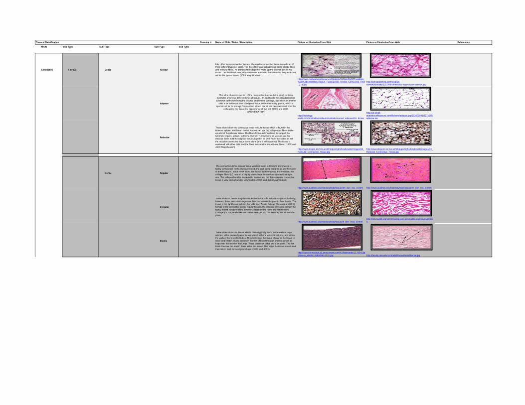
Connective Fibrous Loose Areolar
Like other loose connective tissues, the areolar connective tissue is made up of
three different types of fibers. The three fibers are collagenous fibers, elastic fibers
and reticular fibers. All of these fibers together make up the distinct look of this
tissue. The little black dots with extensions are called fibroblast and they are found
within this type of tissue. (100X Magnification)
http://www.noelways.com/courses/Anatomy%20and%20Physiology
%20I/Labs/Histology/Tissue_Types/Loose_Areolar_Con/Loose_Areo
_2_b.jpg
http://sohoparenting.com/blog/wp-
content/uploads/2011/06/connective-tissue-loose-areolar.jpg
Adipose
This slide of a cross section of the mammalian trachea (wind pipe) contains
examples of several different kinds of tissues. In addition to the pseudostratified
columnar epithelium lining the trachea and hyaline cartilage, also seen on another
slide is an extensive area of adipose tissue in the mammary glands, which is
specialized for fat storage.On prepared slides, the fat has been removed from the
cells giving the tissue the appearance of fish net. (100X and 400X
MAGNIFICATION)
http://histology-
world.com/photoalbum/albums/uploads/normal_adipose20X_lbl.jpg
http://dr-small-
anatomy.wikispaces.com/file/view/adipose.jpg/291602931/327x279/
adipose.jpg
Reticular
These slides show the connective loose reticular tissue which is found in the
kidneys, spleen, and lymph nodes. As you can see the collagenous fibers make
up a lot of the reticular tissue. The fibers form a soft ‘skeleton’ to support the
lymphoid organs, spleen, red bone marrow. Furthermore, as we can see the
reticular fibers hold the adipose tissues together as well. From the slides as well
the reticular connective tissue is not alone (and it will never be). The tissue is
combined with other cells and the fibers in its matrix are reticular fibers. (100X and
400X Magnification) http://www.stegen.k12.mo.us/tchrpges/sghs/ksulkowski/images/32_
Reticular_Connective_Tissue.jpg
http://www.stegen.k12.mo.us/tchrpges/sghs/ksulkowski/images/32_
Reticular_Connective_Tissue.jpg
Dense Regular
The connective dense regular tissue which is found in tendons and muscle is
tightly compacted. In the slides provided, the dark spots that pop up are the nuclei
of the fibroblasts. In the 400X slide, the ‘fb nuc’ is the nucleus. Furthermore, the
collagen fibers (cf) take on a slightly wavy shape rather than a perfectly straight
one. The collagen bundles in a parallel fashion and the dense regular connective
tissue is very strong but also very flexible. (100X and 400X Magnification)
http://www.austincc.edu/histologyhelp/tissues/tm_den_reg_ct.html http://www.austincc.edu/histologyhelp/tissues/tm_den_reg_ct.html
Irregular
These slides of Dense irregular connective tissue is found all throughout the body;
however, these particular images are from the skin on the palms of our hands. The
tissue is the light brown color in the slide from Austin College (the ones at 400 X).
Similar to the connective dense regular tissues, the irregular ones also contain the
tightly bound collagen fibers; however, based off the name the matrix fibers
(collagen) is not parallel like the others were. As you can see they are all over the
place.
http://www.austincc.edu/histologyhelp/tissues/tl_den_irreg_ct.html
http://stevegallik.org/sites/histologyolm.stevegallik.org/images/dict.jp
g
Elastic
These slides show the dense, elastic tissue typically found in the walls of large
arteries, within certain ligaments associated with the vertebral column, and within
the walls of the bronchial tubes. The elasticity of the tissue allows for the tissue to
recoil and stretch. It also assists in the flow of blood through arteries as well as
helps with the recoil of the lungs. These particular slides are of an aorta. The thin
black lines are the elastin fibers within the tissue. This helps the tissue stretch and
then return back to its original shape. (100X and 400X)
http://classconnection.s3.amazonaws.com/42/flashcards/1178042/jp
g/dense_elastic1329009610032.jpg http://faculty.une.edu/com/abell/histo/elasticfibersw.jpg

Tissues Classification Drawing # Name of Slide / Notes / Description Picture or Illustration From Web Picture or Illustration From Web References
MAIN Sub Type Sub Type Sub Type Sub Type
Supportive Cartilge Hyaline
The bar in one of the images shows us how much cartilage is in the tracheal wall.
Furthermore, as you can see in the slide with the bar there is some
pseudostratified ciliated epithelium as well. Cartilage contains cells that are deeply
embedded in the matrix of fibers. The cells are called chondrocytes (ch) and empty
space within the cartilage is called lacunae. There are not a lot of fibers in the
hyaline cartilage as you can see by the slides; however, there is fibrous lining on
the outside which is called perichondrium . (100X and 400X)
http://www.austincc.edu/histologyhelp/tissues/to_hy_cart.html http://www.austincc.edu/histologyhelp/tissues/to_hy_cart.html
Elastic
Elastic cartilage is commonly found in the outer ear of an individual. The ear is very
bendable but still is able to hold its shape and that is because if this type of
cartilage. The elastic, or yellow, cartilage is also found in the eustachian tube, and
the epiglottis. The elastic and the hyaline cartilage are very similar histologically,
but the elastic cartilage has many fibers in the solid matrix. These fibers give the
cartilage the great elasticity and ability to move and bend and still retain its shape.
(100X and 400X)
http://www2.palomar.edu/users/ggushansky/histology/images/elastic
%20cartilage%202_tif.jpg
https://meded.ucsd.edu/hist-img-
bank/chapter_2/Slide_14_elastic/images/b.3.14.1.3.jpg
http://www.gwc.maricopa.edu/class/bio201/Histology/21E
lasticCartilage1_400X_rev.jpg
Fibro
While Hyaline and Fibro has some similarities, fibrocartilage looks different
because of the number of collagen fibers embedded in the matrix. From the slides
you can see the nuclei within the chondrocytes which are located in lacunae.
Fibrocartilage is found in the pubic symphysis, the anulus fibrosus of intervertebral
discs, menisci and the TMJ. The cartilage needs to be flexible as well but also very
strong and durable.
http://www.austincc.edu/histologyhelp/tissues/tp_fibro_cart.html http://www.austincc.edu/histologyhelp/tissues/tp_fibro_cart.html
Bone Compact
In these slides, there are several structural units of bone tissue (osteons or
Haversian systems). Each osteon looks like a ring with a light spot in the center.
The center is a canal that carries a blood vessel and a nerve fiber. The outer ring,
which is also the darker part is made of bone matrix from cells. We are able to see
the layered structure of the bone in these slides. Moreover, the more purple and
pink slide shows compact bone that is decalcified. We wanted to show this
because there is a difference in appearance. The bone has been removed of its
calcium salts. To the right is some bone marrow as well. http://www2.sunysuffolk.edu/pickenc/Compact%20Bone%20Osteon
%20400X.JPG
http://www.austincc.edu/histologyhelp/tissues/tr_bone_com_dcal.ht
ml
Spongy
With these slides and diagrams you can see how much space is in between the
bone. It is much less dense than compact bone and therefore, it takes up a lot of
surface space. This type of bone is typically found at the end of a large bone and
you can see this is one of the pictures. Spongy bone also contains a lot of red
bone marrow and blood vessels.
http://fau.pearlashes.com/anatomy/Chapter%209/Chapter%209_file
s/cancellous_bone.jpg
http://classconnection.s3.amazonaws.com/856/flashcards/655856/jp
g/3._cancellous_(spongy_bone_(400x)1315253703008.jpg
Tissues Classification Drawing # Name of Slide / Notes / Description Picture or Illustration From Web Picture or Illustration From Web References
MAIN Sub Type Sub Type Sub Type Sub Type
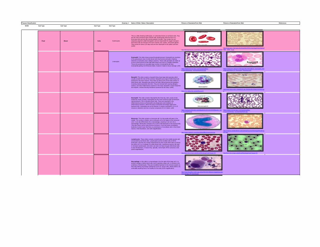
Fluid Blood Cells Erythrocytes
This is a slide showing erythrocytes, or commonly known as red blood cells. They
are the most numerous type of blood cells. Erythrocytes have the form of a
biconcave disk and are also narrow-waisted in profile. They contain the red
respiratory pigment called hemoglobin and are responsible for oxygen transport.
Red blood cells are formed in the bone marrow, liver, spleen, and lymph glands.
They survive for about 120 days and are then destroyed in the spleen and liver.
(x400)
http://biology-forums.com/index.php?action=gallery;sa=view;id=8498
http://www.gwc.maricopa.edu/class/bio202/Hematology/histo/Normal
RBC_400X_A.jpg
Leukocytes
Eosinophil: This slide shows several Eosinophil present. Eosinophil are members
of the granulocytic class of white blood cells that function primarily in fighting
infections of parasites and in allergic reactions. These cells generally are made up
of one to three percent of the total white blood cell count in a healthy individual.
Those who possess an unusually large number of eosinophils will have
eosinophilia getting rid of illnesses faster, however it leads to tissue damage. (x40)
https://www.microscopyu.com/assets/gallery-
images/pathology_eosinophilia40x03.jpg
https://commons.wikivet.net/images/thumb/a/a8/LH_Eosinophil_Hist
ology.jpg/150px-LH_Eosinophil_Histology.jpg3
Basophil: This slide contains a basophil (they have large dark granules which
usually occlude the view of the nucleus). Basophils are a type of white blood cell
produced in the bone marrow. They make up about 0.5% of the total number of
white blood cells. Basophils also protect the body, killing bacteria and parasites,
including external parasites like ticks. They are involved with causing allergies,
asthma, and other inflammatory reactions in the body. Basophils contain histamine
and heparin, a blood thinning substance produced by the body. (x400)
https://en.wikipedia.org/wiki/Basophil
http://www.iupui.edu/~anatd502/Labs.f04/blood%20lab/s18100nb.jp
g
Neutrophil: This slide contains Neutrophils (the three big, dark, purple circles
toward the center of slide). Neutrophils are the most numerous type of leukocyte,
representing for 70% of all white blood cells. These are important in the
inflammatory process as Neutrophils are phagocytes and mediators of
inflammatory reactions. When the body is breached and foreign objects like
bacteria enter, neutrophils eat up the bacteria. A mature neutrophil is 12 to 15
microns in diameter and has a nucleus composed of 2 to 5 lobes. (x40)
https://upload.wikimedia.org/wikipedia/commons/3/33/Blausen_067
6_Neutrophil.png
https://www.microscopyu.com/assets/gallery-
images/pathology_neutrophilia40x04.jpg
Monocyte: This slide contains a monocyte cell: it is the purple cell seen in the
middle. The nucleus is folded, and a monocyte cell is the largest of the leukocyte
cells. It can also differentiate into a macrophage, meaning it can turn into a
macrophage. Monocytes compose 2% to 10% of all leukocytes in the human body.
They also help to serve many immune functions. If one has acute monoblastic
leukemia it would mean that there is a dominance of monoblast cells in their blood
marrow. Artist illustration, and x100 magnification)
http://www.mabtech.com/sites/default/files/monocyte.jpg
http://classes.midlandstech.edu/carterp/Courses/histology/blood/img
011.jpg
Lymphocytre: These slides contains a lymphocyte cell in the middle (purple cell)
surrounded by red blood cells. Lymphocytes are the smallest cells of all the
leukocytes, and they are mostly characterized by their nuclei which take up almost
the whole cell. It is a subtype of a white blood cells. Lymphoma cancers can start
in immature lymphocytes, and when one does not have enough white blood cells
in their bloodstream, it means they, typically, cannot fight off the cancerous cells.
(x100 magnification)
http://bcrc.bio.umass.edu/bestofhistology/node?page=34 http://bcrc.bio.umass.edu/bestofhistology/node?page=34
Macrophage: In this slide is a macrophage is too the right of the large cell. It is
another subtype of white blood cells, and is typically a large cell, in contrast to the
smallness of lymphocytes. They are incredibly important to the immune system as
they help destroy any foreign substances such as: cancer cells, cellular debris, etc;
essentially anything that is not healthy for the body (x100 magnification)
http://new.bloodline.net/image-atlas/2661064-Mature-megakaryocyte-
macrophage-ITP-marrow.jpg

Tissues Classification Drawing # Name of Slide / Notes / Description Picture or Illustration From Web Picture or Illustration From Web References
MAIN Sub Type Sub Type Sub Type Sub Type
Cell Fragments Platelets
In this slide, the platelets are the smaller blue dots surrounded by red blood cells.
Platelets stop bleeding by clotting and clumping blood vessel injuries. You have
just cut your hand? Platelets to the rescue! While you may need to put a bandage
on it for a little while, the platelets will do the rest for you. They are typically about
20% the diameter of a red blood cell. The normal range of platelets in a healthy
Caucasian is 150,000 to 450,000 per cubic millimeter. That is a lot.
https://upload.wikimedia.org/wikipedia/commons/thumb/5/51/Platele
ts2.JPG/250px-Platelets2.JPG
Plasma
In this slide, the plasma is the fluid that the cells are suspended in. It is clear and
makes up about 55% of the human body’s total blood volume. When preparing
plasma, it is what is left when all the other cells are gone. (x40 magnification)
http://www.austincc.edu/histologyhelp/tissues/tt_blood.html

Tissues Classification Drawing # Name of Slide / Notes / Description Picture or Illustration From Web Picture or Illustration From Web References
MAIN Sub Type Sub Type Sub Type Sub Type
Muscle Skeletal
Skeletal muscle moves joints by strong and rapid contractions.
Each muscle is a bundle of muscle fibres, which are long multinucleated cells. To
the right we see an artist's illustration of this, and longitudinally viewed skeletal
muscle at different magnification levels
Black Bar = 50 microns & 20 microns
http://www.histology.leeds.ac.uk/tissue_types/muscle/muscle_skelet
al.php
http://www.histology.leeds.ac.uk/tissue_types/muscle/Three_muscle
_types.php
http://www.histology.leeds.ac.uk/tissue_types/muscle/mu
scle_skeletal.php
Smooth
Smooth muscle is made up of cells that contain a single central nucleus. The cells
are spindle shaped, and the nucleus is central. Can be found in Urinary bladder,
Intestines, Airways and the Gallbladder. This diagram shows a few of the cells that
can be seen in Longitudinal Section & in Transverse Section.
Black Bar = 20 microns. Final Figure is under 100X magnification.
http://www.histology.leeds.ac.uk/tissue_types/muscle/muscle_smoot
h.php
http://www.histology.leeds.ac.uk/tissue_types/muscle/muscle_smoot
h.php
http://education.med.nyu.edu/Histology/courseware/mod
ules/muscle/muscle.08.html
Cardiac
Cardiac muscle is striated, like skeletal muscle. Cardiac muscle cells usually have
a single (central) nucleus. The cells are often branched, and are tightly connected
by specialised junctions. The region where the ends of the cells are connected to
another cell is called an intercalated disc. The intercalated disc contains gap
junctions that allow the muscle cells to be electrically coupled, so they beat in
synchronization
To the Right of the Diagram is a 100X magnification image of Cardiac Muscle,
followed by a 400X magnification image of Cardiac Musclehttp://www.histology.leeds.ac.uk/tissue_types/muscle/muscle_cardia
c.php http://imgarcade.com/1/cardiac-muscle-100x/ http://pinstake.com/cardiac-muscle-400x-11/
Tissues Classification Drawing # Name of Slide / Notes / Description Picture or Illustration From Web Picture or Illustration From Web References
MAIN Sub Type Sub Type Sub Type Sub Type
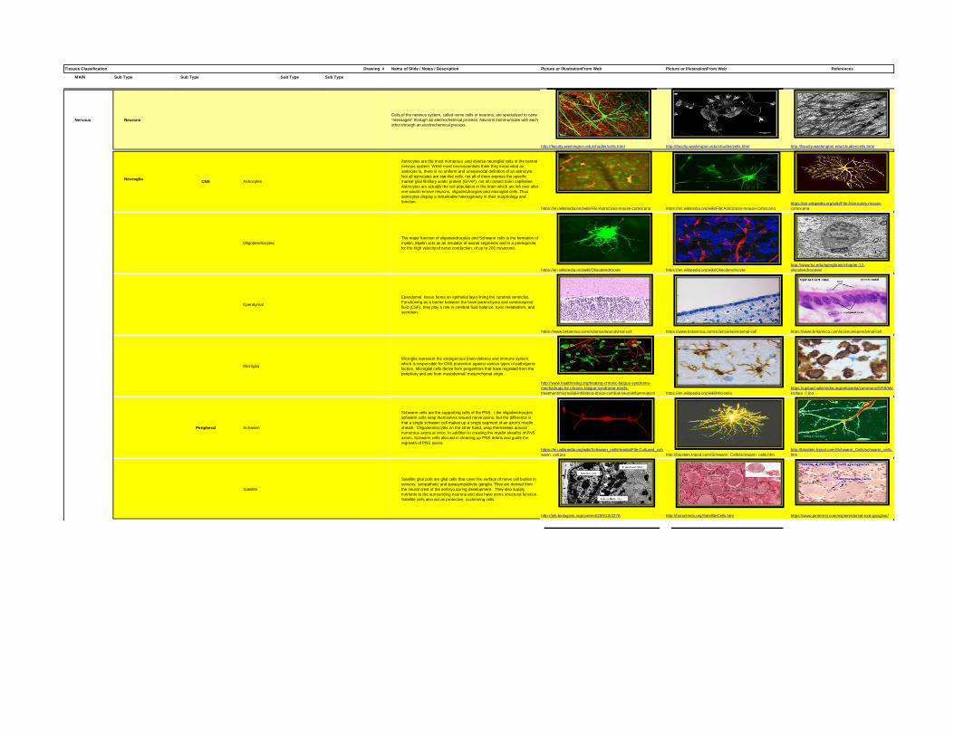
Nervous Neurons
Cells of the nervous system, called nerve cells or neurons, are specialized to carry
"messages" through an electrochemical process. Neurons communicate with each
other through an electrochemical process.
http://faculty.washington.edu/chudler/cells.html http://faculty.washington.edu/chudler/cells.html http://faculty.washington.edu/chudler/cells.html
NeurogliaCNS Astrocytes
Astrocytes are the most numerous and diverse neuroglial cells in the central
nervous system. While most neuroscientists think they know what an
astrocyte is, there is no uniform and unequivocal definition of an astrocyte.
Not all astrocytes are star-like cells, not all of them express the specific
marker glial fibrillary acidic protein (GFAP), not all contact brain capillaries.
Astrocytes are actually the cell population in the brain which are left over after
one would remove neurons, oligodendrocytes and microglial cells. Thus
astrocytes display a remarkable heterogeneity in their morphology and
function.
https://en.wikipedia.org/wiki/File:Astrocytes-mouse-cortex.png https://en.wikipedia.org/wiki/File:Astrocytes-mouse-cortex.png
https://en.wikipedia.org/wiki/File:Astrocytes-mouse-
cortex.png
Oligodendrocytes
The major function of oligodendrocytes and Schwann cells is the formation of
myelin. Myelin acts as an insulator of axonal segments and is a prerequisite
for the high velocity of nerve conduction, of up to 200 m/second.
https://en.wikipedia.org/wiki/Oligodendrocyte https://en.wikipedia.org/wiki/Oligodendrocyte
http://www.bu.edu/agingbrain/chapter-12-
oligodendrocytes/
Ependymal
Ependymal tissue forms an epithelial layer lining the cerebral ventricles.
Functioning as a barrier between the brain parenchyma and cerebrospinal
fluid (CSF), they play a role in cerebral fluid balance, toxin metabolism, and
secretion.
https://www.britannica.com/science/ependymal-cell https://www.britannica.com/science/ependymal-cell https://www.britannica.com/science/ependymal-cell
Microglia
Microglia represent the endogenous brain defence and immune system,
which is responsible for CNS protection against various types of pathogenic
factors. Microglial cells derive from progenitors that have migrated from the
periphery and are from mesodermal/ mesenchymal origin.
http://www.healthrising.org/treating-chronic-fatigue-syndrome-
mecfs/drugs-for-chronic-fatigue-syndrome-mecfs-
treatment/microglial-inhibiting-drugs-combat-neuroinflammation/ https://en.wikipedia.org/wiki/Microglia
https://upload.wikimedia.org/wikipedia/commons/5/59/Ma
krofagi_2.jpg
Peripheral Schwann
Schwann cells are the supporting cells of the PNS. Like oligodendrocytes
schwann cells wrap themselves around nerve axons, but the difference is
that a single schwann cell makes up a single segment of an axon's myelin
sheath. Oligodendrocytes on the other hand, wrap themselves around
numerous axons at once. In addition to creating the myelin sheaths of PNS
axons, Schwann cells also aid in cleaning up PNS debris and guide the
regrowth of PNS axons.
https://en.wikipedia.org/wiki/Schwann_cell#/media/File:Cultured_sch
wann_cell.jpg http://blustein.tripod.com/Schwann_Cells/schwann_cells.htm
http://blustein.tripod.com/Schwann_Cells/schwann_cells.
htm
Satellite
Satellite glial cells are glial cells that cover the surface of nerve cell bodies in
sensory, sympathetic and parasympathetic ganglia. They are derived from
the neural crest of the embryo during development. They also supply
nutrients to the surrounding neurons and also have some structural function.
Satellite cells also act as protective, cushioning cells.
http://jeb.biologists.org/content/209/12/2276 http://horsehints.org/SatelliteCells.htm https://www.pinterest.com/explore/dorsal-root-ganglion/
Tissues Classification Drawing # Name of Slide / Notes / Description Picture or Illustration From Web Picture or Illustration From Web References
MAIN Sub Type Sub Type Sub Type Sub Type
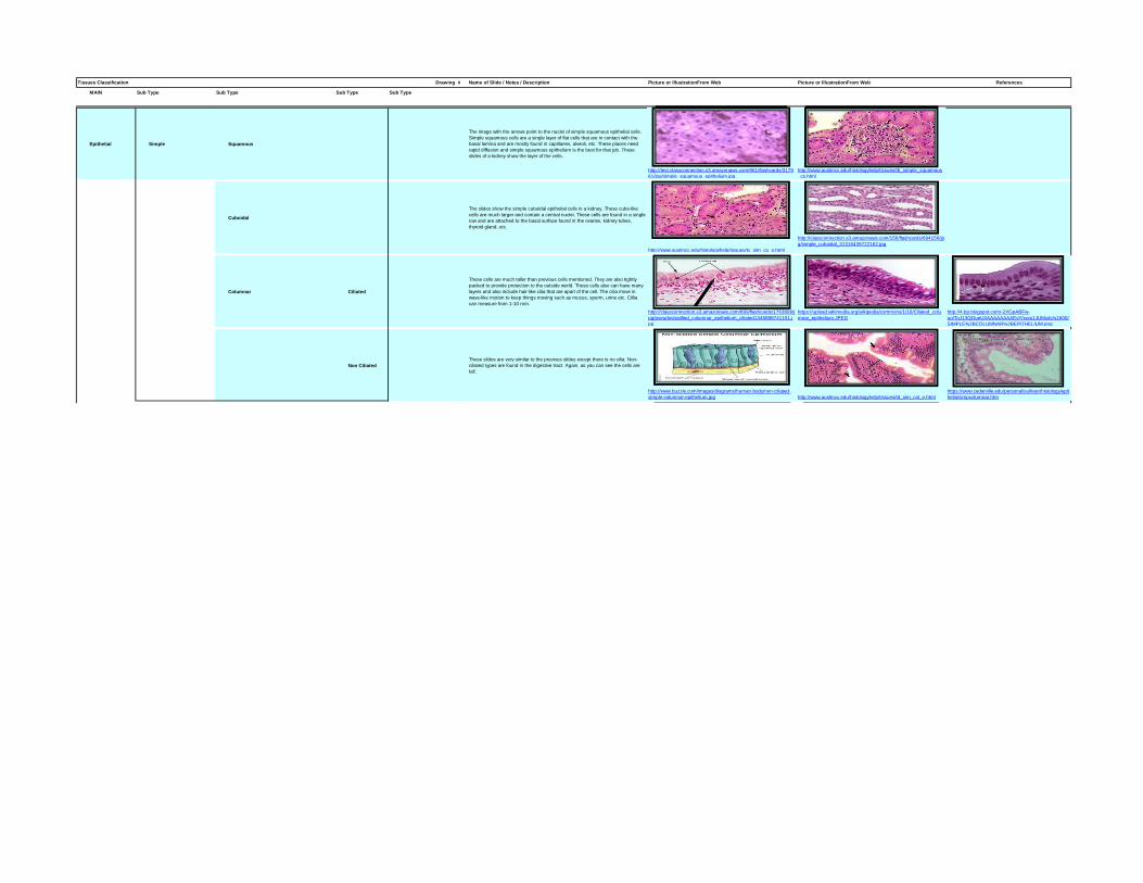
Epithelial Simple Squamous
The image with the arrows point to the nuclei of simple squamous epithelial cells.
Simple squamous cells are a single layer of flat cells that are in contact with the
basal lamina and are mostly found in capillaries, alveoli, etc. These places need
rapid diffusion and simple squamous epithelium is the best for that job. These
slides of a kidney show the layer of the cells.
http://test.classconnection.s3.amazonaws.com/961/flashcards/3179
61/jpg/simple_squamous_epithelium.jpg
http://www.austincc.edu/histologyhelp/tissues/tb_simple_squamous
_cs.html
Cuboidal
The slides show the simple cuboidal epithelial cells in a kidney. These cube-like
cells are much larger and contain a central nuclei. These cells are found in a single
row and are attached to the basal surface found in the ovaries, kidney tubes,
thyroid gland, etc.
http://www.austincc.edu/histologyhelp/tissues/tc_sim_cu_e.html
http://classconnection.s3.amazonaws.com/156/flashcards/694156/jp
g/simple_cuboidal_31316439722162.jpg
Columnar Ciliated
These cells are much taller than previous cells mentioned. They are also tightly
packed to provide protection to the outside world. These cells also can have many
layers and also include hair-like cilia that are apart of the cell. The cilia move in
wave-like motion to keep things moving such as mucus, sperm, urine etc. Cillia
can measure from 1-10 mm.
http://classconnection.s3.amazonaws.com/899/flashcards/1763899/j
pg/pseudostratified_columnar_epithelium_ciliated1346888741151.j
pg
https://upload.wikimedia.org/wikipedia/commons/1/16/Ciliated_colu
mnar_epithelium.JPEG
http://4.bp.blogspot.com/-2XGpABFw-
uo/TnJ13QOueUI/AAAAAAAAEvY/xxvu1JUMxdc/s1600/
SIMPLE%2BCOLUMNAR%2BEPITHELIUM.png
Non Ciliated
These slides are very similar to the previous slides except there is no cilia. Non-
ciliated types are found in the digestive tract. Again, as you can see the cells are
tall.
http://www.buzzle.com/images/diagrams/human-body/non-ciliated-
simple-columnar-epithelium.jpg http://www.austincc.edu/histologyhelp/tissues/td_sim_col_e.html
https://www.cedarville.edu/personal/sullivan/histology/epit
helia/simpcolumnar.htm

Tissues Classification Drawing # Name of Slide / Notes / Description Picture or Illustration From Web Picture or Illustration From Web References
MAIN Sub Type Sub Type Sub Type Sub Type
Stratified Squamous Keratinized
These slides show the stratified squamous keratinized epithelium and how it
covers the entire surface of the skin. To be able to find stratified squamous
keratinized epithelium, you need to look at the skin on areas on your body that do
not have hair. This tissue shown is from the palm of the hand. These cells are flat
and no longer alive, with the addition of no nucleus nor organelles. Instead that are
filled with keratin which is what makes our skin waterproof. The cells on the surface
are dead and are continually lost as well as continuously being replaced by
mitosis. They start out as cuboidal, then irregular, and finally become flat.
(x40,x100,x400)
http://www.austincc.edu/histologyhelp/tissues/images/tg040t.jpg http://www.austincc.edu/histologyhelp/tissues/images/tg100t.jpg
http://www.austincc.edu/histologyhelp/tissues/images/tg4
00combo.jpg
Non Keratinized
These slides show the stratified squamous non-keratinized epithelium. Underneath
its layers are composed mainly of connective tissue and muscle. As you can see,
the nuclei are arranged in more than one layer and how the cells seem to be
separating from the surface of the tissue. The cells form coverings and linings and
is a process called sloughing. Stratified squamous nonkeratinized epithelium can
be found as the corneas, the lining mucosa of the mouth, esophagus, and the
internal portion of the lips. (x40,x100,x400)
http://www.austincc.edu/histologyhelp/tissues/images/tf040t.jpg http://www.austincc.edu/histologyhelp/tissues/images/tf100t.jpg
http://www.austincc.edu/histologyhelp/tissues/images/tf4
00t_s.jpg
Cuboidal
This slide contains the stratified cuboidal epithelia, it is a rare type of epithelial
tissue composed of cuboidal shaped cells arranged in multiple layers. Its functions
include protection, support, secretion and absorption. This tissue is found in sweat
gland ducts, egg-producing vesicles and follicles of the ovaries, and sperm-
producing ducts and seminiferous tubules of the testis. It lines the inner and outer
surfaces of the body and its cavities. (x100, x400)
http://medcell.med.yale.edu/systems_cell_biology/epithelium_lab.ph
p
https://upload.wikimedia.org/wikipedia/commons/thumb/b/bd/WVSO
M_Parotid_Gland1.JPG/250px-WVSOM_Parotid_Gland1.JPG
Columnar
This slide contains the stratified columnar epithelia, it is also a rare type of
epithelial tissue composed of column shaped cells arranged in multiple layers.
Stratified columnar epithelia are found in the conjunctiva of the eye, in parts of the
pharynx, uterus, the male urethra and vas deferen, and the in lobar ducts in
salivary glands. The tissue functions in secretion and protection. Stratified
columnar epithelium occurs in transition areas between other epithelial types.
(x400)
http://www.mhhe.com/biosci/ap/histology_mh/strcol2.jpg
https://secure.health.utas.edu.au/intranet/cds/histoten/images/strat_
columnar01a.jpg
Pseudostratified Ciliated
These slides contain the pseudostratified ciliated epithelial tissue. Notice that this
tissue is not really stratified, hence it being named “pseudostratified”. This tissue
also has numerous goblet cells. At x100 magnification, the pseudostratified ciliated
epithelium tissue can be seen next to the lumen. The epithelium has a darker stain
than the underlying connective tissue. It’s located in the lines of the upper
respiratory tract, the epididymis, and part of the male urethra. Its functions are
secretion and movement of mucus by ciliary action. (x40,x100,x400)
http://www.austincc.edu/histologyhelp/tissues/images/te040t.jpg http://web.clark.edu/rrausch/biolabs/histo/epithelia/PCCE_100.jpg
http://www.austincc.edu/histologyhelp/tissues/images/te4
00t.jpg
Transitional
These are slides of the transitional epithelial tissue, you can identify them by
looking noticing that the shape of the cells next to the lumen, the presence of
scalloping, and the fact that in the nucleus of the cells next to the lumen the
nucleolus can be seen. This tissue has features common to stratified cuboidal and
stratified squamous. This tissue is located in the lines in the urinary bladder,
ureter, and part of the urethra. Its functions are that it stretches readily and permits
distension of urinary organ by contained urine. (x40,x100,x400)
http://www.austincc.edu/histologyhelp/tissues/images/th040t.jpg http://www.austincc.edu/histologyhelp/tissues/images/th100t.jpg
http://web.clark.edu/rrausch/biolabs/histo/epithelia/transiti
onal_400.jpg

![Histology Slides - mediconotes.commediconotes.com/freenotes/basic/histology_laboratory_slides.pdf[Histology] Histology Slides MedicoNotes provides real laboratory Histological slides](https://img.pdfslide.us/doc/110x75/5ae110e87f8b9a5a668e6aa3/histology-slides-histology-histology-slides-mediconotes-provides-real-laboratory.jpg)