Embed Size (px)
Citation preview
Biokinetics and inhalation toxicity of cerium dioxide and barium sulfate
nanoparticles after 1, 4, 13 and 52 weeks of exposure
INAUGURAL – DISSERTATION
zur Erlangung des Grades eines
Dr. med. vet.
beim Fachbereich Veterinärmedizin
der Justus-Liebig-Universität Gießen
Jana Keller

Aus dem Institut für Veterinär-Pathologie, Fachbereich Veterinärmedizin,
Justus-Liebig-Universität, Gießen
Betreuerin: Prof. Dr. med. vet. Christiane Herden
und
Aus der School of Medicine and Dentistry,
University of Rochester Medical Center, New York, USA
Betreuer: Prof. Dr. med. vet. Günter Oberdörster
Biokinetics and inhalation toxicity of cerium dioxide and barium sulfate
nanoparticles after 1, 4, 13 and 52 weeks of exposure
INAUGURAL-DISSERTATION zur Erlangung des Grades eines
Dr. med. vet. beim Fachbereich Veterinärmedizin
der Justus-Liebig-Universität, Gießen
Eingereicht von
Jana Keller
Tierärztin aus Karlsruhe
Gießen, 2015

Mit Genehmigung des Fachbereiches Veterinärmedizin der Justus-Liebig-Universität Gießen
Dekan: Prof. Dr. Dr. h. c. med. vet. M. Kramer
Gutachter:
Prof. Dr. med. vet. Christiane Herden
Prof. Dr. med. vet. Günter Oberdörster
Tag der Disputation: 13. Juli 2015

This dissertation work was performed at the laboratories of the
Experimental Toxicology and Ecology of BASF SE in Ludwigshafen, Germany.
Part of this work was already published in peer-reviewed journals as follows:
Keller, J., Wohlleben, W., Ma-Hock, L., Strauss, V., Groeters, S., Küttler, K., Wiench, K., Herden, C.,
Oberdörster, G., van Ravenzwaay, B., Landsiedel, R.: Time course of lung retention and toxicity of inhaled particles: short-term exposure to nano-Ceria, Archives of Toxicology, 2014, 88:2033-2059
Konduru, N., Keller, J., Ma-Hock, L., Gröters, S., Landsiedel, R., Donaghey, T. C., Brain, J. D.,
Wohlleben, W. and Molina, R. M: Biokinetics and effects of barium sulfate nanoparticles, Part
Fibre Toxicol, 2014, 11:55
The papers have been accepted for inclusion in this dissertation work by BASF SE.

Contents 1. Foreword 1
2. Summary 2
3. Zusammenfassung 4
4. Introduction 6
4.1. Engineered nanomaterials and their potential health concerns 6
4.2. Poorly soluble, low toxicity particles (PSLT) 12
4.2.1. Particle deposition, clearance and overload 13
4.2.2. Pulmonary cellular response to inhaled particles 15
4.2.3. Pulmonary effects of inhaled nano-PSLT 20
4.2.4. Systemic Effects of inhaled nano-PSLT 21
4.2.5. Mechanism of action of inhaled PSLT 22
4.2.6. Relevance of rat model to humans 24
4.3. Inhalation toxicology of cerium dioxide (CeO2) and barium sulfate (BaSO4) nanoparticles 25
4.4. Nanotechnology in veterinary medicine 28
5. Aim of this work 29
5.1. Description of work 32
6. Paper 1: Time course of lung retention and toxicity of inhaled particles: short-term exposure to nano-Ceria 34
7. Paper 2: Biokinetics and effects of barium sulfate nanoparticles 62
8. Inhalation exposure to cerium dioxide and barium sulfate nanoparticles for 1, 4, 13 and 52 weeks: Additional examinations 78
8.1. One and 4 weeks of inhalation exposure to nano-CeO2 and –BaSO4 78
8.1.1. Pathology 78
8.1.2. Immunohistology (cell proliferation, BrdU) 89
8.1.3. Statistical analysis 92
8.2. Thirteen and 52 weeks of exposure within the long-term inhalation study 93
8.2.1. Aerosol characterization 93
8.2.2. Results: Lung and lymph node burdens 94
8.2.3. Results: Bronchoalveolar lavage 96

8.2.4. Results: Hematology and acute phase proteins in blood 100
8.3. Summary of relevant results after 1, 4, 13 and 52 weeks of exposure to nano-CeO2 (NM-212) 103
9. Discussion 105
9.1. Biokinetics and effects of CeO2 in the lung 105
9.2. Biokinetics and effects of BaSO4 in the lung 108
9.3. Systemic effects 112
10. Conclusion 114
11. References 116
12. Annex 127
12.1. Abbrevations 127
12.2. Erklärung/Declaration 128
12.3. Acknowledgment 129

Foreword1.
Nanomaterials are used in various applications, within consumer products (cosmetics, textiles, food)
and for medical (tumor therapy, drug delivery) and technical (catalyst, batteries) purposes (Blasco &
Pico 2011; Lewicka et al. 2011; Stark et al. 2015). Production and applications of engineered
nanomaterials have increased during the last two decades.
During production, handling and use of nanomaterials, a release into the air may occur. For airborne
nanoparticles, inhalation exposure is the major route of concern. There has been raised the issue that
nanomaterials yield new risks for human health. Therefore, possible toxic effects and underlying
mechanisms after inhalation of engineered nanoparticles have to be studied in detail. So far, no
“nano-specific toxicity”, the toxicity found with nanomaterials, but not seen with other materials,
has been observed; there are rather different toxic effects of different nanoparticles. Nevertheless,
nanoparticles can be grouped according to certain similarities (Arts et al. 2014). Several industrial
nanomaterials of high production volume belong to the group of granular, poorly soluble low toxicity
particles (PSLT); another group has been classified as biopersistent fibrous materials.
Only little is known about long-term effects after inhalation of nanoparticles (Becker et al., 2011). From
studies with micron-scale, non-nano PSLT, it is known, that high lung burdens can lead to impairment
of macrophage mediated clearance (ECETOC 2013). Under these lung overload conditions, inhaled
PSLT can cause chronic inflammation, increase in lung weights, epithelial cell proliferation, fibrosis,
and possibly lung cancer in rats (Cullen et al. 1999; Cullen et al. 2000; Lee et al. 1986). So far, only
two nanoparticles, nano-TiO2 and Carbon black, have been tested in long-term inhalation studies
(Heinrich et al. 1995, Mauderly et al. 1994, Nikula et al. 1995). However, these studies were not
conducted according to OECD test guidelines and only one single high aerosol concentration was
tested. Therefore, a chronic and carcinogenicity inhalation study with two different nanoparticles was
initiated in 2013. As a preparation work for the long-term study, two short-term studies with 1 and 4
weeks of exposure were performed to select the aerosol concentrations. The tested materials were
nano-CeO2 (NM-212) and -BaSO4 both assumed to be PSLT – based on their morphology,
chemical water-solubility and previous short-term studies. The long-term study was performed
according to OECD test guideline no. 453 (OECD 2009), under GLP (Good Laboratory Practice) and
with aerosol concentrations range set according to the outcome of the short-term studies. The results
of this long-term study will be used to set (occupational) exposure limit values for humans.
Furthermore, these results will help to classify the carcinogenic potential of certain nanomaterials.
This dissertation work contains the results of the short-term studies with 1 and 4 weeks of exposure as
well as the interim results of the long-term study after 13 and 52 weeks of inhalation exposure to nano-
CeO2 (NM-212) and BaSO4. This work aims to investigate the lung deposition and clearance of inhaled
nanomaterials and the resulting effects on the organism at different time points. The results will be the
basis to understand and assess the results of the long-term study after two years of exposure.
1
Foreword

Summary 2.
The aim of this work was to compare lung clearance and retention kinetics as well as biological effects
of two poorly soluble, low toxicity nanoparticles (nano-PSLT) after 1, 4, 13 and 52 weeks of inhalation
exposure. The two tested nano-PSLT may represent a range of biological responses within the group.
Cerium dioxide (CeO2; NM-212) and barium sulfate (BaSO4) nanoparticles were tested for toxic effects
and organ burden after inhalation exposure. Female Wistar rats inhaled nano-CeO2 or - BaSO4 by
whole-body exposure, 6 hours per day, 5 days per week for a total of two years. Interim results after
13 and 52 weeks of exposure including bronchoalveolar lavage fluid (BALF) analysis and
determination of lung- and lymph node burdens are presented in this dissertation work. The tested
aerosol concentrations were 0.1, 0.3, 1 and 3 mg/m³ CeO2 and 50 mg/m³ BaSO4. The aerosol
concentrations were selected based on the results of short-term inhalation studies with 1 and 4 weeks
of exposure to 0.5, 5 and 25 mg/m³ CeO2 and 50 mg/m³ BaSO4. Lung, lymph node and liver burdens
(by inductively coupled plasma optical emission spectrometry), histopathology of lung and
extrapulmonary organs and examination of BALF and blood were assessed after short-term exposure
and post-exposure periods up to 129 days.
In the short-term studies with 1 and 4 weeks of exposure, inhaled nano-CeO2 at low aerosol
concentrations of 0.5 mg/m³ were cleared from the lung at physiological rates whereas higher aerosol
concentrations above 5 mg/m³ retarded the lung clearance of the deposited particles and caused
pulmonary inflammation already after one week of exposure. This pulmonary inflammation was still
apparent by increased BALF neutrophils after long-term exposure.
After inhalation of nano-CeO2, the first sign of a pulmonary inflammation was the increase of
neutrophils in BALF. Neutrophils in the initial inflammation phase were later supplemented by
macrophages, and the inflammation progressed towards a granulomatous type after 4 weeks plus 4
weeks post-exposure. This effect was only observable by histopathology. Comparing inflammation
upon sustained exposure and after post-exposure periods, it seems that the dose-rate drove the initial
inflammation phase whereas the continuous presence of deposited particles in the lungs was driving
the granulomatous inflammation. Nano-CeO2 seems to be a nano-PSLT with an inherent toxicity in the
lung; lung inflammation and retarded particle clearance in the lung concurred. Its toxicity mechanism
needs further investigations.
In the short-term studies, inhaled nano-BaSO4 showed an unusually fast clearance. This may be
explained by higher in vivo dissolution compared to cell-free in vitro solubility studies. The observed
fast lung clearance and high systemic bioavailability of inhaled BaSO4 in vivo cannot be achieved by
any known physiological process but dissolution of the particles. High BaSO4 aerosol concentrations
of 50 mg/m³ achieved a lung burden of 0.8 mg/lung after 4 weeks of exposure. At these burdens,
BaSO4 caused, however, no pulmonary inflammation and no morphological changes in the lung while
similar lung burdens of nano-CeO2 (achieved, however, by lower aerosol concentrations) caused
already significant lung effects. After 52 weeks, lung burdens of BaSO4 were, however, strongly
2
Summary

increased indicating a change in the clearance rate and eventually, pulmonary inflammation indicated
by increased BALF parameters occurred. There were no indications of barium-ion toxicity and the
effects are regarded as being particle effects in the lung. The mechanisms of the fast lung clearance
of BaSO4 and the basis of its low toxicity need further investigations.
Cell proliferation rates in the lung examined by immunohistochemistry (BrdU stain of epithelial cells)
were moderately increased after short-term exposure to nano-CeO2, but not after exposure to BaSO4.
Cell proliferation rates in young, growing animals are, however, rather variable and the biological
relevance of this moderate increase remains questionable.
Systemic effects were evaluated by analysis of blood including hematology, clinical chemistry and
acute phase proteins. Inhalation exposure to nano-CeO2 and -BaSO4 elicited no or only minimal
systemic effects after short-term and long-term exposure, respectively.
In summary, the two nanoparticles examined in this body of work differed in their particle kinetics and
effects in the lung already after one week and up to 52 weeks of exposure. Within the group of nano-
PSLT, nano-CeO2 represents the biopersistent and higher toxicity end. Nano-BaSO4 could be
regarded as not being a classical PSLT, but rather soluble in vivo albeit indicated to be insoluble by its
chemical properties and behavior in vitro. The remarkably low toxicity of inhaled nano-BaSO4 could
only partly be assigned to its fast lung clearance, but may also be the result of its low inherent toxicity.
Whether the lung burdens and biological effects observed within the first 52 weeks of exposure will
lead to lung tumour formation will be revealed by histopathology after 2 years of exposure in the
current long-term inhalation study.
3
Summary

Zusammenfassung 3.
Das Ziel dieser Arbeit war der Vergleich zweier gering löslicher und gering toxischer Nanopartikel
(Nano-PSLT) bezüglich ihrer Clearance- und Retentionskinetik in der Lunge und ihrer biologischen
Effekte nach 1, 4, 13 und 52 Wochen Inhalationsexposition. Hiermit sollte die Bandbreite der
Biopersistenz und Toxizität in der Gruppe der inhalierten PSLT-Nanopartikel untersucht werden.
Die Inhalationstoxizität und die Organbeladung (Gehalt der Testmaterialien in verschiedenen
Organen) von Ceriumdioxid- (CeO2) und Bariumsulfat- (BaSO4) Nanopartikeln wurden nach
Langzeitexposition getestet. Weibliche Wistar-Ratten wurden in Ganzkörperinhalationskammern für 6
Stunden pro Tag an fünf Tage pro Woche für insgesamt zwei Jahre exponiert. Die in dieser Arbeit
präsentierten Zwischenergebnisse nach 13 und 52 Wochen Exposition beinhalten die Analyse der
bronchoalveolären Lavageflüssigkeit (BALF) und die Untersuchung der Partikelbeladungen der Lunge
und der assoziierten Lymphknoten. In der Langzeitstudie waren die getesteten
Aerosolkonzentrationen 0.1, 0.3, 1 und 3 mg/m³ für CeO2 und 50 mg/m³ für BaSO4. Diese
Konzentrationen wurden anhand der Ergebnisse der vorherigen Kurzzeitstudien mit 1 und 4 Wochen
Exposition festgelegt. Die Aerosolkonzentrationen dieser Kurzzeitstudien waren 0.5, 5 und 25 mg/m³
für CeO2 und 50 mg/m³ für BaSO4. In den Kurzzeitstudien wurden die Tiere direkt nach der Exposition
und nach einer Nachbeobachtungszeit von bis zu 129 Tagen untersucht. Die Partikelbeladungen der
Lunge, Lymphknoten und Leber wurden mit Massenspektrometrie mit induktiv gekoppeltem Plasma
gemessen. Die Histopathologie der Lunge und extrapulmonalen Organe sowie die bronchoalveoläre
Lavageflüssigkeit und das Blut wurden weiterhin untersucht.
Die Inhalation von niedrigen Aerosolkonzentrationen von 0.5 mg/m³ Nano-CeO2 in den
Kurzzeitstudien führte zu Partikelbeladungen, die mit einer physiologischen Rate aus der Lunge
gereinigt wurde. Aerosolkonzentrationen von 5 mg/m³ und höher verlangsamten die Partikelreinigung
und verursachten bereits nach einwöchiger Exposition eine Entzündung in der Lunge. Dies war auch
nach der Langzeitexposition anhand von erhöhten BALF-Neutrophilenzahlen erkennbar. Nano-CeO2
repräsentiert innerhalb der Gruppe der Nano-PSLT Nanopartikel mit einer gewissen inhärenten
Toxizität in der Lunge, die noch detaillierter untersucht werden muss. Nach der Inhalation der
untersuchten CeO2-Nanopartikel war die Erhöhung der neutrophilen Granulozyten in der BALF das
erste Anzeichen einer Entzündung in der Lunge. Dieser Entzündungsprozess wurde später durch
Makrophagen ergänzt und entwickelte einen granulomatösen Charakter nach vierwöchiger Exposition
und vierwöchiger Nachbeobachtungszeit ohne Exposition. Die spätere granulomatöse Entzündung
war nur in der Histopathologie erkennbar. Wenn man den Verlauf der Entzündungen bei anhaltender
Exposition und nach dem Ende der Exposition vergleicht, ergaben sich Unterschiede. Die Dosisrate
(der Partikeleintrag in die Lunge pro Zeiteinheit) schien die initiale Entzündung zu bestimmten; dieser
Prozess klang nach dem Ende der Exposition ab. Die Entwicklung der Entzündung zur
granulomatösen Entzündung dagegen schien durch die andauernde Präsenz der Partikel in der Lunge
verursacht zu sein; hohe Partikelbeladungen in der Lunge trugen auch nach dem Ende der Exposition
zu diesem Prozess bei.
4
Zusammenfassung

In der Kurzzeitstudie wurden die BaSO4-Nanopartikel ungewöhnlich schnell aus der Lunge entfernt.
Dies kann möglicherweise durch eine höhere Löslichkeitsrate der BaSO4-Nanopartikel begründet
werden, da die schnelle Clearance aus der Lunge und hohe systemische Bioverfügbarkeit nicht durch
einen bekannten physiologischen Reinigungsprozess erklärt werden kann. Nach vierwöchiger
Inhalation hoher Aerosolkonzentrationen von 50 mg/m³ BaSO4 wurden Lungengehalte von 0.8 mg pro
Lunge gemessen, die keine Entzündung oder morphologische Veränderungen in der Lunge zur Folge
hatten. Hingegen verursachten vergleichbare Lungengehalte von Nano-CeO2 bereits deutliche Effekte
in der Lunge. Nach 52-wöchiger Exposition von Nano-BaSO4 stiegen die Lungengehalte jedoch
deutlich an. Mit dieser verringerten Partikelreinigungskapazität der Lunge ging ein
Entzündungsgeschehen in der Lunge einher, das durch eine Erhöhung der BALF-Parameter
gekennzeichnet war. Dies sprach eher für Partikeleffekte in der Lunge. Zudem gab es keinen Hinweis
auf toxische Effekte durch Barium-Ionen. Die schnelle Clearance und der ihr zugrundeliegende
Mechanismus sowie die geringe Toxizität der BaSO4-Nanopartikel benötigen weitere und detailliertere
Untersuchungen.
Die Zellproliferationsraten in der Lunge wurden mittels einer immunhistochemischen Färbung von
BrdU-markierten Epithelzellen erfasst. Sie zeigten einen moderaten Anstieg nach der
Kurzzeitexposition mit Nano-CeO2, jedoch keinen Anstieg mit Nano-BaSO4. Jedoch können
Zellproliferationsraten in jungen und wachsenden Tieren variieren. Die biologische Relevanz dieses
Anstiegs ist daher fraglich.
Neben lokalen Effekten in der Lunge wurden auch systemische Effekte anhand der Hämatologie und
klinisch-chemischer Untersuchungen des Blutes, einschließlich der Messung von Akute-Phase-
Proteinen, untersucht. Nach Kurzzeit- und Langzeitinhalationsexpositionen zeigten Nano-CeO2 und –
BaSO4 keine oder nur geringgradige systemische Effekte.
Die beiden hier untersuchten Nanopartikel unterschieden sich sowohl in ihrer Partikelkinetik als auch
in ihren biologischen Effekten in der Lunge. Dies war bereits nach einer Woche und dann über die
Expositionszeit von 52 Wochen zu beobachten. Innerhalb der Nano-PSLT repräsentiert CeO2
Nanopartikel mit hoher Biopersistenz und inhärenter Toxizität. Nano-BaSO4 kann nicht als klassischer
PSLT-Nanopartikel angesehen werden, obwohl es in abiotischen Systemen wasserunlöslich ist. Die
bemerkenswert geringe Toxizität inhalierter BaSO4-Nanopartikel kann nur teilweise auf dessen
schnellere Clearance zurückgeführt werden; Nano-BaSO4 hat daneben auch eine geringe inhärente
Toxizität.
Ob die Partikelbeladungen der Lunge und die biologischen Effekte, die innerhalb der ersten 52
Wochen der Exposition beobachtet wurden, sich zu Lungentumoren weiterentwickeln, kann erst durch
die histopathologischen Untersuchungen nach zweijähriger (Lebenszeit-)Exposition geklärt werden.
5
Zusammenfassung

Introduction 4.
Engineered nanomaterials and their potential health concerns 4.1.
Nanomaterials are used in various applications, within consumer products (cosmetics, textiles, food)
and for medical (tumor therapy, drug delivery) and technical (catalyst, batteries) purposes (Blasco &
Pico 2011; Lewicka et al. 2011). Production and applications of engineered nanomaterials have
accelerated in the last decades. By 2015, the European Commission predicts a global volume of € 2
trillion for “nanotechnology involved products” (European Commission 2013b).
An overview of major definitions of the term “nanomaterial” in Europe is presented in table 1. Common
to all of these definitions is “nanoscale” but they differ in regard of mass or number size distribution,
focusing on natural, engineered or intentionally manufactured nanoparticles, and unique nanospecific
properties. Especially European Regulators ask for a harmonized definition to be used in the
regulatory environment (Joint Research Centre of the European Commission 2010). The proposed
definition of the European Commission (EC) includes number size distribution of 1 to 50 %, size of
internal structural elements and surface area (European Commission 2011a; Liden 2011). However,
these requirements also cover other materials which may otherwise not be considered to be
nanomaterials. Conventional particulate materials (pigments, catalysts) of micron and submicron size
have size distributions with a tail of primary particles below 100 nm; this nanoparticle fraction
represents a small mass fraction but may account for a large number fraction (Brown et al. 2013). The
European Commission aims to create a consistent definition and nomenclature of nanomaterials being
the prerequisite for their regulation (Bleeker et al. 2013).
The European chemical regulation REACH (Registration, Evaluation, Authorisation and Restriction of
Chemicals) and CLP (Classification, Labelling and Packaging) addresses substances, on their own, in
preparations or in articles, in whatever size, shape or physical state (European Commission 2013a). It
further states: “The term ‘nanomaterial’ can be used as a synonym for a substance at the
nanoscale/nanoform, on its own, in a preparation or in an article.” However, to date, there are no
specific requirements for nanomaterials. Therefore, the European Commission plans to modify the
REACH Annexes. A “Study for Impact Assessment of relevant regulatory options for nanomaterials”
within REACH contains an assessment of future options to address nanomaterials under REACH with
the aim “to ensure further clarity on how nanomaterials are addressed and safety demonstrated in
registration dossiers” (Matrix Insigh Ltd 2014). Specific regulations (for food and cosmetics) demand
nanomaterial ingredients to be labelled (European Commission 2011c). The information that products
contain nanomaterials may, however, give no information on their human health hazards (Gebel et al.
2014).
Material properties of nanomaterials can be different from properties of the corresponding larger
microscale materials. This difference is exploited in many technical applications of nanomaterials, and
has raised the concern that they may have novel toxicological properties compared to larger materials
(Nel et al. 2006; Oberdörster et al. 2005). Due to their small size, larger surface area per mass or
6
Introduction

volumes and increased surface reactivity, particulate nanomaterials (nanoparticles) may have a higher
biological activity on a mass basis and affect other compartments of the body compared to larger size
particles with identical composition (Oberdörster et al. 2005). One option of this feature can be utilized
to develop more effective and efficient formulations of therapeutic drugs (as discussed in chapter 4).
With regard to toxicology, no “nano-specific-effect” could be detected. Effects observed with
nanoparticles were also seen with other particles of the same or a different composition and size
(Donaldson & Poland 2013; Gebel et al. 2013; Oomen et al. 2014). Hence, there is no general
“nanotoxicity” exhibited by all nanoparticles, but rather different toxic effects of different nanoparticles.
Not unlike the toxicity of different microscale particles, the same toxicological concerns are raised for
nanoparticles and the type of effect in primary target organs seems to be similar to microscale
particles.
During production, handling and use of nanomaterials at the workplace, a release into the air may
occur. For airborne nanoparticles, inhalation exposure is the major route of concern. Toxicological
data for airborne particles can be derived from human studies (epidemiological studies, volunteers
inhaling particles), animal studies and in vitro studies (Valberg et al. 2009). For broad application and
prospective information, in vivo inhalation studies are the method of choice. In vivo instillation studies
in rodents use bolus exposure conditions which are not reflecting the actual human exposure and are
not applicable for long-term, repeated exposure studies. Likewise, in vitro systems most often do not
adequately reflect the real-life concentration and form of nanoparticles and their read-out often
interferes with the nanoparticles (Landsiedel et al. 2012b). Instillation and in vitro studies may be
valuable tools to guide testing strategies and provide data to support grouping of nanomaterials (Arora
et al. 2012; Arts et al. 2014; Kroll et al. 2012; Oomen et al. 2014; Stone et al. 2009).
Extensive safety research within the last decade on nanomaterials improved the data base and
knowledge on their effects after inhalation. But still, for many nanomaterials, the experimental
toxicological database is not complete. Especially information on long-term effects, including
carcinogenicity, is missing (Becker et al. 2011). Long-term inhalation studies are only available for
titanium dioxide and carbon black nanoparticles (and diesel exhaust) (Heinrich et al. 1995, Mauderly
et al. 1994, Nikula et al. 1995). Carbon black, however, is used since the early 20th century and an
increased cancer risk from human exposure to these nanoparticles has not yet been observed
(Anderson 2010). In terms of common particle toxicity, exposure to carbon-containing dusts (coal
miners) can induce pneumoconiosis which is characterized by particle accumulation in the lungs,
inflammation and macrophage involvement leading to granuloma formation (De Capitani et al. 2007;
Wang & Christiani 2000; Szozda 1996). Silicosis and lung cancer induced by crystalline silica particles
in the lungs occurred in humans working in coal- and metal-mining and building industries (IARC
1997). The mechanism involved impaired particle lung clearance together with macrophage activation
and persistent pulmonary inflammation leading to inflammation-driven secondary genotoxicity (Borm
et al. 2011; IARC 1997). Respirable crystalline silica (quartz) particles are considered to be high-
toxicity particles and, inhaled from occupational sources, they are classified by the International
Agency for Research on Cancer (IARC) as Group 1 human carcinogens (IARC 1997). These
7
Introduction

mentioned dust materials have been studied in detail and are known to be able to cause lung tumors
in both humans and animals. Transferring conventional particle toxicology to the nanoscale feature,
inhaled nanoparticles may cause adverse health effects in the lung as well as in extra-pulmonary
organs. The lung is the main portal of entry and primary target site for inhaled particles. However,
inhaled nanoparticles have also shown to translocate via the blood system to extrapulmonary organs,
such as spleen and liver, and to provoke systemic effects (Elder & Oberdörster 2006; Kreyling et al.
2002). Neuronal translocation via the olfactory nerve to the brain and inflammatory changes in the
olfactory bulb have also been reported following inhalation exposure to nanosized carbon and
manganese oxide (Elder et al. 2006; Oberdörster et al. 2004). On the other hand, high concentrations
of intravenously injected TiO2 nanoparticles did not cause toxicity in rats (Fabian et al. 2008).
Nanomaterials differ in composition (core and shell chemistry and crystal structure) and geometry
(shape and size). Moreover, exposure to nanoparticles usually involves the primary particles as well
as aggregates and agglomerates of these particles. Their physico-chemical characteristics can
influence their uptake and distribution in the body and the subsequent biological effects. Data on
modes of action of nanoparticles, mechanisms leading to toxicity and related aspects of biokinetics are
still required (European Commission 2004). To avoid extensive toxicological testing of each single
nanomaterial, grouping or categorization strategies are needed (Arts et al. 2014). This will allow an
efficient safety assessment of different nanomaterials and help to reduce animal testing according to
the 3R-concept (Reduce, Refine, and Replace) (Russel & Burch 1959). Material properties are linked
to biological effects and can be used for grouping (e.g. in classical QSAR approaches) (Burello &
Worth 2011a; Burello & Worth 2011b). The biological pathway leading from material properties to the
apical toxic effect may be complex and is often not yet fully understood. Rather than correlating
material properties to adverse outcome, additional grouping criteria along the biological pathway can
be applied.
Arts et al. (2014) reviewed the available grouping strategies and recommended a “source-to-adverse-
outcome pathway” in which relevant physico-chemical properties, exposure, biokinetics and hazard
endpoints are covered (Arts et al. 2014; Oomen et al. 2014). Grouping should include all aspects of
the whole life-cycle of a nanomaterial. This comprehensive “multiple perspective framework”
considered nanomaterial properties and biophysical interactions, their use, exposure and disposal,
their uptake and kinetics, possible early and apical biological effects. These approaches aid to simplify
a future hazard assessment because it is difficult to correlate nanomaterial induced effects to one
special material property. Further proposals and concepts for grouping of nanomaterials are currently
emerging.
Many industrial relevant nanomaterials belong to the group of respirable granular biodurable particles
without known significant specific toxicity (GBS) (as mentioned in chapter 4.2) (Gebel 2012; Roller &
Pott 2006). For those dusts, a general threshold value at workplaces has been assigned by the
Commission for the Investigation of Health Hazards of Chemical Compounds in the Work Area (DFG,
German Research foundation) (DFG Deutsche Forschungsgemeinschaft 2013a). However, the
recommendations apply to bulk-, not specifically to nano-dusts (also called “ultrafine”).
8
Introduction

The German Maximum Workplace Concentration (MAK; “Maximale Arbeitsplatzkonzentration”) value
is a threshold limit value (TVL) to which workers can be exposed daily for working-life time (8
hours/day, average of 40 hours/week) without known adverse health effects. The MAK value is often
derived from a no observed adverse effect level (NOAEL) of a 90-day inhalation study with animals
(Deutsche Forschungsgesellschaft (DFG) 2014). In 2011, the Commission lowered the general
threshold value for dust for the respirable fraction (dust particles reaches the alveoli, formerly called
“fine dust” until 1996) from 1.5 mg/m³ to a new MAK value of 0.3 mg/m³ (respirable (R)-fraction) for a
density of 1 g/cm³. The R-fraction of biopersistent granular dusts is categorized in Carcinogen
Category 4 which includes substances with typically non-genotoxic mechanisms. At the international
level, however, occupational exposure limits (OEL) of specific nanomaterials already exist.
Recommended exposure limit values of titanium dioxide are 2.4 mg/m³ (fine (> 0.1 µm)) and 0.3
mg/m³ (ultrafine, including “engineered nanoscale”) recommended by the US National Institute for
Occupational Safety and Health (NIOSH) (National Institute for Occupational Safety and Health
(NIOSH) 2011). The value covers up to 10 working hours per day during a 40-hour work week.
However, generic OELs of nanomaterials are not yet available in Germany (DFG Deutsche
Forschungsgemeinschaft 2013b; Packroff & Baron 2013). OELs for bulk materials applied to
nanomaterials may provide no adequate protection for workers (Dankovic et al. 2007; Schulte et al.
2010). Schulte et al. (2010) recommended that OELs should be developed for specific groups of
nanomaterials rather than for individual nanomaterials (Schulte et al. 2010). For this, grouping
approaches using mode of action and material properties can be applied together with concentration-
response data from animal studies.
9
Introduction

Tabl
e 1
Ove
rvie
w o
f def
initi
ons
of “
nano
mat
eria
l” o
r “na
no”
rela
ted
term
s
Term
D
efin
ition
So
urce
N
anom
ater
ial
“Mat
eria
l with
any
ext
erna
l dim
ensi
on in
the
nano
scal
e or
hav
ing
inte
rnal
stru
ctur
e or
sur
face
st
ruct
urei
n th
e na
nosc
ale.
”
Inte
rnat
iona
l Org
anis
atio
n fo
r Sta
ndar
disa
tion
(ISO
), Te
chni
cal S
peci
ficat
ions
(TS
) IS
O/T
S 80
004-
1:20
10
http
://w
ww
.iso.
org/
iso/
hom
e/st
ore/
cata
logu
e_tc
/cat
alog
ue_d
etai
l.htm
?csn
umbe
r=51
240,
09
.03.
15
Nan
osca
le
“Siz
e ra
nge
from
app
roxi
mat
ely
1 nm
to 1
00 n
m.”
ISO
/TS
2768
7:20
08
http
://w
ww
.iso.
org/
iso/
hom
e/st
ore/
cata
logu
e_tc
/cat
alog
ue_d
etai
l.htm
?csn
umbe
r=44
278,
09
.03.
15
Man
ufac
ture
d na
no-o
bjec
t “N
ano-
obje
ct in
tent
iona
lly p
rodu
ced
for
com
mer
cial
pur
pose
s to
hav
e sp
ecifi
c pr
oper
ties
or c
ompo
sitio
n”
ISO
/TS
1280
5:20
11
http
://w
ww
.iso.
org/
iso/
hom
e/st
ore/
cata
logu
e_tc
/cat
alog
ue_d
etai
l.htm
?csn
umbe
r=51
766,
09
.03.
15
Nan
omat
eria
l “A
nat
ural
, inc
iden
tal o
r man
ufac
ture
d m
ater
ial
cont
aini
ng p
artic
les,
in a
n un
boun
d st
ate
or a
s an
ag
greg
ate
or a
s an
agg
lom
erat
e an
d w
here
, for
50
% o
r mor
e of
the
parti
cles
in th
e nu
mbe
r siz
e di
strib
utio
n, o
ne o
r mor
e ex
tern
al d
imen
sion
s is
in
the
size
rang
e 1
nm -
100
nm.
In s
peci
fic c
ases
and
whe
re w
arra
nted
by
conc
erns
for t
he e
nviro
nmen
t, he
alth
, saf
ety
or
com
petit
iven
ess
the
num
ber s
ize
dist
ribut
ion
thre
shol
d of
50
% m
ay b
e re
plac
ed b
y a
thre
shol
d be
twee
n 1
and
50 %
.”
Euro
pean
Com
mis
sion
(Eur
opea
n C
omm
issi
on 2
011a
) C
OM
MIS
SIO
N R
ECO
MM
EN
DAT
ION
of 1
8 O
ctob
er 2
011
on th
e de
finiti
on o
f nan
omat
eria
l, 20
11/6
96/E
U, 2
011
10
Introduction

Con
tinua
tion
of T
able
1 O
verv
iew
of d
efin
ition
s of
“na
nom
ater
ial”
or “
nano
” re
late
d te
rms
Term
D
efin
ition
So
urce
N
anom
ater
ial
“An
inso
lubl
e or
bio
pers
iste
nt a
nd in
tent
iona
lly m
anuf
actu
red
mat
eria
l with
one
or
mor
e ex
tern
al d
imen
sion
s, o
r an
inte
rnal
stru
ctur
e, o
n th
e sc
ale
from
1 to
100
nm
.” Eu
rope
an U
nion
Cos
met
ic P
rodu
cts
Reg
ulat
ion
Reg
ulat
ion
(EC
) No
1223
/200
9 on
cos
met
ic p
rodu
cts.
–
OJ
L342
, 22.
12.2
009,
p. 5
9
Engi
neer
ed
nano
mat
eria
l “A
ny in
tent
iona
lly p
rodu
ced
mat
eria
l tha
t has
one
or m
ore
dim
ensi
ons
of th
e or
der o
f 10
0 nm
or l
ess
or is
co
mpo
sed
of d
iscr
ete
func
tiona
l par
ts, e
ither
inte
rnal
ly o
r at t
he s
urfa
ce, m
any
of
whi
ch h
ave
one
or m
ore
dim
ensi
ons
of th
e or
der o
f 100
nm
or l
ess,
incl
udin
g st
ruct
ures
, agg
lom
erat
es o
r agg
rega
tes,
whi
ch m
ay h
ave
a si
ze a
bove
the
orde
r of
100
nm b
ut re
tain
pro
perti
es th
at a
re c
hara
cter
istic
to th
e na
nosc
ale.
”
Euro
pean
Uni
on N
ovel
Foo
ds, C
omm
issi
on p
ropo
sal
for a
Reg
ulat
ion
on n
ovel
food
s an
d am
endi
ng
Reg
ulat
ion
(EC
) No
258/
97. –
14.
1.20
08, C
OM
(200
7)
872
final
, 200
8/00
02 (C
OD
)
11
Introduction

Poorly soluble, low toxicity particles (PSLT)
Respirable granular biodurable particles (GBS) (Gebel 2012; Roller & Pott 2006) (as mentioned in
chapter 4.1) are also known as poorly soluble, low toxicity particles (PSLT) (Dankovic et al. 2007;
Maynard & Kuempel 2005), poorly soluble particles (PSP) (Oberdörster 2002a) or low-solubility low-
toxicity particles (LSLTP) (Monteiller et al. 2007). They are further termed as low toxicity (LT) dusts
(Cherrie et al. 2013). PSLT include nano- and larger particles.
Within the group of PSLT, there are high production volume and industrial relevant materials such as
titanium dioxide, carbon black, coal dust, barium sulfate (BaSO4), zirconium oxide (ZrO2) and cerium
dioxide (CeO2).
PSLT are solid, granular, non-fibrous particles which are poorly soluble (“PS”) or insoluble in biological
fluids and therefore biopersistent. Chemical dissolution is observed for biosoluble nanoparticles such
as Zinc oxide with rapid clearance and release of toxic ions (Zn2+) (Xia et al. 2008).
Due to their surface energy, dispersed nanoparticles tend to agglomerate. This was also shown for
nano-PSLT. Aerosolized and inhaled TiO2 particles at the workplace comprise of a 20% fraction of
particles <100 nm and 80 % larger agglomerates (Ma-Hock et al. 2007; Morfeld et al. 2012). However,
only agglomerates >100 nm were found in the lungs and lymph nodes after 1 week (5 days) of
inhalation. Therefore, deposition of inhaled nanoscaled particles in the lung seems to play a minor role
and associated potential effects may be rather caused by agglomerated than by nanoscaled particles.
On the other hand, disintegration of larger agglomerates to nanosized structures in the lung appears
to be unlikely since agglomerated nanostructured TiO2 particles do not seem to disintegrate into
smaller structures after exposure to biological fluids similar to lung surfactant (Maier et al. 2006;
Morfeld et al. 2012). However, there are also other studies available which indicate a certain
deagglomeration in the lung, e.g. with multi-walled carbon nanotubes (Mercer et al. 2013a, Mercer et
al. 2013b; Oberdörster et al. 1992).
In general, inhaled PSLT exhibit no or low cytotoxicity and no or low acute toxicity (“LT”). Compared to
insoluble particles with known specific toxicity, such as the chemically active, cytotoxic crystalline silica
(quartz), PSLT are rather non-reactive, without a known, specific toxicity. They exhibit no specific
functional chemical surface groups and their toxicity is not based on specific substances present in the
particles (Gebel et al. 2014). Furthermore, PSLT exhibit no primary genotoxicity which is defined as
“genetic damage elicited by particles in the absence of pulmonary inflammation” (ILSI Risk Science
Institute Workshop Participants 2000; Schins & Knaapen 2007).
Prolonged exposure and high lung burdens of PSLT can lead to impairment of macrophage mediated
clearance (as mentioned in chapter 4.2.1.) (ECETOC 2013). This overload condition is accompanied
by increased particle transfer to the lymph nodes and accumulation of particles in the lung. Under
overload conditions, inhaled PSLT can cause chronic inflammation, increases in lung weights,
Introduction
12
epithelial cell proliferation, fibrosis, and possibly lung cancer in rats (Cullen et al. 1999; Cullen et al.
2000; Lee et al. 1986).
4.2.1. Particle deposition, clearance and overload
Deposition of inhaled particles in the respiratory tract is determined by particle characteristics (size,
shape, density, aerodynamic diameter and aggregation), the geometry of the airways and breathing
pattern. Mechanisms of deposition are diffusion (Brownian motion), sedimentation, impaction and
interception. The latter three are more relevant for larger particles while deposition of nanoparticles is
mostly driven by diffusion (Oberdörster et al. 2005).
Inhaled particle deposition in the lung is not uniformly distributed. In human and rats, more particles
are deposited near the bifurcation and the entry of the alveolar ducts (Brain et al. 1976; Brody & Roe
1983; Holma 1969; Snipes 1989). The mass median aerodynamic diameter (MMAD) of particles
affects aerosol deposition in the lung. In humans, about 50% of the particles with an aerodynamic
diameter of > 20 µm (inhalable fraction) can enter the respiratory system by the nose or mouth. With a
50% probability, particles < 4 µm aerodynamic diameter can reach the alveoli (respirable fraction) (ISO
2008; Maynard & Kuempel 2005). In the alveolar region, particles with a 20 nm size have the highest
deposition rate whereas 1 nm particles have nearly no deposition (Oberdörster et al. 2005).
Clearance of particles depends on the region where they are deposited (nasopharyngeal,
tracheobronchial or pulmonary) (Snipes 1989). Particle removal can include dissolution-absorption
processes or physical transport (e.g. uptake by phagocytic cells like alveolar macrophages). The latter
is the most prevalent for PSLT in the lower respiratory tract.
In the upper respiratory tract, coughing, sneezing and the mucociliary system are the first line of
defense. The mucociliary escalator rapidly clears particles deposited in the tracheobronchial region
with retention half-times from 24 to 48 hours (fast clearance) (IARC 1996). In the alveolar region,
alveolar macrophages (AM) (and neutrophils) are attracted by chemotactic stimuli and migrate into the
alveolar space with the aim to phagocytize, degrade or transport deposited particles. Via migration to
terminal bronchioles, particles are transported to the mucociliary escalator and further to the larynx
(Oberdörster 1988). These particles are swallowed and then excreted after passage through the
gastro-intestinal tract. A smaller particle fraction may also be translocated to the lung-associated
lymph nodes either by transepithelial migration of particle-containing alveolar macrophages or by
translocation of free particles to the interstitium (IARC 2010; Ma-Hock et al. 2009; Ravenzwaay et al.
2009). Furthermore, certain conditions, e.g. pulmonary inflammation, can influence mucociliary
clearance, phagocytosis, uptake and transport of particles to or through the epithelium (Oberdörster et
al. 1994a).
13
Introduction

Phagocytosis of deposited particles by macrophages in the alveolar space starts within the first 6-12
hours. After phagocytosis of the particles, the macrophages slowly move toward the mucociliary
escalator. The subsequent migration of the laden macrophages is, however, rather slow. Retention
half-times for rats and humans are around 70 and 700 days, respectively (Bailey et al. 1989;
Landsiedel et al. 2012a ; Oberdörster et al. 2005).
Retention is a “time-dependent distribution pattern” of deposited but not yet cleared particles, and is
associated with potential lung effects (Morrow 1988; Snipes 1989). The particle content of the lung
achieved by inhalation at given time is called lung burden. If the deposition rate is less or equal to the
clearance rate at low inhaled concentrations, a steady-state lung burden develops (Oberdörster et al.
1992). The steady state of lung burdens can be reached after approximately 5 retention half-times
have passed (Oberdörster 1995). High aerosol concentrations and/or prolonged, chronic exposure of
PSLT can result in high lung burdens which overwhelm the mechanical macrophage-mediated
clearance as observed by the decreased migration of macrophages to the mucociliary escalator.
Clearance retardation with a constant exposure results in increasing retention half-times and
consequently increasing lung burdens. This general condition was called dust overload and assigned
to animals (e.g. rats, dogs) and possibly humans (Morrow 1988).
Morrow (1988) estimated that volumetric overload starts when 6% of the average macrophage volume
is filled with particles indicating a threshold above which macrophage clearance may decrease
(Morrow 1988; Morrow et al. 1996). The 6% volume load of a macrophage can be expressed as a
volumetric lung burden of 1 µL particles/ g lung of rats (Oberdörster 1995). Retardation of
macrophage-mediated clearance and prolonged particle retention can lead to macrophage
aggregates, higher access of particles to the interstitium and increased translocation rate to the lung
associated lymph nodes (Tran et al. 2000). Overall, overload conditions have been related to
pulmonary responses such as increased lung weights, pulmonary inflammation and increased cell
proliferation with subsequent fibrosis and tumor formation in rats (ECETOC 2013; ILSI Risk Science
Institute Workshop Participants 2000; Muhle et al. 1991).
This concept was established in reference to larger PSLT; however, the overload concept may also
applicable to inhaled nano-PSLT. Twelve weeks of inhalation of a 23.5 mg/m³ TiO2 aerosol (Degussa,
anatase, 25 nm) and a burden of 5.22 mg/lung resulted in a retention half-time of 501 days, increased
accumulation of nano-TiO2 in the regional lymph nodes and lung inflammation (based on analysis of
bronchoalveolar lavage) (Oberdörster et al. 1994). Pulmonary inflammation was correlated with a large
surface area and high interstitialization of nano-TiO2. In the same 12-week inhalation study, micron-
TiO2 (Degussa, 250 nm) with a similar concentration of 22.3 mg/m³ (burden of 6.62 mg/lung) was
tested as a separate dose group and showed a lower potential for clearance retardation, particle
translocation and inflammation.
Volumetric macrophage loads of micron- and nano-TiO2 were 9% and 2.6%, respectively, with a
clearance of the latter prolonged by a factor of 4 and no correlation to volumetric overload. The
authors stated that observed effects and retardation of clearance cannot only be explained by volume
14
Introduction

overload of macrophages but that particle surface correlated better with the prolongation of clearance.
There may be additional factors which elicited responses similar to those observed with overload
conditions (IARC 2010). Macrophages may also be affected by increased oxidative stress due to the
large surface area of the nanoparticles or generation of ROS on the surface. In cases of inhaled nano-
PSLT, the retained concentration in the alveolar macrophages expressed as particle surface area may
be more predictive for retarded clearance than the volume load formerly proposed by Morrow
(Oberdörster et al. 1994, Tran et al., 2000) (as mentioned in chapter 4.2.5.).
Furthermore, some nano-PSLT may escape macrophage clearance mechanisms and get access to
the interstitium (Ferin et al. 1992). Twelve week inhalation of two TiO2 particles (around 21 nm and
250 nm diameters) showed that, at equal masses, more nano-TiO2 than micron-TiO2 translocated to
the interstitium resulting in prolonged lung retention of these nanoparticles. A neutrophil based
inflammation was observed directly after exposure but decreased to control values after a post-
exposure period; however, the lung burdens were still elevated.
4.2.2. Pulmonary cellular response to inhaled particles
Inhaled particles are deposited on the alveolar epithelial surface which consists of type I and II alveolar
cells. In close contact with alveolar epithelial cells, alveolar macrophages (and also other phagocytes)
have the task to remove particles by phagocytosis, their degradation via released mediators and
transport to the mucociliary escalator. Particle transport to the blood or lymph nodes by macrophages
may also play a role, depending on size and concentration.
Macrophages belong to the mononuclear phagocytic system (MPS). They originate from short-lived
blood monocytes or have an embryonic origin without involving blood monocytes (Scott et al. 2014).
Four different types of pulmonary macrophages are described: pleural, intravascular, interstitial and
alveolar (Geiser 2010). Growth and differentiation from stem cells in the bone marrow to monocytes
and later macrophages depends on cytokines like macrophage colony-stimulation factor (M-CSF) and
granulocyte-macrophage colony-stimulation factor (GM-CSF) (Gordon 2003). Alveolar macrophages
are long-lived, resident tissue cells scattered in the (healthy) lung with a turnover rate of 40% in one
year (Janssen et al. 2011; Maus et al. 2006). Macrophages can be rapidly recruited from circulating
monocytes and migrate along a chemoattractant gradient (chemotaxis) into the alveolar space.
However, distinction between originally resident macrophages from recently recruited macrophages
may be difficult since they adapt to their pulmonary environment (Gordon 2003).
With their surface receptors (toll-like, G-protein coupled receptors, receptors for opsonins and
cytokines), macrophages recognize endogenous and exogenous agents such as inhaled particles
(Arredouani et al. 2004; Conner & Schmid 2003). Regulation, inhibition and activation of blood born
macrophages include a cascade of events involving interactions between alveolar macrophages,
epithelial cells and soluble mediators (table 2). To maintain homeostasis, type II alveolar epithelial
15
Introduction

cells with their CD200 receptor, their αvβ6 Integrin for transforming-growth factor β (TGF-β)
attachment and secreted Interleukin-10 (IL-10) balance macrophage activation with inhibitory signals
(Hussell & Bell 2014). Activation consists of the classical M1 and the alternative M2 macrophage
pathways (Gordon 2003). The M1 is activated by microbes, toll-like receptor ligands or Interferon-ɣ
(IFN-ɣ) which is produced by natural killer cells and activated T-lymphocytes.
The synthesis and release of chemokines, cytokines (such as IL-1, IL-12, IL-23, tumor necrosis factor
TNF), growth factors, and proteases by macrophages and other (epithelial) cells prolong inflammation
(table 2). This results in an influx of other inflammatory cells, proliferation of fibroblasts and collagen
deposition. TNFα as chemoattractant is mainly involved in inflammatory cell recruitment and activation
after particle exposure (Driscoll 2000). Monocyte chemoattractant protein-1 (MCP-1) has shown to be
chemoattractant for mononuclear phagocytes (e.g. macrophages), lead to their recruitment and
stimulate their oxidant production (Driscoll et al. 1996; Rollins et al. 1991). Therefore, this pathway is
responsible for triggering inflammation. The alternative modulators of macrophage activation (M2,
alternative way) are IL-4 and IL-13. This way limits inflammation by release of IL-10 and Transforming
Growth Factor (TGF-β). However, the M2 macrophage also contributes to repair and fibrosis by TGF-
β, the inflammatory resistin-like secreted protein FIZZ1 and chitinase-like secretory lectin YM1
(Hussell & Bell 2014). In general, activated macrophages possess a higher phagocytic capacity,
oxidative burst and proinflammatory cytokine production than inactivated (Lohmannmatthes et al.
1994; Steinmuller et al. 2000). During chronic inflammation, macrophages may persist due to local
proliferation and sustained recruitment from the blood (Gentek et al. 2014).
Depending on the concentration, inhaled (nano)particles cause inflammation. They may directly
interact with and affect alveolar macrophages. Particulate nano-TiO2 (size of 20 nm) was taken up by
alveolar macrophages and stored as aggregates in lysosome-like vesicles (Liu et al. 2010b). After
instillation of 5 and 50 mg/kg nano-TiO2 in rats, alveolar macrophages showed membrane damage,
decreased phagocytic and chemotactic ability and an increase in nitric oxide (NO)- and TNFα-levels.
These effects could be explained by the high exposure concentration of 50 mg/kg, surface area and
crystal structure of nano-TiO2 (Liu et al. 2010a). Other nanoparticles, such as nano-SiO2, were shown
to be M1-polarising proinflammatory stimuli by increasing the production of IL-1β and TNF-α, both
major cytokines for acute inflammation (Lucarelli et al. 2004).
The optimum size of particles which are effectively phagocytized is 1.5 - 3 µm. After particle
engulfment, the alveolar macrophages exert to kill microorganisms or degrade particles in the
phagolysosome by release of reactive oxygen species (ROS), NO and lysosomal enzymes. ROS can
have intracellular impact on macrophages and other phagocytes, but they can also affect epithelial
cells resulting in DNA damage, influencing signal transduction pathways and altering gene expression
(Deutsche Forschungsgesellschaft (DFG) 2014; Driscoll et al. 1995a; Driscoll et al. 1995b; Driscoll et
al. 1995c; Driscoll 2000).
For example, ROS increase the synthesis and release of IL-1, IL-6 and TNF-α in alveolar
macrophages and epithelial cells (Driscoll et al. 1995a). TNF-α, in turn, increases the synthesis of
16
Introduction

prostaglandin E2 and prostacyclin and the synthesis of ROS and NO radicals in leukocytes (Deutsche
Forschungsgesellschaft (DFG) 2014). ROS also activate transcription factor NF-κB which stimulates
inflammatory and proliferative gene expression. For instance, the gene promotor of neutrophil
chemoattractant macrophage inflammatory protein-2 (MIP-2α) has a NF-κB-binding site and activation
of them by ROS leads to formation of MIP-2α. Increased lung mRNA expression of MIP-2 and
increased neutrophil numbers in BAL were observed after 13 weeks of inhalation exposure to 7.1 and
52.8 mg/m³ carbon black (Monarch 880, particle diameter of 0.016 µm) (Driscoll et al. 1996). MIP-2
and also interleukin 8 (IL-8)/ cytokine-induced-neutrophil-chemoattractant (CINC-1; rat homologue of
IL-8) further appeared to be proliferative and mitogenic stimuli for epithelial cells since IL-8 can
increase mRNA synthesis in airway epithelial cells (Donaldson et al. 2008; Driscoll et al. 1995b;
Standiford et al. 1990). The chemokine CINC-1 which is released by activated macrophages is also
responsible for activation and chemotaxis of neutrophils (Driscoll et al. 1997; Grommes & Soehnlein
2011) (table 2).
Further recruitment of inflammatory cells includes the lymphocytes and polymorph nuclear neutrophils
involved in acute and subchronic inflammatory processes. Since neutrophils respond faster to
chemotaxis and inflammatory signals evoked by deposited particles in the lung, they appear earlier
than recruited macrophages. Peak numbers of neutrophils are described within 6-48 hours after
particle deposition and they are replaced by monocytes and lymphocytes thereafter (Alber et al. 2012).
TNF-α from surveillance macrophages increases expression of E-selectin and vascular cell adhesion
molecule (VCAM-1) on capillary endothelial cells which enable trapping of neutrophils and monocytes
at the blood wall and migration through the endothelial pores (Hussell & Bell 2014). Depending on the
degree of neutrophil activation, neutrophil recruitment into the interstitium and alveolar airspace of the
lung can result in tissue injury (Grommes & Soehnlein 2011).
However, mechanistic details of nanoparticle macrophage interaction leading to inflammation and
secondary development of oxidative stress have not been sufficiently studied (Deutsche
Forschungsgesellschaft (DFG) 2014).In cases of macrophage damage and death, the formerly
phagocytized particles are re-released into the alveolar space. Free (non-phagocytized), persistent
and small sized particles on the alveolar surface can be re-uptaken by other alveolar macrophages
which can lead to particle redistribution among the alveolar macrophages of the lungs (Lehnert 1992).
However, particles can also get in contact with epithelial cells and be taken up by alveolar epithelial
cells, especially by type I cells (Ferin et al. 1992). Uptake by alveolar type II cells is not or rarely
observed. Particle-epithelial contact can also result in the release of cytokines and chemokines (e.g.
MCP-1, CINC-1) stimulating inflammatory cell infiltration. In case of damage (necrosis or apoptosis) of
epithelial cells, type II cells divide and differentiate to replace type I cells. However, the process is not
instantaneous and the epithelial surface can be left bare. Hyperplasia and compensatory cell
proliferation of type II alveolar and bronchial epithelial cells follow (Barlow et al. 2005). Furthermore,
epithelial cells (type II) are considered to be cell of origin for tumours related to PSLT exposure (IARC
2010; Nikula et al. 1995).
17
Introduction

Under overload conditions (as mentioned in chapter 4.2.1.), the phagocytosis efficiency of
macrophages, including their mobility and function, is decreased or even fails with increasing particle
burdens (Morrow 1988). Increased numbers of particles reaching the interstitium (interstitialization)
remain there and stimulate interstitial cells or transfer to the lymph nodes (Morrow 1988). Beyond the
epithelium, interstitial macrophages (IM) represent around 40% of the total macrophages in total tissue
and are able to phagocytize particles (below 1 µm diameter) (Crowell et al. 1992; Geiser 2002). They
are close to matrix and connective-tissue cells (e.g. fibroblasts) which is important for effects elicited
by their released mediators (Laskin et al. 2001). Their activation may result in a change of the
chemotactic gradient from the alveoli towards the interstitium after which inflammatory cell infiltration
follow (Oberdörster et al. 1992). Since interstitial macrophages may possess a higher proliferative
capacity than alveolar macrophages, these particle-macrophage cell interaction can lead to release of
fibrotic mediators (fibroblast growth factors, FGF; TNF-α) and initiation of fibrosis (Bowden et al. 1989;
Laskin et al. 2001). Phagocytosis of deposited silica particles by interstitial macrophages was
associated with increased fibroblastic proliferation and collagen deposition resulting in fibrosis and
granuloma formation (Adamson et al. 1989).
Dendritic cells (DC) in the lungs also have nanoparticle phagocytizing capabilities but their function
here is rather unknown (Geiser 2002). Interstitial macrophages may be able to regulate and inhibit
maturation and migration of pulmonary DCs (Bedoret et al. 2009). Monocyte-derived alveolar
macrophages may re-enter the blood circulation and differentiate into DCs (Randolph 2001). DCs can
further enter efferent lymphatics or the thoracic duct which may be important for particle clearance
mechanisms (Gordon 2003). At increasing lung burdens, nanoparticles can be drained to a higher
extent into the lung associated lymph nodes or in the blood circulation reaching extra-pulmonary
organs (Semmler et al. 2004).
Overall, inhaled PSLT may lead to activation and accumulation of particle loaded alveolar
macrophages, infiltration of inflammatory cells such as neutrophils and lymphocytes, release of pro-
inflammatory mediators, oxidant generation, alveolar hypertrophy and cell proliferation of the
epithelium.
Biomarkers for initial inflammation (neutrophils, macrophages, cytokines and chemokines; see chapter
8.2.3.) in the lungs can partly be assessed by bronchoalveolar lavage fluid (BALF) analysis, even after
only 1 week of exposure (5-days, short-term inhalation test), depending on the inhaled concentration
(Landsiedel et al. 2014). These markers indicate early morphological changes in the lung (Henderson
et al. 1987). Morphological changes appear rather late after exposure to low toxicity nanoparticles.
Sustained inflammation can be characterized by persistent elevation of the number of neutrophils (in
BALF) and by an increased number of inflammatory cells (e.g. macrophages) in lung (detected by
histopathology). Therefore, a combination of different methods (histopathology, BAL) enables an early
detection as well as progression and regression of effects over time.
18
Introduction

Table 2 Selected mediators relevant for macrophage function and inflammation
Macrophage colony-stimulation factor (M-CSF)/granulocyte-macrophage colony-stimulation factor (GM-CSF):
Differentiation from stem cells in the bone marrow to monocytes and later macrophages (Gordon 2003)
Monocyte chemoattractant protein-1 (MCP-1):
Chemoattractant; macrophage recruitment and stimulation of their oxidant production
(Driscoll et al. 1996; Rollins et al. 1991).
Tumor necrosis factor α (TNFα): Inflammatory cell recruitment and macrophage activation after particle exposure (Driscoll 2000)
Increased expression of E-selectin and vascular cell adhesion molecules on capillary endothelial cells
(relevant for monocyte migration into tissue) (Hussel & Bell 2014)
Cytokine-induced-neutrophil-chemoattractant (CINC-1)/Interleukin IL-8 (rat homologue) Chemokine, released by activated macrophages, responsible for activation and chemotaxis of
neutrophils (Grommes & Soehnlein 2011; Driscoll et al. 1997)
Proliferative and mitogenic to epithelial cells (Standiford et al. 1990, Donaldson et al. 2008)
Neutrophil chemoattractant macrophage inflammatory protein (MIP-2α): Proliferative and mitogenic to epithelial cells (Standiford et al. 1990, Donaldson et al. 2008)
Fibroblast growth factor (FGF):
Fibrotic mediator released by macrophages; initiation of fibrosis (Browden et al. 1989, Laskin et al. 2001)
19
Introduction

4.2.3. Pulmonary effects of inhaled nano-PSLT
Effects of inhaled PSLT in rats seem not to be fundamentally different from those observed for
crystalline silica (quartz) besides the much higher potency of the latter (Roller & Pott 2006).
Furthermore, the type of pulmonary effects of nano- and larger PSLT seem to be comparable.
Therefore, this section will focus mainly on inhalation studies with nano-PSLT and their induced
pulmonary effects.
Multiple short-term rat inhalation studies of nano-PSLT are available. Nano-TiO2 and -carbon black
were studied in short-term as well as subchronic studies (Driscoll et al. 1996; Johnston et al. 2000;
Klein et al. 2012; Landsiedel et al. 2014; Ma-Hock et al. 2009; Noel et al. 2012). One week (5 days) of
inhalation exposure of 10 mg/m³ nano-TiO2 (P25, Degussa, 25 nm, around 80% anastase, 20 % rutile)
resulted in a lung burden of 1.6 mg/lung and caused an increase of inflammatory cells and mediators
in BALF (e.g. elevated neutrophils, LDH) and alveolar infiltration with histiocytes (alveolar
macrophages) (Landsiedel et al. 2014; Ma-Hock et al. 2009). Nano-ZrO2 (BASF, pyrolytic research
product of NanoCare, 25-60 nm), on the other hand, caused no adverse effects at 0.5, 2.5 and 10
mg/m³ (Kuhlbusch et al. 2009, Landsiedel et al. 2014, Wohlleben et al. 2013). After 13 weeks
inhalation of 10 mg/m³ (resulting in burdens of about 11 mg/g lung), nano-TiO2 (P25, Degussa, 21 nm)
induced increases in neutrophils, macrophages and lymphocytes in BALF and proliferation of type II
alveolar cells, epithelial metaplasia and interstitial fibrosis in histopathology (Bermudez et al. 2004).
The lung retention half-time was 395 days indicating overload conditions. Rats developed interstitial
lung fibrosis after 13 weeks of inhalation exposure to 23 mg/m³ nano-TiO2 (P25, Degussa, 20 nm) and
a post-exposure period of 6 months (Baggs et al. 1997). Thirteen week inhalation of 7 and 50 mg/m³
carbon black (Printex 90, 16 nm) in rats caused burdens of 0.97 and 4.87 mg/lung, respectively, and
an increase in neutrophils in BAL indicating inflammation (Gallagher et al. 2003). Exposure to 50
mg/m³ carbon black induced also an increase in 8-OH-2’-deoxyguanosine (8-oxo-dG) in pulmonary
epithelial cells indicating oxidative stress and secondary genotoxicity. Oxidative stress in the lungs can
be caused by retained particles provoking the influx of immune cells into the lung (as mentioned in
chapter 4.2.2.). Overwhelmed antioxidant capacities and chronic inflammation in the lung can lead to
subsequent oxidant-induced DNA damage as a secondary genotoxic effect (Driscoll 1997).
However, the long-term effects of inhaled nano-PSLT are tentative since only two
chronic/carcinogenicity inhalation studies on nanomaterials are currently available (Becker et al.
2011). Epidemiological studies, e.g. on carbon black, provide no sufficient evidence of a carcinogenic
potential in humans (Deutsche Forschungsgesellschaft (DFG) 2014). In the eighties and nineties of
last century, inhalation studies were performed to investigate potential adverse effects after long-term
exposure to particles. The main focus of these studies was diesel soot particles; nanoparticles were
also simultaneously tested at a single high aerosol concentration. Female Wistar rats inhaled average
aerosol concentrations of 10 mg/m³ nano-TiO2 (P25, Degussa, 80% anatase and 20% rutile, primary
particle diameter 15-40 nm) and 11.6 mg/m³ carbon black (Printex 90, Degussa, primary particle
20
Introduction

diameter 14 nm) (Heinrich et al. 1995). Lung burdens of nano-TiO2 and carbon black were around 40
and 44 mg/lung after 24 months, respectively. Squamous cell carcinomas, bronchioalveolar adenomas
and adenocarcinomas with tumor rates between 32 - 39% were observed in the rat lungs after 24
months of inhalation exposure and a post-exposure period of 6 months. However, high aerosol
concentrations, no dose-response-relationship and non-standardized study design with missing
endpoints limit the use of these long-term inhalation studies for regulatory assessment (Becker et al.
2011). Dose-response relations and long-term effects at low aerosol concentrations remain unknown.
4.2.4. Systemic Effects of inhaled nano-PSLT
Pulmonary exposure to ambient particulate matter seems to be associated with effects on
cardiovascular function. Intratracheal instillation of 0.1 or 0.25 mg/rat micron-TiO2 to rats induced a
dose-dependent impairment of endothelium-dependent arteriolar dilation in the systemic
microcirculation, increased leukocyte rolling and adhesion in paired venules and local oxidative stress
(Nurkiewicz et al. 2006).
Besides local effects in the lung, systemic effects have also been studied after inhalation exposure to
nanoparticles. Translocation of nanoparticles from the lung, across the air-blood barrier, to extra-
pulmonary organs has been observed (Elder & Oberdörster 2006, Kreyling et al. 2002). Increasing
nanoparticle accumulation in extra-pulmonary organs and consequent effects have to be taken into
account after long-term inhalation. However, the translocation rate of inhaled nano-PSLT to the blood
and extrapulmonary organs is low. This was demonstrated after inhalation of insoluble nano-Iridium
(15 nm and 80 nm) in rats (Kreyling et al. 2002; Kreyling et al. 2009). Biodistribution and uptake of
nanoparticles was associated with mononuclear phagocyte system-organs like spleen and liver but
also with the skeleton and soft tissues (Kreyling et al. 2009). Whole-body inhalation exposure of 180
µg/m³ aerosolized nano-13C (count median diameter 20-29 nm) led to particle accumulation in liver 24
hours post-exposure (5-times higher 13C liver burdens than lung) but not in any other extra-pulmonary
organ (Oberdörster et al. 2002). Systemic toxicity seems to be less relevant for inhaled PSLT since
they translocate to extrapulmonary organs less (<10 % by mass) and rarely induce toxic effects in
those organs (Landsiedel et al. 2012a; Molina et al. 2014; Moreno-Horn & Gebel 2014).
21
Introduction

4.2.5. Mechanism of action of inhaled PSLT
PSLT are assumed to share a common mode of toxic action, but the overall mode of action is still
under debate (Greim et al. 2001; Greim & Ziegler-Skylakakis 2007). A sequence of events describes
the biological procedure starting with particle deposition in the lung and related cells and ending with
potential lung tumour formation (IARC 2010). Most of the inhaled PSLT dusts commonly induce
pulmonary inflammation which appears to be the main driver for further pathological changes.
Activation of alveolar macrophages and neutrophils results in acute inflammation which, with
increased particle accumulation, may progress to chronic inflammation and fibrosis (Oberdörster
1995).
ROS exceeding antioxidant defenses (oxidative stress) and secondary DNA damage seem to be also
linked to PSLT exposure (Greim & Ziegler-Skylakakis 2007; Li et al. 2010). Oxidative stress can be
mediated by persistent inflammation; ROS species are generated by alveolar macrophages and
epithelial cells. This can lead to secondary genotoxicity and subsequent target-cell mutations (Driscoll
1997; Madl et al. 2014). Epithelial cell metaplasia, proliferation and mutations seem to be the latest
steps in lung tumour formation (such as adenocarcinomas, squamous cell carcinomas).
For carbon black and crystalline silica particles, cell proliferation as well as inflammation, fibrosis,
epithelial hyperplasia and squamous metaplasia may relate to tumor frequencies (Kolling et al. 2011;
Kreyling et al. 2009; Oberdörster et al. 1992; Oberdörster et al. 1994; Rittinghausen et al. 2013). For
other PSLT, such as TiO2, persistent inflammation, inflammatory cell derived ROS and increased cell
proliferation rather than genotoxicity were associated with transformation of pulmonary epithelial and
lung tumor formation (Deutsche Forschungsgesellschaft (DFG) 2013b; DFG Deutsche
Forschungsgemeinschaft 2014; Driscoll 1997). It was proposed that additional or alternative pathways
can be involved in PSLT-induced tumor formation although few data are available (IARC 2010): PSLT
may also be able to translocate from the alveolar space to epithelial cells, enter them and interfere
with the cytoskeleton during cell division inducing mutagenic effects.
The mode of action of nano- and micron-PSLT is assumed to be the same (Gebel et al. 2014).
Mechanisms, such as inflammation and oxidative stress, are also commonly known for larger particles
(Gebel et al. 2013). The biological effects of nano- and larger PSLT, however, may differ in potency
(Deutsche Forschungsgesellschaft (DFG) 2014). Correlated to the dose metric mass concentration,
nano-PSLT showed a 2 to 3 times higher carcinogenic potency compared to larger PSLT (Gebel
2012), but when adjusted to the dose-metric surface area, the potencies were comparable. This was
explained by the higher surface area per mass of nano-PSLT. However, to date, no long-term
inhalation study comparing nano- and micron-PSLT is available (Becker et al. 2011).
Nano-PSLT are able to induce significantly greater inflammatory effects on a mass basis than their
bulk materials with same chemical composition (Oberdörster et al. 2005). Differences in alveolar
macrophage reaction were also observed: after phagocytosis of nano-PSLT, alveolar macrophages
Introduction
22

initiate oxidative stress and inflammation at lower mass concentrations compared to larger PSLT
(Donaldson et al. 2002; Gebel et al. 2014). The higher inflammatory potency of nano-PSLT might be
explained by increased proinflammatory cytokine levels; carbon black induced TNF-α release by
macrophage via increased intracellular Ca2+ levels (Donaldson et al. 2002). In a 3-month inhalation
study, nano-TiO2 (20 nm) elicited higher fibrotic lung response than its bulk form (250 nm) at similar
aerosol concentrations after 6 months of post-exposure (Baggs et al. 1997).
Across the group of PSLT, there seem to be also a difference in potency to induce overload since
nano-PSLT may impair macrophage clearance at mass doses that are much lower than those
associated with micron-PSLT overload (Bellmann et al. 1991; ILSI Risk Science Institute Workshop
Participants 2000). Here, adjustment of dose metrics is necessary since there seems to be different
dose-response relationships between lung burdens and overload (Donaldson 2000). Lung burdens are
usually expressed as mass burdens since this is the common dose metrics used for regulatory
assessments. Particle surface area seems to be a good marker predicting pulmonary response (e.g.
inflammation) (Driscoll et al. 1996; Oberdörster 2002a; Tran et al. 2000). A correlation was shown for
pulmonary inflammation as well for tumor potential and incidences. Tran and others suggested particle
surface area as better metric for overload, specifically inflammatory response per unit particle surface
area (Oberdörster 2002b; Rushton et al., 2010; Tran et al. 2000).
An increase in potency of overload effects was explained by higher “biologically-accessible surface” of
these nanoparticles (Donaldson & Poland 2012). Possible cytotoxic effects due to generation of ROS
on the large surface area, surface reactivity, coating and crystallinity of nano-PSLT may contribute to
these differences since cytotoxic materials can also affect macrophage mediated particle clearance
(Oberdörster 1995; Sager et al. 2008). Particle volume was the central focus of Morrow’s hypothesis
for overload (Morrow 1988) but was also recognized as appropriate dose-metrics by others (Pauluhn
2011a; Roller & Pott 2006). Lower agglomerate densities of nano-PSLT and higher void space of their
agglomerates lead to a macrophage response by volumetric overload mediating inflammatory
responses (Pauluhn 2011b).
With an appropriate dose-metric representing lung burdens, similar pulmonary responses of PSLT
may be expected (Cherrie et al. 2013). The relationship of (chronic) inflammation and overload is
discussed to be strongly associated with tumour formation (as mentioned in chapter 4.2.1. and 4.2.2.)
(ECETOC 2013). In concentrations below the overload range, no lung inflammation and also no
tumours should occur (Gebel 2012). However, to date, other inhaled nano-PSLT, such as ZrO2 and
BaSO4, showed only low or no inflammation and toxic effects, even at concentrations in the overload
range (Cullen et al. 2000; Landsiedel et al. 2014; Tran et al. 2000). This may indicate a range with a
lower and upper end across the group of PSLT.
Introduction
23

4.2.6. Relevance of rat model to humans
The relevance of a rat model to humans is questionable due differences in particle kinetics and
pulmonary responses (Cherrie et al. 2013; ILSI Risk Science Institute Workshop Participants 2000).
There is no overall agreement whether the rat overload phenomena is applicable to humans (ILSI Risk
Science Institute Workshop Participants 2000), although accumulation of particles in lungs of humans
comparable to the levels observed in rats can be seen, e.g. in coal miners after inhalation of coal dust
(Gregoratto et al. 2011). Stöber et al. (1965) indicated prolonged lung clearance in coal workers
(Stöber et al. 1965). In general, responses of humans comprise higher interstitialization of particles
(Green 2000, Gregoratto et al. 2011).
Human PSLT exposure may be linked to nonmalignant respiratory diseases rather than tumour
formation since high particle lung burdens elicited chronic obstructive lung disease (COPD) and
pneumoconiosis in exposed coal miners (Kuempel et al. 2001). In both, humans and rats, PSLT
exposures lead to an alveolar macrophage response, accumulation of particles in the interstitium and
mild interstitial fibrosis. Epithelial hyperplasia and fibrosis can be seen in humans, even without
impaired macrophage-mediated clearance (Kuempel et al. 2014). In general, rats show a higher acute
inflammatory and proliferative pulmonary response than humans (Green 2000).
Lung tumours in humans differed from tumours found in rats after chronic dust inhalation (Deutsche
Forschungsgesellschaft (DFG) 2014). Rat tumours observed in long-term studies (adenocarcinomas,
squamous cell carcinoma) are mainly located in the peripheral (bronchiolo-alveolar) lung with high
proliferation rates of type II alveolar cells whereas lung tumours found in humans are most frequently
bronchial carcinomas. Thus, a direct comparison between coal-dust-exposed rats and coal miners
may not be feasible. It was concluded that rats may be predictive for humans in case of non-neoplastic
responses after inhalation to PSLT (Donaldson & Tran 2002; ILSI Risk Science Institute Workshop
Participants 2000; ECETOC 2013).
For neoplastic responses at overload conditions, rats seem to show a greater response in developing
lung tumours than humans. However, epidemiological studies or sufficient human data are too sparse
to rule out the risk in lung tumour formation for all inhaled PSLT appropriately, especially with respect
to uncertainties related to nanostructures (Green 2000; Kuempel et al. 2014).
24
Introduction

4.3. Inhalation toxicology of cerium dioxide (CeO2) and barium sulfate (BaSO4) nanoparticles
The Working Party on Manufactered Nanomaterials (WPMN) of the Organisation for Economic Co-
operation and Development (OECD) aims to assess the toxic potential of nanomaterials with a set of
commercially relevant reference materials (OECD 2010). The following nanomaterials are on the list of
the representative manufactured nanomaterials of the OECD sponsorship programme selected to be
tested for human health and environmental safety: fullerenes (C60), single-walled carbon nanotubes
(SWCNTs), multi-walled carbon nanotubes (MWCNTs), silver nanoparticles, iron nanoparticles,
titanium dioxide, aluminium oxide, barium sulfate, cerium oxide, zinc oxide, silicon dioxide,
dendrimers, nanoclays and gold nanoparticles (OECD 2010; OECD 2012; Singh et al. 2011). Data on
measurement, toxicology and risk assessment of these nanomaterials may serve as international
benchmarks. In 2011, the Institute for Health and Consumer Protection of the European Commission
established a repository with samples of representative nanomaterials (European Commission 2011b).
Samples of different nano-CeO2 and -BaSO4 are available within the repository (Singh et al. 2011).
Each nanomaterial got a corresponding nanomaterial (NM) number: two CeO2 were coded as NM-
211and NM-212 and BaSO4 as NM-220 in the list of OECD WPMN Sponsorship Programme for the
Testing of Manufactered Nanomaterials. In 2014, all nanomaterials in the JRC Repository were re-
numbered. Currently, CeO2 NM-211 and NM-212 are coded as JRCNM02101a and JRCNM02102a,
respectively. However, in this body of work, the acronyms NM-211 and NM-212 were still used.
As already mentioned in paper 2, nano-BaSO4 (NM-220) used in the intratracheal (IT) instillation,
gavage and intravenous (IV) injection studies was a reference material for the Nanomaterial Testing
Sponsorship Program of the OECD and its characterization of this original batch was described by
Wohlleben et al. (2013) (Wohlleben et al. 2013). The long-term inhalation study required large
amounts of the test material.Therefore, nano-BaSO4 was reproduced at a different production plant
using the same synthesis protocol and characterized by the same methods. It was used for the studies
with 4, 13 and 52 weeks of exposure.
CeO2 is used in various commercial and industrial applications, e.g. as a diesel fuel combustion
catalyst, as sun protection in UV resistant coatings or as polishing agent for silicon wafers (Antaria
2014; Corma et al. 2004; Murray et al. 1999; Wakefield et al. 2008). Furthermore, CeO2 is also
considered to be a versatile biocompatible material with promising use in therapeutics capturing ROS
in vitro (Hirst et al. 2013). Nano-CeO2 has anti-oxidative and oxidative effects with the coexistence of
tri- (Ce3+) and tetravalent (Ce4+) states resulting in both protective and toxic impacts (Xia et al. 2008;
Kilbourn 2003). Representative test materials of commercially manufactured nano-CeO2 (NM-211 and
NM-212; NM-213 is the bulk reference) were completely characterized by the European Commission's
Joint Research Centre (JRC) (European Commission 2014).
Associated with its industrial and commercial use, exposure of airborne nano-CeO2 has been identified
since it was detected in diesel exhaust emissions (Cassee et al. 2011; PROSPEcT 2010). A male
25
Introduction

patient with a history of 35 years in optical lens grinding (with exposure to CeO2 and other metal dusts)
suffered from rare earth pneumoconiosis and pulmonary fibrosis (McDonald et al. 1995). SEM and
EDX analysis of a lung biopsy showed mainly cerium particles (diameter range from <1 μm to 5 - 10
μm) deposited in the lung associated with pulmonary interstitial fibrosis in this patient.
Nano-CeO2 has been further studied in vitro and in vivo including animal studies with inhalation,
instillation, oral and intravenous exposure (Aalapati et al. 2014; Babin et al. 2013; Environmental
Protection Agency (EPA) 2009; Gosens et al. 2013; Kumari et al. 2014; Ma et al. 2014; Molina et al.
2014; Yokel et al. 2008; Yokel et al. 2012). In vitro, human aortic endothelial cells exposed to 50 μg/ml
nano-CeO2 (spray flame synthesized, median diameter 40 nm) did not exhibit significant inflammation
measured as mRNA levels of inflammatory markers (IL-8, MCP-1, ICAM-1) (Gojova et al. 2009).
Hepatotoxicity due to systemic bioavailability was observed after intratracheal instillation of 1.0, 3.5, or
7.0 mg/kg b.w. of nano-CeO2 (Sigma-Aldrich, 10.14 nm) in male Sprague-Dawley rats (Nalabotu et al.
2011). Nano-CeO2 may translocate via blood from lungs to liver and induced increase liver levels
associated with decreases in liver weight and alterations in blood chemistry. Nano-CeO2 (Sigma-
Aldrich, 20 nm in water) caused significant lung inflammation (increase in AM, PMN, LDH) after
instillation of 0.15, 0.5, 1.3, 5 and 7 mg/kg b.w. in male Sprague Dawley rats (Ma et al. 2011). It also
increased IL-12 production and apoptosis of alveolar macrophages via caspase-3 and -9 activation.
Male Sprague–Dawley rats were exposed to 0.15, 0.5, 1, 3.5 or 7 mg/kg b.w. nano-CeO2 (6.4 - 14.8
nm) by a single intratracheal instillation (Ma et al. 2014). Nano-CeO2 exposure induced a sustained
inflammatory response, macrophage secretion of proinflammatory cytokine IL-12 and phospholipidosis
and fibrosis in histopathology 4 weeks post-exposure. The authors concluded that nano-CeO2 might
be able to shift a proinflammatory to a fibrotic environment, ending up in pulmonary fibrosis (Ma et al.
2012).
In an acute inhalation study with rats, 4 hours of nose-only exposure to 641 mg/m³ nano-CeO2
(primary particle size of 55 nm) resulted in pulmonary inflammation and formation of multifocal lung
microgranulomas explained by impaired lung clearance (Srinivas et al. 2011). In short-term studies
with one week (5 days) of inhalation to 2.5 and 10 mg/m³ of nano-CeO2 (BASF, laboratory batch for
experimental uses, primary particle sizes of 10-200 nm, burdens of 166 and 417 µg/lung, respectively)
caused increased levels of neutrophils, CINC-1, IFN-γ, IL-1α, MCP-1 and M-CSF in BAL and lung
tissue as well as a mild histiocytosis in histopathology (Kuhlbusch et al. 2009; Landsiedel et al. 2014).
Observed effects were not fully reversible after 3 weeks of post-exposure. Four weeks of inhalation
exposure to dose-equivalent concentrations of 1.2 mg/m³ NM-211 and 2.5 mg/m³ NM-212 nano-CeO2
(European Nanomaterial Repository, 5-10 and 40 nm, respectively) induced increases in either
neutrophils and/ or lymphocytes in BAL and only mild morphological changes, such as particles within
alveoli and macrophages, increased septal cellularity (Geraets et al. 2012; Gosens et al. 2013).
Along with nano-TiO2, nano-CeO2 was ranked within substances causing lung effects in the STIS at
aerosol concentrations of 0.5 mg/m³, and may be among the materials with higher biological activity
(Landsiedel et al. 2014).
26
Introduction

BaSO4 is a water-insoluble salt of barium. Hence, it is used for radiographic contrast media (Romero-
Ibarra et al. 2010; Zhang et al. 2011). Nano-BaSO4 particles are used as fillers in coatings (e.g. in
motor vehicles) due to their mechanical, optical and chemical properties. Recently, nano-BaSO4 has
been used in orthopedic medicine, diagnostic imaging and other applications, such as bone cement
additive (Gillani et al. 2010). Incorporation of nano-BaSO4 into pellethane improved its antimicrobial
activity and decreased biofilm formation and hospital-acquired infections (Aninwene et al. 2013).
Human aspiration of radiographic contrast containing BaSO4 elicited thickened interlobular septa,
subpleural cysts, and centrilobular micronodules along with barium particles in a subpleural distribution
indicating clinical fibrosis (Voloudaki et al. 2003). Nine cases of baritosis (benign pneumoconiosis) in
factory workers exposed to barium dusts were reported (Doig 1976). Two patients developed acute
dyspnea after aspiration of the contrast agent BaSO4 into the lung (Tamm & Kortsik 1999).
In a simulated physiological saline, BaSO4 showed negligible dissolution (Cullen et al. 2000; Tran et
al. 2000).
Nano-BaSO4 was found to have no or very low toxicity in short-term inhalation studies, in any case
less toxic than nano-TiO2 and -CeO2 (Klein et al. 2012; Landsiedel et al. 2014). One week of exposure
(5-days, STIS) to 50 mg/m³ nano-BaSO4 (Solvay, primary particle size of 25 nm, burden of 1.1
mg/lung) elicited no effects in BAL and histopathology (besides a negligible, transient increase in IL-1α
in tissue homogenates of the lavaged lungs). No other inhalation study with nano-BaSO4 has been
performed until now. However, a subchronic rat inhalation study with 75 mg/m³ micron-BaSO4 (Aldrich,
burden of 10 mg/lung) did not cause neutrophil response in BAL and showed no retarded clearance
(Cullen et al. 2000; Tran et al. 2000). These studies showed that compared to other PSLT, nano-
BaSO4 is remarkable untoxic, even at high concentrations (Landsiedel et al. 2014). The effects
observed with nano-BaSO4 and -CeO2 nanoparticles illustrate that the inhalation toxicity within the
group of PSLT spans a range of biological effects.
27
Introduction

4.4. Nanotechnology in veterinary medicine
Higher biological activity of nanomaterials can be desirable and intended, as for therapeutics, but can
also be undesirable when humans are unintentionally exposed to airborne nanoparticles released from
industrial products (see previous chapters). Nanoparticles can also be used as delivery systems, e.g.
for pharmaceuticals or bioactive food ingredients (Oberdörster 2010). The delivered compound and
the in vivo internal concentration determine the potential benefit or risk for this type of application.
Nanotechnology will be a promising field in human and veterinary medicine and animal health
(Etheridge et al. 2013). Nanomaterials used in human pharmaceutical applications can also be applied
to veterinary medicine. Pharmaceutical applications are under development for drug delivery, disease
diagnosis and treatment (Chakravarthi & Balaji 2010, Lobatto et al. 2011).
For instance, synthetic polymers, e.g. dendrimers, can be used as pharmacological agents. Due to
their small size, they are able to enter cells easily and deliver drugs directly to the target site, e.g.
direct delivery into tumour microvasculature for tumour treatment and therapy. Polymeric nanoparticles
are also used in pulmonary applications, mostly as drug delivery systems with all the benefits of
modified surface properties, high drug encapsulation and degradation protection of the drug and
prolonged drug delivery (Paranjpe & Muller-Goymann 2014). Loaded with an anti-cancer compound,
polymeric micelles as pulmonary drug carriers were administered to animals via intratracheal
instillation resulting in no toxicity and sustaining high target concentrations in lung over time (Gill et al.
2014).
Barium sulfate has been widely used as contrast agent for the visualization of abnormalities of the
gastrointestinal tract. Mouse studies showed that it can also be used in angiography producing high-
contrast images using basic X-ray imaging (Givvimani et al. 2011). Here, various structural
abnormalities in the vascular system of small animals, such as atherosclerotic coronary arteries, can
be visualized.
The advantages of nanomaterials in the field of diagnosis and treatment are many but at the same
time the toxic potential of nanomaterials have to be studied to prevent harm to humans and animals
(Lynch et al. 2014).
28
Introduction

Aim of this work 5.
Long-term effects of inhaled engineered nanomaterials have been identified as a research priority
(Becker et al., 2011). So far, for the group of PSLT, only nano-TiO2 and carbon black have been
tested in long-term inhalation studies (Heinrich et al. 1995, Mauderly et al. 1994, Nikula et al. 1995).
However, these studies were either not conducted according to OECD test guidelines or only one
single high aerosol concentration was tested. There are no data on pulmonary or extrapulmonary
effects after long-term exposure to low aerosol concentrations of nanomaterials.
Nano-CeO2 and -BaSO4 are industrial relevant nanomaterials. They were further pointed out as high
priority nanomaterials for toxicological testing by the Organisation for Economic Co-operation and
Development (OECD) (OECD 2010; OECD 2012; Singh et al. 2011). Nano-CeO2 is known to possess
a certain toxic potential since short-term inhalation studies showed high particle retention and slow
pulmonary clearance (nearly no elimination from the lung within 48-72 hours), as well as
corresponding biological effects in the lungs, such as pulmonary inflammation and granuloma
formation (Geraets et al. 2012; Srinivas et al. 2011; see chapter 4.3). In the few published data
available, nano-BaSO4 was found to have no or low toxicity with a remarkably fast pulmonary
clearance in short-term inhalation studies, even at high aerosol concentrations (Klein et al. 2012;
Landsiedel et al. 2014). Within this work, inhalation studies were carried out regarding the short-term
(paper 1 and 2) and (sub)chronic effects of nano-CeO2 and BaSO4.
The work addressed the following hypotheses:
1. Inhaled nano-PLSTs are able to cause pulmonary inflammation and further biological effects
after short-term exposure. Nano-CeO2 and -BaSO4 are both assumed to be PSLT – based on
their morphology and chemical water-solubility – and may represent a range of biological
responses.
2. Repeated inhalation of nano-CeO2 and -BaSO4 exerts long-term effects by progression of
these short-term effects.
3. The long-term effects are driven by the particle (bio)kinetics and cellular effects in the lung,
such as lung clearance and retention and inflammation.
4. These effects are not observable below a certain aerosol concentration (no observed adverse
effect concentration; NOAEC) depending on the kinetics and biological potency of the particles
5. Translocation of inhaled nano-CeO2 and -BaSO4 to extra-pulmonary organs and systemic
effects are less relevant at concentrations used in this work.
The experimental approaches consisted of two main phases (table 3).
29
Aim of this work

Phase 1 included two short-term inhalation studies with 1 and 4 weeks of exposure (paper 1 and 2).
The specific objective was:
1. To determine the lung and lymph node burdens as well as pulmonary and extrapulmonary
effects after 1 week of exposure to two concentrations of CeO2 NM-211 and three
concentrations of CeO2 NM-212;
2. To examine the course of lung retention and clearance kinetics of CeO2 NM-212 and BaSO4
after 4 weeks of exposure and a post-exposure period up to 129 days
3. To examine particle burdens of extra-pulmonary organs (in this work based on liver)
4. To assess the inflammatory response and morphological changes in the lung based on
bronchoalveolar lavage and histopathology;
5. To examine cell proliferation in the lung using a bromodeoxyuridine (BrdU) stain of pulmonary
epithelial cells
6. To assess systemic effects by hematology, clinical chemistry and acute phase proteins in
serum, systemic genotoxicity (micronucleus test) in blood and histopathology in
extrapulmonary organs
For the short-term studies, 25 mg/m³ CeO2 were selected as highest test concentration, calculated to
be in the overload range. In contrast, 5 mg/m³ were chosen as mid and 0.5 mg/m³ as low
concentrations, expected not to be in the overload range and without significant biological effects.
Aerosol concentrations of 50 mg/m³ were selected for BaSO4 based on the earlier published 1 week
inhalation study and were expected to be in the overload range (Klein et al. 2012; Landsiedel et al.
2014). The study with 4 weeks of exposure made reference to OECD Guidelines for Testing of
Chemicals, Section 4: Health Effects, No. 412, with additional modifications to the study design
(Organization for Economic Cooperation and Development (OECD) 2009). The two short-term studies
were performed under GLP (Organisation for Economic Cooperation and Development (OECD) 1998).
Part of the 1-week study contributed to the European project “NanoMILE” (“Engineered nanomaterial
mechanisms of interactions with living systems and the environment”).
Phase 2
Based on the lung burdens and toxicological findings after short-term exposure, aerosol
concentrations of 0.1, 0.3, 1 and 3 mg/m³ nano-CeO2 and 50 mg/m³ nano-BaSO4 were selected for a
long-term inhalation study with up to two years of exposure. Results of the subchronic study on nano-
BaSO4 are presented in paper 2. The reported subchronic and chronic studies with 13 and 52 weeks
of exposure were interim results of this long-term study which included
1. Lung retention and clearance kinetics based on lung and associated lymph node burdens
2. Pulmonary effects based on bronchoalveolar lavage fluid analysis
3. Systemic effects by hematology, clinical chemistry and acute phase proteins
30
Aim of this work

The long-term inhalation study with nano-CeO2 (NM-212) and -BaSO4 was initiated in 2013 and
performed within the framework of the EU project “NanoREG” (Teunenbroek et al. 2013). The study
was performed according to OECD test guideline No. 453, under GLP and with a relevant aerosol
concentration range (OECD 2009). The results of this long-term study will be used to set exposure
limit values for humans. The obtained NOAEC will be used as basis of future threshold limit values.
Furthermore, these results will help to classify the carcinogenic potential of nanomaterials which will
be based on mechanisms of tumour formation and incidences. Thus, the interim results after 13 and
52 weeks of exposure have enabled early insights into lung retention, clearance and pulmonary
effects within the course of the long-term study.
Results of the short-term studies with exposure to nano-CeO2 and –BaSO4 are presented in the paper
1 and 2 (chapter 6 and 7). Additional results of the short-term and (sub)chronic studies are presented
in chapter 8, followed by a general discussion (chapter 9) and conclusion (chapter 10).
31
Aim of this work

Des
crip
tion
of w
ork
5.1.
All
endp
oint
s an
d ex
amin
atio
ns o
f thi
s bo
dy o
f wor
k ar
e su
mm
ariz
ed in
tabl
e 3.
Ava
ilabl
e da
ta a
nd th
eir l
ocat
ion
in th
e m
anus
crip
t are
hig
hlig
hted
in th
e ita
lic
brac
kets
.
Tabl
e 3
Stud
y de
sign
of 1
, 4, 1
3 an
d 52
wee
ks o
f exp
osur
e
1 n=
5 an
imal
s pe
r con
cent
ratio
n an
d tim
e po
int;
2 n=
3 or
5; x
=per
form
ed e
ndpo
int;
brac
kets
hig
hlig
ht th
e lo
catio
n of
the
avai
labl
e da
ta in
the
diss
erta
tion;
ND
: not
det
erm
ined
1
wee
k 4
wee
ks
13 w
eeks
52
wee
ks
Day
s of
exp
osur
e 5
20
20
67
67
253
253
Pos
t-exp
osur
e pe
riod
[day
s]
24
129
35
- -
- -
Test
sub
stan
ce
CeO
2 N
M-2
11
CeO
2 N
M-2
12
CeO
2 N
M-2
12
BaS
O4
CeO
2 N
M-2
12
BaS
O4
CeO
2 N
M-2
12
BaS
O4
Test
Con
cent
ratio
ns
[mg/
m³]
0.5
/ 25
0.5
/ 5 /
25
0.5
/ 5 /
25
50
0.1
/ 0.3
/ 1
/
3
50
0.1
/ 0.3
/ 1
/
3
50
Endp
oint
s
Org
an b
urde
n2
[exa
min
ed ti
me-
poin
ts]
2
(pap
er 1
)
7
(pap
er 1
)
3
(pap
er 2
)
1
(cha
pter
8.2
)
1
(pap
er 2
and
chap
ter 8
.2)
1
(cha
pter
8.2
)
1
(cha
pter
8.2
)
BA
LF c
ytol
ogy,
cel
l med
iato
rs, p
rote
in
and
enzy
me
activ
ites1
x
(pap
er 1
)
x
(pap
er 1
)
x
(pap
er 2
)
x
(cha
pter
8.2
)
x
(pap
er 2
and
chap
ter 8
.2)
x
(cha
pter
8.2
)
x
(cha
pter
8.2
)
Nec
rops
y, o
rgan
wei
ghts
and
hist
opat
holo
gy1
x
(pap
er 1
and
cha
pter
8.1
)
x
(pap
er 1
and
chap
ter 8
.1)
x
(pap
er 2
and
chap
ter 8
.1)
ND
ND
ND
ND
Imm
unhi
stoc
hem
istry
lung
(cel
l pro
lifer
atio
n)1
x
(cha
pter
8.1
)
x
(cha
pter
8.1
)
x
(cha
pter
8.1
)
ND
ND
ND
ND
32
Aim of this work

C
ontin
uatio
n of
Tab
le 3
Stu
dy d
esig
n of
1, 4
, 13
and
52 w
eeks
of e
xpos
ure
1 n=
5 an
imal
s pe
r con
cent
ratio
n an
d tim
e po
int;
x=pe
rform
ed e
ndpo
int;
brac
kets
hig
hlig
ht th
e lo
catio
n of
the
avai
labl
e da
ta in
the
diss
erta
tion;
ND
: not
det
erm
ined
.
1
wee
k 4
wee
ks
13 w
eeks
52
wee
ks
Day
s of
exp
osur
e 5
20
20
67
67
253
253
Pos
t-exp
osur
e pe
riod
[day
s]
24
129
35
- -
- -
Test
sub
stan
ce
CeO
2 N
M-2
11
CeO
2 N
M-2
12
CeO
2 N
M-2
12
BaS
O4
CeO
2 N
M-2
12
BaS
O4
CeO
2 N
M-2
12
BaS
O4
Test
Con
cent
ratio
ns
[mg/
m³]
0.5
/ 25
0.5
/ 5 /
25
0.5
/ 5 /
25
50
0.1
/ 0.3
/ 1
/
3
50
0.1
/ 0.3
/ 1
/
3
50
Endp
oint
s
Hem
atol
ogy
acco
rdin
g to
OE
CD
TG
412;
acu
te p
hase
pro
tein
s1
x
(pap
er 1
)
x
(pap
er 1
and
tabl
e 12
)
x
(pap
er 2
and
tabl
e 12
)
x
(cha
pter
8.2
)
x
(pap
er 2
and
chap
ter 8
.2)
x
(cha
pter
8.2
)
x
(cha
pter
8.2
)
Syst
emic
gen
otox
icity
(MN
T)
perip
hera
l blo
od1
x
(pap
er 1
)
x
(pap
er 1
)
x
(pap
er 2
)
ND
ND
ND
ND
33
Aim of this work

Paper 1: Time course of lung retention and toxicity of inhaled particles: 6. short-term exposure to nano-Ceria
34
Paper 1: Time course of lung retention and toxicity of inhaled particles: short-term exposure to nano-Ceria

1 3
Arch ToxicolDOI 10.1007/s00204-014-1349-9
NANOTOXICOLOGY
Time course of lung retention and toxicity of inhaled particles: short-term exposure to nano-Ceria
Jana Keller · Wendel Wohlleben · Lan Ma-Hock · Volker Strauss · Sibylle Gröters · Karin Küttler · Karin Wiench · Christiane Herden · Günter Oberdörster · Bennard van Ravenzwaay · Robert Landsiedel
Received: 27 June 2014 / Accepted: 25 August 2014 © The Author(s) 2014. This article is published with open access at Springerlink.com
the exposure was terminated (during the post-exposure period). The neutrophil number observed in bronchoalveo-lar lavage fluid (BALF) was decreasing and supplemented by mononuclear cells, especially macrophages which were visible in histopathology but not in BALF. Further progres-sion to granulomatous inflammation was observed 4 weeks post-exposure. The surface area of the particles provided a dose metrics with the best correlation of the two Ceria’s inflammatory responses; hence, the inflammation appears to be directed by the particle surface rather than mass or volume in the lung. Observing the time course of lung burden and inflammation, it appears that the dose rate of particle deposition drove an initial inflammatory reaction by neutrophils. The later phase (after 4 weeks) was domi-nated by mononuclear cells, especially macrophages. The progression toward the subsequent granulomatous reaction was driven by the duration and amount of the particles in the lung. The further progression of the biological response will be determined in the ongoing long-term study.
Keywords Nanomaterial (NM) · Inhalation · Ceria · Respiratory tract · Bronchoalveolar lavage
AbbreviationsAAALAC Association for Assessment and Accreditation
of Laboratory Animal Care InternationalAAN Average agglomeration numbersALP Alkaline phosphataseAUC Area under the curveBALF Bronchoalveolar lavage fluidBALT Bronchus-associated lymphoid tissueBET Brunauer–Emmet–TellerCeria Cerium dioxideCINC Cytokine-induced neutrophil chemoattractantFaSSIF Fasted state simulated intestinal fluid
Abstract Two Ceria nanomaterials (NM-211 and NM-212) were tested for inhalation toxicity and organ burdens in order to design a chronic and carcinogenicity inhalation study (OECD TG No. 453). Rats inhaled aero-sol concentrations of 0.5, 5, and 25 mg/m3 by whole-body exposure for 6 h/day on 5 consecutive days for 1 or 4 weeks with a post-exposure period of 24 or 129 days, respectively. Lungs were examined by bronchoalveolar lavage and his-topathology. Inhaled Ceria is deposited in the lung and cleared with a half-time of 40 days; at aerosol concentra-tions higher than 0.5 mg/m3, this clearance was impaired resulting in a half-time above 200 days (25 mg/m3). After 5 days, Ceria (>0.5 mg/m3) induced an early inflamma-tory reaction by increases of neutrophils in the lung which decreased with time, with sustained exposure, and also after
Electronic supplementary material The online version of this article (doi:10.1007/s00204-014-1349-9) contains supplementary material, which is available to authorized users.
J. Keller · W. Wohlleben · L. Ma-Hock · V. Strauss · S. Gröters · K. Küttler · B. van Ravenzwaay · R. Landsiedel (*) Experimental Toxicology and Ecology, BASF SE, 67056 Ludwigshafen am Rhein, Germanye-mail: [email protected]
W. Wohlleben Material Physics, BASF SE, Ludwigshafen am Rhein, Germany
K. Wiench Product Safety, BASF SE, Ludwigshafen am Rhein, Germany
C. Herden Institute of Veterinary Pathology, Justus-Liebig-University, Giessen, Germany
G. Oberdörster Department of Environmental Medicine, University of Rochester, Rochester, NY, USA
35
Paper 1: Time course of lung retention and toxicity of inhaled particles: short-term exposure to nano-Ceria

Arch Toxicol
1 3
GGT γ-GlutamyltransferaseGSD Geometric standard deviationICP-AES Inductively coupled plasma atomic emission
spectrometryICP-MS Inductively coupled plasma optical emission
spectrometryIL InterleukinLDH Lactate dehydrogenaseM-CSF Macrophage colony-stimulating factorMCP Monocyte chemoattractant proteinMMAD Mass median aerodynamic diameterMPPD Multiple-path particle dosimetry modelNAG N-acetyl-β-glucosaminidaseNOAEC No observed adverse effect concentrationOPN OsteopontinPMN Polymorphnuclear neutrophilPSF Phagolysosomal simulant fluidPSP Poorly soluble particles with low toxicitySAD Selected area electron diffractionSEM Scanning electron microscopySIMS Secondary ion mass spectrometrySMPS Scanning mobility particle sizerSTIS Short-term inhalation study protocolTEM Transmission electron microscopyTGA Thermogravimetric analysisTiO2 Titanium dioxideXPS X-ray photoelectron spectroscopy
Introduction
During the production and processing of nanomaterials in the workplace, releases into the air may occur (Kuhlbusch et al. 2011). Therefore, inhalation is considered to be the major route of concern. Furthermore, pulmonary inflamma-tion, fibrosis, and lung cancer are most important adverse effects after chronic inhalation exposure of rats to poorly soluble, non-fibrous (nano)particles of low toxicity (PSP) (Oberdorster 2002). Tumor formation in rat lungs after long-term PSP exposure is thought to be caused by altered particle clearance kinetics involving chronic inflammation associated with oxidative stress, secondary genotoxicity, and cell proliferation (Greim and Ziegler-Skylakakis 2007; ILSI Risk Science Institute Workshop Participants 2000). A previous study observed that excessive amounts of the microscale Titania induced lung tumors in rats after 2-year inhalation exposure to a high concentration of 250 mg/m3 (Lee et al. 1985). Also, nanoscale materials, Titania (P25) and carbon black (Printex 90), caused lung tumors in rats after 2 years of inhalation to 10 and 11.7 mg/m3, respec-tively (Heinrich et al. 1995; Nikula et al. 1995). The tumor incidence observed in these studies was high (32–29 %). Only high concentrations were tested in these studies, and
no information about detailed deposition, clearance, and retention was provided. Thus, the existing studies contrib-ute little to the understanding of the mechanisms of nan-oparticle-induced tumor formation by long-term inhala-tion (Becker et al. 2011). Standardized long-term studies according to the test guidelines with a distinct study design and concentration selection focusing on exposure dose–response relationships are crucial for risk assessment and derivation of occupational safety values, as well as for clas-sification and labeling under REACH (Becker et al. 2011; European Commission 2013).
Therefore, an appropriate long-term inhalation study has been initiated as a collaboration of the German government with BASF within the OECD sponsorship programme and the EU-project NanoREG (Teunenbroek et al. 2013; Fed-eral Ministry for the Environment 2012). This study is per-formed according to OECD test guideline No. 453 (OECD 2009a). Nano-Ceria was selected as a test material repre-senting a poorly soluble particle because of its importance for industrial applications. Nano-Ceria is used as an UV-absorbent, a polishing agent for silicon wafers, and a fuel additive to decrease diesel particle emission (Cargnello et al. 2013). The OECD has added Ceria to the list of representa-tive manufactured nanomaterials for toxicological evalu-ations (NM-211, NM-212, NM-213) (Demokritou et al. 2012; OECD 2010). The short-term studies with 5 days and 4 weeks of exposure presented in this manuscript pro-vide information on biokinetics and effects of nano-Ceria required for the design of a long-term inhalation study.
Till now, the available knowledge concerning inhalation toxicity of Ceria is limited. In an acute study, 4-h nose-only exposure to 641 mg/m3 nano-Ceria in rats resulted in pulmonary inflammation and the formation of multifocal microgranulomas in lung after 14 days of post-exposure. This was explained by the impaired clearance of the par-ticles from the lung (Srinivas et al. 2011). In a whole-body 4-day inhalation study, exposing rats to 2.7 mg/m3 nano-Ceria 2 h/day, lung injury, and inflammation were observed after 1 day post-exposure (Demokritou et al. 2012). In a 28-day inhalation study of micro- and nano-Ceria (10.79 mg/m3 NM-211, 19.95 mg/m3 NM-212, 55.0 mg/m3 NM-213) in rats, high lung burdens, distribution of inhaled Ceria to various extrapulmonary organs, and slow tissue clearance from the lung were observed (Geraets et al. 2012). Furthermore, 28-day nose-only inhalation of 2 mg/m3 nano-Ceria in mice induced severe and chronic inflam-mation including necrosis; proteinosis; fibrosis and granu-lomas in the lungs; and severe changes in kidney, liver, and heart (Aalapati et al. 2014). These strong effects were asso-ciated with significant accumulation of the nanoparticles in the (extra)pulmonary tissues and indicated that inhalation of Ceria can lead to pulmonary and extrapulmonary tox-icity. There were also a few instillation studies assessing
36
Paper 1: Time course of lung retention and toxicity of inhaled particles: short-term exposure to nano-Ceria

Arch Toxicol
1 3
toxicity of nano-Ceria (He et al. 2010; Nalabotu et al. 2011; Ma et al. 2011, 2014; Molina et al. 2014). After sin-gle intratracheal instillation of 0.15, 0.5, 1.3, 5, and 7 mg/kg in male Sprague–Dawley rats, Ceria caused concentra-tion-dependent alveolar macrophage functional change, significant lung inflammation, and cytotoxicity, indicating a potential shift from a proinflammatory environment to final pulmonary fibrosis (Ma et al. 2011). However, instillation studies usually have a bolus of high dose and high dose rate in the lung and are therefore less suitable to determine the exposure concentrations, biokinetics, and biological effects toward the design of a long-term inhalation study (Baisch et al. 2014). The majority of the published studies indi-cate an inflammatory potential of Ceria but lack appropri-ate dose metrics and biokinetic information (Cassee et al. 2011; Becker et al. 2011).
The short-term studies with 5 days and 4 weeks of expo-sure described in this manuscript provide data on bioki-netics as well as pulmonary and potential extrapulmonary effects of two nano-Ceria (NM-211 and NM-212) using established and standardized study protocols. Whole-body exposure is the only option for exposure routes in long-term studies for animal welfare reasons because the ani-mals are not restrained during the exposure. Furthermore, oral and dermal uptake of different micro- and nano-sized test materials has been proven to be negligible (Landsiedel et al. 2012; Gamer et al. 2006; Pflucker et al. 2001; Molina et al. 2014).
Physicochemical properties of nanoparticles can have a significant influence on their fate and biological effects (Oberdorster et al. 1994a; Anjilvel and Asgharian 1995). Therefore, the two nanoparticles were characterized in accordance with the nano-specific guidance in REACH, as validated earlier with other materials from the OECD spon-sorship programme (Wohlleben et al. 2013). Persistence and solubility in different media were investigated to study the bioavailability of the tested Ceria. Comprehensive characterizations of the test atmospheres were performed according to the respective guidelines and OECD guidance documents (ECHA 2012; Hussain et al. 2009).
Materials and methods
General
Two short-term studies (5 days and 4 weeks of exposure) were designed using two different nano-Ceria. In the short-term study with 5 days of exposure, we compared two Ceria (NM-212 and NM-211) with regard to their inha-lation toxicity, deposition, and clearance kinetics. Ceria (NM-212) was tested further after 4 weeks of exposure. In order to compare the effects after different exposure
durations, same target concentrations were selected for the two studies. Biological effects of Ceria were studied by the analysis of bronchoalveolar lavage fluid (BALF) and blood and histopathology of the respiratory tract. Biokinet-ics were assessed by the determination of lung and lung-associated lymph node burdens at different time points. The short-term inhalation studies were performed according to the OECD Principles of Good Laboratory Practice (GLP) [Organization for Economic Cooperation and Development (OECD) 1998]. The study design of the study with 4 weeks of exposure was carried out according to the OECD guide-lines for testing of chemicals, Section 4: Health Effects, No. 412, with additional modifications [Organization for Economic Cooperation and Development (OECD) 2009b]. To increase the comparability, the short-term studies were performed in the same inhalation laboratory under the same test conditions, determining same end points for assessing biological effects and deposition and clearance kinetics. This facilitated a direct comparison between the measured parameters.
Test materials and characterization
The two Ceria test materials in this work are NM-211 and NM-212 received from the OECD sponsorship programme for safety testing of manufactured nanomaterials (Hussain et al. 2009). Very recently, the European Chemicals Agency (ECHA) drafted a nano-specific guidance document, des-ignated as Appendix R7-1 (Hermans 1963). The following end points for the characterization of the aerosol materials were considered as follows: agglomeration/aggregation, particle size distribution, water solubility/dispersability, crystalline phase and size, representative electron micros-copy (TEM) for morphology, specific surface area, zeta-potential (surface charge), surface chemistry, photocatalytic activity, purity and impurities, and porosity. The methods to characterize these properties are described in detail in a previous work (Wohlleben et al. 2013). The agglomer-ate density of Ceria NM-212 was derived from mercury porosimetry (see Fig. S1). By integrating all void volumes up to the agglomerate outer dimensions of 1.1 μm, the agglomerate density of NM-212 would be 2.0 g/m3 based on the mercury porosimetry measurement. The density of NM-211 was determined consistently. The agglomerate density was used in the calculation of expected lung burden by multiple-path particle dosimetry (MPPD) model (Anjil-vel and Asgharian 1995).
Animals
This study was approved by the local authorizing agency for animal experiments (Landesuntersuchungsamt Kob-lenz, Germany) as referenced by the approval number G
37
Paper 1: Time course of lung retention and toxicity of inhaled particles: short-term exposure to nano-Ceria

Arch Toxicol
1 3
12-3-028. Animals were housed in an AAALAC-accredited facility in accordance with the German Animal Welfare Act and the effective European Council Directive. Female Wistar rats [strain: Crl:WI(Han)] were obtained at an age of 7 weeks from Charles River Laboratories (Sulzfeld, Ger-many). The animals were maintained in groups up to five animals in a polysulfone cage [H-Temp (PSU)], TECNI-PLAST, Germany) with a floor area of about 2,065 cm2 (610 × 435 × 215 mm) with access to wooden gnawing blocks, GLP-certified feed (Kliba laboratory diet, Provimi Kliba SA, Kaiseraugst, Basel Switzerland), and water ad libitum. Animal room was kept at 20–24 °C and relative humidity 30–70 % with 15 air changes/h. A light/dark cycle of 12 h each was kept throughout the study periods. To adapt to the exposure conditions, the animals were acclima-tized to fresh air under the study flow conditions in whole-body inhalation chambers for 2 days before the start of the exposure period. Up to 2 animals/cage were exposed in wire cages, type DKIII (BECKER & Co., Castrop-Rauxel, Germany) in a whole-body chamber. During the exposure, feed and drinking water were withdrawn from the animals.
Inhalation system
The animals were exposed in wire cages that were located in a stainless steel whole-body inhalation chamber (V = 2.8 m3 or V = 1.4 m3). The inhalation atmospheres were passed into the inhalation chambers with the supply air and were removed by an exhaust air system with 20 air changes/h. For the control animals, the exhaust air system was adjusted in such a way that the amount of exhaust air was lower than the filtered clean, supply air (positive pres-sure) to ensure that no laboratory room air reaches the con-trol animals. For the treated animals, the amount of exhaust air was higher than the supply air (negative pressure) to prevent the contamination of the laboratory as a result of potential leakages from the inhalation chambers.
Aerosol generation and monitoring
Ceria aerosols were produced by dry dispersion of powder pellets with a brush dust generator (developed by the Tech-nical University of Karlsruhe in cooperation with BASF, Germany) using compressed air (1.5 m3/h). The so gener-ated dust aerosol was diluted by conditioned air (54.5 m3/h) passed into whole-body inhalation chambers. The control group was exposed to conditioned, clean air. The desired concentrations were achieved by varying the feeding speed of the substance pellet and by varying the rotating speed of the brush. Based on the data of a comprehensive technical trial, the aerosol concentrations within the chambers were considered to be homogenous (data not shown). Neverthe-less, the positions of the exposure cages were rotated within
each chamber. Generated aerosols were continuously moni-tored by scattered light photometers (VisGuard, Sigrist). Particle concentrations in the inhalation atmospheres were analyzed by gravimetric measurement of air filter samples. Particle size distribution was determined gravimetrically by cascade impactor analysis using eight stages Marple per-sonal cascade impactor (Sierra Anderson, USA). In addi-tion, light-scattering aerosol spectrometer (WELAS® 2000, Palas, Karlsruhe, Germany) was used to measure particles from 0.24 to 10 μm. To measure particles in the submi-crometer range, scanning mobility particle sizer (SMPS 5.400, Grimm Aerosoltechnik, Ainring, Germany) was used. The sampling procedures and measurements to char-acterize the generated aerosols were described in detail in a previous work (Ma-Hock et al. 2007).
Study design of short-term studies (5 days and 4 weeks of exposure)
In general, the animals were whole-body exposed to dust aerosols for 6 h/day on 5 consecutive days/week with a respective post-exposure period (see Fig. 1). The highest aerosol concentration was 25 mg/m3, which was expected to cause biological effects and should lead to lung over-load at least for 20 exposures. The mid and low aerosol concentrations were 5 and 0.5 mg/m3. The low aerosol concentration with an expected lung burden far below the overload condition should not lead to any adverse effects. The mid aerosol concentration, which was spaced tenfold higher than the low concentration, was expected to cause some biological effects. The post-exposure period and the examination time points were scheduled to address the progression or regression of the biological effects, with their correlation to lung burden and lung clearance kinetics.
Clinical examinations
Clinical observations of the animals were recorded for each animal at least three times per day on exposure days and once a day during the pre-exposure and post-exposure periods. Signs and findings were recorded for each ani-mal. During exposure, examination was possible only on a group basis. The animals were weighed prior to the pre-exposure period, at the start of the exposure period (day 0), and twice weekly until killing or twice within the 5 expo-sure days.
Hematology and clinical chemistry
Blood sampling of five fasted rats per test group was per-formed in the morning by retro-orbital venous plexus punc-ture under isoflurane anesthesia (Isoba®, Essex GmbH
38
Paper 1: Time course of lung retention and toxicity of inhaled particles: short-term exposure to nano-Ceria
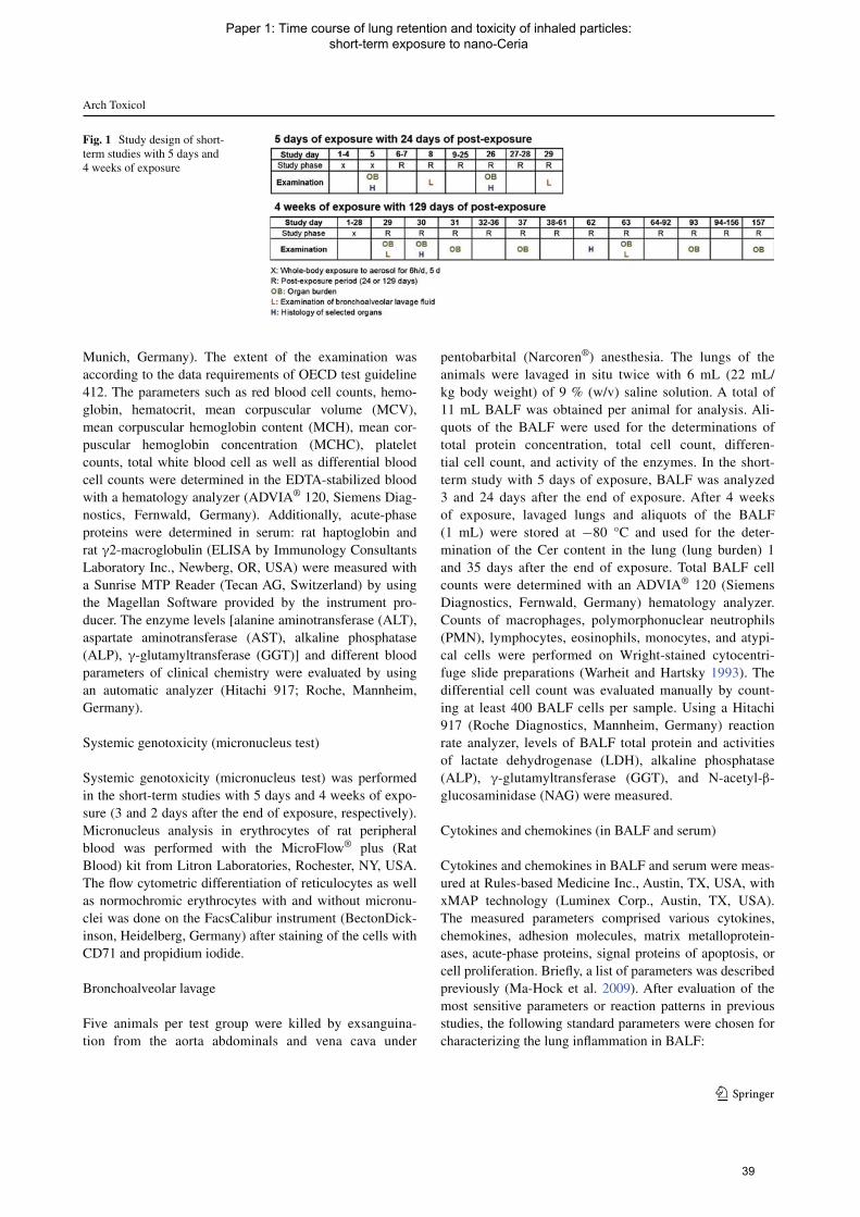
Arch Toxicol
1 3
Munich, Germany). The extent of the examination was according to the data requirements of OECD test guideline 412. The parameters such as red blood cell counts, hemo-globin, hematocrit, mean corpuscular volume (MCV), mean corpuscular hemoglobin content (MCH), mean cor-puscular hemoglobin concentration (MCHC), platelet counts, total white blood cell as well as differential blood cell counts were determined in the EDTA-stabilized blood with a hematology analyzer (ADVIA® 120, Siemens Diag-nostics, Fernwald, Germany). Additionally, acute-phase proteins were determined in serum: rat haptoglobin and rat γ2-macroglobulin (ELISA by Immunology Consultants Laboratory Inc., Newberg, OR, USA) were measured with a Sunrise MTP Reader (Tecan AG, Switzerland) by using the Magellan Software provided by the instrument pro-ducer. The enzyme levels [alanine aminotransferase (ALT), aspartate aminotransferase (AST), alkaline phosphatase (ALP), γ-glutamyltransferase (GGT)] and different blood parameters of clinical chemistry were evaluated by using an automatic analyzer (Hitachi 917; Roche, Mannheim, Germany).
Systemic genotoxicity (micronucleus test)
Systemic genotoxicity (micronucleus test) was performed in the short-term studies with 5 days and 4 weeks of expo-sure (3 and 2 days after the end of exposure, respectively). Micronucleus analysis in erythrocytes of rat peripheral blood was performed with the MicroFlow® plus (Rat Blood) kit from Litron Laboratories, Rochester, NY, USA. The flow cytometric differentiation of reticulocytes as well as normochromic erythrocytes with and without micronu-clei was done on the FacsCalibur instrument (BectonDick-inson, Heidelberg, Germany) after staining of the cells with CD71 and propidium iodide.
Bronchoalveolar lavage
Five animals per test group were killed by exsanguina-tion from the aorta abdominals and vena cava under
pentobarbital (Narcoren®) anesthesia. The lungs of the animals were lavaged in situ twice with 6 mL (22 mL/kg body weight) of 9 % (w/v) saline solution. A total of 11 mL BALF was obtained per animal for analysis. Ali-quots of the BALF were used for the determinations of total protein concentration, total cell count, differen-tial cell count, and activity of the enzymes. In the short-term study with 5 days of exposure, BALF was analyzed 3 and 24 days after the end of exposure. After 4 weeks of exposure, lavaged lungs and aliquots of the BALF (1 mL) were stored at −80 °C and used for the deter-mination of the Cer content in the lung (lung burden) 1 and 35 days after the end of exposure. Total BALF cell counts were determined with an ADVIA® 120 (Siemens Diagnostics, Fernwald, Germany) hematology analyzer. Counts of macrophages, polymorphonuclear neutrophils (PMN), lymphocytes, eosinophils, monocytes, and atypi-cal cells were performed on Wright-stained cytocentri-fuge slide preparations (Warheit and Hartsky 1993). The differential cell count was evaluated manually by count-ing at least 400 BALF cells per sample. Using a Hitachi 917 (Roche Diagnostics, Mannheim, Germany) reaction rate analyzer, levels of BALF total protein and activities of lactate dehydrogenase (LDH), alkaline phosphatase (ALP), γ-glutamyltransferase (GGT), and N-acetyl-β-glucosaminidase (NAG) were measured.
Cytokines and chemokines (in BALF and serum)
Cytokines and chemokines in BALF and serum were meas-ured at Rules-based Medicine Inc., Austin, TX, USA, with xMAP technology (Luminex Corp., Austin, TX, USA). The measured parameters comprised various cytokines, chemokines, adhesion molecules, matrix metalloprotein-ases, acute-phase proteins, signal proteins of apoptosis, or cell proliferation. Briefly, a list of parameters was described previously (Ma-Hock et al. 2009). After evaluation of the most sensitive parameters or reaction patterns in previous studies, the following standard parameters were chosen for characterizing the lung inflammation in BALF:
Fig. 1 Study design of short-term studies with 5 days and 4 weeks of exposure
39
Paper 1: Time course of lung retention and toxicity of inhaled particles: short-term exposure to nano-Ceria

Arch Toxicol
1 3
• Rat monocyte chemoattractant protein-1 level (rat MCP-1; Instant ELISA, Bender MedSystems, Vienna, Austria (cat. no BMS631INST);
• Rat cytokine-induced polymorphonuclear neutrophil chemoattractant-1 level (rat CINC-1/IL-8; ELISA, R&D Systems Inc., Minneapolis, USA (Quantikine rat CINC-1, cat. no. RCN100);
• Macrophage colony-stimulating factor (M-CSF; Quan-tikine Mouse M-CSF ELISA, R&D Systems Inc., Min-neapolis, USA (cat no. MMC00);
• Rodent osteopontin; ELISA, R&D Systems, Inc., Min-neapolis, US (Quantikine mouse osteopontin, cat. no. MOST00).
• The cell mediators were measured at a sunrise MTP reader (Tecan AG, Switzerland) by using the Magellan Software provided by the instrument producer. Four chemokines and cytokines were measured using ELISA test kits: MCP-1, IL-8/CINC-1, M-CSF, and osteopon-tin. The detailed procedure was described previously (Ma-Hock et al. 2009).
Pathology
In the short-term studies, necropsy and histopathology were performed after 5 days of exposure and 21 days after the end of exposure (5 days of exposure) and 2 and 34 days after the end of exposure (4 weeks of exposure). In general, five animals per test group were investigated for pathologi-cal examination in both short-term studies. For the short-term study with 4 weeks of exposure, however, ten animals were examined for pathological examination of the respira-tory tract and all gross lesions. At necropsy, animals were exsanguinated by opening of the abdominal great vessels under deep pentobarbital anesthesia. All organs were pre-served according to OECD TG No. 412. Following organs were weighed: adrenal glands, brain, heart, ovaries, uterus with cervix, kidney, liver, lungs, spleen, thymus, and thy-roid glands. The lungs were instilled (30 cm water column) with and fixed in 10 % neutral-buffered formalin (NBF). After 24- to 48-h fixation in 10 % NBF, the lungs were transferred to 70 % ethanol. All other organs were fixed in 10 % NBF. All the organs and tissues described in the OECD TG No. 412 were trimmed according to the RITA trimming guides for inhalation studies (Kittel et al. 2004; Ruehl-Fehlert et al. 2003). After paraplast-embedding, the blocks were cut at 2- to 3-μm thickness, mounted on glass slides and stained with hematoxylin and eosin. Extrapul-monary organs and the respiratory tract compromising nasal cavity (four levels), larynx (three levels), trachea (transverse and longitudinal with carina), lung (five lobes), and mediastinal and tracheobronchial lymph nodes were assessed by light microscopy. For the lungs, whole histo-pathological examination was performed in animals of all
test groups. For all other tissues, only the animals of the control and high concentration group of Ceria were ini-tially examined. When changes were observed in the high concentration group, respective organs and tissues of the animals exposed to low and intermediate aerosol concen-trations were also examined by light microscopy. All his-topathological examinations were performed by a well-experienced board-certified veterinarian toxicopathologist followed by an internal pathology peer review.
Organ burden analysis
Lung burden of the two different Ceria was evaluated twice, immediately after 5 days of exposure and after 21 days after the exposure end. In the short-term study with 4 weeks of exposure, Cerium content was determined at seven time points over 129 days of post-exposure period. Cerium content in the lungs, lung-associated lymph nodes, and liver of either three or five animals per test group were examined. 1 and 35 days after the end of exposure (4 weeks of exposure), the lavaged lungs and aliquots of BALF of five animals per group were used for the determination of lung burden. This examination method has likely caused a loss of the test material during preparation and handling of the lungs. Furthermore, lung burdens were measured 2 days after the end of exposure using the left half lungs of five animals/test group, only. On the basis of the availability of total lung weights, lung burdens were calculated up from the half lung burden values with the corresponding weight of the half lungs. Lung burden of the remaining time points was determined using the whole (not lavaged) lung. After digestion with mixed acid, samples of each lung or lymph node were dissolved in sulfuric acid and ammonium sul-fate. 140Cer content in the obtained solution was analyzed by inductively coupled plasma mass spectrometry (ICP-MS, Agilent 7500 C) or by inductively coupled plasma optical emission spectrometry (ICP-OES, Varian 720-ES) with a wavelength of 419 nm. With this method, limit of detection for Cer is 0.3 μg. The amounts of Ceria in the respective tissues were calculated by measuring elemental Cerium with ICP-MS.
Statistical analysis
For body weight changes, Dunnett’s test was used for a comparison of each test group with the control group test (Dunnett 1955; Dudewicz et al. 1975). Clinical pathology parameters (BALF cytology, enzyme data, and BALF and serum cell mediator data) were analyzed by nonparamet-ric one-way analysis using the Kruskal–Wallis test (two-sided). If the resulting p value was ≤0.05, a pair-wise comparison of each test group with the control group was performed using the Wilcoxon test or the Mann–Whitney U
40
Paper 1: Time course of lung retention and toxicity of inhaled particles: short-term exposure to nano-Ceria
Arch Toxicol
1 3
test (both two-sided) (p ≤ 0.05 for statistical significance). Comparison of organ weights among test groups was per-formed by nonparametric one-way analysis using the two-sided Kruskal–Wallis test, followed by a two-sided Wil-coxon test for the hypothesis of equal medians in case of p ≤ 0.05.
Results
Characterization of test material
Both Ceria were yellowish white powders that were pro-duced by precipitation and were nominally uncoated. We completely re-characterized the Ceria NM-211 and NM-212 based on the nano-specific guidance on physical–chemical properties (Wohlleben et al. 2013). Table 1 indi-cates the techniques chosen for each end point (see “Mate-rials and methods” section) and summarizes the results. The Ceria NM-212 material had an average primary parti-cle diameter of 40 nm, in excellent accordance of electron microscopy with the crystallite size derived from the dif-fraction peak width and with the sphere-equivalent diam-eter derived from the BET-specific surface area of 27 m2/g (see Fig. 2). The Ceria NM-211 material consisted of con-siderably smaller primary particles with number-based median diameter of 8.2 nm (from TEM) and correspond-ingly larger specific surface (53 m2/g, from BET). Primary particles of both materials were crystalline with cubic lat-tice as it is the characteristic for cerianite and had irregular, but roughly globular shapes. In the as-produced powder, porosimetry by Hg intrusion indicated that these particles were aggregated and agglomerated to sizes that range from a few hundred nm to tens of μm.
The surface of Ceria NM-212 beared organic contami-nations. An assessment by thermogravimetry (TGA) con-firmed that the total organic content remained below 0.7 %, but the photoelectron signal of XPS, which had an infor-mation depth between 3 and 10 nm, indicated 80 % carbon atoms on the surface. The contamination was hence a very thin and homogeneous layer around the purely inorganic particles. According to the fit of photoelectron energies from C(1s) level and from O(1s) level, the contamination could be an ester with a long alkyl chain. In contrast, the Ceria NM-211 had only 14 % carbon atoms on the surface (by XPS), and the even more sensitive SIMS techniques confirmed that Ceria dominated the surface, with vanish-ing amounts of nitrates and alkyls. The Cerium atoms at crystalline edges were known to be redox-active, which was confirmed by the detection of 14 % of the Cerium as Ce(III) (within the XPS-accessible surface layer) in Ceria NM-212, respectively, 22 % for the Ceria NM-211 with its decreased radius of curvature.
If dispersed in clean water, the Ceria NM-212 surface was positively charged across the entire physiological pH range, with a zeta-potential of +42 mV at pH 7. The sur-face of Ceria NM-212 was rather reactive in a photocata-lytic assay with a photon efficiency of 1.3 × 10⁻², which was 5 times higher than the well-known Titania P25 (NM-105) and an order of magnitude higher than for the Ceria NM-211, leaving the possibility of the organic contamina-tions being responsible for that catalytic activity, observed only under UV irradiation (Wohlleben et al. 2013; Molina et al. 2014). After dispersion in water by stirring only, the dispersability was limited with average agglomeration numbers (AAN = the ratio of agglomerate diameter to pri-mary particle diameter) above 10, based on the measure-ment of the resulting agglomerates that were just below the micron range (by analytical ultracentrifugation), and tended to be larger for the Ceria NM-211 than for NM-212, con-firming earlier findings of the PROSPECT consortium and the European Commission’s JRC report (Hermans 1963; PROSPEcT: Ecotoxicology Test Protocols for Representa-tive Nanomaterials in Support of the OECDSponsorship Programme 10 A.D.; European Commission 2014).
The solubility in physiological media was investigated by 28-day abiotic incubation in different fluids and detec-tion of both the remaining particulate fraction (by SEM, centrifugation, LD, zeta-potential) and of the released metal ions (by ICP-MS). Both Ceria were insoluble except for marginal solubility in 0.1 N HCl (simulate oral inges-tion), whereas a nanoscale precipitated Silica as positive control dissolved readily and Titania (NM-105) showed marginal solubility in phagolysosomal simulant fluid (PSF, simulate uptake and digestion in macrophages) (Wohlle-ben et al. 2013). In buffers with organic constituents (PSF, fasted state simulated intestinal fluid = FaSSIF), the sur-face charge reversed to negative zeta-potentials, which was identified earlier as indicator for the adsorption of constituents of the buffer for a range of the nano-Ceria (Wohlleben et al. 2013). Under all conditions, primary par-ticles remained recognizable in TEM scans, but both Ceria NM-211 and NM-212 formed large structures after 28-day incubation in the acidic PSF medium that simulates the lys-osome of macrophages (see Fig. 3). We found by selected area electron diffraction (SAD) that these aged structures retained the same cubic cerianite crystalline phase of the as-produced powder for both Ceria NM-211 and NM-212. Under identical conditions, also other nanomaterials, e.g., the Titania (NM-105), aged into similar structures, and we added the respective TEM and SAD for comparison (Wohlleben et al. 2013).
To summarize the comparative physical–chemical characterization, Ceria NM-211 and NM-212 had identi-cal composition, agglomeration state, crystallinity, and insolubility. Compared to NM-212, the Ceria NM-211
41
Paper 1: Time course of lung retention and toxicity of inhaled particles: short-term exposure to nano-Ceria

Arch Toxicol
1 3
Tabl
e 1
Phy
sica
l–ch
emic
al c
hara
cter
izat
ion
of C
eria
NM
-212
and
NM
-211
OE
CD
end
poi
nts
Cer
ia N
M-2
12C
eria
NM
-211
Part
icle
siz
e di
stri
butio
n (T
EM
: pri
mar
y pa
rtic
le d
iam
eter
)/
stat
e of
agg
lom
erat
ion
(SE
M: a
gglo
mer
ate
diam
eter
)40
nm
SEM
: 3,0
00–1
50,0
00 n
mH
g po
re s
izes
: 35
nm, a
roun
d 7
μm
4–15
nm
D50
: 8.2
nm
Hg
pore
siz
es: 5
, 70
nm, a
roun
d 10
μm
Cry
stal
lite
size
(X
RD
)40
.0 n
m12
.5 n
m
Cry
stal
lite
phas
e (X
RD
)C
eria
nite
, Cer
ia-c
ubic
Cer
iani
te, C
eria
-cub
ic
Spec
ific
surf
ace
area
(H
g, B
ET
)30
m2 /g
(H
g), 2
7 m
2 /g (
BE
T)
33 m
2 /g (
Hg)
, 53
m2 /g
(B
ET
)
Surf
ace
chem
istr
y (X
PS)
Ato
m p
erce
ntC
79.
9(C
–C 6
2.6;
C–O
7.0
; C=O
3.5
; CO
OH
6.9
)O
17.
7C
e 2.
4W
ith o
xida
tion
stat
eC
e(II
I) 1
4 %
, Ce(
IV)
86 %
Ce
28.7
O 5
7.2
C 1
4.1
With
oxi
datio
n st
ate
Ce(
III)
22
%, C
e(IV
) 78
%
Surf
ace
char
geIs
oele
ctri
c po
int =
IE
Pζ-
Pot a
t pH
7 f
rom
ele
ctro
phor
etic
mob
ility
IEP:
>pH
10
(alw
ays
catio
nic)
+42
mV
IEP
= pH
8.3
+16
mV
Phot
ocat
alyt
ic a
ctiv
ity
Pho
ton
effic
ienc
y (m
ethy
lene
blu
e)0.
01 ±
0.0
050.
0005
± 0
.000
2
Dis
pers
abili
ty
D50
and
ave
rage
agg
lom
. num
ber
(cen
trif
ugat
ion)
D50
= 4
32 n
m/A
AN
= 1
1(in
wat
er)
D50
= 2
,839
nm
/AA
N =
346
(in
wat
er)
Solu
bilit
y (I
CP-
MS)
In
wat
er0.
002
wt%
<0.
001
wt%
In
DM
EM
/FC
S<
0.00
1 w
t%<
0.00
1 w
t% (
recr
ysta
llize
s)
In
PSF
<0.
001
wt%
(re
crys
talli
zes)
<0.
001
wt%
In
PBS
<0.
001
wt%
In
Fass
iF<
0.00
1 w
t%
In
0.1
N H
Cl
0.02
wt%
(ri
peni
ng)
Impu
ritie
s (T
GA
, XPS
)To
tal c
onte
nt o
f 0.
7 %
org
anic
con
tam
inat
ions
, ide
ntifi
ed a
s es
ter +
alky
l gro
ups,
fou
nd m
ostly
on
the
part
icle
sur
face
Tota
l con
tent
of
1.6
% c
onta
min
atio
ns, t
here
of s
mal
l am
ount
s of
al
kyls
fou
nd o
n th
e pa
rtic
le s
urfa
ce
42
Paper 1: Time course of lung retention and toxicity of inhaled particles: short-term exposure to nano-Ceria

Arch Toxicol
1 3
had considerably smaller primary particles, larger specific surface, significantly fewer organic contaminations on the surface, and much reduced photocatalytic activity. Despite these differences, Ceria NM-211 and NM-212 shared the same tendency to recrystallize in PSF.
Characterization of test atmosphere
The target concentrations were 0.5, 5, and 25 mg/m3 in the short-term studies (5 days and 4 weeks of exposure) with nano-Ceria NM-212. In the study with 5 days of exposure,
Fig. 2 Structure of Ceria. Electron microscopy images of a representative ensemble of particles of Ceria NM-211 (a) and NM-212 (b) (see Table 1 for size characterization by complementary methods)
Fig. 3 Persistence of Ceria NM-211 (a), NM-212 (b), and Titania NM-105 (c) as benchmark. Transmission electron microscopy (TEM) and selected area diffraction (SAD) after 28-day incubation in phago-lysosomal simulant fluid (PSF) [see Table 1 for dissolution detected
by ions in the supernatant (ICP-MS)]. The large almost spherical structures are no contaminations, but are confirmed as Ceria in the same crystalline phase
43
Paper 1: Time course of lung retention and toxicity of inhaled particles: short-term exposure to nano-Ceria

Arch Toxicol
1 3
0.5 and 25 mg/m3 were selected for nano-Ceria NM-211. Analyzed concentrations and particle size distributions are summarized in Table 2. The target concentrations were met and maintained well throughout the studies. Particle size distribution demonstrated that Ceria particles were in the respirable range for rats.
Clinical examination
Five days and 4 weeks of inhalation exposure to Ceria NM-212 and NM-211 did not affect the body weight devel-opment of the animals (data not shown). The animals exposed to Ceria NM-211 and NM-212 showed no clinical signs or findings compared to the control animals (data not shown).
Hematology, clinical chemistry, cytokines, and chemokines in serum
Five days of exposure
Absolute and relative neutrophil counts in blood were increased 3 days after the end of exposure to 25 mg/m3 Ceria NM-212 and NM-211, whereas relative lymphocyte counts were decreased (see Table S2). The changes were in a concentration-related manner. 24 days after the end of exposure, all parameters had returned to near control val-ues. No other blood parameters were affected.
Four weeks of exposure
Hematological, clinical chemistry parameters, and acute-phase protein levels were not affected in rats exposed to Ceria NM-212 (see Table S2; the other data are not shown).
Systemic genotoxicity (micronucleus test)
Micronuclei in the erythrocytes of peripheral blood cells were counted to assess chromosomal aberrations of
hematopoietic cells in the bone marrow. Peripheral blood cells were examined in the short-term studies with 5 days and 4 weeks of exposure (3 and 2 days after the end of exposure, respectively). All examined parameters were in the range of control values (see Table S3).
Bronchoalveolar lavage (BAL)
In the short-term studies with 5 days and 4 weeks of expo-sure, BALF analysis of five animals per test group was obtained 3 days or 1 day after the end of exposure and 24 or 35 days after the end of exposure, respectively (see Fig. 1). The resulting BALF parameters are presented in Table 3.
Five days of exposure
In animals exposed to Ceria NM-212, the majority of BALF parameters were increased at aerosol concentra-tions of 5 mg/m3. At 0.5 mg/m3, the neutrophil counts and cytokine-induced neutrophil chemoattractant-1 (CINC-1) were both statistically increased and were slightly above the historical control range (see Table S4). With Ceria NM-211, but not NM-212, monocyte chemoattractant pro-tein-1 (MCP-1) and macrophage colony-stimulating fac-tor (M-CSF) were increased at aerosol concentrations of 0.5 mg/m3 and above. 24 days after the end of exposure, a full recovery was observed at aerosol concentrations of 0.5 mg/m3 and a partial recovery at aerosol concentrations of 5 and 25 mg/m3. The recovery of animals exposed to 25 mg/m3 Ceria NM-211 seems to be slower than those exposed to NM-212.
4 weeks of exposure
Four weeks of inhalation exposure to 5 and 25 mg/m3 Ceria NM-212 resulted in an increase in total cells in BALF due to increases in polymorph nuclear neutrophils, lympho-cytes, and monocytes in BALF (see Table 3). Consistent
Table 2 Measured concentrations and particle size distributions of Ceria
Study Test substance
Targeted concentrations (mg/m3)
Measured concentrations mean ± SD (mg/m3)
MMAD (μm)/ GSD mean
Particle count concentration measured by SMPS (particle/cm3)
Particle count median (nm)
5 days of exposure
Ceria NM-212 0.5 0.5 ± 0.2 1.4/2.3 2,583 283
5 5.3 ± 0.9 1.2/2.1 137,096 206
25 25.9 ± 6.0 1.0/2.5 537,548 171
Ceria NM-211 0.5 0.48 ± 0.0 1.6/2.1 5,689 256
25 5.2 ± 1.1 1.3/2.1 84,655 167
4 weeks of exposure
Ceria NM-212 0.5 25.6 ± 6.0 0.9/2.5 514,193 203
5 0.45 ± 0.1 1.9/2.9 1,503 242
25 25.8 ± 1.7 2.2/2.4 160,162 188
44
Paper 1: Time course of lung retention and toxicity of inhaled particles: short-term exposure to nano-Ceria

Arch Toxicol
1 3
Tabl
e 3
Clin
ical
pat
holo
gy p
aram
eter
s in
BA
LF
of s
hort
-ter
m s
tudi
es w
ith 5
day
s an
d 4
wee
ks o
f ex
posu
re
Targ
et c
onc.
(m
g/m
3 )5
days
of
expo
sure
4 w
eeks
of
expo
sure
Con
trol
Cer
ia N
M-2
12C
eria
NM
-211
Con
trol
Cer
ia N
M-2
12
00.
55
250.
525
00.
55
25
Mea
sure
d co
nc.
(mg/
m3 )
+ SD
00.
48 ±
0.0
5.2
± 1.
125
.6 ±
6.0
0.45
± 0
.125
.8 ±
1.7
00.
5 ±
0.2
5.3
± 0.
925
.9 ±
6.0
BA
LF
cell
coun
ts (
cn/μ
L)
Tot
al c
ells
Tim
e po
int 1
a63
.73
± 11
.03
69.7
6 ±
26.1
598
.24*
* ±
16.3
833
7.35
** ±
125
.24
71.3
2 ±
22.6
937
4.98
** ±
77.
8476
.42
± 23
.97
75.1
0 ±
21.5
113
3.44
* ±
48.4
029
6.90
** ±
124
.92
Tim
e po
int 2
b53
.09
± 13
.33
44.2
0 ±
2.45
43.9
8 ±
17.7
046
.71
± 8.
1647
.01
± 11
.34
81.2
0 ±
38.8
775
.29
± 14
.10
62.2
3 ±
11.5
997
.44
± 34
.23
220.
50**
± 1
05.2
7
Neu
trop
hils
(PM
N)
Tim
e po
int 1
a0.
82 ±
0.8
03.
70**
± 2
.53
49.1
9**
± 17
.68
272.
30**
± 1
06.6
24.
41*
± 3.
3829
7.21
** ±
59.
230.
85 ±
0.3
51.
75 ±
1.1
365
.66*
* ±
50.2
322
2.29
** ±
99.
25
Tim
e po
int 2
b0.
69 ±
0.3
81.
34 ±
2.0
15.
15 ±
9.9
14.
63**
± 3
.83
2.27
** ±
0.5
625
.57*
* ±
33.9
52.
44 ±
1.0
12.
52 ±
1.3
741
.70*
* ±
22.0
161.
69**
± 8
7.82
Lym
phoc
ytes
Tim
e po
int 1
a0.
22 ±
0.3
20.
17 ±
0.2
21.
57*
± 0.
979.
20**
± 4
.51
0.39
± 0
.34
19.1
4**
± 15
.13
0.55
± 0
.50
0.63
± 0
.74
5.41
** ±
3.2
18.
93**
± 6
.04
Tim
e po
int 2
b0.
32 ±
0.3
30.
11 ±
0.1
00.
43 ±
0.3
11.
03 ±
0.9
50.
51 ±
0.2
52.
74**
± 2
.17
1.65
± 1
.13
0.77
± 0
.49
7.43
* ±
9.02
10.8
9**
± 3.
48
Mac
roph
ages
Tim
e po
int 1
a62
.65
± 11
.56
75.7
2 ±
15.1
046
.83
± 23
.88
51.2
1 ±
24.7
666
.47
± 22
.13
52.9
9 ±
42.8
974
.94
± 23
.79
72.7
1 ±
21.1
660
.25
± 20
.82
59.3
2 ±
26.2
8
Tim
e po
int 2
b52
.05
± 13
.00
42.3
2 ±
3.66
38.3
4 ±
11.9
240
.93
± 6.
3144
.23
± 11
.53
52.6
1 ±
15.5
171
.14
± 13
.31
58.7
8 ±
11.0
447
.58
± 16
.48
45.1
7 ±
25.3
3
Mon
ocyt
es
Tim
e po
int 1
a0.
04 ±
0.0
80.
00 ±
0.0
00.
52*
± 0.
373.
50 ±
1.8
6**
0.05
± 0
.11
4.40
** ±
2.6
80.
00 ±
0.0
00.
00 ±
0.0
01.
65*
± 1.
903.
95*
± 4.
65
Tim
e po
int 2
b0.
00 ±
0.0
00.
00 ±
0.0
00.
03 ±
0.0
70.
07 ±
0.1
10.
00 ±
0.0
00.
28 ±
0.3
40.
06 ±
0.0
90.
04 ±
0.0
80.
68*
± 0.
432.
46*
± 3.
03
Eos
inop
hils
Tim
e po
int 1
a0.
00 ±
0.0
00.
00 ±
0.0
00.
00 ±
0.0
00.
39 ±
0.5
40.
00 ±
0.0
00.
00 ±
0.0
00.
09 ±
0.1
20.
00 ±
0.0
00.
00 ±
0.0
00.
34 ±
0.4
9
Tim
e po
int 2
b0.
03 ±
0.0
60.
00 ±
0.0
00.
03 ±
0.0
60.
00 ±
0.0
00.
00 ±
0.0
00.
00 ±
0.0
00.
00 ±
0.0
00.
12 ±
0.1
10.
05 ±
0.1
10.
00 ±
0.0
0
Aty
pica
l cel
ls
Tim
e po
int 1
a0.
00 ±
0.0
00.
00 ±
0.0
00.
12 ±
0.1
70.
76 ±
0.7
30.
00 ±
0.0
01.
25*
± 0.
850.
00 ±
0.0
00.
00 ±
0.0
00.
48 ±
0.7
22.
07*
± 1.
71
Tim
e po
int 2
b0.
00 ±
0.0
00.
00 ±
0.0
00.
00 ±
0.0
00.
05 ±
0.0
70.
00 ±
0.0
00.
00 ±
0.0
00.
00 ±
0.0
00.
00 ±
0.0
00.
00 ±
0.0
00.
29 ±
0.4
0
Tota
l pro
tein
/enz
ymes
Tot
al p
rote
in (
mg/
L)
Tim
e po
int 1
a39
± 1
547
± 1
675
* ±
2319
8**
± 52
44 ±
720
7**
± 54
60 ±
483
** ±
594
** ±
21
245*
* ±
77
Tim
e po
int 2
b97
± 1
2259
± 2
851
± 1
660
* ±
992
± 9
182
± 1
481
± 2
360
± 2
298
± 4
917
5**
± 98
GG
T (
nkat
/L)
Tim
e po
int 1
a20
± 1
536
± 2
010
6**
± 26
125*
* ±
1537
± 2
612
8**
± 16
37 ±
17
51 ±
11
111*
* ±
2114
9**
± 31
Tim
e po
int 2
b30
± 7
28 ±
17
33 ±
14
61**
± 1
625
± 1
087
** ±
20
42 ±
12
44 ±
20
83**
± 3
012
3**
± 30
LD
H (
μkat
/L)
Tim
e po
int 1
a0.
46 ±
0.0
90.
58 ±
0.1
70.
89**
± 0
.25
2.17
** ±
0.2
10.
672.
60**
± 0
.31
0.51
± 0
.18
0.55
± 0
.15
1.08
** ±
0.3
72.
28**
± 0
.52
Tim
e po
int 2
b0.
36 ±
0.1
40.
47 ±
0.2
50.
42 ±
0.0
40.
72**
± 0
.20
0.58
± 0
.40
1.08
** ±
0.3
20.
58 ±
0.0
80.
50 ±
0.2
00.
84*
± 0.
221.
88**
± 1
.20
45
Paper 1: Time course of lung retention and toxicity of inhaled particles: short-term exposure to nano-Ceria

Arch Toxicol
1 3
* St
atis
tical
ly s
igni
fican
t, p
< 0
.05
** S
tatis
tical
ly s
igni
fican
t, p
< 0
.01;
n =
5; S
D s
tand
ard
devi
atio
na T
ime
poin
t 1: 3
day
s af
ter
the
end
of e
xpos
ure
(5 d
ays
of e
xpos
ure)
and
1 d
ay a
fter
the
end
of e
xpos
ure
(4 w
eeks
of
expo
sure
)b T
ime
poin
t 2: 2
4 da
ys a
fter
the
end
of e
xpos
ure
(5 d
ays
of e
xpos
ure)
and
35
days
aft
er th
e en
d of
exp
osur
e (4
wee
ks o
f ex
posu
re)
Tabl
e 3
con
tinue
d
Targ
et c
onc.
(m
g/m
3 )5
days
of
expo
sure
4 w
eeks
of
expo
sure
Con
trol
Cer
ia N
M-2
12C
eria
NM
-211
Con
trol
Cer
ia N
M-2
12
00.
55
250.
525
00.
55
25
AL
P (μ
kat/L
)
Tim
e po
int 1
a0.
43 ±
0.1
00.
53 ±
0.0
81.
31**
± 0
.53
1.56
** ±
0.1
50.
55*
± 0.
091.
56**
± 0
.35
0.83
± 0
.16
0.84
± 0
.35
1.16
** ±
0.1
01.
53**
± 0
.21
Tim
e po
int 2
b1.
01.
5*1.
9**
0.51
± 0
.20
0.87
** ±
0.1
20.
70 ±
0.0
90.
67 ±
0.0
81.
05**
± 0
.16
1.09
** ±
0.2
5
NA
G (
nkat
/L)
Tim
e po
int 1
a46
± 9
47 ±
11
57 ±
12
74**
± 7
51 ±
19
94**
± 1
645
± 5
55 ±
15
53*
± 8
86*
± 26
Tim
e po
int 2
b41
± 1
037
± 1
239
± 4
46 ±
746
± 7
59*
± 10
47 ±
838
± 6
47 ±
771
± 3
5
Cel
l med
iato
rs (
pg/m
L)
MC
P-1
Tim
e po
int 1
a14
.7 ±
0.6
19.1
± 5
.510
1.1*
* ±
31.3
1,34
2.9*
* ±
530.
017
.5*
± 1.
61,
581.
0**
± 77
1.3
14.0
± 0
.019
.6 ±
11.
055
9.4*
* ±
444.
43,
587.
2**
± 28
1.0
Tim
e po
int 2
b48
.2 ±
59.
122
.8 ±
12.
526
.7 ±
10.
814
4.1*
± 8
2.8
30.6
± 2
6.0
329.
2**
± 29
0.5
17.3
± 2
.615
.4 ±
3.1
492.
5**
± 55
3.1
1,85
4.2*
* ±
1,18
4.0
CIN
C-1
/IL
-8
Tim
e po
int 1
a59
.8 ±
17.
783
.4*
± 24
.634
8.6*
* ±
185.
432
2.4*
* ±
93.9
88.3
* ±
21.1
436.
0**
± 22
8.3
104.
2 ±
26.7
103.
8 ±
14.0
506.
7**
± 19
5.9
1,19
0.9*
* ±
294.
9
Tim
e po
int 2
b88
.3 ±
25.
787
.5 ±
24.
794
.3 ±
35.
715
5.1*
* ±
13.5
99.7
± 3
9.4
254.
3**
± 70
.415
8.8
± 38
.113
3.9
± 45
.344
9.4*
* ±
226.
783
1.0*
* ±
497.
1
M-C
SF
Tim
e po
int 1
a41
± 1
752
± 2
334
± 2
591
** ±
28
61*
± 16
114*
* ±
4826
± 1
722
± 1
227
± 1
848
± 2
9
Tim
e po
int 2
b45
± 1
948
± 5
71*
± 13
49 ±
351
± 1
260
± 2
446
± 2
655
± 2
941
± 1
453
± 1
2
Ost
eopo
ntin
Tim
e po
int 1
a17
2.28
± 4
8.04
194.
81 ±
131
.24
301.
40 ±
238
.96
849.
04**
± 3
86.4
413
4.32
± 8
2.72
1,11
6.02
** ±
653
.35
391.
44 ±
187
.39
288.
80 ±
110
.90
755.
44*
± 20
6.21
592.
14 ±
336
.47
Tim
e po
int 2
b32
0.38
± 1
77.6
419
1.47
± 1
16.0
924
7.44
± 1
89.4
649
5.92
± 2
51.9
311
1.92
± 8
0.28
398.
72 ±
140
.88
337.
36 ±
282
.91
284.
40 ±
292
.66
1,00
3.18
* ±
434.
2083
8.48
± 5
29.4
5
46
Paper 1: Time course of lung retention and toxicity of inhaled particles: short-term exposure to nano-Ceria

Arch Toxicol
1 3
with these findings, several other parameters including the examined cell mediators were increased. 35 days after the end of exposure, some of the BALF parameters returned to control levels, whereas several of them were still signifi-cantly increased at 5 and 25 mg/m3 (e.g., total cells, lym-phocytes, neutrophils; GGT, LDH, ALP; MCP-1, CINC-1).
Five days of exposure caused slightly higher neutrophil and lymphocyte counts at aerosol concentrations of 25 mg/m3 Ceria NM-212 compared to 4 weeks of exposure (see Fig. 4). CINC-1 was already increased at 0.5 mg/m3 after 5 days but not after 4 weeks of exposure. The regression of the BALF parameters was faster after 5 days than after 4 weeks of inhalation exposure. Cell mediators, especially MCP-1, were higher elevated at concentrations of 5 and 25 mg/m3 after 4 weeks than after 5 days of exposure.
Pathology
Organ weights and macroscopic findings
Five days of exposure After 5 days of exposure, no increase in lung weights was observed after exposure to Ceria NM-212 (see Table S5). An aerosol concentration of 25 mg/m3 Ceria NM-211, however, resulted in significant increases in absolute and relative lung weights (+20 and 24 %, respectively, p < 0.01). The increase in lung weight was no longer present 21 days after the end of exposure. After 5 days of exposure, the macroscopic finding enlarged mediastinal lymph nodes were found in individual animals of all test groups (see Table S5). 21 days after the end of the exposure, enlarged mediastinal lymph nodes were observed in almost all animals exposed to Ceria NM-211 or NM-212. The tracheobronchial lymph nodes and all other examined organs did not show any macroscopic findings.
Four weeks of exposure Absolute and relative lung weights were significantly increased at aerosol concentra-tions of 25 mg/m3 Ceria NM-212 (+30 and 29 %, respec-tively) 2 days after the end of the exposure and were still significantly elevated (+16 and 20 %) 34 days after the end of the exposure (see SI, S5). 2 days after the end of the exposure, absolute and relative lung weights of ani-mals exposed to 5 mg/m3 were increased significantly by +13 and 10 %, respectively. They returned to control levels within the following 34 days. No effects on organ weights were observed after inhalation of 0.5 mg/m3. 2 days after the end of exposure, mediastinal lymph nodes of two ani-mals (out of ten) were enlarged at aerosol concentrations of 25 mg/m3 Ceria NM-212. 34 days after the end of exposure, the incidence of animals with enlarged, yellow white-colored mediastinal lymph nodes increased consid-erably from two to eight (out of ten) per group at aerosol concentrations of 25 mg/m3 Ceria NM-212, respectively.
Mediastinal lymph nodes of animals exposed to 5 mg/m3 were firstly enlarged 34 days after the end of exposure.
All other extrapulmonary organs including the tracheo-bronchial lymph nodes revealed no macroscopical findings after inhalation exposure to Ceria.
Histopathology
Overall, histopathological evaluations were consistent in the short-term studies with 5 days and 4 weeks of exposure. The findings assessed in the short-term study with 5 days showed lower severities (see Table 4; Fig. 5a–c).
Lungs
Five days of exposure
After 5 days of exposure, single or accumulated mac-rophages were located in the lumen of the alveoli and a few macrophages occurred also in the alveolar wall and ducts. They were distributed multifocally in all lobes over the whole lung. Most of them were loaded with amber-like colored particles of different sizes (below 1 μm diameter). These particles were considered to represent agglomer-ated or aggregated Ceria particles (“alveolar histiocytosis with particles”). Alveolar histiocytosis and free eosino-philic granular material with particles, interpreted as rem-nants of destroyed macrophages, were found in the lung of almost all animals exposed to 25 mg/m3 Ceria NM-211 and NM-212. At aerosol concentrations of 0.5 mg/m3 Ceria NM-211, 0.5 and 5 mg/m3 Ceria NM-212, amber-colored particles were noted within single histiocyte in the alveoli. In the bronchus-associated lymphoid tissue (BALT), parti-cles were detected free or in single macrophages, mostly at 5 mg/m3 Ceria and above. Findings regressed but were still present 21 days after the end of exposure. The find-ing “eosinophilic granular material with particles” was no longer visible after the post-exposure period. Particles were detected free or in single macrophages in BALT, even at the lowest concentration of 0.5 mg/m3 Ceria NM-211. Only one animal exposed to 25 mg/m3 Ceria NM-211 showed macrophage aggregates in BALT 21 days after the end of exposure.
Four weeks of exposure
Two days after the end of the exposure, a concentration-related increase in alveolar macrophages was observed in the lungs in all Ceria NM-212-exposed animals (Table 4; Fig. 5a). Eosinophilic granular material and small parti-cles were distributed in the alveoli of animals exposed to 5 and 25 mg/m3 Ceria NM-212. The occurrence of alveo-lar histiocytosis and of eosinophilic granular material was
47
Paper 1: Time course of lung retention and toxicity of inhaled particles: short-term exposure to nano-Ceria

Arch Toxicol
1 3
correlated with increased lung weights in animals exposed to 5 and 25 mg/m3 Ceria NM-212. In BALT, single, small macrophage aggregates with particles occurred in ani-mals exposed to 25 mg/m3 (see Fig. 5b). In addition, sin-gle or a few amber-like colored particles occurred extra-cellularly in BALT without any macrophage activation at aerosol concentrations of 5 and 25 mg/m3. 34 days after the end of exposure, alveolar histiocytosis and eosino-philic granular material with particles were still observed
at concentrations of 5 and 25 mg/m3 Ceria NM-212. At 0.5 mg/m3, in contrast, amber-like colored particles could only be noted within single histiocyte. In one animal of 5 mg/m3 and in five out of ten animals exposed to 25 mg/m3 Ceria NM-212, a multifocal granulomatous inflamma-tion appeared first. In BALT, single or few amber-colored particles at 0.5, 5, and 25 mg/m3 Ceria NM-212 as well as an increasing number of animals with macrophage aggre-gates with particles at 5 and 25 mg/m3 were still observed
Fig. 4 Comparison of changes in BALF parameters after 5 days (a, b) and 4 weeks (c, d) of exposure to Ceria NM-211 and NM-212: short-term study with 5 days of exposure: a 3 days after the end of exposure, b 24 days after the end of exposure; short-term study with
4 weeks of exposure, c 1 day after the end of exposure, d 35 days after the end of exposure. Changes are shown as x-fold differences compared to controls using a logarithmic scaling
48
Paper 1: Time course of lung retention and toxicity of inhaled particles: short-term exposure to nano-Ceria

Arch Toxicol
1 3
Table 4 Histopathology of short-term studies with 5 days and 4 weeks of exposure
Target conc. (mg/m3) 5 days of exposure to Ceria NM-212 5 days of exposure to Ceria NM-211
4 weeks of exposure to Ceria NM-212
Time point 1a Time point 2b Time point 1a
Time point 2b
Time point 1a Time point 2b
0.5 5 25 0.5 5 25 0.5 25 0.5 25 0.5 5 25 0.5 5 25
Histopathology
Lungs
Alveolar histiocytosis with particles – – 5 – 3 5 – 5 – 4 10 10 10 – 10 10
Grade 1 1 3 5 3 4 10 4 3
Grade 2 4 2 6 5 7
Grade 3 5 10
Eosinophilic granular material/particles – – 5 – – – – 5 – – – 10 10 – 2 10
Grade 1 2 5 10 2 5
Grade 2 3 6 4
Grade 3 4 1
BALT: macrophage aggregates particles – – – – – – – – – 1 – – 5 – 1 8
Grade 1 1 5 3
Grade 2 1 2
Grade 3 3
BALT: occurrence of particles – 2 5 – 3 5 – 5 2 4 – 8 5 3 8 2
Inflammation, granulomatous – – – – – – – – – – – – – – 1 5
Grade 1 1
Grade 2 5
Particles within histiocytes 5 5 – 5 2 – 5 – 5 – – – – 10 – –
Mediastinal lymph nodes
Macrophage aggregates particle. – – – – – 4 – – – 2 – – 4 – 9 9
Grade 1 2 1 2
Grade 2 1 2
Grade 3 1 2 6 2
Grade 4 1 1 7
Occurrence of particles – – 2 – 1 1 – 3 – 2 – 6 6 1 – 1
Hyperplasia, lympho-reticulocellular 1 – 3 – – 3 3 3 – 1 1 1 4 – 2 7
Grade 1 1 3 2 3 1 1
Grade 2 1 1 1 1 1 3 2 6
Grade 3 1 1
Tracheobronchial lymph nodes
Macrophage aggregates particle. – – – – – 4 – – – 3 – 1 8 – 10 10
Grade 1 1 1 3 1 1
Grade 2 2 1 5 1 1
Grade 3 2 5 2
Grade 4 1 3 6
Occurrence of particles – – 5 – 3 1 – 4 – 2 – 7 2 1 – –
49
Paper 1: Time course of lung retention and toxicity of inhaled particles: short-term exposure to nano-Ceria

Arch Toxicol
1 3
(see Fig. 5b). All compound-related findings after exposure of 5 and 25 mg/m3 Ceria NM-212 were correlated with increased lung weights in these test groups.
Lung-associated lymph nodes (mediastinal and tracheobronchial lymph nodes)
Five days of exposure
After 5 days of exposure to 25 mg/m3 Ceria NM-211 and NM-212, comparable to the finding of particles in BALT of the lung, amber-like colored particles were seen partly within macrophages or extracellularly in the lymphoid tissue, without any macrophage activation or aggrega-tion. 21 days after the end of exposure to 25 mg/m3 Ceria NM-211, findings in both lymph nodes progressed. In the mediastinal and the tracheobronchial lymph nodes, mul-tifocal macrophage aggregates with amber-like colored particles were noted in animals exposed to Ceria NM-211 and NM-212 (see Table 4). In both lymph nodes, lympho-reticulocellular hyperplasia was observed after 5 days of exposure and 21 days after the end of exposure to Ceria NM-211 and NM-212.
Four weeks of exposure
Two days after the end of exposure to 5 and 25 mg/m3 Ceria NM-212, multifocal macrophage aggregates with particles were observed in the mediastinal as well as in the tracheo-bronchial lymph nodes (see Fig. 5c). A lympho-reticulocel-lular hyperplasia was present in both lymph nodes, mostly seen in animals in the group of 25 mg/m3. The hyperplasia of the mediastinal lymph nodes was correlated with their corresponding macroscopic enlargement after exposure to
25 mg/m3 Ceria NM-212. 34 days after the end of expo-sure, the number of animals with macrophage aggregates (incidence and grading) and with hyperplasia in both lymph nodes was higher compared to the animals examined 2 days after the end of the exposure. Nearly all other findings were still present 34 days after the end of the exposure.
Upper respiratory tract
Five days of exposure
After 5 days of exposure to 25 mg/m3 Ceria NM-211 and NM-212, extracellular, amber-like colored particles with diameters up to 1.5 μm were found in the lamina propria mucosae of the dorsal area of the larynx (level III). At aerosol concentrations of 25 mg/m3 Ceria NM-211 and NM-212, resembling amber-like colored particles were detected within the subepithelial tissue in the carina of the trachea. 21 days after the end of exposure, findings in the larynx were only observed for animals exposed to Ceria NM-212, whereas particles in the carina (trachea) were still present in animals exposed to Ceria NM-211 and NM-212.
Four weeks of exposure
Two days after the end of the exposure to 5 and 25 mg/m3 Ceria NM-212, amber-like colored particles occurred similarly in the dorsal area of the larynx (level III). Ani-mals exposed to 25 mg/m3 showed particles in the carina of the trachea. At aerosol concentrations of 25 mg/m3 Ceria NM-212, single amber-like colored particles were firstly found in the nasal-associated lymphoid tissue (NALT), inside single macrophages or extracellularly. These find-ings were still present 34 days after the end of exposure.
Target conc. (mg/m3) 5 days of exposure to Ceria NM-212 5 days of exposure to Ceria NM-211
4 weeks of exposure to Ceria NM-212
Time point 1a Time point 2b Time point 1a
Time point 2b
Time point 1a Time point 2b
0.5 5 25 0.5 5 25 0.5 25 0.5 25 0.5 5 25 0.5 5 25
Hyperplasia, lympho-reticulocellular – – 4 1 1 2 – 1 1 1 – 1 7 – 1 6
Grade 1 4 1 1 2 1 1
Grade 2 1 1 2 1 1 3 4
Grade 3 2 1
Number of examined organs per test group: n = 10 (4 weeks of exposure); n = 5 (5 days of exposure)
Grade 1: minimal, grade 2: slight, grade 3: moderate, grade 4: severe, grade 5: extreme; whenever a grading was not used, the microscopic find-ing was indicated to be presenta Time point 1: after the end of exposure (5 days of exposure) and 2 days after the end of exposure (4 weeks of exposure)b Time point 2: 21 days after the end of exposure (5 days of exposure) and 34 days after the end of exposure (4 weeks of exposure)
Table 4 continued
50
Paper 1: Time course of lung retention and toxicity of inhaled particles: short-term exposure to nano-Ceria

Arch Toxicol
1 3
Extrapulmonary organs
Five days of exposure
Extrapulmonary organs of animals exposed for 5 days to Ceria NM-211 and NM-212 were not examined.
Four weeks of exposure
Histological examination of extrapulmonary organs, e.g., liver, spleen, and kidneys, did not show any substance-related morphological changes in animals exposed to 0.5, 5, and 25 mg/m3 Ceria NM-212 in the short-term study with 4 weeks of exposure (data not shown).
Organ burden analysis
In the short-term studies with 5 days and 4 weeks of expo-sure, Ceria burden of lung, lymph nodes, and partly liver is summarized in Tables S6–S8. The time course of lung and lymph node burdens is presented in Fig. 6.
Five days of exposure
Exposure to 0.5 mg/m3 Ceria NM-212 resulted in a lung burden of 0.011 mg/lung, directly after 5 days of expo-sure and decreased to 0.006 mg/lung 21 days after the end of the exposure, whereas exposure to 5 and 25 mg/m3 yielded higher lung burdens (0.1 and 0.53 mg/lungs, respectively) with only little decrease (0.088 and 0.4 mg/lung, respectively) within 21 days after the end of the exposure.
Lung burdens of Ceria NM-211 were around twofold lower compared to those of Ceria NM-212 (see Table S6). Cerium content in the lung-associated lymph nodes at aerosol concentrations of 25 mg/m3 increased from 1.7 to 5 μg for Ceria NM-212 and from 1.4 μg to 3 μg for Ceria NM-211 in the short-term study with 5 days of exposure (see Table S7).
Four weeks of exposure
The time course of the lung burdens is presented in Fig. 6 (for detailed values, see Table S6). Inhalation exposure of 0.5, 5, or 25 mg/m3 Ceria NM-212 resulted in mean lung burdens of 0.04, 0.52, or 2.62 mg 1 day after the end of exposure. 2 days after the end of exposure, higher lung bur-dens of the left lungs were measured and these data were disregarded for half-time calculations. Applying the fol-lowing equation, retention half-times of Ceria in the lungs were calculated as
N(t) = N0 × e−�t
where N(t) was the lung burden at time point t, N0 was the lung burden shortly after last exposure, t as days after last exposure.
At aerosol concentrations of 0.5 mg/m3 Ceria NM-212, a retention half-time of 40 days was determined. Higher aer-osol concentrations of 25 mg/m3 Ceria NM-212 resulted, however, in a much longer half-time above 200 days.
The Cerium burden in the lung-associated lymph nodes (tracheobronchial and mediastinal lymph nodes) was 10 μg 3 days after the end of the exposure to 25 mg/m3 Ceria NM-212 and increased to 350 μg 129 days after the end of exposure (see Table S7).
After the exposure to 25 mg/m3 Ceria NM-212, Cerium was also detected in the liver (1.56 and 1.93 μg) 3 and 65 days after the end of the exposure, respectively (see Table S8).
Discussion
Objectives of this body of work were to determine lung deposition and clearance kinetics as well as the inhala-tion toxicity of two nano-Ceria (NM-211 and NM-212) in short-term studies with 5 days and 4 weeks of inhala-tion exposure. The results should serve as basis for select-ing appropriate exposure concentrations for a subsequent long-term inhalation study. The following sections focus on a discussion of (1) deposition and clearance, (2) local and systemic effects, (3) comparison of the two Ceria NM-211 and NM-212, and (4) comparison of 5 days and 4 weeks of exposure.
Deposition and clearance
Four weeks of exposure to 0.5 mg/m3 Ceria NM-212 resulted in a lung burden of 41 μg/lung; the post-exposure retention half-time was 40 days. This is in the range of physiological retention half-times of poorly soluble parti-cles between 60 and 70 days (Oberdorster 2002). A higher aerosol concentration of 25 mg/m3 elicited a lung burden of 2,620 μg/lung resulting in a retarded retention half-time above 200 days. At the mid concentration of 5 mg/m3, the lung burdens at three time points indicated a retarded reten-tion half-time as the lung burden (500 μg/lung) stayed at a constant level during 4 weeks of post-exposure. A pro-longed lung clearance of the same Ceria NM-212 was also observed in a rat instillation study with neutron-activated 141Ceria NM-212 at an instilled dose of 1 mg/kg body weight, corresponding to 200–300 μg/lung (Molina et al. 2014). During 28 days post-exposure, only 12 % of the 141Cerium in the lung has been cleared.
Pulmonary inflammation was only observed at concen-trations of 5 and 25 mg/m3, which also caused significant
51
Paper 1: Time course of lung retention and toxicity of inhaled particles: short-term exposure to nano-Ceria

Arch Toxicol
1 3
52
Paper 1: Time course of lung retention and toxicity of inhaled particles: short-term exposure to nano-Ceria

Arch Toxicol
1 3
retardation of pulmonary clearance. Concerning the thresh-old for overload conditions, mass lung burden of 41 μg, achieved at 0.5 mg/m3, was well below the overload threshold proposed by Morrow, while the lung burden of 2,620 μg, achieved at 25 mg/m3, was above it (Morrow 1988). At 25 mg/m3; a strong pulmonary inflammation was apparent. The mid concentration of 5 mg/m3 Ceria NM-212 elicited pulmonary inflammation at a constant lung burden of around 520 μg, which is slightly below or at the border of the overload threshold.
Morrow assumed overload conditions when particle volume exceeds 60 μm3/alveolar macrophage (Morrow 1988). Oberdoerster et al. (1994a) suggested retained sur-face area as appropriate metrics for correlating overload with retarded clearance, particularly if nanoparticles are involved. Tran et al. (2000) proposed overload thresh-old values of 0.02–0.03 m2/g lung. In the current study, 4 weeks of exposure to an aerosol concentration of 25 mg/m3 caused a particle surface burden of 0.07 m2/lung and 5 mg/m3 resulted in 0.014 m2 surface burden per lung (see Fig. 7; Table S9). This is slightly below the surface bur-den threshold proposed by Tran et al. and caused already inflammation and retarded clearance in the lung. Therefore, it can be assumed that a threshold, however, is highly mate-rial-dependent and may not be based on one or two particle types alone. The thresholds published previously may need adaptations for the specific type of nanomaterial in order to address its toxicological properties.
Lung burdens after 5 days of exposure were about one-fourth of those after 4 weeks of exposure reflecting a linear kinetics. This is the consequence of a linear deposition and a slow clearance. In the long term, however, the kinetics will not be linear due to clearance.
In the lung-associated lymph nodes, 350 μg Ceria was found 129 days after the exposure to 25 mg/m3 for 4 weeks. The lung burden decreases from 2,620 to 1,800 μg in the same time period. The lymphatic clearance of Ceria was around 13 % of the initial retained burden after the end of the exposure. Lymphatic clearances of 1–5 % were previ-ously reported for microscale particles but were also shown to be higher for nanoscale particles (Kreyling and Scheuch
2000; Kreyling et al. 1986; Oberdorster et al. 1994b). Most of the Ceria nanoparticles were presumably cleared by mucociliary clearance and subsequent fecal excretion, which was not evaluated in this work.
A very low content of Ceria was detected in the liver in the presented inhalation study with 4 weeks of exposure to 25 mg/m3 Ceria NM-212. Consistent with our findings, accumulation of Ceria NM-212 in extrapulmonary tissue and associated toxicity appeared to be low as reported in a rat study using intravenous injection of neutron-activated 141Cer NM-212 (Molina et al. 2014). Moreover, dissolu-tion of Ceria within alveolar macrophages or other extra-cellular fluids, and subsequent absorption/bioavailability
Fig. 6 Biokinetics of lung (a–c) and lymph node burdens (c) in the short-term study with 4 weeks of exposure. Lung burden after expo-sure to 0.5 mg/m3 (a), 5 mg/m3 (b), and 25 mg/m3 (c) Ceria NM-212 and burden of lung-associated lymph nodes after exposure to 25 mg/m3 Ceria NM-212 (c, green) after 4 weeks of exposure and during 129 days post-exposure
Fig. 5 a Microscopic appearance of lungs (a–d) in the short-term study with 4 weeks of exposure. a Control, lung; b 2 days after the end of exposure to 25 mg/m3 Ceria NM-212: lung, alveolar histiocy-tosis with particles; c, d 34 days after the end of exposure to 25 mg/m3 Ceria NM-212: lung, granulomatous inflammation. b Microscopic appearance of lungs (e, f) in the short-term study with 4 weeks of exposure; e control, BALT; f 34 days after the end of exposure to 25 mg/m3 Ceria NM-212: BALT macrophage aggregates with par-ticles. c Microscopic appearance of lung-associated lymph nodes (g, h) in the short-term study with 4 weeks of exposure; g control, lymph node; h 34 days after the end of exposure to 25 mg/m3 Ceria NM-212: tracheobronchial lymph node, macrophage aggregation with particles
◂
53
Paper 1: Time course of lung retention and toxicity of inhaled particles: short-term exposure to nano-Ceria

Arch Toxicol
1 3
of ionic Cerium into the blood and transport to extrapul-monary organs, may occur (Oberdörster et al. 2005). The exploration in simulant fluids indicated structural changes mediated by marginal solubility in (acidic) PSF, but not in (neutral) PBS (see Table 1). Hence, there is only a minor, if any, contribution of ionic Cerium.
Local effects
Local pulmonary effects after inhalation of nano-Ceria were consistent after 5 days and 4 weeks of exposure. The reported pulmonary inflammation was assessed by the changes in BALF parameters (e.g., neutrophils) and his-topathological findings (alveolar histiocytosis and granu-lomatous inflammation).
In BALF, proinflammatory cytokines are also indi-cators of an inflammatory process in the lung (Hender-son 1984). Increased activities of the enzymes ALP and NAG indicated the proliferation of pneumocyte type II cells and activation of macrophages, respectively (Hen-derson 2005; Capelli 1997). Consistently, the chemokine MCP-1 released by macrophages was concentration-related increased indicating a recruitment of blood monocytes into the alveolar compartment. The pro-inflammatory and neutrophil-attractant chemokine CINC-1 were increased in parallel but did not exceed MCP-1. Both parameters were assessed as predictive markers for early pulmonary inflam-mation and macrophage recruitment into the lungs in pre-vious inhalation studies testing 14 different nanomaterials (Landsiedel et al. 2014). However, MCP-1 was the only cell mediator, which could be correlated with the progres-sion of alveolar histiocytosis in histopathology. Consist-ently to ascending gradings and incidences in histopathol-ogy after 4 weeks exposure, MCP-1 levels were higher increased after 4 weeks than 5 days of exposure. The other cell mediators were not able to predict or indicate any of the observed morphological changes. The cytokine M-CSF is a marker for the differentiation of monocytes into histio-cytes. Produced by macrophages, it may be responsible for increases in macrophage numbers (Landsiedel et al. 2014), but was not increased overall. Osteopontin is known to be involved in pulmonary granuloma development in rodents (Chiba et al. 2000), but was not increased in our studies, although a granulomatous inflammation was observed.
In BALF, macrophages counts were not elevated sig-nificantly. However, increase in alveolar macrophage number, different stages of (recruited, activated, highly particle loaded, destroyed) macrophages, and granuloma-tous inflammation was apparent in histopathology. This discrepancy between macrophage numbers in BALF and histopathology during an inflammatory state in the lungs was already recognized in different other inhalation stud-ies (Porter et al. 2002, 2007). Most probably, the lung
macrophages were not detached and washed out using the lavage technique with only two washes in our studies (Henderson 2005). Other studies with Ceria reported higher macrophage counts in BALF using the same or a higher number of washes (Gosens et al. 2013; Ma et al. 2011). The BAL method used in the here-presented studies is highly standardized and yields highly reproducible results compared to more rigorous lavage techniques but is obvi-ously not capable of detaching more sessile macrophages of the lung. Hence, neutrophil counts in BALF were used as sensitive parameter to indicate an inflammatory response (Henderson 2005; Henderson et al. 1979a, b).
Fig. 7 Dose–response curves using different dose metrics after 5 days of exposure to 0.5 and 25 mg/m3 Ceria NM-211 and 0.5, 5, and 25 mg/m3 Ceria NM-212; a mass, b surface, c volume
54
Paper 1: Time course of lung retention and toxicity of inhaled particles: short-term exposure to nano-Ceria

Arch Toxicol
1 3
In pathology, lung weights were increased in the studies with 5 days (25 mg/m3 Ceria NM-211) and 4 weeks (5 and 25 mg/m3 Ceria NM-212) of exposure. By light micros-copy, Ceria particles were primarily seen extracellularly and intra-alveolar or engulfed by alveolar macrophages. Different to observations after inhalation to other nanopar-ticles such as Titania, Ceria was not detected within alveo-lar epithelial cells in the current study (Warheit and Hart-sky 1993). This supports the hypothesis that macrophage activation and immobilization together with cytokine and fibrogenic effects may be the mode of action for micro-scale Ceria inhalation toxicity [Environmental Protection Agency (EPA) 2009]. Furthermore, apoptosis of alveolar macrophages, their stimulation (pro-inflammatory cytokine and ROS release), and switch from the inflammatory M1 to a fibrotic M2 type was shown by the interaction of nano-Ceria with these cells after an intratracheal instillation study with rats (Ma et al. 2011).
At the lowest concentration of 0.5 mg/m3 in the present studies, the histopathological findings alveolar histiocytosis and particles, either free or within macrophages, reflect an expected physiological response (Brain et al. 1976). How-ever, after 4 weeks of exposure, the alveolar histiocytosis had progressed to a multifocal granulomatous inflammation at 5 mg/m3 and at 25 mg/m3 within 4 weeks after the end of exposure. The combination of moderate alveolar histiocy-tosis with particles and the presence of eosinophilic mate-rial, potentially precursors of granulomatous inflammation, are thus considered to be adverse. Granulomatous inflam-mation, multifocal microgranulomas, or granulomas due to inhalation of nano-Ceria have previously been reported in a number of animal studies (Srinivas et al. 2011; Cho et al. 2010; Snipes 1989; Aalapati et al. 2014). Granulomatous inflammation represents one type of chronic inflammation which is characterized by inflammatory lesions with large numbers of macrophages forming interlacing palisades, lymphocytes, and plasma cells (Renne et al. 2009). Other cells of the mononuclear phagocyte system (epithelioid and multinucleated cells) also contribute to maintain the inflammatory process. However, in the present studies, later stages of granuloma formation, transformation of mac-rophages to epithelioid, or giant cells were not observed. However, granulomatous inflammation can lead to tissue damage and fibrosis at later stages as observed by Ma et al. 4 weeks after a single intratracheal instillation of 7 mg/kg nano-Ceria in rats (Ma et al. 2014).
Concerning the two different methods (BAL and histo-pathology) in the study with 4 weeks of exposure, BALF parameters showed a regression during the post-exposure period. Histopathological findings, in contrast, progressed after the end of the exposure at concentrations of 5 mg/m3 Ceria and above. A regression of the BALF parameter (e.g., neutrophils) and a certain progression of histopathological
findings during the post-exposure were already recognized in previous works with Ceria (Gosens et al. 2013; Srini-vas et al. 2011). It was concluded that during a sustained inflammation in the lungs, parameters in BALF do not rep-resent the current inflammatory state and damage in the lungs.
Lymphatic clearance of inhaled Ceria via the lymphatic vessels from the pulmonary region to the lung-associated lymph nodes was demonstrated by histological evaluations and confirmed by measured Cerium lymph node burdens. Accumulation of free particles or particles within mac-rophage aggregates was detected in the BALT or mediasti-nal and tracheobronchial lymph nodes. Increasing Cerium clearance into the lung-associated lymph nodes was cor-related with an increase in grading and severity of mac-rophage aggregates during the post-exposure period (see pathological score, Table 4). Moderate macrophage aggre-gates with particles in the lung-associated lymph nodes, combined with lympho-reticulocellular hyperplasia, were considered to be adverse. It is more likely that the particles reached the associated lymph nodes by phagocytizing mac-rophages than by entering the interstitium.
Aerosol concentrations of 5 and 25 mg/m3 caused inflammatory effects in the lung, which can be considered adverse. At 0.5 mg/m3, slight effects were still detected by BALF after 5 days of exposure but not by histopathology or after 4 weeks of exposure. BALF parameters proved to be more sensitive to short-term effects but less predictive for the progression of these effects.
The observations of the current study resemble the find-ings of previously published inhalation (and instillation) studies with different Ceria (Gosens et al. 2013; Ma et al. 2011; Demokritou et al. 2012; Aalapati et al. 2014). In an 28-day inhalation study with the Ceria NM-212, Gos-ens et al. (2013) observed pulmonary effects at an aero-sol concentration equivalent to 2.5 mg/m3. These effects were less pronounced compared to the current study. The dose-equivalent concentration of 2.5 mg/m3 was achieved by the exposure of the animals to 50 mg/m3 for different daily exposure durations, whereas the studies reported here exposed animals to actual 0.5, 5, and 25 mg/m3 for 6 h/day. The difference in daily exposure times and dose rate may account for the differences in the severity of the effects. Aalapati et al. (2014) observed pulmonary inflam-mation, necrosis, fibrosis, and granulomas in the lung of mice exposed to 2 mg/m3 nano-Ceria for 14 and 28 days resulting in a burden of 1,200 mg/g lung after exposure for 28 days. Rats exposed for 4 weeks in the study reported here inhaled 12.5-fold higher aerosol concentrations, which yielded a lung burden of around 3,000 μg/lung (equiva-lent to 2,730 μg/g lung) and resulted in less morphological changes. The results of the 2 mg/m3 Ceria inhalation study to mice leave open questions which need to be discussed.
55
Paper 1: Time course of lung retention and toxicity of inhaled particles: short-term exposure to nano-Ceria

Arch Toxicol
1 3
Systemic effects
Substance-related adverse effects after inhalation expo-sure to two Ceria in the short-term studies were limited to the lung. Potential systemic genotoxicity was assessed by micronuclei test (MNT). The MNT is a frequently used genotoxicity test on nanomaterials (Oesch and Lands-iedel 2012). This in vivo assay is able to demonstrate the potential interaction of nanomaterials with chromosomes or mitotic apparatus of replicating erythroblasts as well as their influence on erythropoiesis in the bone marrow (LeBaron et al. 2013). No genotoxic potential after inha-lation of Ceria NM-211 or NM-212 was observed in both short-term studies. Cerium bone marrow content was not determined in our inhalation studies, but a tissue concentra-tion of 6.61 ng/g bone marrow was reported 7 days after intratracheal instillation of 1 mg/kg of neutron-activated Ceria NM-212 (Molina et al. 2014). Ceria inhalation expo-sure has been reported to result in a wide range of systemic responses ranging from no effects to severe effects in blood or extrapulmonary organs, such as liver or kidneys (Aala-pati et al. 2014; Gosens et al. 2013; Srinivas et al. 2011). In the study reported here, 5 days of exposure to 25 mg/m3 (retained lung burden of 0.53 mg) resulted in higher abso-lute and relative neutrophil cell counts in the blood, with-out an increase in total cell counts. This slight neutrophilia was detected directly after the end of exposure and was no longer present after 3 weeks of post-exposure. Consider-ing the pronounced inflammation in the lung at 25 mg/m3, the neutrophilia in blood was considered to be secondary to the local effects (systemic acute-phase response). The inflammatory response in the lung based on the increase in neutrophil counts in BALF was also lower after 4 weeks compared to 5 days of exposure. And no altered blood parameters could be detected after 4 weeks of inhalation exposure. Furthermore, a whole panel of extrapulmonary organs and tissues was examined histologically as required by OECD test guideline 412. Very low Cerium contents were detected in the liver at two time points after 4 weeks of inhalation exposure to 25 mg/m3 Ceria NM-212 (which is a general finding for inhaled nanoparticles) without any related morphological abnormalities. None of the other extrapulmonary organs showed any morphological abnor-malities. The absence of systemic effects is consistent with the very low extrapulmonary tissue Ceria concentrations in this and other studies (Gosens et al. 2013; Geraets et al. 2012; Molina et al. 2014). After instillation of 1 mg/kg body weight neutron-activated 141Cer NM-212, only 0.3 % of the administered dose was found in the liver after 28 days post-instillation (Molina et al. 2014). However, after a sin-gle intravenous (i.v.) administration of 30 nm nano-Ceria (87 mg/kg body weight) into rats, oxidative stress was seen
in liver and spleen and granulomas were observed in the liver (Yokel et al. 2012). The Ceria-induced adverse effects were explained by mechanical irritation or its chemical (catalytic) activity (Yokel et al. 2012). The high Cerium concentrations in the liver after i.v. administration were far beyond the concentrations achievable by oral, dermal, or inhalation exposure. In contrast, severe systemic find-ings were reported in mice after 28-day inhalation expo-sure to 2 mg/m3 nano-Ceria plus 28 days of post-exposure (lung burden of 500 mg/g tissue). The findings comprised necrosis in kidneys, hepatocytomegaly, and cytoplasmatic vacuolization in the heart (Aalapati et al. 2014), which was claimed to be associated with moderate accumulation of Cerium in extrapulmonary organs. However, even i.v. injec-tion of high doses of Ceria did not lead to similar changes in rat kidney and heart (Yokel et al. 2012).
Comparison of NM-211 with NM-212
The effects of two different Ceria (NM-211 and NM-212) were compared after 5 days of inhalation exposure. Ceria NM-211 and NM-212 have identical composition, agglom-eration state, crystallinity, and insolubility. Compared to Ceria NM-212, NM-211 has considerably smaller primary particles, larger specific surface, significantly fewer organic contaminations on the surface, and reduced photocatalytic activity. Despite these differences, Ceria NM-211 and NM-212 share the same tendency to recrystallize in acidic PSF, which simulates the lysosome of macrophages. These properties are comparable to each other, but significantly different from other metal oxide nanomaterials and seem to be primarily substance-related themselves. Compared to Ceria NM-212, Ceria NM-211 elicited higher increases in lymphocytes, cell mediators (e.g., MCP-1), and neutro-phils in BALF at 25 mg/m3 with a slower recovery during the post-exposure period. Moreover, significant increase in lung weights was noted in animals exposed to 25 mg/m3 Ceria NM-211, but not NM-212. When biological effects (e.g., inflammation) of two particles are compared, the lung burden may be more appropriately expressed as particle volume, particle surface area (Oberdorster et al. 1994b). Morrow first introduced volume as dose metric for overload (Morrow 1988). Pauluhn modified the overload hypothesis by introducing agglomerate density (instead of physical density) into the calculation of volumetric loading (Pauluhn 2011). The effective particle density is needed to calculate the particle volume. In a guidance document pub-lished by Webb (2001), various definitions of the powder volumes are given (e.g., apparent density and tapping den-sity). In the current study, an agglomerate density of 2 g/cm3 for NM-212 and 0.6 g/cm3 for NM-211 was selected. The densities were extracted by integrating the porosity
56
Paper 1: Time course of lung retention and toxicity of inhaled particles: short-term exposure to nano-Ceria

Arch Toxicol
1 3
below 1 μm (from Hg intrusion porosimetry) and then derived empirically by approaching MPPD model calcula-tions together with measured mass lung burden. Accord-ing to our calculation, volumetric lung overload can only be assumed after 4 weeks exposure to 25 mg/m3. Impaired lung clearance—which is one of the consequences of lung overload conditions—was, however, already observed after inhalation of 5 mg/m3 Ceria.
It is not fully understood which characteristics of nano-Ceria lead to the effects observed in previous and current studies [Environmental Protection Agency (EPA) 2009]. Assessing the immune toxicity of another member of the group of poorly soluble particle namely Titania after instil-lation in rats, lower phagocytic ability and disruption of pulmonary alveolar macrophage function were correlated with surface reactivity and size of surface area (Liu et al. 2010). A number of studies over the past 15 years suggested that the smaller the particle size (the greater the surface area dose), the greater the induced inflammatory response (Oberdorster et al. 1994a; Cullen et al. 2000; Sager and Castranova 2009; Sager et al. 2008). Furthermore, surface area of retained particles seems to correlate with acute inflammatory reactions after inhalation. The correlation of lung burden, expressed as mass, volume, and surface area, and neutrophil (PMN) counts in BALF as parameter for biological effects is presented in Fig. 7 (Table S9).
In the present study with 5 days of exposure, the larger specific surface area of Ceria NM-211 seems to contrib-ute to the higher biological activity as compared to Ceria NM-212. Surface area normalized the dose response of the two Ceria materials. This result is consistent with other studies, which used surface area as dose metric to quantify the toxicity of the PSPs Titania and barium sulfate (Tran et al. 2000; Oberdorster et al. 1994a, b).
Comparison of 5 days and 4 weeks of exposure
Rats were exposed to Ceria NM-212 for 5 days and 4 weeks. Exposure to 25 mg/m3 for 5 days resulted in the same lung burden as 4 weeks of exposure to 5 mg/m3. In consideration of the same lung burdens, the dose rates are 106 μg (5 days of exposure to 25 mg/m3) and 26 μg/day (4 weeks of exposure to 5 mg/m3) (see Table 5). One
should note that the calculated dose rates still include the clearance and, therefore, reflect rather the retained than the deposited Ceria concentration in the lung.
The dose rate of particles seems to influence the acute inflammatory reaction in the lung (Baisch et al. 2014). Despite the same lung burdens but different dose rates, a stronger neutrophil response in BALF was observed after the shorter exposure time (Figs. 8, 9). The decay in neu-trophil numbers after 4 weeks was by far slower than after 5 days, suggesting that inflammation developing at lower dose rate is longer lasting and more persistent. Moreover, BALF effects almost completely regressed after the end of the 5 days exposure to 5 mg/m3 even though the lung bur-den was only cleared by about 10 % (Fig. 9).
Hence, the dose rate rather than the lung burden seems to drive the neutrophil count in BALF (Fig. 9; Table 5).
Neutrophils are one of the first cell types recruited by inflammatory signals evoked by deposited particles in the lung, with peak numbers within 48 h (Alber et al. 2012). Neutrophils in BALF are a sensitive measure of this initial phase. With sustained inflammatory conditions, neutrophil recruitment undergoes a certain physiological adaptation in the body. The number of neutrophils observed in BALF was decreasing and supplemented by mononuclear cells, especially macrophages that were visible in histopathol-ogy but not in BALF. The sustained presence of particles in the lung can drive macrophages to form granulomatous lesions. Although a small dose rate over a long exposure time achieved the same lung burden as a high dose rate over a short time period, the effects observed may be more severe due to consistent irritation of the tissue by small dose rates of fresh materials. Apparently, the initial neutro-phil reaction is directed by the dose rate while the progres-sion of the inflammation in the lung is also driven by the continuing presence of particles in the lung.
Conclusion
Inhaled nano-Ceria was deposited in the lung. The par-ticles were cleared from the lung with a retention half-time of 40 days at a concentration of 0.5 mg/m3; higher aerosol concentrations impaired this clearance. A smaller
Table 5 Dose rate of Ceria NM-212
a Lung burden values after 5 days of exposure (5 days exposure) and 2 days after the end of exposure (4 weeks exposure)
Study Test substance Concentration (mg/m3) Lung burdena (μg) Dose rate (μg/day)
5 days (5 exposures) Ceria NM-212 0.5 11 2.2
5 100 20
25 530 106
4 weeks (20 exposures) Ceria NM-212 0.5 41 2.05
5 520 26
25 2,620 131
57
Paper 1: Time course of lung retention and toxicity of inhaled particles: short-term exposure to nano-Ceria

Arch Toxicol
1 3
fraction of the particles was transferred to the lung-asso-ciated lymph nodes. The initial inflammatory reaction was observed by an increase in neutrophils which corre-lated with dose rate rather than absolute lung burden. With time, the neutrophil number decreased. The decrease was observed after the exposure ended but also with sustained exposure (longer than 5 days in the current studies). In the later phase (after 4 weeks of exposure), the inflammatory reaction was dominated by mononuclear cells, especially macrophages. The inflammatory response was driven by the surface of the particles presented to the lung, as this was the dose metrics with the best correlation of the two Ceria materials. An aerosol concentration of 0.5 mg/m3 did not cause inflammatory responses in the lung. 5 mg/m3 was the lowest aerosol concentration at which the early as well as the later inflammatory response was observed, even though lung burdens were different at the onset of the two phases. The progression of the later inflammatory reaction toward a granulomatous type depended on the duration and amount of the particle (surface) burden in the lung (Fig. 10). Thus, the potency of an inhaled particle to induce inflammatory responses in the lung could be assessed by the aerosol con-centration causing the initial neutrophil response, whereas the later granulomatous phase could only be detected by histopathology. The further progression of the biological response was determined by the continuing presence of the particles in the lung. Ultimately, the further progression of the biological responses needs to be studied by the long-term inhalation study assessing the effects as well as the time course of the lung burdens. The results of the short-term studies presented here were used to set the aerosol concentrations of the long-term study with Ceria which is currently being performed. Four aerosol concentrations are being tested, 0.1, 0.3, 1, and 3 mg/m3, expected to result in no inflammation and no overload (two concentrations),
Fig. 8 Inflammatory potency of the short-term studies with 5 days and 4 weeks of exposure over post-exposure period: absolute neutro-phil counts in BALF in the short-term studies: 3 and 24 days after end of exposure to 25 mg/m3 Ceria NM-211 (orange 5-day exposure) and NM-212 (light blue 5-day exposure) or one and 35 days after the end of exposure to 25 mg/m3 NM-212 (dark blue 4-week exposure)
Fig. 9 Neutrophil counts in BALF correlated with deposited particle per surface lung after 5-day and 4-week exposure using the aerosol concentrations of 0.5, 5, and 25 mg/m3 CeO2 NM-212 for each point
Fig. 10 Summary of results after 5 days and 4 weeks of exposure to Ceria
5 day 4 week4 week
+ 4 weeklong-term
PMNdominated
AMgranulo-matous ?
dose-rate
lungburden
clearance
deposition
INFLAMMATION
AM
aerosolconc. > 0.5 mg/m³
58
Paper 1: Time course of lung retention and toxicity of inhaled particles: short-term exposure to nano-Ceria

Arch Toxicol
1 3
inflammation without overload, and inflammation and overload, respectively.
Acknowledgments The authors wish to thank the inhalation team and pathology team of BASF for their excellent technical assistance in performing the inhalation study, the clinical, and histopathologi-cal examinations. The study with 5 days of exposure (NM-212) was funded via the European Commission’s 7th Framework Programme project NanoMILE (Contract No. NMP4-LA-2013-310451). The rest of the work was sponsored by BASF SE.
Conflict of interest BASF SE is a company producing and market-ing nanomaterials.
Open Access This article is distributed under the terms of the Crea-tive Commons Attribution License which permits any use, distribu-tion, and reproduction in any medium, provided the original author(s) and the source are credited.
References
Aalapati S, Ganapathy S, Manapuram S, Anumolu G, Prakya BM (2014) Toxicity and bio-accumulation of inhaled cerium oxide nanoparticles in CD1 mice. Nanotoxicology 8:786–798
Alber A, Howie SEM, Wallace WAH, Hirani N (2012) The role of macrophages in healing the wounded lung. Int J Exp Pathol 93:243–251
Anjilvel S, Asgharian B (1995) A multiple-path model of particle dep-osition in the rat lung. Fundam Appl Toxicol 28:41–50
Baisch BL, Corson NM, Wade-Mercer P, Gelein R, Kennell AJ, Oberdorster G, Elder A (2014) Equivalent titanium dioxide nanoparticle deposition by intratracheal instillation and whole body inhalation: the effect of dose rate on acute respiratory tract inflammation. Part Fibre Toxicol 11:5
Becker H, Herzberg F, Schulte A, Kolossa-Gehring M (2011) The carcinogenic potential of nanomaterials, their release from prod-ucts and options for regulating them. Int J Hyg Environ Health 214:231–238
Brain JD, Knudson DE, Sorokin SP, Davis MA (1976) Pulmonary distribution of particles given by intratracheal instillation or by aerosol inhalation. Environ Res 11:13–33
Capelli A (1997) Lung alkaline phosphatase as a marker of fibro-sis in chronic interstitial disorders. Am J Respir Crit Care Med 155(1):249–253
Cargnello M, Doan-Nguyen V, Gordon T, Diaz R, Stach E, Gorte R, Fornasiero P, Murray C (2013) Control of metal nanocrystal size reveals metal-support interface role for ceria catalysts. Science 341(6147):771–773
Cassee FR, van Balen EC, Singh C, Green D, Muijser H, Weinstein J, Dreher K (2011) Exposure, health and ecological effects review of engineered nanoscale cerium and cerium oxide associated with its use as a fuel additive. Crit Rev Toxicol 41:213–229
Chiba S, Rashid MM, Okamoto H, Shiraiwa H, Kon S, Maeda M, Murakami M, Inobe M, Kitabatake A, Chambers AF, Uede T (2000) The role of osteopontin in the development of granuloma-tous lesions in lung. Microbiol Immunol 44:319–332
Cho WS, Duffin R, Poland CA, Howie SEM, MacNee W, Bradley M, Megson IL, Donaldson K (2010) Metal oxide nanoparticles induce unique inflammatory footprints in the lung: important implications for nanoparticle testing. Environ Health Perspect 118:1699–1706
Cullen RT, Tran CL, Buchanan D, Davis JMC, Searl A, Jones AD, Donaldson K (2000) Inhalation of poorly soluble particles. I.
Differences in inflammatory response and clearance during expo-sure. Inhal Toxicol 12:1089–1111
Demokritou P, Gass S, Pyrgiotakis G, Cohen JM, Goldsmith W, McKinney W, Frazer D, Ma J, Schwegler-Berry D, Brain J, Cas-tranova V (2012) An in vivo and in vitro toxicological characteri-sation of realistic nanoscale CeO2 inhalation exposures. Nano-toxicology 7:1338–1350
Dudewicz EJ, Ramberg JS, Chen HJ (1975) New tables for multiple comparisons with a control (unknown variances). Biometrische Zeitschrift 17:13–26
Dunnett CW (1955) A Multiple comparison procedure for comparing several treatments with a control. J Am Stat Assoc 50:1096–1121
ECHA (2012) Guidance on information requirements and chemi-cal safety assessment: appendix R7-1 recommendations for nanomaterials applicable to chapter R7a—endpoint specific guidance. Eurpean Chemical Agency. http://echa.europa.eu/documents/10162/13632/appendix_r7a_nanomaterials_en.pdf
Environmental Protection Agency (EPA) (2009) Toxicological review of cerium oxide and cerium compounds, Report, EPA/635/R-08/002F. Environmental Protection Agency, Washington, DC. http://www.epa.gov/iris/toxreviews/1018tr.pdf,EPA/635/R-08/002F
European Commission (2013) Classification, labeling and packaging of nanomaterials in REACH and CLP (Doc. CA/90/2009 Rev2). Euro-pean Commission, Brussels, Report. http://ec.europa.eu/enterprise/sectors/chemicals/files/reach/nanos_in_reach_and_clp_en.pdf
European Commission (2014) Science and policy report by the joint research centre, the European Commission, JRC89825, EUR 26649 EN. Publications office of the European union, Luxem-bourg. doi:10.2788/80203
Federal Ministry for the Environment (2012) NCBaNS Lang-zeitforschungsprojekt zur Sicherheit von Nanomaterial-ien gestartet, press release no. 066/12. Federal Ministry for the Environment, Berlin. http://www.bmub.bund.de/presse/pressemitteilungen/pm/artikel/langzeitforschungsprojekt-zur-sicherheit-von-nanomaterialien-gestartet-1/
Gamer AO, Leibold E, van Ravenzwaay B (2006) The in vitro absorp-tion of microfine zinc oxide and titanium dioxide through porcine skin. Toxicol In Vitro 20:301–307
Geraets L, Oomen AG, Schroeter JD, Coleman VA, Cassee FR (2012) Tissue distribution of inhaled micro- and nano-sized cerium oxide particles in rats: results from a 28-day exposure study. Tox-icol Sci 127:463–473
Gosens I, Mathijssen L, Bokkers B, Muijser H, Cassee FR (2013) Comparative hazard identification of nano- and micro-sized cerium oxide particles based on 28-day inhalation studies in rats. Nanotoxicology 8(6):643–653
Greim H, Ziegler-Skylakakis K (2007) Risk assessment for biopersis-tent granular particles. Inhal Toxicol 19(Suppl 1):199–204
He X, Zhang H, Ma Y, Bai W, Zhang Z, Lu K, Ding YY, Zhao YL, Chai Z (2010) Lung deposition and extrapulmonary translocation of nano-ceria after intratracheal instillation. Nanotechnology 21 (28):285103
Heinrich U, Fuhst R, Rittinghausen S, Creutzenberg O, Bellmann B, Koch M, Levsen K (1995) Chronic inhalation exposure of Wistar rats and two different strains of mice to diesel engine exhaust, carbon black, and titanium dioxide. Inhal Toxicol 7:533–556
Henderson R (1984) Use of bronchoalveolar lavage to detect lung damage. Environ Health Perspect 56:115–129
Henderson R (2005) Use of bronchoalveolar lavage to detect res-piratory tract toxicity of inhaled material. Exp Toxicol Pathol 57:155–159
Henderson R, Rebar A, Pickrell JA, Newton GJ (1979a) Early damage indicators in the lung. 3. Biochemical and cytological response of the lung to inhaled metal-salts. Toxicol Appl Pharmacol 50:123–136
Henderson R, Rebar AH, Denicola DB (1979b) Early damage indi-cators in the lungs. 4. Biochemical and cytologic response of
59
Paper 1: Time course of lung retention and toxicity of inhaled particles: short-term exposure to nano-Ceria

Arch Toxicol
1 3
the lung to lavage with metal-salts. Toxicol Appl Pharmacol 51:129–135
Hermans JJ (1963) Density gradient centrifugation of a mixture of polymers differing in molecular weight and specific volume. J Polym Sci Part C 1:179–86. ISSN: 0022-3832
Hussain SM, Braydich-Stolle L, Schrand AM, Murdock RC, Yu KO, Mattie DM, Schlager JJ, Terrones M (2009) Toxicity evaluation for safe use of nanomaterials: recent achievements and technical challenges. Adv Mater 21:1–11
ILSI Risk Science Institute Workshop Participants (2000) The rele-vance of the rat lung response to particle overload for human risk assessment: a workshop consensus report. Inhal Toxicol 12:1–17
Kittel B, Ruehl-Fehlert C, Morawietz G, Klapwijk J, Elwell MR, Lenz B, O’Sullivan MG, Roth DR, Wadsworth PF (2004) Revised guides for organ sampling and trimming in rats and mice—part 2—a joint publication of the RITA and NACAD groups. Exp Toxicol Pathol 55:413–431
Kreyling W, Scheuch G (2000) Clearance of particles deposited in the lungs. In: Gehr P, Heyder J (eds) Particle lung interactions, chap 7. CRC Press, pp 323–376
Kreyling WG, Ferron GA, Haider B (1986) Metabolic-fate of inhaled co aerosols in beagle dogs. Health Phys 51:773–795
Kuhlbusch TA, Asbach C, Fissan H, Göhler D, Stintz M (2011) Nano-particle exposure at nanotechnology workplaces: a review. Part Fibre Toxicol 8(1):22
Landsiedel R, Fabian E, Ma-Hock L, Wohlleben W, Wiench K, Oesch F, van Ravenzwaay B (2012) Toxico-/biokinetics of nanomateri-als. Arch Toxicol 86:1021–1060
Landsiedel R, Ma-Hock L, Hofmann T, Wiemann M, Strauss V, Treu-mann S, Wohlleben W, Groeters S, Wiench K, van Ravenzwaay B (2014) Application of short-term inhalation studies to assess the inhalation toxicity of nanomaterials. Part Fibre Toxicol 11:16
LeBaron MJ, Schisler MR, Torous DK, Dertinger SD, Gollapudi BB (2013) Influence of counting methodology on erythrocyte ratios in the mouse micronucleus test. Environ Mol Mutagen 54:222–228
Lee KP, Trochimowicz HJ, Reinhardt CF (1985) Pulmonary response of rats exposed to titanium dioxide (TiO2) by inhalation for two years. Toxicol Appl Pharmacol 79:179–192
Liu R, Zhang XY, Pu YP, Yin LH, Li YH, Zhang XQ, Liang GY, Li XB, Zhang J (2010) Small-sized titanium dioxide nanoparticles mediate immune toxicity in rat pulmonary alveolar macrophages in vivo. J Nanosci Nanotechnol 10:5161–5169
Ma JY, Zhao H, Mercer RR, Barger M, Rao M, Meighan T, Schwe-gler-Berry D, Castranova V, Ma JK (2011) Cerium oxide nan-oparticle-induced pulmonary inflammation and alveolar mac-rophage functional change in rats. Nanotoxicology 5(3):312–325
Ma J, Young S, Mercer R, Barger M, Schwegler-Berry D, Ma JK, Castranova V (2014) Interactive effects of cerium oxide and die-sel exhaust nanoparticles in inducing pulmonary fibrosis. Toxicol Appl Pharmacol
Ma-Hock L, Gamer AO, Landsiedel R, Leibold E, Frechen T, Sens B, Linsenbuehler M, van Ravenzwaay B (2007) Generation and characterization of test atmospheres with nanomaterials. Inhal Toxicol 19:833–848
Ma-Hock L, Burkhard S, Strauss V, Gamer AO, Wiench K, van Ravenzwaay B, Landsiedel R (2009) Development of a short-term inhalation test in the rat using nano-titanium dioxide as a model substance. Inhal Toxicol 21:102–118
Molina RM, Konduru N, Jimenez R, Wohlleben W, Brain JD (2014) Bioavailability, distribution and clearance of tracheally instilled, gavaged or injected cerium dioxide nanoparticles and ionic cerium. Nano Environ Sci (submitted)
Morrow PE (1988) Possible mechanisms to explain dust overloading of the lungs. Fundam Appl Toxicol 10:369–384
Nalabotu SK, Kolli MB, Triest WE, Ma JY, Manne NDPK, Katta A, Addagarla HS, Rice KM, Blough ER (2011) Intratracheal instil-lation of cerium oxide nanoparticles induces hepatic toxicity in male Sprague–Dawley rats. Int J Nanomed 6:2327–2335
Nikula KJ, Snipes MB, Barr EB, Griffith WC, Henderson RF, Maud-erly JL (1995) Comparative pulmonary toxicities and carcino-genicities of chronically inhaled diesel exhaust and carbon black in F344 rats. Fundam Appl Toxicol 25:80–94
Oberdorster G (2002) Toxicokinetics and effects of fibrous and nonfi-brous particles. Inhal Toxicol 14:29–56
Oberdorster G, Ferin J, Lehnert BE (1994a) Correlation between par-ticle size, in vivo particle persistence, and lung injury. Environ Health Perspect 102(Suppl. 5):173–179
Oberdorster G, Ferin J, Soderholm SC, Gelein R, Cox C, Baggs R, Morrow PE (1994b) Increased pulmonary toxicity of inhaled ultrafine particles: due to lung overload alone? Ann Occup Hyg 38:295–302
Oberdörster G, Oberdörster E, Oberdörster J (2005) Nanotoxicology: an emerging discipline evolving from studies of ultrafine parti-cles. Environ Health Perspect 113:823–839
Oesch F, Landsiedel R (2012) Genotoxicity investigations on nano-materials. Arch Toxicol 86:985–994
Organisation for Economic Cooperation and Development (OECD) (1998) OECD principles on good laboratory practice (as revised in 1997). ENV/MC/CHEM(98)17. Organisation for Economic Cooperation and Development, Paris
OECD (2009a) Test no. 453: combined chronic toxicity/carcinogenic-ity studies, OECD guidelines for the testing of chemicals, sect 4. OECD Publishing. doi:10.1787/9789264071223-en
Organization for Economic Cooperation and Development (OECD) (2009b) Test No. 412: Subacute inhalation toxicity: 28-day study, OECD Guidelines for the testing of chemicals, sect 4, OECD Publishing. doi:10.1787/9789264070783-en
OECD (2010) List of manufactured nanomaterials and list of end-points for phase one of the OECD testing programme. Series on the safety of manufactured nanomaterials, environment directo-rate OECD, Paris, Report ENV/JM/MONO(2010)46. http://www.oecd.org/officialdocuments/publicdisplaydocumentpdf/?cote=env/jm/mono(2010)46&doclanguage=en
Pauluhn J (2011) Poorly soluble particulates: searching for a unifying denominator of nanoparticles and fine particles for DNEL estima-tion. Toxicology 279:176–188
Pflucker F, Wendel V, Hohenberg H, Gartner E, Will T, Pfeiffer S, Wepf R, Gers-Barlag H (2001) The human stratum corneum layer: an effective barrier against dermal uptake of different forms of topically applied micronised titanium dioxide. Skin Pharmacol Appl Skin Physiol 14:92–97
Porter DW, Ye J, Ma J, Barger M, Robinson VA, Ramsey D, McLau-rin J, Khan A, Landsittel D, Teass A, Castranova V (2002) Time course of pulmonary response of rats to inhalation of crystalline silica: NF-kappa B activation, inflammation, cytokine production, and damage. Inhal Toxicol 14:349–367
PROSPEcT: Ecotoxicology Test Protocols for Representa-tive Nanomaterials in Support of the OECDSponsorship Programme (2010) Toxicological review of nano cerium oxide. http://www.nanotechia.org/sites/default/files/files/PROSPECT_Nano-CeO2_Literature_Review.pdf
Porter DW, Hubbs AF, Baron PA, Millecchia LL, Wolfarth MG, Bat-telli LA, Schwegler-Berry DE, Beighley CM, Andrew ME, Cas-tranova V (2007) Pulmonary toxicity of Expancel microspheres in the rat. Toxicol Pathol 35:702–714
Renne R, Brix A, Harkema J, Herbert R, Kittel B, Lewis D, March T, Nagano K, Pino M, Rittinghausen S, Rosenbruch M, Tellier P, Wohrmann T (2009) Proliferative and nonproliferative lesions of the rat and mouse respiratory tract. Toxicol Pathol 37:5S–73S
60
Paper 1: Time course of lung retention and toxicity of inhaled particles: short-term exposure to nano-Ceria

Arch Toxicol
1 3
Ruehl-Fehlert C, Kittel B, Morawietz G, Deslex P, Keenan C, Mahrt CR, Nolte T, Robinson M, Stuart BP, Deschl U (2003) Revised guides for organ sampling and trimming in rats and mice—part 1—a joint publication of the RITA and NACAD groups. Exp Toxicol Pathol 55:91–106
Sager TM, Castranova V (2009) Surface area of particle administered versus mass in determining the pulmonary toxicity of ultrafine and fine carbon black: comparison to ultrafine titanium dioxide. Part Fibre Toxicol 6:15
Sager TM, Kommineni C, Castranova V (2008) Pulmonary response to intratracheal instillation of ultrafine versus fine titanium diox-ide: role of particle surface area. Part Fibre Toxicol 5:17
Snipes MB (1989) Long-term retention and clearance of particles inhaled by mammalian-species. Crit Rev Toxicol 20:175–211
Srinivas A, Rao PJ, Selvam G, Murthy PB, Reddy PN (2011) Acute inhalation toxicity of cerium oxide nanoparticles in rats. Toxicol Lett 205:105–115
Teunenbroek T, Dijkzeul A, Hoehener K (2013) NANoREG-A common European approach to the regulatory testing of nano-materials, Press release, NANoREG. http://www.nanoreg.eu/images/NANOREG_PressRelease_final.pdf
Tran CL, Buchanan D, Cullen RT, Searl A, Jones AD, Donaldson K (2000) Inhalation of poorly soluble particles. II. Influence of par-ticle surface area on inflammation and clearance. Inhal Toxicol 12:1113–1126
Warheit DB, Hartsky MA (1993) Role of alveolar macrophage chem-otaxis and phagocytosis in pulmonary clearance responses to inhaled particles: comparisons among rodent species. Microsc Res Tech 26:412–422
Webb PA (2001) Volume and density determinations for particle tech-nologists. Micromeritics Instrument Corp 2(16):01. http://www. particletesting.com/Repository/Files/density_determinations.pdf
Wohlleben W, Ma-Hock L, Boyko V, Cox G, Egenolf H, Freiberger H, Hinrichsen B, Hirth S, Landsiedel R (2013) Nanospecific Guidance IN REACH: a comparative physical–chemical charac-terization of 15 materials with methodical correlations. J Ceram Sci Technol 4:93–104
Yokel RA, Au TC, MacPhail R, Hardas SS, Butterfield DA, Sultana R, Goodman M, Tseng MT, Dan M, Haghnazar H, Unrine JM, Graham UM, Wu P, Grulke EA (2012) Distribution, elimination, and biopersistence to 90 days of a systemically introduced 30 nm ceria-engineered nanomaterial in rats. Toxicol Sci 127:256–268
61
Paper 1: Time course of lung retention and toxicity of inhaled particles: short-term exposure to nano-Ceria

Paper 2: Biokinetics and effects of barium sulfate nanoparticles 7.
62
Paper 2: Biokinetics and effects of barium sulfate nanoparticles

RESEARCH Open Access
Biokinetics and effects of barium sulfatenanoparticlesNagarjun Konduru1†, Jana Keller2†, Lan Ma-Hock2, Sibylle Gröters2, Robert Landsiedel2*, Thomas C Donaghey1,Joseph D Brain1, Wendel Wohlleben2 and Ramon M Molina1*
Abstract
Background: Nanoparticulate barium sulfate has potential novel applications and wide use in the polymer andpaint industries. A short-term inhalation study on barium sulfate nanoparticles (BaSO4 NPs) was previously published[Part Fibre Toxicol 11:16, 2014]. We performed comprehensive biokinetic studies of 131BaSO4 NPs administered viadifferent routes and of acute and subchronic pulmonary responses to instilled or inhaled BaSO4 in rats.
Methods: We compared the tissue distribution of 131Ba over 28 days after intratracheal (IT) instillation, and over 7days after gavage and intravenous (IV) injection of 131BaSO4. Rats were exposed to 50 mg/m3 BaSO4 aerosol for 4 or13 weeks (6 h/day, 5 consecutive days/week), and then gross and histopathologic, blood and bronchoalveolarlavage (BAL) fluid analyses were performed. BAL fluid from instilled rats was also analyzed.
Results: Inhaled BaSO4 NPs showed no toxicity after 4-week exposure, but a slight neutrophil increase in BAL after13-week exposure was observed. Lung burden of inhaled BaSO4 NPs after 4-week exposure (0.84 ± 0.18 mg/lung)decreased by 95% over 34 days. Instilled BaSO4 NPs caused dose-dependent inflammatory responses in the lungs.Instilled BaSO4 NPs (0.28 mg/lung) was cleared with a half-life of ≈ 9.6 days. Translocated 131Ba from the lungswas predominantly found in the bone (29%). Only 0.15% of gavaged dose was detected in all organs at 7 days.IV-injected 131BaSO4 NPs were predominantly localized in the liver, spleen, lungs and bone at 2 hours, but redistributedfrom the liver to bone over time. Fecal excretion was the dominant elimination pathway for all three routes of exposure.
Conclusions: Pulmonary exposure to instilled BaSO4 NPs caused dose-dependent lung injury and inflammation.Four-week and 13-week inhalation exposures to a high concentration (50 mg/m3) of BaSO4 NPs elicited minimalpulmonary response and no systemic effects. Instilled and inhaled BaSO4 NPs were cleared quickly yet resulted inhigher tissue retention than when ingested. Particle dissolution is a likely mechanism. Injected BaSO4 NPs localized inthe reticuloendothelial organs and redistributed to the bone over time. BaSO4 NP exhibited lower toxicity andbiopersistence in the lungs compared to other poorly soluble NPs such as CeO2 and TiO2.
Keywords: Lung absorption, Bioavailability, Particokinetics, Particle dissolution, Translocation, Inhalation
BackgroundBarium sulfate nanoparticles (BaSO4 NPs) are usedas fillers in coatings (e.g. in motor vehicles) due to theirmechanical, optical and chemical properties. Recently,BaSO4 NPs have also been used in orthopedic medicine,diagnostic imaging and other applications [1-5]. It has
been reported that pellethane, a polyurethane elastomer,when incorporated with BaSO4 NPs exhibited antimi-crobial properties in vitro [6]. Exposure to aerosolizedBaSO4 NPs may occur during their production, shipping,handling, incorporation into final products, and the useand disposal of those products. Chronic exposure tohigh levels of micron-scale BaSO4 sulfate may inducepneumoconiosis (baritosis) in miners [7-9].Barium sulfate is considered a member of the poorly
soluble particles (PSP) or poorly soluble low toxicity(PSLT) particle groups, as are cerium dioxide (CeO2)and titanium dioxide (TiO2) [10-12]. These biodurable
* Correspondence: [email protected]; [email protected]†Equal contributors2Experimental Toxicology and Ecology, BASF SE, GV/TB - Z470,Carl-Bosch-Straße 38, Ludwigshafen 67056, Germany1Department of Environmental Health, Molecular and IntegrativePhysiological Sciences Program, Harvard School of Public Health, 665Huntington Avenue, Boston, MA 02115, USA
© 2014 Konduru et al.; licensee BioMed Central Ltd. This is an Open Access article distributed under the terms of the CreativeCommons Attribution License (http://creativecommons.org/licenses/by/4.0), which permits unrestricted use, distribution, andreproduction in any medium, provided the original work is properly credited. The Creative Commons Public DomainDedication waiver (http://creativecommons.org/publicdomain/zero/1.0/) applies to the data made available in this article,unless otherwise stated.
Konduru et al. Particle and Fibre Toxicology 2014, 11:55http://www.particleandfibretoxicology.com/content/11/1/55
63
Paper 2: Biokinetics and effects of barium sulfate nanoparticles
nanomaterials are usually poorly absorbed after oral andinhalation exposure [13-15]. Particokinetics of nano-particles are influenced by particle size and route ofexposure [16]. Poorly soluble particles may also differ inclearance and biological effects compared to soluble par-ticles [12,17-19]. It is not established whether the bioki-netics of inhaled BaSO4 NPs are similar to other PSLTNPs. Therefore, it is of interest whether the biokineticsof inhaled BaSO4 NPs are different from other PSLT NPs.Previous studies have described the lung clearance ofintratracheally instilled micron-sized radioactive BaSO4
and showed that the particle size influences lung clearanceof Ba [20,21]. A subchronic inhalation study in ratsshowed a neutrophil increase in bronchoalveolar lavage(BAL) with micron-scale TiO2 but not with BaSO4 atcomparable overload lung burdens (~10 mg Ba) [12,17].The difference was attributed to the lower surface area ofBaSO4 than TiO2. Toxicity of nanoparticles is influencedby particle physicochemical properties [16,22-24]. Thebiological responses to small particles differ from biggerparticles of the same composition [25,26]. Furthermore, ashort-term inhalation study on BaSO4 NPs has been re-ported recently [27,28]. Rats were exposed (nose-only) to50 mg/m3 BaSO4 (NM-220) for 6 hours/day for 5 days. Itwas found that the lung burden of BaSO4 at the end of ex-posure was 1.1 mg/lung which decreased to 0.24 mg/lungwithin 21 days. This short-term exposure to BaSO4 didnot elicit significant pulmonary or systemic responsesconsistent with previous reports in various in vitro andin vivo test systems [26]. The mechanisms underlying thelower toxicity and rapid lung clearance of BaSO4 NPs arenot fully understood. For example, more research isneeded to quantify the components of clearance attri-butable to intact particles versus particle dissolution andclearance of barium ions. Thus, there is continuing inte-rest in the biokinetics and effects of BaSO4 NPs, especiallyafter pulmonary exposure. A two-year inhalation study ofBaSO4 and CeO2 has been initiated in collaboration bet-ween the German Federal Ministry for the EnvironmentGerman government and BASF (Ludwigshafen, Germany).The project is within the Organization for EconomicCooperation and Development (OECD) sponsorship pro-gram and the European Union Project NANoREG(a European approach to the regulatory testing of manu-factured nanomaterials).The data presented here were used in designing this
long-term inhalation study. Our objective was to cha-racterize the pulmonary and systemic effects of inhaledBaSO4 NPs after short-term and subchronic exposure. Inaddition, we report here a comprehensive study on thebiokinetics of 131Ba after intratracheal instillation (IT),intravenous injection (IV) and gavage administration ofradiolabeled 131BaSO4 NPs. These studies are importantin assessment of risks from exposure to BaSO4 NPs.
ResultsPhysicochemical characterization of NM-220 and thereproduced batch of BaSO4 nanoparticlesBarium sulfate NPs (NM-220) used in all IT instillation,gavage and IV injection studies were obtained from BASFSE (Ludwigshafen, Germany). This sample was a referencematerial for the Nanomaterial Testing Sponsorship Pro-gram of the OECD. The characterization of this originalbatch was published recently [29]. Since the chronic inhal-ation study requires large amounts (>100 kg), BaSO4 NPswere reproduced at a different production plant using thesame synthesis protocol. This reproduced batch was cha-racterized by the same methods and was used for the4-week and 13-week inhalation studies. All physico-chemical endpoints are summarized in Additional file 1:Table S1 (online Supporting Information), which includesthe previously published characterization of NM-220 forcomparison [29]. Transmission and scanning electronmicrographs show that BaSO4 NPs in both batches werenonspherical globular with no fiber, rod or platelet im-purities. The primary particle size was 25 nm for bothbatches (Figure 1A and 1B). The NPs form larger sphericalagglomerates (2–15 μm diameter) in the as-producedpowder (Additional file 1: Table S1). This agglomeratestructure was confirmed by porosimetry which showeddominant pore sizes of 30 nm and 5 μm for both batches(Additional file 1: Figure S1A). X-ray diffraction (XRD)analysis showed that the particle mineralogy was ortho-rhombic barite (Additional file 1: Figure S1B) for bothbatches. Photocatalytic activity of both batches was ex-tremely low as shown by the absence of methylene bluedegradation (Additional file 1: Figure S2).The shape, particle size distribution, primary particle
diameter, state of agglomeration (powder), crystallinephase, specific surface area, surface charge, photocatalyticactivity, and dispersability in water and in Dulbecco’smodified Eagle/fetal calf serum (DMEM/FCS) media weresimilar in both batches. Analyses by several methods (EM,minimal pore size, specific surface area) indicate that thetwo batches have similar primary particle sizes. The pro-perties that were determined by surface chemistry such asdispersability, charge/zeta potential and photocatalytic re-activity were also similar (Additional file 1: Figure S2).However, significant differences were observed in crys-tallite size (36 nm for NM-220 vs. 23 nm for reproducedbatch). The reproduced material also had an inter-mediate pore size of 200 nm (agglomerate structure)which was absent in the original NM-220 material. XPSanalyses showed significantly less carbon atoms exposedon the surface of the reproduced material (2 vs. 17%).Additionally, elemental analysis by neutron activationshowed that the NM-220 batch had 599 μg Ba/mgmaterial (59.9 wt%), as expected for relatively pureBaSO4 (Table 1).
Konduru et al. Particle and Fibre Toxicology 2014, 11:55 Page 2 of 15http://www.particleandfibretoxicology.com/content/11/1/55
64
Paper 2: Biokinetics and effects of barium sulfate nanoparticles

In physiological simulant fluids, BaSO4 NPs (NM-220)dissolved only slightly at pH 1 (1% dissolution in 0.1 NHCl) although its particle shapes changed (Additionalfile 1: Table S1, Figure 1E) [29]. Very low (0.1%) disso-lution was observed in phosphate buffered saline (PBS)or phagolysosomal simulant fluid (PSF, pH 4.5) after
28 days incubation (Additional file 1: Table S1). No mor-phologic changes were seen in PBS (Figure 1C). How-ever, the non-spherical BaSO4 NPs lost their structuralfeatures with lowest radius of curvature and recrystal-lized to spherical structures in PSF (Figure 1D) [30]. Itwas confirmed by selected area electron diffraction thatthe crystallinity was retained (data not shown). BaSO4
NPs remained in a low agglomeration state and retained asignificant dispersed fraction (80%) of ≤1 μm diameter inall simulant buffer conditions. The zeta potential rangedfrom −20 mV to −32 mV.The agglomerate size of BaSO4 NPs in deionized water
suspension employed in IT instillation (0.67 mg/ml),gavage (10 mg/ml) and IV injection (1 mg/ml) was as-sessed using dynamic light scattering (DLS). We foundthat BaSO4 NP agglomerate size was influenced by par-ticle concentration (Table 2): the higher the concen-tration, the larger the hydrodynamic diameter. For theinhalation studies, the particle concentrations and size
Figure 1 Structural characterization by representative SEM scans of as-produced BaSO4 nanomaterial and after incubation for testingof persistence. (A) reproduced batch, as-produced powder; (B) NM-220 batch, as-produced powder; (C) pellet of NM-220 after 28d incubation inPBS; (D) pellet of NM-220 after 28d incubation in PSF; (E) pellet of NM-220 after 1d incubation in 0.1 N HCl.
Table 1 Neutron activation analysis of BaSO4 NM-220
Element Concentration (μg/g)
Ba 599,000 ± 33,000
Sr 9,820 ± 570
Na 3,022 ± 69
Zn 31.1 ± 2
Mn 0.41 ± 0.21
Co 0.164 ± 0.038
Au 0.04 ± 0.003
Sc 0.0206 ± 0.0055
Data are mean ± standard deviation.
Konduru et al. Particle and Fibre Toxicology 2014, 11:55 Page 3 of 15http://www.particleandfibretoxicology.com/content/11/1/55
65
Paper 2: Biokinetics and effects of barium sulfate nanoparticles

distributions are summarized in Table 3. The target con-centration of 50 mg/m3 was achieved and maintainedthroughout the inhalation exposures. Particle size distri-bution of aerosolized BaSO4 NPs was in the respirablerange for rats.
Pulmonary responses to instilled or inhaled BaSO4
nanoparticlesTo determine whether BaSO4 NPs elicit toxic or inflam-matory response in rats and to identify a suitable dosefor the IT biokinetic studies, groups of six rats wereIT-instilled with BaSO4 NP suspension (NM-220) at 0, 1,2, and 5 mg/kg body weight. We found that BaSO4 NPscaused an acute dose-dependent inflammatory responseevidenced by significant increases in BAL parameters(Table 4). Neutrophils, myeloperoxidase (MPO) and lac-tate dehydrogenase (LDH) levels in bronchoalveolarlavage (BAL) were elevated 24 hours post-instillation. Wealso found that 2 and 5 mg/kg doses caused pulmonaryhemorrhage and edema as indicated by increased BALhaemoglobin and albumin levels. Based on these data,we concluded that 1 mg/kg was the maximum safe dosefor the IT biokinetic study, since injury and inflammationwere minimal, yet it was sufficient for gamma detectionof 131Ba in the lungs and other tissues over a period of28 days.To assess pulmonary responses of rats after short-term
and subchronic inhalation of BaSO4 NPs, BAL analysiswas performed one day (4- and 13-week groups) and 35days (4-week group) after the end of each exposure pro-tocol. Results for all BAL parameters are presented inTable 5. After 4 weeks of exposure, neutrophils weresignificantly increased compared to concurrent controls(filtered air-exposed) one day after the end of exposure.However, these values were within the historical controlrange in our previous studies. Rats exposed for 13 weeksshowed significant increases in BAL total cells and neutro-phils compared to control. These neutrophil counts were
significantly lower than those seen in instilled rats (Table 4,Additional file 1: Figure S3). Cytokine levels of monocytechemoattractant protein-1 (MCP-1) and cytokine-inducedneutrophil chemoattractant-1 (CINC-1) were elevated inboth exposure groups (Table 5). The longer 13-weekexposure to BaSO4 NPs induced higher levels of the cyto-kine MCP-1 compared to the 4-week exposure. All BALparameters elevated at 1 day post-exposure returned tocontrol levels in the 4-week exposure group at 35 days.No morphological changes were detected by histopath-ology in the lungs (Additional file 1: Figure S4) and extra-pulmonary organs. Other parameters such as bodyweights, micronucleus test of erythrocytes in peripheralblood, showed no significant change. Rats exposed for 13weeks showed significantly higher gamma glutamyl trans-ferase (GGT) and alkaline phosphatase (ALP) levels thantheir corresponding controls (Table 5).
In vivo clearance and translocation of 131BaSO4
nanoparticles after IT instillation in ratsThe clearance of 131BaSO4 NPs from the lungs post-instillation is shown in Figure 2A. Approximately 47% ofthe total dose was cleared from the lungs by day 7 and84% by day 28. A linear regression on the natural loga-rithm of the lung 131BaSO4 levels (% dose) over time wasperformed (y = e-0.003011x , R2 = 0.96, p = <0.0001). The es-timated clearance half-life was 9.6 days. Extrapulmonarytranslocation of 131Ba is shown in Figure 2B. A significantfraction of 131Ba radioactivity was found in the bones(29% of dose) and lower fractions in all other tissues com-bined (7%). The rest of the 131Ba was excreted mostly inthe feces (30%) and to a lesser extent in the urine (3.9%)(Figure 3). The complete distribution data of 131Ba afterinstillation of 131BaSO4 are summarized in Additionalfile 1: Table S2.
Fate of 131BaSO4 nanoparticles after oral administrationin ratsThe tissue distribution of 131Ba activity following oraladministration is summarized in Figure 4 and listed inAdditional file 1: Table S3. Nearly 100% of the adminis-tered dose was measured in the stomach at 5 minutespost-gavage (Figure 4A). At 7 days, very low percentagesof the total dose were detected in blood, bone and bonemarrow (<0.1%) (Figure 4B). Gavaged 131BaSO4 NPs weremostly cleared from the GI tract and eliminated in the
Table 2 Dynamic light scattering analysis of BaSO4
NM-220 suspensions
Concentration (mg/ml dH2O) dH (nm) PdI ζ (mV)
0.66 144 ± 4 0.20 ± 0.03 −18.5
1 154 ± 21 0.24 ± 0.07 −18.7
10 354 ± 3 0.46 ± 0.03 −16.5
Data are mean ± standard deviation, n = 3.dH, hydrodynamic diameter, PdI, polydispersity index, ζ, zeta potential.
Table 3 Aerosol concentrations and particle size distributions of BaSO4 NM-220
Duration ofexposure
Targetedconcentrations (mg/m3)
Measuredconcentrations (mg/m3)
MMAD(μm)/GSD mean
Particle countconcentration (particle/cm3)
Particle countmedian diameter (nm)
4 weeks 50 46.2 ± 5.9 2.3/2.0 92752 326
13 weeks 50 50.1 ± 5.6 1.9/2.1 77992 304
Konduru et al. Particle and Fibre Toxicology 2014, 11:55 Page 4 of 15http://www.particleandfibretoxicology.com/content/11/1/55
66
Paper 2: Biokinetics and effects of barium sulfate nanoparticles

feces (Figure 5A). Only 0.02% was excreted in the urine(Figure 5B).
Tissue distribution of 131BaSO4 nanoparticles afterintravenous injection in ratsAt 2 hours after intravenous injection of 131BaSO4 NPs,the blood levels of 131Ba were less than 0.5% of the
administered dose (Figure 6). The complete distributiondata at various time point post-injection are summarizedin Additional file 1: Table S4. The tissue distribution wastypical of circulating particles that are taken up in organscomprising the mononuclear phagocyte system with ac-cess to the circulation [31]. Notably, 131BaSO4 NPs werepredominantly localized in the liver, spleen, bone and
Table 4 Bronchoalveolar lavage analysis at 1 day after intratracheal instillation of BaSO4 NPs
Dose Control 1 mg/kg 2 mg/kg 5 mg/kg
BaSO4 lung burden (mg) 0 0.28 ± 0.004 0.56 ± 0.004 1.4 ± 0.004
Total Cells (million) 8.31 ± 1.01 12.59 ± 1.71 14.26 ± 0.74* 16.76 ± 1.15*
Macrophage (million) 8.31 ± 1.01 12.28 ± 1.62 11.41 ± 0.67 10.31 ± 1.40
Neutrophils (million 0.001 ± 0.00 0.29 ± 0.10 2.85 ± 0.29* 6.45 ± 1.09*
LDH (mU/ml) 38.10 ± 0.30 57.23 ± 4.98 171.52 ± 33.05 167.68 ± 18.53*
MPO (mU/ml) 0.94 ± 0.07 0.80 ± 0.21 26.48 ± 8.50 34.95 ± 8.82
Albumin (μg/ml) 3.28 ± 1.39 6.12 ± 0.38* 11.88 ± 1.20* 14.53 ± 1.12*
Hemoglobin (μg) 9.44 ± 1.39 16.56 ± 0.89* 24.79 ± 2.78* 37.24 ± 2.64*
Data are mean ± SE, n = 4-5/group.*p <0.05, BaSO4 versus 0 mg/kg (Control, equivalent volume of distilled water).
Table 5 Bronchoalveolar lavage analysis at 1 or 35 days after inhalation exposure to BaSO4 NPs
4-week exposure 13-week exposure
Control BaSO4 Control BaSO4
BaSO4 lung burden (mg) 0.84 ± 0.18 1.73 ± 0.85
Total cells (million)
1 day 0.649 ± 0.20 0.562 ± 0.12 0.610 ± 0.20 0.836 ± 0.15*
35 days 0.580 ± 0.11 0.454 ± 0.12 ND ND
Neutrophils (million)
1 day 0.007 ± 0.003 0.021 ± 0.010* 0.016 ± 0.006 0.204 ± 0.175*
35 days 0.019 ± 0.008 0.032 ± 0.024 ND ND
Total protein (mg/L)
1 day 60 ± 4 77 ± 13* 52 ± 10 64 ± 13
35 days 81 ± 23 59 ± 13 ND ND
GGT (nkat/L)
1 day 37 ± 17 40 ± 10 41 ± 7 64 ± 13*
35 days 42 ± 12 37 ± 12 ND ND
ALP (μkat/L)
1 day 0.83 ± 0.16 0.83 ± 0.20 0.51 ± 0.10 0.87 ± 0.21*
35 days 0.70 ± 0.09 0.70 ± 0.12 ND ND
MCP-1 (pg/ml)
1 day 14.0 ± 0.0 54.7 ± 14.3* 24.2 ± 8.4 176.7 ± 126.1*#
35 days 17.3 ± 2.6 14.7 ± 1.7 ND ND
CINC-1/IL-8 (pg/ml)
1 day 104.2 ± 26.7 158.7 ± 22.4* 93.7 ± 18.7 223.8 ± 125.7*
35 days 158.8 ± 38.1 167.6 ± 41.1 ND ND
Data are mean ± SD, n = 5/group. Control rats were exposed to filtered air.ND, not determined.*p ≤0.05, BaSO4-exposed vs. control; #p ≤0.05, 13-week vs. 4-week exposure.Neutrophils counts were significantly much lower compared to data from rats instilled with 1.4 mg BaSO4 (5 mg/kg BaSO4) (Table 4).
Konduru et al. Particle and Fibre Toxicology 2014, 11:55 Page 5 of 15http://www.particleandfibretoxicology.com/content/11/1/55
67
Paper 2: Biokinetics and effects of barium sulfate nanoparticles

bone marrow. Interestingly, a significant fraction was alsomeasured in the lungs. This may represent the largeragglomerates that may be lodged within pulmonary capil-laries. Over the period of 7 days after IV administration,131Ba in the liver significantly decreased and redistributedinto lungs, bone, and bone marrow (Figure 6). 131Baactivity in the lungs also significantly decreased over time(Figure 6). By day 7, a significant fraction of 131Ba ra-dioactivity was found in the bones (46%). The cumula-tive fecal and urinary excretions of 131Ba are shown inAdditional file 1: Figure S5. The cumulative fecal excretionwas 17% while only 4% of the total injected dose wasexcreted in the urine over a period of 7 days (Additionalfile 1: Figure S5B).
Barium tissue concentration - influence of route ofexposureWe examined how the route of exposure affects tissuebarium concentrations after dosing with 131BaSO4 NPs.Using the measured specific activity of 131BaSO4 NPs andeach tissue 131Ba concentration, we estimated Ba concen-tration in ng Ba/g tissue. The Ba concentrations at 7 dayspost-dosing are shown in Table 6. These data demonstratethat IT instillation resulted in significantly higher tissueconcentrations than gavage, especially in the bone. Bariumtissue levels ranged from very low to not detectable post-gavage despite dosing the animals with a higher mass dose(1 v. 5 mg/kg). As expected, IV injection resulted in higherBa concentrations in most tissues compared to IT andgavage administration.
Lung and lymph node barium analysis after inhalationexposure to BaSO4 nanoparticlesThe amounts of BaSO4 in the lungs and lymph nodeswere estimated by measuring Ba with ICP-MS. Inhalation
Figure 2 Clearance and translocation of 131BaSO4 NPs following IT instillation. (A) Lung clearance of 131BaSO4 over time. The clearance halflife was approximately 9.6 days. By 28 days, 84% of dose has been cleared from the lungs. (B) Translocated 131Ba from the lungs gradually accumulatedin other organs. By 28 days, 29% of the instilled 131Ba dose was retained in the bone and 7% in all the other organs. Data are mean ± standard error ofthe mean, n = 5 per group.
Figure 3 Cumulative fecal and urinary excretion of 131Bafollowing IT instillation. Elimination of 131Ba was mainly via thefeces. By 28 days post-dosing, 30% of the instilled dose was excreted inthe feces (A) and only 4.4% in the urine (B). Data are mean ± standarderror of the mean, n = 5 per group.
Konduru et al. Particle and Fibre Toxicology 2014, 11:55 Page 6 of 15http://www.particleandfibretoxicology.com/content/11/1/55
68
Paper 2: Biokinetics and effects of barium sulfate nanoparticles

exposure to 50 mg/m3 resulted in an equivalent BaSO4
lung burden of 0.84 ± 0.18 mg at 1 day after the end ofa 4-week exposure. Lung BaSO4 burden decreased by95% (0.84 ± 0.18 to 0.04 mg) on day 1 versus day 35after exposure. After 13 weeks of exposure, the lung,
tracheobronchial and mediastinal lymph node burdenswere 1.73 ± 0.85 mg, 5.92 ± 6.52 μg, 2.72 ± 3.38 μg BaSO4,respectively.
DiscussionOur studies examined the effects of short-term (4-week)and subchronic (13-week) inhalation exposure and a sin-gle IT instillation of BaSO4 NPs in rats. We also per-formed comprehensive biokinetic studies of 141Ba when141BaSO4 NPs were administered in rats via differentroutes. Four weeks of inhalation of 50 mg/m3 BaSO4 re-sulted in no pulmonary toxicity by 35 days post-exposure.BAL parameters were comparable to control values afterthe post-exposure period. Delayed onset of adverse effectsbeyond this post-exposure period is unknown. Histo-pathologic examination performed in 4-week exposed ani-mals showed no morphological changes in lungs andextrapulmonary organs (e.g. brain, heart, liver, spleen,kidneys). These results are consistent with our previousshort-term inhalation study that tested a variety of nano-materials including BaSO4 NPs [27]. A 13-week exposureelicited a slight inflammatory response in rat lungs. Thelong-term effects of inhalation exposure to BaSO4 NPs arebeing evaluated in an ongoing two-year study. Our instil-lation data showed a moderate dose-dependent inflamma-tory response to BaSO4 NPs at 24 hours. Lung burdens at1 day after 4 or 13 weeks of inhalation exposure were0.84 ± 0.18 and 1.73 ± 0.85 mg BaSO4/lung, respectively.At the 5 mg/kg instilled dose (1.4 mg BaSO4 lung burden)the neutrophil response was significantly higher than at24 hours after the last inhalation exposure. The differencein neutrophil response may be due to the differences indose rate, particle distribution, particle clearance, agglom-erate surface properties and gender between the twostudies. That the two exposure methods yield different re-sponses is also consistent with previous reports [32,33]. A
Figure 4 Tissue distribution of 131Ba following gavage. A. Immediately post-gavage, 100% of dose was recovered in the stomach. B. At 7 dayspost-gavage, 131Ba was negligible in all tissues. Very low percentages of the dose were detected in bone and bone marrow. Data are mean ± standarderror of the mean, n = 5 per group.
Figure 5 Cumulative fecal and urinary excretion of 131Bapost-gavage. A. Elimination of 131Ba was nearly 100% via the feces.B. By 7 days post-gavage, only 0.02% of the administered dose wasexcreted in the urine. Data are mean ± standard error of the mean,n = 5 per group.
Konduru et al. Particle and Fibre Toxicology 2014, 11:55 Page 7 of 15http://www.particleandfibretoxicology.com/content/11/1/55
69
Paper 2: Biokinetics and effects of barium sulfate nanoparticles

Figure 6 Tissue distribution of 131Ba post-IV injection. Two hours post-injection, 58% of the injected dose was recovered in the liver and lowerpercentages in the spleen, bone, bone marrow and the lungs. Over time, 131Ba levels in the liver and lungs decreased with accompanying increases inbone and bone marrow. Data are mean ± standard error of the mean, n = 5 per group. *P <0.05, decrease over time, #P <0.05, increase over time,MANOVA).
Table 6 Comparison of tissue barium concentrations at 7 days after dosing
Route (dose) IT Instillation (1 mg/kg) Gavage (5 mg/kg) IV Injection (1 mg/kg)
ng/g ± SE ng/g ± SE ng/g ± SE
Lungs 69074.5 ± 4993.8 < 0.01 9847.5 ± 983.8
Bone 2271.9 ± 124.6* 0.079 ± 0.014 3788.5 ± 156.6#
Bone marrow 1018.3 ± 71.4* 0.13 ± 0.12 1641.3 ± 62.2#
Cecum 119.3 ± 23.8 < 0.01 61.2 ± 4.2#
TB LN 113.1 ± 16.3 < 0.01 64.8 ± 14.1
Large Intestine 106.6 ± 31.2 < 0.01 72.8 ± 6.1#
Small Intestine 43.2 ± 6.7 < 0.01 28.5 ± 2.3
Spleen 37.5 ± 1.5 < 0.01 3256.4 ± 391.2#
Stomach 30.3 ± 18.6* 0.016 ± 0.016 46.3 ± 10.1
Kidneys 7.8 ± 2.7 < 0.01 140.8 ± 12.9#
Plasma 3.6 ± 1.2 < 0.01 0.1 ± 0.1
Heart 3.6 ± 1.2 < 0.01 77.5 ± 10.8#
Brain 3.5 ± 0.8 < 0.01 18.6 ± 4.5#
RBC 3.1 ± 0.5 < 0.01 < 0.01
Skeletal Muscle 3.0 ± 0.8 < 0.01 8.3 ± 0.5#
Liver 2.5 ± 0.5 < 0.01 1760.0 ± 221.9#
Skin 2.5 ± 1.0 < 0.01 2.5 ± 0.8
Testes 0.7 ± 0.7 < 0.01 3.3 ± 3.3
Data are mean ± SE, n = 5/group.*P <0.05, IT instillation higher than gavage. All other tissues post-gavage were lower than detection limit ~0.01 ng/g Barium.#P <0.05, IV injection higher than IT instillation.TB LN, tracheobronchial lymph nodes.
Konduru et al. Particle and Fibre Toxicology 2014, 11:55 Page 8 of 15http://www.particleandfibretoxicology.com/content/11/1/55
70
Paper 2: Biokinetics and effects of barium sulfate nanoparticles

study by Baisch et al. reported a higher inflammatoryresponse to a similar deposited dose of TiO2 NPs whendelivered via IT instillation rather than whole body inhala-tion [32]. It is clear that although IT instillation is a reli-able method for administering a precise dose to the lungs,it does not model inhalation exposure. Particle distri-bution and dose rate are different between these twoexposure methods. However, IT instillation is useful inbiokinetic studies that require precise dosing and timingespecially for radioactive materials such as 131BaSO4 NPs.Our use of radiolabelled NPs provided a very sensitivemethod that measured only 131Ba from the nanoparticlesand excluded background Ba from other sources, such asfood and water. The sensitivity of 131Ba detection alsoavoided the use of high BaSO4 doses while allowing us tomeasure very low levels in tissues.Pulmonary clearance kinetics post-inhalation was simi-
lar to the previous 5-day inhalation study [28]. Weobserved a 95% clearance of Ba from the lungs in34 days. This is consistent with the previously observed77% clearance over 21 days [28]. The lung burden ofBaSO4 after 13-week exposure to a high concentrationof BaSO4 was also similar to those of rats exposedto lower concentrations of TiO2 and CeO2 [28,34]. Thisshows that clearance of BaSO4 is much faster than theseother two nanomaterials. But the similar lung burdensfrom exposure to TiO2 and CeO2 resulted in greater in-flammatory responses [10,28]. Our data suggest that thelow toxicity of inhaled BaSO4 is inherent to the nanoma-terial as well as its relatively faster clearance.The biokinetic data based on radioactive 131BaSO4
showed a fast clearance of 131Ba from the lungs. We ob-served that 50% of the initial dose was cleared from thelungs after 9.6 days. By 28 days only 16% of the initialdose was retained in the lungs. This clearance rate alsoroughly correlated with that obtained from our in-halation experiment. Based on a linear regression on thenatural logarithm of lung 131Ba levels (% of instilled dose)over time, the extrapolated clearance of instilled 131BaSO4
dose at 35 days is 92%. The lung burden post-instillationwas 0.28 ± 0.004 which was lower than lung burden after4-week inhalation (0.84 ± 0.18 mg BaSO4). Despite thisdifference in initial lung burden, the clearance rate ofBaSO4 NPs was not different between the two exposuremethods.The lung clearance of BaSO4 NPs was similar to that
shown for micron-sized radiolabeled BaSO4 where 17%of radiolabeled barium remained at 22 days post-instillation in rat lungs [20,21]. The fate of the 16% of131Ba remaining in the lungs at the end of our observa-tion needs longer-term studies. Since lung epithelial in-jury may alter the fate of instilled NPs, we chose a dosethat would not cause significant injury that might affectthe outcome of our IT biokinetic study. Our data
suggest that 131Ba from instilled 131BaSO4 NPs wascleared from the entire animal mainly via the gastro-intestinal route. The excreted fraction in the feces mightinclude contributions from both the mucociliary andbiliary clearance pathways. Although lung clearance of131BaSO4 NPs is relatively fast and only 16% of the admi-nistered dose remained 4 weeks post-instillation, a sub-stantial fraction (37%) was retained elsewhere in the body.The tissue distribution of 131Ba following IT instillationshowed a significant translocation to bone, consistent withother heavy earth alkaline metals like calcium and stron-tium [35] as well as to the thoracic lymph nodes. Whetherthe 131Ba measured in these extrapulmonary organs was131BaSO4 NPs or ionic 131Ba could not be ascertained inthis study. A previous study showed that IT-instilled ionicbarium cleared much more rapidly than BaSO4 particles[36]. We have also demonstrated that ionic cerium wasmore toxic and was cleared more rapidly than CeO2
NPs [37].The clearance of inhaled BaSO4 NPs was fast as evi-
denced by the decrease in BaSO4 lung burden over time.Only 5% of retained BaSO4 in the lungs (4-week-exposure)remained 35 days after the end of exposure. The relativelyhigh bioavailability of inhaled or instilled BaSO4 does notcorrelate with its very low dissolution rate in phagolyso-somal simulant fluid, a proposed model of macrophagedissolution/clearance of particles [30]. This strongly sug-gests that PSF does not fully simulate the complex kineticprocesses of lung transport and clearance, especially themechanism of particle dissolution within macrophage pha-golysosomes. Our cell-free in vitro dissolution studiesshowed very low dissolution in PSF even after 28 days.However, we observed that the non-spherical BaSO4 NPslost their feature of lowest radius of curvature andlater recrystallized over this period. Interestingly, it hasbeen shown that the NP surface charge and interactiveproperties may vary with the local radius of curvature[38]. The regions of the particle surface with differentcurvature become charged at differing pH values of thesurrounding solution [38]. Previous studies showed thatnon-spherical nanomaterial may exhibit different toxicityfrom that of spherically shaped nanomaterial of the samecomposition due to the varying local charge density [39].Likewise, quartz and vitreous silica NPs, with irregular sur-faces and sharp edges were more toxic than spherical silica[40]. The significance of the noted structural changes ofBaSO4 NPs in vitro remains to be studied in the phagoly-sosomal compartment of lung macrophages. How thesestructural changes relate to cytotoxicity is likewise yet tobe determined.Whole-body exposure of rats to NP aerosols results in
not only pulmonary deposition but also in ingestion ofNPs due to the grooming behavior of rats. This ingestioncan complicate the pattern of bioavailability from whole-
Konduru et al. Particle and Fibre Toxicology 2014, 11:55 Page 9 of 15http://www.particleandfibretoxicology.com/content/11/1/55
71
Paper 2: Biokinetics and effects of barium sulfate nanoparticles

body inhalation exposures. However, for animal welfareconsiderations, whole-body is more convenient than nose-only exposure for long-term inhalation studies. For someapplications of BaSO4, the gastrointestinal tract is also acommon route for human exposures. Thus, we investi-gated the fate of orally administered 131BaSO4 NPs. Sincethe GI transit time is generally less than one day, it is lesslikely for nanoparticles to remain in the GI tract for aprolonged period of time. Even particles adherent to oringested by columnar epithelial cells are eliminated rapidlysince the epithelium sloughs off and regenerates con-stantly. Our data showed that 88% of the dose was elimi-nated in the feces within 24 hours, and almost 100%by 7 days post-gavage. Since very low radioactivity was de-tected in other organs, we conclude that neither 131BaSO4
NPs nor 131Ba ions significantly crossed the intestinalbarrier. This low oral bioavailability correlates with our ob-servation of very low dissolution of BaSO4 NPs in simulatedgastric and intestinal fluid (Additional file 1: Table S1).This indicates that there is negligible contribution from furdeposition and ingestion during inhalation exposure. It alsomeans that barium detected in extrapulmonary organs afterinhalation translocates from the lungs to the blood.As we and others have shown, a small fraction of
inhaled nanoparticles may translocate into the systemiccirculation [41]. Although our study focused on normallungs, when they are compromised by injury or inflam-mation increased rates of NP translocation may occur.Therefore, we evaluated the biokinetics and tissue dis-tribution of intravenously injected 131BaSO4 NPs to elu-cidate their fate in the circulation. When we sacrificedanimals at 2 hours post-IV injection of 131BaSO4 NPs,we found very low radioactivity in the blood. Since thefirst time point we examined was at 2 hours, we couldnot determine the vascular clearance rate. Previously,we have shown that clearance half-lives of circulatingparticles are on the order of minutes even for nanoparti-culates such as gold colloid [31]. Initially, a significanthepatic accumulation of 131Ba was observed but liver re-tention was decreased by 7 days. This likely reflectsrapid ingestion of 131BaSO4 NPs by the abundant he-patic macrophages (Kupffer cells) and possibly subse-quent dissolution followed by release of barium ionsinto the blood. The decrease was accompanied by in-creasing accumulation in bone similar to that observedfollowing IT instillation. Despite the significant uptakeof Ba in the bone, no evidence of genotoxicity in thebone marrow was noted. We found no micronucleusformation in peripherial blood cells that originate fromhematopoiesis in the bone marrow.
ConclusionsOur data show that inhaled BaSO4 NPs elicited minimalpulmonary response and no systemic effects. Equivalent
lung burdens of CeO2 and TiO2 elicit more pulmonaryresponse than BaSO4 [28]. This difference might be dueto its lower inherent toxicity and also to its faster lungclearance. The mechanism of this faster clearance needsfurther investigation. There is no direct correlation bet-ween abiotic in vitro dissolution of BaSO4 in severalcell-free biological simulation fluids and actual in vivobiopersistence and bioavailability of barium from BaSO4
NPs. Our data suggest that cell-free in vitro assays eitherlack crucial constituents or do not adequately simulatethe processes that facilitate particle dissolution and in-crease bioavailability. The Ba in BaSO4 from the lungstranslocates to many tissues, especially the bones. Thecomparison of pulmonary versus ingestion routes of ex-posure provides a quantitative measure of relative dosesto a variety of non-pulmonary tissues. From our data, itis evident that the bioavailability of Ba from ingestion ofBaSO4 NPs is very low and that no significant contribu-tion from ingestion should occur during whole-body in-halation studies in rats.Our study underscores the high Ba bioavailability and
clearance of BaSO4 NPs deposited in the lungs. UnlikeCeO2 and TiO2, BaSO4 NPs are retained to a lesser ex-tent in the lungs after inhalation. Even at lung burdenssimilar to CeO2 and TiO2, BaSO4 NPs cause lower pul-monary toxicity. Barium sulfate exhibits lower toxicityand biopersistence in the lungs compared to poorlysoluble CeO2 and TiO2.
MethodsPhysicochemical characterization of BaSO4 nanoparticlesBaSO4 NPs (NM-220) used for IT instillation studies wereobtained from BASF SE (Ludwigshafen, Germany). It was areference material for the Nanomaterial Testing Spon-sorship Program of the Organization for Economic Co-operation and Development (OECD). The characterizationof the original batch distributed as “NM-220” was pub-lished recently [29]. The reproduced BaSO4 used in inha-lation studies was characterized by the same methods (SeeSupporting Information).
Animals for intratracheal instillation, gavage andintravenous injection studiesThe protocols used in this study were approved bythe Harvard Medical Area Animal Care and Use Com-mittee. Male Wistar rats (8 weeks old) were obtainedfrom Charles River Laboratories (Wilmington, MA) andwere housed in standard microisolator cages under con-trolled conditions of temperature, humidity, and light atthe Harvard Center for Comparative Medicine. Theywere fed commercial chow (PicoLab Rodent Diet 5053,Framingham, MA) and reverse-osmosis purified waterwas provided ad libitum. The animals were acclimatizedin the facility for 7 days before the start of experiments.
Konduru et al. Particle and Fibre Toxicology 2014, 11:55 Page 10 of 15http://www.particleandfibretoxicology.com/content/11/1/55
72
Paper 2: Biokinetics and effects of barium sulfate nanoparticles
Preparation of BaSO4 suspension for animal dosingSuspensions of BaSO4 NPs were prepared at appropriateconcentrations in sterile polyethylene tubes. The criticaldispersion sonication energy (DSEcr) required to achievethe lowest reported particle agglomeration was used aspreviously reported [42]. Suspensions in sample tubeswere sonicated with a Branson Sonifier S-450A (BransonUltrasonics, Danbury, CT) fitted with a cup sonicatorat 242 J/ml, the critical dispersive energy shown tomaximally disperse these particles in water [42] whileimmersed in running cold water to minimize heating of theparticles. The hydrodynamic diameter (dH), polydispersityindex (PdI), and zeta potential (ζ) of each suspension weremeasured by dynamic light scattering using a ZetasizerNano-ZS (Malvern Instruments, Worcestershire, UK).
Pulmonary responses to intratracheally instilled BaSO4
nanoparticles – Bronchoalveolar lavage and analysesThis experiment was performed to determine a particledose for pulmonary particle instillation that does not causesignificant injury or inflammation. Twenty rats (mean wt ±standard deviation, 280 ± 15 g) were IT-instilled withBaSO4 suspension at 1, 2 and 5 mg/kg dose (5 rats perdose) to determine the acute pulmonary effects of BaSO4
particles. The nanoparticle concentrations were 0.67, 1.33,and 3.33 mg/ml for the 1, 2, and 5 mg/kg dose, respec-tively. Rats instilled with an equivalent volume of steriledistilled water served as controls. The volume dose was1.5 ml/kg. The particle suspensions were delivered tothe lungs through the trachea, as described earlier [43].Twenty-four hours later, the rats were anesthetized andeuthanized via exsanguination. The trachea was exposedand cannulated. The lungs were then lavaged 12 timeswith 3 mL of Ca- and Mg-free 0.9% sterile PBS. The cellsof all washes were separated from the supernatant by cen-trifugation (350 × g at 4°C for 10 min). Total cell countand hemoglobin measurements were made from the cellpellets. After smearing and staining the cells, a differentialcell count was performed. The supernatant of the two firstwashes was clarified via centrifugation (14,500 × g at 4°Cfor 30 min), and used for spectrophotometric assays forlactate dehydrogenase (LDH), myeloperoxidase (MPO)and albumin.
Neutron activation of BaSO4 nanoparticles forpharmacokinetic studiesBarium sulfate NM-220 particles were neutron activatedat the MIT Nuclear Reactor Laboratory (Cambridge, MA)with a thermal neutron flux of 5 × 10¹³ n/cm2s for 24hours. The process generated 131Ba, which decays with ahalf life of 10.5 days and emits multiple gamma rays withvarying energies. The specific activity was 2.6 μCi 131Baper mg BaSO4 NPs.
Pharmacokinetics of tracheally-instilled, gavaged orintravenously-injected 131BaSO4 nanoparticlesFifty rats (mean wt ± standard deviation, 270 ± 12 g) wereused for this study. Neutron-activated 131BaSO4 NPs weresuspended in sterile distilled water at 0.67 mg/ml for intra-tracheal instillation (IT), 10 mg/ml for gavage, and 1 mg/mlfor intravenous (IV) injection. The mass and volume doseswere 1) IT - 1 mg/kg (1.5 ml/kg), 2) gavage - 5 mg/kg(0.5 ml/kg), and 3) IV - 1 mg/kg (1 ml/kg). The particlesuspensions were dispersed as described earlier. Aliquotsof each suspension were measured in a WIZARD gammacounter (PerkinElmer, Inc., Waltham, MA) to estimateeach rat’s 131Ba dose. Gamma energies at 200–270 KeVwere utilized for 131Ba quantitation. Each rat was anesthe-tized with isoflurane (Piramal Healthcare, Bethlehem, PA)during particle administration. After dosing, each rat wasplaced in a metabolic cage with food and water ad libitum.Twenty-four-hour samples of urine and feces were col-lected at 0–1, 2–3, 6–7, 9–10, 13–14, 20–21, and 27–28days after dosing.The 131BaSO4 NP suspension was delivered to the lungs
through the trachea as described earlier. For gavage,131BaSO4 NPs were delivered into the stomach via theesophagus. IV injection was done using the penile vein insimilarly anesthetized animals. Five rats from the IT groupwere humanely killed at each time point: 5 minutes and 2,7, 14 and 28 days post-dosing. Analysis of rats at 5 minutespost-instillation was performed to get an accurate measureof the initial deposited dose. Equal numbers of rats (5 pertimepoint) were analyzed at 5 minutes and 7 days post-gavage, and at 2 hours, 2 days, and 7 days post-IV injec-tion. At each time point, rats were anesthetized and bloodcollected from the abdominal aorta. Plasma and red bloodcells were separated by centrifugation. The lungs, brain,heart, spleen, kidneys, gastrointestinal tract, liver, testes,and samples of skeletal muscle, bone marrow, skin, andfemoral bone were collected and placed in pre-weighedtubes. Sample weight was recorded and radioactivity(200–270 KeV) was measured in a WIZARD gamma coun-ter (PerkinElmer, Inc., Waltham, MA). Disintegrations perminute were calculated from the counts per minute andthe counter efficiency. The limit of detection for 131Ba was0.05 nCi. All radioactivity data were adjusted for physicaldecay over the entire observation period. Data wereexpressed as μCi/g and as a percentage of the administereddose retained in each organ. Total radioactivity in organsand tissues not measured in their entirety was computedusing the following estimates of tissue percentage of totalbody weight: skeletal muscle, 40%; bone marrow, 3.2%;peripheral blood, 7%; skin, 19%; and bone, 6% [44,45].
Animals for inhalation studiesProtocols for the inhalation studies were approved by thelocal authorizing agency in Landesuntersuchungsamt
Konduru et al. Particle and Fibre Toxicology 2014, 11:55 Page 11 of 15http://www.particleandfibretoxicology.com/content/11/1/55
73
Paper 2: Biokinetics and effects of barium sulfate nanoparticles

Koblenz, Germany. Animals were housed in an AAALAC-accredited facility in accordance with the German AnimalWelfare Act and the effective European Council Directive.Female Wistar Han rats were obtained at 5 or 7 weeks ofage from Charles River Laboratories (Sulzfeld, Germany).The animals were maintained in groups of up to 5 animalsin a polysulfon cage (H-Temp [PSU], TECNIPLAST,Germany) with a floor area of about 2065 cm2 with accessto wooden gnawing blocks, GLP certified diet (Klibalaboratory diet, Provimi Kliba SA, Kaiseraugst, BaselSwitzerland) and water ad libitum. Animal rooms werekept under controlled conditions (20 - 24°C tempera-ture, 30-70% relative humidity, 15 air changes per hour,12-hour light/dark cycle). To adapt to the exposure con-ditions, the animals were acclimatized to exposure con-ditions over two days (3 and 6 hours, respectively). Up totwo animals per wire cage type DK III (BECKER & Co.,Castrop-Rauxel, Germany) were exposed in the whole-body exposure chamber.
Study design - inhalation exposure for four and thirteenweeksThirty female rats (in groups of five) were whole-body ex-posed to 50 mg/m3 BaSO4 NPs for 6 hours per day on fiveconsecutive days for 4 weeks (15 rats). Another cohort of15 rats was exposed for 13 weeks. Body weights were re-corded before and every week throughout the duration ofthe experiments. After 4 weeks of exposure, one groupwas examined and another after a post-exposure period of35 days. The short-term inhalation study with 4 weeks ofexposure was performed according to the OECD Princi-ples of Good Laboratory Practice (GLP) [46], according toOECD Guidelines for Testing of Chemicals, Section 4:Health Effects, No. 412 [47]. This study provides informa-tion on biokinetics and effects of BaSO4 NPs required forthe design of the long-term inhalation study. Bariumburden in lungs was measured at three time points to de-termine the retention half-life. BAL analysis and histo-pathology of the lungs were performed. In addition,systemic effects were investigated with histopathology ofextrapulmonary organs, examination of blood and systemicgenotoxicity by micronucleus test (MNT). Based on theresult of the short-term study with 4 weeks of exposure,the long-term study was started at the same concentrationof 50 mg/m3 BaSO4. The long-term inhalation study isperformed according to OECD Guidelines for Testing ofChemicals, Section 4: Health Effects, No. 453 [48].
Inhalation systemThe animals were exposed while in wire cages that werelocated in a stainless-steel whole-body inhalation chamber(V = 2.8 m3 or V = 1.4 m3). The aerosols were passed intothe inhalation chambers with the supply air and were re-moved by an exhaust air system with 20 air changes per
hour. For the control animals, the exhaust air system wasadjusted in such a way that the amount of exhaust air waslower than the filtered clean, supply air (positive pressure)to ensure that no laboratory room air reaches the controlanimals. For the BaSO4-exposed rats, the amount ofexhaust air was higher than the supply air (negative pres-sure) to prevent contamination of the laboratory as a re-sult of potential leakages from the inhalation chambers.
Aerosol generation and monitoringBaSO4 aerosols were produced by dry dispersion of pow-der pellets with a brush dust generator using compressedair at 1.5 m3/h (developed by the Technical University ofKarlsruhe in cooperation with BASF, Germany). The dustaerosol was diluted by conditioned air passed into thewhole-body inhalation chambers. The control group wasexposed to conditioned clean air. The desired concen-trations were achieved by varying the feeding speed of thepowder pellet or by varying the rotation speed of thebrush. Based on a comprehensive technical trial, atmos-pheric concentrations within the chambers were found tobe homogenous (Table 3). Nevertheless, exposure cageswere rotated within each chamber daily for the 4-week,and weekly for the 13-week group.Generated aerosols were continuously monitored by
scattered light photometers (VisGuard, Sigrist). Particleconcentrations in the inhalation chambers were analyzedby gravimetric measurement of air filter samples. Particlesize distribution was determined gravimetrically by cas-cade impactor analysis using eight stages Marple PersonalCascade Impactor (Sierra-Anderson, USA). In addition, alight-scattering aerosol spectrometer (WELAS 2000, Palas,Karlsruhe, Germany) was used to measure particle sizesfrom 0.24 to 10 μm. To measure particles in the sub-micrometer range, a scanning mobility particle sizer(SMPS 5.400, Grimm Aerosoltechnik, Ainring, Germany)was used. The sampling procedures and measurements tocharacterize the generated aerosols were previously de-scribed [49].
Pulmonary responses to inhaled BaSO4
nanoparticles - Bronchoalveloar lavage and analysisFive animals per group were examined. After euthanasia,the lungs were lavaged twice in situ with 22 mL/kg bodyweight (4 to 5 ml) of normal saline. The recovered vo-lume ranged from 8 to 10 ml per animal. Aliquots ofBAL were used for determinations of total protein con-centration, total cell count, differential cell count andenzyme activities. In the 4 week-exposure group and itscontrol, BAL analysis was performed twice (1 and 35days after the end of exposure) but only at 1 day post-exposure in the 13-week exposure group. Lavaged lungtissue and aliquots of the BAL fluid (1 ml) were storedat −80°C and used for determination of barium content.
Konduru et al. Particle and Fibre Toxicology 2014, 11:55 Page 12 of 15http://www.particleandfibretoxicology.com/content/11/1/55
74
Paper 2: Biokinetics and effects of barium sulfate nanoparticles

Total BAL cell counts were determined with an Advia 120(Siemens Diagnostics, Fernwald, Germany) hematologyanalyzer. Differential cell counts were made on Wright-stained cytocentrifuge slide preparations. Using a Hitachi917 (Roche Diagnostics, Mannheim, Germany) reactionrate analyzer, levels of BAL total protein and activities oflactate dehydrogenase (LDH), alkaline phosphatase (ALP),γ-glutamyltransferase (GGT) and N-acetyl-β-glucosami-nidase (NAG) were measured. Inflammatory cytokines(MCP-1, IL-8/CINC-1, M-CSF, osteopontin) in BALwere measured using ELISA test kits as described pre-viously [50].
Tissue analysis of barium contentBa levels were measured in the lungs and lung-associatedlymph nodes of exposed animals and controls. Thelavaged lungs and aliquots of BAL of five animals pergroup were used. Barium content in the 4 week-exposuregroup lungs was measured three times (1, 2 and 35 daysafter the end of exposure) but only once (1 day post-exposure) in the 13-week exposure group. Each tissuesample was dried and sulfuric acid was added. The samplewas then ashed and acid was vaporized at 500°C for 15min. Sulfuric and nitric acid were added to the residue.Then a mixture of nitric acid, sulfuric acid and perchloricacid ( 2:1:1 v/v/v) was added and the solution was heatedto oxidize organic matter. After evaporation, the residuewas dissolved in concentrated sulfuric acid. The resultingsolution was analyzed for 137Ba content by inductivelycoupled plasma mass spectrometry (ICP-MS) usingAgilent 7500C (Agilent, Frankfurt, Germany). The limit ofdetection for Ba is 0.3 μg per tissue sample.
Necropsy and histopathologyAfter 4 weeks of exposure, necropsy and histopathologywere performed on selected rats at 1 day and 34 days afterthe end of exposure. Gross and histopathological exa-mination of the lungs and extrapulmonary organs wereperformed on ten rats per group. The animals were eutha-nized by cutting the abdominal aorta and vena cava undersodium pentobarbital anesthesia. According to OECD no.412, the following organs were weighed: adrenal glands,brain, heart, ovaries, uterus with cervix, kidney, liver, lungs,spleen, thymus, thyroid glands. The lungs were IT-instilledwith neutral buffered 10% formalin at 30 cm water pres-sure. All other organs were fixed in the same fixative. Theorgans and tissues were trimmed, paraffin embedded andsectioned according to RITA trimming guides for inha-lation studies [51-53]. Paraffin sections were stained withhematoxylin and eosin. Extrapulmonary organs and the re-spiratory tract, comprised of the nasal cavity (four levels),larynx (three levels), trachea (transverse and longitudinalwith carina), lungs (five lobes), and mediastinal and
tracheobronchial lymph nodes, were examined by lightmicroscopy.
Statistical analysesPharmacokinetic and single instillation studiesAll BAL parameters and tissue 131Ba distribution datawere analyzed using multivariate analysis of variance(MANOVA) followed by Bonferroni (Dunn) post hoc testsusing SAS Statistical Analysis software (SAS Institute,Cary, NC). Lung clearance data were analyzed by linearregression of the natural logarithm of the lung 131BaSO4
levels (% dose) over time using R Program v. 3.1.0 (The RFoundation for Statistical Computing, Vienna, Austria).
Inhalation studiesBody weight differences were compared betweenBaSO4-exposed and control groups using Dunnett’s test.Bronchoalveolar lavage cytology, enzyme and cell me-diator data were analyzed by non-parametric one-wayanalysis of variance using the Kruskal-Wallis test (two-sided). If the resulting p value was equal or less than0.05, a pair-wise comparison of each test group with thecontrol group was performed using the Wilcoxon testor the Mann–Whitney U-test. Comparison of organweights was performed by nonparametric one-way ana-lysis using the two-sided Kruskal–Wallis test, followedby a two-sided Wilcoxon test for the hypothesis of equalmedians.
Additional file
Additional file 1: Online Supporting Information. Table S1.Physicochemical characterization of BaSO4 nanoparticles. Table S2.Distribution of recovered 131Ba post-intratracheal instillation of 131BaSO4
nanoparticles. Table S3. Distribution of recovered 131Ba post-gavage of131BaSO4 nanoparticles. Table S4. Distribution of recovered 131Bapost-intravenous injection of 131BaSO4 nanoparticles. Figure S1. Structureof BaSO4 NM-220. A) Pore size distribution by Hg intrusion. B) Crystallinityby XRD (black line), with assignment of peaks to reference spectrum ofbulk BaSO4 orthorombic (red). There were no unexpected peaks.Figure S2. Photocatalytic reactivity of A) NM-220 batch, B) reproducedbatch: shown are UV–vis absorption spectra at 0, 2, 6 and 22 h incubationof Methylene Blue with BaSO4, irradiated with 1 mW/cm2 UV (350 nm) asspecified in DIN 52980:2008–10, adapted for dispersed surfaces [1]. Theblue curves are spectra of samples kept in the dark, the red-yellow curvesare spectra of irradiated samples. The evaluation is compatible with zerodegradation of the dye. Figure S3. Total lavaged neutrophils at 1 daypost-instillation (A) or after the end of 4 and 13 weeks inhalationexposure of BaSO4 NPs (B). The x-axis represents the lung burden of bariumat the end of 4-week (0.8 mg) or 13-week inhalation exposure (1.7 mg) (B).The neutrophil counts were significantly higher in instilled (A) thanaerosol-exposed rats (B). Figure S4. Microscopic appearance of lungs after 4weeks of inhalation exposure. Lung section of control animal (A) and animalexposed to 50 mg/m3 BaSO4 (B). Figure S5. Cumulative fecal and urinaryexcretion of 131Ba post-IV injection. Elimination of 131Ba was 17% via thefeces (A) and only 4.4% of dose via the urine (B).
AbbreviationsALP: Alkaline phosphatase; BAL: Bronchoalveolar lavage; BALF: Bronchoalveolarlavage fluid; CINC: Cytokine-induced neutrophil chemoattractant; DLS: Dynamic
Konduru et al. Particle and Fibre Toxicology 2014, 11:55 Page 13 of 15http://www.particleandfibretoxicology.com/content/11/1/55
75
Paper 2: Biokinetics and effects of barium sulfate nanoparticles

light scattering; FaSSIF: Fasted state simulated intestinal fluid; GGT: γ-Glutamyl-transpeptidase; GSD: Geometric standard deviation; ICP-MS: Inductively coupledplasma mass spectrometry; IL: Interleukin; LDH: Lactate dehydrogenase;M-CSF: Macrophage colony stimulating factor; MCP: Monocyte chemoattractantprotein; MMAD: Mass median aerodynamic diameter; MPO: Myeloperoxidase;NAG: N-Acetyl-β-glucosaminidase; NM: Nanomaterial; OPN: Osteopontin;PBS: Phosphate buffered saline; PMN: Polymorphonuclear; PSF: Phagolysosomalsimulant fluid; PSLT: Poorly soluble low toxicity; SEM: Scanning electronmicroscopy; SMPS: Scanning mobility particle sizer; TEM: Transmission electronmicroscopy; TGA: Thermogravimetric analysis.
Competing interestsJK, LM, SG, RL and WW are employees of BASF SE, a company that producesand markets nanomaterials. NVK was a BASF fellow for the duration of thestudy. All other authors declare that they have no competing interests.
Authors’ contributionsRL, RMM and JDB designed the project and evaluated the experimentalresults. RMM, NVK, TCD and JDB carried out the biokinetic studies withradioactive barium sulfate and pulmonary toxicity experiments afterintratracheal instillation. JK, LM, SG, RL and WW performed the inhalationtoxicity studies and physico-chemical characterizations of barium sulfatenanoparticles. NVK and JK drafted the manuscript. All authors read, revisedand approved the manuscript.
AcknowledgmentThis study was funded by BASF SE (Ludwigshafen, Germany) and by theNational Institute of Environmental Health Sciences (ES000002). NagarjunKonduru was supported with a BASF Fellowship. We thank Thomas Bork fortechnical assistance with neutron activation, the inhalation and pathologyteam of BASF for their technical support, Christa Watson for technical helpwith DLS analysis and Melissa Curran for her editorial assistance.
Received: 7 August 2014 Accepted: 4 October 2014
References1. Gillani R, Ercan R, Qiao A, Webster TJ: Nanofunctionalized zirconia and
barium sulfate particles as bone cement additives. Int J Nanomed 2010,5:1–11.
2. Gomoll AH, Fitz W, Scott RD, Thornhill TS, Bellare A: Nanoparticulate fillersimprove the mechanical strength of bone cement. Acta Orthop 2008,79:421–427.
3. Mohn D, Zehnder M, Imfeld T, Stark WJ: Radio-opaque nanosizedbioactive glass for potential root canal application: evaluation ofradiopacity, bioactivity and alkaline capacity. Int Endod J 2010,43:210–217.
4. Noreen R, Pineau R, Chien CC, Cestelli-Guidi M, Hwu Y, Marcelli A, MoennerM, Petibois C: Functional histology of glioma vasculature by FTIRimaging. Anal Bioanal Chem 2011, 401:795–801.
5. Villalobos-Hernandez JR, Muller-Goymann CC: Novel nanoparticulate carriersystem based on carnauba wax and decyl oleate for the dispersion ofinorganic sunscreens in aqueous media. Eur J Pharm Biopharm 2005,60:113–122.
6. Aninwene GE 2nd, Stout D, Yang Z, Webster TJ: Nano-BaSO4: a novelantimicrobial additive to pellethane. Int J Nanomed 2013,8:1197–1205.
7. Doig AT: Baritosis: a benign pneumoconiosis. Thorax 1976, 31:30–39.8. Dosios T, Karydas AG: Baritosis of the mediastinal lymph nodes. Ann
Thorac Surg 2003, 76:297.9. Levi-Valensi P, Drif M, Dat A, Hadjadj G: Apropos of 57 cases of pulmonary
baritosis. (Results of a systematic investigation in a baryta factory).J Fr Med Chir Thorac 1966, 20:443–455.
10. Maynard AD, Kuempel ED: Airborne nanostructured particles andoccupational health. J Nanopart Res 2005, 7:587–614.
11. Oberdorster G: Toxicokinetics and effects of fibrous and nonfibrousparticles. Inhal Toxicol 2002, 14:29–56.
12. Tran CL, Buchanan D, Cullen RT, Searl A, Jones AD, Donaldson K: Inhalationof poorly soluble particles. II. Influence of particle surface area oninflammation and clearance. Inhal Toxicol 2000, 12:1113–1126.
13. He X, Zhang H, Ma Y, Bai W, Zhang Z, Lu K, Ding Y, Zhao Y, Chai Z: Lungdeposition and extrapulmonary translocation of nano-ceria afterintratracheal instillation. Nanotechnology 2010, 21:285103.
14. Park E-J, Park Y-K, Park K: Acute toxicity and tissue distribution of ceriumoxide nanoparticles by a single oral administration in rats. Toxicol Res2009, 25:79–84.
15. Shi H, Magaye R, Castranova V, Zhao J: Titanium dioxide nanoparticles:a review of current toxicological data. Part Fibre Toxicol 2013, 10:15.
16. Buzea C, Pacheco II, Robbie K: Nanomaterials and nanoparticles: sourcesand toxicity. Biointerphases 2007, 2:MR17–MR71.
17. Cullen RT, Tran CL, Buchanan D, Davis JM, Searl A, Jones AD, Donaldson K:Inhalation of poorly soluble particles. I. Differences in inflammatoryresponse and clearance during exposure. Inhal Toxicol 2000, 12:1089–1111.
18. Dhawan A, Sharma V, Parmar D: Nanomaterials: A challenge fortoxicologists. Nanotoxicology 2008, 3:1–9.
19. Konduru NV, Murdaugh KM, Sotiriou GA, Donaghey TC, Demokritou P, BrainJD, Molina RM: Bioavailability, distribution and clearance of tracheally-instilledand gavaged uncoated or silica-coated zinc oxide nanoparticles. Part FibreToxicol 2014, 11:44.
20. Cember H, Hatch TF, Watson JA, Grucci T: Pulmonary effects fromradioactive barium sulfate dust. AMA Arch Ind Health 1955, 12:628–634.
21. Cember H, Hatch TF, Watson JA, Grucci T, Bell P: The elimination ofradioactive barium sulfate particles from the lung. AMA Arch Ind Health1956, 13:170–176.
22. Reijnders L: The release of TiO2 and SiO2 nanoparticles fromnanocomposites. Polym Degrad Stab 2009, 94:873–876.
23. Song G, Wang Q, Wang Y, Lv G, Li C, Zou R, Chen Z, Qin Z, Huo K, Hu R,Hu J: A low-toxic multifunctional nanoplatform based on Cu9S5@mSiO2
core-shell nanocomposites: combining photothermal- and chemotherapieswith infrared thermal imaging for cancer treatment. Ad Func Mat 2013,23:4281–4292.
24. Stec AA, Hull TR: Toxic Combustion of Products from Fire Retarded NanocompositePolymers. Edinburgh, UK: Proceedings of the 5th International Seminar on Fireand Explosion Hazards; 2007. 23–27 April 2007, Edinburgh, UK.
25. Johnston H, Pojana G, Zuin S, Jacobsen NR, Moller P, Loft S, Semmler-BehnkeM, McGuiness C, Balharry D, Marcomini A, Wallin H, Kreyling W, Donaldson K,Tran L, Stone V: Engineered nanomaterial risk. Lessons learnt fromcompleted nanotoxicology studies: potential solutions to current andfuture challenges. Crit Rev Toxicol 2013, 43:1–20.
26. Landsiedel R, Ma-Hock L, Kroll A, Hahn D, Schnekenburger J, Wiench K,Wohlleben W: Testing metal-oxide nanomaterials for human safety.Adv Mater 2010, 22:2601–2627.
27. Klein CL, Wiench K, Wiemann M, Ma-Hock L, Van Ravenzwaay B, Landsiedel R:Hazard identification of inhaled nanomaterials: making use of short-terminhalation studies. Arch Toxicol 2012, 86:1137–1151.
28. Landsiedel R, Ma-Hock L, Hofmann T, Wiemann M, Strauss V, Treumann S,Wohlleben W, Groters S, Wiench K, van Ravenzwaay B: Application of short-terminhalation studies to assess the inhalation toxicity of nanomaterials. Part FibreToxicol 2014, 11:16.
29. Wohlleben W, Ma-Hock L, Boyko V, Cox G, Egenolf H, Freiberger H, HinrichsenB, Hirth S, Landsiedel R: Nanospecific guidance in REACH: a comparativephysical-chemical characterization of 15 materials with methodicalcorrelations. J Ceram Sci Tech 2013, 4:93–104.
30. Stefaniak AB, Guilmette RA, Day GA, Hoover MD, Breysse PN, Scripsick RC:Characterization of phagolysosomal simulant fluid for study of berylliumaerosol particle dissolution. Toxicol In Vitro 2005, 19:123–134.
31. Brain JD, Molina RM, DeCamp MM, Warner AE: Pulmonary intravascularmacrophages: their contribution to the mononuclear phagocyte systemin 13 species. Am J Physiol 1999, 276:L146–L154.
32. Baisch BL, Corson NM, Wade-Mercer P, Gelein R, Kennell AJ, Oberdorster G,Elder A: Equivalent titanium dioxide nanoparticle deposition by intratrachealinstillation and whole body inhalation: the effect of dose rate on acuterespiratory tract inflammation. Part Fibre Toxicol 2014, 11:5.
33. Osier M, Oberdorster G: Intratracheal inhalation vs intratracheal instillation:differences in particle effects. Fundam Appl Toxicol 1997, 40:220–227.
34. Keller J, Wohlleben W, Ma-Hock L, Strauss V, Groters S, Kuttler K, Wiench K,Herden C, Oberdorster G, van Ravenzwaay B, Landsiedel R: Time course oflung retention and toxicity of inhaled particles: short-term exposure tonano-Ceria. Arch Toxicol 2014. doi:10.1007/s00204-014-1349-9.
35. Moore W Jr: Comparative metabolism of barium-133 and calcium-45 byembryonic bone grown in vitro. Radiat Res 1964, 21:376–382.
Konduru et al. Particle and Fibre Toxicology 2014, 11:55 Page 14 of 15http://www.particleandfibretoxicology.com/content/11/1/55
76
Paper 2: Biokinetics and effects of barium sulfate nanoparticles

36. Cember H, Watson JA, Novak ME: The influence of radioactivity and lungburden on the pulmonary clearance rate of barium sulfate. Am Ind HygAssoc J 1961, 22:27–32.
37. Molina RM, Konduru NV, Jimenez RJ, Pyrgiotakis G, Demokritou P,Wohlleben W, Brai JD: Bioavailability, distribution and clearance oftracheally instilled, gavaged or injected cerium dioxide nanoparticlesand ionic cerium. Part Fibre Toxicol 2014. doi:10.1039/c4en00034j.
38. Walker DA, Leitsch EK, Nap RJ, Szleifer I, Grzybowski BA: Geometric curvaturecontrols the chemical patchiness and self-assembly of nanoparticles.Nat Nanotechnol 2013, 8:676–681.
39. Wani MY, Hashim MA, Nabi F, Malik MA: Nanotoxicity: dimensionaland morphological concerns. Adv Phys Chem 2011, 2011:1–15.
40. Ghiazza M, Polimeni M, Fenoglio I, Gazzano E, Ghigo D, Fubini B: Doesvitreous silica contradict the toxicity of the crystalline silica paradigm?Chem Res Toxicol 2010, 23:620–629.
41. Choi HS, Ashitate Y, Lee JH, Kim SH, Matsui A, Insin N, Bawendi MG, Semmler-Behnke M, Frangioni JV, Tsuda A: Rapid translocation of nanoparticles fromthe lung airspaces to the body. Nat Biotechnol 2010, 28:1300–1303.
42. Cohen J, Deloid G, Pyrgiotakis G, Demokritou P: Interactions of engineerednanomaterials in physiological media and implications for in vitrodosimetry. Nanotoxicology 2013, 7:417–431.
43. Brain JD, Knudson DE, Sorokin SP, Davis MA: Pulmonary distribution ofparticles given by intratracheal instillation or by aerosol inhalation.Environ Res 1976, 11:13–33.
44. Brown RP, Delp MD, Lindstedt SL, Rhomberg LR, Beliles RP: Physiologicalparameter values for physiologically based pharmacokinetic models.Toxicol Ind Health 1997, 13:407–484.
45. Schoeffner DJ, Warren DA, Muralidara S, Bruckner JV, Simmons JE: Organweights and fat volume in rats as a function of strain and age. J ToxicolEnviron Health A 1999, 56:449–462.
46. Organization for Economic Cooperation and Development (OECD): OECDPrinciples on Good Laboratory Practice (as revised in 1997). ENV/MC/CHEM(98)17,Guidance Manual for the testing of manufactured nanomaterials. Paris: OECD;1998.
47. Organization for Economic Cooperation and Development (OECD): OECDGuidelines for Testing of Chemicals, Section 4: Health Effects, No.412, "RepeatedDose Inhalation Toxicity: 28day or 14day Study", Guidance Manual for theTesting of Manufactured Nanomaterials: OECD's Sponsorship Programme.Paris: OECD; 2009.
48. Organization for Economic Cooperation and Development (OECD): OECDGuidelines for the Testing of Chemicals, Section 4, Test No. 453: CombinedChronic Toxicity/Carcinogenicity Studies. Paris: OECD; 2009.
49. Ma-Hock L, Gamer AO, Landsiedel R, Leibold E, Frechen T, Sens B,Linsenbuehler M, Van Ravenzwaay B: Generation and characterization oftest atmospheres with nanomaterials. Inhal Toxicol 2007, 19:833–848.
50. Ma-Hock L, Burkhardt S, Strauss V, Gamer AO, Wiench K, Van Ravenzwaay B,Landsiedel R: Development of a short-term inhalation test in the rat usingnano-titanium dioxide as a model substance. Inhal Toxicol 2009, 21:102–118.
51. Kittel B, Ruehl-Fehlert C, Morawietz G, Klapwijk J, Elwell MR, Lenz B, O'SullivanMG, Roth DR, Wadsworth PF: Revised guides for organ sampling andtrimming in rats and mice–part 2. A joint publication of the RITA andNACAD groups. Exp Toxicol Pathol 2004, 55:413–431.
52. Morawietz G, Ruehl-Fehlert C, Kittel B, Bube A, Keane K, Halm S, Heuser A,Hellmann J: Revised guides for organ sampling and trimming in rats andmice–part 3. A joint publication of the RITA and NACAD groups.Exp Toxicol Pathol 2004, 55:433–449.
53. Ruehl-Fehlert C, Kittel B, Morawietz G, Deslex P, Keenan C, Mahrt CR, NolteT, Robinson M, Stuart BP, Deschl U: Revised guides for organ samplingand trimming in rats and mice–part 1. Exp Toxicol Pathol 2003, 55:91–106.
doi:10.1186/s12989-014-0055-3Cite this article as: Konduru et al.: Biokinetics and effects of bariumsulfate nanoparticles. Particle and Fibre Toxicology 2014 11:55.
Submit your next manuscript to BioMed Centraland take full advantage of:
• Convenient online submission
• Thorough peer review
• No space constraints or color figure charges
• Immediate publication on acceptance
• Inclusion in PubMed, CAS, Scopus and Google Scholar
• Research which is freely available for redistribution
Submit your manuscript at www.biomedcentral.com/submit
Konduru et al. Particle and Fibre Toxicology 2014, 11:55 Page 15 of 15http://www.particleandfibretoxicology.com/content/11/1/55
77
Paper 2: Biokinetics and effects of barium sulfate nanoparticles

Inhalation exposure to cerium dioxide and barium sulfate nanoparticles for 8.1, 4, 13 and 52 weeks: Additional examinations
The here presented inhalation studies were approved by the local authorizing agency for animal
experiments (Landesuntersuchungsamt Koblenz, Germany) as referenced by the approval number
G12-3-028. Animals were housed in an AAALAC-accredited facility in accordance with the German
Animal Welfare Act and the effective European Council Directive. Female Wistar rats [strain:
Crl:WI(Han)] were obtained from Charles River Laboratories (Sulzfeld, Germany; for further details see
paper 1 and 2).
8.1 One and 4 weeks of inhalation exposure to nano-CeO2 and -BaSO4
The following chapter 8.1 complements the paper 1 and 2 concerning (histo-)pathology and
immunohistochemistry after short-term exposure of 1 and 4 weeks to nano-CeO2 and -BaSO4.
8.1.1. Pathology
Table 4 Overview of histopathological examinations
1 week 4 weeks
Test substance CeO2
NM-211 CeO2
NM-212 CeO2
NM-212 BaSO4
Test concentrations
[mg/m³] 0.5 / 25 0.5 / 5 / 25 0.5 / 5 / 25 50
Days after exposure Directly after last exposure and
3 weeks after exposure end
2 days and 4 weeks after
exposure end
Number of animals for each
group 5 5
In the studies with 1 and 4 weeks of exposure, necropsy and histopathology were performed after the
last exposure and after an exposure free period of 3 or 4 weeks, respectively (table 4). Five animals
per test group were investigated for pathological examination of the respiratory tract. Extrapulmonary
organs were examined additionally (paper 1).
78
Inhalation exposure to cerium dioxide and barium sulfate nanoparticles for 1, 4, 13 and 52 weeks: Additional examinations

Materials and methods of the histopathological examination were described in paper 1 and 2. The
histotechnical processing, examination by light microscopy and assessment of findings after 1 and 4
weeks of exposure were described in table 5 and 6. All the organs and tissues described in the OECD
TG No. 412 were trimmed according to the Registry of Industrial Toxicology Animal-data (RITA)
trimming guides for inhalation studies (Kittel et al. 2004).
Results and summary
In the upper and lower respiratory tract, amber-like colored particles were observed freely or within
macrophages in tissues of the trachea (figure 8-1), nasal cavity (figure 8-2) and larynx (figure 8-3),
without any inflammatory response or morphological changes in the corresponding tissues after 1 and
4 weeks of exposure to CeO2 NM-212 (see also paper 1). These findings have already been reported
earlier: particles within the epithelium or in subepithelial tissue of larynx and nasal cavity were seen
after a 21 day inhalation study to 9 and 45 mg/m³ TiO2 (MMAD 1.1 µm) and after a 1 week inhalation
study to 88 mg/m³ nano-TiO2, respectively (Eydner et al. 2012; Ma-Hock et al. 2009). In contrast to our
studies, slight focal alterations of the epithelium at the base of the epiglottis of the larynx were
observed together with the particles at aerosol concentrations of 50 mg/m³ nano-TiO2 (Ma-Hock et al.
2009).
In addition to the histopathology pictures in the papers, further histopathology pictures of nano-CeO2
and -BaSO4 exposed animals are presented in figure 8-4 for demonstration of inflammatory changes
in lung tissue sections and 8-5, respectively.
Few CeO2- and BaSO4- particle-laden macrophages were also seen in pulmonary blood vessels, in
the neighborhood of the bronchi (figure 8-6 and 8-7).
79
Inhalation exposure to cerium dioxide and barium sulfate nanoparticles for 1, 4, 13 and 52 weeks: Additional examinations

Ta
ble
5 Sc
hem
e of
his
tote
chni
cal p
roce
ssin
g, e
xam
inat
ion
by li
ght m
icro
scop
y an
d as
sess
men
t of f
indi
ngs
(1 w
eek)
Met
hods
/Sco
pe o
f exa
min
atio
ns:
A =
Hem
atox
ylin
-eos
in s
tain
I =
Imm
unoh
isto
logy
(Brd
U)
1 =
All
anim
als/
test
gro
up
2 =
All
anim
als
affe
cted
/test
gro
up
a one
leve
l doe
s in
clud
e th
e ba
se o
f the
epi
glot
tis, b o
ne le
vel w
ill in
clud
e na
soph
aryn
geal
duc
t; th
e 4
leve
ls a
llow
ade
quat
e ex
amin
atio
n of
the
squa
mou
s,
trans
ition
al, r
espi
rato
ry a
nd o
lfact
ory
epith
eliu
m, a
nd th
e dr
aini
ng ly
mph
atic
tiss
ue (N
ALT)
, c one
tran
sver
se s
ectio
n an
d on
e lo
ngitu
dina
l sec
tion
thro
ugh
the
carin
a of
the
bifu
rcat
ion
of th
e ex
trapu
lmon
ary
bron
chi
1
wee
k of
exp
osur
e
Dire
ctly
afte
r exp
osur
e en
d 3
wee
ks a
fter e
xpos
ure
end
Org
ans
Con
trol
0.
5 m
g/m
³ C
eO2
NM
-212
5
mg/
m³
CeO
2 N
M-2
12
25
mg/
m³
CeO
2 N
M-2
12
0.5
mg/
m³
CeO
2 N
M-2
11
25
mg/
m³
CeO
2 N
M-2
11
Con
trol
0.5
mg/
m³
CeO
2 N
M-2
12
5
mg/
m³
CeO
2 N
M-2
12
25
mg/
m³
CeO
2 N
M-2
12
0.5
mg/
m³
CeO
2 N
M-2
11
25
mg/
m³
CeO
2 N
M-2
11
1.
All
gros
s le
sion
s A
2 A
2 A
2 A
2 A
2 A
2 A
2 A
2 A
2 A
2 A
2 A
2
2.
Lary
nx (3
leve
ls)a
A1
A1
A
1 A
1
A
1
A1
3.
Lung
s (5
lobe
s)
A1,
I1
A1,
I1
A1,
I1
A1,
I1
A1,
I1
A1,
I1
A1
A1
A1
A1
A1
A1
4.
Med
iast
inal
ly
mph
nod
es
A1
A1
A1
A1
A1
A1
A1
A1
A1
A1
A1
A1
5.
Nas
al c
avity
(4
leve
ls)b
A1
A1
A
1 A
1
A
1
A1
6.
Pha
rynx
A
1
A
1
A1
A1
A1
A
1
7.
Trac
heac
A1
A1
A
1 A
1
A
1
A1
8.
Trac
heo-
bron
chia
l ly
mph
nod
es
A1
A1
A1
A1
A1
A1
A1
A1
A1
A1
A1
A1
80
Inhalation exposure to cerium dioxide and barium sulfate nanoparticles for 1, 4, 13 and 52 weeks: Additional examinations
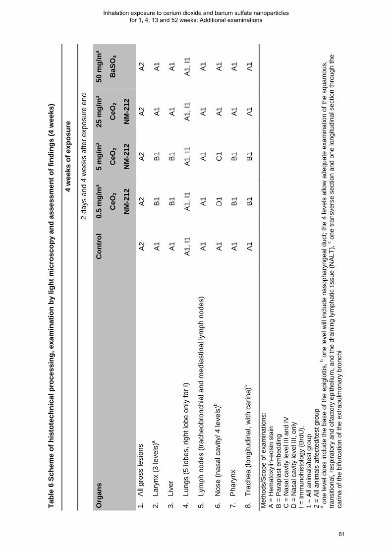
Ta
ble
6 Sc
hem
e of
his
tote
chni
cal p
roce
ssin
g, e
xam
inat
ion
by li
ght m
icro
scop
y an
d as
sess
men
t of f
indi
ngs
(4 w
eeks
)
4
wee
ks o
f exp
osur
e
2
days
and
4 w
eeks
afte
r exp
osur
e en
d
Org
ans
Con
trol
0.
5 m
g/m
³
CeO
2
NM
-212
5 m
g/m
³
CeO
2
NM
-212
25 m
g/m
³
CeO
2
NM
-212
50 m
g/m
³
BaS
O4
1.
All
gros
s le
sion
s A
2 A
2 A
2 A
2 A
2
2.
Lary
nx (3
leve
ls)a
A1
B1
B1
A1
A1
3.
Live
r A
1 B
1 B
1 A
1 A
1
4.
Lung
s (5
lobe
s, ri
ght l
obe
only
for I
) A
1, I1
A
1, I1
A
1, I1
A
1, I1
A
1, I1
5.
Lym
ph n
odes
(tra
cheo
bron
chia
l and
med
iast
inal
lym
ph n
odes
) A
1 A
1 A
1 A
1 A
1
6.
Nos
e (n
asal
cav
ity/ 4
leve
ls)b
A1
D1
C1
A1
A1
7.
Pha
rynx
A
1 B
1 B
1 A
1 A
1
8.
Trac
hea
(long
itudi
nal,
with
car
ina)
c A
1 B
1 B
1 A
1 A
1
Met
hods
/Sco
pe o
f exa
min
atio
ns:
A =
Hem
atox
ylin
-eos
in s
tain
B =
Par
apla
st e
mbe
ddin
g C
= N
asal
cav
ity le
vel I
II an
d IV
D
= N
asal
cav
ity le
vel I
II, o
nly
I = Im
mun
ohis
tolo
gy (B
rdU
),
1 =
All
anim
als/
test
gro
up
2 =
All
anim
als
affe
cted
/test
gro
up
a one
leve
l doe
s in
clud
e th
e ba
se o
f the
epi
glot
tis, b o
ne le
vel w
ill in
clud
e na
soph
aryn
geal
duc
t; th
e 4
leve
ls a
llow
ade
quat
e ex
amin
atio
n of
the
squa
mou
s,
trans
ition
al, r
espi
rato
ry a
nd o
lfact
ory
epith
eliu
m, a
nd th
e dr
aini
ng ly
mph
atic
tiss
ue (N
ALT)
, c one
tran
sver
se s
ectio
n an
d on
e lo
ngitu
dina
l sec
tion
thro
ugh
the
carin
a of
the
bifu
rcat
ion
of th
e ex
trapu
lmon
ary
bron
chi
81
Inhalation exposure to cerium dioxide and barium sulfate nanoparticles for 1, 4, 13 and 52 weeks: Additional examinations

Figu
re 8
-1 H
isto
path
olog
ical
imag
es o
f bifu
rcat
ion
of tr
ache
a af
ter 4
wee
ks o
f exp
osur
e
A
Con
trol a
nim
al, B
Ani
mal
exp
osed
to 5
mg/
m³ C
eO2 (
NM
-212
): O
ccur
renc
e of
par
ticle
s (a
rrow
), sm
all i
mag
e: h
ighe
r
mag
nific
atio
n
82
Inhalation exposure to cerium dioxide and barium sulfate nanoparticles for 1, 4, 13 and 52 weeks: Additional examinations

Figu
re 8
-2 H
isto
path
olog
ical
imag
es o
f nas
al c
avity
afte
r 4 w
eeks
of e
xpos
ure
A C
ontro
l ani
mal
, B A
nim
al e
xpos
ed to
25
mg/
m³ C
eO2 (
NM
-212
): O
ccur
renc
e of
par
ticle
s (a
rrow
), C
Hig
her m
agni
ficat
ion
of
i
mag
e B
83
Inhalation exposure to cerium dioxide and barium sulfate nanoparticles for 1, 4, 13 and 52 weeks: Additional examinations
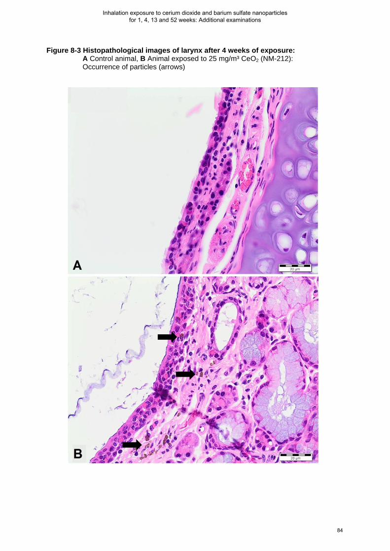
Figure 8-3 Histopathological images of larynx after 4 weeks of exposure: A Control animal, B Animal exposed to 25 mg/m³ CeO2 (NM-212): Occurrence of particles (arrows)
84
Inhalation exposure to cerium dioxide and barium sulfate nanoparticles for 1, 4, 13 and 52 weeks: Additional examinations

Figu
re 8
-4 H
isto
path
olog
ical
imag
es o
f the
lung
afte
r 4 w
eeks
inha
latio
n pl
us 4
wee
ks p
ost-e
xpos
ure:
A C
ontro
l ani
mal
,
B
-D A
nim
al e
xpos
ed to
25
mg/
m³ C
eO2 (
NM
-212
); B
-C A
lveo
lar h
istio
cyto
sis
with
par
ticle
s,
D G
ranu
lom
atou
s in
flam
mat
ion
85
Inhalation exposure to cerium dioxide and barium sulfate nanoparticles for 1, 4, 13 and 52 weeks: Additional examinations

Figu
re 8
-5 H
isto
path
olog
ical
imag
es o
f lun
gs a
fter 4
wee
ks o
f inh
alat
ion
expo
sure
:
A
Con
trol a
nim
al a
nd B
Ani
mal
exp
osed
to 5
0 m
g/m
³ BaS
O4
86
Inhalation exposure to cerium dioxide and barium sulfate nanoparticles for 1, 4, 13 and 52 weeks: Additional examinations

Figure 8-6 Histopathological images of pulmonary blood vessels after 4 weeks of exposure: A and B Animal exposed to 5 mg/m³ CeO2 (NM-212): Occurrence of macrophages with particles (arrows), small image with higher magnification
87
Inhalation exposure to cerium dioxide and barium sulfate nanoparticles for 1, 4, 13 and 52 weeks: Additional examinations

Figure 8-7 Histopathological images of pulmonary blood vessels after 4 weeks of exposure: A and B Animal exposed to 25 mg/m³ CeO2 (NM-212): Occurrence of macrophages with particles (arrows), smaller image with higher magnification
88
Inhalation exposure to cerium dioxide and barium sulfate nanoparticles for 1, 4, 13 and 52 weeks: Additional examinations

8.1.2. Immunohistology (cell proliferation, BrdU)
Material and Methods
Cell proliferation was determined in large/medium bronchi, terminal bronchioli and alveoli (table 7).
For evaluation of cell proliferation in the lung, rats were subcutaneously implanted with Alzet (Alzet
Corporation, Palo Alto, CA, USA) osmotic pumps (Model 2ML1) three days prior to necropsy. The
pumps contained 20 mg/mL 5-bromo-2-deoxyuridine (BrdU, Sigma Chem.), a thymidine structural
analog, which can be incorporated into DNA as a substitute for thymidine. Implantation was
subcutaneously to the dorsal region under Isofluran anesthesia (Essex GmbH, Munich, Germany). In
order to reduce pain during and after the implantation, the animals received a subcutaneous injection
of an appropriate analgetic compound (Buprenorphine, 0.01 mg/kg body weight) 10 to 30 minutes
prior to surgery.
Cell proliferation was determined after immunostaining of dewaxed sections and was performed with a
mouse anti-BrdU antibody (BioGenex, Hamburg, Germany; see protocol below). The BrdU-staining
identified cells passed through the S-phase of the cell cycle during the BrdU-labeling period. This
enabled detection of DNA replication in proliferating cells. Dividing cells in a defined time period and
area were related to non-dividing cells. At least 1000 cells per compartment and overall 3000 cells per
lung were counted (see below). For the microscope, 400x and 100x magnification were used for
counting type 1 cells (alveoli) and cells in the bronchioles, respectively. Numbers of dividing cells were
expressed as Labeling Index (LI). The percentage of nuclei was counted which underwent replicative
DNA synthesis (cells located in the S-phase of the cell cycle).
For the 1 week study, the left and accessory lung lobes of animals were used (one time point only). In
the 4 week study, the right half lungs (three lobes) were examined in order to reduce the animal
number per examination method. Two slides of each lung were prepared: one serving as test slides
and the other as a negative control slide. Jejunum served as positive control for the BrdU stain.
Immunhistological staining:
Sections were dewaxed by Xylol (2x 5 min) and rehydrated with descending 100, 96, 70 % alcohol (2 x
5 min each). Afterwards sections were rinsed in deionized water. For antigen retrieval, samples were
predigested with 0.05 % protease (2 min at 37° C) and hydrolysed with HCL (4N) for 20 min. After
washing with deionized water, 6 % H2O2 was added for 10 min. Samples were washed again and PBS
buffer were added. To reduce the areas which were moistened by the antibody, the tissue were traced
with a wax pencil (Pap-pen, BioGenex, USA). Then the primary monoclonal antibody (mouse anti-
BrdU; dilution of 1:1200) was added for 2 h. After rinsing twice with PBS buffer, the secondary
antibody (BioGenex, USA; link, anti-mouse, biotinilated; dilution of 1:20) were added for 20 min. After
rinsing again, the HR-Peroxidase/Streptavidin enzyme complex (BioGenex, Germany) for labelling
were added (20 min, 1:20). The secondary antibody system was used to visualize antibody binding.
89
Inhalation exposure to cerium dioxide and barium sulfate nanoparticles for 1, 4, 13 and 52 weeks: Additional examinations

Staining was developed with diaminobenzidine (DAB; Zytomed Systems GmbH, Germany) and the
slides were counterstained with Mayer’s hematoxylin (30 sec). For the negative control slides, PBS
replaced the primary antibody.
Quantitative assessment:
Performing quantitative assessment, positively stained cells were identified by red/brown pigment over
the nuclei. An image analysis system (KS 400, ZEISS, Germany) were used. Type 1 cells (alveoli) and
cells in the bronchioles were discriminated from mesenchymal cell on the base of their shape and size.
To facilitate comparability of counted cells, comparable lung compartments were selected for all
animals. Labeling indices (percentage of nuclei counted undergoing replicative DNA synthesis) were
determined for the lung compartments, the large/medium sized bronchi, the terminal bronchiole and
the alveoli (pneumocytes type I cells). In each compartment a minimum of 1 000 cells were counted.
For the alveoli, a minimum of 15 areas were used to count 1 000 cells. For the large/medium sized
bronchi and the terminal bronchiole, cell counts were based on a previously defined line of epithelial
cells including 1 000 cells.
The Labeling index (LI) in BrdU immunostained sections were calculated as follows:
BrdU LI (%) = (labeled cells) / (unlabeled cells + labeled cells) x 100
Results
After 1 week of exposure to CeO2 NM-212, increased cell proliferation rates were observed in the
large/medium bronchi (5 and 25 mg/m³), terminal bronchioli (25 mg/m³) and alveoli (> 0.5 mg/m³)
(table 7). After 4 weeks of exposure, increased cell proliferation rates were comparable as after 1
week of exposure, besides a higher increase in the terminal bronchioli at 25 mg/m³ after 1 week. Two
days after 4 weeks of exposure, increased cell proliferation rates were observed in the large/medium
bronchi, terminal bronchioli and alveoli at aerosol concentrations of 25 mg/m³ CeO2 (NM-212). After 4
weeks plus 4 weeks post-exposure, cell proliferation rates were concentration-dependent increased in
all three compartments. Higher proliferation rates of large/medium bronchi and terminal bronchiole
were recorded after the post-exposure period than after exposure end whereas the proliferation rates
of the alveoli were comparable for both time points.
BaSO4 did not cause any increased cell proliferation rates (4 weeks of exposure) (table 7).
90
Inhalation exposure to cerium dioxide and barium sulfate nanoparticles for 1, 4, 13 and 52 weeks: Additional examinations

Tabl
e 7
Effe
cts
on c
ell p
rolif
erat
ion
rate
s in
the
lung
afte
r 1 a
nd 4
wee
ks o
f inh
alat
ion
expo
sure
to C
eO2 a
nd B
aSO
4
1
wee
k of
exp
osur
e+ 4
wee
ks o
f exp
osur
e++
D
irect
ly a
fter l
ast e
xpos
ure
2 da
ys a
fter e
xpos
ure
end
4 w
eeks
afte
r the
end
of e
xpos
ure
Aer
osol
con
cent
ratio
n [m
g/m
³]
Con
trol
0.
5
CeO
2
NM
-21
2
5
CeO
2
NM
-21
2
25
CeO
2
NM
-21
2
0.5
CeO
2
NM
-21
1
25
CeO
2
NM
-21
1
Con
trol
0.
5
CeO
2
NM
-21
2
5
CeO
2
NM
-21
2
25
CeO
2
NM
-21
2
50
BaS
O4
Con
trol
0.
5
CeO
2
NM
-21
2
5
CeO
2
NM
-21
2
25
CeO
2
NM
-21
2
50
BaS
O4
Larg
e/m
ediu
m b
ronc
hi
1.59
±0.7
6
(100
)
1.95
±0.5
4
(123
)
2.58
*
±0.6
0
(162
)
3.70
*
±2.3
1
(181
)
1.42
±0.5
5
(89)
2.05
±0.7
4
(129
)
1.44
±1.0
4
(100
)
0.66
±0.4
6
(46)
1.64
±0.7
4
(113
)
3.29
*
±1.2
7
(228
)
1.36
±0.6
4
(94)
0.24
±0.2
0
(100
)
1.80
**
±0.7
0
(750
)
2.87
**
±1.0
9
(119
6)
4.92
**
±1.7
7
(205
0)
0.65
±0.4
2
(270
)
Term
inal
bro
nchi
oli
1.67
±0.7
5
(100
)
1.92
±0.6
0
(115
)
1.96
±0.7
7
(117
)
9.83
**
±1.2
0
(589
)
1.34
±0.5
3
(80)
4.38
**
±0.7
9
(262
)
1.69
±0.9
4
(100
)
1.08
±0.6
5
(64)
1.94
±0.5
0
(115
)
3.30
**
±0.4
1
(195
)
1.41
±0.4
3
(83)
0.34
±0.2
3
(100
)
1.34
*
±0.9
4
(394
)
3.90
**
±1.2
9
(114
7)
5.24
**
±1.9
5
(154
10)
0.44
±0.2
7
(129
)
Alv
eoli
2.22
±0.4
4
(100
)
3.80
**
±0.7
7
(171
)
5.92
**
±0.6
4
(267
)
5.76
**
±0.5
2
(258
)
2.50
±0.1
6
(113
)
4.09
**
±0.5
7
(184
)
3.02
±1.6
8
(100
)
2.29
±0.5
6
(76)
4.45
±0.8
3
(147
)
6.84
**
±1.7
7
(226
)
2.51
±0.5
5
(83)
2.22
±0.3
1
(100
)
3.96
**
±0.6
1
(178
)
4.62
**
±0.5
1
(208
)
5.45
**
±0.8
6
(250
)
2.22
±0.4
8
(100
)
Res
ults
are
pre
sent
ed a
s m
ean
labe
lling
indi
ces
± st
anda
rd d
evia
tion.
Fig
ures
in p
aren
thes
es s
how
rela
tive
incr
ease
s or
dec
reas
es to
the
corr
espo
ndin
g co
ntro
l lev
els;
n=5
. O
ne s
ided
Wilc
oxon
test
was
use
d: *
p< 0
.05,
p**
< 0.
01;
+ In th
e 1
wee
k st
udy,
the
left
and
acce
ssor
y lu
ng lo
bes
wer
e us
ed (o
ne ti
me
poin
t), ++
In th
e 4
wee
k st
udy,
the
right
hal
f lun
gs (t
hree
lobe
s) w
ere
exam
ined
.
91
Inhalation exposure to cerium dioxide and barium sulfate nanoparticles for 1, 4, 13 and 52 weeks: Additional examinations

8.1.3. Statistical analysis
Clinical pathology parameters (BALF cytology, enzyme data and BALF and serum cell mediator data)
were analyzed by non-parametric one-way analysis using the Kruskal-Wallis test (two-sided). If the
resulting p value was equal or less than 0.05, a pair-wise comparison of each test group with the
control group was performed using the Wilcoxon test or the Mann-Whitney U-test (both two-sided) (p ≤
0.05 for statistical significance). Comparison of lung weights among test groups were performed by
nonparametric one-way analysis using the two-sided Kruskal–Wallis test, followed by a two-sided
Wilcoxon test for the hypothesis of equal medians in case of p ≤ 0.05. The cell proliferation data was
analyzed by a pairwise comparison of each dose group with the control group using the Wilcoxon test
(one-sided, *p ≤ 0.05, **p ≤ 0.01).
92
Inhalation exposure to cerium dioxide and barium sulfate nanoparticles for 1, 4, 13 and 52 weeks: Additional examinations

8.2 Thirteen and 52 weeks of exposure within the long-term inhalation study
A part of the results obtained after 13 weeks of exposure to 50 mg/m³ BaSO4 was already presented
in paper 2: Konduru, N., Keller, J. et al. "Biokinetics and effects of barium sulfate nanoparticles". All
other results obtained after inhalation exposure of 13 and 52 weeks to CeO2 and BaSO4 are described
in this chapter.
8.2.1. Aerosol characterization
The aerosols were generated by brush dust generators (Ma-Hock et al. 2007). The aerosols were
analysed as described in the Material and Methods of paper 1 and 2. The particle concentrations and
particle size distributions are summarized in table 8. The target concentrations were met and
maintained well throughout the studies. Particle size distribution demonstrated that all particles were in
the respirable range for rats. The particles were largely agglomerated.
Table 8 Aerosol concentrations and particle size distributions after 52 weeks of exposure
Test material Targeted concentrations [mg/m³] Measured concentrations
[mg/m³] ± SD
MMAD [µm] / GSD
CeO2 NM-212
0.1 0.1 ± 0.1 2.3 / 2.4
CeO2 NM-212
0.3 0.3 ± 0.1 1.7 / 2.3
CeO2 NM-212
1 1.0 ± 0.1 1.5 / 2.3
CeO2 NM-212
3 3.0 ± 0.4 1.4 / 2.1
BaSO4 50 49.9 ± 5.6 2.0 / 2.0
Presented mean values of >14 measurements (MMAD, GSD); MMAD: mass median aerodynamic diameter, GSD: geometric standard deviation
93
Inhalation exposure to cerium dioxide and barium sulfate nanoparticles for 1, 4, 13 and 52 weeks: Additional examinations

8.2.2. Results: Lung and lymph node burdens
Directly after 13 and 52 weeks of exposure, the lavaged lungs, aliquots of lavage fluids and
tracheobronchial and mediastinal lymph nodes of five animals per group were used to determine lung
burden (as described in the Material and Methods of paper 2). The amounts of BaSO4 in lungs and
lymph nodes were estimated by measuring Ba with Inductively coupled plasma optical emission
spectrometry (ICP-MS) and extrapolating to BaSO4 (assuming that it was still particulate). The same
method was applied to lung burdens of CeO2. During the exposure period, lung burdens increased
with longer exposure duration (table 9). Only a slight translocation of the nanoparticles to the
tracheobronchial and mediastinal lymph nodes was observed (0.5 and 1.9 % of the BaSO4 and CeO2
lung burden, respectively) (table 9).
94
Inhalation exposure to cerium dioxide and barium sulfate nanoparticles for 1, 4, 13 and 52 weeks: Additional examinations

Tabl
e 9
Ove
rvie
w o
f org
an b
urde
n an
alys
is a
fter 1
3 an
d 52
wee
ks o
f exp
osur
e to
CeO
2 (N
M-2
12) a
nd B
aSO
4
13
wee
ks
52 w
eeks
Stud
y da
ys
94
367
Tim
e po
int
One
day
afte
r las
t exp
osur
e
Num
ber o
f ani
mal
s 5
5
Exam
ined
org
ans
Lung
,
trach
eobr
onch
ial a
nd m
edia
stin
al ly
mph
nod
es
Lung
Test
mat
eria
l C
eO2
NM
-212
B
aSO
4
C
eO2
NM
-212
B
aSO
4
Te
st C
once
ntra
tions
[mg/
m³]
0.1
3 50
0.
1 3
50
Lung
bur
den
[µg]
±SD
11
.96
±2.8
2
42
.07
±11.
05
Lung
bur
den
[mg]
±SD
1.39
±0
.16
1.73
±0
.85
2.
61
±0.5
2
10.2
1
±2.6
9
Trac
heob
ronc
hial
lym
ph n
ode
burd
en [µ
g] ±
SD
ND
11
.93
±14.
21
5.92
±6.5
2 N
D
ND
N
D
Med
iast
inal
lym
ph n
ode
burd
en [µ
g] ±
SD
ND
13
.78
±15.
85
2.27
±3.3
8 N
D
ND
N
D
ND
: not
det
erm
ined
, SD
: sta
ndar
d de
viat
ion,
n=5
; bur
den
is m
g or
µg
per l
ung;
lava
ged
lung
s an
d co
rres
pond
ing
aliq
uots
wer
e us
ed fo
r det
erm
inat
ion
of lu
ng
burd
en b
y IC
P-M
S.
95
Inhalation exposure to cerium dioxide and barium sulfate nanoparticles for 1, 4, 13 and 52 weeks: Additional examinations

8.2.3. Results: Bronchoalveolar lavage
Bronchoalveolar lavage fluids were assessed for 5 animals per group after 13 (paper 2: nano-BaSO4
BALF results) and 52 weeks of exposure (table 10). The procedure and analysis of BALF is described
in paper 1 and 2.
Exposure to nano-CeO2
After 13 weeks, exposure to 0.1, 0.3, 1 and 3 mg/m³ CeO2 induced a dose-dependent increase in
neutrophil cell counts with a statistically significant increase in total cells (table 10). Lymphocytes and
monocytes were significantly increased only at 3 mg/m³. Macrophage numbers and total cell counts
were statistically significantly increased also at lower concentrations (0.1 and 0.3 mg/m³). The
enzymes GGT, LDH and ALP were statistically significantly increased by aerosol concentrations of 1
mg/m³ and above. Total protein and NAG were increased only at 3 mg/m³. Cell mediators (MCP-1,
CINC-1 and Osteopontin) were statistically significant increased at 1 mg/m³ and above.
After 52 weeks of exposure to aerosol concentrations of 0.3 mg/m³ CeO2 and above, neutrophils and
lymphocytes were more elevated than after 13 weeks, but total cell counts were only increased at 3
mg/m³. Exposure of 0.1 mg/m³ CeO2 caused no changes in BALF cell numbers. Total protein and the
enzyme LDH were significantly increased at 1 and 3 mg/m³. ALP and NAG levels were not affected at
any aerosol concentration whereas GGT were significantly increased at all concentrations. MCP-1 and
CINC-1 levels were still elevated but to a lower extent compared with 13 weeks of exposure.
Exposure to nano-BaSO4
After 13 weeks of 50 mg/m³ nano-BaSO4 exposure, neutrophil and total cell numbers were
significantly increased (table 10). The enzyme ALP and the cell mediators MCP-1 and CINC-1 were
elevated as well.
After 52 weeks of exposure, neutrophil and total cells were even more increased than after 13 weeks.
Additionally, monocyte and lymphocytes levels were elevated. The levels of ALP were elevated after
13 weeks of exposure but declined to near control levels after 52 weeks whereas all other enzymes
were significantly increased. With the exception of MCS-F, all other cell mediators were more elevated
after 52 weeks of exposure than after 13 weeks.
Comparison of exposure to nano-CeO2 and -BaSO4
After 13 weeks of exposure, 3 mg/m3 nano-CeO2 elicited higher increases in (pro-)inflammatory
parameters (neutrophils, lymphocytes, total cells, MCP-1, NAG) than 50 mg/m³ BaSO4. However, this
reversed after 52 weeks of exposure where nearly all other parameters (neutrophil, lymphocytes, total
protein and enzymes) were more increased at 50 mg/m³ BaSO4 compared to 3 mg/m³ CeO2.
96
Inhalation exposure to cerium dioxide and barium sulfate nanoparticles for 1, 4, 13 and 52 weeks: Additional examinations

A co
mpa
rison
of
neut
roph
il nu
mbe
rs i
n BA
L flu
id (
x-fo
ld o
f co
ncur
rent
con
trol)
afte
r 1,
4,
13 a
nd 5
2 w
eeks
of
expo
sure
to
nano
- C
eO2
and
- Ba
SO4
is
pres
ente
d in
figu
re 8
-8.
Figu
re 8
-8 N
eutr
ophi
l num
bers
in B
AL fl
uid
(x-fo
ld o
f con
curr
ent c
ontr
ol) a
fter A
1 a
nd 4
wee
ks o
f exp
osur
e, B
13
and
52
wee
ks o
f exp
osur
e to
CeO
2 (N
M-2
12) a
nd B
aSO
4. M
ean
abso
lute
val
ues
with
sta
ndar
d de
viat
ion
of B
ALF
neut
roph
ils a
re
pres
ente
d in
tabl
e 10
.
97
Inhalation exposure to cerium dioxide and barium sulfate nanoparticles for 1, 4, 13 and 52 weeks: Additional examinations

Tabl
e 10
Bro
ncho
alve
olar
lava
ge fl
uid
anal
ysis
afte
r 13
and
52 w
eeks
of e
xpos
ure
to C
eO2 a
nd B
aSO
4
Test
mat
eria
l
CeO
2 NM
-212
B
aSO
4
Test
Con
cent
ratio
ns [m
g/m
³] C
ontr
ol
0.1
0.3
1 3
50
Mea
n va
lues
± S
D
Tota
l cel
ls (c
ount
s/µL
)
13 w
eeks
66
.3±2
2.04
11
5.22
**±2
3.72
98
.70*
±30.
49
117.
51*±
43.9
9 16
3.33
**±5
7.29
96
.08*
±16.
89
52 w
eeks
54
.27±
20.2
7 59
.95±
26.5
2 60
.97±
20.1
8 68
.56±
28.6
9 96
.06*
±29.
29
222.
73*±
95.1
8
Neu
troph
ils (c
ount
s/µL
)
13 w
eeks
1.
71±0
.66
0.53
±0.5
8 2.
85±3
.45
18.5
6**±
11.6
8 72
.59*
*±54
.65
23.4
6*±2
0.06
52 w
eeks
0.
58±0
.69
1.05
±0.9
9 4.
82**
±3.9
4 16
.07*
*±5.
17
31.9
7**±
14.2
3 15
2.75
**±7
4.45
Lym
phoc
ytes
(cou
nts/
µL)
13 w
eeks
0.
55±0
.42
0.27
±0.2
6 0.
38±0
.63
2.21
±2.2
3 5.
43**
±3.5
2 1.
84±1
.89
52 w
eeks
0.
56±0
.36
1.30
±1.1
2 9.
79*±
18.0
6 6.
69**
±2.1
6 9.
72**
±5.9
7 14
.35*
*±4.
25
Mac
roph
ages
(cou
nts/
µL)
13 w
eeks
63
.99±
22.3
6 11
4.37
**±2
4.06
95
.43*
±27.
12
96.3
0±46
.85
83.5
2±15
.72
70.3
7±23
.68
52 w
eeks
53
.01±
19.7
7 57
.49±
25.8
5 46
.10±
12.5
9 45
.33±
26.1
7 53
.57±
12.3
1 53
.54±
37.0
5
Mon
ocyt
es (c
ount
s/µL
)
13 w
eeks
0.
00±0
.00
0.00
±0.0
0 0.
00±0
.00
0.13
±0.3
0 0.
94**
±0.6
7 0.
10±0
.13
52 w
eeks
0.
04±0
.06
0.04
±0.0
6 0.
16±0
.18
0.29
*±0.
24
0.43
*±0.
40
1.14
*±1.
73
Eos
inop
hils
(cou
nts/
µL)
13 w
eeks
0.
04±0
.08
0.00
±0.0
0 0.
04±0
.08
0.13
±0.2
0 0.
25±0
.39
0.06
±0.1
3
52 w
eeks
0.
00±0
.00
0.00
±0.0
0 0.
00±0
.00
0.00
±0.0
0 0.
05±0
.10
0.06
±0.1
3
* st
atis
tical
ly s
igni
fican
t, p
< 0.
05; *
* st
atis
tical
ly s
igni
fican
t, p
< 0.
01; n
=5; m
ean
valu
es a
nd s
tand
ard
devi
atio
n (S
D);
lung
s w
ere
lava
ged
one
day
afte
r las
t exp
osur
e.
98
Inhalation exposure to cerium dioxide and barium sulfate nanoparticles for 1, 4, 13 and 52 weeks: Additional examinations

Con
tinua
tion
of T
able
10
Bro
ncho
alve
olar
lava
ge fl
uid
anal
ysis
afte
r 13
and
52 w
eeks
of e
xpos
ure
to C
eO2 a
nd B
aSO
4
Test
mat
eria
l
CeO
2 NM
-212
B
aSO
4
Test
Con
cent
ratio
ns [m
g/m
³] C
ontr
ol
0.1
0.3
1 3
50
Tota
l pro
tein
(mg/
L)
13 w
eeks
52
±10
57
±9
60±2
3 60
±18
84*±
26
64 ±
13
52 w
eeks
31
±8
33±1
2 67
±73
61**
±11
73**
±28
135*
*±72
GG
T (n
kat/L
)
13 w
eeks
41
±7
40±5
53
±13
80**
±18
106*
*±26
41
±7
52 w
eeks
26
±4
37**
±6
38*±
9 70
**±2
3 65
**±1
4 12
6**±
66
LDH
(µka
t/L)
13 w
eeks
0.
38±0
.06
0.43
±0.0
3 0.
52±0
.16
0.61
**±0
.18
0.95
**±0
.33
0.53
±0.1
7
52 w
eeks
0.
35±0
.04
0.37
±0.0
8 0.
44±0
.13
0.66
**±0
.14
0.80
**±0
.19
1.67
**±0
.69
ALP
(µka
t/L)
13 w
eeks
0.
51±0
.10
0.45
±0.0
6 0.
58±0
.09
0.73
*±0.
17
0.84
**±0
.19
0.87
**±0
.21
52 w
eeks
0.
44±0
.12
0.54
±0.1
0 0.
48±0
.13
0.58
±0.1
9 0.
50±0
.06
0.60
±0.2
1
NA
G (n
kat/L
)
13 w
eeks
35
±7
35±5
37
±13
43±9
51
**±8
41
±14
52 w
eeks
37
±7
42±1
4 38
±8
47±9
47
±12
73**
±30
MC
P-1
(pg/
mL)
13 w
eeks
24
.2±8
.4
19.1
±7.5
30
.8±2
6.4
111.
4**±
100.
1 62
0**±
613.
5 17
6.7*
* ±1
26.1
52 w
eeks
15
.3±8
.2
17.9
±7.1
49
.3*±
37.6
16
9.4*
*±11
3.9
378.
2**±
245.
5 23
00.7
**±1
463.
1
CIN
C-1
/IL-8
(pg/
mL)
13 w
eeks
93
.7±1
8.7
60.6
±38.
9 77
.7±3
8.9
172.
3*±1
00.7
28
0.6*
±190
.8
223.
8*±1
25.7
52 w
eeks
82
.4±2
5.4
68.8
±37.
1 69
.7±2
6.5
132.
9*±4
9.1
180.
6*±1
14.8
51
4.2*
±333
.6
M-C
SF
(pg/
mL)
13 w
eeks
14
±0
14±0
14
±0
14±0
14
±0
14±0
52 w
eeks
21
±12
18±3
18
±4
27±1
2 25
±15
31±1
6
Ost
eopo
ntin
(pg/
mL)
13 w
eeks
49
.02±
40.8
6 37
.40±
32.6
9 11
8.49
±133
.12
136.
87*±
72.8
9 30
0.41
*±25
3.01
23
4.02
±240
.06
52 w
eeks
16
2.79
±98.
61
166.
71±1
87.1
4 13
5.32
±61.
53
175.
72±8
8.75
22
8.80
±217
.96
1092
.70*
*±60
7.68
* st
atis
tical
ly s
igni
fican
t, p
< 0.
05; *
* st
atis
tical
ly s
igni
fican
t, p
< 0.
01; n
=5; m
ean
valu
es a
nd s
tand
ard
devi
atio
n (S
D);
lung
s w
ere
lava
ged
one
day
afte
r las
t exp
osur
e.
99
Inhalation exposure to cerium dioxide and barium sulfate nanoparticles for 1, 4, 13 and 52 weeks: Additional examinations

8.2.4. Results: Hematology and acute phase proteins in blood
Hematology, clinical chemistry and acute phase proteins were measured in the blood of 5 animals per
group (see detailed procedure in paper 1).
In blood, a slight increase of neutrophil counts was observed for CeO2 NM-212 after 1 week (5 days)
of exposure to 25 mg/m³ (see paper 1). Exposure of 4 weeks to CeO2 NM-212 and BaSO4 elicited no
increase of neutrophil counts (table 11 and 12).
Thirteen and 52 weeks of inhalation exposure to nano-CeO2 did not affect hematology and clinical
chemistry parameters in blood at any tested concentration apart from increased haptoglobin (HAPT)
and α2-macroglobulin (A2M) levels in serum after 52 weeks of exposure to 3 mg/m³ CeO2 (table 11). A
dose-dependent increase was observed for A2M after 52 weeks of exposure using the median values
rather than the means (median values of 10.94 and 11.64 for 1 and 3 mg/m³, respectively). After 13
weeks, the acute phase protein A2M was only significantly increased at 1 mg/m³, but had returned to
near control values by 52 weeks. The change was not dose-dependent and regarded as incidental.
Nano-BaSO4 inhalation exposure showed no changes in hematology and clinical chemistry
parameters in blood apart from a slight increase in (absolute and relative) neutrophil numbers and a
not statistically significant elevation in HAPT and A2M levels after 52 weeks of exposure (table 11).
A summary of neutrophil levels in BALF (x-fold of control) and of neutrophil levels and acute phase
proteins in blood (x-fold of control) after 4, 13 and 52 weeks of exposure to CeO2 and BaSO4 is
presented in table 12.
100
Inhalation exposure to cerium dioxide and barium sulfate nanoparticles for 1, 4, 13 and 52 weeks: Additional examinations

Tabl
e 11
Mea
n ne
utro
phil
leve
ls a
nd a
cute
pha
se p
rote
ins
in b
lood
afte
r 13
and
52 w
eeks
of e
xpos
ure
to C
eO2 a
nd B
aSO
4
Test
mat
eria
l
CeO
2
NM
-212
BaSO
4
Test
Con
cent
ratio
ns [m
g/m
³] C
ontr
ol
0.1
0.3
1 3
50
Mea
n va
lues
± S
D
Neu
troph
ils (g
iga/
L)
13 w
eeks
0.
63±0
.25
0.95
±0.1
6 0.
78±0
.10
0.93
±0.3
0 1.
09±0
.35
0.76
±0.0
6
52 w
eeks
0.
67±0
.17
0.79
±0.1
9 0.
70±0
.17
1.02
±0.2
0 0.
97±0
.29
1.23
*±0.
61
Neu
troph
ils (%
)
13 w
eeks
15
.2±4
.60
23.4
±4.3
19
.8±5
.0
22.4
±7.6
25
.4±9
.8
20.7
±1.8
52 w
eeks
26
.7±5
.80
33.8
±5.8
28
.1±4
.5
32.6
±7.1
32
.3±4
.8
38.5
*±9.
6
Acut
e ph
ase
prot
eins
(µg/
mL)
HAP
T
13 w
eeks
20
4.00
±47.
01
223.
60±6
7.11
22
8.60
±55.
86
192.
20±5
1.45
22
9.20
±58.
14
239.
00±5
6.14
52 w
eeks
13
9.60
±74.
26
215.
40±5
4.45
18
4.20
±73.
96
173.
60±3
7.35
25
1.40
*±63
.06
295.
8*±1
13.1
7
A2M
13 w
eeks
14
.55±
5.90
18
.18±
4.22
20
.69±
4.55
22
.66*
±7.0
6 19
.65±
4.45
21
.63±
8.33
52 w
eeks
8.
86±2
.14
9.52
±2.1
7 9.
88±4
.48
12.3
9±3.
98
12.1
9*±1
.61
14.4
4±9.
63
* sta
tistic
ally
sig
nific
ant,
p <
0.05
; ** s
tatis
tical
ly s
igni
fican
t, p
< 0.
01; n
=5; S
D: s
tand
ard
devi
atio
n, H
APT:
hap
togl
obin
, A2M
: α2-
mac
rogl
obul
in; b
lood
w
ere
colle
cted
dire
ctly
afte
r 13
wee
ks o
f exp
osur
e an
d on
e da
y af
ter 5
2 w
eeks
of e
xpos
ure.
101
Inhalation exposure to cerium dioxide and barium sulfate nanoparticles for 1, 4, 13 and 52 weeks: Additional examinations

Table 12 Neutrophil levels in BAL and neutrophil levels and acute phase proteins in blood after 4, 13 and 52 weeks of exposure to CeO2 and BaSO4
CeO2 (NM-212)
Study 4 weeks 13 weeks 52 weeks
Aerosol concentration
[mg/m³] 25 3 3
Lung burden [mg] 2.6 1.4 2.6
Neutrophil BAL [x-fold of control]
262** 43** 55**
Neutrophil blood [x-fold of control]
1.0 2.0 1.0
Haptoglobin [x-fold of control]
1.0 1.0 2.0*
A2M [x-fold of control]
1.0 1.0 1.0*
50 mg/m³ BaSO4
Study 4 weeks 13 weeks 52 weeks
Lung burden [mg] 0.8 1.7 10.2
Neutrophil BAL [x-fold of control]
3.0 14.0* 263.0**
Neutrophil blood [x-fold of control]
1.0 1.0 2.0*
Haptoglobin [x-fold of control]
1.0 1.0 2.0*
A2M [x-fold of control]
1.0** (0.6) 1.0 2.0
Values of neutrophils and acute phase proteins are rounded to the first decimal place.
102
Inhalation exposure to cerium dioxide and barium sulfate nanoparticles for 1, 4, 13 and 52 weeks: Additional examinations

8.3.
Sum
mar
y of
rele
vant
resu
lts a
fter 1
, 4, 1
3 an
d 52
wee
ks o
f exp
osur
e to
nan
o-C
eO2 (N
M-2
12)
To c
ompa
re to
tal l
ung
burd
en (
base
d on
mas
s or
par
ticle
sur
face
are
a) a
nd d
ose
rate
, neu
troph
ils in
BA
LF a
nd a
bsol
ute
lung
wei
ghts
afte
r 1,
4, 1
3 an
d 52
wee
ks o
f exp
osur
e to
CeO
2 N
M-2
12 w
ere
put i
nto
rela
tion
to th
e lu
ng b
urde
n or
lung
bur
den
per
expo
sure
tim
e (ta
ble
13 a
nd fi
gure
8-9
). R
etai
ned
parti
cle
surfa
ce a
rea
was
cal
cula
ted
base
d on
the
sur
face
are
a de
rived
fro
m B
runa
uer-
Emm
et-T
elle
r (B
ET)
mea
sure
men
ts (
see
pape
r 1
and
2, p
hysi
co-c
hem
ical
char
acte
rizat
ion)
.
Tabl
e 13
: Inh
alat
ion
expo
sure
to n
ano-
CeO
2 (N
M-2
12):
Cor
rela
tion
betw
een
dose
-rat
e an
d as
soci
ated
resp
onse
usi
ng m
ass
lung
bu
rden
and
reta
ined
par
ticle
sur
face
are
a in
the
lung
as
dose
met
rics
Stud
y/
Expo
sure
da
ys
Aer
osol
co
ncen
trat
ion
[mg/
m³]
Lung
bu
rden
[µ
g] ±
SD
Ret
aine
d pa
rtic
le
surf
ace
area
[m
²]
Dos
e ra
te
[µg/
day]
Neu
trop
hil
BA
L [c
n/µL
] ±SD
Neu
trop
hil
BA
L [x
-fold
of
cont
rol]
Abs
olut
e lu
ng
wei
ght
[g] ±
SD
Abs
olut
e lu
ng
wei
ght
[%]+
1 w
eek/
5
0.5
11±1
.0
0.00
027
2.2
3.70
±2.5
3 5
0.89
±0.2
96
5 10
0±9.
0 0.
0027
20
49
.19±
17.6
8 60
0.
91±0
.05
98
25
530±
120
0.01
431
106
272.
30±1
06.6
2 33
0 1.
048±
0.1
113
4 w
eeks
/ 20
0.5
41±7
0.
0010
8 2.
05
1.75
±1.1
3 2
1.01
±0.1
3 10
2
5 52
0±60
0.
0140
4 26
65
.66±
50.2
3 77
1.
11**
±0.0
7 11
3
25
2620
±290
0.
0707
4 13
1 22
2.29
±99.
25
262
1.28
**±0
.15
130
13 w
eeks
/ 67
0.
1 11
.96±
2.82
0.
0002
97
0.18
0.
53±0
.58
0.3
1.53
±0.1
3 10
3
3 13
90±0
.16
0.03
753
20.7
5 72
.59±
54.6
5 43
1.
57±0
.18
106
52 w
eeks
/ 25
3
0.1
42.0
7±11
.05
0.00
1134
0.
17
1.05
±0.9
9 2
1.88
±0.1
2 10
8
3 26
10±0
.52
0.07
02
10.3
2 31
.97±
14.2
3 55
1.
96±0
.15
113
+ com
pare
d to
the
cont
rol g
roup
(set
to 1
00%
); W
ilcox
on (t
wo-
side
d): *
p ≤
0.05
, **p
≤ 0
.01;
abs
olut
e lu
ng w
eigh
ts: n
=5 (1
wee
k) a
nd n
=10
(4 w
eeks
)
103
Inhalation exposure to cerium dioxide and barium sulfate nanoparticles for 1, 4, 13 and 52 weeks: Additional examinations

Retained particle surface [m²] in the lung
Figure 8-9 Neutrophil counts in BALF correlated with retained particle surface in the lung after 1, 4, 13 and 52 weeks of exposure to CeO2 NM-212 using the aerosol concentrations of 0.5, 5, and 25 mg/m³ (1 and 4 weeks) and 0.1 and 3 mg/m³ (13 and 52 weeks) CeO2 NM-212 for each point
104
Inhalation exposure to cerium dioxide and barium sulfate nanoparticles for 1, 4, 13 and 52 weeks: Additional examinations

9. Discussion
In this body of work, biokinetics and toxicity of inhaled cerium dioxide and barium sulfate nanoparticles
were examined after 1, 4, 13 and 52 weeks of exposure. The following sections discuss the results on
biokinetics and effects of nano-CeO2 (9.1) and -BaSO4 (9.2) in the lung and systemic effects of both
nanoparticles (9.3).
9.1 Biokinetics and effects of CeO2 in the lung
Before starting the inhalation exposure, the tested nano-CeO2 was studied in cell-free in vitro solubility
assays as reported previously (Molina et al. 2014). In these assays, CeO2 (NM-212) demonstrated a
very low solubility in fluids simulating different compartments of the body (e.g. below 0.001 wt% in
phagolysosomal simulant fluid (PSF), 0.02 wt% in 0.1 N HCl) and recrystallized in PSF, a model of
macrophage dissolution and clearance of particles. However, nano-CeO2 is not completely insoluble
and is able to release cerium ions (Schwabe et al. 2014; Yokel et al. 2014a; Yokel et al. 2014b). This
process is highly dependent on the particle size and composition of the surrounding medium. This was
valid at a pH below 4.6 and even at a pH of 8 in the presence of chelating agents. In the environment,
the following four steps seem to be involved in chemical transformation of nano-CeO2: dissolution of
Ce in the (3+) oxidation state, reduction of Ce4+ to Ce3+ in acidic media and release of Ce3+,
complexation with chelating agents and precipitation on the surface (Kakuwa & Matsumoto 2006,
Schwabe et al. 2014). These pathways may also play a role in vivo.
Consistently to the in vitro dissolution assays, inhaled CeO2 showed high biopersistence in the lungs
and a low bioavailability in the presented inhalation studies. Intratracheal instillation of the same, but
neutron activated 141CeO2 (dose of around 250 µg per lung) showed prolonged lung retention and a
slow clearance with an estimated half-time of 140 days (Molina et al. 2014). In our short-term study
with 1 week (5 days) of exposure, lung burdens were about one-fourth of those after 4 weeks of
exposure. In the short-term study with 4 weeks of exposure, inhaled CeO2 was deposited in the lungs
and cleared with a retention half-time of 40 days at aerosol concentrations of 0.5 mg/m³ (lung burden
of 0.041±0.007 mg/lung decreased to 0.009±0.004 mg 129 days post-exposure). At higher aerosol
concentrations of 25 mg/m³, the clearance was retarded with a retention half-life above 200 days (lung
burden of 2.62±0.20 mg/lung decreased to 1.8±0.26 mg 129 days post-exposure). Retardation of
clearance was already observed at aerosol concentrations of 5 mg/m³ since the lung burdens stayed
nearly constant during 4 weeks of post-exposure (lung burden of around 0.5 mg/lung). In the long-term
study, the lung burden of CeO2 after 13 weeks was nearly doubled after 52 weeks of exposure at
aerosol concentrations of 0.1 and 3 mg/m³. However, no information on time course of lung retention
and clearance kinetics has been available during post-exposure in the long-term study so far.
105
Discussion

Lymph node burdens after short-term exposure were higher than after long-term exposure. In general,
lymph node burdens were low (below 14 and 2 % of the initial lung burden after 4 and 13 weeks,
respectively) indicating that, at least at this lung burden range, it plays no major role for particle
clearance.
In the lung, inflammation was associated with retardation of pulmonary clearance in the short-term
studies. In the long-term study, local inflammation in the lung was observed by increases in BAL
neutrophils, lymphocytes and monocytes after 13 weeks of exposure. BAL changes (cell counts,
enzyme activities, total protein and cell mediator levels) after 52 weeks of exposure to 3 mg/m³ CeO2
were comparable to those after 13 weeks. Minor changes in BAL were observed at lower aerosol
concentrations of 1 mg/m³. Exposure to 0.3 mg/m³ elicited no BAL changes after 13 weeks and only
minor changes after 52 weeks (neutrophils, GGT, MCP-1). In animals exposed to lower aerosol
concentrations rather than in those exposed to higher, BAL lymphocytes were higher increased than
neutrophils after 52 weeks of exposure indicating a later phase of inflammation.
The strongest increase in BAL neutrophils indicating pulmonary inflammation was observed after 1
week of exposure. The increase in BAL neutrophils appeared to be attenuated after 4 and 13 weeks of
exposure. One week of exposure to 25 mg/m³ CeO2 caused a lung burden of 0.5 mg and elicited a
330-fold increase of BAL neutrophils whereas a lung burden of 2.6 mg was observed after 4 and 52
weeks of exposure to 25 and 3 mg/m³ CeO2, respectively. This burden caused 262-fold and 55-fold
increases in BALF neutrophils, respectively (figure 8-8 and table 13). The dose-rate at 3 mg/m³ for 52
weeks (10.3 µg/day) is 13 times lower than that of 4 weeks exposure to 25 mg/m³ (131 µg/day). The
initial pulmonary inflammation after 1 week of exposure correlates with the dose rate rather than with
the lung burden (see paper 1, conclusion).
For CeO2, the particle surface area was the dose metrics correlating best with pulmonary inflammation
based on BAL neutrophils. This was shown by dose-response curves using different dose-metrics
(surface area, mass, volume) for the two tested CeO2 (NM-211 and NM-212) in the 1 week study (see
paper 1, figure 7). To compare total lung burden and dose rate, neutrophils in BALF after 1, 4, 13 and
52 weeks of exposure to CeO2 NM-212 were put into relation to the lung burden or lung burden per
exposure time (table 13 and figure 8-9). The dose rate was linearly correlated with the observed lung
effects at all exposure times and aerosol concentrations. The neutrophil response observed in BALF
was obviously driven by the dose rate of particle surface entering the lung. Dose rate did not obviously
correlate with other lung response parameters (absolute lung weight) (table 13). The increase of
absolute lung weights after 1 and 4 weeks of exposure was rather affected by the exposure duration
than by dose rate.
After exposure to 5 and 25 mg/m³ CeO2, the initial pulmonary inflammation based on BAL neutrophils
was supplemented and dominated by mononuclear cells, especially macrophages. In the 4 week
study, alveolar histiocytosis progressed towards a granulomatous inflammation after 4 weeks plus 4
weeks post-exposure at aerosol concentrations of 5 and 25 mg/m³. The later inflammatory phase was
only detectable by histopathology. The progression of the later reaction towards a granulomatous type
106
Discussion

was clearly driven by the continuous presence of the particles in the lung. Histopathology after 13 and
52 weeks of exposure was not part of this body of work.
Despite the differences in the early and later phase of the reaction (neutrophil vs. macrophage; dose-
rate vs dose), the observations made in the short-term studies have relevance for the longer
exposure. At lung burdens of 2.6 mg after 4 and 52 weeks of exposure and an increasing duration of
particle presence in the lung, biological responses in the lung (e.g. granulomatous inflammation) may
show a further progression after 52 weeks. However, the further development of the granulomatous
inflammation and the occurrence of possible morphological changes will be studied in another project
within the long-term study.
Besides pulmonary inflammation, increased epithelial cell proliferation in the lung may be an indicator
for potential lung tumor formation (ECETOC 2013; ILSI Risk Science Institute Workshop Participants
2000). For assessment of cell proliferation in paraffin-embedded tissue sections, 5-bromodeoxyuridine
(BrdU) immunohistochemistry is the method of choice (Muskhelishvili et al. 2003). In our short-term
studies, statistically significant increased proliferation rates of epithelial cells, especially in the terminal
bronchioli and alveoli, were shown after 1 and 4 weeks of exposure (table 7). At lung burdens of
around 0.5 mg after 1 week of exposure, cell proliferation rates of inhaled CeO2 (aerosol concentration
of 25 mg/m³) were comparable or slightly higher than those of nano-TiO2 (10 mg/m³) although the
deposited particle surface per lung was 2 times lower for CeO2 (0.014 m²) than for TiO2 (0.028 m²)
(Ma-Hock et al. 2009). At lung burdens of around 0.5 mg after 4 week of exposure, inhaled CeO2 (5
mg/m³) elicited a slightly lower proliferation rate than after 1 week of exposure (25 mg/m³). The
increased response after shorter exposure may be explained by the higher dose-rate after 1 week
than after 4 weeks of exposure. The age of the animals may have a further impact since younger
animals may show higher cell turnover rates than older due to ongoing organ development and
growth. In our short-term studies, epithelial cells of the alveoli and terminal bronchioli proliferated
stronger than those of the large/medium bronchi, especially in the test groups exposed to CeO2. This
may be explained by higher particle deposition and particle-cell interaction in these areas since most
of the particles were found within the alveoli. No other particle induced changes of epithelial cells were
observed by histopathology.
Cell proliferation rates can also be influenced by increased cell turnover rates due to tissue growth.
Further investigations such as evaluation of epithelial cell apoptosis would be necessary to clarify this
question. Furthermore, comparative data on cell proliferation in the lung after inhalation of
nanoparticles is limited to a few studies. Statistically significant cell proliferation rates of the 1 and 4
week study were measured. The biological relevance of this increase remains questionable as cell
proliferation rates in young, growing animals are rather variable. In this case, the endpoint tumor
formation after inhalation of nanoparticles can only be revealed by histopathology after 2 years of
inhalation exposure. Cell proliferation data may be used in the future to establish a test system for
predicting possible tumour development already after shorter exposure.
For neoplastic responses in rats after inhalation of PSLT, the mode of action may require particle
accumulation in the lungs, persistent (neutrophilic) inflammation and epithelial cell proliferation
107
Discussion

(ECETOC 2013; ILSI Risk Science Institute Workshop Participants 2000). In our inhalation studies, all
three processes were observed. Whether the extent and interaction of these processes will lead to
lung tumour formation or not can only be revealed by histopathology after 2 years of exposure in the
current long-term inhalation study.
9.2. Biokinetics and effects of BaSO4 in the lung
Barium sulfate nanoparticles were also characterized and tested in cell-free in vitro solubility studies
(see paper 2). The cell-free in vitro studies showed very low dissolution (0.1 wt%) of nano-BaSO4 in
PSF. A consistent negligible dissolution in simulated physiological saline fluid was previously observed
for micron-BaSO4 (Cullen et al. 2000; Cullen et al. 1999). But in the short-term in vivo studies inhaled
nano-BaSO4 showed an unusual fast short-term clearance which may be explained by higher in vivo
biosolubility compared to our in vitro cell-free static system since the observed fast lung clearance and
high systemic bioavailability of inhaled BaSO4 cannot be explained by any known physiological
process. The available cell-free biological simulation fluids may lack critical constituents of the lung
which facilitate a more rapid dissolution. Moreover, the cell-free system determined the dissolution in a
static mode which may not adequately reflect the dynamics of an in vivo system and kinetics of the
dissolution process (e.g. removal of barium ions from the dissolution compartment resulting in a
steady state condition). Such limitations of abiotic, static solubility assays to estimate in vivo
biosolubility have been previously described (ECETOC 2013).
Differences in biosolubility of particles can affect particle clearance kinetics and overload conditions. In
our 4 week study, 95 % of BaSO4 initial lung burden was cleared within 35 days post-exposure (a lung
burden of 0.84 mg/lung 1 day post-exposure decreased to a lung burden of 0.04 mg/lung after 35 days
post-exposure). The retention half-time of inhaled BaSO4 was calculated with around 7 days (see
equation used for calculations of CeO2 retention half-times in paper 1). The retention half-time of
instilled 131BaSO4 (0.28 ± 0.004 mg/lung) was 9.6 days (see paper 2). Pulmonary micron-BaSO4
clearance was reported to consist of 52 % mucociliary clearance and 48 % transport from the lung via
blood to extrapulmonary tissues and organs, assuming macrophage involvement and disposal of
sulfate particles (Spritzer & Watson 1964; IPCS 1990). Dissolution in vivo is a known mechanism for
particle clearance (ECETOC 2013). Dissolution of BaSO4 in the lungs (e.g. in the phagolysosome of
macrophages) may contribute to their rapid clearance and translocation to extrapulmonary organs and
tissues. After instillation, 131BaSO4 was found in faeces (30 % of instilled dose) and bone (29 %).
Whether the instilled 131Ba measured in the bones was particulate 131BaSO4 or ionic 131Ba could not be
determined in this study.
In our inhalation study, lung burdens increased strongly with a longer exposure period (from 1.73
±0.85 mg after 13 weeks to 10.21 ±2.69 mg after 52 weeks, see table 9). This may indicate a change
in clearance from short-term to long-term exposure. This could be due to overwhelmed macrophage-
108
Discussion

dependent particle clearance, it may indicate a saturation of the process facilitating the dissolution of
BaSO4 in the lung or could be a combination of both.
After inhalation, deposited micron-BaSO4 has been reported to be cleared from the lung and elevated
barium levels were detected in the bone (Einbrodt et al. 1972; Cuddihy et al. 1974). This clearance
may also be transferable to BaSO4 nanoparticles. After 10 days (50 hours) of inhalation exposure to
micron-BaSO4, the barium burden in the lung was 83 µg/g dried tissue whereas the barium burden in
the femur was 1300 µg/g dried tissue, around 16-fold higher than in the lung (Einbrodt et al. 1972).
Whether the translocated barium was in an ionic or particulate form was not assessed in this study.
The majority of inhaled BaSO4 may be retained in the skeleton and to a much lesser extent in the soft
tissues. Barium translocation to the bone had already been observed to be similar to other heavy earth
alkaline metals like calcium and strontium (Moore 1964).
In the periodic table, barium neighbors calcium in the group of alkaline earth metals. The ratio of
calcium to barium atoms in the body is 1000 to 1 (Straube & Donate 2013). In the bone, calcium is
embedded in hydroxyapatite crystals. But as barium may mimic calcium in the body, barium ions may
also be incorporated in the bone matrix, mainly in areas of active bone growth in the distal regions
(Bligh & Taylor 1963; IPCS 1990; Moore 1964). In an in vitro model for bone metabolism, the
replacement of calcium by barium seems to be limited to the surface of the hydroxyapatite crystal
whereas strontium is able to replace calcium also in the interior of the crystal (Stark 1968). Barium
uptake into the bone seems to be rapid and, 1.5 till 5 times faster than calcium or strontium (Reeves
1986). Barium incorporation into the bone was reported to be increased in young growing animals and
reduced in mature animals (Bligh & Taylor 1963). This may help to explain the results of our studies
since the rats were around 6 weeks old, still developing, at the start of inhalation exposure and will be
around 2 years old after long-term exposure ends. At this time, it is not known why BaSO4 has such as
fast clearance after short-term exposure and if there is a (specific, active) process for barium transport
from the lung to the bone. The half-time of barium in the bone is around 50 days (Machata 1988).
Saturation of a specific process may also partly explain the decrease of barium clearance out of the
lung and the corresponding high barium burden in the lung after 52 weeks of exposure.
Variations in pulmonary barium burden and clearance mechanisms were already observed after 40
inhalation exposures to 40 mg/m³ micron-BaSO4 (Einbrodt et al. 1972). After the first 14 exposures,
barium content in the lungs increased but decreased rapidly during the next 4 weeks. It increased
again during the post-exposure period of 4 weeks. The author stated that barium redistribution from
extrapulmonary organs via blood to the lung might play a role. Barium translocation to rat femora and
jaw was also reported. Barium content in the femora peaked after 10 exposures and decreased
thereafter during further exposures and the post-exposure period.
To follow up the distribution and clearance mechanism of inhaled BaSO4 in our studies, further
investigations have to include the analysis of barium content in blood, bone and bone marrow. The
mechanism of barium deposition in the bone is also unclear. Proposed mechanisms include ionic
exchange with calcium at actively calcifying surfaces and rapid process of adsorption of colloidal
particles on to the bone surfaces (Bligh & Taylor 1963). It is necessary to determine in which form -
109
Discussion

ionic or particle - BaSO4 was rapidly cleared from the lung and translocated to the bone as well as how
and where it was integrated in the bone. Since barium is mainly excreted in the faeces and to a lesser
extent in the urine (see paper 2: by 28 days post-dosing, 30% of the 131Ba instilled dose was excreted
in the faeces and only 4.4% in the urine) (Cember et al. 1961), analysis of faeces and urine of
exposed animals may also contribute to excretion and clearance mechanisms of inhaled BaSO4 and
has to be investigated in the ongoing long-term study.
BaSO4 (nano)particles are used as filler in poly methyl methacrylate (PMMA) bone cements for
orthopedic reasons (Gillani et al. 2010). BaSO4 particles which are used as radiopaque agent within
the bone cement seems to promote osteolysis at the bone-implant interface (Wimhurst et al. 2001).
Increased inflammatory response and pathological bone resorption by enhancing macrophage-
osteoclast differentiation were also reported with BaSO4 containing cement (concentrations of 5 μg/ml
added to murine monocytes culture with osteoblast-like UMR106 cells) (Lazarus et al. 1994; Sabokbar
et al. 2000). Therefore, biological effects in bone by translocated BaSO4 after inhalation have to be
further evaluated in the long-term inhalation study.
After inhalation and deposition in the lung, nano-PSLT can be taken up by macrophages which then
can translocate to the lymph nodes (as mentioned in chapter 4.2.1.-4.2.3.). This mechanism has
generally only a minor contribution to the total lung clearance of particles (Ma-Hock et al. 2009; IARC
2010; Ravenzwaay et al. 2009). In the present work, after 13 weeks of exposure, barium burden in the
tracheobronchial and mediastinal lymph nodes was generally low (around 0.5 % of the initial lung
burden). This is consistent with other inhalation studies with micron-BaSO4 (Einbrodt et al. 1972).
Other inhaled PSLT nanoparticles, such as nano-TiO2 and -CeO2, were found within macrophages
and also free in pulmonary blood vessels (figure 8-6 and 8-7) (Eydner et al. 2012). There are two
possible mechanisms which may underlay this observation: nanoparticles may be phagocytized by
pulmonary macrophages and translocated within macrophages from the lung tissue into the blood. Or
free nanoparticles translocated from the lung to the blood vessels and were phagocytized by
pulmonary intravascular macrophages; these macrophages were, however, reported to be rare in rats
(Brain et al. 1999; Geiser 2010; Lehnert 1992). It is of interest whether nano-BaSO4 may translocate
freely or cell-associated (within macrophages) from the lungs to the blood and further into
extrapulmonary tissues. The mechanistic basis of the observed nanoparticles which were found within
macrophages and also free in pulmonary blood vessels warrants further investigations.
In the lung, inhaled BaSO4 elicited vanishing low effects after 4 weeks of exposure to high aerosol
concentrations. These observations were consistent with the previous short-term inhalation study after
1 week of exposure (Landsiedel et al. 2014). This may be explained by the low inherent toxicity and
the remarkable fast clearance of BaSO4 from the lungs.
With a burden of 0.84 mg/lung, nano-BaSO4 elicited no inflammation and no morphological changes in
the lung (and extra-pulmonary organs) (see figure 8-5) after short-term exposure of 4 weeks and a
subsequent post-exposure period of 4 weeks, besides a negligible elevation of some BAL parameters
(neutrophils, MCP-1, total protein). A moderate neutrophilic response in BAL (13-fold of control) was
110
Discussion

observed at a burden of 1.73 mg/lung after 13 weeks of exposure. This change led to corresponding
increases of enzyme levels (GGT, ALP) indicating slight pulmonary inflammation. Increased numbers
of neutrophils (263-fold of concurrent control), lymphocytes and monocytes showed a stronger
pulmonary inflammation after 52 weeks of exposure (burden of 10 mg/lung). Additional elevation of
total protein and all enzyme levels and MCP-1 may indicate a strong response to recruit and activate
monocytes and macrophages into the lung.
In another study, prolonged exposure to high aerosol concentrations led to high lung burdens and,
eventually, pulmonary inflammation occurred. In a 118 day-inhalation study, pulmonary response and
lung retention and clearance were compared for two micron-TiO2 and -BaSO4 tested at 50 mg/m³ and
75 mg/m³, respectively (Cullen et al. 1999). After 90 days with 64 exposures, micron-BaSO4 elicited a
burden of around 7.5 mg in the lung and 0.5 mg in the lymph nodes. Neutrophil response in BAL was
generally low (at background levels), besides a slight peak after 90 days (around 600 000 neutrophils,
12 % of total cells). These findings are in contrast to our effect data. In the same study, at equal mass
burdens of the two dusts, micron-TiO2 elicited a pulmonary inflammation based on increased
neutrophil numbers in BALF (around 28 % of total cells). Differences of the two particles were
explained by the smaller surface area of micron-BaSO4 (3.13 m²/g) compared to micron-TiO2
(6.68 m²/g) (Tran et al. 2000).
The change in inflammatory potential and toxicity after 52 weeks of exposure parallels the increase of
the test material in the lungs. In the phagolysosomal simulation fluid of our cell free studies, BaSO4
nanoparticles changed their physico-chemical properties and showed difference in charge at different
pH and recrystallized later over 28 days of incubation. These altered properties may lead to
differences in clearance and pulmonary toxicity.
Overall, BaSO4 nanoparticles are clearly deposited in the lung in our inhalation studies, especially
after 52 weeks of exposure. On the other hand, the clearance rate is faster after short-term exposure
than the clearance of other PSLT nanoparticles indicating a yet unknown clearance mechanism of
BaSO4 particles from the lung or dissolution of BaSO4 particles in vivo (albeit there was no indication
of this in vitro). The question on which of the two mechanisms are involved is of utmost interest and
needs to be addressed in future investigations.
The first case (clearance of undissolved BaSO4 nanoparticles) implies a hitherto unknown particle
clearance mechanism. Additionally, this would point out the need to re-frame the concept of PSLT
effects in the lung, since, in the case of the presented results, biopersistence would not correlate with
high lung retention or lung burdens.
The latter case (bio-dissolution of BaSO4 nanoparticles) implies that the in vivo biopersistence (or
rather the non-biopersistence) of a (nano-)particle cannot always be predicted from in vitro dissolution
(or rather insolubility) in biolological simulation fluids. With the presented results, BaSO4
(nano)particles may not be considered to be a classical PSLT (albeit indicated to be one by chemical
properties and initial in vitro dissolution tests). On the other hand, there are certain indications of
111
Discussion

particle effects in the lung (especially after 52 weeks of exposure) but no indication of barium
intoxication as described in the literature (Bhoelan et al. 2014).
The actual lung burden is always a function of both: the deposition and the clearance rate. The
aerosol concentration was quite high for BaSO4 in our studies. Low aerosol concentrations of
biopersistent particles may result in the same lung burden as high aerosol concentrations of rapidly-
cleared particles: aerosol concentrations of 3 mg/m³ CeO2 and 50 mg/m³ BaSO4 resulted in similar
lung burdens after 13 weeks of exposure (table 9). The effects of the (nano-)particles in the lung,
however, depend on the properties of the deposited material, the dose-rate and the toxicity of the
released ions: aerosol concentrations of 3 mg/m³ CeO2 and 50 mg/m³ BaSO4 resulted in similar lung
burdens after 13 weeks of exposure, but the effects (based on BALF parameters) differed. Different
(nano-)particles will span a continuous range of solubilities in the lung without a distinct threshold
dividing poorly soluble from soluble (nano-)particles. Likewise, most (nano-)particle may not exhibit
mere particle effects or mere released-ion effects. Hence, BaSO4 (nano-)particles could be regarded
as not being a classical PSLT or representing the lower toxicity and biopersistence end of PSLT.
9.3. Systemic effects
Systemic toxicity for inhaled PSLT seems to be less relevant since translocation rates to extra-
pulmonary organs are low and very rarely associated with toxic effects (Landsiedel et al. 2012a;
Moreno-Horn & Gebel 2014). This was also observed in our studies with 1, 4, 13 and 52 weeks of
exposure to nano-CeO2 and -BaSO4. There were, however, small amounts of particles translocating
from the lung and slight effects secondary to local pulmonary inflammation.
In blood, a slight neutrophilia was observed for CeO2 NM-212 after 1 week (5 days) of exposure to 25
mg/m³ (see paper 1). The neutrophilia in blood was considered to be secondary since a pronounced
inflammation was found in the lung at these aerosol concentrations. Exposure of 4 and 13 weeks to
CeO2 NM-212 and BaSO4 elicited no neutrophilia. After 52 weeks of exposure, a slight neutrophilia in
blood was apparent for 50 mg/m³ BaSO4 (table 11 and 12). Consistent to the 1 week study, the
neutrophilia in blood after 52 weeks of BaSO4 exposure seems to be rather a concomitant effect of the
corresponding local inflammation in the lung.
There is a concern that occupational exposure to nanoparticles can lead to an increased risk in
cardiovascular diseases (Donaldson et al. 2001). Various inhaled nanoparticles (e.g. carbon black,
TiO2) seem to be capable to induce an acute phase response in the blood (Halappanavar et al. 2011;
Saber et al. 2009; Saber et al. 2013). The major acute phase protein of the rat is alpha-2-
macroglobulin which is a proteinase inhibitor in serum. It is able to bind foreign substances or particles
as humoral defense (Borth 1992). Alpha-2-macroglobulin is further a carrier of growth factors with
regulatory functions of growth factor activity and is increased in inflammation (Bhattacharjee et al.
2005). Its expression is furthermore enhanced by Interleukin-6. The second acute phase protein
haptoglobin is able to scavenge hemoglobin which is released into blood by hemolysis or red blood
112
Discussion

cell turnover (Quaye 2008). Its increase is linked to diseases which exhibit inflammatory causes.
Haptoglobin is also a novel specific granule protein of neutrophils and its synthesis takes place during
differentiation of granulocytes (Theilgaard-Monch et al. 2006). After storage, it is released by activated
neutrophils. Furthermore, haptoglobin is involved in free radical quenching, tissue repair and
regeneration (Quaye 2008). Increase of haptoglobin levels in plasma were already observed after
intravenous exposure to 0.8 mg silica nanoparticles (particle diameter of 70 nm) in mice (Higashisaka
et al. 2011). The alteration of acute phase protein levels in blood may reflect the level of inflammation
(Donaldson et al. 2001; Halappanavar et al. 2011; Saber et al. 2009; Saber et al. 2013). Activated
leukocytes migrate to the site of inflammation in the lung. They pass the blood stream and release
cytokines which are able to trigger acute phase response (Gabay 1999). This acute phase response is
generated during inflammation over time leading to altered serum levels of acute phase proteins.
In our inhalation studies, the acute phase proteins haptoglobin and alpha-2-macroglobulin (A2M) (3
mg/m³ CeO2, 50 mg/m³ BaSO4) were slightly increased in blood after 52 weeks but not after 4 or 13
weeks of exposure (table 11 and 12). For 50 mg/m³ BaSO4, a late onset of pulmonary inflammation
occurred after 52 weeks accompanied by a slight increase of neutrophils.
Four weeks exposure of 25 mg/m³ CeO2 showed a pronounced lung inflammation, but no increase in
neutrophils and acute phase proteins in blood (table 12). After 52 weeks of exposure to 3 mg/m³, lung
burdens (of 2.6 mg) were comparable to those after 4 weeks of exposure to 25 mg/m³. Although
pulmonary inflammation based on increased BALF neutrophils was around 5-times lower after 52
weeks than after 4 weeks and no neutrophilia in blood was apparent, both acute phase proteins were
slightly increased even after 52 weeks. Since altered levels of acute-phase proteins reflect the
presence and intensity of inflammation (Gabay 1999; Davidson 2013), the duration of pulmonary
inflammation may be also a decisive parameter. The increase in acute phase proteins may be
explained by the longer lasting pulmonary inflammation after 52 weeks compared to 4 weeks of
exposure. A good correlation was shown for dose-rate and initial inflammation in the lung (based on
BALF neutrophils) after short-term exposure. Here, the dose rate seems not to be the driver for the
increase of the acute phase proteins after 52 weeks. This may indicate a different mechanism.
Overall, the increase in acute phase proteins in blood seems to be no nanoparticle-specific effect, but
rather, together with the slight neutrophilia in blood, a concomitant effect of recruitment of neutrophils
into the lung and of pulmonary inflammation and released mediators.
After 4 weeks of exposure to 25 mg/m³ CeO2, translocation of CeO2 from the lung to the liver was low
since liver burdens were below 2 µg at two time points and during 8 weeks of post-exposure (see
paper 1) unless there was a rapid hepato-biliary excretion of CeO2 from the liver. Cerium in bile was,
however, not measured. Low translocation of CeO2 deposited in the lung was also confirmed by an
intratracheal instillation study where less than 1 % of the total instilled 141Ce concentration was found
in extrapulmonary tissues after 28 days (Molina et al. 2014). Furthermore, no morphological changes
of extra-pulmonary organs could be detected in histopathology after short-term exposure of both
nanoparticles.
113
Discussion

10. Conclusion
Differences in toxic potency of various PSLT (nano-)particles were reported earlier (ECETOC 2013;
ILSI Risk Science Institute Workshop Participants 2000). In this body of work, two different
nanoparticles were tested in our inhalation studies.
In the short-term studies, inhaled nano-BaSO4 showed an unusually fast short-term clearance. This
may be explained by higher in vivo dissolution compared to our cell-free in vitro solubility studies since
the observed fast lung clearance and high systemic bioavailability of inhaled BaSO4 cannot be
achieved by any known physiological process unless there may be a specific rapid translocation of
nano-BaSO4. It is of utmost interest which mechanisms are involved; this needs to be addressed in
future investigations. In the long-term study, lung burdens increased strongly with longer exposure
period indicating a change in clearance from short-term to long-term exposure. Long-term exposure to
high aerosol concentrations of nano-BaSO4 led to high lung burdens and, eventually, pulmonary
inflammation based on increased BALF parameters. There were no indications of barium-ion toxicity
and the effects are regarded as being particle effects in the lung. Nano-BaSO4 seems to exhibit
peculiar characteristics concerning kinetics and effects compared to nano-PSLT. Nano-BaSO4 could
be regarded as not being a classical PSLT, but rather soluble in vivo albeit indicated to be insoluble by
its chemical properties. The remarkably low toxicity of inhaled nano-BaSO4 could only partly be
assigned to its fast lung clearance, but may also be the result of its low inherent toxicity.
Nano-CeO2, on the other hand, was biopersistent. Low lung burdens were cleared at physiological
rates whereas higher burdens induced retarded clearance and toxicity already after short-term
exposure. Comparing the observed effects in our studies with the literature data, CeO2 seems to
possess a higher toxicity than TiO2 (Ma-Hock et al. 2009; Eydner et al. 2012). It may represent the
higher toxicity and high biopersistence end of the group of PSLT.
Comparing the two tested nanoparticles, aerosol concentrations of 3 mg/m³ CeO2 and 50 mg/m³
BaSO4 resulted in similar lung burdens after 13 weeks of exposure, but the effects (based on BALF
parameters) were different; this probably depends on the properties of the deposited material, the
dose-rate and the toxicity of the released ions. Different (nano-)particles will span a continuous range
of solubilities in the lung without a distinct threshold dividing poorly soluble from soluble
(nano-)particles. Likewise, most particle may not exhibit mere particle effects or mere released-ion
effects.
The local no observed adverse effect concentrations in the lung (NOAEC) of CeO2 - based on BALF in
female rats after 13 and 52 weeks of exposure - are 0.3 and 0.1 mg/m³, respectively. The local
NOAEC of BaSO4 in the lung - based on BALF in female rats after 13 and 52 weeks of exposure – is
below 50 mg/m³.
Inhalation exposure to CeO2 and BaSO4 elicited no or only minimal systemic effects after short-term
and long-term exposure.
114
Conclusion

The two nanoparticles examined in this body of work differed in their particle kinetics and effects in the
lung already after one week and up to 52 weeks of exposure. Whether the lung burdens and effects
observed within the first 52 weeks of exposure will lead to lung tumour formation will be revealed by
histopathology after 2 years of exposure in the current long-term inhalation study.
115
Conclusion

11. References
Aalapati, S., Ganapathy, S., Manapuram, S., Anumolu, G. & Prakya, B. M. (2014). Toxicity and bio-accumulation of inhaled cerium oxide nanoparticles in CD1 mice. Nanotoxicology, 8, 786-798.
Adamson, I. Y. R., Letourneau, H. L. & Bowden, D. H. (1989). Enhanced Macrophage Fibroblast Interactions in the Pulmonary Interstitium Increases Fibrosis After Silica Injection to Monocyte-Depleted Mice. American Journal of Pathology, 134, 411-418.
Alber, A., Howie, S. E. M., Wallace, W. A. H. & Hirani, N. (2012). The role of macrophages in healing the wounded lung. International Journal of Experimental Pathology, 93, 243-251.
Anderson, A. H. (2010). Carbon Black Industry, Handbook of Texas Online. Texas State Historical Association, http://www.tshaonline.org/handbook/online/articles/doc01
Aninwene, G. E., Stout, D., Yang, Z. F. & Webster, T. J. (2013). Nano-BaSO4:a novel antimicrobial additive to pellethane. International Journal of Nanomedicine, 8, 1197-1205.
Antaria . Antaria: Ceria. 2014. 18-8-2014. Ref Type: Online Source, http://www.antaria.com/IRM/Company/ShowPage.aspx/PDFs/1096-68184674/AntariaStrengthensEuropeanDistribution Arora, S., Rajwade, J. M. & Paknikar, K. M. (2012). Nanotoxicology and in vitro studies: The need of the hour. Toxicol Appl Pharmacol, 258, 151-165.
Arts, J. H., Hadi, M., Keene, A. M., Kreiling, R., Lyon, D., Maier, M., Michel, K., Petry, T., Sauer, U. G., Warheit, D., Wiench, K. & Landsiedel, R. (2014). A critical appraisal of existing concepts for the grouping of nanomaterials. Regul.Toxicol.Pharmacol., 70, 492-506.
Babin, K., Antoine, F., Goncalves, D. M. & Girard, D. (2013). TiO2, CeO2 and ZnO nanoparticles and modulation of the degranulation process in human neutrophils. Toxicology Letters, 221, 57-63.
Baggs, R. B., Ferin, J. & Oberdörster, G. (1997). Regression of pulmonary lesions produced by inhaled titanium dioxide in rats. Veterinary Pathology, 34, 592-597.
Bailey, M. R., Kreyling, W. G., Andre, S., Batchelor, A., Collier, C. G., Drosselmeyer, E., Ferron, G. A., Foster, P., Haider, B., Hodgson, A., Masse, R., Metivier, H., Morgan, A., Muller, H. L., Patrick, G., Pearman, I., Pickering, S., Ramsden, D., Stirling, C. & Talbot, R. J. (1989). An Interspecies Comparison of the Lung Clearance of Inhaled Monodisperse Cobalt Oxide Particles .1. Objectives and Summary of Results. Journal of Aerosol Science, 20, 169-188.
Barlow, P. G., Clouter-Baker, A., Donaldson, K., Maccallum, J. & Stone, V. (2005). Carbon black nanoparticles induce type II epithelial cells to release chemotaxins for alveolar macrophages. Part Fibre.Toxicol., 2, 11.
Becker, H., Herzberg, F., Schulte, A. & Kolossa-Gehring, M. (2011). The carcinogenic potential of nanomaterials, their release from products and options for regulating them. International Journal of Hygiene and Environmental Health, 214, 231-238.
Bedoret, D., Wallemacq, H., Marichal, T., Desmet, C., Calvo, F. Q., Henry, E., Closset, R., Dewals, B., Thielen, C., Gustin, P., de Leval, L., Van Rooijen, N., Le Moine, A., Vanderplasschen, A., Cataldo, D., Drion, P. V., Moser, M., Lekeux, P. & Bureau, F. (2009). Lung interstitial macrophages alter dendritic cell functions to prevent airway allergy in mice. Journal of Clinical Investigation, 119, 3723-3738.
Bellmann, B., MUHLE, H., Creutzenberg, O., Dasenbrock, C., Kilpper, R., MacKenzie, J. C., Morrow, P. & Mermelstein, R. (1991). Lung clearance and retention of toner, utilizing a tracer technique, during chronic inhalation exposure in rats. Fundam Appl Toxicol, 17, 300-313.
Bermudez, E., Mangum, J. B., Wong, B. A., Asgharian, B., Hext, P. M., Warheit, D. B. & Everitt, J. I. (2004). Pulmonary responses of mice, rats, and hamsters to subchronic inhalation of ultrafine titanium dioxide particles. Toxicological Sciences, 77, 347-357.
Bhattacharjee, G., Arandjelovic, S., Mackman, N., Gonias, S. L. & Edgington, T. S. (2005). Enhancement of tissue factor dependent procoagulant activity and factor xa generation by alpha(2)-macroglobulin. Blood, 106, 550A.
Bhoelan, B. S., Stevering, C. H., van der Boog, A. T. J., & van der Heyden, M. A. G. (2014). Barium toxicity and the role of the potassium inward rectifier current. Clinical Toxicology, 52(6), 584-593. Blasco, C. & Pico, Y. (2011). Determining nanomaterials in food. Trac-Trends in Analytical Chemistry, 30, 84-99.
References
116

Bleeker, E. A. J., De Jong, W. H., Geertsma, R. E., Groenewold, M., Heugens, E. H. W., Koers-Jacquemijns, M., Van De Meent, D., Popma, J. R., Rietveld, A. G., Wijnhoven, S. W. P., Cassee, F. R. & Oomen, A. G. (2013). Considerations on the EU definition of a nanomaterial: Science to support policy making. Regulatory Toxicology and Pharmacology, 65, 119-125.
Bligh, P. H. & Taylor, D. M. (1963). Comparative Studies of Metabolism of Strontium and Barium in Rat. Biochemical Journal, 87, 612
Borm, P. J. A., Tran, L. & Donaldson, K. (2011). The carcinogenic action of crystalline silica: A review of the evidence supporting secondary inflammation-driven genotoxicity as a principal mechanism. Critical Reviews in Toxicology, 41, 756-770.
Borth, W. (1992). Alpha(2)-Macroglobulin, A Multifunctional Binding-Protein with Targeting Characteristics. Faseb Journal, 6, 3345-3353.
Bowden D.H., Hedgecock C. & Adamson I.Y. (1989). Silica-induced pulmonary fibrosis involves the reaction of particles with interstitial rather than alveolar macrophages. Journal of Pathology, 73-80.
Brain, J. D., Knudson, D. E., Sorokin, S. P. & Davis, M. A. (1976). Pulmonary Distribution of Particles Given by Intratracheal Instillation Or by Aerosol Inhalation. Environmental Research, 11, 13-33.
Brain, J. D., Molina, R. M., DeCamp, M. M., & Warner, A. E. (1999). Pulmonary intravascular macrophages: their contribution to the mononuclear phagocyte system in 13 species. American Journal of Physiology-Lung Cellular and Molecular Physiology, 276(1), L146-L154. Brody, A. R. & Roe, M. W. (1983). Deposition Pattern of Inorganic Particles at the Alveolar Level in the Lungs of Rats and Mice. American Review of Respiratory Disease, 128, 724-729.
Brown, S. C., Boyko, V., Meyers, G., Voetz, M. & Wohlleben, W. (2013). Towards Advancing Nano-object Count Metrology – A Best Practice Framework. Environ Health Perspect, doi:10.1289/ehp.1306957.
Burello, E. & Worth, A. (2011a). Computational Nanotoxicology Predicting Toxicity of Nanoparticles. Nature Nanotechnology, 6, 138-139.
Burello, E. & Worth, A. P. (2011b). QSAR modeling of nanomaterials. Wiley Interdisciplinary Reviews-Nanomedicine and Nanobiotechnology, 3, 298-306.
Cassee, F. R., van Balen, E. C., Singh, C., Green, D., Muijser, H., Weinstein, J. & Dreher, K. (2011). Exposure, health and ecological effects review of engineered nanoscale cerium and cerium oxide associated with its use as a fuel additive. Crit Rev.Toxicol., 41, 213-229.
Cember, H., Watson, J. A. & Novak, M. E. (1961). The influence of radioactivity and lung burden on the pulmonary clearance rate of barium sulfate. American Industrial Hygiene Association Journal, 22(1), 27-32.
Chakravarthi VP & Balaji SN (2010). Application of Nanotechnology in Veterinary Medicine. Veterinary World, 3, 477-480.
Cherrie, J. W., Brosseau, L. M., Hay, A. & Donaldson, K. (2013). Low-toxicity dusts: current exposure guidelines are not sufficiently protective. Ann.Occup.Hyg., 57, 685-691.
Corma, A., Atienzar, P., Garcia, H. & Chane-Ching, J. Y. (2004). Hierarchically mesostructured doped CeO2 with potential for solar-cell use. Nature Materials, 3, 394-397.
Crowell, R. E., Heaphy, E., Valdez, Y. E., Mold, C. & Lehnert, B. E. (1992). Alveolar and Interstitial Macrophage Populations in the Murine Lung. Experimental Lung Research, 18, 435-446.
Cuddihy, R. G., Hall, R. P. & Griffith, W. C. (1974). Inhalation Exposures to Barium Aerosols: Physical, Chemical and Mathematical-Analysis. Health Physics, 26(5), 405-416.
Cullen, R. T., Tran, C. L., Buchanan, D., Davis, J. M., Donaldson, K., Jones, A. D. & Searl, A. Health and Safety Executive Contract Research Report: Investigations into the pulmonary effects of low toxicity dusts. Part I: Relative toxicological potency of dusts. Health and Safety Executive Contract Research Report 216/1999. 1999. Sudbury, UK, HSE Books. Ref Type: Report Cullen, R. T., Tran, C. L., Buchanan, D., Davis, J. M., Searl, A., Jones, A. D. & Donaldson, K. (2000). Inhalation of poorly soluble particles. I. Differences in inflammatory response and clearance during exposure. Inhal Toxicol, 12, 1089-1111.
Dankovic, D., Kuempel, E. & Wheeler, M. (2007). An approach to risk assessment for TiO2. Inhalation Toxicology, 19, 205-212.
Davidson, S. J. (2013). Inflammation and Acute Phase Proteins in Haemostasis, chapter 2, Acute Phase Proteins. In Sabina Janciauskiene (Ed) Acute Phase Proteins (book), DOI: 10.5772/55998.
References
117

De Capitani, E. M., Algranti, E., Handar, A. M. Z., Altemani, A. M. A., Ferreira, R. G., Balthazar, A. B., Cerqueira, E. M. F. P. & Ota, J. S. (2007). Wood charcoal and activated carbon dust pneumoconiosis in three workers. American Journal of Industrial Medicine, 50, 191-196.
Deutsche Forschungsgesellschaft (DFG) . General threshold limit value for dust (R fraction) (Biopersistent granular dusts) [MAK Value Documentation, 2012]. 2014. Wiley-VCH Verlag GmbH & Co. KGaA. Ref Type: Report
DFG Deutsche Forschungsgemeinschaft (2013a). List of MAK and BAT Values 2013: Maximum Concentrations and Biological Tolerance Values at the Workplace. Wiley-VCH Verlag GmbH & Co. KGaA.
DFG Deutsche Forschungsgemeinschaft (2013b). Nanomaterials; report of the Commission for the Investigation of Health Hazards of Chemical Compounds in the Work Area . Wiley-VCH Verlag.
Doig A.T. (1976). Baritosis: a benign pneumoconiosis. Thorax, 31, 30-39.
Donaldson, K. (2000). Nonneoplastic lung responses induced in experimental animals by exposure to poorly soluble nonfibrous particles. Inhalation Toxicology, 12, 121-139.
Donaldson, K., Borm, P. J., Oberdörster, G., Pinkerton, K. E., Stone, V. & Tran, C. L. (2008). Concordance between in vitro and in vivo dosimetry in the proinflammatory effects of low-toxicity, low-solubility particles: the key role of the proximal alveolar region. Inhal Toxicol, 20, 53-62.
Donaldson, K. & Poland, C. A. (2013). Nanotoxicity: challenging the myth of nano-specific toxicity. Current Opinion in Biotechnology, 24, 724-734.
Donaldson, K., Stone, V., Seaton, A. & MacNee, W. (2001). Ambient particle inhalation and the cardiovascular system:potential mechanisms. Environ Health Perspect [109], 523-527. Donaldson, K. & Tran, C. L. (2002). Inflammation caused by particles and fibers. Inhal Toxicol, 14, 5-27.
Donaldson, K., Brown, D., Clouter, A., Duffin, R., MacNee, W., Renwick, L., Tran, L. & Stone, V. (2002). The pulmonary toxicology of ultrafine particles. Journal of Aerosol Medicine, 15, 213-220.
Donaldson, K. & Poland, C. A. (2012). Inhaled nanoparticles and lung cancer - what we can learn from conventional particle toxicology. Swiss medical weekly, 142, 13547.
Driscoll, K. E. (2000). TNF alpha and MIP-2: role in particle-induced inflammation and regulation by oxidative stress. Toxicology Letters, 112, 177-183.
Driscoll, K. E., Carter, J. M., Hassenbein, D. G. & Howard, B. (1997). Cytokines and particle-induced inflammatory cell recruitment. Environ.Health Perspect., 105 Suppl 5, 1159-1164.
Driscoll, K. E., Carter, J. M., Howard, B. W., Hassenbein, D. G., Pepelko, W., Baggs, R. B. & Oberdörster, G. (1996). Pulmonary inflammatory, chemokine, and mutagenic responses in rats after subchronic inhalation of carbon black. Toxicol Appl Pharmacol, 136, 372-380.
Driscoll, K. E., Hassenbein, D. G., Carter, J. M., Kunkel, S. L., Quinlan, T. R. & Mossman, B. T. (1995a). TNF alpha and increased chemokine expression in rat lung after particle exposure. Toxicology Letters, 82-3, 483-489.
Driscoll, K. E., Hassenbein, D. G., Howard, B. W., Isfort, R. J., Cody, D., Tindal, M. H., Suchanek, M. & Carter, J. M. (1995b). Cloning, Expression, and Functional-Characterization of Rat Mip-2 - A Neutrophil Chemoattractant and Epithelial-Cell Mitogen. Journal of Leukocyte Biology, 58, 359-364.
Driscoll, K. E., Maurer, J. K., Higgins, J. & Poynter, J. (1995c). Alveolar Macrophage Cytokine and Growth-Factor Production in A Rat Model of Crocidolite-Induced Pulmonary Inflammation and Fibrosis. Journal of Toxicology and Environmental Health, 46, 155-169.
Driscoll,K.E.,Deyo,l.C.,Howard,B.W.,Hassenbein,D.G.,Bertram,T. (1997). Effects of particle exposure and particle-elicited inflammatory cells on mutation in rat alveolar type II cells. Carcinogenesis, 18, 423–430.
ECETOC . ECETOC: Poorly Soluble Particles / Lung Overload, Technical Report No. 122. European Centre for Ecotoxicology and Toxicology of Chemicals. 122. 2013. Brussels, European Centre for Ecotoxicology and Toxicology of Chemicals. Ref Type: Report Einbrodt, H. J., Wobker, F. & Klippel, H. G. (1972). Experimental Studies on Accumulation and Distribution of Barium Sulfate in Rat Following Inhalation. Internationales Archiv fur Arbeitsmedizin, 30, 237-&.
References
118

Elder, A., Gelein, R., Silva, V., Feikert, T., Opanashuk, L., Carter, J., Potter, R., Maynard, A., Ito, Y., Finkelstein, J. & Oberdörster, G. (2006). Translocation of inhaled ultrafine manganese oxide particles to the central nervous system. Environ Health Perspect, 114, 1172-1178.
Elder, A. & Oberdörster, G. (2006). Translocation and effects of ultrafine particles outside of the lung. Clin.Occup.Environ Med, 5, 785-796.
Environmental Protection Agency (EPA). (2009) Report: Toxicological review of Cerium Oxide and Cerium Compounds. Environmental Protection Agency EPA/635/R-08/002F . 15-4-2014.
Etheridge, M. L., Campbell, S. A., Erdman, A. G., Haynes, C. L., Wolf, S. M., & McCullough, J. (2013). The big picture on nanomedicine: the state of investigational and approved nanomedicine products. Nanomedicine: Nanotechnology, Biology and Medicine, 9(1), 1-14.
European Commission . (2004) Report: Community Health and Consumer Protection: Nanotechnologies: A preliminary risk analysis on the basis of a workshop organized in Brussels by the Health and Consumer Protection Directorate General of the European Commission.
European Commission . (2011a) Report: COMMISSION RECOMMENDATION of 18 October 2011 on the definition of nanomaterial 2011/696/EU .
European Commission . (2011b) Report: List of materials in the JRC Nanomaterials (NM) Repository, Institute of Health and Consumer Protection. European Commission. 26-9-2014b.
European Commission . (2011c) Regulation (EU) No 1169/2011 of the European Parliament and of the Council of 25 October 2011 on the provision of food information to consumers, amending Regulations (EC) No 1924/2006 and (EC) No 1925/2006 of the European Parliament and of the Council, and repealing Commission Directive 87/250/EEC, Council Directive 90/496/EEC, Commission; Directive 1999/10/EC, Directive 2000/13/EC of the European Parliament and of the Council, Commission Directives 2002/67/EC and 2008/5/EC and Commission Regulation (EC) No 608/2004. L 304/18. Off J Eur Union.
European Commission . (2013a) Report: Classification, Labeling and packaging of nanomaterials in REACH and CLP. [Doc. CA/90/2009 Rev2].
European Commission . (2013b) Report: Introduction to nanotechnologies. 2013b. 7-4-2014b.
European Commission . (2014) JRC Science and Policy Reports: JRC Repository: NM-Series of Representative Manufactured Nanomaterials: Cerium Dioxide, NM-211, NM-212, NM-213. Characterisation and Test Item Preparation. JRC89825. Publications Office of the European Union.
Eydner, M., Schaudien, D., Creutzenberg, O., Ernst, H., Hansen, T., Baumgartner, W. & Rittinghausen, S. (2012). Impacts after inhalation of nano- and fine-sized titanium dioxide particles: morphological changes, translocation within the rat lung, and evaluation of particle deposition using the relative deposition index. Inhal.Toxicol., 24, 557-569.
Fabian, E., Landsiedel, R., Ma-Hock, L., Wiench, K., Wohlleben, W. & van Ravenzwaay, B. (2008). Tissue distribution and toxicity of intravenously administered titanium dioxide nanoparticles in rats. Archives of Toxicology, 82, 151-157.
Ferin, J., Oberdörster, G. & Penney, D. P. (1992). Pulmonary retention of ultrafine and fine particles in rats. Am J Respir.Cell Mol.Biol, 6, 535-542.
Gabay, C. (1999). Mechanisms of disease - Acute-phase proteins and other systemic responses to inflammation (vol 340, pg 448, 1999). New England Journal of Medicine, 340, 1376.
Gallagher, J., Sams, R., Inmon, J., Gelein, R., Elder, A., Oberdörster, G. & Prahalad, A. K. (2003). Formation of 8-oxo-7,8-dihydro-2 '-deoxyguanosine in rat lung DNA following subchronic inhalation of carbon black. Toxicol Appl Pharmacol, 190, 224-231.
Gebel, T. (2012). Small difference in carcinogenic potency between GBP nanomaterials and GBP micromaterials. Archives of Toxicology, 86, 995-1007.
Gebel, T., Marchan, R. & Hengstler, J. (2013). The nanotoxicology revolution. Archives of Toxicology, 87, 2057-2062.
Gebel, T., Foth, H., Damm, G., Freyberger, A., Kramer, P. J., Lilienblum, W., Röhl, C., Schupp, T., Weiss, C., Wollin, K. M. & Hengstler, J. G. (2014). Manufactered nanomaterials: categorisation and approaches to hazard assessment. Critical Reviews in Toxicology, 88(12):2191-211.
Geiser, M. (2002). Morphological aspects of particle uptake by lung phagocytes. Microscopy Research and Technique, 57, 512-522.
Geiser, M. (2010). Update on Macrophage Clearance of Inhaled Micro- and Nanoparticles. Journal of Aerosol Medicine and Pulmonary Drug Delivery, 23, 207-217.
Gentek, R., Molawi, K. & Sieweke, M. H. (2014). Tissue macrophage identity and self-renewal. Immunol.Rev., 262, 56-73.
References
119

Geraets, L., Oomen, A. G., Schroeter, J. D., Coleman, V. A. & Cassee, F. R. (2012). Tissue distribution of inhaled micro- and nano-sized cerium oxide particles in rats: results from a 28-day exposure study. Toxicol.Sci., 127, 463-473.
Gill, K. K., Nazzal, S. & Kaddourni, A. (2014). Paclitaxel loaded PEG5000-DSPE micelles as pulmonary delivery platform: Formulation characterization, tissue distribution, plasma pharmacokinetics, and toxicological evaluation. European Jorunal of Pharmaceutics and Biopharmaceutics, 79, 276-284.
Gillani, R., Ercan, B., Qiao, A. & Webster, T. J. (2010). Nanofunctionalized zirconia and barium sulfate particles as bone cement additives. International Journal of Nanomedicine, 5, 1-11.
Givvimani S., Leevy, M. & Tyagi S.C. (2011) Report: Barium Angiography in Small Animals. Application Note # AP0108. Bruker.
Gojova, A., Lee, J. T., Jung, H. S., Guo, B., Barakat, A. I. & Kennedy, I. M. (2009). Effect of cerium oxide nanoparticles on inflammation in vascular endothelial cells. Inhal.Toxicol., 21 Suppl 1, 123-130.
Gordon, S. (2003). Alternative activation of macrophages. Nature Reviews Immunology, 3, 23-35.
Gosens, I., Mathijssen, L., Bokkers, B., Muijser, H. & Cassee, F. R. (2013). Comparative hazard identification of nano- and micro-sized cerium oxide particles based on 28-day inhalation studies in rats. Nanotoxicology, 8:6, 643-653.
Green, F. H. Y. (2000). Pulmonary responses to inhaled poorly soluble particulate in the human. Inhalation Toxicology, 12, 59-95.
Gregoratto, D., Bailey, M. R. & Marsh, J. W. (2011). Particle Clearance in the Alveolar-Interstitial Region of the Human Lungs: Model Validation. Radiation Protection Dosimetry, 144, 353-356.
Greim, H., Borm, P., Schins, R., Donaldson, K., Driscoll, K., Hartwig, A., Kuempel, E., Oberdörster, G. & Speit, G. (2001). Toxicity of fibers and particles. Report of the workshop held in Munich, Germany, 26-27 October 2000. Inhal Toxicol, 13, 737-754.
Greim, H. & Ziegler-Skylakakis, K. (2007). Risk assessment for biopersistent granular particles. Inhal.Toxicol., 19 Suppl 1, 199-204.
Grommes, J. & Soehnlein, O. (2011). Contribution of neutrophils to acute lung injury. Mol.Med., 17, 293-307.
Halappanavar, S., Jackson, P., Williams, A., Jensen, K. A., Hougaard, K. S., Vogel, U., Yauk, C. L. & Wallin, H. (2011). Pulmonary Response to Surface-Coated Nanotitanium Dioxide Particles Includes Induction of Acute Phase Response Genes, Inflammatory Cascades, and Changes in MicroRNAs: A Toxicogenomic Study. Environmental and Molecular Mutagenesis, 52, 425-439.
Heinrich, U., Fuhst, R., Rittinghausen, S., Creutzenberg, O., Bellmann, B., Koch, M. & Levsen, K. (1995). Chronic inhalation exposure of Wistar rats and two different strains of mice to diesel engine exhaust, carbon black, and titanium dioxide. Inhal Toxicol, 7, 533-556.
Henderson, R., Mauderly, JL., Pickrell, JA., Hahn, F., Muhle, H. & Rebar, AH. (1987). Comparative study of bronchoalveolar lavage fluid-effect of species, age and mehtod of lavage. Experimental Lung Research, 13, 329-342.
Higashisaka, K., Yoshioka, Y., Yamashita, K., Morishita, Y., Fujimura, M., Nabeshi, H., Nagano, K., Abe, Y., Kamada, H., Tsunoda, S., Yoshikawa, T., Itoh, N. & Tsutsumi, Y. (2011). Acute phase proteins as biomarkers for predicting the exposure and toxicity of nanomaterials. Biomaterials, 32, 3-9.
Hirst, S. M., Karakoti, A., Singh, S., Self, W., Tyler, R., Seal, S. & Reilly, C. M. (2013). Bio-distribution and in vivo antioxidant effects of cerium oxide nanoparticles in mice. Environ.Toxicol., 28, 107-118.
Holma, B. (1969). Scanning Electron Microscopic Observation of Particles Deposited in Lung. Archives of Environmental Health, 18, 330-339.
Hussell, T. & Bell, T. J. (2014). Alveolar macrophages: plasticity in a tissue-specific context. Nature Reviews Immunology, 14, 81-93.
IARC (1996). Report: IARC monographs on the evaluation of carcenogenic risks to humans. Volume 65. Printing processes and printing inks, carbon black and some nitro compounds. Monogr Eval Carcinog Risks Hum, 65, 1-578.
IARC (1997). Report: IARC monographs on the evaluation of carcenogenic risks to humans. Volume 68. Silica, some silicates, coal dust and para-amid fibrils. Volume 68.
IARC (2010). Report: IARC Monograohs on the Evaluation of Carcinogenic Risks to Humans: Volume 93, Carbon black, Titanium Dioxide and Talc. WHO Press.
References
120

ILSI Risk Science Institute Workshop Participants (2000). The relevance of the rat lung response to particle overload for human risk assessment: a workshop consensus report. ILSI Risk Science Institute Workshop Participants. Inhal.Toxicol., 12, 1-17.
IPCS (1990). Report: Barium, International Programme on Chemical Safety (Environmental Health Criteria 107). Geneva, World Health Organization.
ISO (2008). Report: ISO 7780:1995 Air quality - Particle size fraction defintions for health-related sampling.
Janssen, W. J., Barthel, L., Muldrow, A., Oberley-Deegan, R. E., Kearns, M. T., Jakubzick, C. & Henson, P. M. (2011). Fas Determines Differential Fates of Resident and Recruited Macrophages during Resolution of Acute Lung Injury. American Journal of Respiratory and Critical Care Medicine, 184, 547-560.
Johnston, C. J., Driscoll, K. E., Finkelstein, J. N., Baggs, R., O'Reilly, M. A., Carter, J., Gelein, R. & Oberdörster, G. (2000). Pulmonary chemokine and mutagenic responses in rats after subchronic inhalation of amorphous and crystalline silica. Toxicological Sciences, 56, 405-413.
Joint Research Centre of the European Commission (2010). Report: Considerations on a definition of nanomaterial for regulatory purposes (JRC Reference Reports).
Kakuwa, Y., & Matsumoto, R. (2006). Cerium negative anomaly just before the Permian and Triassic boundary event—the upward expansion of anoxia in the water column. Palaeogeography, Palaeoclimatology, Palaeoecology, 229(4), 335-344.
Kilbourn, B. (2003). Kirk-Othmer encyclopedia of chemical technology: Cerium and Cerium compounds. New York: John Wiley and Sons, Inc.
Kittel, B., Ruehl-Fehlert, C., Morawietz, G., Klapwijk, J., Elwell, M. R., Lenz, B., O'Sullivan, M. G., Roth, D. R. & Wadsworth, P. F. (2004). Revised guides for organ sampling and trimming in rats and mice - Part 2 - A joint publication of the RITA and NACAD groups. Experimental and Toxicologic Pathology, 55, 413-431.
Klein, C. L., Wiench, K., Wiemann, M., Ma-Hock, L., van, Ravenzwaay, B. & Landsiedel, R. (2012). Hazard identification of inhaled nanomaterials: making use of short-term inhalation studies. Arch.Toxicol., 86, 1137-1151.
Kolling, A., Ernst, H., Rittinghausen, S. & Heinrich, U. (2011). Relationship of pulmonary toxicity and carcinogenicity of fine and ultrafine granular dusts in a rat bioassay. Inhalation Toxicology, 23, 544-554.
Kreyling, W. G., Semmler, M., Erbe, F., Mayer, P., Takenaka, S., Schulz, H., Oberdörster, G. & Ziesenis, A. (2002). Translocation of ultrafine insoluble iridium particles from lung epithelium to extrapulmonary organs is size dependent but very low. Journal of Toxicology and Environmental Health-Part A, 65, 1513-1530.
Kreyling, W. G., Semmler-Behnke, M., Seitz, J., Scymczak, W., Wenk, A., Mayer, P., Takenaka, S. & Oberdörster, G. (2009). Size dependence of the translocation of inhaled iridium and carbon nanoparticle aggregates from the lung of rats to the blood and secondary target organs. Inhalation Toxicology, 21 Suppl 1, 55-60.
Kroll, A., Pillukat, M. H., Hahn, D. & Schnekenburger, J. (2012). Interference of engineered nanoparticles with in vitro toxicity assays. Archives of Toxicology, 86, 1123-1136.
Kuempel, E. D., Attfield, M. D., Stayner, L. T. & Castranova, V. (2014). Human and Animal Evidence Supports Lower Occupational Exposure Limits for Poorly-Soluble Respirable Particles: Letter to the Editor re: 'Low-Toxicity Dusts: Current Exposure Guidelines Are Not Sufficiently Protective' by Cherrie, Brosseau, Hay and Donaldson. Ann.Occup.Hyg..
Kuempel, E. D., O'Flaherty, E. J. & Stayner, L. T. (2001). A biomathematical model of particle clearance and retention in the lungs of coal miners. Regul.Toxicol Pharmacol, 34, 67-87.
Kumari, M., Singh, S. P., Chinde, S., Rahman, M. F., Mahboob, M. & Grover, P. (2014). Toxicity Study of Cerium Oxide Nanoparticles in Human Neuroblastoma Cells. Int J Toxicol, 33, 86-97.
Landsiedel, R., Fabian, E., Ma-Hock, L., Wohlleben, W., Wiench, K., Oesch, F. & van Ravenzwaay, B. (2012a). Toxico-/biokinetics of nanomaterials. Archives of Toxicology, 86, 1021-1060.
Landsiedel, R., Ma-Hock, L., Haussmann, H. J., van Ravenzwaay, B., Kayser, M. & Wiench, K. (2012b). Inhalation studies for the safety assessment of nanomaterials: status quo and the way forward. Wiley Interdisciplinary Reviews-Nanomedicine and Nanobiotechnology, 4, 399-413.
Landsiedel, R., Ma-Hock, L., Hofmann, T., Wiemann, M., Strauss, V., Treumann, S., Wohlleben, W., Groeters, S., Wiench, K. & Ravenzwaay, B. v. (2014). Application of short-term inhalation studies to assess the inhalation toxicity of nanomaterials. Particle and Fibre Toxicology, 11:16.
Laskin, D. L., Weinberger, B. & Laskin, J. D. (2001). Functional heterogeneity in liver and lung macrophages. Journal of Leukocyte Biology, 70, 163-170.
References
121

Lazarus, M. D., Cuckler, J. M., Schumacher, H. R., Ducheyne, P. & Baker, D. G. (1994). Comparison of the Inflammatory Response to Particulate Polymethylmethacrylate Debris with and Without Barium-Sulfate. Journal of Orthopaedic Research, 12, 532-541.
Lee, K. P., Henry, N. W., III, Trochimowicz, H. J. & Reinhardt, C. F. (1986). Pulmonary response to impaired lung clearance in rats following excessive TiO2 dust deposition. Environ Res, 41, 144-167.
Lehnert, B. E. (1992). Pulmonary and thoracic macrophage subpopulations and clearance of particles from the lung. Environmental health perspectives, 97, 17.
Lewicka, Z. A., Benedetto, A. F., Benoit, D. N., Yu, W. W., Fortner, J. D. & Colvin, V. L. (2011). The structure, composition, and dimensions of TiO2 and ZnO nanomaterials in commercial sunscreens. Journal of Nanoparticle Research, 13, 3607-3617.
Li, J. J., Muralikrishnan, S., Ng, C. T., Yung, L. Y. L. & Bay, B. H. (2010). Nanoparticle-induced pulmonary toxicity. Experimental Biology and Medicine, 235, 1025-1033.
Liden, G. (2011). The European Commission Tries to Define Nanomaterials. Annals of Occupational Hygiene, 55, 1-5.
Lobatto, M. E., Fuster, V., Fayad, Z. A., & Mulder, W. J. (2011). Perspectives and opportunities for nanomedicine in the management of atherosclerosis. Nature Reviews Drug Discovery, 10(11), 835-852.
Liu, R., Yin, L. H., Pu, Y. P., Li, Y. H., Zhang, X. Q., Liang, G. Y., Li, X. B., Zhang, J. A., Li, Y. F. & Zhang, X. Y. (2010a). The Immune Toxicity of Titanium Dioxide on Primary Pulmonary Alveolar Macrophages Relies on their Surface Area and Crystal Structure. Journal of Nanoscience and Nanotechnology, 10, 8491-8499.
Liu, R., Zhang, X. Y., Pu, Y. P., Yin, L. H., Li, Y. H., Zhang, X. Q., Liang, G. Y., Li, X. B. & Zhang, J. (2010b). Small-Sized Titanium Dioxide Nanoparticles Mediate Immune Toxicity in Rat Pulmonary Alveolar Macrophages In Vivo. Journal of Nanoscience and Nanotechnology, 10, 5161-5169.
Lohmannmatthes, M. L., Steinmuller, C. & Frankeullmann, G. (1994). Pulmonary Immune Cells - Pulmonary Macrophages. European Respiratory Journal, 7, 1678-1689.
Lucarelli, M., Gatti, A. M., Savarino, G., Quattroni, P., Martinelli, L., Monari, E. & Boraschi, D. (2004). Innate defence functions of macrophages can be biased by nano-sized ceramic and metallic particles. European Cytokine Network, 15, 339-346.
Lynch, I., Feitshans, I. L., & Kendall, M. (2015). ‘Bio-nano interactions: new tools, insights and impacts’: summary of the Royal Society discussion meeting. Philosophical Transactions of the Royal Society B: Biological Sciences, 370(1661), 20140162.
Ma, J., Young, S., Mercer, R., Barger, M., Schwegler-Berry, D., Ma, J. K. & Castranova, V. (2014). Interactive effects of cerium oxide and diesel exhaust nanoparticles in inducing pulmonary fibrosis. Toxicol Appl Pharmacol.
Ma, J. Y., Mercer, R. R., Barger, M., Schwegler-Berry, D., Scabilloni, J., Ma, J. K. & Castranova, V. (2012). Induction of pulmonary fibrosis by cerium oxide nanoparticles. Toxicol Appl Pharmacol, 262, 255-264.
Ma, J. Y., Zhao, H. W., Mercer, R. R., Barger, M., Rao, M., Meighan, T., Schwegler-Berry, D., Castranova, V. & Ma, J. K. (2011). Cerium oxide nanoparticle-induced pulmonary inflammation and alveolar macrophage functional change in rats. Nanotoxicology, 5, 312-325.
Ma-Hock, L., Burkhard, S., Strauss, V., Gamer, A. O., Wiench, K., van Ravenzwaay, B. & Landsiedel, R. (2009). Development of a short-term inhalation test in the rat using nano-titanium dioxide as a model substance. Inhalation Toxicology, 21, 102-118.
Ma-Hock, L., Gamer, A. O., Landsiedel, R., Leibold, E., Frechen, T., Sens, B., Linsenbuehler, M. & van Ravenzwaay, B. (2007). Generation and characterization of test atmospheres with nanomaterials. Inhalation Toxicology, 19, 833-848.
Machata, G. (1988). Handbook on Toxicity of Inorganic Compounds: Barium. New York: Marcel Dekker, Incop.
Madl, A. K., Plummer, L. E., Carosino, C. & Pinkerton, K. E. (2014). Nanoparticles, Lung Injury, and the Role of Oxidant Stress. Annual Review of Physiology, Vol 76, 76, 447-465.
Maier, M., Hannebauer, B., Holldorff, H. & Albers, P. (2006). Does lung surfactant promote disaggregation of nanostructured titanium dioxide? Journal of Occupational and Environmental Medicine, 48, 1314-1320.
Matrix Insigh Ltd (2014). Report: A Study to support the Impact Assessment of relevant regulatory options for nanomaterials in the framework of REACH.
Mauderly, J. L., Snipes, M. B., Barr, E. B., Belinsky, S. A., Bond, J. A., Brooks, A. L., ... & Griffith, W. C. (1994). Pulmonary toxicity of inhaled diesel exhaust and carbon black in chronically exposed rats. Part I: Neoplastic and nonneoplastic lung lesions. Research Report (Health Effects Institute), (68 Pt 1), 1.
References
122

Maus, U. A., Janzen, S., Wall, G., Srivastava, M., Blackwell, T. S., Christman, J. W., Seeger, W., Welte, T. & Lohnneyer, J. (2006). Resident alveolar macrophages are replaced by recruited monocytes in response to endotoxin-induced lung inflammation. Am J Respir Cell Mol Biol, 35, 227-235.
Maynard, A. D. & Kuempel, E. D. (2005). Airborne nanostructured particles and occupational health. Journal of Nanoparticle Research, 7, 587-614.
McDonald, J. W., Ghio, A. J., Sheehan, C. E., Bernhardt, P. F. & Roggli, V. L. (1995). Rare-Earth (Cerium Oxide) Pneumoconiosis - Analytical Scanning Electron-Microscopy and Literature-Review. Modern Pathology, 8, 859-865.
Mercer, R. R., Scabilloni, J. F., Hubbs, A. F., Battelli, L. A., McKinney, W., Friend, S., ... & Porter, D. W. (2013a). Distribution and fibrotic response following inhalation exposure to multi-walled carbon nanotubes. Particle and fibre toxicology, 10(1), 33.
Mercer, R. R., Scabilloni, J. F., Hubbs, A. F., Wang, L., Battelli, L. A., McKinney, W., ... & Porter, D. W. (2013b). Extrapulmonary transport of MWCNT following inhalation exposure. Particle and fibre toxicology, 10(1), 38.
Molina, R. M., Konduru, N., Jimenez, R., Wohlleben, W. & Brain, J. D. (2014). Bioavailability, distribution and clearance of tracheally instilled, gavaged or injected cerium dioxide nanoparticles and ionic cerium. Environmental Science: Nano.
Monteiller, C., Tran, L., MacNee, W., Faux, S., Jones, A., Miller, B. & Donaldson, K. (2007). The pro-inflammatory effects of low-toxicity low-solubility particles, nanoparticles and fine particles, on epithelial cells in vitro: the role of surface area. Occupational and Environmental Medicine, 64, 609-615.
Moore, W. (1964). Comparative Metabolism of Barium-133 + Calcium-45 by Embryonic Bone Grown in Vitro. Radiation Research, 21, 376-382.
Moreno-Horn, M. & Gebel, T. Granular biodurable nanomaterials: No convincing evidence for systemic toxicity. Critical Reviews in Toxicology [26], 1-27. 2014.
Morfeld, P., Treumann, S., Ma-Hock, L., Bruch, J. & Landsiedel, R. (2012). Deposition behavior of inhaled nanostructured TiO2 in rats: fractions of particle diameter below 100 nm (nanoscale) and the slicing bias of transmission electron microscopy. Inhalation Toxicology, 24, 939-951.
Morrow, P. E. (1988). Possible Mechanisms to Explain Dust Overloading of the Lungs. Fundamental and Applied Toxicology, 10, 369-384.
Morrow, P. E., Haseman, J. K., Hobbs, C. H., Driscoll, K. E., Vu, V. & Oberdörster, G. (1996). The maximum tolerated dose for inhalation bioassays: Toxicity vs overload. Fundamental and Applied Toxicology, 29, 155-167.
Muhle, H., Bellmann, B., Creutzenberg, O., Dasenbrock, C., Ernst, H., Kilpper, R., MacKenzie, J. C., Morrow, P., Mohr, U., Takenaka, S. & . (1991). Pulmonary response to toner upon chronic inhalation exposure in rats. Fundam Appl Toxicol, 17, 280-299.
Murray, E. P., Tsai, T. & Barnett, S. A. (1999). A direct-methane fuel cell with a ceria-based anode. Nature, 400, 649-651.
Muskhelishvili, L., Latendresse, J. R., Kodell, R. L. & Henderson, E. B. (2003). Evaluation of cell proliferation in rat tissues with BrdU, PCNA, Ki-67(MIB-5) immunohistochemistry and in situ hybridization for histone mRNA. Journal of Histochemistry & Cytochemistry, 51, 1681-1688.
Nalabotu, S. K., Kolli, M. B., Triest, W. E., Ma, J. Y., Manne, N. D. P. K., Katta, A., Addagarla, H. S., Rice, K. M. & Blough, E. R. (2011). Intratracheal instillation of cerium oxide nanoparticles induces hepatic toxicity in male Sprague-Dawley rats. International Journal of Nanomedicine, 6, 2327-2335.
National Institute for Occupational Safety and Health (NIOSH) (2011). Current Intelligence Bulletin: Evaluation of Health Hazards and Recommendations for Occupational Exposure to Titanium Dioxide. 2011.160.
Nel, A., Xia, T., Madler, L. & Li, N. (2006). Toxic potential of materials at the nanolevel. Science, 311, 622-627.
Nikula, K. J., Snipes, M. B., Barr, E. B., Griffith, W. C., HENDERSON, R. F. & MAUDERLY, J. L. (1995). Comparative pulmonary toxicities and carcinogenicities of chronically inhaled diesel exhaust and carbon black in F344 rats. Fundam.Appl.Toxicol., 25, 80-94.
Noel, A., Maghni, K., Cloutier, Y., Dion, C., Wilkinson, K. J., Halle, S., Tardif, R. & Truchon, G. (2012). Effects of inhaled nano-TiO2 aerosols showing two distinct agglomeration states on rat lungs. Toxicology Letters, 214, 109-119.
Nurkiewicz, T. R., Porter, D. W., Barger, M., Millecchia, L., Rao, K. M. K., Marvar, P. J., Boegehold, M. A. (2006). Systemic microvascular dysfunction and inflammation after pulmonary particulate matter exposure. Environmental health perspectives, 412-419.
Oberdörster, G. (1988). Lung clearance of inhaled insoluble and soluble particles. J Aerosol Med, 1, 289-330.
Oberdörster, G. (1995). Lung particle overload: implications for occupational exposures to particles. Regul.Toxicol Pharmacol, 21, 123-135.
References
123

Oberdörster, G. (2002a). Toxicokinetics and effects of fibrous and nonfibrous particles. Inhalation Toxicology, 14, 29-56.
Oberdörster, G., Sharp, Z., Atudorei, V., Elder, A., Gelein, R., Lunts, A., Kreyling, W. & Cox, C. (2002b). Extrapulmonary translocation of ultrafine carbon particles following whole-body inhalation exposure of rats. Journal of Toxicology and Environmental Health-Part A, 65, 1531-1543.
Oberdörster, G. (2010). Safety assessment for nanotechnology and nanomedicine: concepts of nanotoxicology. Journal of Internal Medicine, 267, 89-105.
Oberdörster, G., Ferin, J., Gelein, R., Soderholm, S. C. & Finkelstein, J. (1992). Role of the Alveolar Macrophage in Lung Injury - Studies with Ultrafine Particles. Environmental Health Perspectives, 97, 193-199.
Oberdörster, G., Ferin, J. & Lehnert, B. E. (1994). Correlation between particle size, in vivo particle persistence, and lung injury. Environ.Health Perspect., 102 Suppl 5, 173-179.
Oberdörster, G., Ferin, J., Soderholm, S. C., Gelein, R., Cox, C., Baggs, R. & Morrow, P. E. (1994). Increased pulmonary toxicity of inhaled ultrafine particles: Due to lung overload alone? The Annals of Occupational Hygiene, 38, 295-302.
Oberdörster, G., Oberdörster, E. & Oberdörster, J. (2005). Nanotoxicology: An Emerging Discipline Evolving from Studies of Ultrafine Particles. Environ.Health Perspect., 113, 823-839.
Oberdörster, G., Sharp, Z., Atudorei, V., Elder, A., Gelein, R., Kreyling, W. & Cox, C. (2004). Translocation of inhaled ultrafine particles to the brain. Inhalation Toxicology, 16, 437-445.
OECD (2009). Test No. 453: Combined Chronic Toxicity/Carcinogenicity Studies, OECD Guidelines for the Testing of Chemicals, Section 4. OECD Publishing.
OECD (2010). List of manufactured nanomaterials and list of endpoints for phase one of the OECD testing programme. Series on the Safety of Manufactured Nanomaterials No. 6. OECD publishing.
OECD (2012). List of representative manufactered nanomaterials for testing. 26-9-2014.
Oomen, A. G., Bos, P. M., Fernandes, T. F., Hund-Rinke, K., Boraschi, D., Byrne, H. J., Aschberger, K., Gottardo, S., von der, K. F., Kuhnel, D., Hristozov, D., Marcomini, A., Migliore, L., Scott-Fordsmand, J., Wick, P. & Landsiedel, R. (2014). Concern-driven integrated approaches to nanomaterial testing and assessment - report of the NanoSafety Cluster Working Group 10. Nanotoxicology.
Organisation for Economic Cooperation and Development (OECD) (1998). OECD Principles on Good Laboratory Practice (as revised in 1997). ENV/MC/CHEM(98)17. Organisation for Economic Cooperation and Development, Paris. 26-1-1998.
Organization for Economic Cooperation and Development (OECD) (2009). OECD Guidelines for Testing of Chemicals, Section 4: Health Effects, No.412, "Repeated Dose Inhalation Toxicity: 28day or 14day Study".
Packroff, R. & Baron, M. (2013). Nanomaterials at the Workplace: Occupational Safety and Health. In W. Luther & A. Zweck (Eds) Safety Aspects of Engineered Nanomaterials (pp. 235-257). Pan Stanford Publishing Pte. Ltd.
Paranjpe, M. & Muller-Goymann, C. C. (2014). Nanoparticle-Mediated Pulmonary Drug Delivery: A Review. International Journal of Molecular Sciences, 15, 5852-5873.
Pauluhn, J. (2011a) Common mechanism-based study design for inhalation toxicity testing of poorly soluble (nano-/ultrafine-) particles: Are new testing strategies and interpretation guidelines needed? Nanocon 2011.
Pauluhn, J. (2011b). Poorly soluble particulates: Searching for a unifying denominator of nanoparticles and fine particles for DNEL estimation. Toxicology, 279, 176-188.
PROSPEcT (2010). Report: Ecotoxicology Test Protocols for Representative Nanomaterials in Support of the OECDSponsorship Programme: Toxicological Review of Nano Cerium Oxide.
Quaye, I. K. (2008). Haptoglobin, inflammation and disease. Transactions of the Royal Society of Tropical Medicine and Hygiene, 102, 735-742.
Randolph, G. J. (2001). Dendritic cell migration to lymph nodes: cytokines, chemokines, and lipid mediators. Seminars in Immunology, 13, 267-274.
Ravenzwaay, B. v., Landsiedel, R., Fabian, E., Burkhard, S., Strauss, V. & Ma-Hock, L. (2009). Comparing fate and effects of three particles of different surface properties: Nano-TiO2, pigmentary TiO2 and quartz. Toxicology Letters, 186, 152-159.
Reeves, A. L. (1986). Book: Handbook on the toxicology of metals. Vol II. Specific metals. Barium. Amsterdam: Elsevier Science Publishers.
References
124

Rittinghausen, S., Bellmann, B., Creutzenberg, O., Ernst, H., Kolling, A., Mangelsdorf, I., Kellner, R., Beneke, S. & Ziemann, C. (2013). Evaluation of immunohistochemical markers to detect the genotoxic mode of action of fine and ultrafine dusts in rat lungs. Toxicology, 303, 177-186.
Roller, M. & Pott, F. (2006). Lung tumor risk estimates from rat studies with not specifically toxic granular dusts. Living in A Chemical World: Framing the Future in Light of the Past, 1076, 266-280.
Rollins, B. J., Walz, A. & Baggiolini, M. (1991). Recombinant Human Mcp-1/Je Induces Chemotaxis, Calcium Flux, and the Respiratory Burst in Human Monocytes. Blood, 78, 1112-1116.
Romero-Ibarra, I. C., Rodriguez-Gattorno, G., Garcia-Sanchez, M. F., Sanchez-Solis, A. & Manero, O. (2010). Hierarchically Nanostructured Barium Sulfate Fibers. Langmuir, 26, 6954-6959.
Rushton, E. K., Jiang, J., Leonard, S. S., Eberly, S., Castranova, V., Biswas, P., Oberdörster, G. (2010). Concept of assessing nanoparticle hazards considering nanoparticle dosemetric and chemical/biological response metrics. Journal of Toxicology and Environmental Health, Part A, 73(5-6), 445-461.
Russel, W. M. S. & Burch, R. L. (1959). Book: The Principales of Human Experimental Technique. London.
Saber, A. T., Halappanavar, S., Folkmann, J. K., Bornholdt, J., Boisen, A. M., Moller, P., Williams, A., Yauk, C., Vogel, U., Loft, S. & Wallin, H. (2009). Lack of acute phase response in the livers of mice exposed to diesel exhaust particles or carbon black by inhalation. Part Fibre Toxicol, 6, 12.
Saber, A. T., Lamson, J. S., Jacobsen, N. R., Ravn-Haren, G., Hougaard, K. S., Nyendi, A. N., Wahlberg, P., Madsen, A. M., Jackson, P., Wallin, H. & Vogel, U. (2013). Particle-Induced Pulmonary Acute Phase Response Correlates with Neutrophil Influx Linking Inhaled Particles and Cardiovascular Risk. PLOS ONE, 8(7), e69020.
Sabokbar, A., Hirayama, T., Diaz, J., Itonaga, I., Murray, D. W., Lidgren, L. & Athanasou, N. A. Effect on osteoclast differntiation and bone resoprtion of bone cement containing two new radio-opaque contrast media. 46th Annual Meeting, Orthopaedic Research Society, March 12-15, 2000. 2000. Conference Proceeding.
Sager, T. M., Kommineni, C. & Castranova, V. (2008). Pulmonary response to intratracheal instillation of ultrafine versus fine titanium dioxide: role of particle surface area. Particle and Fibre Toxicology, 5.
Schins, R. P. F. & Knaapen, A. M. (2007). Genotoxicity of poorly soluble particles. Inhalation Toxicology, 19, 189-198.
Schulte, P. A., Murashov, V., Zumwalde, R., Kuempel, E. D. & Geraci, C. L. (2010). Occupational exposure limits for nanomaterials: state of the art. Journal of Nanoparticle Research, 12, 1971-1987.
Schwabe, F., Schulin, R., Rupper, P., Rotzetter, A., Stark, W. & Nowack, B. (2014). Dissolution and transformation of cerium oxide in plant growth media. Journal of Nanoparticle Research, 16[2668].
Scott, C. L., Henri, S. & Guilliams, M. (2014). Mononuclear phagocytes of the intestine, the skin, and the lung. Immunol.Rev., 262, 9-24.
Semmler, M., Seitz, J., Erbe, F., Mayer, P., Heyder, J., Oberdörster, G., & Kreyling, W. G. (2004). Long-term clearance kinetics of inhaled ultrafine insoluble iridium particles from the rat lung, including transient translocation into secondary organs. Inhalation toxicology, 16(6-7), 453-459.
Singh, C., Friedrichs, S., Gibson, N., Jensen, K. A., Levin, M., Birkedal, R., Pojana, G., Wohlleben, W., Schulte, S., Wiench, K., Marshall, D., Hund-Rinke, K., Kördel, W. & Klein, C. L. NM-Series of Representative Manufactured Nanomaterials: Zinc Oxide NM-110, NM-111, NM-112, NM-113. Characterisation and test item preparation. EUR Report 25066. 2011.
Snipes, M. B. (1989). Long-Term Retention and Clearance of Particles Inhaled by Mammalian-Species. Critical Reviews in Toxicology, 20, 175-211.
Spritzer, A. A. & Watson, J. A. (1964). The Measurement of Ciliary Clearance in the Lungs of Rats. Health Physics, 10, 1093-1097.
Srinivas, A., Rao, P. J., Selvam, G., Murthy, P. B. & Reddy, P. N. (2011). Acute inhalation toxicity of cerium oxide nanoparticles in rats. Toxicol.Lett., 205, 105-115.
Standiford, T. J., Kunkel, S. L., Basha, M. A., Chensue, S. W., Lynch, J. P., Toews, G. B., Westwick, J. & Strieter, R. M. (1990). Interleukin-8 Gene-Expression by A Pulmonary Epithelial-Cell Line - A Model for Cytokine Networks in the Lung. Journal of Clinical Investigation, 86, 1945-1953.
Stark, W. J., Stoessel, P.R., Wohlleben, W., Hafner, A. (2015). Industrial Appilcations of Nanomaterials. Chemical Society Reviews.
Stark, G. (1968). Thesis/Dissertation: Untersuchungen an synthetischem Hydroxylapatit im Hinblick auf den Knochenstoffwechsel von Calcium, Strontium, Barium und Radium. Biophysik 5[1], 55-65. Springer.
References
125

Steinmuller, C., Franke-Ullmann, G., Lohmann-Matthes, M. L. & Emmendorffer, A. (2000). Local activation of nonspecific defense against a respiratory model infection by application of interferon-gamma - Comparison between rat alveolar and interstitial lung macrophages. Am J Respir Cell Mol Biol, 22, 481-490. Stöber, W., Einbrodt, H. J., & Klosterkötter, W. (1965). Quantitative studies of dust retention in animal and human lungs after chronic inhalation. Inhaled Particles and Vapours, II (Davies CN, ed). Oxford, UK: Pergamon Press, 409-418. Stone, V., Johnston, H. & Schins, R. P. F. (2009). Development of in vitro systems for nanotoxicology: methodological considerations. Critical Reviews in Toxicology, 39, 613-626.
Straube, R. & Donate, H. P. (2013). Magazine Article: Bariumintoxication in clinical environmental medicine: clinical picture, diagnostic procedure and therapy. Umwelt, Medizin, Gesellschaft 2013 26[3], 194-197. Szozda, R. (1996). Pneumoconiosis in carbon black workers. Journal of UOEH, 18, 223-228. Tamm, I. & Kortsik, C. (1999). Severe barium sulfate aspiration into the lung: Clinical presentation, prognosis and therapy. Respiration, 66, 81-84.
Teunenbroek, T., Dijkzeul, A. & Hoehener, K. NANoREG-A common European approach to the regulatory testing of nanomaterials, press release. 2013. 9-4-2014. Kuhlbusch, T. A., Krug, H. F., & Nau, K. (2009). NanoCare-Health Related Aspects of Nanomaterials: Final Scientific Report. DECHEMA, ISBN: 978-3-89746-108-6. Theilgaard-Monch, K., Jacobsen, L. C., Nielsen, M. J., Rasmussen, T., Udby, L., Gharib, M., Arkwright, P. D., Gombart, A. F., Calafat, J., Moestrup, S. K., Porse, B. T. & Borregaard, N. (2006). Haptoglobin is synthesized during granulocyte differentiation, stored in specific granules, and released by neutrophils in response to activation. Blood, 108, 353-361.
Tran, C. L., Buchanan, D., Cullen, R. T., Searl, A., Jones, A. D. & Donaldson, K. (2000). Inhalation of poorly soluble particles. II. Influence Of particle surface area on inflammation and clearance. Inhal Toxicol, 12, 1113-1126.
Valberg, P. A., Bruch, J. & McCunney, R. J. (2009). Are rat results from intratracheal instillation of 19 granular dusts a reliable basis for predicting cancer risk? Regulatory Toxicology and Pharmacology, 54, 72-83.
Voloudaki, A., Ergazakis, N. & Gourtsoyiannis, N. (2003). Late changes in barium sulfate aspiration: HRCT features. European Radiology, 13, 2226-2229.
Wakefield, G., Wu, X. P., Gardener, M., Park, B. & Anderson, S. (2008). Envirox fuel-borne catalyst: Developing and launching a nano-fuel additive. Technology Analysis & Strategic Management, 20, 127-136.
Wang, X. R. & Christiani, D. C. (2000). Respiratory symptoms and functional status in workers exposed to silica, asbestos, and coal mine dusts. Journal of Occupational and Environmental Medicine, 42, 1076-1084.
Wimhurst, J. A., Brooks, R. A. & Rushton, N. (2001). The effects of particulate bone cements at the bone-implant interface. Journal of Bone and Joint Surgery-British Volume, 83B, 588-592.
Wohlleben, W., Ma-Hock, L., Boyko, V., Cox, G., Egenolf, H., Freiberger, H., Hinrichsen, B., Hirth, S. & Landsiedel, R. (2013). Nanospecific Guidance in REACH: A Comparative Physical-Chemical Characterization of 15 Materials with Methodical Correlations. Journal of ceramic science and technology, 4, 93-104.
Xia, T., Kovochich, M., Liong, M., Madler, L., Gilbert, B., Shi, H. B., Yeh, J. I., Zink, J. I. & Nel, A. E. (2008). Comparison of the Mechanism of Toxicity of Zinc Oxide and Cerium Oxide Nanoparticles Based on Dissolution and Oxidative Stress Properties. Acs Nano, 2, 2121-2134.
Yokel, R. A., Au, T. C., MacPhail, R., Hardas, S. S., Butterfield, D. A., Sultana, R., Goodman, M., Tseng, M. T., Dan, M., Haghnazar, H., Unrine, J. M., Graham, U. M., Wu, P. & Grulke, E. A. (2012). Distribution, Elimination, and Biopersistence to 90 Days of a Systemically Introduced 30 nm Ceria-Engineered Nanomaterial in Rats. Toxicological Sciences, 127, 256-268.
Yokel, R. A., Florence, R., Tseng, M., Graham, U., Sultana, R., Butterfield, D. A., Wu, P. & Grulke, E. (2008). Conference Proceeding: Biodistribution and toxicity of systemically-introdiced nanoscale ceria. Nanotox 2008 Zürich , 87. Yokel, R. A., Hussain, S., Garantziotis, S., Demokritou, P., Castranova, V. & Cassee, F. R. (2014a). The Yin: An adverse health perspective of nanoceria: uptake, distribution, accumulation, and mechanisms of its toxicity. Environmental Science: Nano, 1, 406-428. Yokel, R. A., Unrine, J. M., Wu, P., Wang, B. & Grulke, E. A. (2014b). Nanoceria biodistribution and retention in the rat after its intravenous administration are not greatly influenced by dosing schedule, dose, or particle shape. Environmental Science: Nano, 1, 549-560.
Zhang, M., Zhang, B., Li, X. H., Yin, Z. L. & Guo, X. Y. (2011). Synthesis and surface properties of submicron barium sulfate particles. Applied Surface Science, 258, 24-29.
References
126
12. Annex
12.1. Abbrevations
AAALAC, Association for Assessment and Accreditation of Laboratory Animal Care International
AAN, Average Agglomeration Numbers
ALP, Alkaline phosphatase
AUC, Area under the curve
BALF, Bronchoalveolar lavage fluid
BALT, Bronchus-associated lymphoid tissue
BET, Brunauer-Emmet-Teller
Ceria, Cerium dioxide
CINC, Cytokine-induced neutrophil chemoattractant
FaSSIF, Fasted State Simulated Intestinal Fluid
GGT, γ-Glutamyl-transferase
GSD, Geometric standard deviation
ICP-AES, Inductively coupled plasma atomic emission spectrometry
ICP-MS, Inductively coupled plasma optical emission spectrometry
IL, interleukin
LDH, lactate dehydrogenase
M-CSF, macrophage colony stimulating factor
MCP, monocyte chemoattractant protein
MMAD, mass median aerodynamic diameter
MPPD, multiple-path particle dosimetry model
NAG, N-acetyl-β-glucosaminidase
NOAEC, no observed adverse effect concentration
OPN, osteopontin
PMN, polymorphnuclear neutrophil
PSF, phagolysosomal simulant fluid
PSP, poorly soluble particles with low toxicity
SAD, selected area electron diffraction
SEM, scanning electron microscopy
SIMS, secondary ion mass spectrometry
SMPS, scanning mobility particle sizer
STIS, short term inhalation study protocol
TEM, transmission electron microscopy
TGA, thermogravimetric analysis
TiO2, titanium dioxide
XPS, X-ray photoelectron spectroscopy
127
Annex

12.2. Erklärung/Declaration
"Ich erkläre: Ich habe die vorgelegte Dissertation selbständig und ohne unerlaubte fremde Hilfe und
nur mit den Hilfen angefertigt, die ich in der Dissertation angegeben habe. Alle Textstellen, die wörtlich
oder sinngemäß aus veröffentlichten oder nicht veröffentlichten Schriften entnommen sind, und alle
Angaben, die auf mündlichen Auskünften beruhen, sind als solche kenntlich gemacht. Bei den von mir
durchgeführten und in der Dissertation erwähnten Untersuchungen habe ich die Grundsätze guter
wissenschaftlicher Praxis, wie sie in Promotionsordnung des Fachbereichs Veterinärmedizin
06.11.2012 7.40.10 Nr. 1 S. 8 der "Satzung der Justus-Liebig-Universität Gießen zur Sicherung guter
wissenschaftlicher Praxis" niedergelegt sind, eingehalten."
“I declare that I have completed this dissertation single-handedly without the unauthorized help of a
second party and only with the assistance acknowledged therein. I have appropriately acknowledged
and referenced all text passages that are derived literally from or are based on the content of
published or unpublished work of others, and all information that relates to verbal communications. I
have abided by the principles of good scientific conduct laid down in the charter of the Justus-Liebig-
University of Giessen in carrying out the investigations described in the dissertation.”
Jana Keller
128
Annex

12.3. Acknowledgment
I would like to thank all those people, who have been involved into this period of life with my doctoral
thesis in every possible way.
Grateful thanks to Christiane Herden and Günter Oberdörster for their supervision, guidance and
helpful suggestions and corrections.
I would like to thank the NanoMILE and NanoREG (EU-projects) and project partners allowing me to
do the research within their framework.
Thanks to the colleagues at University of Harvard, Joe Brain and Ramon Molina, for inspiring ideas
and the collaboration.
Deep thanks to my colleagues and team at BASF:
Robert Landsiedel, my supervisor, for his support, patience and guidance, for fruitful (and heated)
discussions and his willingness to let me grow and leave.
Sibylle Gröters for her veterinary friendship and guidance, always encouraging me and supporting me.
Lan Ma-Hock, Wendel Wohlleben and Karin Wiench for their support in (nano)toxicology questions, I
had a great time with you.
Thomas Hoffmann for his advice in difficult questions.
Karin Küttler and Volker Strauß for their help in (histo)- and clinical pathological questions
The inhalation lab team, the (histo-)pathology team, the cell proliferation guys (;-)), for their
professional technical assistance, for the great atmosphere, wonderful moments and (too) many
cookies, cakes and sweets.
Thanks to my current lab team and group, to all the vets, colleagues and other students at BASF and
all my supporters during my PhD. I had a wonderful time and will take on new challenges.
Thanks to my friends and colleagues at home.
Finally, I would like to thank my family for their love and unending support over the course of my PhD,
my time at BASF and throughout my entire life.
129
Annex

![Antioxidant Cerium Oxide Nanoparticles in Biology and … · Antioxidant Cerium Oxide Nanoparticles in Biology ... dermal burn cream (Flammacerium) [5] ... Antioxidant Cerium Oxide](https://img.pdfslide.us/doc/110x75/5ade477c7f8b9ae1408e286b/antioxidant-cerium-oxide-nanoparticles-in-biology-and-cerium-oxide-nanoparticles.jpg)