Embed Size (px)
Citation preview
RESEARCH Open Access
Quantitative biokinetics over a 28 dayperiod of freshly generated, pristine, 20 nmsilver nanoparticle aerosols in healthy adultrats after a single 1½-hour inhalationexposureWolfgang G. Kreyling1,2* , Uwe Holzwarth3 , Stephanie Hirn1, Carsten Schleh1,4, Alexander Wenk1,5,Martin Schäffler1, Nadine Haberl1 and Neil Gibson3
Abstract
Background: There is a steadily increasing quantity of silver nanoparticles (AgNP) produced for numerousindustrial, medicinal and private purposes, leading to an increased risk of inhalation exposure for both professionalsand consumers. Particle inhalation can result in inflammatory and allergic responses, and there are concerns aboutother negative health effects from either acute or chronic low-dose exposure.
Results: To study the fate of inhaled AgNP, healthy adult rats were exposed to 1½-hour intra-tracheal inhalations ofpristine 105Ag-radiolabeled, 20 nm AgNP aerosols (with mean doses across all rats of each exposure group ofdeposited NP-mass and NP-number being 13.5 ± 3.6 μg, 7.9 ± 3.2•1011, respectively). At five time-points (0.75 h, 4 h,24 h, 7d, 28d) post-exposure (p.e.), a complete balance of the [105Ag]AgNP fate and its degradation products werequantified in organs, tissues, carcass, lavage and body fluids, including excretions.Rapid dissolution of [105Ag]Ag-ions from the [105Ag]AgNP surface was apparent together with both fast particulateairway clearance and long-term particulate clearance from the alveolar region to the larynx. The results arecompatible with evidence from the literature that the released [105Ag]Ag-ions precipitate rapidly to low-solubility[105Ag]Ag-salts in the ion-rich epithelial lining lung fluid (ELF) and blood. Based on the existing literature, thedegradation products rapidly translocate across the air-blood-barrier (ABB) into the blood and are eliminated via theliver and gall-bladder into the small intestine for fecal excretion. The pathway of [105Ag]Ag-salt precipitates wascompatible with auxiliary biokinetics studies at 24 h and 7 days after either intravenous injection or intratracheal ororal instillation of [110mAg]AgNO3 solutions in sentinel groups of rats. However, dissolution of [105Ag]Ag-ions(Continued on next page)
© The Author(s). 2020 Open Access This article is licensed under a Creative Commons Attribution 4.0 International License,which permits use, sharing, adaptation, distribution and reproduction in any medium or format, as long as you giveappropriate credit to the original author(s) and the source, provide a link to the Creative Commons licence, and indicate ifchanges were made. The images or other third party material in this article are included in the article's Creative Commonslicence, unless indicated otherwise in a credit line to the material. If material is not included in the article's Creative Commonslicence and your intended use is not permitted by statutory regulation or exceeds the permitted use, you will need to obtainpermission directly from the copyright holder. To view a copy of this licence, visit http://creativecommons.org/licenses/by/4.0/.The Creative Commons Public Domain Dedication waiver (http://creativecommons.org/publicdomain/zero/1.0/) applies to thedata made available in this article, unless otherwise stated in a credit line to the data.
* Correspondence: [email protected] of Epidemiology, Helmholtz Center Munich – German ResearchCenter for Environmental Health, Ingolstaedter Landstrasse 1, 85764Neuherberg / Munich, Germany2Comprehensive Pneumology Center, Institute of Lung Biology and Disease,Helmholtz Zentrum München – German Research Center for EnvironmentalHealth, Ingolstaedter Landstrasse 1, 85764 Neuherberg / Munich, GermanyFull list of author information is available at the end of the article
Kreyling et al. Particle and Fibre Toxicology (2020) 17:21 https://doi.org/10.1186/s12989-020-00347-1
(Continued from previous page)
appeared not to be complete after a few hours or days but continued over two weeks p.e. This was due to theadditional formation of salt layers on the [105Ag]AgNP surface that mediate and prolonge the dissolution process.The concurrent clearance of persistent cores of [105Ag]AgNP and [105Ag]Ag-salt precipitates results in theelimination of a fraction > 0.8 (per ILD) after one week, each particulate Ag-species accounting for about half ofthis. After 28 days p.e. the cleared fraction rises marginally to 0.94 while 2/3 of the remaining [105Ag]AgNP areretained in the lungs and 1/3 in secondary organs and tissues with an unknown partition of the Ag speciesinvolved. However, making use of our previous biokinetics studies of poorly soluble [195Au]AuNP of the same sizeand under identical experimental and exposure conditions (Kreyling et al., ACS Nano 2018), the kinetics of the ABB-translocation of [105Ag]Ag-salt precipitates was estimated to reach a fractional maximum of 0.12 at day 3 p.e. andbecame undetectable 16 days p.e. Hence, persistent cores of [105Ag]AgNP were cleared throughout the studyperiod. Urinary [105Ag]Ag excretion is minimal, finally accumulating to 0.016.
Conclusion: The biokinetics of inhaled [105Ag]AgNP is relatively complex since the dissolving [105Ag]Ag-ions (a)form salt layers on the [105Ag]AgNP surface which retard dissolution and (b) the [105Ag]Ag-ions released from the[105Ag]AgNP surface form poorly-soluble precipitates of [105Ag]Ag-salts in ELF. Therefore, hardly any [105Ag]Ag-ionclearance occurs from the lungs but instead [105Ag]AgNP and nano-sized precipitated [105Ag]Ag-salt are cleared viathe larynx into GIT and, in addition, via blood, liver, gall bladder into GIT with one common excretional pathway viafeces out of the body.
Keywords: Spark ignition generated silver nanoparticle (NP) aerosols, Characterization of physicochemical NPproperties, Intratracheal inhalation of freshly generated aerosols, Ionic silver released from the NP surface precipitateas low-solubility silver-salt precipitates in body fluids, Prolonged dissolution in body fluids due to low-solubilitysilver-salt layers on the surface of NP cores, Competing translocation of silver NP cores versus low-solubility silver-salt precipitates across the air-blood-barrier, Clearance of low-solubility silver-salt precipitates from to blood via liverand gall bladder into the small intestine, Accumulation in secondary organs and tissues, Minimal urinary excretionof any silver species
BackgroundBy range of applications, silver nanoparticles (AgNP) arethe most frequently used nanomaterial due to their anti-microbial, cytotoxic and electrical properties. In 2014the Nanotechnology Consumer Products Inventory(CPI) of the Woodrow Wilson International Center forScholars listed 435 (out of 1814) products containingnano-silver [1]. Silver nanoparticles (AgNP) have beenused extensively (a) in electronic products (b) as anti-microbial and anti-bacterial agents used in wound dress-ings, sprays, textiles, and medical devices, (c) in foodstorage and food packaging, (d) as textile coatings and(e) in a number of environmental applications [1–3]such as water treatment [4]. Moreover, the textile indus-try has started to use AgNP in different textile fabrics [2,3]. Recently, inkjet technology has been used to produceflexible electronic circuits, using nano-sized metal parti-cles such as Au or Ag of high electrical conductivity [2,3, 5] uniformly dispersed in the inks. Overall 25% of theabove-mentioned products apply or contain nanomater-ials in a possibly inhalable way for example as sprays [1],which deserves special attention since AgNP may pene-trate the air-blood barrier (ABB) and even subsequentbarriers such as the placental barrier [6].A recent review [7] summarizes the numerous ways to
generate AgNP: currently, Ag-NP are being fabricated
on an industrial scale utilizing physico-chemical tech-niques such as chemical reduction, gamma-ray radiation,microemulsion techniques, electrochemical methods,laser ablation, autoclaving, microwaving, and photo-chemical reduction. These methods are all effective butsuffer from several limitations such as the use of toxicingredients, high operational cost, and high energy con-sumption. In order to address these weaknesses, recently“green methods” to synthesize AgNP have been ad-vanced. These methods make use of the capabilities ofsome microorganisms such as certain bacteria, fungi,yeasts, algae, or plant extracts to reduce and/or stabilizecertain silver compounds thereby forming AgNP. De-pending on the synthesis methods and the synthesisconditions, AgNP produced by green methods vary insize, shape, surface electric charge, and in other physico-chemical characteristics. Like other nanomaterials, nano-sized Ag-particles are several times more reactive thanthe corresponding bulk particles and exhibit much morepronounced catalytic effects. However, these desiredproperties may also increase the toxicity of the nano-forms due to their capability to generate reactive oxygenspecies (ROS).In vivo and in vitro toxicity of AgNP has been re-
ported in numerous research papers and reviews [8–13].The present work deals with the effects of pristine
Kreyling et al. Particle and Fibre Toxicology (2020) 17:21 Page 2 of 28
AgNP, generated by spark ignition aerosol generarionbetween two pure silver electrodes and immediately usedfor inhalation experiments with rats. Therefore, we refermainly to investigations that refrained from applyingcoatings or capping agents in order to prevent AgNP ag-glomeration in suspension. Here we investigate ratherthe interactions of inhaled pristine AgNP with the bio-logical environment, and their subsequent fate in thelungs and the entire organism of healthy rats [14–22].Substantial evidence exists to suggest that the adverse
effects induced by AgNP are predominantly mediatedvia Ag+ ions that are released from the particle surface[7, 21, 23, 24]. Therefore, and in contrast to our earlierexperiments with inhaled 20 nm nanoparticle-sized aero-sols of gold [25], iridium [26–28], elemental carbon [29],or titanium dioxide [30] we expect clearly observable dif-ferences in the translocation across the air-blood-barrier(ABB), the biodistribution in the entire organism andthe excretion kinetics of AgNP due to the instability ofAgNP which is mediated by gradual oxidation leading toAg+ ion release and AgNP dissolution [31]. However,the thermodynamically possible full oxidation and dis-solution of AgNP has never been observed in biologicalsystems, and the fast, initial Ag+ ion release graduallyslows down [32]. Therefore, these authors conclude thatAgNP are protected against complete dissolution by theformation of very stable Ag6O octahedra on their surfacewhen oxygen radicals penetrate the nanoparticle surface.Furthermore, Li and co-workers [33] showed that in
biological fluids containing high Cl− concentrations thereleased Ag+ ions precipitate as solid AgCl (with a verylow solubility constant (Ksp = 1.77 10− 11 mol/L)) on theAgNP. The precipitated AgCl can form both, nano-sizedsilver clusters in the body fluid or they can form smoothsurface layers on AgNP thereby dramatically altering theAgNP morphology [31]. TEM images confirmed by X-ray diffraction showed clusters formed by partiallydissolved Ag which adhere to each other and form ag-glomerates of irregular shape and size. The smoothsurface of the adhered Ag-clusters is attributed to con-tinued AgCl precipitation on their surface [31]. There-fore, the decrease in the soluble silver speciesconcentration in body fluids containing chloride ions isattributed to the formation of AgCl clusters and toAgCl(s) precipitation as shells on persistent cores ofAgNP [34, 35]. When exposed to human synthetic stom-ach fluid containing HCl and pepsin, a pronounced re-lease of Ag+ ions has been reported as well as apronounced agglomeration of the AgNP with AgCl be-ing present on the surfaces and interfaces between theAgNP [36, 37]. Levard and coworkers [38] also reportedsulfidation of AgNP surfaces since silver readily reactswith sulfide to form Ag(0)/Ag2S core−shell particles; i.e.elemental silver in the AgNP is oxidized to Ag+, which
then reacts with inorganic sulfide abundantly present inbiological fluids to form secondary Ag2S NPs or core−shell Ag:Ag2S particles. As the concentration ratio ofsulfur to silver increases, the release of Ag+ ions fromthe AgNP decreases [3, 31].The formation of secondary nanoparticles containing
silver has been reported by Juling et al. [39] after oral de-livery of 15 nm AgNP to rats as well as after intravenousinjection of silver ions in the form of silver acetate. ByTEM and EDX analyses of liver tissue after oral AgNPdelivery, particles containing silver and sulfur in a sizerange of 5 to 12 nm were observed inside different typesof liver cells. Small silver particles of about 6 nm werealso detected at high numbers in the livers of rats evenafter intravenous injection of silver acetate, which showsthat nano-sized Ag-clusters can be formed in vivo fromAg+ ions de novo [39].Further evidence for the formation of secondary nano-
particles comes from an oral exposure study using either20 nm AgNP or AgNO3 solution [40]. In this study, Ag-clusters were detected using single-particle ICPMS in or-gans of both groups of animals that were orally instilledwith either 20 nm AgNP or with AgNO3 solution. Simi-lar results were found by [31, 41] as well as in argyriapatients who had ingested soluble forms of silver only[42, 43] where also selenium-rich nanoparticles were de-tected. The concentration of Se in living organisms ismuch lower than that of S, but Se binds more stronglyto Ag than S and may, therefore, replace S in Ag2S grad-ually over time forming more stable Ag2Se nanoparticles.The capability of Ag+ ions to form nanoparticulate Ag2Sand Ag2Se in vivo and the capability of AgNP to dissolveimplies that the toxicity of Ag is subject to an interplaybetween various chemical processes. The formation ofsecondary nanoparticles and nano-sized silver salt clus-ters [44] from Ag+ ions, consisting of precipitated,poorly soluble AgCl3, Ag2S, Ag2Se, Ag2PO4 [44], ex-plains well why similar effects are observed irrespectivewhether Ag+ ions or AgNP are applied [43]. This is alsoin line with similar organ distribution patterns of Agfound in rats after oral exposure to AgNP and to Agacetate [40, 41].In an early study published in 2001, Takenaka and co-
workers [45] described similar clearance kinetics pat-terns of inhaled AgNP – generated in a manner similarto the methodology of the current study – and anAgNO3-solution instilled into the lungs of rats. Since atthat time the authors were not aware of the subsequentwork discussed above, they were not able to fully inter-pret the results of their study.In order to analyze the results and deduce the bioki-
netics and competitive clearance pathways of AgNP afterinhalation of freshly produced, pristine, radiolabeled, 20nm-sized [105Ag]AgNP by healthy rats, three auxiliary
Kreyling et al. Particle and Fibre Toxicology (2020) 17:21 Page 3 of 28
biokinetics studies were additionally performed in thepresent study by applying [110mAg]AgNO3-solutions, (i)by intravenous injection (IV) into blood, (ii) by intratra-cheal instillation (IT) into the lungs and (iii) by oral in-stillation (GAV) into the gastro-intestinal-tract (GIT).This biokinetics study on inhaled 20 nm [105Ag]AgNP
is the fifth in a series that used the same inhalation ap-paratus and methodology, the same strain of adulthealthy rats and the same biokinetics analysis method-ology as for four different, 20-nm-sized, poorly solubleradio-labeled NP materials applied in exactly the sameway. This allowed us the unique possibility of comparingthe in vivo fate of inhaled AgNP - which are expected todissolve partially during the experiments – with that ofthe poorly soluble, more stable nanoparticles.
ResultsAims and rationaleThe present study was designed to investigate the quan-titative biokinetics of AgNP after a single 1½ -hourintratracheal inhalation exposure of 105Ag radio-labeled,20 nm [105Ag]AgNP over a period of up to 28 days. Fe-male, adult Wistar-Kyoto rats inhaled an [105Ag]AgNPaerosol freshly generated by spark ignition between twopure, proton-irradiated silver electrodes. The study de-sign is presented in Table 1. The study is part of a seriesof studies comparing the biokinetics and accumulationof five different inhaled 20-nm NP-materials – AgNP(this study), IrNP [26–28], AuNP [25], elemental carbonNP [29] and TiO2-NP [30] – including their transloca-tion kinetics across the air-blood-barrier (ABB) and theirsubsequent gradual uptake from the blood into second-ary organs and tissues up to 28 days post-exposure (p.e.).In order to ensure comparability, the same strain of rats,the same inhalation technology and the same analyticalmethodology of the biokinetics was used for all experi-ments. From the previous biokinetics results, it is knownthat nanoparticle translocation and accumulation occursrather rapidly during the first 24 h. Therefore, thepresent investigation covers early accumulation with
three time-points of 0.75 h, 4 h, and 24 h, followed bytwo time-points after 7 days and 28 days to investigatepossibly slower processes of accumulation, redistribu-tion, and clearance of nanoparticles.The added value of these studies is related to the small
size of the agglomeration-controlled nanoparticles in theaerosols which were inhaled immediately after gener-ation. In many other inhalation studies, the applied NPagglomerates were much larger than 20 nm when in-haled by the rats. Moreover, the use of radiolabeled NPin this study provides the required precision and an-easy-to-use analytical methodology to study transloca-tion kinetics across the ABB and also to quantify minorNP accumulations in secondary organs and tissues andin excretions.Since we have previously observed intrapulmonary re-
location of NP after intratracheal inhalation of 20 nm[195Au]AuNP [25], [192Ir]IrNP [26–28] and [48V]TiO2-NP [30] into the alveolar interstitium and subsequentre-entrainment back onto the lung epithelial surface, wehypothesized that the same kinetics would not necessar-ily be observable for the more soluble inhaled[105Ag]AgNP of the same size. Moreover, we have tokeep in mind that the balance of the radioactivity mea-surements of the 105Ag and of 110mAg represent the ap-plied dose of [105Ag]AgNP and of [110mAg]AgNO3,respectively. However, the 105Ag activity in individual or-gans and those from feces and urine will be the sum ofthree contributions: (i) 105Ag still incorporated in re-sidual [105Ag]AgNP, which may exhibit a slightly smallersize due to their partial dissolution (ii) the activity of[105Ag]Ag+ ions released from the [105Ag]AgNP, and (iii)the activity of [105Ag]Ag+ ions incorporated in precipi-tates of low solubility formed in epithelial lining fluid(ELF) and/or in blood containing high amounts of ionssuch as chloride, phosphate, and sulfide. These precipi-tates can be considered as secondary nanoparticles. How-ever, poorly soluble [105Ag]Ag salts may also create surface-coatings on primarily deposited [105Ag]AgNP. Due to therapidly occurring biochemical transformations of
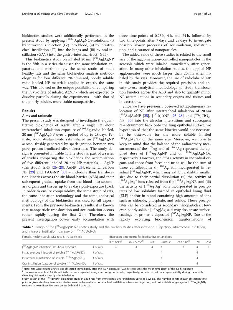
Table 1 Design of the [105Ag]AgNP biokinetics study and the auxiliary studies after intravenous injection, intratracheal instillation,and intra-oral instillation (gavage) of [110mAg]AgNO3
Female, healthy, adult WKY rats, 8–10 weeks old dissection time-points for biodistribution analyses
0.75 h/1sta 0.75 h/2ndb 4 h 24 h/1st 24 h/2ndb 7d 28d
[105Ag]AgNP Inhalation, 1½ -hour exposure # of rats 4 4 4 4 4 4 4
Intratravenous injection of soluble [110mAg]AgNO3 # of rats 4 4
Intratracheal instillation of soluble [110mAg]AgNO3 # of rats 4 4
Oral instillation (gavage) of soluble [110mAg]AgNO3 # of rats 4 4a Note: rats were exsanguinated and dissected immediately after the 1.5-h exposure; “0.75 h” represents the mean time-point of the 1.5-h exposureb The measurements at 0.75 h and 24 h p.e. were repeated using a second group of rats, respectively, in order to test data reproducibility during the rapidlychanging biokinetics directly after inhalationStudy design of the [105Ag]AgNP biokinetics study in adult rats from immediately after inhalation up to 28 days p.e. The number of rats at each dissection time-point is given. Auxiliary biokinetics studies were performed after intratracheal instillation, intravenous injection, and oral instillation (gavage) of [110mAg]AgNO3
solutions at two dissection time points 24 h and 7 days p.e.
Kreyling et al. Particle and Fibre Toxicology (2020) 17:21 Page 4 of 28
[105Ag]Ag+ ions in body fluids with their abundant presenceof chloride, phosphate and sulfide ions, it is rather unlikelyto find 105Ag activity in purely ionic form as summarized inthe Background section. It should be noted, however, thatfrom radioactivity measurements of 105Ag or 110mAg in or-gans, tissues and excretions it is not possible to distinguishbetween Ag+-ions or primary or secondary nanoparticles,which is a major difference with earlier experiments usingde facto unsoluble nanoparticles.
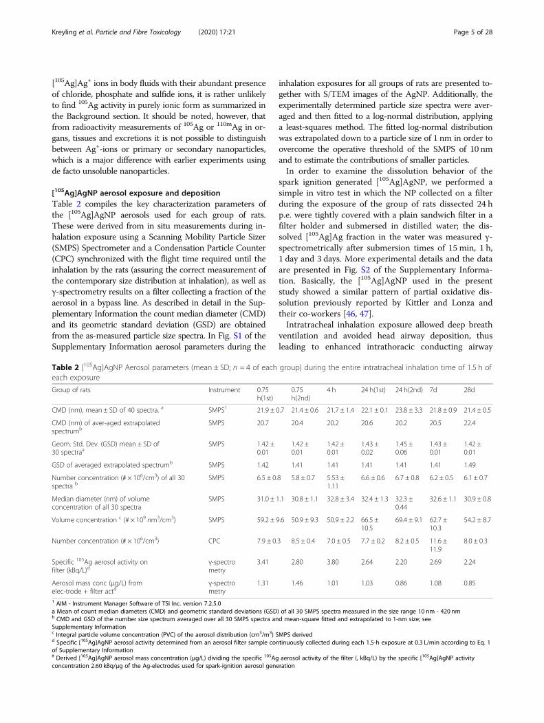
[105Ag]AgNP aerosol exposure and depositionTable 2 compiles the key characterization parameters ofthe [105Ag]AgNP aerosols used for each group of rats.These were derived from in situ measurements during in-halation exposure using a Scanning Mobility Particle Sizer(SMPS) Spectrometer and a Condensation Particle Counter(CPC) synchronized with the flight time required until theinhalation by the rats (assuring the correct measurement ofthe contemporary size distribution at inhalation), as well asγ-spectrometry results on a filter collecting a fraction of theaerosol in a bypass line. As described in detail in the Sup-plementary Information the count median diameter (CMD)and its geometric standard deviation (GSD) are obtainedfrom the as-measured particle size spectra. In Fig. S1 of theSupplementary Information aerosol parameters during the
inhalation exposures for all groups of rats are presented to-gether with S/TEM images of the AgNP. Additionally, theexperimentally determined particle size spectra were aver-aged and then fitted to a log-normal distribution, applyinga least-squares method. The fitted log-normal distributionwas extrapolated down to a particle size of 1 nm in order toovercome the operative threshold of the SMPS of 10 nmand to estimate the contributions of smaller particles.In order to examine the dissolution behavior of the
spark ignition generated [105Ag]AgNP, we performed asimple in vitro test in which the NP collected on a filterduring the exposure of the group of rats dissected 24 hp.e. were tightly covered with a plain sandwich filter in afilter holder and submersed in distilled water; the dis-solved [105Ag]Ag fraction in the water was measured γ-spectrometrically after submersion times of 15 min, 1 h,1 day and 3 days. More experimental details and the dataare presented in Fig. S2 of the Supplementary Informa-tion. Basically, the [105Ag]AgNP used in the presentstudy showed a similar pattern of partial oxidative dis-solution previously reported by Kittler and Lonza andtheir co-workers [46, 47].Intratracheal inhalation exposure allowed deep breath
ventilation and avoided head airway deposition, thusleading to enhanced intrathoracic conducting airway
Table 2 [105Ag]AgNP Aerosol parameters (mean ± SD; n = 4 of each group) during the entire intratracheal inhalation time of 1.5 h ofeach exposure
Group of rats Instrument 0.75h(1st)
0.75h(2nd)
4 h 24 h(1st) 24 h(2nd) 7d 28d
CMD (nm), mean ± SD of 40 spectra. a SMPS1 21.9 ± 0.7 21.4 ± 0.6 21.7 ± 1.4 22.1 ± 0.1 23.8 ± 3.3 21.8 ± 0.9 21.4 ± 0.5
CMD (nm) of aver-aged extrapolatedspectrumb
SMPS 20.7 20.4 20.2 20.6 20.2 20.5 22.4
Geom. Std. Dev. (GSD) mean ± SD of30 spectraa
SMPS 1.42 ±0.01
1.42 ±0.01
1.42 ±0.01
1.43 ±0.02
1.45 ±0.06
1.43 ±0.01
1.42 ±0.01
GSD of averaged extrapolated spectrumb SMPS 1.42 1.41 1.41 1.41 1.41 1.41 1.49
Number concentration (# × 106/cm3) of all 30spectra b
SMPS 6.5 ± 0.8 5.8 ± 0.7 5.53 ±1.11
6.6 ± 0.6 6.7 ± 0.8 6.2 ± 0.5 6.1 ± 0.7
Median diameter (nm) of volumeconcentration of all 30 spectra
SMPS 31.0 ± 1.1 30.8 ± 1.1 32.8 ± 3.4 32.4 ± 1.3 32.3 ±0.44
32.6 ± 1.1 30.9 ± 0.8
Volume concentration c (# × 109 nm3/cm3) SMPS 59.2 ± 9.6 50.9 ± 9.3 50.9 ± 2.2 66.5 ±10.5
69.4 ± 9.1 62.7 ±10.3
54.2 ± 8.7
Number concentration (# × 106/cm3) CPC 7.9 ± 0.3 8.5 ± 0.4 7.0 ± 0.5 7.7 ± 0.2 8.2 ± 0.5 11.6 ±11.9
8.0 ± 0.3
Specific 105Ag aerosol activity onfilter (kBq/L)d
γ-spectrometry
3.41 2.80 3.80 2.64 2.20 2.69 2.24
Aerosol mass conc (μg/L) fromelec-trode + filter actd
γ-spectrometry
1.31 1.46 1.01 1.03 0.86 1.08 0.85
1 AIM - Instrument Manager Software of TSI Inc. version 7.2.5.0a Mean of count median diameters (CMD) and geometric standard deviations (GSD) of all 30 SMPS spectra measured in the size range 10 nm - 420 nmb CMD and GSD of the number size spectrum averaged over all 30 SMPS spectra and mean-square fitted and extrapolated to 1-nm size; seeSupplementary Informationc Integral particle volume concentration (PVC) of the aerosol distribution (cm3/m3) SMPS derivedd Specific [105Ag]AgNP aerosol activity determined from an aerosol filter sample continuously collected during each 1.5-h exposure at 0.3 L/min according to Eq. 1of Supplementary Information# Derived [105Ag]AgNP aerosol mass concentration (μg/L) dividing the specific 105Ag aerosol activity of the filter (, kBq/L) by the specific [105Ag]AgNP activityconcentration 2.60 kBq/μg of the Ag-electrodes used for spark-ignition aerosol generation
Kreyling et al. Particle and Fibre Toxicology (2020) 17:21 Page 5 of 28
deposition as well as alveolar deposition, with long-termalveolar retention being the dominant outcome. Parame-ters of aerosol inhalation and deposition are compiled inTable 3 for each group of rats. In addition, the activityfractions cleared from the lungs that can be attributed toearly clearance into the gastro-intestinal-tract (GIT) andfeces up to 2 days p.e. and the long-term clearance of[105Ag]AgNP and their degradation products formed inthe retention period from 3 days up to 28 days p.e. aregiven for each group of rats. Note that the retained al-veolar [105Ag]AgNP fraction at the time of dissection di-minishes rapidly at days 7 and 28 p.e., due to the rapidtransformation and clearance of the [105Ag]AgNP. Witha mean rat bodyweight of 204 ± 13 g, the mean depositedAgNP mass per body weight is 125 μg•kg− 1.The estimated mean deposited [105Ag]AgNP fractions
relative to the inhaled aerosol show considerable inter-subject variability (as shown by their standard devia-tions) indicating that the tidal volume calculated fromEq. (2) of the Supplementary Information is only a roughestimate. In addition, the deposited fractions (per in-haled [105Ag]AgNP activity) in line 6 of Table 3 are con-sistently lower (0.3–0.4 for 24 h until 28d groups of rats)than the total deposited fraction of 0.6 as calculated bythe MPPD software 3.04 (see Fig. S5 of the Supplemen-tary Information). This may have resulted from low-pressure ventilation in the plethysmograph.
Auxiliary studies of the biokinetics after intravenousinjection and intratracheal instillation of soluble[105Ag]AgNO3 salt solutionsIn the auxiliary biokinetics study after IV injection of an[110mAg]AgNO3-solution in rats, the silver was mainlycleared into the GIT and almost completely excreted in
the feces with negligible urinary excretion. After IT in-stillation of an [110mAg]AgNO3-solution, we observed anAg-fraction in the GIT and feces which was muchhigher than expected for fast mucociliary clearance.Figure 1 shows the biodistributions of intravenously
injected, (IV, panel A) and of intratracheally instilled(IT, panel B), and of intraesophageal instilled (GAV,panel C) [110mAg]AgNO3 salt solutions (fully dissociatedat the time of application) in four groups of rats (n = 4)after 24 h and 7 days, respectively. Already 24 h after IVinjection (Fig. 1a), a fraction of 0.96 was eliminated fromthe blood circulation. Small fractions (< 0.04) wereretained in secondary organs and the tissues of theremaining carcass. In contrast, predominant fractionswere found in GIT (0.63) and feces (0.24). When distin-guishing the GIT into its compartments - stomach, smallintestine, and hindgut -, 110mAg activity fractions of0.002, 0.017 and 0.61, respectively, were found 24 h p.e.These data confirm that after IV-injection the passageinto and through the small intestine is fast and almostcomplete within 24 h p.e. Noteworthy is the minimalurinary excretion of 0.005 24 h p.e. (and 0.0005 afterGAV). After 1 week the fractions in all secondary organsand the carcass had decreased tenfold and more, but inthe GIT the decrease was even 1000-fold due to almostcomplete fecal excretion of [110mAg]Ag.Only an [110mAg]Ag fraction of about 0.14 of the intra-
tracheally instilled material is retained in the lungs andBAL 24 h p.e. (Fig. 1b) which continues to slightly de-crease during the following week to 0.07. Thus 24 h afterIT instillation, a fraction of 0.86 has been translocatedacross the ABB into blood and fractions of 0.48 and 0.22were found in the GIT in feces, respectively. In contrast,retained fractions in the liver, kidneys (~ 0.02 each) and
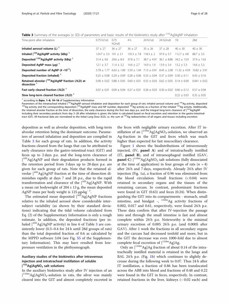
Table 3 Summary of the averages (± SD) of parameters and basic results of the biokinetics study after [105Ag]AgNP inhalation
Time-point after inhalation 0.75 h(1st) 0.75h(2nd)
4 h 24 h(1st) 24 h(2nd) 7d 28d
Inhaled aerosol volume (L) 1 37 ± 27 36 ± 27 36 ± 27 35 ± 26 37 ± 28 40 ± 30 40 ± 30
Inhaled [105Ag]AgNP activity (kBq) 1 124.7 ± 3.3 101 ± 3.1 135.3 ± 7.8 118.3 ± 2 97.9 ± 5.7 112.7 ± 4.8 88.7 ± 3.5
Deposited [105Ag]AgNP activity (kBq) 1 31.4 ± 9.6 29.6 ± 8.4 37.8 ± 7.1 38.7 ± 4.91 36.1 ± 8.86 34.2 ± 13.9 37.9 ± 13.6
Deposited AgNP mass (μg) 1 12.1 ± 3.7 11.4 ± 3.2 14.6 ± 2.7 14.9 ± 1.9 13.9 ± 3.4 13.2 ± 5.3 14.6 ± 5.3
Deposited number of AgNP (# •1011) 5.78 ± 1.77 6.63 ± 1.89 5.59 ± 1.04 7.13 ± 0.91 8.45 ± 2.08 11.32 ± 4.59 10.82 ± 3.91
Deposited fraction (inhaled) 1 0.25 ± 0.08 0.29 ± 0.09 0.28 ± 0.06 0.33 ± 0.04 0.37 ± 0.09 0.30 ± 0.11 0.43 ± 0.16
Retained alveolar [105Ag]AgNP fraction (/ILD) atdissection 1
0.90 ± 0.02 0.88 ± 0.05 0.69 ± 0.01 0.55 ± 0.03 0.62 ± 0.05 0.14 ± 0.09 0.041 ± 0.02
Fast early cleared fraction (/ILD) 1 0.07 ± 0.01 0.09 ± 0.04 0.27 ± 0.01 0.36 ± 0.03 0.30 ± 0.02 0.60 ± 0.12 0.57 ± 0.04
Slow long-term cleared fraction (/ILD) 1 0.25 ± 0.03 0.35 ± 0.051 according to Eqns. 1–8, 10–16 of Supplementary InformationParameters of the intratracheal inhaled [105Ag]AgNP aerosol inhalation and deposition for each group of rats: inhaled aerosol volume and 105Ag activity, deposited105Ag activity and the corresponding deposited [105Ag]AgNP mass and NP number, deposited 105Ag activity as a fraction of the inhaled 105Ag activity. Additionally,the retained alveolar fraction at the time of dissection, the early clearance during the first two days p.e. and the integral long-term clearance of [105Ag]AgNPincluding their secondary products from day 3–28 after inhalation is given; the latter is calculated based on fecal excretion and retention in the gastro-intestinal-tract (GIT). All fractional data are normalized to the Initial Lung Dose (ILD), i.e. the sum of 105Ag radioactivities of all organs and tissues including excretion
Kreyling et al. Particle and Fibre Toxicology (2020) 17:21 Page 6 of 28
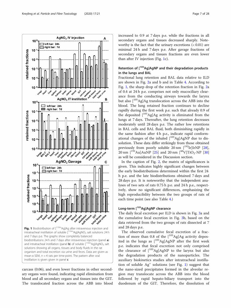
carcass (0.06), and even lower fractions in other second-ary organs were found, indicating rapid elimination fromblood and all secondary organs and tissues into the GIT.The translocated fraction across the ABB into blood
increased to 0.9 at 7 days p.e. while the fractions in allsecondary organs and tissues decreased sharply. Note-worthy is the fact that the urinary excretions (< 0.01) areminimal 24 h and 7 days p.e. After gavage fractions ofsecondary organs and tissues fractions are even lowerthan after IV injection (Fig. 1c).
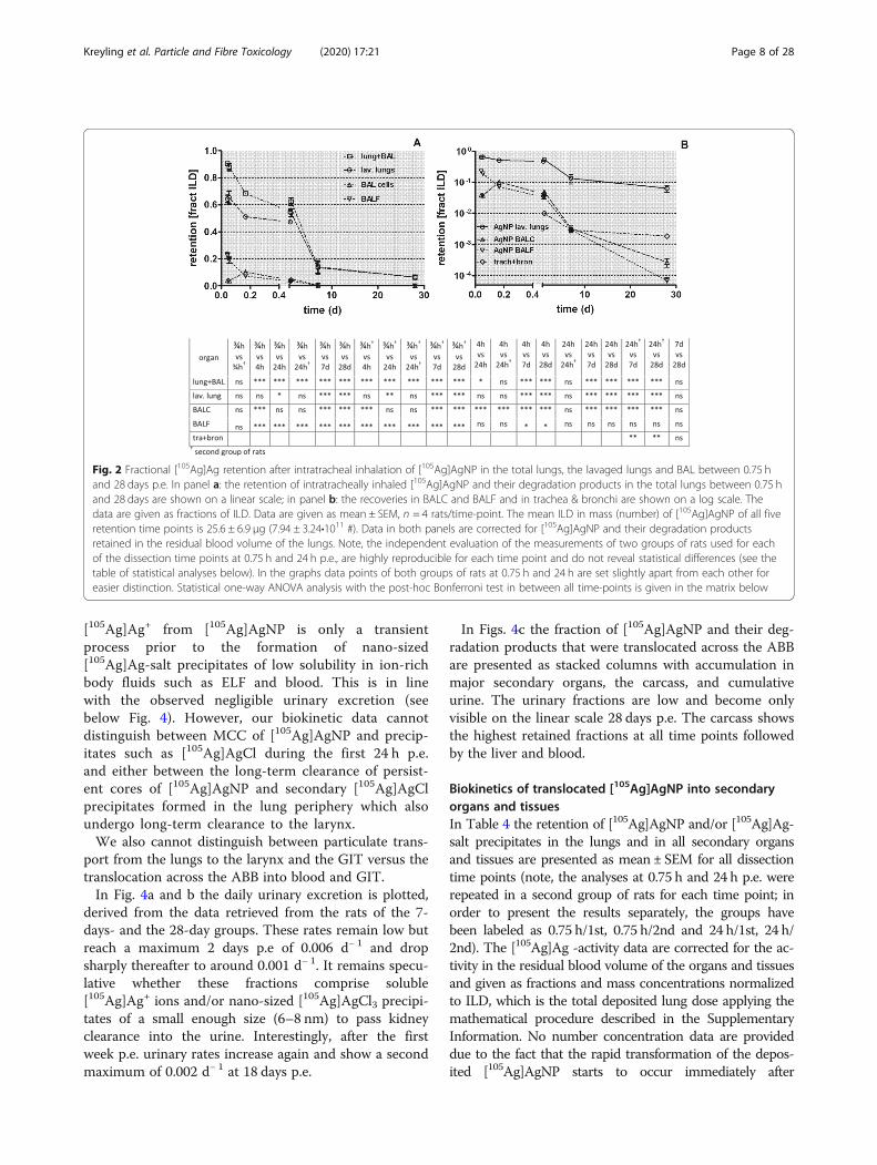
Retention of [105Ag]AgNP and their degradation productsin the lungs and BALFractional lung retention and BAL data relative to ILDare shown in Fig. 2a and b and in Table 4. According toFig. 1, the sharp drop of the retention fraction in Fig. 2aof 0.4 at 24 h p.e. comprises not only mucociliary clear-ance from the conducting airways towards the larynxbut also [105Ag]Ag translocation across the ABB into theblood. The lung retained fraction continues to declinerapidly during the first week p.e. such that already 0.9 ofthe deposited [105Ag]Ag activity is eliminated from thelungs at 7 days. Thereafter, the lung retention decreasesmoderately until 28 days p.e. The rather low retentionsin BAL cells and BAL fluid, both diminishing rapidly inthe same fashion after 4 h p.e., indicate rapid conform-ational changes of the inhaled [105Ag]AgNP due to dis-solution. These data differ strikingly from those obtainedpreviously from poorly soluble 20 nm [192Ir]IrNP [28],20 nm [195Au]AuNP [25] and 20 nm [48V]TiO2-NP [30]as will be considered in the Discussion section.In the caption of Fig. 2, the matrix of significances is
given. This indicates highly significant changes betweenthe early biodistributions determined within the first 24h p.e. and the late biodistributions obtained 7 days and28 days p.e. It is noteworthy that the independent ana-lyses of two sets of rats 0.75 h p.e. and 24 h p.e., respect-ively, show no significant differences, emphasizing thehigh reproducibility between the two groups of rats ofeach time point (see also Table 4.)
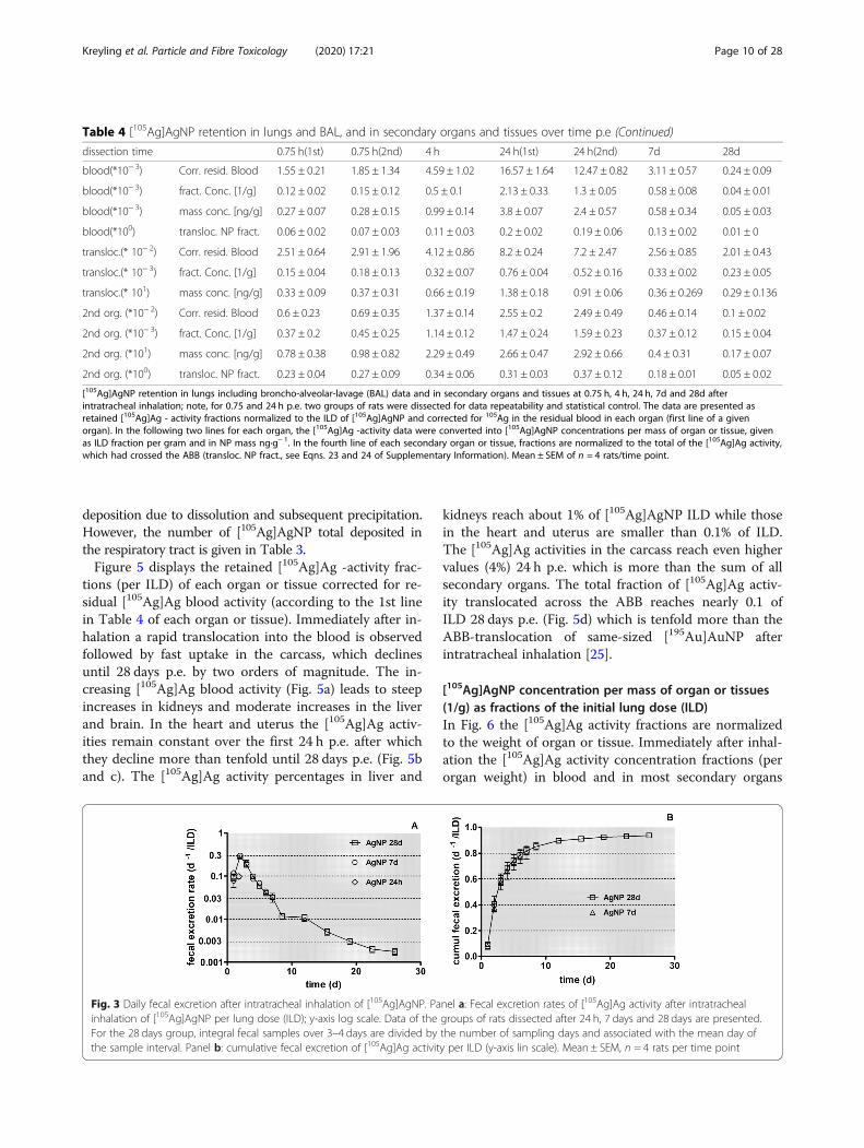
Long-term [105Ag]AgNP clearanceThe daily fecal excretion per ILD is shown in Fig. 3a andthe cumulative fecal excretion in Fig. 3b, based on thedata retrieved from the two groups of rats dissected at 7and 28 days p.e.The observed cumulative fecal excretion of a frac-
tion of more than 0.8 of the [105Ag]Ag activity depos-ited in the lungs as [105Ag]AgNP after the first weekp.e. indicates that fecal excretion not only comprisedthe clearance of [105Ag]AgNP to the larynx but alsothe degradation products of the nanoparticles. Theauxiliary biokinetics studies after intratracheal instilla-tion of soluble Ag+ solutions (see Fig. 1) suggest thatthe nano-sized precipitates formed in the alveolar re-gion may translocate across the ABB into the bloodfollowed by rapid hepato-biliary transport into theduodenum of the GIT. Therefore, the dissolution of
Fig. 1 Biodistribution of [110mAg]Ag after intravenous injection andintratracheal instillation of soluble [110mAg]AgNO3 salt solutions 24 hand 7 days p.e. The graphs show completely balancedbiodistributions 24 h and 7 days after intravenous injection (panel a)and intratracheal instillation (panel b) of soluble [110mAg]AgNO3 saltsolutions showing all organs, tissues and body fluids in the ratorganism and total excretion via urine and feces. Data are given asmean ± SEM, n = 4 rats per time-point. The pattern after oralinstillation is given given in panel c
Kreyling et al. Particle and Fibre Toxicology (2020) 17:21 Page 7 of 28
[105Ag]Ag+ from [105Ag]AgNP is only a transientprocess prior to the formation of nano-sized[105Ag]Ag-salt precipitates of low solubility in ion-richbody fluids such as ELF and blood. This is in linewith the observed negligible urinary excretion (seebelow Fig. 4). However, our biokinetic data cannotdistinguish between MCC of [105Ag]AgNP and precip-itates such as [105Ag]AgCl during the first 24 h p.e.and either between the long-term clearance of persist-ent cores of [105Ag]AgNP and secondary [105Ag]AgClprecipitates formed in the lung periphery which alsoundergo long-term clearance to the larynx.We also cannot distinguish between particulate trans-
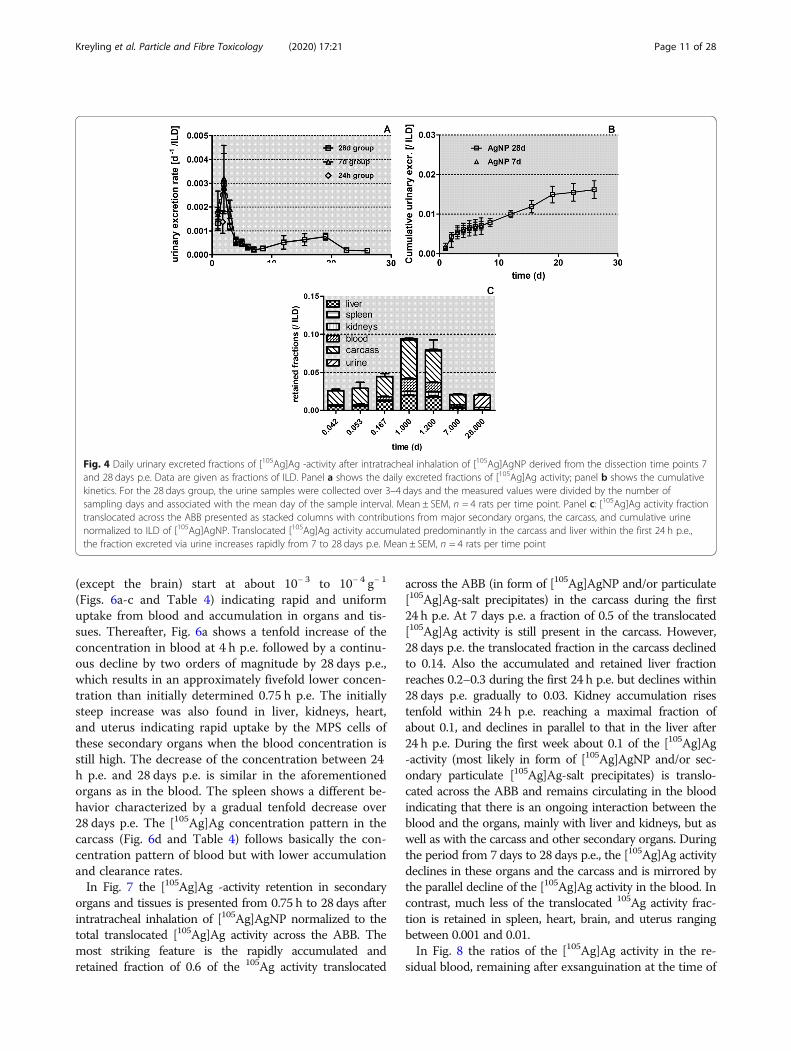
port from the lungs to the larynx and the GIT versus thetranslocation across the ABB into blood and GIT.In Fig. 4a and b the daily urinary excretion is plotted,
derived from the data retrieved from the rats of the 7-days- and the 28-day groups. These rates remain low butreach a maximum 2 days p.e of 0.006 d− 1 and dropsharply thereafter to around 0.001 d− 1. It remains specu-lative whether these fractions comprise soluble[105Ag]Ag+ ions and/or nano-sized [105Ag]AgCl3 precipi-tates of a small enough size (6–8 nm) to pass kidneyclearance into the urine. Interestingly, after the firstweek p.e. urinary rates increase again and show a secondmaximum of 0.002 d− 1 at 18 days p.e.
In Figs. 4c the fraction of [105Ag]AgNP and their deg-radation products that were translocated across the ABBare presented as stacked columns with accumulation inmajor secondary organs, the carcass, and cumulativeurine. The urinary fractions are low and become onlyvisible on the linear scale 28 days p.e. The carcass showsthe highest retained fractions at all time points followedby the liver and blood.
Biokinetics of translocated [105Ag]AgNP into secondaryorgans and tissuesIn Table 4 the retention of [105Ag]AgNP and/or [105Ag]Ag-salt precipitates in the lungs and in all secondary organsand tissues are presented as mean ± SEM for all dissectiontime points (note, the analyses at 0.75 h and 24 h p.e. wererepeated in a second group of rats for each time point; inorder to present the results separately, the groups havebeen labeled as 0.75 h/1st, 0.75 h/2nd and 24 h/1st, 24 h/2nd). The [105Ag]Ag -activity data are corrected for the ac-tivity in the residual blood volume of the organs and tissuesand given as fractions and mass concentrations normalizedto ILD, which is the total deposited lung dose applying themathematical procedure described in the SupplementaryInformation. No number concentration data are provideddue to the fact that the rapid transformation of the depos-ited [105Ag]AgNP starts to occur immediately after
Fig. 2 Fractional [105Ag]Ag retention after intratracheal inhalation of [105Ag]AgNP in the total lungs, the lavaged lungs and BAL between 0.75 hand 28 days p.e. In panel a: the retention of intratracheally inhaled [105Ag]AgNP and their degradation products in the total lungs between 0.75 hand 28 days are shown on a linear scale; in panel b: the recoveries in BALC and BALF and in trachea & bronchi are shown on a log scale. Thedata are given as fractions of ILD. Data are given as mean ± SEM, n = 4 rats/time-point. The mean ILD in mass (number) of [105Ag]AgNP of all fiveretention time points is 25.6 ± 6.9 μg (7.94 ± 3.24•1011 #). Data in both panels are corrected for [105Ag]AgNP and their degradation productsretained in the residual blood volume of the lungs. Note, the independent evaluation of the measurements of two groups of rats used for eachof the dissection time points at 0.75 h and 24 h p.e., are highly reproducible for each time point and do not reveal statistical differences (see thetable of statistical analyses below). In the graphs data points of both groups of rats at 0.75 h and 24 h are set slightly apart from each other foreasier distinction. Statistical one-way ANOVA analysis with the post-hoc Bonferroni test in between all time-points is given in the matrix below
Kreyling et al. Particle and Fibre Toxicology (2020) 17:21 Page 8 of 28
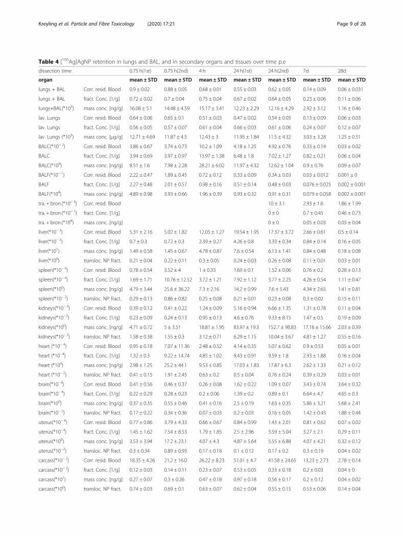
Table 4 [105Ag]AgNP retention in lungs and BAL, and in secondary organs and tissues over time p.e
dissection time 0.75 h(1st) 0.75 h(2nd) 4 h 24 h(1st) 24 h(2nd) 7d 28d
organ mean ± STD mean ± STD mean ± STD mean ± STD mean ± STD mean ± STD mean ± STD
lungs + BAL Corr. resid. Blood 0.9 ± 0.02 0.88 ± 0.05 0.68 ± 0.01 0.55 ± 0.03 0.62 ± 0.05 0.14 ± 0.09 0.06 ± 0.031
lungs + BAL fract. Conc. [1/g] 0.72 ± 0.02 0.7 ± 0.04 0.75 ± 0.04 0.67 ± 0.02 0.64 ± 0.05 0.23 ± 0.06 0.11 ± 0.06
lungs+BAL(*103) mass conc. [ng/g] 16.08 ± 5.1 14.48 ± 4.59 15.17 ± 3.41 12.23 ± 2.29 12.16 ± 4.29 2.92 ± 3.12 1.16 ± 0.46
lav. Lungs Corr. resid. Blood 0.64 ± 0.06 0.65 ± 0.1 0.51 ± 0.03 0.47 ± 0.02 0.54 ± 0.05 0.13 ± 0.09 0.06 ± 0.03
lav. Lungs fract. Conc. [1/g] 0.56 ± 0.05 0.57 ± 0.07 0.61 ± 0.04 0.66 ± 0.03 0.61 ± 0.06 0.24 ± 0.07 0.12 ± 0.07
lav. Lungs (*103) mass conc. [μg/g] 12.71 ± 4.69 11.87 ± 4.5 12.43 ± 3 11.95 ± 1.84 11.5 ± 4.32 3.03 ± 3.28 1.25 ± 0.51
BALC(*10− 2) Corr. resid. Blood 3.86 ± 0.67 3.74 ± 0.73 10.2 ± 1.09 4.18 ± 1.25 4.92 ± 0.76 0.33 ± 0.14 0.03 ± 0.02
BALC fract. Conc. [1/g] 3.94 ± 0.69 3.97 ± 0.97 13.97 ± 1.38 6.48 ± 1.8 7.02 ± 1.27 0.82 ± 0.21 0.06 ± 0.04
BALC(*104) mass conc. [ng/g] 8.51 ± 1.6 7.98 ± 2.28 28.21 ± 6.02 11.97 ± 4.32 12.62 ± 1.04 0.9 ± 0.76 0.09 ± 0.07
BALF(*10− 1) Corr. resid. Blood 2.22 ± 0.47 1.89 ± 0.45 0.72 ± 0.12 0.33 ± 0.09 0.34 ± 0.03 0.03 ± 0.012 0.001 ± 0
BALF fract. Conc. [1/g] 2.27 ± 0.48 2.01 ± 0.57 0.98 ± 0.16 0.51 ± 0.14 0.48 ± 0.03 0.076 ± 0.025 0.002 ± 0.001
BALF(*104) mass conc. [ng/g] 4.89 ± 0.98 3.93 ± 0.66 1.96 ± 0.39 0.93 ± 0.32 0.91 ± 0.31 0.079 ± 0.058 0.002 ± 0.001
tra. + bron.(*10− 3) Corr. resid. Blood 10 ± 3.1 2.93 ± 1.6 1.86 ± 1.99
tra. + bron.(*10− 1) fract. Conc. [1/g] 0 ± 0 0.7 ± 0.45 0.46 ± 0.73
tra. + bron.(*104) mass conc. [ng/g] 0 ± 0 0.05 ± 0.03 0.03 ± 0.04
liver(*10− 3) Corr. resid. Blood 5.31 ± 2.16 5.02 ± 1.82 12.05 ± 1.27 19.54 ± 1.95 17.37 ± 3.72 2.66 ± 0.61 0.5 ± 0.14
liver(*10− 3) fract. Conc. [1/g] 0.7 ± 0.3 0.72 ± 0.3 2.39 ± 0.27 4.26 ± 0.8 3.33 ± 0.34 0.84 ± 0.14 0.16 ± 0.05
liver(*101) mass conc. [ng/g] 1.49 ± 0.58 1.45 ± 0.67 4.78 ± 0.87 7.6 ± 0.54 6.13 ± 1.41 0.84 ± 0.48 0.18 ± 0.08
liver(*100) transloc. NP fract. 0.21 ± 0.04 0.22 ± 0.11 0.3 ± 0.05 0.24 ± 0.03 0.26 ± 0.08 0.11 ± 0.01 0.03 ± 0.01
spleen(*10− 4) Corr. resid. Blood 0.78 ± 0.54 3.52 ± 4 1 ± 0.33 1.69 ± 0.1 1.52 ± 0.06 0.76 ± 0.2 0.28 ± 0.13
spleen(*10− 4) fract. Conc. [1/g] 1.69 ± 1.71 10.76 ± 12.52 3.72 ± 1.21 7.92 ± 1.12 3.77 ± 2.25 4.26 ± 0.54 1.11 ± 0.47
spleen(*100) mass conc. [ng/g] 4.79 ± 3.44 25.6 ± 36.22 7.3 ± 2.16 14.2 ± 0.99 7.6 ± 5.43 4.34 ± 2.65 1.41 ± 0.81
spleen(*10− 2) transloc. NP fract. 0.29 ± 0.13 0.86 ± 0.82 0.25 ± 0.08 0.21 ± 0.01 0.23 ± 0.08 0.3 ± 0.02 0.15 ± 0.11
kidneys(*10− 3) Corr. resid. Blood 0.39 ± 0.12 0.41 ± 0.22 1.24 ± 0.09 5.16 ± 0.94 6.66 ± 1.35 1.31 ± 0.78 0.11 ± 0.04
kidneys(*10−3) fract. Conc. [1/g] 0.23 ± 0.09 0.24 ± 0.13 0.95 ± 0.13 4.6 ± 0.76 9.33 ± 8.15 1.47 ± 0.5 0.19 ± 0.09
kidneys(*100) mass conc. [ng/g] 4.71 ± 0.72 5 ± 3.51 18.81 ± 1.95 83.97 ± 19.3 152.7 ± 98.83 17.16 ± 15.66 2.03 ± 0.39
kidneys(*10− 2) transloc. NP fract. 1.58 ± 0.38 1.55 ± 0.3 3.12 ± 0.71 6.29 ± 1.15 10.04 ± 3.67 4.81 ± 1.27 0.55 ± 0.16
heart (*10− 4) Corr. resid. Blood 0.95 ± 0.18 7.07 ± 11.36 2.48 ± 0.52 4.14 ± 0.35 5.07 ± 0.62 0.9 ± 0.53 0.05 ± 0.01
heart (*10−4) fract. Conc. [1/g] 1.32 ± 0.3 9.22 ± 14.74 4.85 ± 1.02 9.43 ± 0.91 9.59 ± 1.8 2.93 ± 1.88 0.16 ± 0.04
heart (*100) mass conc. [ng/g] 2.98 ± 1.25 25.2 ± 44.1 9.53 ± 0.85 17.03 ± 1.83 17.87 ± 6.3 2.62 ± 1.33 0.21 ± 0.12
heart (*10− 2) transloc. NP fract. 0.41 ± 0.15 1.91 ± 2.45 0.63 ± 0.2 0.5 ± 0.04 0.76 ± 0.24 0.39 ± 0.29 0.03 ± 0.01
brain(*10−4) Corr. resid. Blood 0.41 ± 0.56 0.46 ± 0.37 0.26 ± 0.08 1.62 ± 0.22 1.09 ± 0.07 3.43 ± 0.74 3.64 ± 0.32
brain(*10− 4) fract. Conc. [1/g] 0.22 ± 0.29 0.28 ± 0.23 0.2 ± 0.06 1.39 ± 0.2 0.89 ± 0.1 6.64 ± 4.7 4.65 ± 0.3
brain(*100) mass conc. [ng/g] 0.37 ± 0.35 0.55 ± 0.46 0.41 ± 0.16 2.5 ± 0.19 1.63 ± 0.35 5.86 ± 3.21 5.68 ± 2.41
brain(*10− 2) transloc. NP fract. 0.17 ± 0.22 0.34 ± 0.36 0.07 ± 0.03 0.2 ± 0.03 0.16 ± 0.05 1.42 ± 0.43 1.88 ± 0.44
uterus(*10− 4) Corr. resid. Blood 0.77 ± 0.86 3.79 ± 4.33 0.66 ± 0.67 0.84 ± 0.99 1.43 ± 2.01 0.81 ± 0.62 0.07 ± 0.02
uterus(*10−4) fract. Conc. [1/g] 1.45 ± 1.62 7.54 ± 8.53 1.79 ± 1.85 2.5 ± 2.96 3.59 ± 5.04 3.27 ± 2.1 0.29 ± 0.11
uterus(*100) mass conc. [ng/g] 3.53 ± 3.94 17.2 ± 23.1 4.07 ± 4.3 4.87 ± 5.64 5.55 ± 6.88 4.07 ± 4.21 0.32 ± 0.12
uterus(*10−2) transloc. NP fract. 0.3 ± 0.34 0.89 ± 0.93 0.17 ± 0.18 0.1 ± 0.12 0.17 ± 0.2 0.3 ± 0.19 0.04 ± 0.02
carcass(*10− 2) Corr. resid. Blood 18.35 ± 4.26 21.2 ± 16.0 26.22 ± 8.23 51.01 ± 4.7 41.58 ± 24.65 13.23 ± 2.73 2.78 ± 0.14
carcass(*10− 2) fract. Conc. [1/g] 0.12 ± 0.03 0.14 ± 0.11 0.23 ± 0.07 0.53 ± 0.05 0.33 ± 0.18 0.2 ± 0.03 0.04 ± 0
carcass(*101) mass conc. [ng/g] 0.27 ± 0.07 0.3 ± 0.26 0.47 ± 0.18 0.97 ± 0.18 0.56 ± 0.17 0.2 ± 0.12 0.04 ± 0.02
carcass(*100) transloc. NP fract. 0.74 ± 0.03 0.69 ± 0.1 0.63 ± 0.07 0.62 ± 0.04 0.55 ± 0.15 0.53 ± 0.06 0.14 ± 0.04
Kreyling et al. Particle and Fibre Toxicology (2020) 17:21 Page 9 of 28
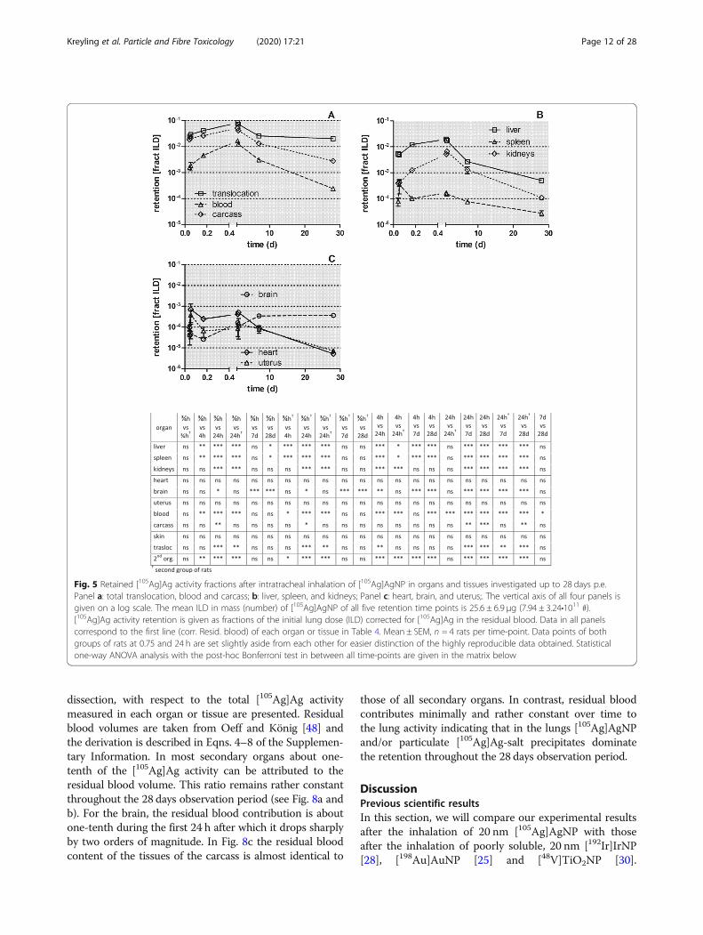
deposition due to dissolution and subsequent precipitation.However, the number of [105Ag]AgNP total deposited inthe respiratory tract is given in Table 3.Figure 5 displays the retained [105Ag]Ag -activity frac-
tions (per ILD) of each organ or tissue corrected for re-sidual [105Ag]Ag blood activity (according to the 1st linein Table 4 of each organ or tissue). Immediately after in-halation a rapid translocation into the blood is observedfollowed by fast uptake in the carcass, which declinesuntil 28 days p.e. by two orders of magnitude. The in-creasing [105Ag]Ag blood activity (Fig. 5a) leads to steepincreases in kidneys and moderate increases in the liverand brain. In the heart and uterus the [105Ag]Ag activ-ities remain constant over the first 24 h p.e. after whichthey decline more than tenfold until 28 days p.e. (Fig. 5band c). The [105Ag]Ag activity percentages in liver and
kidneys reach about 1% of [105Ag]AgNP ILD while thosein the heart and uterus are smaller than 0.1% of ILD.The [105Ag]Ag activities in the carcass reach even highervalues (4%) 24 h p.e. which is more than the sum of allsecondary organs. The total fraction of [105Ag]Ag activ-ity translocated across the ABB reaches nearly 0.1 ofILD 28 days p.e. (Fig. 5d) which is tenfold more than theABB-translocation of same-sized [195Au]AuNP afterintratracheal inhalation [25].
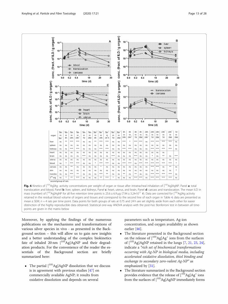
[105Ag]AgNP concentration per mass of organ or tissues(1/g) as fractions of the initial lung dose (ILD)In Fig. 6 the [105Ag]Ag activity fractions are normalizedto the weight of organ or tissue. Immediately after inhal-ation the [105Ag]Ag activity concentration fractions (perorgan weight) in blood and in most secondary organs
Table 4 [105Ag]AgNP retention in lungs and BAL, and in secondary organs and tissues over time p.e (Continued)
dissection time 0.75 h(1st) 0.75 h(2nd) 4 h 24 h(1st) 24 h(2nd) 7d 28d
blood(*10− 3) Corr. resid. Blood 1.55 ± 0.21 1.85 ± 1.34 4.59 ± 1.02 16.57 ± 1.64 12.47 ± 0.82 3.11 ± 0.57 0.24 ± 0.09
blood(*10− 3) fract. Conc. [1/g] 0.12 ± 0.02 0.15 ± 0.12 0.5 ± 0.1 2.13 ± 0.33 1.3 ± 0.05 0.58 ± 0.08 0.04 ± 0.01
blood(*10− 3) mass conc. [ng/g] 0.27 ± 0.07 0.28 ± 0.15 0.99 ± 0.14 3.8 ± 0.07 2.4 ± 0.57 0.58 ± 0.34 0.05 ± 0.03
blood(*100) transloc. NP fract. 0.06 ± 0.02 0.07 ± 0.03 0.11 ± 0.03 0.2 ± 0.02 0.19 ± 0.06 0.13 ± 0.02 0.01 ± 0
transloc.(* 10− 2) Corr. resid. Blood 2.51 ± 0.64 2.91 ± 1.96 4.12 ± 0.86 8.2 ± 0.24 7.2 ± 2.47 2.56 ± 0.85 2.01 ± 0.43
transloc.(* 10− 3) fract. Conc. [1/g] 0.15 ± 0.04 0.18 ± 0.13 0.32 ± 0.07 0.76 ± 0.04 0.52 ± 0.16 0.33 ± 0.02 0.23 ± 0.05
transloc.(* 101) mass conc. [ng/g] 0.33 ± 0.09 0.37 ± 0.31 0.66 ± 0.19 1.38 ± 0.18 0.91 ± 0.06 0.36 ± 0.269 0.29 ± 0.136
2nd org. (*10− 2) Corr. resid. Blood 0.6 ± 0.23 0.69 ± 0.35 1.37 ± 0.14 2.55 ± 0.2 2.49 ± 0.49 0.46 ± 0.14 0.1 ± 0.02
2nd org. (*10− 3) fract. Conc. [1/g] 0.37 ± 0.2 0.45 ± 0.25 1.14 ± 0.12 1.47 ± 0.24 1.59 ± 0.23 0.37 ± 0.12 0.15 ± 0.04
2nd org. (*101) mass conc. [ng/g] 0.78 ± 0.38 0.98 ± 0.82 2.29 ± 0.49 2.66 ± 0.47 2.92 ± 0.66 0.4 ± 0.31 0.17 ± 0.07
2nd org. (*100) transloc. NP fract. 0.23 ± 0.04 0.27 ± 0.09 0.34 ± 0.06 0.31 ± 0.03 0.37 ± 0.12 0.18 ± 0.01 0.05 ± 0.02
[105Ag]AgNP retention in lungs including broncho-alveolar-lavage (BAL) data and in secondary organs and tissues at 0.75 h, 4 h, 24 h, 7d and 28d afterintratracheal inhalation; note, for 0.75 and 24 h p.e. two groups of rats were dissected for data repeatability and statistical control. The data are presented asretained [105Ag]Ag - activity fractions normalized to the ILD of [105Ag]AgNP and corrected for 105Ag in the residual blood in each organ (first line of a givenorgan). In the following two lines for each organ, the [105Ag]Ag -activity data were converted into [105Ag]AgNP concentrations per mass of organ or tissue, givenas ILD fraction per gram and in NP mass ng·g− 1. In the fourth line of each secondary organ or tissue, fractions are normalized to the total of the [105Ag]Ag activity,which had crossed the ABB (transloc. NP fract., see Eqns. 23 and 24 of Supplementary Information). Mean ± SEM of n = 4 rats/time point.
Fig. 3 Daily fecal excretion after intratracheal inhalation of [105Ag]AgNP. Panel a: Fecal excretion rates of [105Ag]Ag activity after intratrachealinhalation of [105Ag]AgNP per lung dose (ILD); y-axis log scale. Data of the groups of rats dissected after 24 h, 7 days and 28 days are presented.For the 28 days group, integral fecal samples over 3–4 days are divided by the number of sampling days and associated with the mean day ofthe sample interval. Panel b: cumulative fecal excretion of [105Ag]Ag activity per ILD (y-axis lin scale). Mean ± SEM, n = 4 rats per time point
Kreyling et al. Particle and Fibre Toxicology (2020) 17:21 Page 10 of 28
(except the brain) start at about 10− 3 to 10− 4 g− 1
(Figs. 6a-c and Table 4) indicating rapid and uniformuptake from blood and accumulation in organs and tis-sues. Thereafter, Fig. 6a shows a tenfold increase of theconcentration in blood at 4 h p.e. followed by a continu-ous decline by two orders of magnitude by 28 days p.e.,which results in an approximately fivefold lower concen-tration than initially determined 0.75 h p.e. The initiallysteep increase was also found in liver, kidneys, heart,and uterus indicating rapid uptake by the MPS cells ofthese secondary organs when the blood concentration isstill high. The decrease of the concentration between 24h p.e. and 28 days p.e. is similar in the aforementionedorgans as in the blood. The spleen shows a different be-havior characterized by a gradual tenfold decrease over28 days p.e. The [105Ag]Ag concentration pattern in thecarcass (Fig. 6d and Table 4) follows basically the con-centration pattern of blood but with lower accumulationand clearance rates.In Fig. 7 the [105Ag]Ag -activity retention in secondary
organs and tissues is presented from 0.75 h to 28 days afterintratracheal inhalation of [105Ag]AgNP normalized to thetotal translocated [105Ag]Ag activity across the ABB. Themost striking feature is the rapidly accumulated andretained fraction of 0.6 of the 105Ag activity translocated
across the ABB (in form of [105Ag]AgNP and/or particulate[105Ag]Ag-salt precipitates) in the carcass during the first24 h p.e. At 7 days p.e. a fraction of 0.5 of the translocated[105Ag]Ag activity is still present in the carcass. However,28 days p.e. the translocated fraction in the carcass declinedto 0.14. Also the accumulated and retained liver fractionreaches 0.2–0.3 during the first 24 h p.e. but declines within28 days p.e. gradually to 0.03. Kidney accumulation risestenfold within 24 h p.e. reaching a maximal fraction ofabout 0.1, and declines in parallel to that in the liver after24 h p.e. During the first week about 0.1 of the [105Ag]Ag-activity (most likely in form of [105Ag]AgNP and/or sec-ondary particulate [105Ag]Ag-salt precipitates) is translo-cated across the ABB and remains circulating in the bloodindicating that there is an ongoing interaction between theblood and the organs, mainly with liver and kidneys, but aswell as with the carcass and other secondary organs. Duringthe period from 7 days to 28 days p.e., the [105Ag]Ag activitydeclines in these organs and the carcass and is mirrored bythe parallel decline of the [105Ag]Ag activity in the blood. Incontrast, much less of the translocated 105Ag activity frac-tion is retained in spleen, heart, brain, and uterus rangingbetween 0.001 and 0.01.In Fig. 8 the ratios of the [105Ag]Ag activity in the re-
sidual blood, remaining after exsanguination at the time of
Fig. 4 Daily urinary excreted fractions of [105Ag]Ag -activity after intratracheal inhalation of [105Ag]AgNP derived from the dissection time points 7and 28 days p.e. Data are given as fractions of ILD. Panel a shows the daily excreted fractions of [105Ag]Ag activity; panel b shows the cumulativekinetics. For the 28 days group, the urine samples were collected over 3–4 days and the measured values were divided by the number ofsampling days and associated with the mean day of the sample interval. Mean ± SEM, n = 4 rats per time point. Panel c: [105Ag]Ag activity fractiontranslocated across the ABB presented as stacked columns with contributions from major secondary organs, the carcass, and cumulative urinenormalized to ILD of [105Ag]AgNP. Translocated [105Ag]Ag activity accumulated predominantly in the carcass and liver within the first 24 h p.e.,the fraction excreted via urine increases rapidly from 7 to 28 days p.e. Mean ± SEM, n = 4 rats per time point
Kreyling et al. Particle and Fibre Toxicology (2020) 17:21 Page 11 of 28
dissection, with respect to the total [105Ag]Ag activitymeasured in each organ or tissue are presented. Residualblood volumes are taken from Oeff and König [48] andthe derivation is described in Eqns. 4–8 of the Supplemen-tary Information. In most secondary organs about one-tenth of the [105Ag]Ag activity can be attributed to theresidual blood volume. This ratio remains rather constantthroughout the 28 days observation period (see Fig. 8a andb). For the brain, the residual blood contribution is aboutone-tenth during the first 24 h after which it drops sharplyby two orders of magnitude. In Fig. 8c the residual bloodcontent of the tissues of the carcass is almost identical to
those of all secondary organs. In contrast, residual bloodcontributes minimally and rather constant over time tothe lung activity indicating that in the lungs [105Ag]AgNPand/or particulate [105Ag]Ag-salt precipitates dominatethe retention throughout the 28 days observation period.
DiscussionPrevious scientific resultsIn this section, we will compare our experimental resultsafter the inhalation of 20 nm [105Ag]AgNP with thoseafter the inhalation of poorly soluble, 20 nm [192Ir]IrNP[28], [198Au]AuNP [25] and [48V]TiO2NP [30].
Fig. 5 Retained [105Ag]Ag activity fractions after intratracheal inhalation of [105Ag]AgNP in organs and tissues investigated up to 28 days p.e.Panel a: total translocation, blood and carcass; b: liver, spleen, and kidneys; Panel c: heart, brain, and uterus;. The vertical axis of all four panels isgiven on a log scale. The mean ILD in mass (number) of [105Ag]AgNP of all five retention time points is 25.6 ± 6.9 μg (7.94 ± 3.24•1011 #).[105Ag]Ag activity retention is given as fractions of the initial lung dose (ILD) corrected for [105Ag]Ag in the residual blood. Data in all panelscorrespond to the first line (corr. Resid. blood) of each organ or tissue in Table 4. Mean ± SEM, n = 4 rats per time-point. Data points of bothgroups of rats at 0.75 and 24 h are set slightly aside from each other for easier distinction of the highly reproducible data obtained. Statisticalone-way ANOVA analysis with the post-hoc Bonferroni test in between all time-points are given in the matrix below
Kreyling et al. Particle and Fibre Toxicology (2020) 17:21 Page 12 of 28
Moreover, by applying the findings of the numerouspublications on the mechanisms and transformations ofvarious silver species in vivo - as presented in the Back-ground section – this will allow us to gain new insightsand a better understanding of the complex biokineticsfate of inhaled 20 nm [105Ag]AgNP and their degrad-ation products. For the convenience of the reader the es-sentials of the Background section are brieflysummarized here:
� The partial [105Ag]AgNP dissolution that we discussis in agreement with previous studies [47] oncommercially available AgNP; it results fromoxidative dissolution and depends on several
parameters such as temperature, Ag-ionconcentration, and oxygen availability as shownearlier [46].
� The literature presented in the Background sectionon the release of [105Ag]Ag+ ions from the surfacesof [105Ag]AgNP retained in the lungs [7, 21, 23, 24],indicate a “rich set of biochemical transformationsoccurring with Ag-NP in biological media, includingaccelerated oxidative dissolution, thiol binding andexchange to secondary zero-valent Ag-NP” asemphasized by [31].
� The literature summarized in the Background sectionprovides evidence that the release of [105Ag]Ag+ ionsfrom the surfaces of [105Ag]AgNP immediately forms
Fig. 6 Kinetics of [105Ag]Ag -activity concentrations per weight of organ or tissue after intratracheal inhalation of [105Ag]AgNP. Panel a: totaltranslocation and blood, Panel b: liver, spleen, and kidneys, Panel c: heart, uterus, and brain, Panel d: carcass and translocation. The mean ILD inmass (number) of [105Ag]AgNP for all five retention time points is 25.6 ± 6.9 μg (7.94 ± 3.24•1011 #). Data are corrected for [105Ag]Ag activityretained in the residual blood volume of organs and tissues and correspond to the second line of each organ in Table 4; data are presented asmean ± SEM; n = 4 rats per time point. Data points for both groups of rats at 0.75 and 24 h are set slightly aside from each other for easierdistinction of the highly reproducible data obtained. Statistical one-way ANOVA analysis with the post-hoc Bonferroni test in between all time-points are given in the matrix below
Kreyling et al. Particle and Fibre Toxicology (2020) 17:21 Page 13 of 28
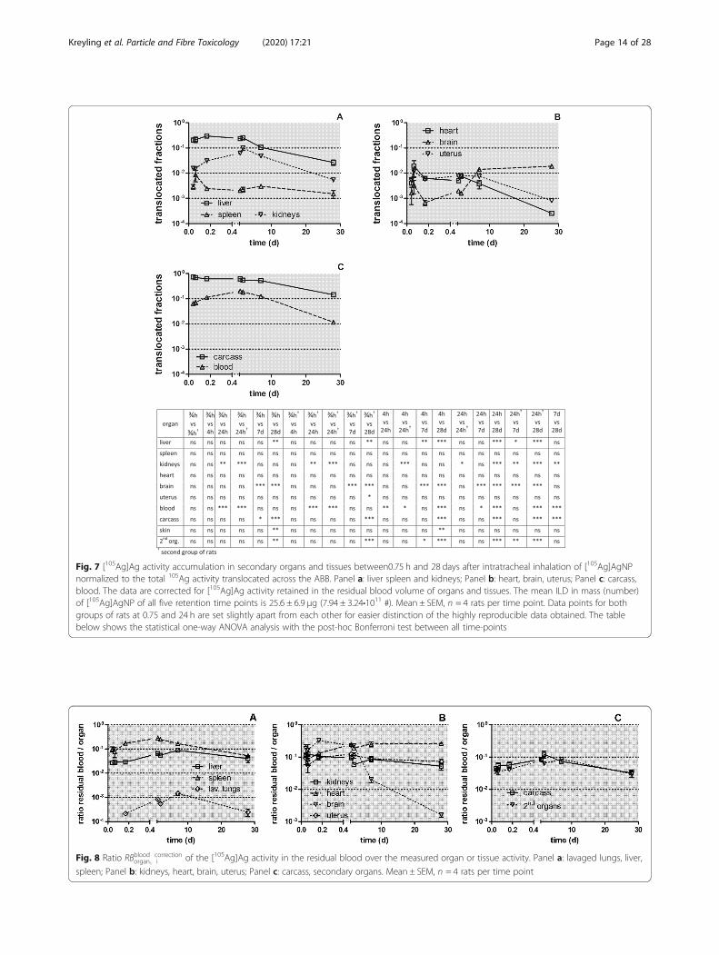
Fig. 8 Ratio RBblood correctionorgan; i of the [105Ag]Ag activity in the residual blood over the measured organ or tissue activity. Panel a: lavaged lungs, liver,
spleen; Panel b: kidneys, heart, brain, uterus; Panel c: carcass, secondary organs. Mean ± SEM, n = 4 rats per time point
Fig. 7 [105Ag]Ag activity accumulation in secondary organs and tissues between0.75 h and 28 days after intratracheal inhalation of [105Ag]AgNPnormalized to the total 105Ag activity translocated across the ABB. Panel a: liver spleen and kidneys; Panel b: heart, brain, uterus; Panel c: carcass,blood. The data are corrected for [105Ag]Ag activity retained in the residual blood volume of organs and tissues. The mean ILD in mass (number)of [105Ag]AgNP of all five retention time points is 25.6 ± 6.9 μg (7.94 ± 3.24•1011 #). Mean ± SEM, n = 4 rats per time point. Data points for bothgroups of rats at 0.75 and 24 h are set slightly apart from each other for easier distinction of the highly reproducible data obtained. The tablebelow shows the statistical one-way ANOVA analysis with the post-hoc Bonferroni test between all time-points
Kreyling et al. Particle and Fibre Toxicology (2020) 17:21 Page 14 of 28
[105Ag]Ag-salt molecules in the abundant presence ofCl−, S2−, PO4
2− and Se2− ions of the surrounding bodyfluids including ELF; this causes the precipitation ofpoorly soluble, [105Ag]Ag-salt clusters in the sizerange between 1 and 10 nm [23, 24, 34, 35, 39].
� The literature discussed in the Background sectionnotes that the fast, initial Ag+ ion release graduallyslows down [32] due to poorly soluble Ag-salt layerswhich are formed on the surface of the remaining[105Ag]AgNP [31, 38, 40].
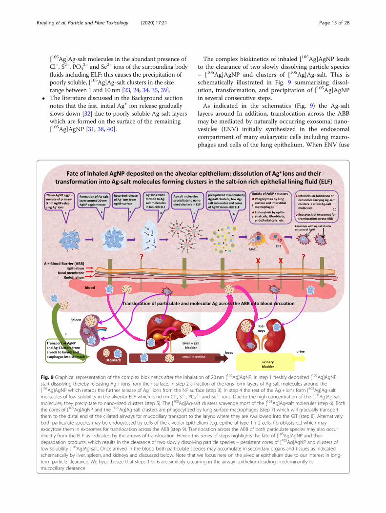
The complex biokinetics of inhaled [105Ag]AgNP leadsto the clearance of two slowly dissolving particle species– [105Ag]AgNP and clusters of [105Ag]Ag-salt. This isschematically illustrated in Fig. 9 summarizing dissol-ution, transformation, and precipitation of [105Ag]AgNPin several consecutive steps.As indicated in the schematics (Fig. 9) the Ag-salt
layers around In addition, translocation across the ABBmay be mediated by naturally occurring exosomal nano-vesicles (ENV) initially synthesized in the endosomalcompartment of many eukaryotic cells including macro-phages and cells of the lung epithelium. When ENV fuse
Fig. 9 Graphical representation of the complex biokinetics after the inhalation of 20 nm [105Ag]AgNP. In step 1 freshly deposited [105Ag]AgNPstart dissolving thereby releasing Ag + ions from their surface. In step 2 a fraction of the ions form layers of Ag-salt molecules around the[105Ag]AgNP which retards the further release of Ag+ ions from the NP surface (step 3). In step 4 the rest of the Ag + ions form [105Ag]Ag-saltmolecules of low solubility in the alveolar ELF which is rich in Cl−, S2−, PO4
2− and Se2− ions. Due to the high concentration of the [105Ag]Ag-saltmolecules, they precipitate to nano-sized clusters (step 5). The [105Ag]Ag-salt clusters scavenge most of the [105Ag]Ag-salt molecules (step 6). Boththe cores of [105Ag]AgNP and the [105Ag]Ag-salt clusters are phagocytized by lung surface macrophages (step 7) which will gradually transportthem to the distal end of the ciliated airways for mucociliary transport to the larynx where they are swallowed into the GIT (step 8). Alternativelyboth particulate species may be endocytosed by cells of the alveolar epithelium (e.g. epithelial type 1 + 2 cells, fibroblasts et.) which mayexocytose them in exosomes for translocation across the ABB (step 9). Translocation across the ABB of both particulate species may also occurdirectly from the ELF as indicated by the arrows of translocation. Hence this series of steps highlights the fate of [105Ag]AgNP and theirdegradation products, which results in the clearance of two slowly dissolving particle species – persistent cores of [105Ag]AgNP and clusters oflow solubility [105Ag]Ag-salt. Once arrived in the blood both particulate species may accumulate in secondary organs and tissues as indicatedschematically by liver, spleen, and kidneys and discussed below. Note that we focus here on the alveolar epithelium due to our interest in long-term particle clearance. We hypothesize that steps 1 to 6 are similarly occurring in the airway epithelium leading predominantly tomucociliary clearance
Kreyling et al. Particle and Fibre Toxicology (2020) 17:21 Page 15 of 28
with the inner cell surface they are released extracellu-larly. They are considered to serve “as a mechanism todischarge unwanted material from the cells, but theyalso could form the basis of an efficient cell-cell commu-nication mechanism” [49]. For example, when 20 nmAuNP were applied to cultured primary human macro-phages the AuNP were rapidly taken up intracellularlyand released within ENV [50]. Moreover, very recentlyENV received much attention as natural, non-cytotoxic,nanotherapeutic carriers for specific cell targeting [51].
Comparison of 24 h excretions after IV injection, ITinstillation and GAVage of soluble ions of 192Ir, 48V, 198Auand of [110mAg]Ag ionsIn Fig. 1a we showed that 24 h p.e. IV injected soluble[110mAg]AgNO3 was rapidly eliminated from the bloodinto the GIT and feces (fraction of 0.87) with only smallfractions found in secondary organs and tissues. Evenmore surprising, in Fig. 1b we found that 24 h p.e. a frac-tion of IT instilled soluble [110mAg]AgNO3 was also rap-idly eliminated (0.70) from the lungs into the blood andfurther into the GIT and feces with only small fractionsfound in secondary organs and tissues and also in urine.Therefore, the question arises, how such a rapid elimin-ation from the blood to the GIT is possible after IV in-jection of soluble [110mAg]AgNO3, and what leads tosuch a rapid translocation across the ABB into the bloodand further into the GIT after IT instillation of soluble[110mAg]AgNO3 The literature cited in the Backgroundsection suggests rapid precipitation of low-solubility Ag-salts in blood and/or ELF, respectively, [31, 39, 41]which is sketched in Fig. 9. It basically excludes the pres-ence of significant amounts of [110mAg]Ag-ions in solu-tion due to the abundance of salt-ions in blood and ELFwhich lead to the precipitation of nano-sized clusters.However, nano-sized clusters smaller than 6–8 nm
and/or macromolecules in blood would be subject torenal glomerular filtration and urinary excretion [52],which we did not observe. Instead, the [110mAg]Ag-saltprecipitates were rapidly eliminated into the GIT – mostlikely by liver uptake and the hepato-biliary clearancepathway. The size of those precipitates may play an im-portant role. After IT instillation of monodisperse, tri-phenylphosphine surface-coated gold NP of 1.4 nm, 2.8nm, 5 nm, 18 nm, 80 nm, and 200 nm diameter thetranslocated fraction across the ABB during the first 24h p.e. declined rapidly with increasing NP size from al-most 0.1 (of the initially delivered NP mass) for 1.4 nmNP by a hundred-fold decline for 200 nm sized particles[53]. Furthermore, after IV injection of the same set ofgold NP resulting in predominant liver retention, wequantitated the hepato-biliary cleared fraction (HBC) of1.4 nm gold NP to be 0.05 while the HBC of 2.8 nm goldNP was 0.008 and all larger-sized gold NP from 5 nm to
80 nm were only cleared by 0.005 during the first 24 hp.e [54]. This is in clear contrast to the predominantelimination of the nano-sized [110mAg]Ag-salt clustersfrom the blood into the GIT. The results of the IT in-stilled and IV injected gold NP together with the ab-sence of urinary [110mAg]Ag excretion implies that theAg-salt precipitates may (a) either have translocatedacross the ABB as extremely small particulates (a few-nanometers) and were scavenged rapidly in the liver or(b) the small Ag-salt precipitates increased their sizeduring circulation to larger than about 10 nm (whichseems not very plausible) or (c) that such extremelysmall Ag-salt precipitates are protected against renal fil-tration by unknown surface modifications of their bio-molecular corona and/or (d) by exosomal mediation.However, the current literature does not provide anysuitable candidate biomolecules blocking renal filtrationand concurrently allowing hepatocytes to transcytose the[110mAg]Ag-salt clusters into the Space of Dissé for fur-ther elimination through the gall-bladder into the smallintestine. These remain urgent questions for futureinvestigations.In Table 5 the urinary and fecal excretion data after
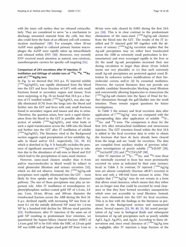
application of [110mAg]Ag+ ions are compared with thecorresponding data after application of soluble 192Ir-,198Au- and 48V-ions. The comparison is done for thefirst 24 h after intratracheal instillation and intravenousinjection. The GIT retention found within the first 24 his added to the fecal excretion data in order to obtainthe fractions that have de facto already been clearedfrom the lungs and are ‘ready’ for excretion. The dataare compiled from auxiliary studies of previous inhal-ation investigations of poorly soluble [192Ir]IrNP [28],[198Au]AuNP [25] and [48V]TiO2NP [30].After IV injection of 192Ir-, 198Au- and 48V-ions these
are minimally excreted in feces but more prominantlyexcreted via urine as indicated by their ratio (urinary:fecal) in Table 5. In contrast, IV injected [110mAg]Agions are almost completely (fraction of0.87) excreted infeces and only a 100-fold lower amount in urine. Thisimplies that [110mAg]Ag+ ions do not remain in a formthat allows renal clearance, which means that they eitherare no longer ions that could be excreted by renal clear-ance or that they have formed secondary nanoparticleswhich were not accessible to renal filtration. Instead,they were eliminated into the GIT for fecal excretion.This is in line with the findings in the literature as pre-sented in the Background section and summarizedabove. Several reports [33, 39, 40, 55, 56] noted precipi-tation of Ag+-ions in biological fluids and subsequentformation of Ag-salt precipitates such as poorly solubleAgCl, Ag2S, Ag2PO4, and Ag2Se. According to these ob-servations and, since renal clearance of [110mAg]Ag ionsis negligible, after IV injection a large fraction of the
Kreyling et al. Particle and Fibre Toxicology (2020) 17:21 Page 16 of 28
Ag+-ions must have formed poorly soluble, nano-sizedAg-salt precipitates. The predominant fecally excretedfraction indicates that these precipitates were cleared viathe hepato-biliary pathway; i.e. they were metabolizedmainly by liver hepatocytes and released into the bilefluid of the Space of Dissé for further elimination via thegall bladder into the small intestine [57, 58] as sketchedin Fig. 9. Note, hepatocytes do not metabolize metalliccations like [110mAg]Ag [57, 58].Twenty-four hours after IT instillation of 192Ir, 198Au
and 48V ions only small fractions of between 0.06 and0.2 (Table 5) are fecally excreted (including GIT reten-tion), while 24 h after IT instillation of [110mAg]Ag-ions,a fraction of 0.7 of the instilled dose is excreted in feces.This is almost the same fraction as after IV injection(0.87). Urinary excretion was similarly low after IVinjection.After IT instillation, rapid mucociliary clearance of
[110mAg]Ag deposited on the airway epithelium is ex-pected to contribute to the fecally excreted fraction.However, based on the 24 h data after the inhalation of[105Ag]AgNP compiled in Table 3 it is not plausible toattribute a fraction of more than 0.3 to fast clearance upto this time point. According to literature presented inthe Background section, it appears reasonable to attri-bute the additional fecal fraction of 0.3–0.4 to the rapidclearance of poorly soluble, nano-sized Ag-salt precipi-tates which had been formed with abundant Cl−-, S2−-,PO4
2−-, ions as well as less abundant but more stablybinding Se2−-ions present in ELF. For their rapid elimin-ation via feces within 24 h, they were first translocatedacross the ABB into the blood and from there via thehepato-biliary pathway into the GIT and feces. Further-more, after intratracheal instillation of [110mAg]Ag -ionsthe differential fraction of 0.21 between the fecal excre-tion after 24 h (0.70, including the GIT content) and thatafter 7 days (0.91) is likely due to the hepato-biliaryclearance pathway (HBC). Hence, there is a biphasic
clearance after either IT instillation or IV injection ofsoluble [110mAg]AgNO3. Additional confirmation comesfrom the daily fecal excretion measurements after allthree instillation applications (IV, IT, GAV) which areshown in Fig. S7 of the Supplementary Information.Since mucociliary clearance to the larynx and ABB
translocation into blood lead both to fecal excretion of[110mAg]Ag it is not directly possible to distinguish be-tween both clearance pathways after IT instillation ofsoluble [110mAg]Ag. The negligible amount of urinaryexcretion indicates that free 110mAg-ions are virtually ab-sent due to their precipitation in ELF, while a slightlyhigher fraction of 198Au-ions and large fractions of 192Ir-and 48V-ions were found in urine 24 h p.e. (see Table 5).Additionally, the third auxiliary biokinetics study in Fig.1c after oral instillation of an [110mAg]Ag-solutionshowed almost exclusive fecal and minimal urinary ex-cretion, which is compatible with results reported earlier[40], and suggests the dominant precipitation of Ag-saltin the GIT and negligible uptake from the GIT throughthe intestinal barrier into the blood.Therefore it is plausible to conclude that the differen-
tial 0.21 fraction is the upper limit of [105Ag]Ag-salt pre-cipitates after [105Ag]AgNP inhalation which may havebeen translocated across the ABB from day 2 to day 7into the blood and eliminated via HBC into the GIT andfeces.
Comparison of lung retention of inhaled, poorly soluble,20 nm [192Ir]IrNP, [195Au]AuNP [48V]TiO2-NP and ofinhaled, partially soluble, 20 nm [105Ag]AgNPLung retention data of inhaled 20 nm [105Ag]AgNP differstrikingly from those previously obtained from poorlysoluble 20 nm [192Ir]IrNP [28], 20 nm [195Au]AuNP [25]and 20 nm [48V]TiO2-NP [30]. While differences be-tween the three inhaled, poorly soluble NP have beendiscussed previously, here we focus on the differ-ences between [105Ag]AgNP and [195Au]AuNP in
Table 5 urinary and fecal excretion 24 h after either IV injection or IT instillation of solutions of 192Ir, 48V, 198Au, and 110mAg ions
Ionic solutions 24 h excretion Fraction of applied ions Ratio urine/feces Fraction of applied ions Ratio urine/feces
IV IV IT IT192Ir-ions GIT + feces 0.007 ± 0.012 0.062 ± 0.019198Au-ions GIT + feces 0.059 ± 0.006 0.201 ± 0.02948V-ions GIT + feces 0.088 ± 0.041 0.119 ± 0.020110mAg-ions GIT + feces 0.873 ± 0.009 0.699 ± 0.062192Ir-ions urine 0.277 ± 0.013 41.26 0.426 ± 0.112 6.911198Au-ions urine 0.087 ± 0.031 1.472 0.07 ± 0.01 0.35048V-ions urine 0.365 ± 0.077 4.127 0.319 ± 0.055 2.672110mAg-ions urine 0.005 ± 0.006 0.006 0.009 ± 0.014 0.013
Comparison of fractional urinary and fecal excretions 24 h after IV injection and IT instillation of soluble 192Ir-ions [28], 48V-ions [30], 198Au-ions [25] and [110mAg]Agions (cf. Figure 1) applied in the auxiliary studies in the present work. Note, in the data of 24 h fecal excretion, the 24 h GIT fractions are added due to the delayedpassage through the GIT into feces. Data are given as mean ± SD, n ≥ 4 rats per group.
Kreyling et al. Particle and Fibre Toxicology (2020) 17:21 Page 17 of 28
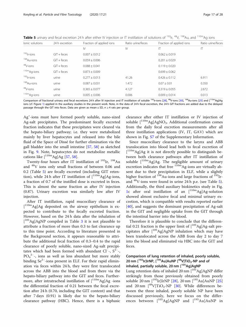
Figs. 10, 11, 12, 13 and 14 since both NP show asimilar NP condensation dynamics immediately afterspark ignition and evaporation. Initially, in the hotzone close to the igniting spark both, vaporized Ag andAu, condense and coalesce, forming liquid droplets up to5–8 nm. Thereafter, when the droplets have escaped to-wards colder zones downstream, they solidify while con-tinuing to coagulate until agglomeration is essentiallystopped by dilution with clean air adjusted to maintainNP aerosol concentrations of about 1•107 NP/cm3. Theinhalation of these aerosols by the rats after sufficientcooling - occurs within 5–10 s after generation (see theexperimental setup in Fig. S2 of Supplemental Informa-tion). Since we cannot distinguish later between the reten-tion and clearance of the inhaled [105Ag]AgNP and theirdegradation products formed after inhalation we simplywrite “[105Ag]Ag activities”.Figure 10b (adopted from [25]) shows that a fraction
of 0.22 of the applied [195Au]AuNP dose is cleared fromthe lungs during the first 24 h after inhalation by MCC,while the cleared fraction of 0.4 for [105Ag]AgNP (seeFig. 10a) during the same period of time is nearly twiceas high. We may reasonably assume that MCC will notonly occur for the [105Ag]AgNP but also for theirparticulate degradation products – i.e. [105Ag]Ag-saltprecipitates also formed on the airway epithelium. Assum-ing a similar mucociliary cleared fraction (of 0.22) of[105Ag]AgNP and precipitated [105Ag]Ag-salt as observedafter [195Au]AuNP inhalation, the other half of the 0.4 de-creases of [105Ag]Ag lung retention must be attributed to
a second clearance pathway. Since the fraction of 105Agretained in the blood after [105Ag]AgNP inhalation(Fig. 10c) during the first 24 h after inhalation is morethan 100 times higher than the corresponding [195Au]Auactivity after [195Au]AuNP inhalation (Fig. 10d), we con-clude that translocation of [105Ag]AgNP and their degrad-ation products across the ABB accounts for this clearedfraction. From the blood, the material is eliminated viahepato-biliary clearance into the GIT and subsequentlyexcreted in feces similar to the fecal clearance after IT in-stillation of soluble [110mAg]AgNO3.Similarly, Fig. 10a and b show that from day 2 until
day 7, long-term macrophage-mediated clearance (LT-MC) of [195Au]AuNP causes a fractional decrease oflung retention by 0.2, while after inhalation of[105Ag]AgNP the fractional lung retention decreases by0.45. This suggests an association of a 0.25 fraction tothe translocation pathway of the precipitated [105Ag]Ag-salt across the ABB which is supported by the persisting100-fold higher [105Ag]Ag content in blood when com-pared to the circulating [195Au]AuNP.
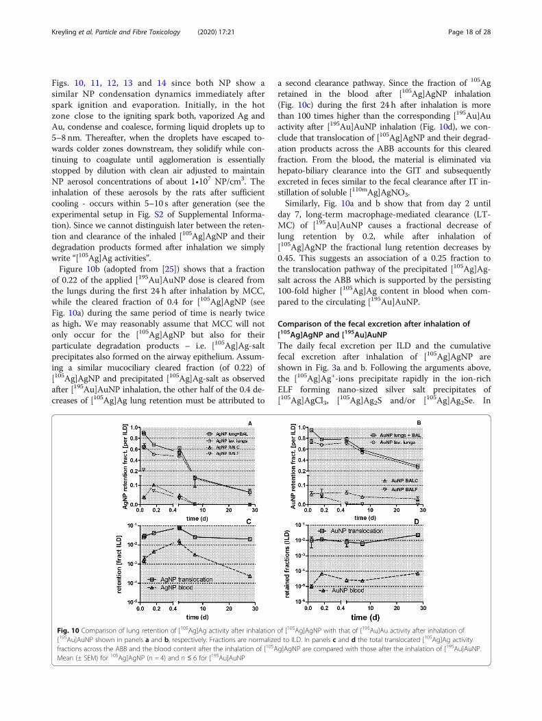
Comparison of the fecal excretion after inhalation of[105Ag]AgNP and [195Au]AuNPThe daily fecal excretion per ILD and the cumulativefecal excretion after inhalation of [105Ag]AgNP areshown in Fig. 3a and b. Following the arguments above,the [105Ag]Ag+-ions precipitate rapidly in the ion-richELF forming nano-sized silver salt precipitates of[105Ag]AgCl3, [105Ag]Ag2S and/or [105Ag]Ag2Se. In
Fig. 10 Comparison of lung retention of [105Ag]Ag activity after inhalation of [105Ag]AgNP with that of [195Au]Au activity after inhalation of[195Au]AuNP shown in panels a and b, respectively. Fractions are normalized to ILD. In panels c and d the total translocated [105Ag]Ag activityfractions across the ABB and the blood content after the inhalation of [105Ag]AgNP are compared with those after the inhalation of [195Au]AuNP.Mean (± SEM) for 105Ag]AgNP (n = 4) and n ≤ 6 for [195Au]AuNP
Kreyling et al. Particle and Fibre Toxicology (2020) 17:21 Page 18 of 28
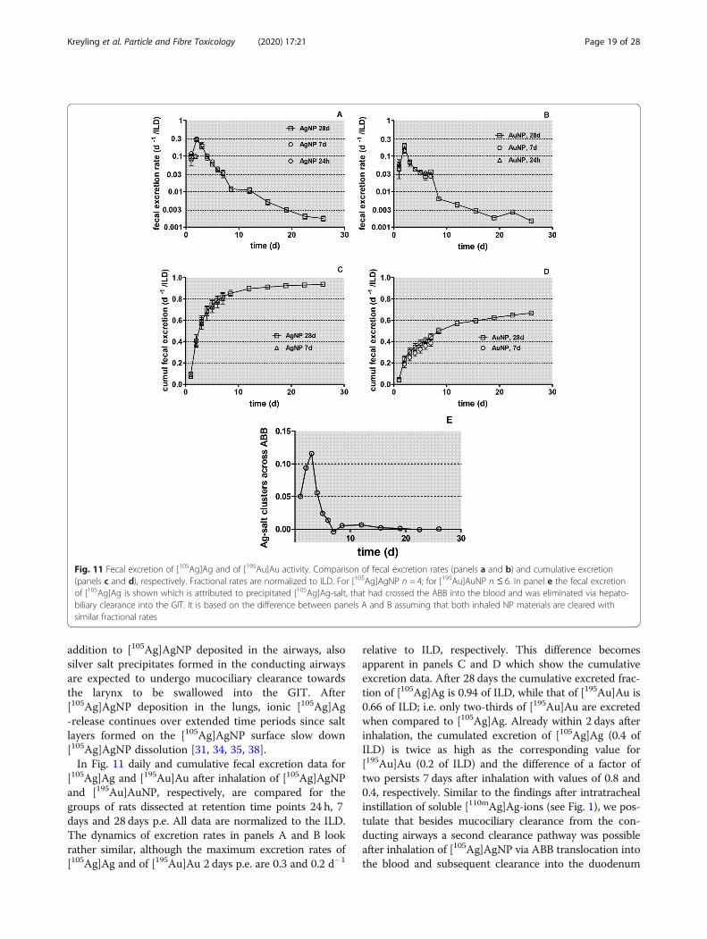
addition to [105Ag]AgNP deposited in the airways, alsosilver salt precipitates formed in the conducting airwaysare expected to undergo mucociliary clearance towardsthe larynx to be swallowed into the GIT. After[105Ag]AgNP deposition in the lungs, ionic [105Ag]Ag-release continues over extended time periods since saltlayers formed on the [105Ag]AgNP surface slow down[105Ag]AgNP dissolution [31, 34, 35, 38].In Fig. 11 daily and cumulative fecal excretion data for
[105Ag]Ag and [195Au]Au after inhalation of [105Ag]AgNPand [195Au]AuNP, respectively, are compared for thegroups of rats dissected at retention time points 24 h, 7days and 28 days p.e. All data are normalized to the ILD.The dynamics of excretion rates in panels A and B lookrather similar, although the maximum excretion rates of[105Ag]Ag and of [195Au]Au 2 days p.e. are 0.3 and 0.2 d− 1
relative to ILD, respectively. This difference becomesapparent in panels C and D which show the cumulativeexcretion data. After 28 days the cumulative excreted frac-tion of [105Ag]Ag is 0.94 of ILD, while that of [195Au]Au is0.66 of ILD; i.e. only two-thirds of [195Au]Au are excretedwhen compared to [105Ag]Ag. Already within 2 days afterinhalation, the cumulated excretion of [105Ag]Ag (0.4 ofILD) is twice as high as the corresponding value for[195Au]Au (0.2 of ILD) and the difference of a factor oftwo persists 7 days after inhalation with values of 0.8 and0.4, respectively. Similar to the findings after intratrachealinstillation of soluble [110mAg]Ag-ions (see Fig. 1), we pos-tulate that besides mucociliary clearance from the con-ducting airways a second clearance pathway was possibleafter inhalation of [105Ag]AgNP via ABB translocation intothe blood and subsequent clearance into the duodenum
Fig. 11 Fecal excretion of [105Ag]Ag and of [195Au]Au activity. Comparison of fecal excretion rates (panels a and b) and cumulative excretion(panels c and d), respectively. Fractional rates are normalized to ILD. For [105Ag]AgNP n = 4; for [195Au]AuNP n ≤ 6. In panel e the fecal excretionof [105Ag]Ag is shown which is attributed to precipitated [105Ag]Ag-salt, that had crossed the ABB into the blood and was eliminated via hepato-biliary clearance into the GIT. It is based on the difference between panels A and B assuming that both inhaled NP materials are cleared withsimilar fractional rates
Kreyling et al. Particle and Fibre Toxicology (2020) 17:21 Page 19 of 28
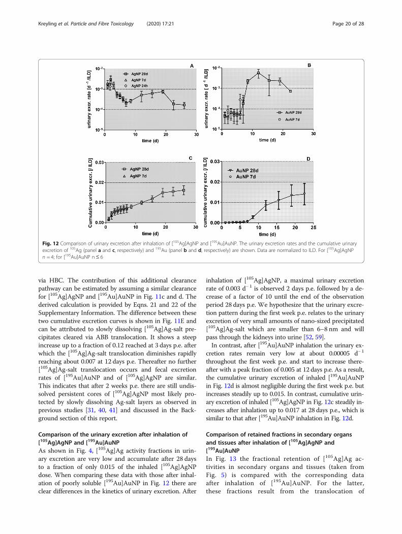
via HBC. The contribution of this additional clearancepathway can be estimated by assuming a similar clearancefor [105Ag]AgNP and [195Au]AuNP in Fig. 11c and d. Thederived calculation is provided by Eqns. 21 and 22 of theSupplementary Information. The difference between thesetwo cumulative excretion curves is shown in Fig. 11E andcan be attributed to slowly dissolving [105Ag]Ag-salt pre-cipitates cleared via ABB translocation. It shows a steepincrease up to a fraction of 0.12 reached at 3 days p.e. afterwhich the [105Ag]Ag-salt translocation diminishes rapidlyreaching about 0.007 at 12 days p.e. Thereafter no further[105Ag]Ag-salt translocation occurs and fecal excretionrates of [195Au]AuNP and of [105Ag]AgNP are similar.This indicates that after 2 weeks p.e. there are still undis-solved persistent cores of [105Ag]AgNP most likely pro-tected by slowly dissolving Ag-salt layers as observed inprevious studies [31, 40, 41] and discussed in the Back-ground section of this report.
Comparison of the urinary excretion after inhalation of[105Ag]AgNP and [195Au]AuNPAs shown in Fig. 4, [105Ag]Ag activity fractions in urin-ary excretion are very low and accumulate after 28 daysto a fraction of only 0.015 of the inhaled [105Ag]AgNPdose. When comparing these data with those after inhal-ation of poorly soluble [195Au]AuNP in Fig. 12 there areclear differences in the kinetics of urinary excretion. After
inhalation of [105Ag]AgNP, a maximal urinary excretionrate of 0.003 d− 1 is observed 2 days p.e. followed by a de-crease of a factor of 10 until the end of the observationperiod 28 days p.e. We hypothesize that the urinary excre-tion pattern during the first week p.e. relates to the urinaryexcretion of very small amounts of nano-sized precipitated[105Ag]Ag-salt which are smaller than 6–8 nm and willpass through the kidneys into urine [52, 59].In contrast, after [195Au]AuNP inhalation the urinary ex-
cretion rates remain very low at about 0.00005 d− 1
throughout the first week p.e. and start to increase there-after with a peak fraction of 0.005 at 12 days p.e. As a result,the cumulative urinary excretion of inhaled [195Au]AuNPin Fig. 12d is almost negligible during the first week p.e. butincreases steadily up to 0.015. In contrast, cumulative urin-ary excretion of inhaled [105Ag]AgNP in Fig. 12c steadily in-creases after inhalation up to 0.017 at 28 days p.e., which issimilar to that after [195Au]AuNP inhalation in Fig. 12d.
Comparison of retained fractions in secondary organsand tissues after inhalation of [105Ag]AgNP and[195Au]AuNPIn Fig. 13 the fractional retention of [105Ag]Ag ac-tivities in secondary organs and tissues (taken fromFig. 5) is compared with the corresponding dataafter inhalation of [195Au]AuNP. For the latter,these fractions result from the translocation of
Fig. 12 Comparison of urinary excretion after inhalation of [105Ag]AgNP and [195Au]AuNP. The urinary excretion rates and the cumulative urinaryexcretion of 105Ag (panel a and c, respectively) and 195Au (panel b and d, respectively) are shown. Data are normalized to ILD. For [105Ag]AgNPn = 4; for [195Au]AuNP n ≤ 6
Kreyling et al. Particle and Fibre Toxicology (2020) 17:21 Page 20 of 28
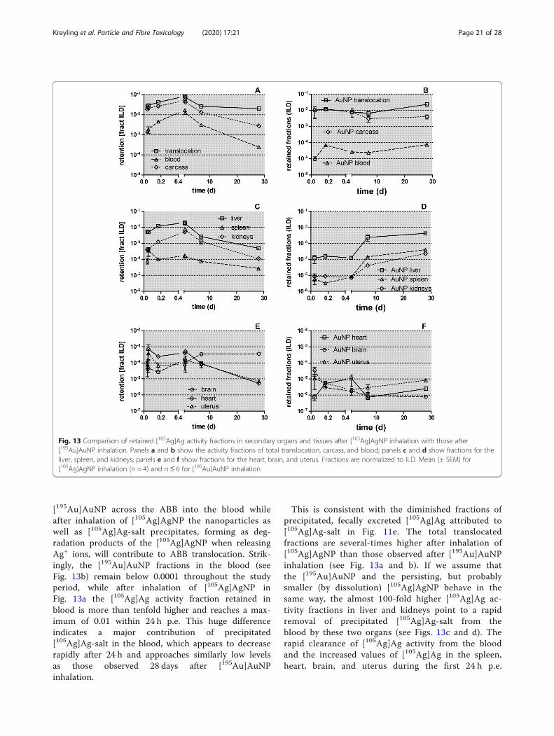
[195Au]AuNP across the ABB into the blood whileafter inhalation of [105Ag]AgNP the nanoparticles aswell as [105Ag]Ag-salt precipitates, forming as deg-radation products of the [105Ag]AgNP when releasingAg+ ions, will contribute to ABB translocation. Strik-ingly, the [195Au]AuNP fractions in the blood (seeFig. 13b) remain below 0.0001 throughout the studyperiod, while after inhalation of [105Ag]AgNP inFig. 13a the [105Ag]Ag activity fraction retained inblood is more than tenfold higher and reaches a max-imum of 0.01 within 24 h p.e. This huge differenceindicates a major contribution of precipitated[105Ag]Ag-salt in the blood, which appears to decreaserapidly after 24 h and approaches similarly low levelsas those observed 28 days after [195Au]AuNPinhalation.
This is consistent with the diminished fractions ofprecipitated, fecally excreted [105Ag]Ag attributed to[105Ag]Ag-salt in Fig. 11e. The total translocatedfractions are several-times higher after inhalation of[105Ag]AgNP than those observed after [195Au]AuNPinhalation (see Fig. 13a and b). If we assume thatthe [195Au]AuNP and the persisting, but probablysmaller (by dissolution) [105Ag]AgNP behave in thesame way, the almost 100-fold higher [105Ag]Ag ac-tivity fractions in liver and kidneys point to a rapidremoval of precipitated [105Ag]Ag-salt from theblood by these two organs (see Figs. 13c and d). Therapid clearance of [105Ag]Ag activity from the bloodand the increased values of [105Ag]Ag in the spleen,heart, brain, and uterus during the first 24 h p.e.
Fig. 13 Comparison of retained [105Ag]Ag activity fractions in secondary organs and tissues after [105Ag]AgNP inhalation with those after[195Au]AuNP inhalation. Panels a and b show the activity fractions of total translocation, carcass, and blood; panels c and d show fractions for theliver, spleen, and kidneys; panels e and f show fractions for the heart, brain, and uterus. Fractions are normalized to ILD. Mean (± SEM) for[105Ag]AgNP inhalation (n = 4) and n ≤ 6 for [195Au]AuNP inhalation
Kreyling et al. Particle and Fibre Toxicology (2020) 17:21 Page 21 of 28
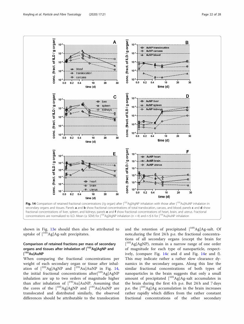
shown in Fig. 13e should then also be attributed touptake of [105Ag]Ag-salt precipitates.
Comparison of retained fractions per mass of secondaryorgans and tissues after inhalation of [105Ag]AgNP and[195Au]AuNPWhen comparing the fractional concentrations perweight of each secondary organ or tissue after inhal-ation of [105Ag]AgNP and [195Au]AuNP in Fig. 14,the initial fractional concentrations after[105Ag]AgNPinhalation are up to two orders of magnitude higherthan after inhalation of [195Au]AuNP. Assuming thatthe cores of the [105Ag]AgNP and [195Au]AuNP aretranslocated and distributed similarly, the observeddifferences should be attributable to the translocation
and the retention of precipitated [105Ag]Ag-salt. Ofnote,during the first 24 h p.e. the fractional concentra-tions of all secondary organs (except the brain for[105Ag]AgNP), remain in a narrow range of one orderof magnitude for each type of nanoparticle, respect-ively, (compare Fig. 14c and d and Fig. 14e and f).This may indicate rather a rather slow clearance dy-namics in the secondary organs. Along this line thesimilar fractional concentrations of both types ofnanoparticles in the brain suggests that only a smallamount of precipitated [105Ag]Ag-salt accumulates inthe brain during the first 4 h p.e. But 24 h and 7 daysp.e. the [105Ag]Ag accumulation in the brain increasesrather rapidly which differs from the rather constantfractional concentrations of the other secondary
Fig. 14 Comparison of retained fractional concentrations (/g organ) after [105Ag]AgNP inhalation with those after [195Au]AuNP inhalation insecondary organs and tissues. Panels a and b show fractional concentrations of total translocation, carcass, and blood; panels c and d showfractional concentrations of liver, spleen, and kidneys; panels e and f show fractional concentrations of heart, brain, and uterus. Fractionalconcentrations are normalized to ILD. Mean (± SEM) for [105Ag]AgNP inhalation (n = 4) and n≤ 6 for [195Au]AuNP inhalation
Kreyling et al. Particle and Fibre Toxicology (2020) 17:21 Page 22 of 28
organs. This may suggest that the blood-brain-barrieris tight for [105Ag]Ag-salt precipitates only for thefirst 4 h p.e.
ConclusionIntratracheal inhalation of freshly generated[105Ag]AgNP aerosols for 1½ hours allows low dose de-position in the lungs of adult healthy rats thereby avoid-ing nasal and pharyngeal deposition. The sensitivityobtained with radiolabeled NP is extremely high. Highlysensitive γ-ray-spectrometry, allows a dynamic, analyticalrange over five orders of magnitude across the differentspecimens. The methodology of radio-analysis is easy tomanage for the large number of samples in order tostudy quantitative biodistributions of [105Ag]AgNP andtheir possible degradation products in the rat organism,as well as complete excretional clearance out of the ratorganism after five different retention time intervals upto 28 days p.e. The repeatability of the initially rapidlyevolving biokinetics proved to be convincingly goodwhen comparing the two independently investigatedgroups of four rats studied at 0.75 h and 24 h p.e.,respectively.Rapid release of [105Ag]Ag-ions from the [105Ag]AgNP
surface appears to be superimposed with both fast MCCfrom the conducting airways and long-term clearance of[105Ag]AgNP from the alveolar region to the larynx. Thecurrent observations are in line with evidence presentedin the literature that released [105Ag]Ag-ions precipitaterapidly to form very small clusters of low solubility[105Ag]Ag-salts in the ion-rich ELF. Besides their clear-ance to the larynx, they translocate across the ABB intothe blood and are predominantly eliminated via thehepato-biliary clearance pathway into the small intestinefor fecal excretion. The pathway of precipitated[105Ag]Ag-salt is supported by the findings of auxiliarybiokinetics studies at 24 h and 7 days after either IV in-jection or IT or oral instillation of [110mAg]AgNO3 solu-tions in sentinel groups of rats. In line with evidence inthe literature, the release of [105Ag]Ag-ions from the[105Ag]AgNP surface may continue for an extended timeat a lower rate due to the simultaneous formation oflow-solubility salt layers on the [105Ag]AgNP surface inthe ion-rich body fluids that mediate and prolonge thedissolution process.The superimposed clearance of (possibly) Ag-salt-
layer-protected, persistent cores of [105Ag]AgNP andprecipitated [105Ag]Ag-salt results in the elimination of afraction of > 0.8 (per ILD) after one-week p.e. Assuminga very similar clearance behavior of the cores of AgNPand AuNP the difference between the results of bothexperiments can be attributed to the clearance of Ag-
precipitates during the first 2 weeks p.e., showing amaximal fractional rate of 0.12 d− 1 at 3 days p.e.
Materials and methodsStudy designTwenty-eight healthy, adult, female Wistar Kyoto rats(WKY/Kyo@Rj rats, Janvier, Le Genest Saint Isle,France) were randomly assigned to seven groups of fourrats and subjected to intratracheal inhalation of an[105Ag]AgNP aerosol for 1½ hours via an endotrachealtube [60]. The biodistribution was analyzed 0.75 h, 4 h,24 h, 7d and 28d after exposure. The first group of rats,which was exsanguinated and dissected immediatelyafter the 1½-hour intratracheal inhalation exposure, wasassigned to the retention time-point 0.75 h since thenanoparticles brought into the rats’ lungs over 1½ hourshad an estimated average time of only 45 min for depos-ition, uptake, and distribution. In order to test the re-peatability of the rapidly changing biokinetics directlyafter inhalation, two additional groups of four rats wereinhalation exposed and analyzed at 0.75 h and 24 h p.e.,respectively. The additional two groups were exposed 1day after the inhalation exposures of the initial 0.75 hand 24 h groups, respectively.Furthermore, twenty-four healthy, adult, female Wistar
Kyoto rats (WKY/Kyo@Rj rats, Janvier, Le Genest SaintIsle, France) were randomly assigned to six groups offour rats and subjected to intratracheal instillation, orintravenous injection, or oral instillation (gavage) of sol-uble [110mAg]AgNO3 solutions and biokinetically studiedat either 24 h or 7 days p.e.
Animals and maintenanceAll Wistar-Kyoto rats (WKY/Kyo@Rj rats, Janvier, LeGenest Saint Isle, France) were housed in relative-humidity and temperature-controlled ventilated cages(VentiRack Bioscreen TM, Biozone, Margate, UK) on a12-h day/night cycle. Rodent diet and water were pro-vided ad libitum. The rats were adapted for at least 2weeks after purchase and then randomly attributed tothe experimental groups. When starting the studies, therats were 8–10 weeks old and exhibited a mean bodyweight of 204 ± 13 g. Some physiological parameters ofthe rats are given in Table 3.All experiments were conducted under German fed-
eral guidelines for the use and care of laboratory animalsin accordance with EU Directive 2010/63/EU for animalexperiments. The studies were approved by the Regier-ung von Oberbayern (Government of District of UpperBavaria, Approval No. 211–2531-94/04) and by theInstitutional Animal Care and Use Committee of theHelmholtz Centre Munich.
Kreyling et al. Particle and Fibre Toxicology (2020) 17:21 Page 23 of 28
Synthesis and characterization of the [105Ag]AgNP aerosolRadiolabeled AgNP aerosols were produced continu-ously during the experiments by spark ignition betweentwo high-purity (99.997%), cylindrical silver electrodes(diameter 3.0 mm, length 5 mm; Cat. No. AG007912;Goodfellow GmbH, Hamburg, Germany) which hadbeen proton irradiated in the cyclotron at JRC (Ispra,Italy). The protons impinging on one of the flat ends ofeach electrode had an energy of about 33.5MeV in orderto achieve the highest possible 105Ag-activity near thesurface. This radioactivity was distributed nearly homo-geneously throughout a layer thickness that would the-oretically be consumed by the spark ignition processduring the experiments. At the time the inhalation ex-periments were performed, the specific 105Ag radioactiv-ity was 2.60MBq/mg in a surface layer of about 250 μmthickness. The radioactive 105Ag decays back to 105Pd viaelectron capture and positron emission, with a half-lifeof 41.3 days, thereby emitting γ-rays with different ener-gies; one γ-emission line of 0.28MeV with a fractionalemission probability of 0.302 was selected for the γ-spectrometrical analyses.For each group of rats, the [105Ag]AgNP aerosol was
freshly generated in the spark ignition aerosol generator(GFG100, Palas, Karlsruhe, Germany) at 250 Hz sparkfrequency in an argon (Ar) gas stream of 3 L/min. Dur-ing spark ignition between the two Ag electrodes, smallamounts of Ag evaporate. The vaporized Ag coalesces,forming liquid droplets up to 5–8 nm. When the drop-lets have escaped towards colder zones downstream,they solidify to solid spheres. The electrically chargedaerosol of these primary spheres is immediately quasi-neutralized by an inline radioactive 85Kr source and thehighly concentrated and continuously agglomerating[105Ag]AgNP pass through a 30 cm long tubular furnacethat is kept at a temperature of 600 °C to form single,densely packed agglomerated particles. Coagulation con-tinues until it is stopped by dilution with clean air whenthe [105Ag]AgNP reach a size of 20 nm. Since denselypacked spheres will occupy 74% and more randomlypacked spheres about 64% of the agglomerate volumewe suggest a density of 7.3 g/cm3 (= 0.7 • 10.49) of thegenerated and heat-treated AgNP.Downstream of the furnace the aerosol was cooled and
diluted in a copper tube (inner diameter 8 mm) by mix-ing with humidified oxygen and nitrogen to obtain afractional oxygen concentration of 0.2–0.25. After dilu-tion to concentrations of about 1•107 NP/cm3 furtheragglomeration was negligible within the few secondsprior to inhalation by the rats. The generated 20 nm-sized [105Ag]AgNP still have a chain agglomerated/ag-gregated structure even after 600 °C heat-treatment al-beit more compact than the non-heat-treated NP. Theflow rate was typically 10 L/min and the fractional
relative humidity of the aerosol was set to about 0.7 be-fore entering the inhalation apparatus; the whole inhal-ation apparatus and the inhalation methodologyincluding the pre-set fractional relative humidity weredescribed earlier [61] and is schematically displayed inFig. S3 of the Supplementary Information. The aerosolparticle concentration and size distribution were con-tinuously sampled and controlled by a condensation par-ticle counter (CPC 3022A, TSI, Aachen, Germany) and ascanning mobility particle spectrometer (SMPS; consist-ing of a model 3071 differential mobility analyzer and aCPC model 3010, TSI, Aachen, Germany), respectively.Averages of the count median diameters (CMD), volumemedian diameters (VMD) and geometric standard devia-tions (GSD) as well as number concentrations and vol-ume concentrations are given as mean ± SD in Table 2.Since the SMPS instrument exhibited a lower particlesize detection limit of 10 nm, the averaged spectra werefitted to a lognormal size distribution using the least-squares method and the fits were extrapolated to a sizeof 1 nm (for details see Supplementary Information).These corrections led to slightly lower CMDs while theGSD changed only negligibly (see Table 2). The charac-teristic parameters of the freshly generated [105Ag]AgNPaerosol were the same as those generated without theradio-label using non-irradiated, pure silver electrodes;TEM images were analyzed from the latter. Similarly,the chemical composition was determined by ElectronEnergy Loss Spectrometry (EELS). The specific 105Ag ac-tivity of the aerosol particles was determined by γ-spectrometric analysis of absolute filters onto which[105Ag]AgNP had been collected at an aerosol flow (0.3L/min) throughout each 1½-h exposure period. Fromthe activity deposited on the filter, an activity concentra-tion of the [105Ag]AgNP aerosol of 2.60 kBq•μg− 1 wasderived. At this activity concentration the atomic ratioof 105Ag: Ag in the nanoparticles is about 1.3 × 10− 6.Hence, statistically, every second AgNP will contain a105Ag -radiolabel. Therefore, the 105Ag-radiolabeling in-volves a minimal impurity of the stable silver matrixwhich would be extremely unlikely to affect the stabilityand the physicochemical characteristics of the[105Ag]AgNP.
Intratracheal inhalation exposureThe four slightly anesthetized adult rats in each groupwere ventilated individually via a flexible endotrachealtube and placed on their left lateral side in an air-tightplethysmograph box of our tailor-made inhalation ap-paratus and connected to the aerosol system, (see Fig. S3of Supplementary Information). They were exposed tothe freshly generated aerosol for 1½ hours. In this report,this exposure method will be called “intratracheal inhal-ation” (see reference [60]).
Kreyling et al. Particle and Fibre Toxicology (2020) 17:21 Page 24 of 28
Treatment of the rats after inhalationAnesthesia of each rat was antagonized immediatelypost-exposure (p.e.) as described in the Supplemen-tary Information and previously in [25, 62]. There-after, each rat was kept individually in a metaboliccage and excreta were collected separately and quanti-tatively. For ethical reasons, the rats of the 28 daysgroup were maintained individually in a normal cageon cotton cloths starting immediately after[105Ag]AgNP inhalation. Each cloth was replaced witha new one every 3–4 days (2 cloths per week), andthe fecal droppings were quantitatively separated fromthe previous one. After separation, the cloth con-tained only [105Ag]Ag activity originating from urinewhich had soaked and dried. In Table S2 the list ofcollected organs, tissues, body fluids, and excretion isgiven. Since the cages of the four rats of each groupwere located next to each other, the rats had the con-tinuous sensory perception of each other.
Evaluation and statistical analysis of [105Ag]AgNPbiokineticsAt 0.75 h, 4 h, 24 h, 7d, and 28d p.e., rats were anesthe-tized (by 5% isoflurane inhalation) and euthanized by ex-sanguination via the abdominal aorta. Blood, all organs,tissues, and excretions were collected and the 105Agradioactivity was determined by γ-spectrometry withoutany further physicochemical processing of the samples,as described in the Supplementary Information and inearlier works [25, 62–64]. Throughout this report nano-particle quantities are calculated from the 105Ag activitydetermined with γ-scintillation detectors, properly cali-brated in γ-ray energy and detection efficiency for 105Ag,and corrected for background and radioactive decay dur-ing the experiments (see Supplementary Information).Samples yielding background-corrected counts in the280 keV region-of-interest of the 105Ag γ-spectrum weredefined to be below the detection limit (<DL; 0.1 Bq)when the number of counts was less than three standarddeviations of the background counts collected during200 min without any sample in the γ-scintillationdetector.BALs were performed by applying 6 × 5 ml of
phosphate-buffered-saline solution (PBS without Ca2+
or Mg2+) under the gentle massage of the thorax. Therecovered fractional BAL fluid (BALF) (about 0.8 ofinstilled PBS) was centrifuged at 500 g for 20 min atroom temperature to separate the lavaged cells fromthe supernatant. The [105Ag]AgNP content was deter-mined by γ-ray-spectrometry.Up to 24 h p.e. early clearance was measured in the GIT
and feces comprising of (a) MCC-cleared [105Ag]AgNPand of precipitated, low-solubility [105Ag]Ag-salt precipi-tates from the conducting airways and (b) of precipitated,
low-solubility [105Ag]Ag-salt precipitates which had trans-located across the respiratory ABB into blood and via liverand gall-bladder into the small intestine. The early clear-ance data of the 7 days- and 28 days-group were derivedfrom fecal excretion measurements during the first 3 daysp.e. The clearance contribution of the [105Ag]Ag+-ionrelease - according to the literature discussed in the Back-ground section - precipitating as low-solubility salt precip-itates was calculated from the 24 h data of the auxiliarystudy after the intratracheal instillation of [110mAg]Agsolution.After 3 days p.e. fecal excretion was considered as
long-term clearance from the peripheral lungs compris-ing of macrophage-mediated LT-MC of (a) persistent,low-solubility Ag-salt-layer protected cores of[105Ag]AgNP in the lung periphery towards the larynxinto the GIT and (b) low-solubility [105Ag]Ag-salt pre-cipitates translocated across the ABB into blood and vialiver and gall-bladder into the small intestine. The clear-ance contribution of the long-term [105Ag]Ag+-ion re-lease precipitating to low-solubility salt precipitates wasestimated from the difference of the 7 days minus 24 hdata of the auxiliary study after the intratracheal instilla-tion of [110mAg]Ag solution.About 0.7 of the fractional blood volume was recov-
ered by exsanguination. Thus, organs and tissues containresidual blood whose 105Ag radioactivity needs to besubtracted to obtain the true content of nanoparticlesand their degradation products. For this purpose, the re-sidual blood contents of organs and tissues after exsan-guination were calculated by making use of the findingsof Oeff & König [48], and the true radioactivities of theorgans and tissues were obtained by subtracting theblood-related 105Ag-radioactivity values. The procedureis outlined (see Eqns. (4–8)) in the SupplementaryInformation.The measured [105Ag]Ag-activity values were
expressed as fractions of the initial lung dose (ILD) i.e.the [105Ag]AgNP radioactivity deposited in the lungs.Fractions were normalized to the sum of all sampled105Ag-radioactivities of a given rat (see SupplementaryInformation). The mathematical procedure is derived inEqns. 11 and 14 of the Supplementary Information. Thefractions for each organ or tissue were averaged over thegroup of rats and were presented with the standard errorof the mean (SEM). All calculated significances are basedon One-Way-ANOVA analyses with the post-hoc Bon-ferroni test. In the case of direct comparisons of twogroups, the unpaired t-test was used. Significance wasconsidered at p ≤ 0.05.The biokinetics data of lung-applied [105Ag]AgNP
were further normalized to the [105Ag]AgNP fractionwhich had translocated across the ABB (see Eqns. 23and 24 in the Supplementary Information).
Kreyling et al. Particle and Fibre Toxicology (2020) 17:21 Page 25 of 28
Biokinetics of soluble [110mAg]Ag after intratrachealinstillation, intravenous injection or oral instillation(gavage)Since [105Ag]AgNP reportedly dissolve in body fluids acertain fraction of the [105Ag]Ag may be released from the[105Ag]AgNP, thus affecting the AgNP biokinetics analysis.In order to estimate such a release from the [105Ag]AgNPsurface and to quantify its effect, auxiliary experimentswith carrier-free ionic 110mAg were carried out. Note, thatwe could not use 105Ag for these studies since this radio-isotope was produced during proton bombardment of themetallic matrix of the solid silver electrodes using the JRCcyclotron; instead, we used 110mAg+ in a 0.1 m HNO3
solution (5mg Ag+ in 0.5ml) which was neutron-activatedat the nuclear research reactor of Research Centre Rez(Husinec-Rez, Czech Republic) yielding 24MBq of110mAg+. The 110mAg isotope has a half-life of 249.9 d andis a gamma emitter emitting with several gamma emissionlines of which we used those in the range of 650–900 keV.The use of both 110mAg and 105Ag isotopes of the chem-ical element Ag is expected to be equivalent in these bio-kinetics studies since both are isotopes of the chemicalelement silver. As reported previously [62], auxiliary stud-ies (AUX) were performed at 24 h and 7 days after intra-tracheal instillation (IT) or intravenous injection (IV) ororal instillation (gavage) with the purpose of correctingthe biodistributions of inhaled [105Ag]AgNP for [105Ag]Ag-ion release. In order to mimic [105Ag]Ag released by[105Ag]AgNP in the auxiliary studies, the following[110mAg]Ag+-solutions were prepared: 0.33 μg•μL− 1 ionicAg(NO3) was added to carrier-free ionic 110mAg+. The pHvalue was adjusted to 5. For the experiments, 60 μL of asolution containing 100 kBq ionic 110mAg+ and 20 μg ofionic, non-radioactive Ag+ were administered per rat.
Supplementary informationSupplementary information accompanies this paper at https://doi.org/10.1186/s12989-020-00347-1.
Caption of Additional File 1 .
Abbreviations*: Level of significance p < 0.05;; **: Level of significance p < 0.01;; ***: Levelof significance p < =0.001;; ABB: AIR-blood barrier; AgNP, [105Ag]AgNP: Silvernanoparticles without or with 105Ag radiolabel; AgNO3, [
110Ag]AgNO3: Silvernitrate solution without or with 110Ag radiolabel; AuNP, [195Au]AuNP: Goldnanoparticles without or with 195Au radiolabel; BAL: Broncho-alveoar-lavage;BALC: Cells of BAL; BALF: Fluid of BAL; CPC: Condensation particle counter;ENV: Exosomal nanovesicle; GAV: Esophageal instillation (gavage); GIT: Gastro-intestinal-tract; HBC: Hepato-biliary clearance; ILD: Initial lung dose;IPLD: Initial peripheral lung dose; IrNP, [192Ir]IrNP: Iridium nanoparticlewithout or with 192Ir radiolabel; IT: Intratracheal instillation; IV: Intravenousinjection; Lav. Lungs: Lavaged lungs; LTMC: Long-term macrophage-mediated particle clearance; Lung-BAL: Total lungs being the sum of the lav.Lungs and BALC and BALF and Tra-bron; ns : Not significant; p.e.: Post-exposure; MCC: Mucociliary clearance; SMPS: Scanning mobility particle sizer;TEM: TRANSMISSION electron microscope; TiO2NP, [
48V]TiO2NP: Titanium
dioxide without or with 48V radiolabel; Tra-bron: Sample of the trachea andthe two main bronchi
AcknowledgmentsWe thank Sebastian Kaidel, Paula Mayer and Nadine Senger from theHelmholtz Center Munich for their excellent technical assistance. The authorsare indebted to Erwin Karg from the Helmholtz Center Munich who advisedus on the extrapolation of the aerosol distribution below 10 nm down to 1nm of size. The authors are most grateful to Marianne Geiser and from theAnatomical Institute of the University of Berne, Switzerland, for hercomments on the manuscript and her colleague, Mircea-Ioan Iacovache, forpreparing the S/TEM images of AgNP generated and sampled in theinhalation lab of the Helmholtz Center Munich.
Authors’ contributionsWGK, UH and NG designed the biokinetics part of the study and generationof [105Ag]AgNP aerosols. Radio-labeling parameters of the irradiation of thesilver electrodes were designed by UH, WGK and NG and performed by UHand NG. The biological experiments of the study were performed by WGK,SH, CS, AW, MS. Data evaluation, and analysis were performed by NH, SH,UH, WGK, AW. The manuscript was drafted by UH and WGK and proof-readby NG. The author(s) read and approved the final manuscript.
FundingThis work was partially supported by the German Research Foundation SPP1313, and the EU FP7 projects NeuroNano (NMP4-SL-2008-214547) andENPRA (NMP4-SL-2009-228789). The funding sources had no involvement inthe study design; in the collection, analysis and interpretation of data; in thewriting of the report; and in the decision to submit the article forpublication.
Availability of data and materialsThe raw/processed data required to reproduce these findings cannot beshared at this time due to technical or time limitations.The datasets supporting the conclusions of this article are included withinthe article and its additional file. Details of the supplementary informationassociated with this paper are provided at the end of this manuscriptdocument under “Additional File 1”.
Ethics approval and consent to participateThis study did not involve any human participants, human data or humantissue.All experiments on rats were conducted under German federal guidelines forthe use and care of laboratory animals in accordance with EU Directive2010/63/EU for animal experiments. They were approved by the Regierungvon Oberbayern (Government of District of Upper Bavaria, Approval No. 211–2531-94/04) and by the Institutional Animal Care and Use Committee of theHelmholtz Centre Munich.
Consent for publicationNot applicable.
Competing interestsThe authors declare that they have no competing interests. The authorsalone are responsible for the content and writing of the manuscript.
Author details1Institute of Epidemiology, Helmholtz Center Munich – German ResearchCenter for Environmental Health, Ingolstaedter Landstrasse 1, 85764Neuherberg / Munich, Germany. 2Comprehensive Pneumology Center,Institute of Lung Biology and Disease, Helmholtz Zentrum München –German Research Center for Environmental Health, Ingolstaedter Landstrasse1, 85764 Neuherberg / Munich, Germany. 3European Commission, JointResearch Centre (JRC), Ispra, Italy. 4Present address: Abteilung Gesundheit,Berufsgenossenschaft Holz und Metall, Am Knie 8, 81241 Munich, Germany.5Present address: Department Infrastructure, Safety, Occupational Protection,German Research Center for Environmental Health, Ingolstaedter Landstrasse1, 85764 Neuherberg / Munich, Germany.
Kreyling et al. Particle and Fibre Toxicology (2020) 17:21 Page 26 of 28
Received: 10 December 2019 Accepted: 4 May 2020
References1. Vance ME, Kuiken T, Vejerano EP, McGinnis SP, Hochella MF, Jr., Rejeski D,
Hull MS: Nanotechnology in the real world: redeveloping the nanomaterialconsumer products inventory. Beilstein J Nanotechnol 2015, 6:1769–1780,(DOI: https://doi.org/https://doi.org/10.3762/bjnano.6.181).
2. Natsuki J, Natsuki T, Hashimoto Y. A Review of Silver Nanoparticles:Synthesis Methods, Properties and Applications. Int J Mater Sci Appl. 2015;4:325–32. https://doi.org/. https://doi.org/10.11648/j.ijmsa.20150405.17.
3. Pareek V, Gupta R, Panwar J: Do physico-chemical properties of silvernanoparticles decide their interaction with biological media and bactericidalaction? A review. Mater Sci Eng C 2018, 90:739–749, (DOI: https://doi.org/https://doi.org/10.1016/j.msec.2018.04.093).
4. Zhang H, Smith JA, Oyanedel-Craver V: The effect of natural waterconditions on the anti-bacterial performance and stability of silvernanoparticles capped with different polymers. Water Res 2012, 46:691–699,(DOI: https://doi.org/https://doi.org/10.1016/j.watres.2011.11.037).
5. Rajan K, Roppolo I, Chiappone A, Bocchini S, Perrone D, A-C: silvernanoparticle ink technology: state of the art. Nanotechnol Sci Appl 2016, 9:1–13, (DOI: https://doi.org/https://doi.org/10.2147/NSA.S68080).
6. Campagnolo L, Massimiani M, Vecchione L, Piccirilli D, Toschi N, Magrini A,Bonanno E, Scimeca M, Castagnozzi L, Buonanno G, et al: Silvernanoparticles inhaled during pregnancy reach and affect the placenta andthe foetus. Nanotoxicology 2017, 11:687–698, (DOI: https://doi.org/https://doi.org/10.1080/17435390.2017.1343875).
7. Akter M, Sikder MT, Rahman MM, Ullah A, Hossain KFB, Banik S, Hosokawa T,Saito T, Kurasaki M: A systematic review on silver nanoparticles-inducedcytotoxicity: physicochemical properties and perspectives. J Adv Res 2018, 9:1–16, (DOI: https://doi.org/https://doi.org/10.1016/j.jare.2017.10.008).
8. Nicholas TP, Kavanagh TJ, Faustman EM, Altemeier WA: The effects of genex environment interactions on silver nanoparticle toxicity in the respiratorysystem. Chem Res Toxicol 2019, 32:952–968, (DOI: https://doi.org/https://doi.org/10.1021/acs.chemrestox.8b00234).
9. Moradi-Sardareh H, Basir HRG, Hassan ZM, Davoudi M, Amidi F, Paknejad M:Toxicity of silver nanoparticles on different tissues of Balb/C mice. Life Sci 2018,211:81–90, (DOI: https://doi.org/https://doi.org/10.1016/j.lfs.2018.09.001).
10. Braeuning A, Oberemm A, Gorte J, Bohmert L, Juling S, Lampen A:Comparative proteomic analysis of silver nanoparticle effects in human liverand intestinal cells. J Appl Toxicol 2018, 38:638–648, (DOI: https://doi.org/https://doi.org/10.1002/jat.3568).
11. Juling S, Bohmert L, Lichtenstein D, Oberemm A, Creutzenberg O,Thunemann AF, Braeuning A, Lampen A: Comparative proteomic analysis ofhepatic effects induced by nanosilver, silver ions and nanoparticle coatingin rats. Food Chem Toxicol 2018, 113:255–266, (DOI: https://doi.org/https://doi.org/10.1016/j.fct.2018.01.056).
12. Dan M, Wen H, Shao A, Xu L: Silver nanoparticle exposure inducesneurotoxicity in the rat Hippocampus without increasing the blood-brainbarrier permeability. J Biomed Nanotechnol 2018, 14:1330–1338, (DOI:https://doi.org/https://doi.org/10.1166/jbn.2018.2563).
13. Scoville DK, Botta D, Galdanes K, Schmuck SC, White CC, Stapleton PL,Bammler TK, MacDonald JW, Altemeier WA, Hernandez M, et al: Geneticdeterminants of susceptibility to silver nanoparticle-induced acute lunginflammation in mice. FASEB J 2017, 31:4600–4611, (DOI: https://doi.org/https://doi.org/10.1096/fj.201700187R).
14. Stebounova LV, Adamcakova-Dodd A, Kim JS, Park H, O'Shaughnessy PT,Grassian VH, Thorne PS: Nanosilver induces minimal lung toxicity orinflammation in a subacute murine inhalation model. Part Fibre Toxicol2011, 8:5, (DOI: https://doi.org/https://doi.org/10.1186/1743-8977-8-5).
15. Sung JH, Ji JH, Park JD, Yoon JU, Kim DS, Jeon KS, Song MY, Jeong J, HanBS, Han JH, et al: Subchronic inhalation toxicity of silver nanoparticles.Toxicol Sci 2008, 108:452–461, (DOI: https://doi.org/https://doi.org/10.1093/toxsci/kfn246).
16. Sung JH, Ji JH, Yoon JU, Kim DS, Song MY, Jeong J, Han BS, Han JH, ChungYH, Kim J, et al: Lung function changes in Sprague-Dawley rats afterprolonged inhalation exposure to silver nanoparticles. InhalToxicol 2008, 20:567–574, (DOI. https://doi.org/https://doi.org/10.1080/08958370701874671).
17. Kwon J-T, Minai-Tehrani A, Hwang S-K, Kim J-E, Shin J-Y, Yu K-N, Chang S-H,Kim D-S, Kwon Y-T, Choi I-J, et al: Acute pulmonary toxicity and body
distribution of inhaled metallic silver nanoparticles. Toxicol Res 2012, 28:25–31, (DOI: https://doi.org/https://doi.org/10.5487/TR.2012.28.1.025).
18. Lee JH, Kim YS, Song KS, Ryu HR, Sung JH, Park JD, Park HM, Song NW, ShinBS, Marshak D, et al: Biopersistence of silver nanoparticles in tissues fromSprague–Dawley rats. Part Fibre Toxicol 2013, 10:36, (DOI: https://doi.org/https://doi.org/10.1186/1743-8977-10-36).
19. Braakhuis HM, Gosens I, Krystek P, Boere JA, Cassee FR, Fokkens PH, Post JA, vanLoveren H, Park MV: Particle size dependent deposition and pulmonaryinflammation after short-term inhalation of silver nanoparticles. Part Fibre Toxicol2014, 11:49, (DOI: https://doi.org/https://doi.org/10.1186/s12989-014-0049-1).
20. Yu KN, Sung JH, Lee S, Kim JE, Kim S, Cho WY, Lee AY, Park SJ, Lim J, Park C,et al: Inhalation of titanium dioxide induces endoplasmic reticulum stress-mediated autophagy and inflammation in mice. Food Chem Toxicol 2015,85:106–113, (DOI: https://doi.org/https://doi.org/10.1016/j.fct.2015.08.001).
21. Guo C, Buckley A, Marczylo T, Seiffert J, Romer I, Warren J, Hodgson A,Chung KF, Gant TW, Smith R, Leonard MO: The small airway epithelium as atarget for the adverse pulmonary effects of silver nanoparticle inhalation.Nanotoxicology 2018, 12:539–553, (DOI: https://doi.org/https://doi.org/10.1080/17435390.2018.1465140).
22. Seiffert J, Buckley A, Leo B, Martin NG, Zhu J, Dai R, Hussain F, Guo C, Warren J,Hodgson A, et al: Pulmonary effects of inhalation of spark-generated silvernanoparticles in Brown-Norway and Sprague–Dawley rats. Respir Res 2016, 17:85, (DOI: https://doi.org/https://doi.org/10.1186/s12931-016-0407-7).
23. Hadrup N, Lam HR: Oral toxicity of silver ions, silver nanoparticles andcolloidal silver – a review. Regul Toxicol Pharmacol 2014, 68:1–7, (DOI:https://doi.org/https://doi.org/10.1016/j.yrtph.2013.11.002).
24. Arai Y, Miyayama T, Hirano S: Difference in the toxicity mechanism betweenion and nanoparticle forms of silver in the mouse lung and inmacrophages. Toxicology 2015, 328:84–92, (DOI: https://doi.org/https://doi.org/10.1016/j.tox.2014.12.014).
25. Kreyling WG, Möller W, Holzwarth U, Hirn S, Wenk A, Schleh C, Schäffler M,Haberl N, Gibson N, Schittny JC: Age-dependent rat lung depositionpatterns of inhaled 20 nanometer gold nanoparticles and their quantitativebiokinetics in adult rats. ACS Nano 2018, 12(8):7771–7790, (DOI: https://doi.org/https://doi.org/10.1021/acsnano.8b01826).
26. Kreyling WG, Semmler M, Erbe F, Mayer P, Takenaka S, Schulz H,Oberdörster G, Ziesenis A: Translocation of ultrafine insoluble iridiumparticles from lung epithelium to extrapulmonary organs is size dependentbut very low. J Toxicol Environ Health-Part A 2002, 65:1513–1530, (DOI.https://doi.org/https://doi.org/10.1080/00984100290071649.
27. Semmler M, Seitz J, Erbe F, Mayer P, Heyder J, Oberdorster G, Kreyling WG:Long-term clearance kinetics of inhaled ultrafine insoluble iridiumparticles from the rat lung, including transient translocation intosecondary organs. Inhal Toxicol 2004, 16:453–459, (DOI. https://doi.org/https://doi.org/10.1080/08958370490439650.
28. Semmler-Behnke M, Takenaka S, Fertsch S, Wenk A, Seitz J, Mayer P,Oberdorster G, Kreyling WG: Efficient elimination of inhaled nanoparticlesfrom the alveolar region: evidence for interstitial uptake and subsequentreentrainment onto airways epithelium. Environ Health Perspect 2007, 115:728–733, (DOI: https://doi.org/https://doi.org/10.1289/ehp.9685).
29. Kreyling WG, Semmler-Behnke M, Seitz J, Scymczak W, Wenk A, Mayer P,Takenaka S, Oberdorster G: Size dependence of the translocation of inhalediridium and carbon nanoparticle aggregates from the lung of rats to theblood and secondary target organs. Inhal Toxicol 2009, 21:55–60, (DOI:https://doi.org/https://doi.org/10.1080/08958370902942517).
30. Kreyling WG, Holzwarth U, Schleh C, Hirn S, Wenk A, Schäffler M, Haberl N,Semmler-Behnke M, Gibson N: Quantitative biokinetics over a 28 dayperiod of freshly generated, pristine, 20 nm titanium dioxidenanoparticle aerosols in healthy adult rats after a single two-hourinhalation exposure. Part Fibre Toxicol 2019, 16:29, (DOI: https://doi.org/https://doi.org/10.1186/s12989-019-0303-7).
31. Liu J, Wang Z, Liu FD, Kane AB, Hurt RH: Chemical transformations ofnanosilver in biological environments. ACS Nano 2012, 6:9887–9899, (DOI:https://doi.org/https://doi.org/10.1021/nn303449n).
32. Molleman B, Hiemstra T: Surface structure of silver nanoparticles as a modelfor understanding the oxidative dissolution of silver ions. Langmuir2015, 31:13361–13372, (DOI: https://doi.org/https://doi.org/10.1021/acs.langmuir.5b03686).
33. Li X, Lenhart JJ, Walker HW: Dissolution-accompanied aggregation kineticsof silver nanoparticles. Langmuir 2010, 26:16690–16698, (DOI: https://doi.org/https://doi.org/10.1021/la101768n).
Kreyling et al. Particle and Fibre Toxicology (2020) 17:21 Page 27 of 28
34. Levard C, Hotze EM, Colman BP, Dale AL, Truong L, Yang XY, Bone AJ,Brown GE, Tanguay RL, Di Giulio RT, et al: Sulfidation of silver nanoparticles:natural antidote to their toxicity. Environ Sci Technol 2013, 47:13440–13448,(DOI: https://doi.org/https://doi.org/10.1021/es403527n).
35. Levard C, Mitra S, Yang T, Jew AD, Badireddy AR, Lowry GV, Brown GE:Effect of chloride on the dissolution rate of silver nanoparticles and toxicityto E. coli. Environ Sci Technol 2013, 47:5738–5745, (DOI: https://doi.org/https://doi.org/10.1021/es400396f).
36. Mwilu SK, El Badawy AM, Bradham K, Nelson C, Thomas D, Scheckel KG,Tolaymat T, Ma L, Rogers KR: Changes in silver nanoparticles exposed tohuman synthetic stomach fluid: effects of particle size and surfacechemistry. Sci Total Environ 2013, 447:90–98, (DOI: https://doi.org/https://doi.org/10.1016/j.scitotenv.2012.12.036).
37. Jiang X, Wu Y, Gray P, Zheng J, Cao G, Zhang H, Zhang X, Boudreau M,Croley TR, Chen C, Yin J-J: Influence of gastrointestinal environment on freeradical generation of silver nanoparticles and implications for theircytotoxicity. NanoImpact 2018, 10:144–152, (DOI: https://doi.org/https://doi.org/10.1016/j.impact.2018.04.001).
38. Levard C, Reinsch BC, Michel FM, Oumahi C, Lowry GV, Brown GE:Sulfidation processes of PVP-coated silver nanoparticles in aqueous solution:impact on dissolution rate. Environ Sci Technol 2011, 45:5260–5266, (DOI:https://doi.org/https://doi.org/10.1021/es2007758).
39. Juling S, Bachler G, von Götz N, Lichtenstein D, Böhmert L, Niedzwiecka A,Selve S, Braeuning A, Lampen A: In vivo distribution of nanosilver in the rat:the role of ions and de novo-formed secondary particles. Food Chem Toxicol2016, 97:327–335, (DOI: https://doi.org/https://doi.org/10.1016/j.fct.2016.08.016).
40. van der Zande M, Vandebriel RJ, Van Doren E, Kramer E, Herrera Rivera Z,Serrano-Rojero CS, Gremmer ER, Mast J, Peters RJ, Hollman PC, et al:Distribution, elimination, and toxicity of silver nanoparticles and silver ionsin rats after 28-day oral exposure. ACS Nano 2012, 6:7427–7442, (DOI:https://doi.org/https://doi.org/10.1021/nn302649p).
41. Loeschner K, Hadrup N, Qvortrup K, Larsen A, Gao X, Vogel U, Mortensen A,Lam HR, Larsen EH: Distribution of silver in rats following 28 days ofrepeated oral exposure to silver nanoparticles or silver acetate. PartFibre Toxicol 2011, 8:18, (DOI: https://doi.org/https://doi.org/10.1186/1743-8977-8-18).
42. Aaseth J, Olsen A, Halse J, Hovig T: Argyria-tissue deposition of silver asselenide. Scand J Clin Lab Invest 1981, 41:247–251, (DOI: https://doi.org/https://doi.org/10.3109/00365518109092041).
43. Hadrup N, Sharma AK, Loeschner K: Toxicity of silver ions, metallic silver,and silver nanoparticle materials after in vivo dermal and mucosal surfaceexposure: a review. Regul Toxicol Pharmacol 2018, 98:257–267, (DOI: https://doi.org/https://doi.org/10.1016/j.yrtph.2018.08.007).
44. Danscher G, Stoltenberg M: Silver enhancement of quantum dots resultingfrom (1) metabolism of toxic metals in animals and humans, (2) in vivo,in vitro and immersion created zinc-sulphur/zinc-selenium nanocrystals, (3)metal ions liberated from metal implants and particles. Prog HistochemCytochem 2006, 41:57–139, (DOI. https://doi.org/https://doi.org/10.1016/j.proghi.2006.06.001.
45. Takenaka S, Karg E, Roth C, Schulz H, Ziesenis A, Heinzmann U, Schramel P,Heyder J: Pulmonary and systemic distribution of inhaled ultrafine silverparticles in rats. Environ Health Perspect 2001, 109:547–551, (DOI: https://doi.org/https://doi.org/10.1289/ehp.01109s4547.
46. Kittler S, Greulich C, Diendorf J, Koller M, Epple M: Toxicity of silvernanoparticles increases during storage because of slow dissolution underrelease of silver ions. Chem Mater 2010, 22:4548–4554, (DO:. https://doi.org/https://doi.org/10.1021/cm100023p.
47. Loza K, Diendorf J, Sengstock C, Ruiz-Gonzalez L, Gonzalez-Calbet JM, Vallet-Regi M, Koller M, Epple M: The dissolution and biological effects of silvernanoparticles in biological media. J Mater Chem B 2014, 2:1634–1643, (DOI:https://doi.org/https://doi.org/10.1039/c3tb21569e).
48. Oeff K, Konig A. Blood volume of rat organs and residual amount of bloodafter blood letting or irrigation; determination with radiophosphorus-labeled erythrocytes. Naunyn Schmiedebergs Arch Exp Pathol Pharmakol.1955;226:98–102.
49. Sancho-Albero M, Navascués N, Mendoza G, Sebastián V, Arruebo M, Martín-Duque P, Santamaría J: Exosome origin determines cell targeting and thetransfer of therapeutic nanoparticles towards target cells. J Nanobiotechnol2019, 17:16, (DOI: https://doi.org/https://doi.org/10.1186/s12951-018-0437-z).
50. Logozzi M, Mizzoni D, Bocca B, Di Raimo R, Petrucci F, Caimi S, Alimonti A,Falchi M, Cappello F, Campanella C, et al: Human primary macrophages
scavenge AuNPs and eliminate it through exosomes. A natural shuttling fornanomaterials. Eur J Pharm Biopharm 2019, 137:23–36, (DOI: https://doi.org/https://doi.org/10.1016/j.ejpb.2019.02.014).
51. Mário F, Ivo L, José T, Cláudia B, Andreia CG: Exosome-like nanoparticles: anew type of nanocarrier. Curr Med Chem 2019, 26:1–15, (DOI: http://dx.doi.org/https://doi.org/10.2174/0929867326666190129142604).
52. Choi HS, Liu W, Misra P, Tanaka E, Zimmer JP, Ipe BI, Bawendi MG, FrangioniJV: Renal clearance of quantum dots. Nat Biotechnol 2007, 25:1165–1170,(DOI. https://doi.org/https://doi.org/10.1038/nbt1340.
53. Kreyling WG, Hirn S, Moller W, Schleh C, Wenk A, Celik G, Lipka J, SchafflerM, Haberl N, Johnston BD, et al: Air-blood barrier translocation of tracheallyinstilled gold nanoparticles inversely depends on particle size. ACS Nano2014, 8:222–233, (DOI: https://doi.org/https://doi.org/10.1021/nn403256v).
54. Hirn S, Semmler-Behnke M, Schleh C, Wenk A, Lipka J, Schaffler M, TakenakaS, Moller W, Schmid G, Simon U, Kreyling WG: Particle size-dependent andsurface charge-dependent biodistribution of gold nanoparticles afterintravenous administration. Eur J Pharm Biopharm 2011, 77:407–416, (DOI:https://doi.org/https://doi.org/10.1016/j.ejpb.2010.12.029).
55. Recordati C, De Maglie M, Bianchessi S, Argentiere S, Cella C, Mattiello S,Cubadda F, Aureli F, D'Amato M, Raggi A, et al: Tissue distribution andacute toxicity of silver after single intravenous administration in mice: nano-specific and size-dependent effects. Part Fibre Toxicol 2016, 13:12, (DOI:https://doi.org/https://doi.org/10.1186/s12989-016-0124-x).
56. Lankveld DPK, Oomen AG, Krystek P, Neigh A, Troost – de Jong a,Noorlander CW, Van Eijkeren JCH, Geertsma RE, De Jong WH: the kinetics ofthe tissue distribution of silver nanoparticles of different sizes. Biomaterials2010, 31:8350–8361, (DOI: https://doi.org/https://doi.org/10.1016/j.biomaterials.2010.07.045).
57. Zhang Y-N, Poon W, Tavares AJ, McGilvray ID, Chan WCW: Nanoparticle–liver interactions: cellular uptake and hepatobiliary elimination. J ControlRelease 2016, 240:332–348, (DOI: https://doi.org/https://doi.org/10.1016/j.jconrel.2016.01.020).
58. Longmire M, Choyke PL, Kobayashi H: Clearance properties of nano-sizedparticles and molecules as imaging agents: considerations and caveats.Nanomedicine (Lond) 2008, 3:703–717, (DOI: https://doi.org/https://doi.org/10.2217/17435889.3.5.703).
59. Hainfeld JF, Slatkin DN, Smilowitz HM: The use of gold nanoparticles toenhance radiotherapy in mice. Phys Med Biol 2004, 49:N309-N315, (DOI.https://doi.org/https://doi.org/10.1088/0031-9155/49/18/n03.
60. Osier M, Baggs RB, Oberdorster G: Intratracheal instillation versusintratracheal inhalation: influence of cytokines on inflammatory response.Environ Health Perspect 1997, 105 Suppl 5:1265–1271, (DOI. https://doi.org/https://doi.org/10.1289/ehp.97105s51265.
61. Kreyling WG, Biswas P, Messing ME, Gibson N, Geiser M, Wenk A, Sahu M,Deppert K, Cydzik I, Wigge C, et al: Generation and characterization ofstable, highly concentrated titanium dioxide nanoparticle aerosols forrodent inhalation studies. J Nanopart Res 2011, 13:511–524, (DOI: https://doi.org/https://doi.org/10.1007/s11051-010-0081-5).
62. Kreyling WG, Holzwarth U, Haberl N, Kozempel J, Wenk A, Hirn S, Schleh C,Schäffler M, Lipka J, Semmler-Behnke M, Gibson N: Quantitative biokineticsof titanium dioxide nanoparticles after intratracheal instillation in rats: part 3.Nanotoxicology 2017, 11:454–464, (DOI: https://doi.org/https://doi.org/10.1080/17435390.2017.1306894).
63. Kreyling WG, Holzwarth U, Haberl N, Kozempel J, Hirn S, Wenk A, Schleh C,Schäffler M, Lipka J, Semmler-Behnke M, Gibson N: Quantitative biokineticsof titanium dioxide nanoparticles after intravenous injection in rats: part 1.Nanotoxicology 2017, 11:434–442, (DOI: https://doi.org/https://doi.org/10.1080/17435390.2017.1306892).
64. Kreyling WG, Holzwarth U, Schleh C, Kozempel J, Wenk A, Haberl N, Hirn S,Schäffler M, Lipka J, Semmler-Behnke M, Gibson N: Quantitative biokineticsof titanium dioxide nanoparticles after oral application in rats: part 2.Nanotoxicology 2017, 11:443–453, (DOI: https://doi.org/https://doi.org/10.1080/17435390.2017.1306893).
Publisher’s NoteSpringer Nature remains neutral with regard to jurisdictional claims inpublished maps and institutional affiliations.
Kreyling et al. Particle and Fibre Toxicology (2020) 17:21 Page 28 of 28