Embed Size (px)
Citation preview
© Endeavour College of Natural Health endeavour.edu.au 1
BIOH111
oCell Module
oTissue Module
oSkeletal system
oMuscle system
oNervous system
oEndocrine system
o Integumentary system
© Endeavour College of Natural Health endeavour.edu.au 2
Textbook and required/recommended
readings
o Sensations: Principles of anatomy and physiology. Tortora
et al; 14th edition: Chapter 16; sections 16.1 and 16.2
o Special senses: Principles of anatomy and physiology.
Tortora et al; 14th edition: Chapter 17; sections 17.1, 17.2,
17.3 and 17.4
© Endeavour College of Natural Health endeavour.edu.au 3
BIOH111 – NERVOUS SYSTEM MODULEo Session 15 (Lectures 23 and 24) – Organisation and
histology of the nervous system
o Session 16 (Lectures 25 and 26) – Function of neurons:
conduction of nerve impulses
o Session 17 (Lectures 27 and 28) – CNS: Brain anatomy
and function
o Session 18 (Lectures 29 and 30) – Sensations and special
senses
o Session 19 (Lectures 31 and 32) – Spinal cord anatomy
and physiology
o Session 20 (Lectures 33 and 34) – Spinal nerves and
somatic sensory and motor pathways
o Session 21 (Lectures 35 and 36) – Autonomic nervous
system: anatomy and function
BIOH111Lectures 29 and 30
Sensations and Special Senses
Department of Bioscience
endeavour.edu.au
© Endeavour College of Natural Health endeavour.edu.au 5
Preparation for next session
o Complete any missing concepts and linking words from
Session 17
o Review:
• plasma membrane and receptors
• neuron structure and function
• structural neuron classification
• sensory area of cerebrum
© Endeavour College of Natural Health endeavour.edu.au 6
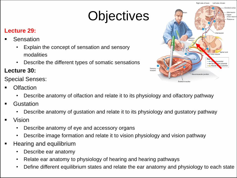
ObjectivesLecture 29:
Sensation
• Explain the concept of sensation and sensory
modalities
• Describe the different types of somatic sensations
Lecture 30:
Special Senses:
Olfaction
• Describe anatomy of olfaction and relate it to its physiology and olfactory pathway
Gustation
• Describe anatomy of gustation and relate it to its physiology and gustatory pathway
Vision
• Describe anatomy of eye and accessory organs
• Describe image formation and relate it to vision physiology and vision pathway
Hearing and equilibrium
• Describe ear anatomy
• Relate ear anatomy to physiology of hearing and hearing pathways
• Define different equilibrium states and relate the ear anatomy and physiology to each state
© Endeavour College of Natural Health endeavour.edu.au 7
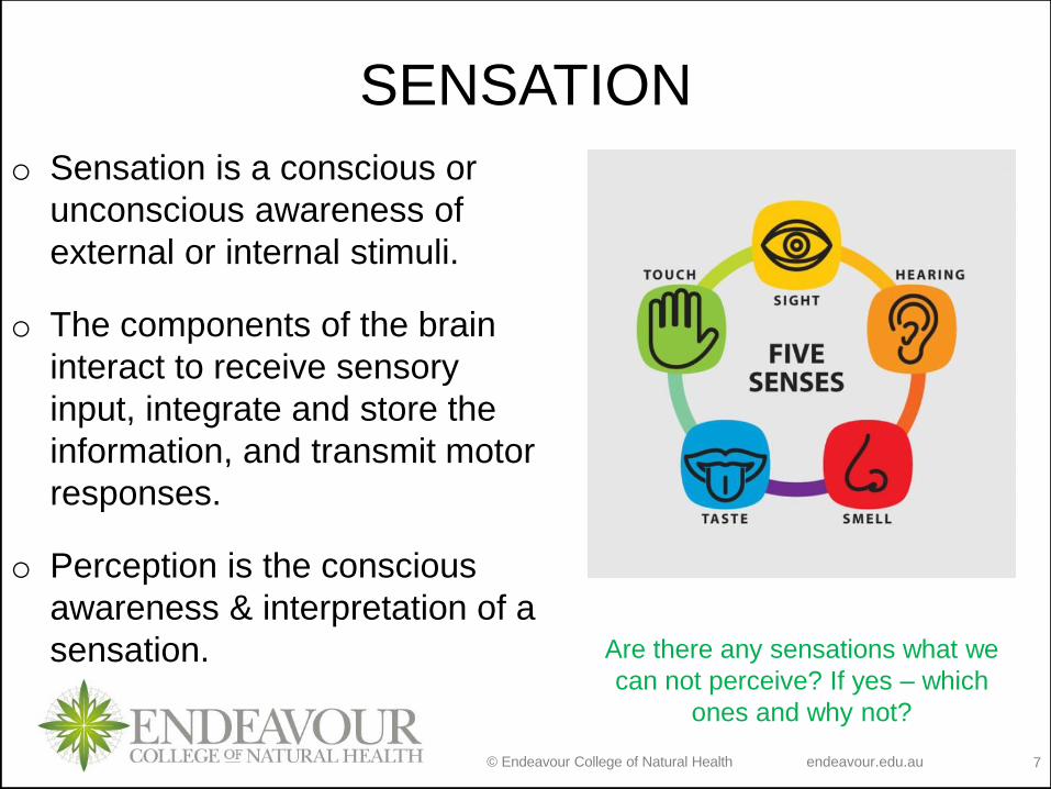
SENSATION
o Sensation is a conscious or
unconscious awareness of
external or internal stimuli.
o The components of the brain
interact to receive sensory
input, integrate and store the
information, and transmit motor
responses.
o Perception is the conscious
awareness & interpretation of a
sensation. Are there any sensations what we
can not perceive? If yes – which
ones and why not?
© Endeavour College of Natural Health endeavour.edu.au 8
SENSORY MODALITIES
o Sensory Modality: unique type of sensation; property by
which one sensation is distinguished from another.
o 2 classes of sensations
1. General senses: include both somatic and visceral senses
Somatic: tactile, thermal, pain and proprioceptive sensations
Visceral: provide information about conditions within internal
organs (e.g. stretch, pressure)
2. Special senses: smell, taste, vision, hearing and
equilibrium (balance); anatomically distinct
© Endeavour College of Natural Health endeavour.edu.au 9
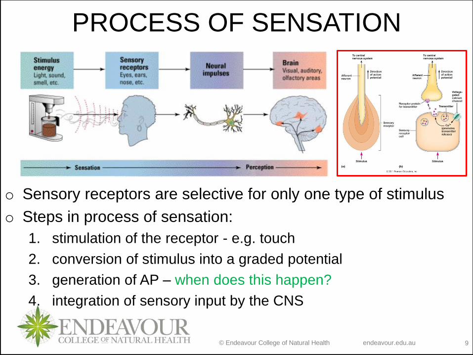
PROCESS OF SENSATION
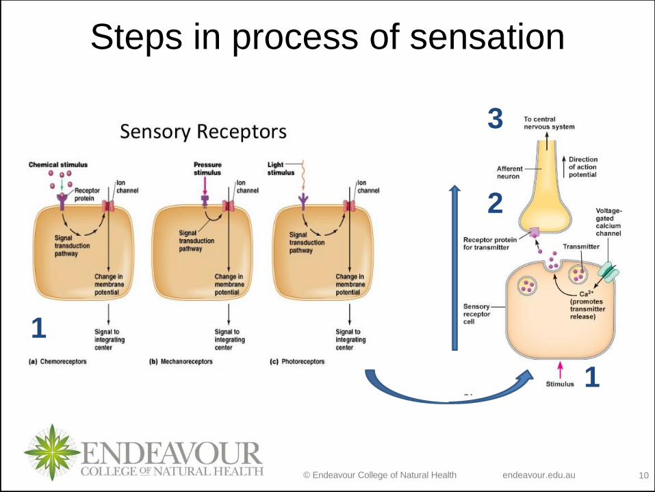
o Sensory receptors are selective for only one type of stimulus
o Steps in process of sensation:
1. stimulation of the receptor - e.g. touch
2. conversion of stimulus into a graded potential
3. generation of AP – when does this happen?
4. integration of sensory input by the CNS
© Endeavour College of Natural Health endeavour.edu.au 10
Steps in process of sensation
1
1
2
3
© Endeavour College of Natural Health endeavour.edu.au 11
SENSORY RECEPTOR
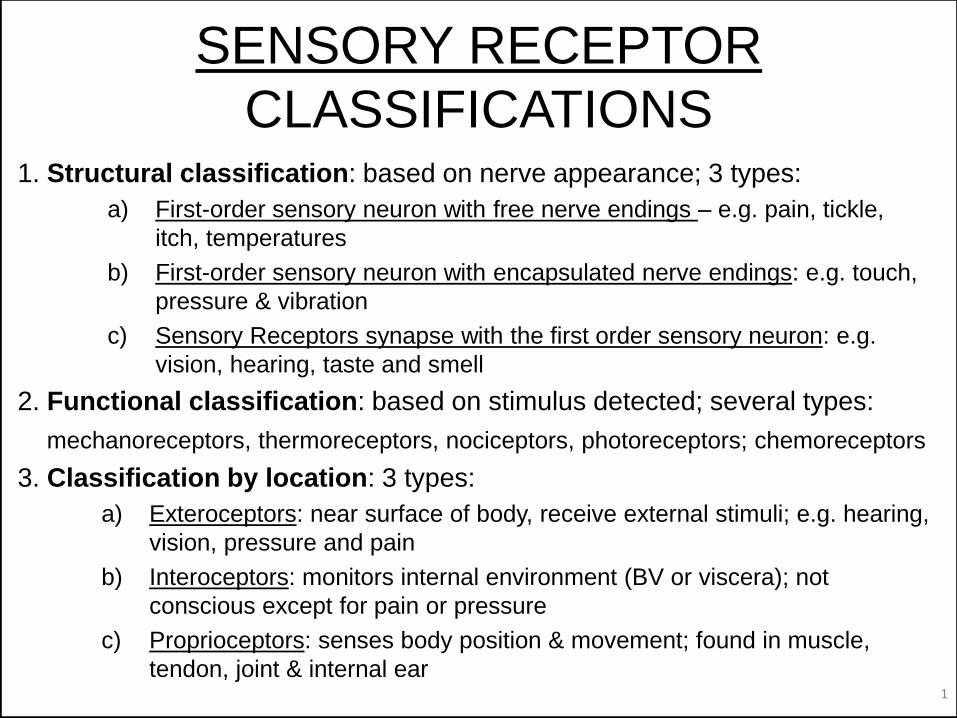
CLASSIFICATIONS1. Structural classification: based on nerve appearance; 3 types:
a) First-order sensory neuron with free nerve endings – e.g. pain, tickle,
itch, temperatures
b) First-order sensory neuron with encapsulated nerve endings: e.g. touch,
pressure & vibration
c) Sensory Receptors synapse with the first order sensory neuron: e.g.
vision, hearing, taste and smell
2. Functional classification: based on stimulus detected; several types:
mechanoreceptors, thermoreceptors, nociceptors, photoreceptors; chemoreceptors
3. Classification by location: 3 types:
a) Exteroceptors: near surface of body, receive external stimuli; e.g. hearing,
vision, pressure and pain
b) Interoceptors: monitors internal environment (BV or viscera); not
conscious except for pain or pressure
c) Proprioceptors: senses body position & movement; found in muscle,
tendon, joint & internal ear
© Endeavour College of Natural Health endeavour.edu.au 12
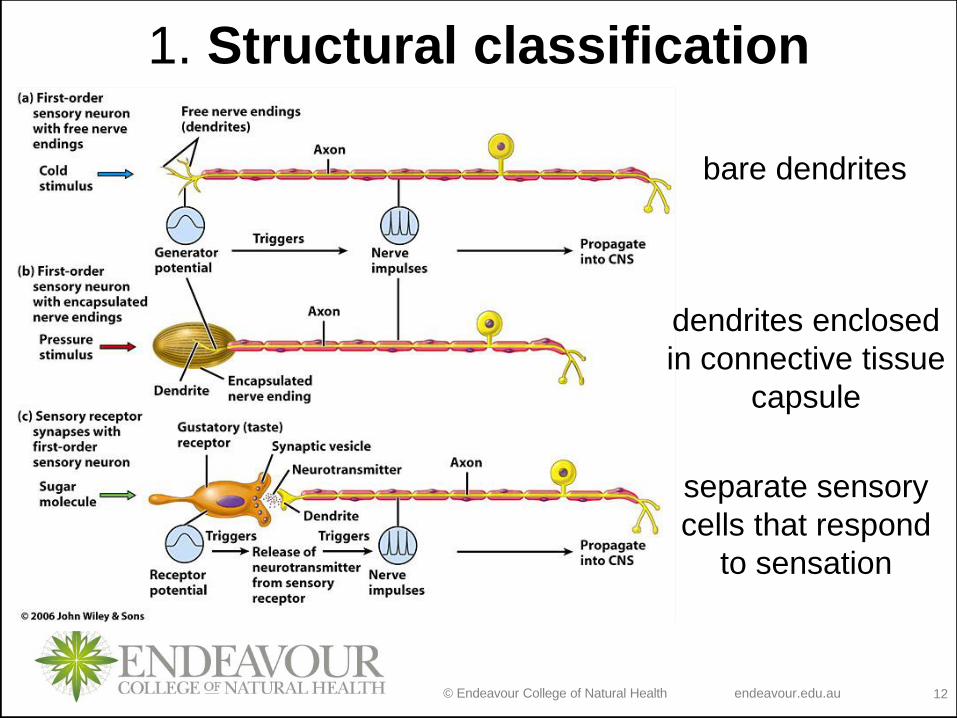
1. Structural classification
bare dendrites
dendrites enclosed
in connective tissue
capsule
separate sensory
cells that respond
to sensation
© Endeavour College of Natural Health endeavour.edu.au 13
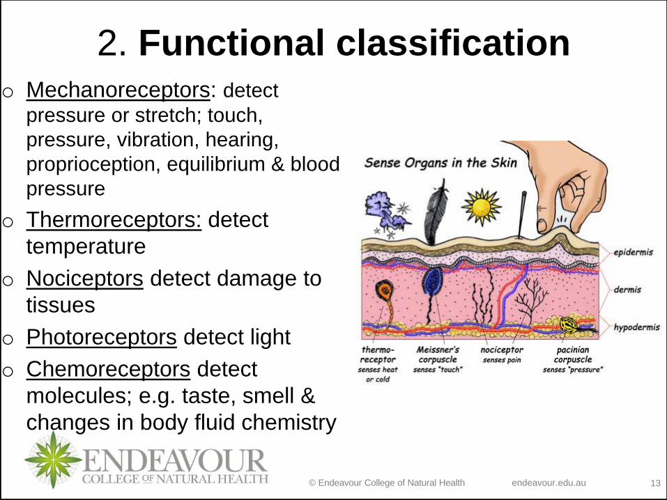
2. Functional classificationo Mechanoreceptors: detect
pressure or stretch; touch,
pressure, vibration, hearing,
proprioception, equilibrium & blood
pressure
o Thermoreceptors: detect
temperature
o Nociceptors detect damage to
tissues
o Photoreceptors detect light
o Chemoreceptors detect
molecules; e.g. taste, smell &
changes in body fluid chemistry
© Endeavour College of Natural Health endeavour.edu.au 14
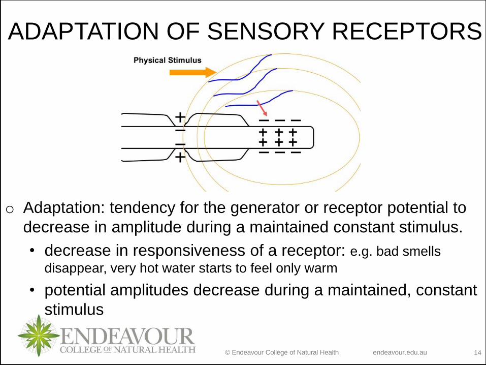
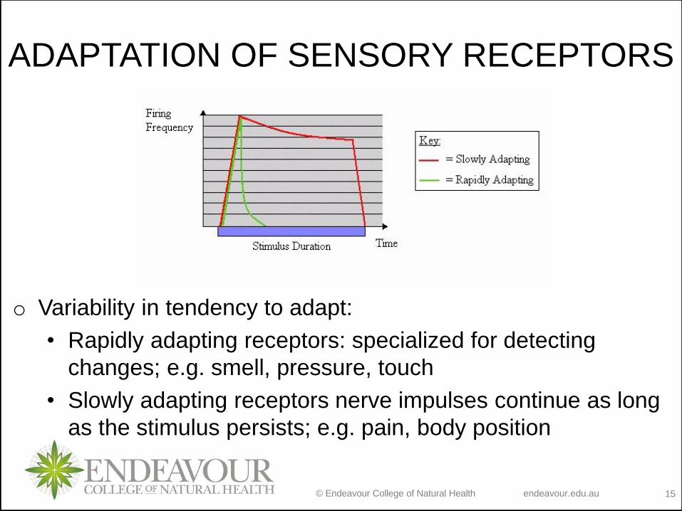
ADAPTATION OF SENSORY RECEPTORS
o Adaptation: tendency for the generator or receptor potential to
decrease in amplitude during a maintained constant stimulus.
• decrease in responsiveness of a receptor: e.g. bad smells
disappear, very hot water starts to feel only warm
• potential amplitudes decrease during a maintained, constant
stimulus
© Endeavour College of Natural Health endeavour.edu.au 15
ADAPTATION OF SENSORY RECEPTORS
o Variability in tendency to adapt:
• Rapidly adapting receptors: specialized for detecting
changes; e.g. smell, pressure, touch
• Slowly adapting receptors nerve impulses continue as long
as the stimulus persists; e.g. pain, body position
© Endeavour College of Natural Health endeavour.edu.au 17
SOMATIC SENSATIONS
o Sensation from the skin, muscles, bones, tendons and
joints. Initiated due to activation of a number of
distinct somatic receptors that respond specifically to
changes in heat, cold, touch, pressure, limb position,
limb movement or pain.
o Classification of somatic sensations:
1. Tactile sensations – touch, pressure, vibration, itch and
tickle
2. Pain
3. Proprioceptive
© Endeavour College of Natural Health endeavour.edu.au 18
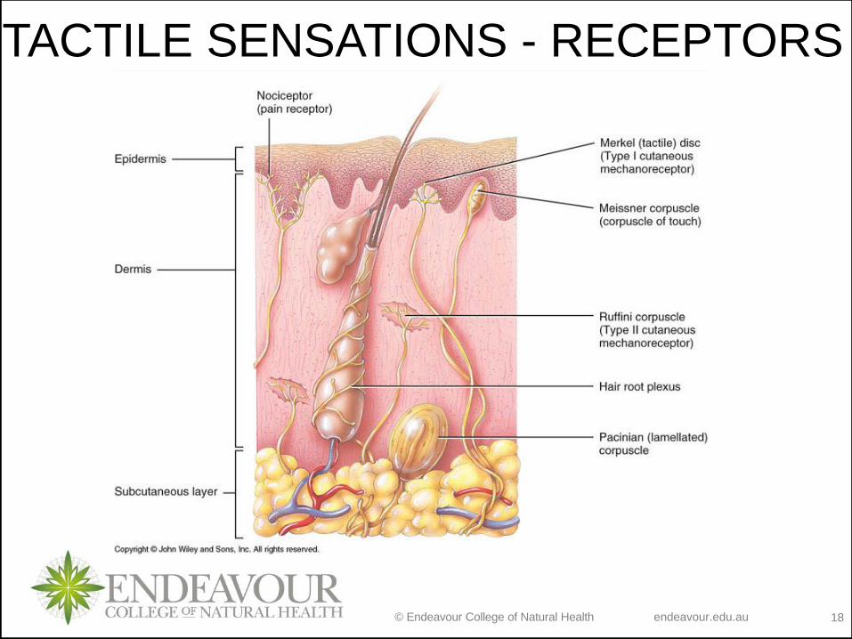
TACTILE SENSATIONS - RECEPTORS
© Endeavour College of Natural Health endeavour.edu.au 19
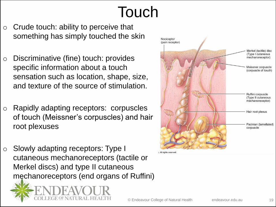
Toucho Crude touch: ability to perceive that
something has simply touched the skin
o Discriminative (fine) touch: provides
specific information about a touch
sensation such as location, shape, size,
and texture of the source of stimulation.
o Rapidly adapting receptors: corpuscles
of touch (Meissner’s corpuscles) and hair
root plexuses
o Slowly adapting receptors: Type I
cutaneous mechanoreceptors (tactile or
Merkel discs) and type II cutaneous
mechanoreceptors (end organs of Ruffini)
© Endeavour College of Natural Health endeavour.edu.au 20
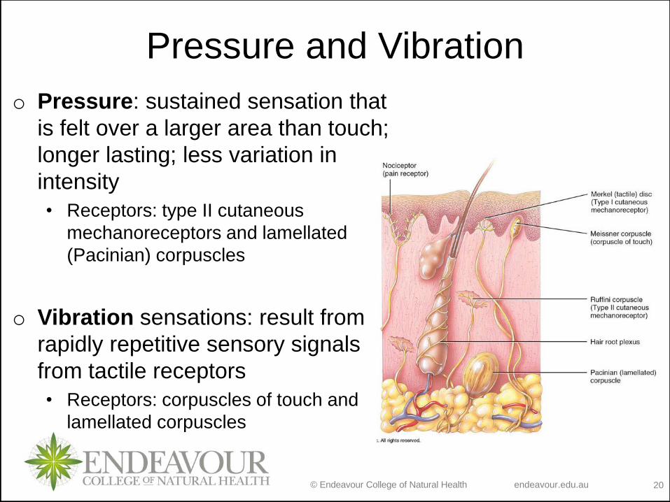
Pressure and Vibration
o Pressure: sustained sensation that
is felt over a larger area than touch;
longer lasting; less variation in
intensity
• Receptors: type II cutaneous
mechanoreceptors and lamellated
(Pacinian) corpuscles
o Vibration sensations: result from
rapidly repetitive sensory signals
from tactile receptors
• Receptors: corpuscles of touch and
lamellated corpuscles
© Endeavour College of Natural Health endeavour.edu.au 21
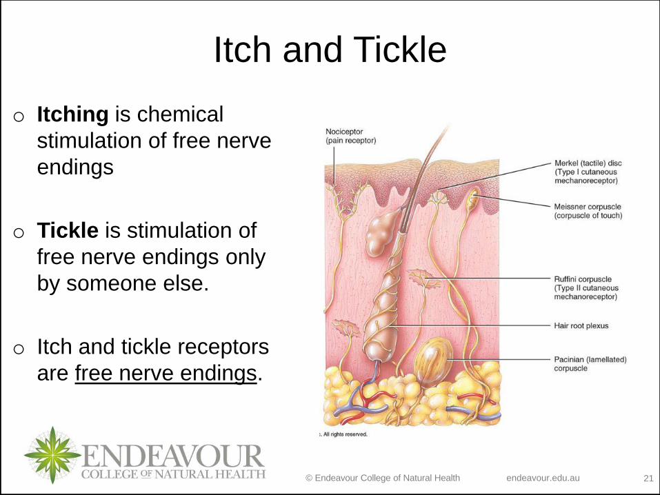
Itch and Tickle
o Itching is chemical
stimulation of free nerve
endings
o Tickle is stimulation of
free nerve endings only
by someone else.
o Itch and tickle receptors
are free nerve endings.

© Endeavour College of Natural Health endeavour.edu.au 22
PAIN
o Types of pain:
1. Acute pain
2. Chronic pain
3. Nerve pain
4. Referred pain
o Provides information about noxious and damaging stimuli;
helps protect from grater damage
o Receptors: nociceptors (free nerve ending); sensory neurons
transmitting messages of painful stimuli, secrete glutamate
and substance P.
© Endeavour College of Natural Health endeavour.edu.au 23
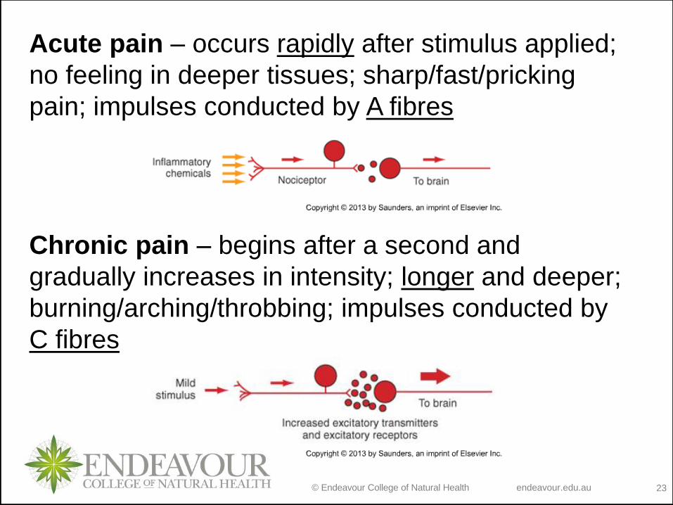
Acute pain – occurs rapidly after stimulus applied;
no feeling in deeper tissues; sharp/fast/pricking
pain; impulses conducted by A fibres
Chronic pain – begins after a second and
gradually increases in intensity; longer and deeper;
burning/arching/throbbing; impulses conducted by
C fibres
© Endeavour College of Natural Health endeavour.edu.au 24
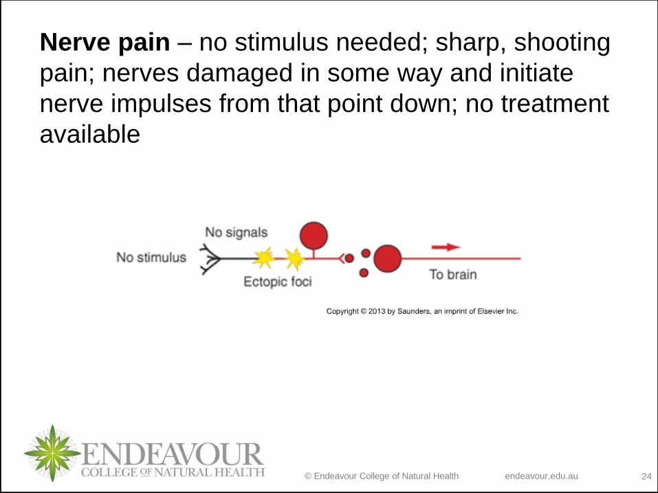
Nerve pain – no stimulus needed; sharp, shooting
pain; nerves damaged in some way and initiate
nerve impulses from that point down; no treatment
available
© Endeavour College of Natural Health endeavour.edu.au 25
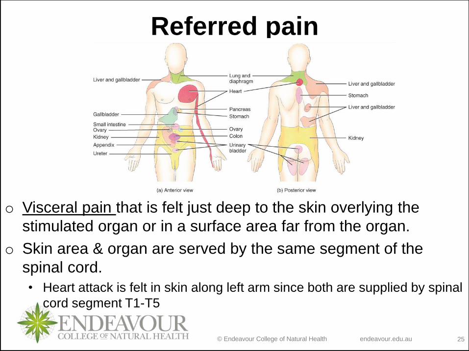
Referred pain
o Visceral pain that is felt just deep to the skin overlying the
stimulated organ or in a surface area far from the organ.
o Skin area & organ are served by the same segment of the
spinal cord.
• Heart attack is felt in skin along left arm since both are supplied by spinal
cord segment T1-T5
© Endeavour College of Natural Health endeavour.edu.au 27
PROPRIOCEPTIVE SENSATIONS
o Proprioceptors allow us to perceive the position of the
body and its parts. Three main types of proprioceptors:
1. muscle spindles: detect muscle movement
2. Golgi tendon organs: determine stretch in tendons
3. joint receptors: detect movement in ligaments
o Sensory information is sent to cerebellum & cerebral cortex
• signals project from muscle, tendon, joint capsules & hair cells in
the vestibular apparatus
© Endeavour College of Natural Health endeavour.edu.au 28
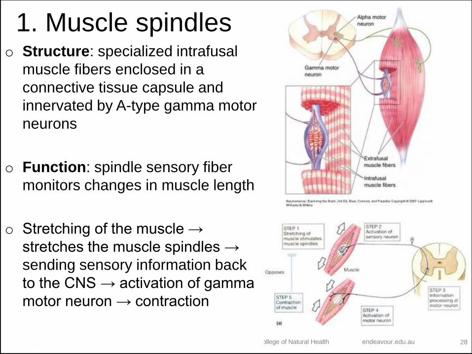
1. Muscle spindleso Structure: specialized intrafusal
muscle fibers enclosed in a
connective tissue capsule and
innervated by A-type gamma motor
neurons
o Function: spindle sensory fiber
monitors changes in muscle length
o Stretching of the muscle →
stretches the muscle spindles →
sending sensory information back
to the CNS → activation of gamma
motor neuron → contraction
© Endeavour College of Natural Health endeavour.edu.au 29
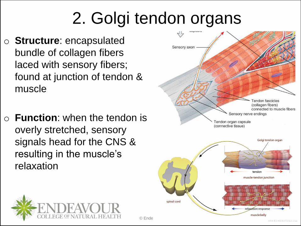
2. Golgi tendon organs
o Structure: encapsulated
bundle of collagen fibers
laced with sensory fibers;
found at junction of tendon &
muscle
o Function: when the tendon is
overly stretched, sensory
signals head for the CNS &
resulting in the muscle’s
relaxation
© Endeavour College of Natural Health endeavour.edu.au 30
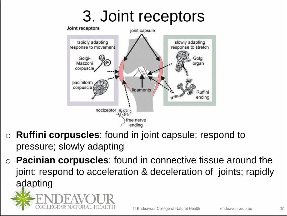
3. Joint receptors
o Ruffini corpuscles: found in joint capsule: respond to
pressure; slowly adapting
o Pacinian corpuscles: found in connective tissue around the
joint: respond to acceleration & deceleration of joints; rapidly
adapting

© Endeavour College of Natural Health endeavour.edu.au 31
ObjectivesLecture 29:
Sensation
• Explain the concept of sensation and sensory modalities
• Describe the different types of somatic sensations
Lecture 30:
Special Senses:
Olfaction
• Describe anatomy of olfaction and relate it to its physiology and olfactory pathway
Gustation
• Describe anatomy of gustation and relate it to its physiology and gustatory pathway
Vision
• Describe anatomy of eye and accessory organs
• Describe image formation and relate it to vision physiology and vision pathway
Hearing and equilibrium
• Describe ear anatomy
• Relate ear anatomy to physiology of hearing and hearing pathways
• Define different equilibrium states and relate the ear anatomy and physiology to each state
© Endeavour College of Natural Health endeavour.edu.au 32
SPECIAL SENSES
o Smell, taste, vision, hearing and equilibrium
o Housed in complex sensory organs
o Chemical senses:
• Interaction of molecules with receptor cells
• Olfaction (smell) and gustation (taste)
• Both project to cerebral cortex & limbic system, so evoke strong
emotional reactions
© Endeavour College of Natural Health endeavour.edu.au 33
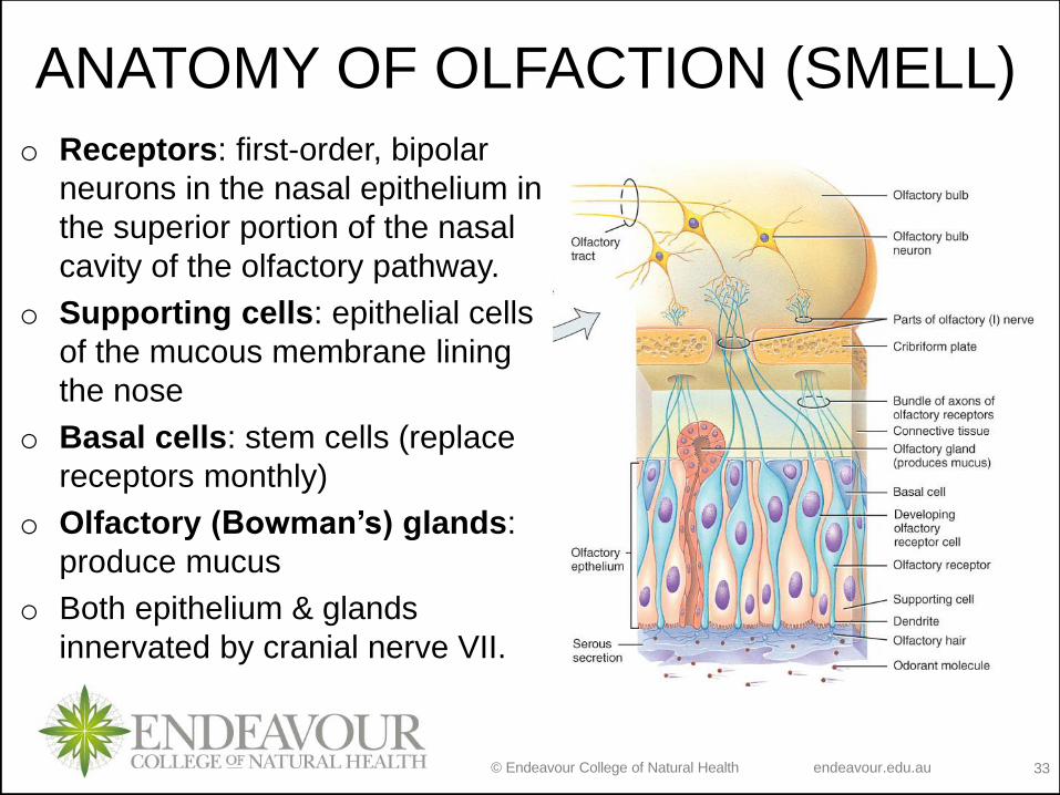
ANATOMY OF OLFACTION (SMELL)
o Receptors: first-order, bipolar
neurons in the nasal epithelium in
the superior portion of the nasal
cavity of the olfactory pathway.
o Supporting cells: epithelial cells
of the mucous membrane lining
the nose
o Basal cells: stem cells (replace
receptors monthly)
o Olfactory (Bowman’s) glands:
produce mucus
o Both epithelium & glands
innervated by cranial nerve VII.
© Endeavour College of Natural Health endeavour.edu.au 34
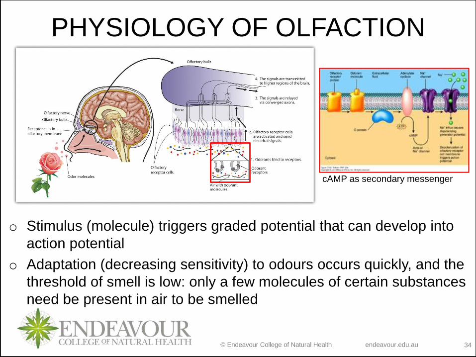
PHYSIOLOGY OF OLFACTION
o Stimulus (molecule) triggers graded potential that can develop into
action potential
o Adaptation (decreasing sensitivity) to odours occurs quickly, and the
threshold of smell is low: only a few molecules of certain substances
need be present in air to be smelled
cAMP as secondary messenger
© Endeavour College of Natural Health endeavour.edu.au 35
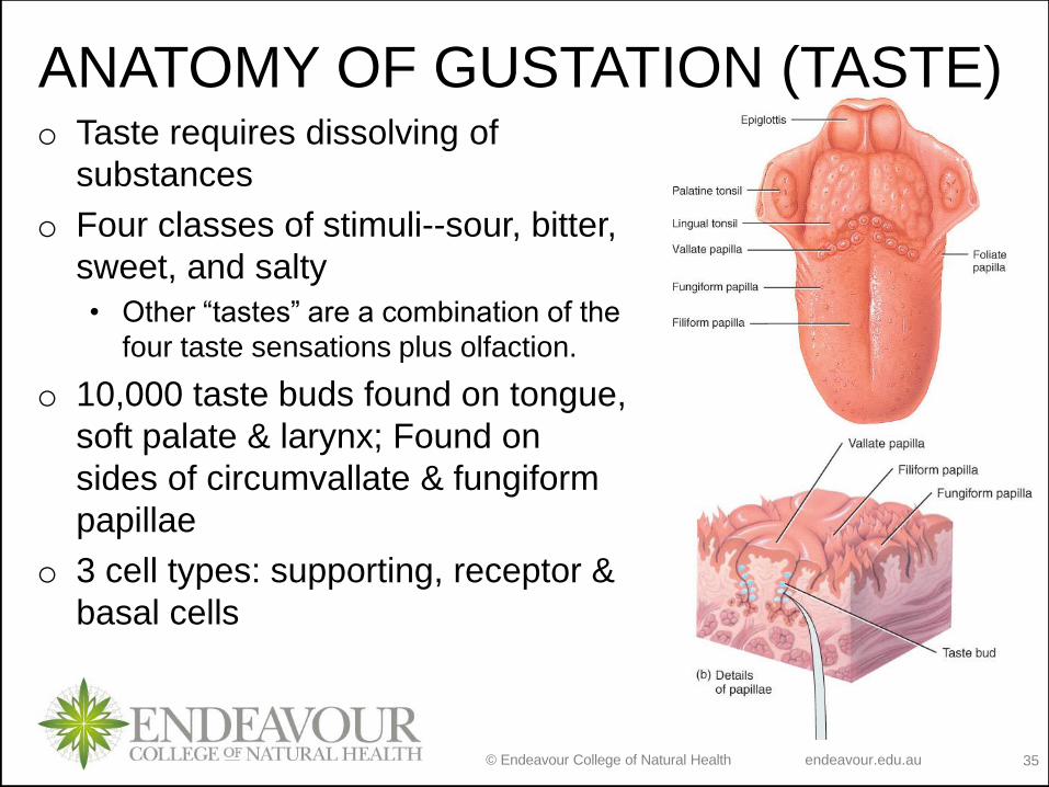
o Taste requires dissolving of
substances
o Four classes of stimuli--sour, bitter,
sweet, and salty
• Other “tastes” are a combination of the
four taste sensations plus olfaction.
o 10,000 taste buds found on tongue,
soft palate & larynx; Found on
sides of circumvallate & fungiform
papillae
o 3 cell types: supporting, receptor &
basal cells
ANATOMY OF GUSTATION (TASTE)
© Endeavour College of Natural Health endeavour.edu.au 36
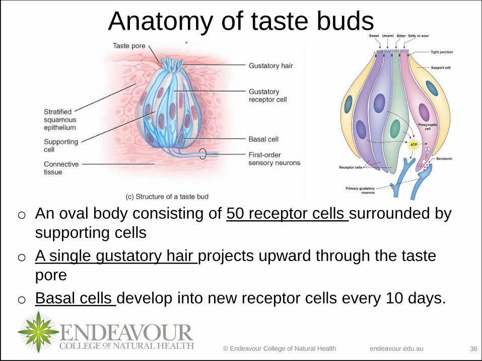
Anatomy of taste buds
o An oval body consisting of 50 receptor cells surrounded by
supporting cells
o A single gustatory hair projects upward through the taste
pore
o Basal cells develop into new receptor cells every 10 days.
© Endeavour College of Natural Health endeavour.edu.au 37
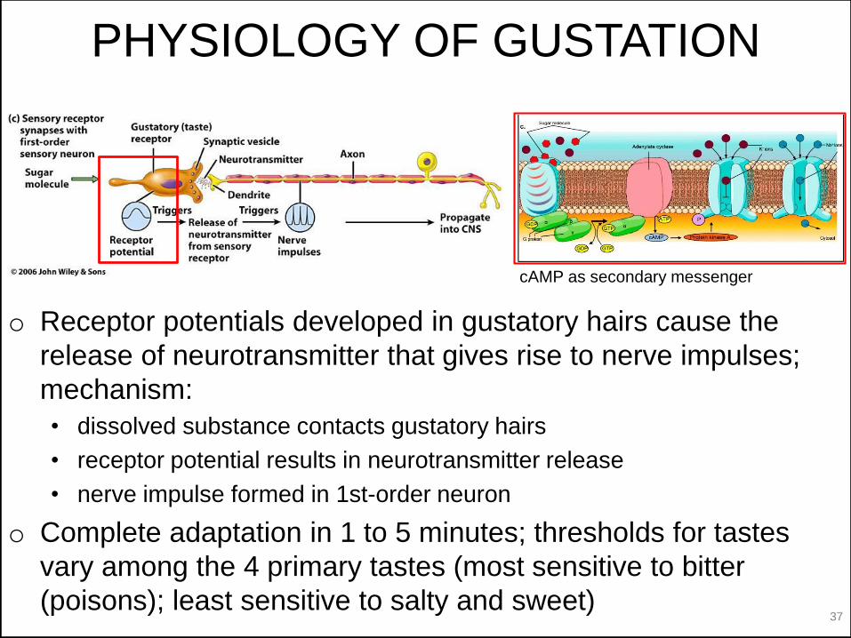
PHYSIOLOGY OF GUSTATION
o Receptor potentials developed in gustatory hairs cause the
release of neurotransmitter that gives rise to nerve impulses;
mechanism:
• dissolved substance contacts gustatory hairs
• receptor potential results in neurotransmitter release
• nerve impulse formed in 1st-order neuron
o Complete adaptation in 1 to 5 minutes; thresholds for tastes
vary among the 4 primary tastes (most sensitive to bitter
(poisons); least sensitive to salty and sweet)
cAMP as secondary messenger
© Endeavour College of Natural Health endeavour.edu.au 38
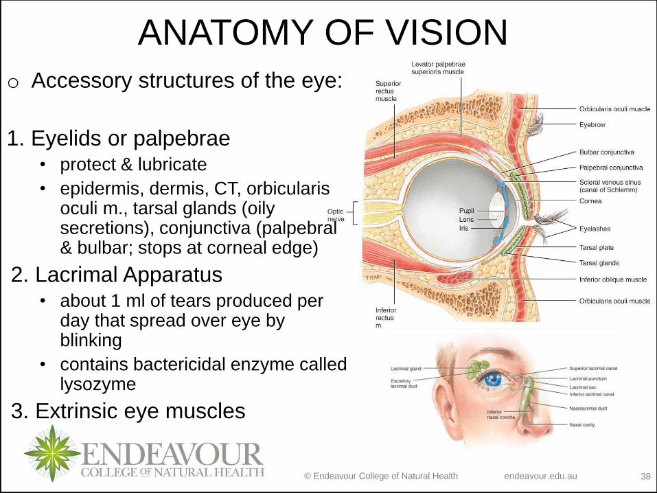
ANATOMY OF VISION
o Accessory structures of the eye:
1. Eyelids or palpebrae• protect & lubricate
• epidermis, dermis, CT, orbicularis oculi m., tarsal glands (oily secretions), conjunctiva (palpebral & bulbar; stops at corneal edge)
2. Lacrimal Apparatus• about 1 ml of tears produced per
day that spread over eye by blinking
• contains bactericidal enzyme called lysozyme
3. Extrinsic eye muscles
© Endeavour College of Natural Health endeavour.edu.au 39
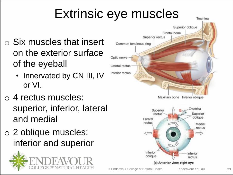
Extrinsic eye muscles
o Six muscles that insert
on the exterior surface
of the eyeball
• Innervated by CN III, IV
or VI.
o 4 rectus muscles:
superior, inferior, lateral
and medial
o 2 oblique muscles:
inferior and superior
© Endeavour College of Natural Health endeavour.edu.au 40
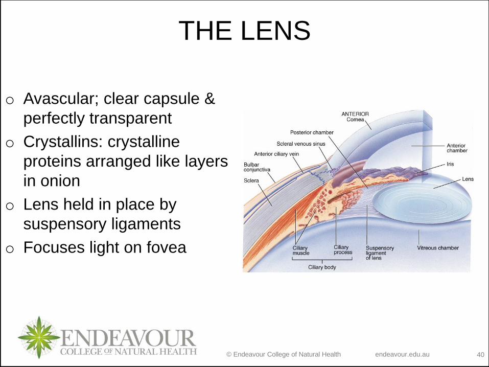
THE LENS
o Avascular; clear capsule &
perfectly transparent
o Crystallins: crystalline
proteins arranged like layers
in onion
o Lens held in place by
suspensory ligaments
o Focuses light on fovea
© Endeavour College of Natural Health endeavour.edu.au 41
CAVITIES OF THE INTERIOR OF
EYEBALL
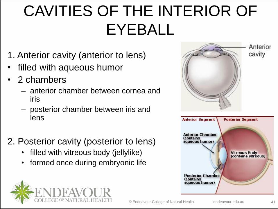
1. Anterior cavity (anterior to lens)
• filled with aqueous humor
• 2 chambers– anterior chamber between cornea and
iris
– posterior chamber between iris and lens
2. Posterior cavity (posterior to lens)• filled with vitreous body (jellylike)
• formed once during embryonic life
© Endeavour College of Natural Health endeavour.edu.au 42
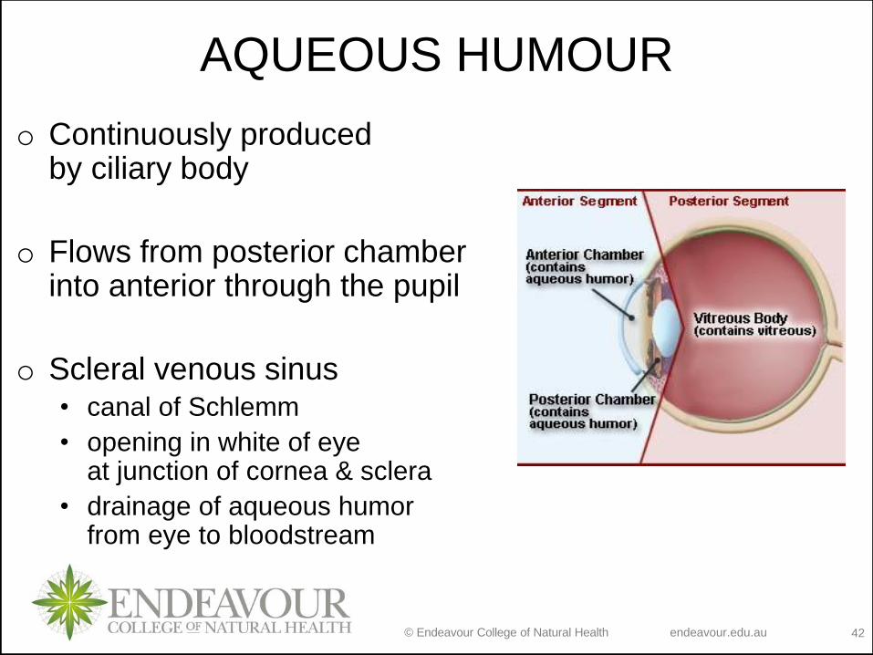
AQUEOUS HUMOUR
o Continuously produced by ciliary body
o Flows from posterior chamberinto anterior through the pupil
o Scleral venous sinus• canal of Schlemm
• opening in white of eyeat junction of cornea & sclera
• drainage of aqueous humor from eye to bloodstream
© Endeavour College of Natural Health endeavour.edu.au 43
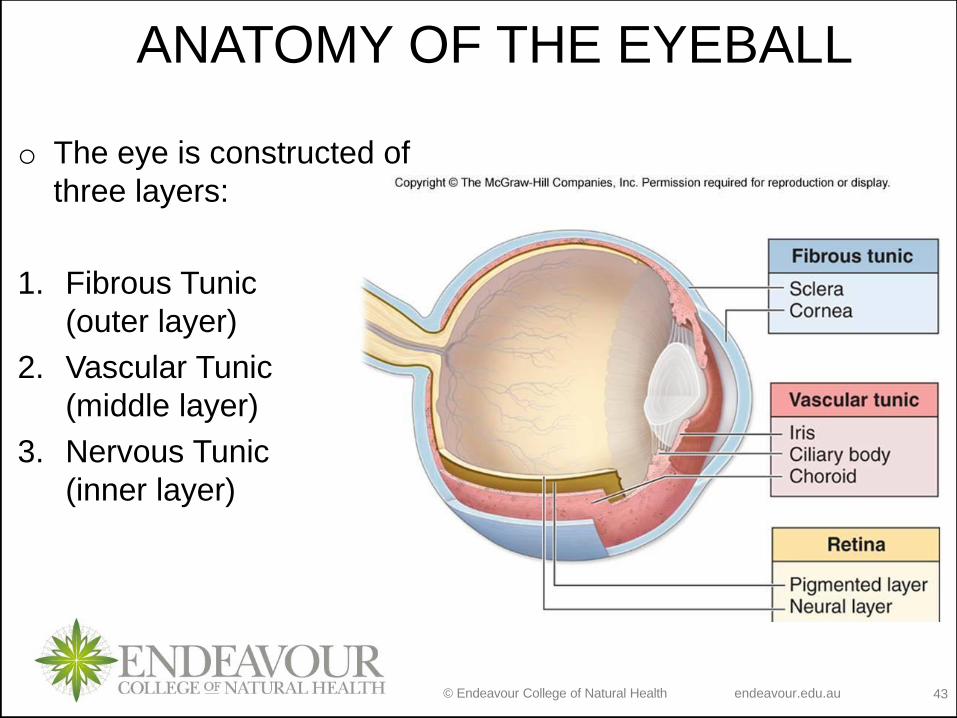
ANATOMY OF THE EYEBALL
o The eye is constructed of
three layers:
1. Fibrous Tunic
(outer layer)
2. Vascular Tunic
(middle layer)
3. Nervous Tunic
(inner layer)
© Endeavour College of Natural Health endeavour.edu.au 44
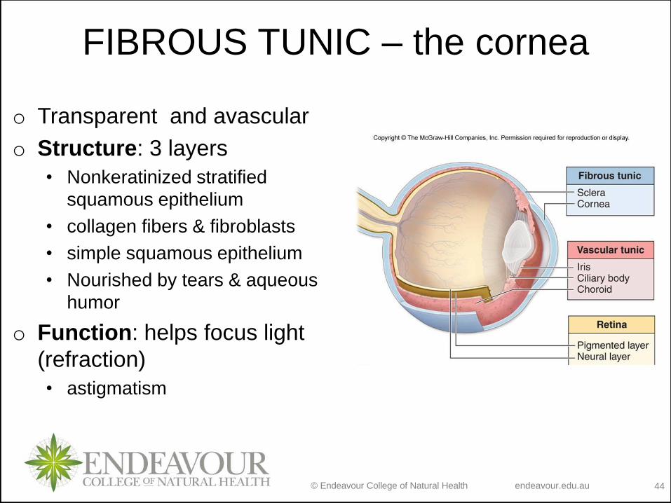
FIBROUS TUNIC – the cornea
o Transparent and avascular
o Structure: 3 layers
• Nonkeratinized stratified
squamous epithelium
• collagen fibers & fibroblasts
• simple squamous epithelium
• Nourished by tears & aqueous
humor
o Function: helps focus light
(refraction)
• astigmatism
© Endeavour College of Natural Health endeavour.edu.au 45
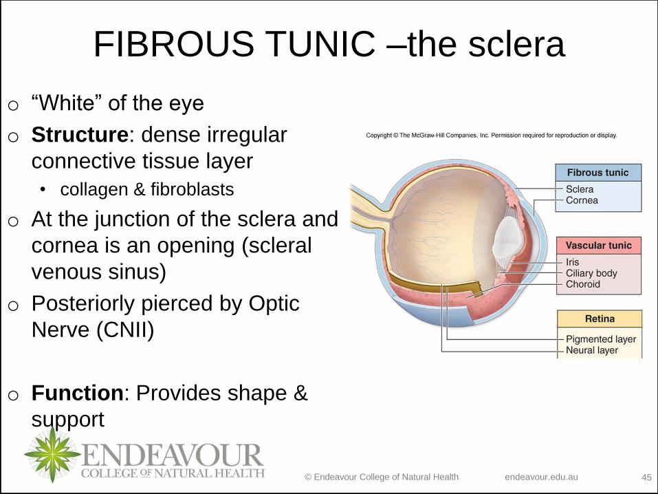
FIBROUS TUNIC –the sclera
o “White” of the eye
o Structure: dense irregular
connective tissue layer
• collagen & fibroblasts
o At the junction of the sclera and
cornea is an opening (scleral
venous sinus)
o Posteriorly pierced by Optic
Nerve (CNII)
o Function: Provides shape &
support
© Endeavour College of Natural Health endeavour.edu.au 46
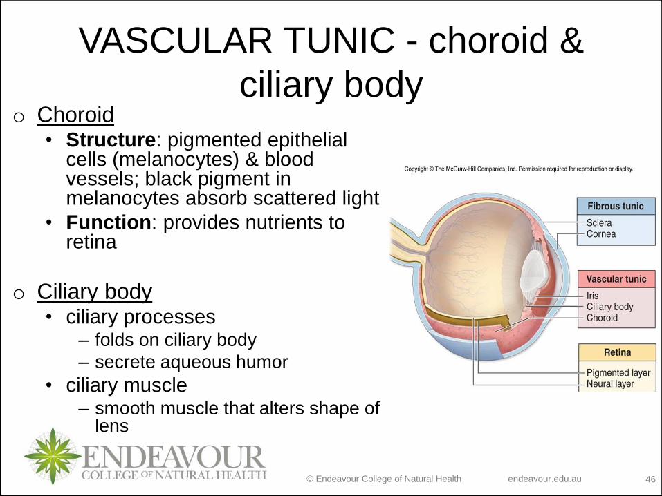
VASCULAR TUNIC - choroid &
ciliary body o Choroid
• Structure: pigmented epithelial cells (melanocytes) & blood vessels; black pigment in melanocytes absorb scattered light
• Function: provides nutrients to retina
o Ciliary body• ciliary processes
– folds on ciliary body
– secrete aqueous humor
• ciliary muscle– smooth muscle that alters shape of
lens

© Endeavour College of Natural Health endeavour.edu.au 47
VASCULAR TUNIC - iris & pupil
o Structure: colored portion of eye; shape of flat donut suspended
between cornea & lens; hole in center of iris is called pupil
o Function: regulation of amount of light entering eye
o Autonomic reflexes:
• Parasympathetic: circular muscle fibers contract in bright light to shrink
pupil
• Sympathetic: radial muscle fibers contract in dim light to enlarge pupil
© Endeavour College of Natural Health endeavour.edu.au 48
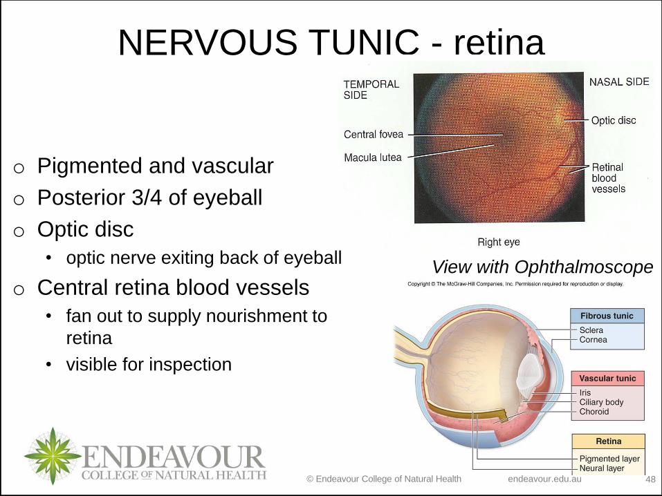
NERVOUS TUNIC - retina
o Pigmented and vascular
o Posterior 3/4 of eyeball
o Optic disc
• optic nerve exiting back of eyeball
o Central retina blood vessels
• fan out to supply nourishment to
retina
• visible for inspection
View with Ophthalmoscope
© Endeavour College of Natural Health endeavour.edu.au 49
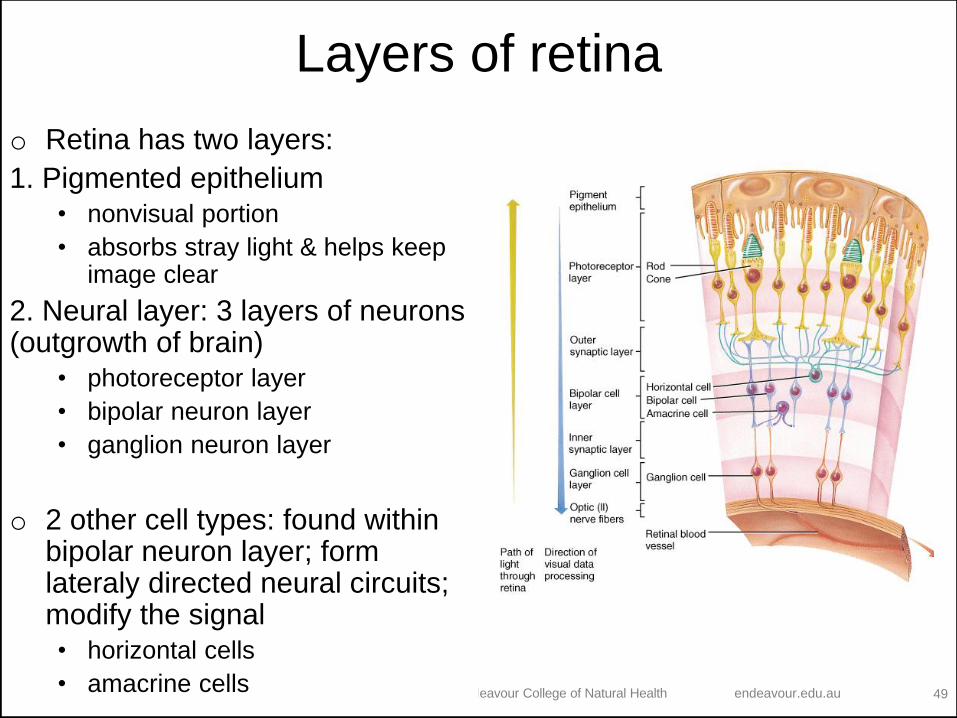
Layers of retina
o Retina has two layers:
1. Pigmented epithelium
• nonvisual portion
• absorbs stray light & helps keep image clear
2. Neural layer: 3 layers of neurons (outgrowth of brain)
• photoreceptor layer
• bipolar neuron layer
• ganglion neuron layer
o 2 other cell types: found within bipolar neuron layer; form lateraly directed neural circuits; modify the signal
• horizontal cells
• amacrine cells
© Endeavour College of Natural Health endeavour.edu.au 50
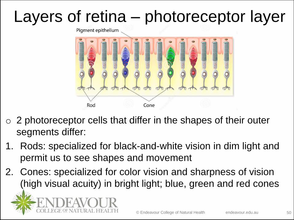
o 2 photoreceptor cells that differ in the shapes of their outer
segments differ:
1. Rods: specialized for black-and-white vision in dim light and
permit us to see shapes and movement
2. Cones: specialized for color vision and sharpness of vision
(high visual acuity) in bright light; blue, green and red cones
Layers of retina – photoreceptor layer
© Endeavour College of Natural Health endeavour.edu.au 51
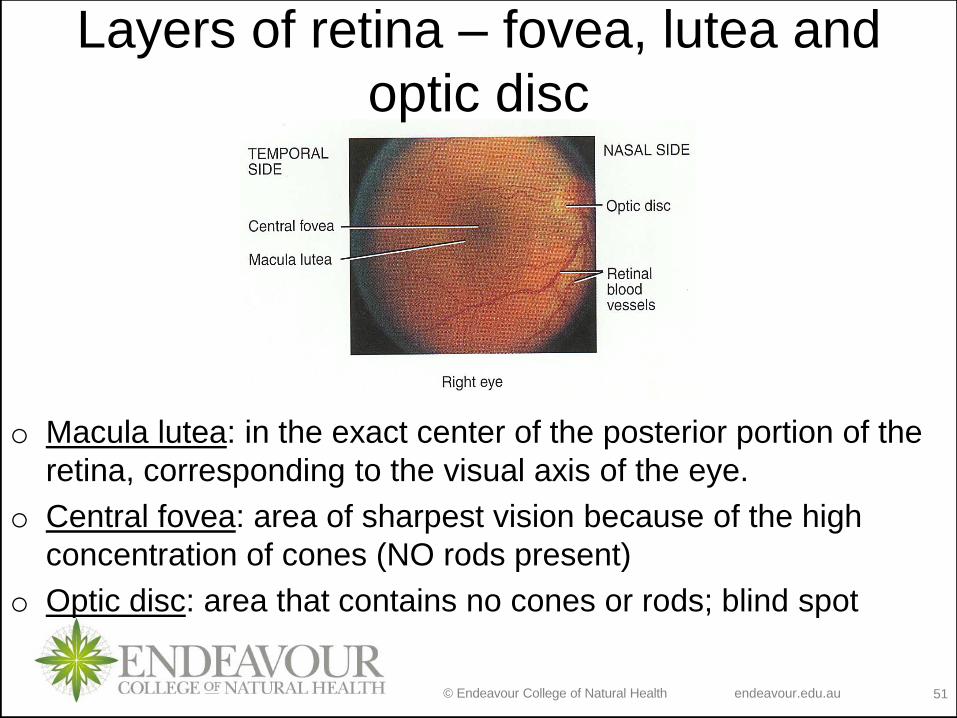
o Macula lutea: in the exact center of the posterior portion of the
retina, corresponding to the visual axis of the eye.
o Central fovea: area of sharpest vision because of the high
concentration of cones (NO rods present)
o Optic disc: area that contains no cones or rods; blind spot
Layers of retina – fovea, lutea and
optic disc
© Endeavour College of Natural Health endeavour.edu.au 53
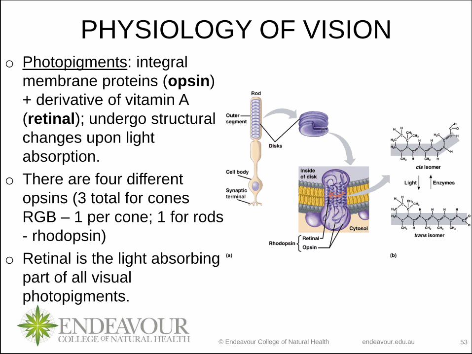
PHYSIOLOGY OF VISIONo Photopigments: integral
membrane proteins (opsin)
+ derivative of vitamin A
(retinal); undergo structural
changes upon light
absorption.
o There are four different
opsins (3 total for cones
RGB – 1 per cone; 1 for rods
- rhodopsin)
o Retinal is the light absorbing
part of all visual
photopigments.
© Endeavour College of Natural Health endeavour.edu.au 54
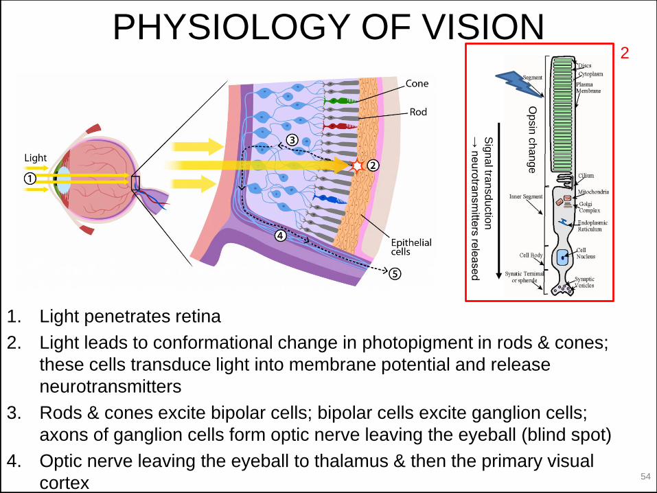
1. Light penetrates retina
2. Light leads to conformational change in photopigment in rods & cones;
these cells transduce light into membrane potential and release
neurotransmitters
3. Rods & cones excite bipolar cells; bipolar cells excite ganglion cells;
axons of ganglion cells form optic nerve leaving the eyeball (blind spot)
4. Optic nerve leaving the eyeball to thalamus & then the primary visual
cortex
PHYSIOLOGY OF VISION
Sig
na
l tran
sd
uctio
n
→ n
eu
rotra
nsm
itters
rele
ase
d
Op
sin
ch
an
ge
2
© Endeavour College of Natural Health endeavour.edu.au 55
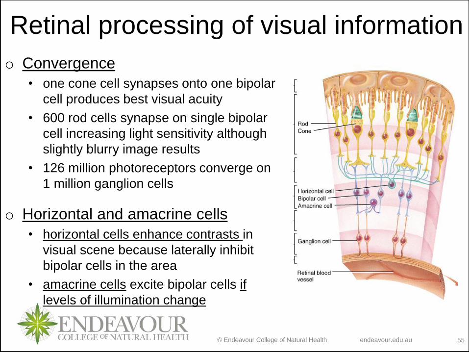
Retinal processing of visual information
o Convergence
• one cone cell synapses onto one bipolar
cell produces best visual acuity
• 600 rod cells synapse on single bipolar
cell increasing light sensitivity although
slightly blurry image results
• 126 million photoreceptors converge on
1 million ganglion cells
o Horizontal and amacrine cells
• horizontal cells enhance contrasts in
visual scene because laterally inhibit
bipolar cells in the area
• amacrine cells excite bipolar cells if
levels of illumination change
© Endeavour College of Natural Health endeavour.edu.au 56
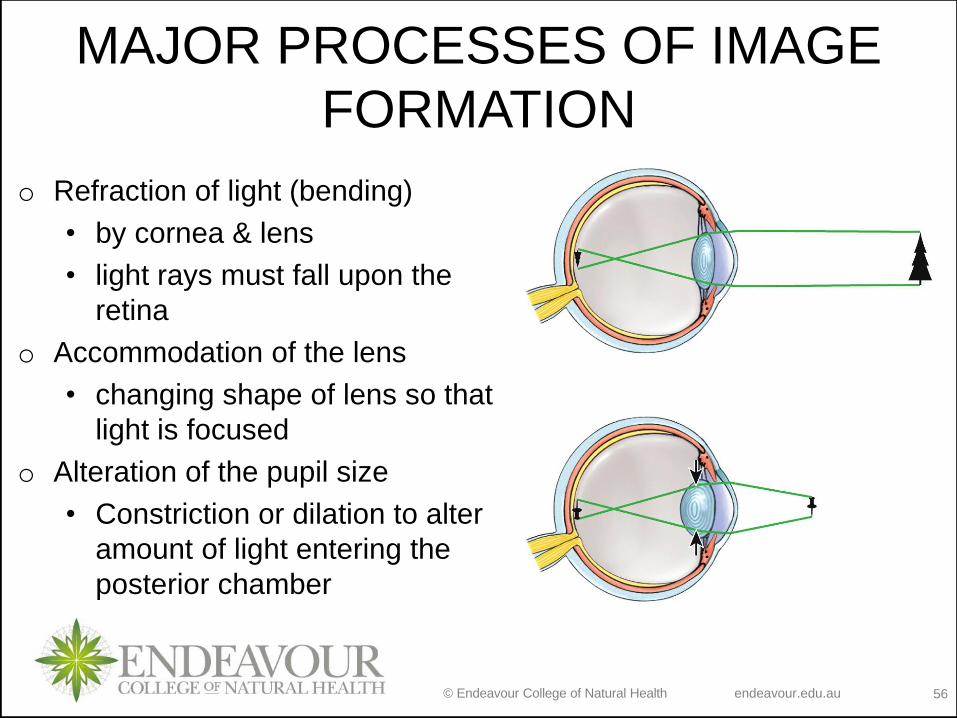
MAJOR PROCESSES OF IMAGE
FORMATION
o Refraction of light (bending)
• by cornea & lens
• light rays must fall upon the
retina
o Accommodation of the lens
• changing shape of lens so that
light is focused
o Alteration of the pupil size
• Constriction or dilation to alter
amount of light entering the
posterior chamber
© Endeavour College of Natural Health endeavour.edu.au 57
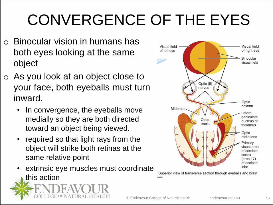
CONVERGENCE OF THE EYES
o Binocular vision in humans has
both eyes looking at the same
object
o As you look at an object close to
your face, both eyeballs must turn
inward.
• In convergence, the eyeballs move
medially so they are both directed
toward an object being viewed.
• required so that light rays from the
object will strike both retinas at the
same relative point
• extrinsic eye muscles must coordinate
this action
© Endeavour College of Natural Health endeavour.edu.au 58
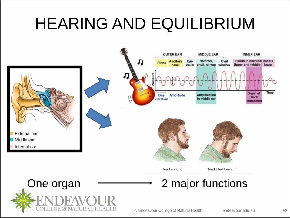
HEARING AND EQUILIBRIUM
One organ 2 major functions
© Endeavour College of Natural Health endeavour.edu.au 59
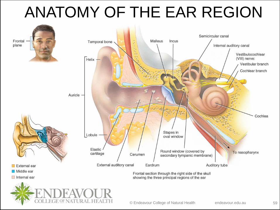
ANATOMY OF THE EAR REGION
© Endeavour College of Natural Health endeavour.edu.au 60
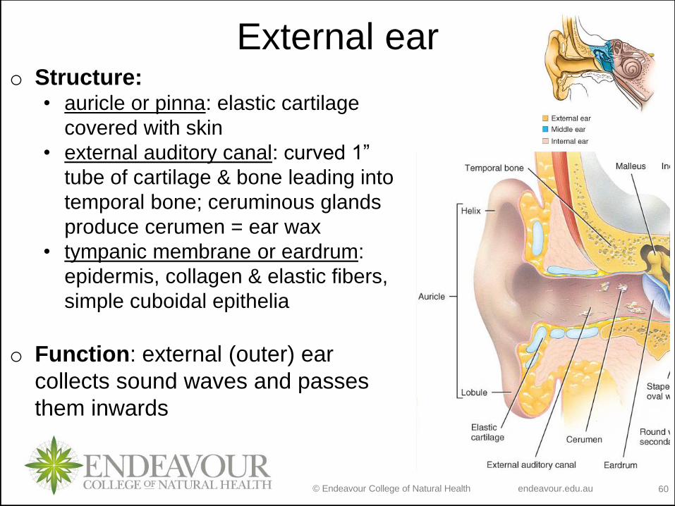
External earo Structure:
• auricle or pinna: elastic cartilage
covered with skin
• external auditory canal: curved 1”
tube of cartilage & bone leading into
temporal bone; ceruminous glands
produce cerumen = ear wax
• tympanic membrane or eardrum:
epidermis, collagen & elastic fibers,
simple cuboidal epithelia
o Function: external (outer) ear
collects sound waves and passes
them inwards
© Endeavour College of Natural Health endeavour.edu.au 61
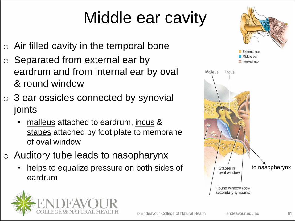
Middle ear cavity
o Air filled cavity in the temporal bone
o Separated from external ear by
eardrum and from internal ear by oval
& round window
o 3 ear ossicles connected by synovial
joints
• malleus attached to eardrum, incus &
stapes attached by foot plate to membrane
of oval window
o Auditory tube leads to nasopharynx
• helps to equalize pressure on both sides of
eardrum
to nasopharynx
© Endeavour College of Natural Health endeavour.edu.au 62
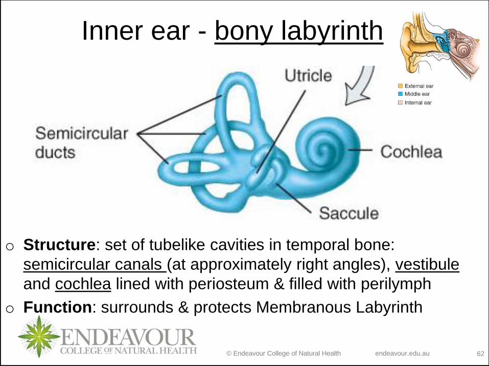
Inner ear - bony labyrinth
o Structure: set of tubelike cavities in temporal bone:
semicircular canals (at approximately right angles), vestibule
and cochlea lined with periosteum & filled with perilymph
o Function: surrounds & protects Membranous Labyrinth
© Endeavour College of Natural Health endeavour.edu.au 63
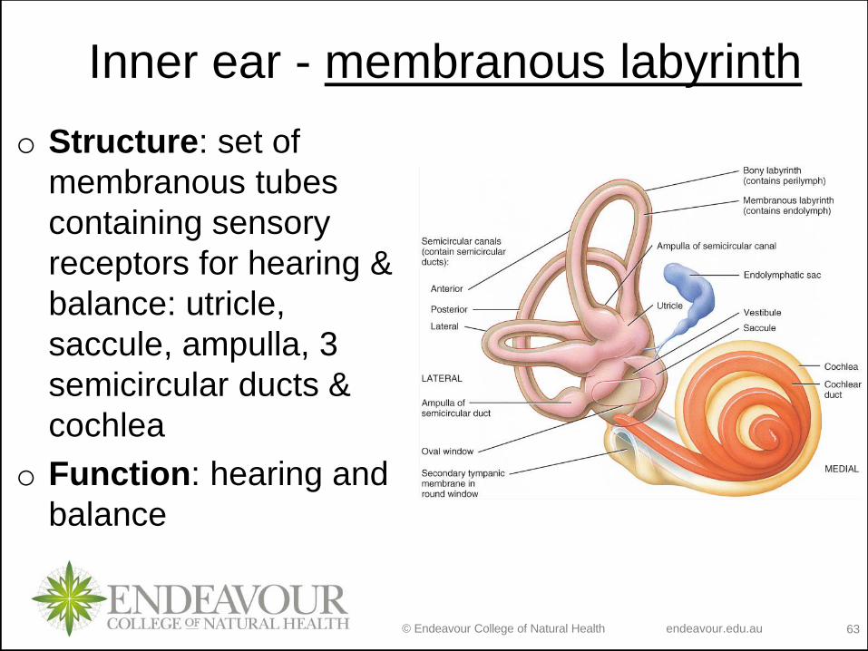
Inner ear - membranous labyrinth
o Structure: set of
membranous tubes
containing sensory
receptors for hearing &
balance: utricle,
saccule, ampulla, 3
semicircular ducts &
cochlea
o Function: hearing and
balance
© Endeavour College of Natural Health endeavour.edu.au 64
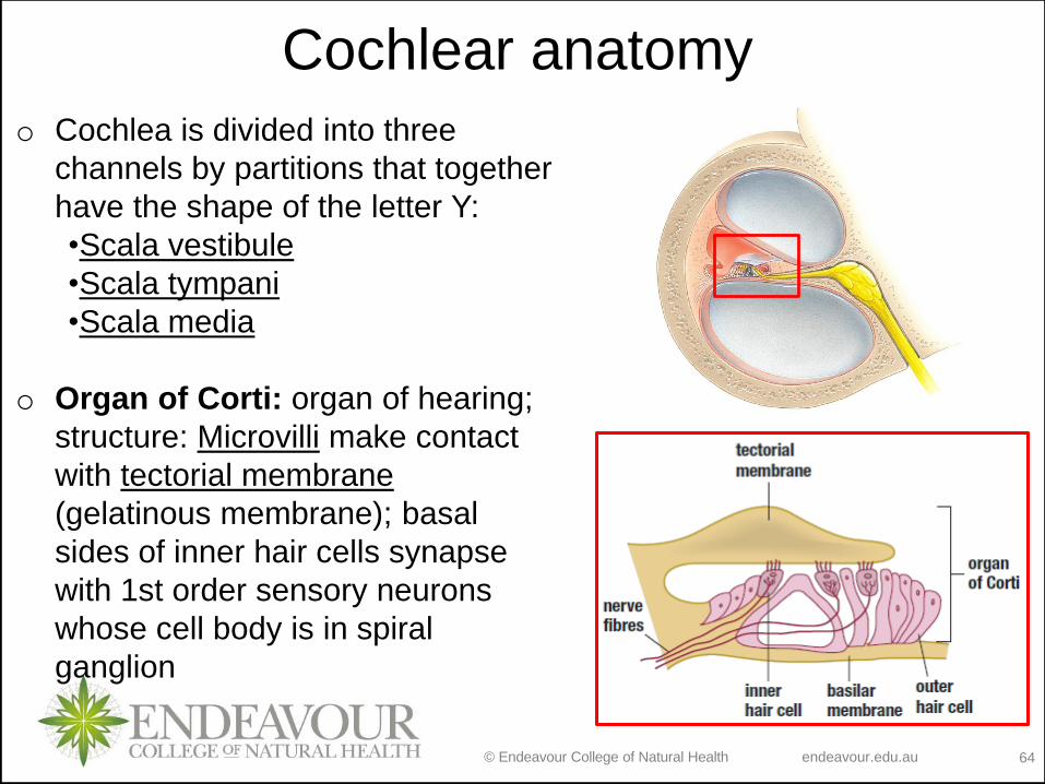
Cochlear anatomy
o Cochlea is divided into three
channels by partitions that together
have the shape of the letter Y:
•Scala vestibule
•Scala tympani
•Scala media
o Organ of Corti: organ of hearing;
structure: Microvilli make contact
with tectorial membrane
(gelatinous membrane); basal
sides of inner hair cells synapse
with 1st order sensory neurons
whose cell body is in spiral
ganglion
© Endeavour College of Natural Health endeavour.edu.au 65
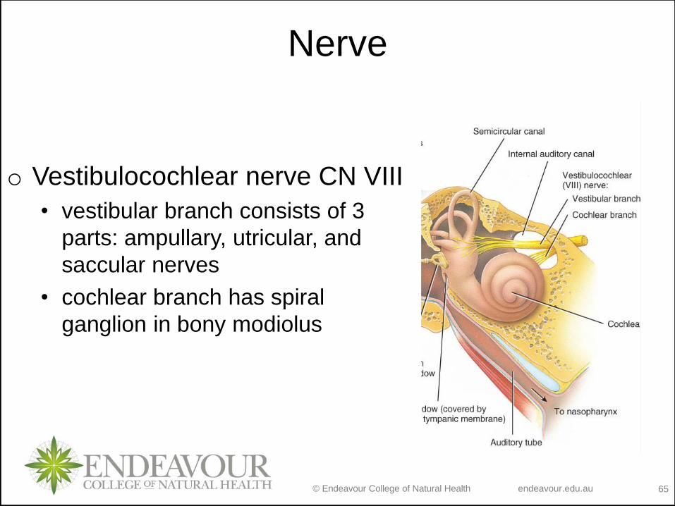
Nerve
o Vestibulocochlear nerve CN VIII
• vestibular branch consists of 3
parts: ampullary, utricular, and
saccular nerves
• cochlear branch has spiral
ganglion in bony modiolus
© Endeavour College of Natural Health endeavour.edu.au 66
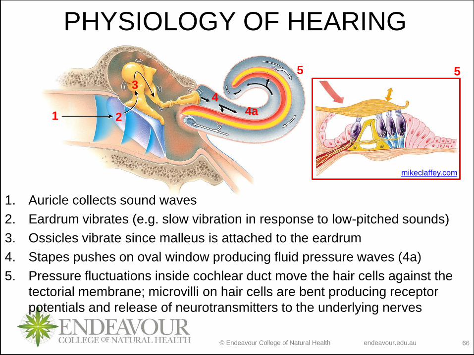
PHYSIOLOGY OF HEARING
1. Auricle collects sound waves
2. Eardrum vibrates (e.g. slow vibration in response to low-pitched sounds)
3. Ossicles vibrate since malleus is attached to the eardrum
4. Stapes pushes on oval window producing fluid pressure waves (4a)
5. Pressure fluctuations inside cochlear duct move the hair cells against the
tectorial membrane; microvilli on hair cells are bent producing receptor
potentials and release of neurotransmitters to the underlying nerves
1 2
34
4a
5
mikeclaffey.com
5
© Endeavour College of Natural Health endeavour.edu.au 67
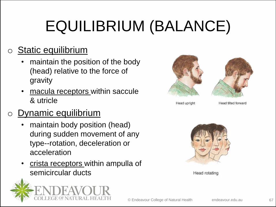
EQUILIBRIUM (BALANCE)
o Static equilibrium
• maintain the position of the body
(head) relative to the force of
gravity
• macula receptors within saccule
& utricle
o Dynamic equilibrium
• maintain body position (head)
during sudden movement of any
type--rotation, deceleration or
acceleration
• crista receptors within ampulla of
semicircular ducts
© Endeavour College of Natural Health endeavour.edu.au 68
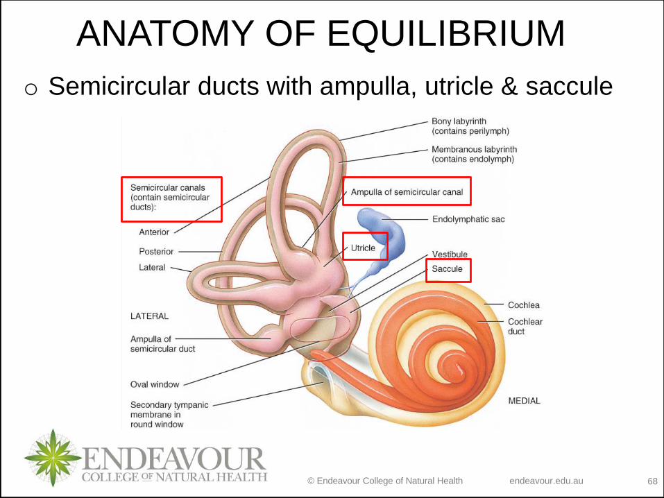
ANATOMY OF EQUILIBRIUM
o Semicircular ducts with ampulla, utricle & saccule
© Endeavour College of Natural Health endeavour.edu.au 69
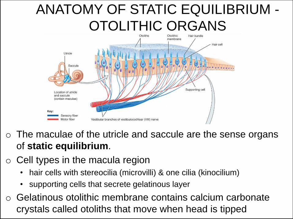
ANATOMY OF STATIC EQUILIBRIUM -
OTOLITHIC ORGANS
o The maculae of the utricle and saccule are the sense organs
of static equilibrium.
o Cell types in the macula region
• hair cells with stereocilia (microvilli) & one cilia (kinocilium)
• supporting cells that secrete gelatinous layer
o Gelatinous otolithic membrane contains calcium carbonate
crystals called otoliths that move when head is tipped
© Endeavour College of Natural Health endeavour.edu.au 70
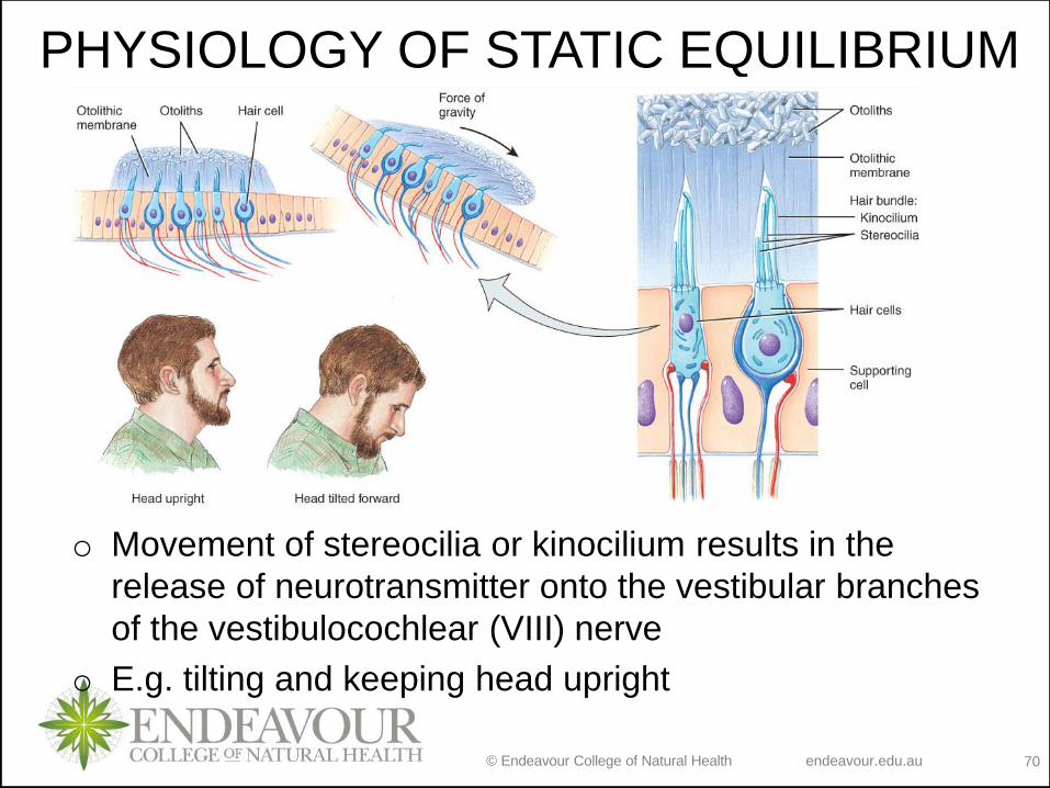
PHYSIOLOGY OF STATIC EQUILIBRIUM
o Movement of stereocilia or kinocilium results in the
release of neurotransmitter onto the vestibular branches
of the vestibulocochlear (VIII) nerve
o E.g. tilting and keeping head upright
© Endeavour College of Natural Health endeavour.edu.au 71
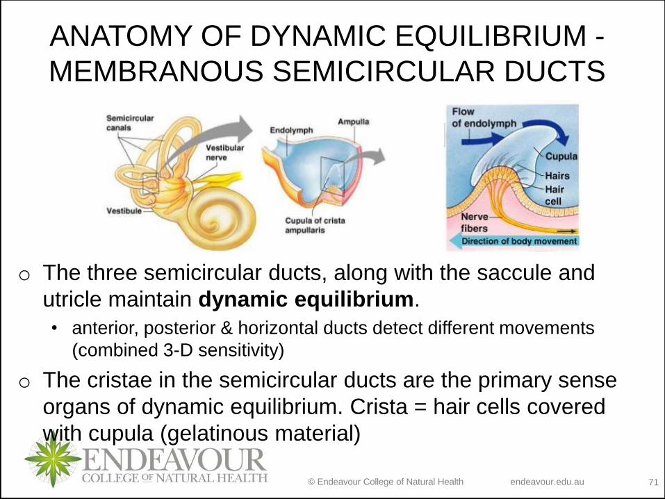
ANATOMY OF DYNAMIC EQUILIBRIUM -
MEMBRANOUS SEMICIRCULAR DUCTS
o The three semicircular ducts, along with the saccule and
utricle maintain dynamic equilibrium.
• anterior, posterior & horizontal ducts detect different movements
(combined 3-D sensitivity)
o The cristae in the semicircular ducts are the primary sense
organs of dynamic equilibrium. Crista = hair cells covered
with cupula (gelatinous material)
© Endeavour College of Natural Health endeavour.edu.au 72
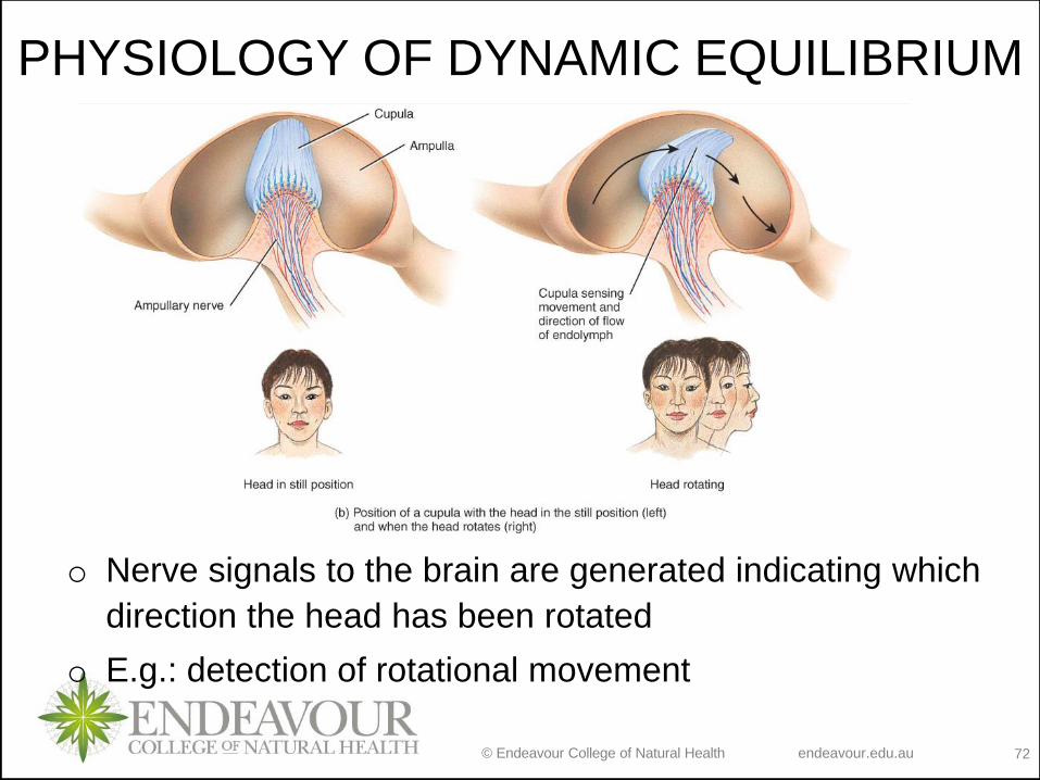
PHYSIOLOGY OF DYNAMIC EQUILIBRIUM
o Nerve signals to the brain are generated indicating which
direction the head has been rotated
o E.g.: detection of rotational movement
© Endeavour College of Natural Health endeavour.edu.au 73
Recap of Session 18
Different types of sensations require different structures to recognise
them
General senses are recognised all over the body and internal organs
and are classified as somatic and visceral; somatic general senses are
classified into tactile, thermal, pain and proprioceptive sensations
Special senses have developed specific organs to receive and
recognize specific sensation; classified into smell, vision, hearing and
equilibrium; for each special sense we have covered anatomy
(structure; ; i.e. where the sensation is received and recognised) and
physiology (function; i.e. how the sensation is received and recognised)
© Endeavour College of Natural Health endeavour.edu.au 74
Preparation for next session
o Complete any missing concepts and linking words from
Session 18
o Review:
• vertebral column
• divisions of nervous system, specifically CNS
• classification of neurons
• myelinated and unmyelinated axons
• ependymal cells
• proprioceptors