Embed Size (px)
Citation preview

HAL Id: hal-01140143https://hal.archives-ouvertes.fr/hal-01140143
Submitted on 7 Apr 2015
HAL is a multi-disciplinary open accessarchive for the deposit and dissemination of sci-entific research documents, whether they are pub-lished or not. The documents may come fromteaching and research institutions in France orabroad, or from public or private research centers.
L’archive ouverte pluridisciplinaire HAL, estdestinée au dépôt et à la diffusion de documentsscientifiques de niveau recherche, publiés ou non,émanant des établissements d’enseignement et derecherche français ou étrangers, des laboratoirespublics ou privés.
Biochemical and structural investigation of twoparalogous glycoside hydrolases from Zobellia
galactanivorans: novel insights into the evolution,dimerization plasticity and catalytic mechanism of the
GH117 family.E Ficko-Blean, D Duffieux, É Rebuffet, R Larocque, A Groisillier, G Michel,
M Czjzek
To cite this version:E Ficko-Blean, D Duffieux, É Rebuffet, R Larocque, A Groisillier, et al.. Biochemical and structuralinvestigation of two paralogous glycoside hydrolases from Zobellia galactanivorans: novel insightsinto the evolution, dimerization plasticity and catalytic mechanism of the GH117 family.. Acta crys-tallographica Section D : Structural biology [1993-..], International Union of Crystallography, 2015,pp.209-223. �10.1107/S1399004714025024�. �hal-01140143�

Enzymatic degradation of red seaweed galactans
1
Biochemical and structural investigation of two paralogous glycoside hydrolases from Zobellia galactanivorans: novel insights into the evolution, dimerization plasticity and catalytic mechanism of
the GH117 family
Elizabeth Ficko-Blean1,2*, Delphine Duffieux1,2, Étienne Rebuffet1,2, Robert Larocque1,2, Agnes Groisillier1,2, Gurvan Michel1,2*, Mirjam Czjzek1,2*
1Sorbonne Universités, UPMC Univ Paris 06, UMR 8227, Integrative Biology of Marine Models, Station
Biologique de Roscoff, CS 90074, F-29688, Roscoff cedex, Bretagne, France
2CNRS, UMR 8227, Integrative Biology of Marine Models, Station Biologique de Roscoff, CS 90074, F-29688, Roscoff cedex, Bretagne, France
*Running title: Enzymatic degradation of red seaweed galactans
*To whom correspondence should be addressed: Station Biologique de Roscoff, Place Georges Teissier, 29680 Roscoff, Bretagne, France. Tel.: 33-298-29-23-30; Fax: 33-298-29-23-24; E-mails: [email protected]; [email protected]; [email protected] Keywords: glycoside hydrolase; GH117; inverting mechanism; red algae; agarose ABSTRACT
The family 117 glycoside hydrolase enzymes (GH117s) have exo-α-1,3-(3,6-anhydro)-L-galactosidase
activity, removing terminal non-reducing α-1,3-linked 3,6-anhydro-L-galactose residues from their
red algal neoagarose substrate. These enzymes have previously been phylogenetically divided into
clades and only the Clade A enzymes have been experimentally studied to date. Investigation into
two GH117 enzymes, Zg3615 and Zg3597, produced by the marine bacterium Zobellia
galactanivorans, reveals structural, biochemical and further phylogenetic diversity between clades.
A product complex with the unusual β-3,6-anhydro-L-galactose residue sheds light on the inverting
catalytic mechanism of the GH117 enzymes as well as into the structure of this unique sugar
produced by hydrolysis of the agarophyte red algal cell wall.
Red seaweeds (Rhodophyta) are ancient photosynthetic eukaryotes and are considered as the first
organisms to have developed advanced multicellularity (1). These red macroalgae are important primary

Enzymatic degradation of red seaweed galactans
2
producers to the marine ecosystem and a large proportion of their organic biomass is recycled through the
food chain. This biomass is composed of approximately 50% polysaccharides, mainly starch and cell wall
components (2). Red seaweeds contain either agars or carrageenans, both of which are sulfated galactans,
and are thus referred to as agarophytes (e.g. Gracillaria, Gelidium, Porphyra spp.) or carrageenophytes
(e.g. Chondrus, Kappaphycus, Eucheuma spp.), respectively. Sulfated galactans contribute to the
flexibility and hydration of the macroalgae which is important given the extreme environmental forces
exerted by the ocean on the organism (3). These polysaccharides consist of a linear backbone of galactose
residues linked by alternating β-1,4 and α-1,3 glycosidic bonds. A specific feature of red algal agars is the
presence of L-sugars, which may in addition contain 3,6-anhydro-bridges, while the equivalent unit is a D-
sugar in carrageenans. Thus, the nomenclature of the α-linked galactose units refers to their configuration,
with the abbreviations LA and L6S referring to L-sugars, or DA and D6S that refer to D-sugars, in agars or
carrageenans, respectively. In red algal polysaccharides the β-linked residues are always classical
galactose units in the D configuration (G monomer), which can also contain different degrees of
sulfatations.
Both agarophyte and carrageenophyte algae synthesize an unusual carbohydrate moiety, the α-1,3-
linked bicyclic 3,6-anhydrogalactose (LA monomer in agars, DA monomer in carrageenans), which they
have incorporated into their cell wall galactans. The synthesis of 3,6-anhydrogalactose is unique to red
algae and imparts important physiochemical properties to the cell wall. These bicyclic sugars are also
crucial for the gelling properties of agars and carrageenans which are widely used as food ingredients (4).
The formation of 3,6-anhydrogalactose moieties is catalyzed at the polymer level by galactose-6-
sulfurylases. These unique enzymes convert galactose 6-sulfate (L6S or D6S monomer) into 3,6-
anhydrogalactose, releasing a free sulfate ion (5,6). However the knowledge on the biosynthesis of
sulfated galactans remains very limited and the first global view of the carbohydrate metabolism of red
seaweeds has been only recently provided by the genome sequencing of the carrageenophyte Chondrus
crispus (7,8).
Enzymatic degradation of red seaweed galactans
3
The regular structure of the agar backbone is often modified with methyl groups and variations in
acidic side groups such as sulfate or pyruvate (2,4). The main repeating disaccharides are the agarobiose
(G-β-1,4-LA) and the porphyranobiose (G-β-1,4-L6S) and these moieties are found in various amounts in
the agar chains of most agarophytes. Complex algal polysaccharides constitute a crucial carbon source for
numerous marine bacteria. These microorganisms produce several glycoside hydrolases (GHs) from
different amino acid sequence based CAZyme (Carbohydrate-Active Enzyme) families (9) in order to
efficiently degrade this unusual polysaccharide. Notably, different endo-hydrolases catalyze the initial
steps of agar degradation. Endo-α-agarases (EC 3.2.1.158) cleave the α-1,3 linkage between two
agarobiose units and belong to a single family, the GH96 family (10). In contrast, the β-agarases (EC
3.2.1.81) which hydrolyze the β-1,4 bond between two neoagarobiose units (LA-α-1,3-G) fall into various
GH families: GH16, GH50, GH86 and GH118 (see for review, (11)). Finally the β-porphyranases which
cleave the β-1,4 bond between two neoporphyranobiose units (L6S-α-1,3-G) constitute new subfamilies
within the families GH16 (12) and GH86 (13).
Recently, the characterization of the enzyme Zg4663 as an α-1,3-(3,6-anhydro)-L-galactosidase
(ZgAhgA) has defined a new GH family which includes agar-specific enzymes, the GH117 family (14).
ZgAhgA specifically removes 3,6-anhydro-L-galactose at the non-reducing end of oligo-agars released by
β-agarases and thus is involved in the terminal steps of agar catabolism.
The marine flavobacterium Zobellia galactanivorans is a model for the biodegradation of most
algal polysaccharides (11). Particularly, it can assimilate sulfated galactans from red seaweeds. Z.
galactanivorans possesses one GH16 kappa-carrageenase (15) and three GH82 iota-carrageenases (16)
(17). This bacterium has also evolved a complex agarolytic system composed of four GH16 β-agarases
and five GH16 β-porphyranases (18). Several of these enzymes have been already biochemically and
structurally characterized: the β-agarases ZgAgaA, ZgAgaB (19-21) and ZgAgaD (18) and the β-
porphyranases ZgPorA and ZgPorB (12,18).

Enzymatic degradation of red seaweed galactans
4
Z. galactanivorans possesses five GH117 enzymes, including the above mentioned ZgAhgA
(Zg4663). The GH117 family has been divided into three phylogenetic clades (A, B, and C), ZgAhgA
belonging to Clade A (14). This enzyme adopts a five-bladed β-propeller fold and forms a dimer in
solution by swapping of a small N-terminal domain. The activity of this enzyme is also cation-dependent
and a unique cation-binding site is found next to the putative catalytic residues. Two other Clade A
GH117s have been characterized since then: SdGH117 from Saccharophagus degradans (22) and
BpGH117 from Bacteroides plebeius (23). These two enzymes are also α-1,3-(3,6-anhydro)-L-
galactosidases and form dimers by swapping of the conserved N-terminal domain but also of a C-terminal
extension absent in ZgAhgA. The structures of SdGH117 and BpGH117 were solved in complex with a D-
galactose at subsite +1 (22) and neoagarobiose at subsites +1 and -1 (23), respectively. A site-directed
mutagenesis analysis identified the potential catalytic residues, and notably a conserved histidine was
proposed to act as a general acid (23). To date, only clade A enzymes have been structurally and
functionally characterized and for the two clade B enzymes little is known. Based on sequence
comparison within the active site region of the Clade C enzymes which suggested replacement of bulky
hydrophobic amino acids with smaller amino acids such as serine and alanine, Hehemann and coworkers
have also proposed that there is an additional -2 subsite within the Clade C enzymes, possibly for
accommodating longer oligo-agars (23).
Besides ZgAhgA, Z. galactanivorans has one Clade B GH117 (Zg185) and three Clade C GH117
enzymes (Zg205, Zg3597 and Zg3615). These proteins are relatively distant from ZgAhgA (35, 37 and
41% sequence identity for Zg205, Zg3597 and Zg3615, respectively), suggesting a potential functional
diversity and/or synergy. New phyologenetic analysis reveals that within the Z. galactanivorans
previously defined Clade C orthologues (14) there are significant differences between enzymes; this
divergence results in Clade C enzymes which might not have the same specificities. With the growth in
the genomic sequences available, and the structural and biochemical characterization done on the GH117

Enzymatic degradation of red seaweed galactans
5
enzymes to date, we have new phylogenetic evidence that there are at least 6 clades within this highly
unusual family.
In order to investigate the fine differences within the clades we chose to study two of the GH117
(previously defined) Clade C enzymes, Zg3615 (now Clade E) and Zg3597 (now Clade D). The structure
of Zg3597 was determined in its native form and Zg3615 was determined in complex with its product, β-
3,6-anhydro-L-galactose (Figure 1). This has important implications on the mechanism of catalysis and it
is the first time, to the best of our knowledge, that the structure of this unusual sugar has been clearly
elucidated.
EXPERIMENTAL PROCEDURES
All materials were obtained from Sigma-Aldrich unless otherwise stated.
Cloning- PCR was used to amplify the gene fragment from Z. galactanivorans genomic DNA
encoding the loci for Zg3615 and Zg3597 without their predicted signal peptide. SignaIP 4.1 (24) predicts
a secretion signal peptide cleavage site between amino acids 20 and 21 for Zg3615 and between amino
acids 24 and 25 for Zg3597. Based on comparison with the structure of ZgAhgA (pdb id 3P2N) (14) and
secondary structure predictions, nucleotides 76-1248 corresponding to amino acids 26-416 of the Zg3615
gene, and nucleotides 79-1299 corresponding to amino acids 27-433 of the Zg3597 gene, were cloned into
the PFO4 vector which encodes an N-terminal hexahistidine tag (25). The following primers and
restriction enzyme sites were used to clone Zg3615: forward primer 5’-
TTTTTTGTCGACCAGCCCGAAGGGTTTCCGTTCA-3’ including SalI (going into XhoI of pFO4) and
reverse primer 5’-AAAAAACTGCAGTTATTTAGGTTGTTTTTCGATGCGTTC-3’ including a PstI site
(going into NsiI site of pFO4). The following primers and restriction enzyme sites were used to clone
Zg3597: forward primer 5'-GGGGGGGGATCCTGTTCCGAACAAAAAACGGAAACCA-3' with a

Enzymatic degradation of red seaweed galactans
6
BamHI site and reverse primer 5'-CCCCCCGAATTCTTATCGTCCTTCGGGACTTAGGTTT-3' with an
EcoRI site.
Protein production and purification- Plasmids were transformed into competent Escherichia coli
strain BL21 (DE3) and grown shaking in 1L LB medium containing 100 µg/mL ampicillin at 37⁰C to an
optical density of ~1.0 whereupon the temperature was lowered to 20⁰C for one hour followed by
overnight induction of protein production with the addition of IPTG to 0.5 mM. Cells were harvested the
following morning by centrifugation at 3000 rpm for 30 minutes. A chemical lysis procedure was used to
lyse the cells. Briefly, cells were resuspended in 20 mL of 50 mM Tris pH 8.0 with 25 % sucrose and 10
µg of lysozyme and left stirring at 4⁰C for 20 minutes. Next, 40 mL of 1% deoxycholate, 1% triton X-
100, 20 mM Tris pH 7.5 and 100 mM NaCl was added and incubated stirring at 4⁰C for 10 minutes.

Enzymatic degradation of red seaweed galactans
7
Finally, MgCl2 was added to 5 mM and 0.1 mg of DNase and the lysis reaction was incubated at room
temperature for 20 minutes, until the viscosity decreased. The lysis solution was clarified at 13000 rpm
over 45 minutes at 4 ⁰C. Supernatant containing the soluble proteins of interest was run on a 5 mL GE His
Trap HP column. Briefly, using an ÄCTA FPLC, the IMAC column was loaded with 0.45 µM filtered
supernatant in Buffer A (20 mM Tris pH 8.0, 100 mM NaCl), washed with Buffer A + 1% Buffer B (20
mM Tris pH 8.0, 100 mM NaCl, and 1M imidazole) and eluted using a gradient of 1%-100% Buffer B.
Fractions containing the protein of interest were pooled and concentrated using an Amicon stir cell
concentrator (MWCO 3 kDa). This was followed by size exclusion chromatography on a Superdex S200
column in 20 mM Tris pH 8.0 and 100 mM NaCl. Again, fractions containing the protein of interest were
pooled and concentrated using an Amicon stir cell concentrator (MWCO 3 kDa). Proteins were assessed
at >95% purity using SDS-PAGE. The concentration was calculated using A280 and the extinction
coefficient calculated by ExPASy ProtParam tool (ε0.1% of 2.1 for Zg3615 and 2.0 for Zg3597). The final
concentration for Zg3615 was 18.0 mg/mL and 10.0 mg/mL for Zg3597.
Thin Layer Chromatography, TLC- Enzymatic reactions were performed in 0.5 % oligo- or
polysaccharide, 100 mM Tris pH 7.5, 0.1 mM ZnSO4, 100 mM NaCl, and 25 mM CaCl2 and 0.1 µg/µL
enzyme. Neoagarooligosaccharides were purified as described previously (20). 2 µL of the reaction
volume was spotted onto Merck TLC Silica Gel 60 plates and developed in 3x butanol, 1x ethanol and 1x
water. The plates were dried then stained in 50 mL of 10% H2SO4 in ethanol added to 25 mL of 0.2%

Enzymatic degradation of red seaweed galactans
8
solution of naphthoresorcinol (1,3-dihydroxynaphthalene) in ethanol just before using. Plates were again
dried and incubated at 60 ⁰C until spots appeared (approximately 45 minutes).
Flurophore Assisted Chromatography Electrophoresis, FACE- Enzymatic reactions were
performed as above. 50 µg of oligo- or polysaccharide reaction volume were dried down using a speed
vacuum. Labelling was done by adding 2 µL of ANTS (8-Aminonaphthalene-1,3,6-Trisulfonic Acid) in
DMSO to the dried sample followed by the addition of 5 µL of freshly made sodium cyanoborohydride in
DMSO. The samples were mixed well, centrifuged and left at 37 ⁰C in the dark overnight. 20µL of 20 %
glycerol was then added as a gel loading aid. 8 µL of sample was loaded on a 27% polyacrylamide gel.
Running buffer used was 0.192 M glycine and 0.025 M Tris pH 8.5. Gels were migrated at 200V for
approximately 3 hours and then visualized under UV light.
Small Angle X-ray Scattering, SAXS- SAXS experiments were carried out on the beamline X33 at
the DORIS-III storage ring at DESY (Deutsches Elektronen-Synchrotron), Hamburg, Germany. The
wavelength λ was 0.15 nm. The sample-to-detector distance was set at 2.7 m, resulting in scattering
vectors q ranging from s = 0.06 nm-1 to 6 nm-1. All experiments were performed at 16°C on a series of
concentrations ranging from 2.0 to 7.9 mg/mL for the heterologously expressed and purified enzyme
Zg3597. The scattering vector is defined as q=4p/λ sinθ, where 2θ is the scattering angle. The detector
used was a 2D Photon counting Pilatus 1M-W pixel X-ray detector and 10 successive frames of 10 second

Enzymatic degradation of red seaweed galactans
9
exposure times were recorded for each sample. Each frame was carefully checked for possible bubble
formation or radiation induced aggregation. If such effects were not observed, the individual frames were
averaged. A 4.8 mg/ml solution of BSA was measured as a reference and for calibration procedures. Up to
five different concentrations of the protein sample were measured to test for consistency and eventually
detect and eliminate concentration dependent effects. Background scattering was quantified before and
after each protein measurement by measuring the corresponding buffer, which is then subtracted from the
protein patterns using the Primus suite (26).
The radius of gyration, Rg, of each measurement was derived by the Guinier approximation:
I(q)=I(0)exp(–q2Rg 2/3) up to qRg<1.0. The radii of gyration Rg, calculated for the different protein
concentrations, displayed no evident concentration dependence. The program GNOM (27) was used to
compute the pair-distance distribution functions P(r). This approach gives the maximum dimension of the
macromolecule Dmax and offers an alternative calculation of Rg, which is based on the entire scattering
curve (see Table 1).
The overall shapes of all assemblies were restored from the experimental data by using the ab
initio modelling programs GASBOR and DAMMIF (28,29). The scattering profiles were used up to
qmax=3 nm-1. Nine low-resolution models obtained from different runs were compared using the program
DAMAVER (30) to give an estimate of the reproducibility of the results inferred from the ab initio shape
calculation and to construct the average model representing the general structural features of all the
reconstruction filtered by DAMFILT. The atomic structures of the individual modules were then
positioned into the low resolution envelopes using the program SUPCOMB (31). For the Zg3597 dimer
obtained from the crystal structure the theoretical SAXS profile, the Rg and the corresponding fit
(represented by χ2 values) to the experimental data were calculated using the program CRYSOL (32).
Crystallization-Initial hits were obtained with sitting drop vapour diffusion and the Cartesian
Dispensing System (Genomic Solution) using four different commercial screens: JCSG+, PEGs suite,

Enzymatic degradation of red seaweed galactans
10
PEGs II and the PACT suite. Hits were optimized using hanging drop vapour diffusion. The optimized
condition for Zg3615 was: 0.1 M sodium acetate pH 5.0, 25% PEG 3350, in a 1:1 ratio of protein (at 10
mg/mL) to mother liquor. Soaks were done on the native protein crystals using 10 mM neoagarotetraose
made up in mother liquor and mixed 1:1 with the crystal drop. The optimized crystallization conditions for
Zg3597 were: 16% glycerol, 0.15 M ammonium sulfate, and 20 % PEG 4000 in a 1:1 ratio with protein at
5 and 2.5 mg/mL and 14% PEG 3350 and 75 mM sodium acetate with a ratio of 2:1 protein (at 7.5
mg/mL) to mother liquor. Crystals were flash frozen in liquid nitrogen using a cryoprotectant of 20 %
glycerol or 20 % ethylene glycol for Zg3597 and 30% ethylene glycol for Zg3615. Crystals for both
proteins grew at 18 ⁰C.
X-ray Crystallography Data Collection and Processing- Diffraction data were collected for
Zg3615 on the PROXIMA 1 line at SOLEIL (Source optimisée de lumière d’énergie intermédiaire du
LURE, Paris). Diffraction data for Zg3597 were collected on the ID14-4 beamline at the ESRF (European
Synchrotron Radiation Facility, Grenoble). The data was processed using MOSFLM (33) and scaled
using SCALA (34). The structures were solved by molecular replacement using the one-to-one threading
tool from PHYRE2 (35) to build a model (100% confidence) using monomer A from the RSCB deposited
GH117 Zg4663 pdb 3P2N (14) and removing any non-conserved amino acids from the MOLREP (36)
input pdb. A library for the co-crystallized sugar was created using ProDrg from the CCP4 suite of
programs (37). Data was refined using REFMAC5 (38) from the CCP4 suite of programs. Waters were
added using the COOT:FIND WATERS option. Successive rounds of model building and refinement were
done using COOT (39) and REFMAC5. All data collection statistics are summarized in Table 2.

Enzymatic degradation of red seaweed galactans
11
Ramachandran statistics were calculated using the program Rampage from the CCP4 suite of programs
(40). Zg3597 has been given the pdb id 4U6B and for Zg3615 the pdb id is 4U6D.
Phylogeny-New sequences of GH117 have been identified using BlastP at the Genbank database.
These new sequences have been added to the previous dataset of GH117 sequences (14) and all sequences
were aligned using MAFFT with the iterative refinement method and the scoring matrix Blosum62 (41).
The MAFFT alignment of these sequences was manually refined using Bioedit (© Tom Hall), on the basis
of the superposition of the five available crystal structures of GH117 (ZgAhgA (Zg4663): PDB code,
3P2N; Zg3597: 4U6B; Zg3615: 4U6D; SdGH117: 3R4Y and BpGH117: 4AK5). This refined alignment
allowed calculating model tests and maximum likelihood trees with MEGA version 6.0.6 (42). Tree
reliability was tested by bootstrap using 100 resamplings of the dataset. The trees were displayed with
MEGA 6.0.6 (42). Accession numbers of the selected sequence homologues are available in
Supplementary Table 1.
RESULTS
Activity Assays- Two representatives (Zg3615 and Zg3597) from the recently discovered GH117
family of enzymes were chosen for investigation. Zg3615 and Zg3597 activities were tested on agarose
and a variety of neoagaro-oligosaccharides. The enzyme activities were also screened in combination with
one another and with Zg4663 (ZgAhgA,(14)), in case the activity of one enzyme is dependent on exposing
the substrate by another. Zg3615 exo-α-1,3-(3,6-anhydro)-L-galactosidase activity was initially
demonstrated using TLC which showed the release of terminal 3,6-anhydro-L-galactose from
neoagarotetraose, neoagarohexaose, and neoagarooctaose (not shown). Enzymatic activity was also tested
on agarose; however, there was no detectable difference between the enzymatic digested and undigested
reactions, consistent with exo-cleavage as there are not sufficient terminal 3,6-anhydro-L-galactose
residues for detection by TLC. In order to release detectable levels of 3,6-anhydro-L-galactose the activity

Enzymatic degradation of red seaweed galactans
12
of a β-agarase would also be required. Thus, by TLC the activity of Zg3615 is indistinguishable from that
of Zg4663. Therefore, FACE was employed to further investigate any potential differences in activity
between the clades. Enzyme reactions using neoagarobiose/neoagarotetraose,
neoagarotetraose/neoagarohexaose (Figure 2), and agarose (not shown) as substrates were migrated on
FACE gels. Both Zg3615 and Zg4663 were active on neoagarooligosaccharides; however, there was no
discernible difference between Zg3615 and Zg4663 activities on these unsubstituted oligo-agars. The
enzymatic release of one 3,6-anhydro-L-galactose residue resulted in a corresponding electrophoretic shift
in migration from neoagarotetraose and neoagarohexaose to 3,6-anhydro-L-galactose, agarotriose and
agaropentaose (Figure 2, Lanes 1-7). The cleavage of neoagarobiose and neoagarotetraose resulted in the
production of agarotriose and the monosaccharides D-galactose and 3,6-anhydro-L-galactose (Figure 2,
Lanes 8-14). The enzymatic digests on agarose polysaccharide showed no visible bands, again consistent
with the exo-activity within this family. No noticeable differences were detected between Zg3615
reactions alone or in combination with Zg3597 or Zg4663. In contrast, and though thorough screening of
activity of Zg3597 on potential agarose substrates was undertaken, it was not possible to discern activity
on any of the oligo- or polysaccharides tested, suggesting a novel and as yet unidentified activity within
the family.
Overall homodimer structural features- The X-ray crystal structure of Zg3615 was determined to
1.7 Å resolution and the structure of Zg3597 to 2.3 Å. The globular Zg3615 and Zg3597 GH117
monomers share the 5-fold β-propeller fold with the other members of the family and each β-sheet has 3 or
4 anti-parallel β-strands. The GH117 enzymes form a structural homodimer, previously shown to be
mediated by both the N- and the C-terminal domains (SdGH117, BpGH117) or just the N-terminal domain
(Zg4663) (Figure 3). The structures of Zg3615 and Zg3597 reveal differences in their N- and the C-
terminal domains. Zg3615 has a helix-random coil N-terminal motif (amino acids 28-61) which interacts
with its other monomer through multiple surface hydrophobic residues. The N-terminal is in a structurally
similar position to the N-terminal helix, followed by a turn, followed by a helix from Zg4663 (amino acids
Enzymatic degradation of red seaweed galactans
13
38-71) and BpGH117 (amino acids 33-65). This N-terminal domain is not visible in the Zg3597 crystal
structure. Zg3615 has a helix, followed by a turn, followed by a helix (amino acids 386-402) within its C-
terminal domain which provides considerable hydrophobic interactions with its partner monomer. This C-
terminal domain is not conserved in the sequence of Zg4663 and its C-terminus is reduced to a short loop
after the last β-strand, whereas BpGH117 and SdGH117 have a large interacting surface area contributed
by their random coil C-termini (amino acids 386-401 and amino acids 350-371). Interestingly, while
spatially the C-terminal domains of the Clade A enzymes are approximately conserved within the clade,
the C-terminal domain is not spatially conserved in Zg3615 or Zg3597. The Zg3597 C-terminal domain
consists only of one helix motif (amino acids 408-425) which is spatially conserved with the Zg3615 C-
terminal end. All contributions to the termini-mediated dimerization of Zg3597 come from the C-terminal
and this has resulted in a different dimer configuration for Zg3597 when compared to the other GH117
enzymes.
Using the webservice PDBePISA, Proteins, Interfaces, Structures and Assemblies
(https://www.ebi.ac.uk/pdbe/pisa/) the overall interaction interface within the Zg3615 dimer in the
asymmetric unit was determined to be ~4100 Å2, this gave a Complex Formation Significance Score
(CSS) of 0.73 indicating it is essential for complex formation. To this, the N-terminal contributes
approximately ~1900 Å2 of surface area and the C-terminal contributes ~1750 Å2 of surface area. Zg3597
has a total interaction interface with its monomeric partner of ~1820 Å2 giving a CSS score of 0.25,
indicating it’s relevant for complex formation. Zg3597 is a tetramer in the asymmetric unit, though the
surface interaction area (~740 Å2) that is not mediated by the N- or C-terminal domains appears to be the
result of crystal packing only (CSS=0).
Active Site Architecture- Both Zg3615 and Zg3597 enzymes have a shallow cavity typical of exo-
acting enzymes. The proposed catalytic residues are conserved (Figure 4), in Zg3615 these are His284,
proposed to be the catalytic acid, Asp89, proposed to be the general base, and Glu285, proposed to
modulate the pKa of the nearby general base (23). Though sequentially conserved (Figure 4), the

Enzymatic degradation of red seaweed galactans
14
proposed catalytic acid (His306) from Zg3597 is shifted compared to Zg3615 (Figure 5 A) and to the
structures of the other enzymes. This is a rather pronounced positional change, putting it well out of
hydrogen bonding position with the glycosidic oxygen seen in the BpGH117 complex (approximately 6.9
Å away compared to between 2.7-3.6 Å for the other enzymes).
Soaks were done on the native Zg3615 crystals with neoagarotetraose. Examination into the active
sites of the homodimer pair revealed an unusual electron density in monomer A. We first attempted to fit
α-3,6-anhydro-L-galactose into the density but our attempts were unsuccessful; however, β-3,6-anhydro-L-
galactose fit the density well in an unconstrained manner (Figure 5 B). This enzyme-product complex
provides a novel glimpse into the structure of the unique β-3,6-anhydro-L-galactose molecule. The
bicyclic sugar moiety sits at the base of the pocket in the -1 subsite in a 4C1 conformation. Its anomeric C1
hydroxyl appears in a ‘flattened’ conformation at the anomeric centre. The 4C1 conformation is stabilized
by several hydrogen bonding interactions with the C1 hydroxyl provided by Zg3615 Arg88, Asp89, and
Glu285. Arg88 also hydrogen bonds to the 6 membered ring’s endocyclic oxygen (Figure 5 A). The axial
C2 hydroxyl interacts via hydrogen bonds to Lys246 and Asp231. Gln181 and Asp192 are within
hydrogen bonding distance of the equatorial C4 hydroxyl group and Thr166 provides a hydrogen bond to
the 5-membered ring’s cyclic oxygen residue. Finally, a remarkable difference is seen when comparing the
two monomers in the Zg3615 homodimer. The loop containing Asp 192 participates in coordinating the
β-3,6-anhydro-L-galactose residue in the active site of monomer A of Zg3615; however, in monomer B
this loop is shifted considerably away from the active site. The contribution of Asp192 to the active site of
monomer A is probably due to interactions with the carbohydrate moiety. Hydrophobic contributions with
the product complex are provided by the His230 imidazole face which interacts with the equatorial C4 arm
of β-3,6-anhydro-L-galactose. Trp128 provides a hydrophobic platform for the sugar’s C5 and C6 atoms
which form part of the 5-membered sugar ring and the arm of Arg88 also provides hydrophobic
contributions to this ring component.

Enzymatic degradation of red seaweed galactans
15
The C-terminal domains of Zg3597 and Zg3615 lie distal to the active site and do not contribute
directly to the +1 subsite. This is in direct contrast with two of the Clade A enzymes, SdGH117 and
BpGH117, who’s C-termini do participate in the active site substructure and interact with the galactose
component in the +1 subsite (BpGH117, His392; SdGH117, Ser358 and Tyr359). The last residues of the
Clade A’s Zg4663 C-terminal domain are disordered in the native, non-complexed crystal structure
(His400-Gln408), though its structural placement is similar to the other Clade A enzymes.
Structural analysis of the enzymes has revealed several important amino acid substitution
differences between Zg3597 and Zg3615 which may affect both active site substructure and N-terminal
dimerization. Firstly, Asp250 from Zg3615 forms a salt bridge with Arg58 from the N-terminal of the
second monomer. Zg3597 has Asn268 at this site, effectively destroying the electrostatic interaction. In
the structure of Zg3615, the phenyl ring of Phe32 inserts between two β-strands of the other monomer and
sits 3.75 Å from the sidechain of Tyr227. Zg3597 replaces these two residues with a histidine residue
(His48- not visible in the crystal structure) and Asp245, suggesting this interaction is abrogated in Zg3597.
A loop comprising Ile322-Tyr329 is flipped in Zg3597 (Figure 6 A and B) which sits just beside
the turn holding His306. One reason for this flip may be the hydrophobic Tyr329 which would otherwise
be surface located. In the +1 subsite, Arg257 from Zg3615 is spatially, though not sequentially, conserved
with a conserved Clade A arginine that interacts with the C4 hydroxyl group of galactose. Arg257 sits
against the imidazole ring of His284 (Figure 6 C), the putative catalytic acid. Zg3597 replaces Arg257
with Val279, a much smaller non-polar residue, leaving a pocket at the C4 site, possibly for
accommodating a substituent on the galactose C4 hydroxyl group. His306, the Zg3597 putative catalytic
acid, is able to shift into the space left by the absence of an arginine residue at this site (e.g. Arg257 in
Zg3615). The consequence of the loop flip and the replacement of Arg257 (Zg3615) with Val279
(Zg3597) is that the Zg3597 His306 lays sandwiched hydrophobically between Val279 and Asp324
(Figure 6 B). In the other structures, this aspartic acid residue is within hydrogen bonding distance to
His306. The change in position of the Zg3597 His306 residue represents a rather large deviation for a

Enzymatic degradation of red seaweed galactans
16
proposed catalytic acid. Though an activity has yet to be described for Zg3597, it is reasonably possible
that this protein has a divergent specificity/function.
Within the -1 subsite there are several rearrangements of bulky hydrophobic groups which again
result in spatial, though not sequential, conservation of amino acid residues. For example, the BpGH117
Phe164 residue is spatially conserved with the Zg3615 Phe183 and Zg3597 Phe201 residues. The residue
BpGH117 Tyr186 is not spatially conserved in Zg3597 or Zg3615, however, the functionality of the Tyr
hydroxyl group is roughly conserved through Zg3615 Ser187 and Zg3597 Ser205. BpGH117 Phe115 is
replaced by Ala116 in Zg3615 and by a more conservative Trp131 in Zg3597 which is disordered at the
enzyme surface. The bulky BpGH117 Phe125 residue is replaced by the smaller Ala26 for Zg3615 and the
polar Thr144 in the case of Zg3597. Zg3615’s C4 hydroxyl interacting residue Asp192 is conserved in the
Zg3597 crystal structure (Asp210); however, it is shifted considerably leaving a more open active site
entrance at the enzyme surface. This residue is not found in any of the Clade A enzyme structures. Both
Zg3615 and Zg3597 enzymes have more open and accessible active site entrances when compared to
BpGH117 and SdGH117 (though not Zg4663), Zg3597 in particular.
Metal binding site- With the exception of SdGH117, divalent metal ions have been modelled into
a conserved metal binding pocket adjacent to the active site. For BpGH117 a Mg2+ ion was modelled and
for Zg4663 a Zn2+ ion. A Ca2+ molecule has been modelled for Zg3615 and Zg3597 at this same site which
is supported by 7 organized water molecules in the solvation shell (43) and the similarity of the B factors
between the metal atoms and their neighboring water molecules. Furthermore, the distances between the
water atoms and the Ca2+ molecule in Zg3615 (monomer A) range from 2.32 Å to 2.60 Å and in Zg3597
(monomer A) from 2.18 Å to 2.58 Å, these values are consistent with the expected distribution range (44).
A water molecule has been modelled in precisely the same site as the metal atoms in the SdGH117 crystal
structure. Examination of the Fo-Fc maps for SdGH117 at this site reveal significant density above 5 sigma
(0.317e-/Å3), consistent with the molecule being modelled incorrectly as a water. Furthermore, the
distorted octahedral geometry of 6 ligands surrounding a central atom is incompatible with the central

Enzymatic degradation of red seaweed galactans
17
atom being a highly coordinated water molecule (43). Regardless, this metal binding site appears to have
some degree of plasticity and is conserved throughout the GH117 structures solved to date.
SAXS- The solution structure of Zg3597 was determined by small angle X-ray scattering. We used
SAXS analyses to address the oligomeric state and conformational plasticity of the Zg3597 enzyme in
solution (Figure 7 D). The slope and intensity at zero angle of the experimental scattering data allowed to
calculate Rg values and estimate the molecular weight in solution (Table 1) that were coherent with the
protein taking a non-spherical, bimodular form. Several forms were calculated to fit the experimental
curves and compared using DAMAVER. The overall shapes were rather similar, although indicative of
variations, as indicated by a mean normalized spatial discrepancy value (NSD) of 1.20±0.01. The best
envelopes that fitted the experimental curve (χ2 = 1.6) are obtained when imposing a 2-fold symmetry for
a dimeric form of the enzyme. These data also allowed determination of an Rg of 34.6 Å. Similar values
were obtained using several distinct calculation methods (Table 1). The distance distribution function P(r)
displayed a biphasic pattern, also indicating an elongated shape with a maximum diameter (Dmax) of
100±2 Å (Figure 7 D). The molecular mass determined from the scattering intensity extrapolated to zero
scattering angle indicated a molecular mass (Mw) of approximately 109 kDa (Table 1), in close agreement
with Zg3597 existing as a homodimer in solution, which is also observed by size-exclusion
chromatography, DLS and in the crystal structure (data not shown). The Mw determined by SAXS also
compares well to the theoretical Mw of a homodimer (~97 kDa), as calculated from the protein sequence.
A theoretical scattering curve of the dimer that exists in the crystal structure of Zg3597 was calculated
using CRYSOL. Although the deduced scattering profile from the crystal structure displayed a similar
shape as the experimental scattering profile (data not shown) and roughly an Rg value (31.9 Å) in the
same order of magnitude as the experimental scattering curve, the χ2 value of 3.4 describing the fit
between experimental and theoretical profiles suggests structural differences between the solution and
crystallographic structures. In agreement with this notion, we found that when the crystallographic
structure was docked onto the SAXS envelope, an additional volume likely corresponding to the catalytic

Enzymatic degradation of red seaweed galactans
18
amino-terminal domains was present in the SAXS envelope (Figure 7 A, B and C). This may reflect
conformational flexibility of the amino-terminus region with respect to the catalytic domains, having a
different position in solution and in the crystal structure. The closer contact with the catalytic domain in
the solution structure might be mediated by weak protein-protein interactions (Figure 7 C). In addition, the
SAXS envelope calculated by GASBOR (Figure 7 A) was of sufficient resolution to show the position of
the crater like active site at the heart of the β -propeller. The calculation of an approximate dihedral angle
between the orientations of these two depressions in the SAXS envelopes appeared to be closer to
perpendicular at roughly 100° (Figure 7 A), whereas this angle estimated from the crystal structure is
about 130° (Figure 7 C). This would imply a rotational rearrangement of the two monomers, most
probably due to constraints in the crystal structure that might disfavor the establishment of the interactions
of the amino-terminal domains with the adjacent catalytic domains in the swapped geometry.
Phylogenetic Analysis- In the initial phylogenetic analysis of GH117 family three clades were
defined: Clade A contained most of the sequences, while Clades B and C only comprised few sequences
and essentially paralogous proteins from Z. galactanivorans (Clade B: Zg185, clade C: Zg205, Zg3597,
and Zg3165 (14). Since then, numerous genomes of marine bacteria have been sequenced and novel
GH117 sequences have been identified, particularly in the initial Clades B and C. Therefore, we have
generated an updated phylogenetic tree (Figure 8 and Supplementary Table 1) in order to revise the
definition of the GH117 subfamilies. In the initial tree (14), Clade B only contained two sequences, Zg185
and RB13146 from Rhodopirellula baltica. In the revised tree, these sequences are much more distant:
Zg185 groups with a new sequence (RspSWK7GH) with a weak statistical support (51%), while RB13146
clusters together with 10 new sequences, forming a solid clade (bootstrap: 100%). To be consistent with
the previous nomenclature, Zg185 remains in the subfamily referred to as Clade B, while RB13146 and its
closely related sequences are defined as the new Clade F. Similarly, the former Clade C has significantly
expanded and changed. The protein LNTAR_17733 from Lentisphaera araneosa was at the root of the
former Clade C and is still an isolated outgroup. In contrast, the remaining sequences of the former Clade

Enzymatic degradation of red seaweed galactans
19
C have now diverged into two solid clades (bootstrap values of 98% and 90% respectively): (i) the first
containing Zg3597 and 5 new sequences and (ii) the second encompassing 28 sequences, including Zg205
and Zg3615. The difference of enzymatic activity between Zg3597 (still unknown) and Zg3615 (exo-α-
1,3-(3,6-anhydro)-L-galactosidase) is consistent with the existence of two distinct subfamilies.
Consequently, one can expect that the outgroup sequence LNTAR_17733 may display an activity different
from Zg3597 and Zg3615. Altogether, we propose that the clade composed of LNTAR_17733 retains the
name Clade C, while the clades of Zg3597 and of Zg3615 are referred to as Clade D and Clade E,
respectively.
DISCUSSION
Z. galactanivorans has evolved a complex GH117 family- Sulfated galactans from red algae are
unique polysaccharides with a complex chemical structure. To assimilate these cell wall polymers, Z.
galactanivorans has evolved a diverse enzymatic system, mainly based on GH16 enzymes including four
β-agarases (18,20), five β-porphyranases (12) and one -carrageenase (15). Alongside the GH16 family, Z.
galactanivorans possesses a multigenic family of five GH117 genes, localized in different clusters
containing sulfatases and additional carbohydrate-related genes (14) and one could expect a similar
functional diversity for this family. The first characterized GH117 enzyme, Zg4663, is an α-1,3-(3,6-
anhydro)-L-galactosidase and thus has been named ZgAhgA (14). In the present work, we have shown
that, despite the sequence divergence, Zg3615 has an activity similar to that of Zg4663, at least on
unsubstituted agarose oligosaccharides (Figure 2). Therefore, Zg3615 and its corresponding gene will be
referred to as ZgAhgB and ahgB, respectively. In contrast, Zg3597 is inactive on oligo-agars. Thus,
Zg3597 is different from ZgAhgA and ZgAhgB, although its exact activity remains to be determined. The
complexity of the GH117 family is confirmed by our revised phylogenetic tree (Figure 8). While the
former Clades B and C initially contained very few sequences (14), these subfamilies are largely expanded

Enzymatic degradation of red seaweed galactans
20
due to the GH117 sequences identified in the numerous genomes of marine bacteria sequenced the last
three years. Our phylogenetic analysis has led to the division of the former clade B into a new Clade B and
Clade F, and of the former Clade C into a new Clade C, Clade D and Clade E. ZgAhgB (Zg3615) aligns
into the new Clade E and Zg3597 aligns into the new clade D, consistent with the differences in
dimerization conformation, active site structure and function. Although Clade E is solidly supported
(bootstrap value: 90%), this clade is still quite large and contains two paralogous enzymes from Z.
galactanivorans, Zg3615 (ZgAhgB) and Zg205. Depending on the biochemical characterization of
additional Clade E enzymes, this clade may be further divided in the future to reflect the difference in
enzymatic specificity.
Implications on catalysis- The GH117 family shares distant sequence similarity with families
GH43 and GH32. The first crystal structure, Zg4663, revealed a zinc ion octahedrally coordinated by
water molecules located at the bottom of the active site pocket (43). The divalent cation was proposed to
have a role in the activation of the catalytic water molecule to provoke hydrolysis. This ion-binding site is
conserved in the structures of all the enzymes solved to date, though the ion type differs. The BpGH117
Michaelis substrate complex trapped the α-1,3-linked 3,6-anhydro-L-galactose moiety in a higher energy
B1,4 boat conformation (Figure 9 B) which aligns the anomeric centre for nucleophilic attack and
demonstrates how the enzymes alter the sugar conformation to favor the oxocarbenium ion transition state
(23). Based on crystallographic and mutational evidence for BpGH117, Hehemann et al proposed Asp90
(Figure 9 B) acts as the general base the adjacent Glu303 as a pKa modulator (23). The positioning of the
BpGH117 active site residues suggests an inverting mechanism (45) and unexpectedly revealed a histidine
residue positioned to act as the putative catalytic acid. This distinguishes this family from the vast majority
of glycoside hydrolases which use a carboxylic acid moiety, though unusual glycoside hydrolase
mechanisms are not unheard of (46). The three proposed catalytic residues, histidine, aspartic acid, and
glutamic acid, are conserved throughout the GH117 family, with the exception of two Clade F (Figure 8)
Enzymatic degradation of red seaweed galactans
21
enzymes (GjGH117-1 and RRSWK_05156) which have an alanine instead of a glutamate in their
predicted active sites.
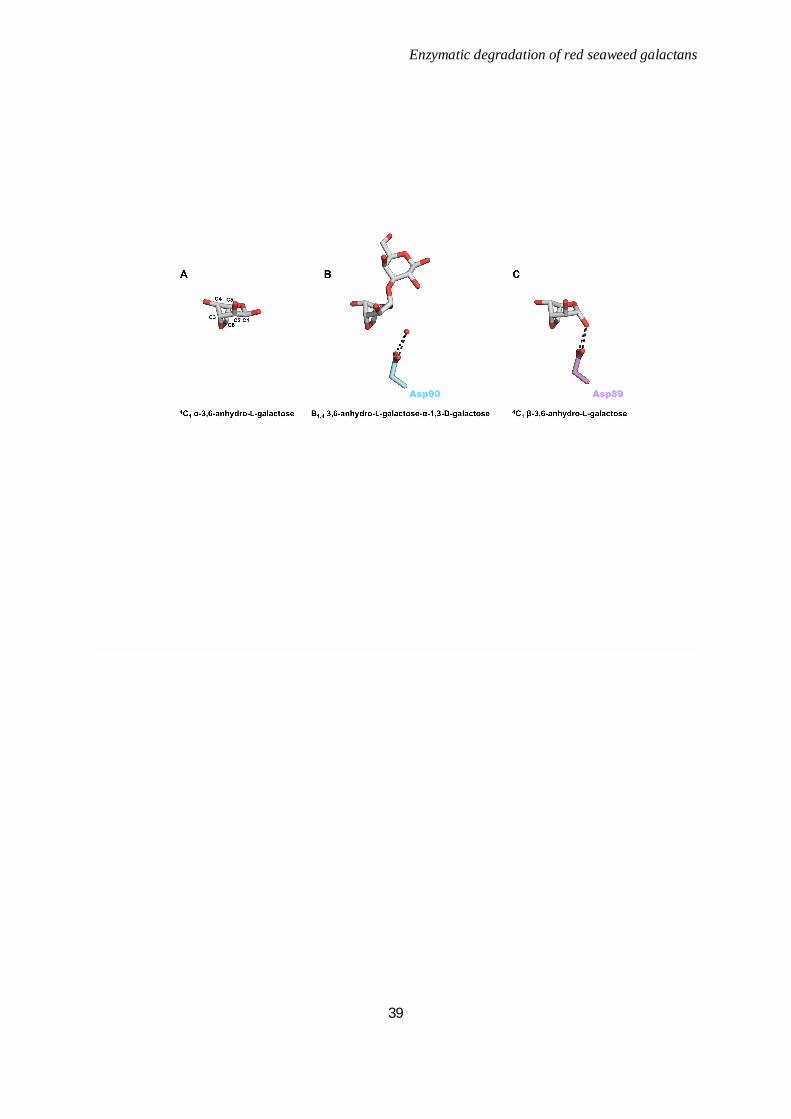
The ZgAhgB 4C1 β-3,6-anhydro-L-galactose chair complex provides a snapshot of the last step of
the conformational itinerary. The chair conformer of the β-sugar closely mimics the 4C1 conformation of
its relaxed α-anomer counterpart (19,47,48) implying this is not a high energy intermediate (Figure 9 A
and C). The unusual positioning of the C1 hydroxyl in a ‘flattened’ conformation is likely due in part to
repulsive steric forces contributed by the rigid constraints of the five-membered 3,6-anhydro ring and the
axial C2 hydroxyl group.
In solution, the 3,6-anhydrogalactose reducing end exists in its aldehyde and hydrated aldehyde
forms and, to date, has not been experimentally demonstrated to undergo mutarotation or to exist in α- or
β-anomeric form in solution (49,50). If GH117 enzymes were to proceed via a retaining mechanism the
product would be an α-sugar, thus, only α-conformers or open chain forms would be expected to be found.
Attempts to model the α-4C1 conformer into the active site revealed there are no residues within H-
bonding distance to the α-C1 hydroxyl group except for Glu285 which is within 2.0 Å (too close for a
hydrogen bond) and at the wrong angle for a hydrogen bond. Sterically, the 4C1 α-conformation at this site
is unlikely to be tolerated without a significant rearrangement of Glu285, a residue that is spatially
conserved in the structures of all the GH117 enzymes. Only the B1,4 conformation of α-1,3-linked 3,6-
anhydro-L-galactose is likely to be accommodated, as seen in the BpGH117 Michaelis complex which is a
snapshot right before catalysis (23). Therefore, the trapped β-3,6-anhydro-L-galactose provides
substantial crystallographic evidence supporting a mechanism of stereochemistry inversion at the
anomeric centre as has been previously suggested (23).
Most of the interactions with the 3,6-anhydro-L-galactose residue are conserved between the
BpGH117 B1,4 α-3,6-anhydro-L-galactose residue and the ZgAhgB’s β-anomeric 4C1 complex. The
exceptions in ZgAhgB are Asp192 which hydrogen bonds to the C4 hydroxyl group of β-3,6-anhydro-L-

Enzymatic degradation of red seaweed galactans
22
galactose and three amino acids (Asp89, Glu285, Arg88) which provide hydrogen-bonds to the β-
anomeric hydroxyl group (Figure 5 A). The three residues which interact with the β-anomeric hydroxyl
are conserved in all the GH117 structures, whereas the C4 hydroxyl binding Asp192 is found only in
Zg3597. An ordered water (HOH 2092) is found 3.1 Å from the BpGH117 B1,4 α-anomeric carbon (Figure
9 B). BpGH117 Asp90 is positioned for in-line activation of the water molecule for direct attack on the
B1,4 anomeric carbon. The BpGH117 water molecule is coordinated by the same conserved residues as the
ZgAhgB 4C1 β-anomer’s C1 hydroxyl. The interactions at this key site likely stabilize the oxocarbenium
ion transition state for the one step single displacement inverting mechanism. An inverting mechanism is
further supported by the distance of 8.5 Å between the putative catalytic acid and base in the ZgAhgB
crystal structure (51).
ZgAhgB and Zg3597 have novel modes of dimerization- Both ZgAhgB and Zg3597 have novel
modes of dimerization compared to the Clade A enzymes. Zg3597 is unusual among the GH117 enzymes
as it is missing its predicted N-terminal dimerization domain in the crystal structure. Interestingly, this
domain appears to be present in solution, as shown by the SAXS experiments, most likely indicating that
this domain displays significant flexibility. This is not unlikely considering the flexibility demonstrated by
the ZgAhgB loop which contributes Asp192 to the active site in the sugar product complex. In the Zg3597
crystal structure dimerization is mediated only by the C-terminal, shifting its relative orientation compared
to ZgAhgB and the Clade A GH117 enzymes. ZgAhgB also dimerizes in a shifted state relative to the
Clade A enzymes, indicating there is some plasticity in dimerization conformations; however, the C-
terminal domains of ZgAhgB and Zg3597 overlap in a similar structural position (Figure 3 B and D).
Zg3597 has a significantly modified active site- There are several possible scenarios that might
arise from the divergent Zg3597 active site sub-structure. First, that the substrate is significantly different
for this enzyme and thus His306 is shifted to accommodate the substrate. Second, that a substrate
stabilized rearrangement occurs upon binding substrate, possibly implicating the N-terminal in
dimerization as, for example, observed in the solution structure. Third, it is possible that this protein is a

Enzymatic degradation of red seaweed galactans
23
non-catalytic protein such as seen in the “inactivated” GH18 chitin binding proteins (52,53). Finally, that
the crystal structure of Zg3597 is in a non-native form induced by crystallization, although the SAXS
analysis does reveal that the protein exists as a dimer in solution (Figure 7 A). In our actual working
hypothesis we assume that the dimeric form of Zg3597 is the biological active one, and the observed
flexibility of the amino-terminal domain in Zg3597 is associated to its catalytic function, but since the
natural substrate is not known yet we are not able to verify this assumption.
GH117 novel subsite structure- ZgAhgB and Zg3597 in particular, have a more open entrance to
their active sites when compared to their Clade A counterparts. This may confer an advantage when
presented with the diverse decorated sugars found in the agar polygalactan chain. Though it has been
postulated that ZgAhgB has an extra -2 subsite (23) we find that it is unlikely, based on this structural
analysis. The ZgAhgB C4 hydroxyl-binding Asp192 impedes the presence of a -2 subsite without
significant structural reorganization; however, Zg3597’s Asp210 is shifted considerably (Figure 5 A)
leaving a more open pocket and some accessibility to the 3,6-anhydro-L-galactose C4 hydroxyl group,
thus, we cannot discount the possibility of a shallow, surface located, -2 subsite in Zg3597. Sequence
analysis reveals that all the new Clade F enzymes have a glycine in place of the tryptophan within their
active sites and the glutamine, which coordinates the C4 hydroxyl group, is replaced by a threonine. These
Clade F enzymes are certainly candidates for a -2 subsite.
The absence of activity for the Clade D Zg3597 on neoagaro-oligosaccharides and the predicted
novel active sites of the Clade F GH117 enzymes suggest further research is required in order to
understand the structure and function of the GH117 enzymes. The red algal agar substrate is truly unique
and classic methods of determining catalytic mechanism are challenging, thus, much remains to be
discovered on the mechanism of GH117 catalysis.
The reported structures of enzymes and CBMs in complex with agarose derivatives, with the
exception of BpGH117 (23), have trapped the 3,6-anhydro-L-galactose in a relaxed α-4C1 conformation

Enzymatic degradation of red seaweed galactans
24
(19,47,48). This is the first time the β-anomer 4C1 conformation has been demonstrated experimentally and
it sheds important insight into the mechanism of the GH117 enzymes as well as into the structure of these
unique bicyclic sugars.
REFERENCES 1. Butterfield, N. (2000) Bangiomorpha pubescens n. gen., n. sp.: implications for the evolution of
sex, multicellularity, and the Mesoproterozoic/Neoproterozoic radiation of eukaryotes. Paleobiology 26, 386-404
2. Popper, Z. A., Michel, G., Herve, C., Domozych, D. S., Willats, W. G., Tuohy, M. G., Kloareg, B., and Stengel, D. B. (2011) Evolution and diversity of plant cell walls: from algae to flowering plants. Annual review of plant biology 62, 567-590
3. Kropf, D. L., Kloareg, B., and Quatrano, R. S. (1988) Cell wall is required for fixation of the embryonic axis in Fucus zygotes. Science 239, 187-190
4. Rees, D. A. (1969) Structure, conformation, and mechanism in the formation of polysaccharide gels and networks. Advances in carbohydrate chemistry and biochemistry 24, 267-332
5. Lawson, C. J., and Rees, D. A. (1970) An enzyme for the metabolic control of polysaccharide conformation and function. Nature 227, 392-393
6. Genicot-Joncour, S., Poinas, A., Richard, O., Potin, P., Rudolph, B., Kloareg, B., and Helbert, W. (2009) The cyclization of the 3,6-anhydro-galactose ring of iota-carrageenan is catalyzed by two D-galactose-2,6-sulfurylases in the red alga Chondrus crispus. Plant physiology 151, 1609-1616
7. Collen, J., Porcel, B., Carre, W., Ball, S. G., Chaparro, C., Tonon, T., Barbeyron, T., Michel, G., Noel, B., Valentin, K., Elias, M., Artiguenave, F., Arun, A., Aury, J. M., Barbosa-Neto, J. F., Bothwell, J. H., Bouget, F. Y., Brillet, L., Cabello-Hurtado, F., Capella-Gutierrez, S., Charrier, B., Cladiere, L., Cock, J. M., Coelho, S. M., Colleoni, C., Czjzek, M., Da Silva, C., Delage, L., Denoeud, F., Deschamps, P., Dittami, S. M., Gabaldon, T., Gachon, C. M., Groisillier, A., Herve, C., Jabbari, K., Katinka, M., Kloareg, B., Kowalczyk, N., Labadie, K., Leblanc, C., Lopez, P. J., McLachlan, D. H., Meslet-Cladiere, L., Moustafa, A., Nehr, Z., Nyvall Collen, P., Panaud, O., Partensky, F., Poulain, J., Rensing, S. A., Rousvoal, S., Samson, G., Symeonidi, A., Weissenbach, J., Zambounis, A., Wincker, P., and Boyen, C. (2013) Genome structure and metabolic features in the red seaweed Chondrus crispus shed light on evolution of the Archaeplastida. Proceedings of the National Academy of Sciences of the United States of America 110, 5247-5252
8. Jonas Collén, M. L. C., James Craigie, Elizabeth Ficko-Blean, Cécile Hervé, Stacy A. Krueger-Hadfield, Catherine Leblanc, Gurvan Michel, Philippe Potin, Thierry Tonon, Catherine Boyen. (2014) Chondrus crispus – A Present and Historical Model Organism for Red Seaweeds. in Advances in Botanical Research, Elsevier Ltd. pp 53-89
9. Cantarel, B. L., Coutinho, P. M., Rancurel, C., Bernard, T., Lombard, V., and Henrissat, B. (2008) The Carbohydrate-Active EnZymes database (CAZy): an expert resource for Glycogenomics. Nucleic acids research
10. Flament, D., Barbeyron, T., Jam, M., Potin, P., Czjzek, M., Kloareg, B., and Michel, G. (2007) Alpha-agarases define a new family of glycoside hydrolases, distinct from beta-agarase families. Applied and environmental microbiology 73, 4691-4694

Enzymatic degradation of red seaweed galactans
25
11. Martin, M., Portetelle, D., Michel, G., and Vandenbol, M. (2014) Microorganisms living on macroalgae: diversity, interactions, and biotechnological applications. Applied microbiology and biotechnology 98, 2917-2935
12. Hehemann, J. H., Correc, G., Barbeyron, T., Helbert, W., Czjzek, M., and Michel, G. (2010) Transfer of carbohydrate-active enzymes from marine bacteria to Japanese gut microbiota. Nature 464, 908-912
13. Hehemann, J. H., Kelly, A. G., Pudlo, N. A., Martens, E. C., and Boraston, A. B. (2012) Bacteria of the human gut microbiome catabolize red seaweed glycans with carbohydrate-active enzyme updates from extrinsic microbes. Proceedings of the National Academy of Sciences of the United States of America 109, 19786-19791
14. Rebuffet, E., Groisillier, A., Thompson, A., Jeudy, A., Barbeyron, T., Czjzek, M., and Michel, G. (2011) Discovery and structural characterization of a novel glycosidase family of marine origin. Environmental microbiology 13, 1253-1270
15. Barbeyron, T., Gerard, A., Potin, P., Henrissat, B., and Kloareg, B. (1998) The kappa-carrageenase of the marine bacterium Cytophaga drobachiensis. Structural and phylogenetic relationships within family-16 glycoside hydrolases. Molecular biology and evolution 15, 528-537
16. Barbeyron, T., Michel, G., Potin, P., Henrissat, B., and Kloareg, B. (2000) iota-Carrageenases constitute a novel family of glycoside hydrolases, unrelated to that of kappa-carrageenases. The Journal of biological chemistry 275, 35499-35505
17. Rebuffet, E., Barbeyron, T., Jeudy, A., Jam, M., Czjzek, M., and Michel, G. (2010) Identification of catalytic residues and mechanistic analysis of family GH82 iota-carrageenases. Biochemistry 49, 7590-7599
18. Hehemann, J. H., Correc, G., Thomas, F., Bernard, T., Barbeyron, T., Jam, M., Helbert, W., Michel, G., and Czjzek, M. (2012) Biochemical and structural characterization of the complex agarolytic enzyme system from the marine bacterium Zobellia galactanivorans. The Journal of biological chemistry
19. Allouch, J., Helbert, W., Henrissat, B., and Czjzek, M. (2004) Parallel substrate binding sites in a beta-agarase suggest a novel mode of action on double-helical agarose. Structure 12, 623-632
20. Jam, M., Flament, D., Allouch, J., Potin, P., Thion, L., Kloareg, B., Czjzek, M., Helbert, W., Michel, G., and Barbeyron, T. (2005) The endo-beta-agarases AgaA and AgaB from the marine bacterium Zobellia galactanivorans: two paralogue enzymes with different molecular organizations and catalytic behaviours. The Biochemical journal 385, 703-713
21. Allouch, J., Jam, M., Helbert, W., Barbeyron, T., Kloareg, B., Henrissat, B., and Czjzek, M. (2003) The three-dimensional structures of two beta-agarases. The Journal of biological chemistry 278, 47171-47180
22. Ha, S. C., Lee, S., Lee, J., Kim, H. T., Ko, H. J., Kim, K. H., and Choi, I. G. (2011) Crystal structure of a key enzyme in the agarolytic pathway, alpha-neoagarobiose hydrolase from Saccharophagus degradans 2-40. Biochemical and biophysical research communications 412, 238-244
23. Hehemann, J. H., Smyth, L., Yadav, A., Vocadlo, D. J., and Boraston, A. B. (2012) Analysis of Keystone Enzyme in Agar Hydrolysis Provides Insight into the Degradation of a Polysaccharide from Red Seaweeds. Journal of Biological Chemistry 287, 13985-13995
24. Petersen, T. N., Brunak, S., von Heijne, G., and Nielsen, H. (2011) SignalP 4.0: discriminating signal peptides from transmembrane regions. Nat Methods 8, 785-786
25. Groisillier, A., Herve, C., Jeudy, A., Rebuffet, E., Pluchon, P. F., Chevolot, Y., Flament, D., Geslin, C., Morgado, I. M., Power, D., Branno, M., Moreau, H., Michel, G., Boyen, C., and Czjzek, M.

Enzymatic degradation of red seaweed galactans
26
(2010) MARINE-EXPRESS: taking advantage of high throughput cloning and expression strategies for the post-genomic analysis of marine organisms. Microbial cell factories 9, 45
26. P. V. Konarev, V. V. V., A. V. Sokolova, M. H. J. Koch and D. I. Svergun. (2003) PRIMUS: a Windows PC-based system for small-angle scattering data analysis. J Appl Crystallogr 36, 1277-1282
27. Svergun, D. I. (1992) Determination of the Regularization Parameter in Indirect-Transform Methods Using Perceptual Criteria. J Appl Crystallogr 25, 495-503
28. Svergun, D. F. a. D. I. (2009) DAMMIF, a program for rapid ab-initio shape determination in small-angle scattering. J Appl Crystallogr 42, 342-346
29. Svergun, D. I., Petoukhov, M. V. & Koch, M. H. (2001). Biophys. J., 2946-2953 30. Volkov, V. V. S., D. I. (2003) Uniqueness of ab initio shape determination in small-angle
scattering. J Appl Crystallogr, 860-864 31. Kozin, M. B., and Svergun, D. I. (2001) Automated matching of high- and low-resolution structural
models. J Appl Crystallogr 34, 33-41 32. Svergun, D., Barberato, C., and Koch, M. H. J. (1995) CRYSOL - A program to evaluate x-ray
solution scattering of biological macromolecules from atomic coordinates. J Appl Crystallogr 28, 768-773
33. Powell, H. R. (1999) The Rossmann Fourier autoindexing algorithm in MOSFLM. Acta crystallographica. Section D, Biological crystallography 55, 1690-1695
34. Evans, P. (2006) Scaling and assessment of data quality. Acta crystallographica. Section D, Biological crystallography 62, 72-82
35. Lawrence Kelley, B. J. Phyre2: Protein Homology/analogY Recognition Engine V 2.0. Structural Bioinformatics Group, Imperial College, London
36. Vagin, A., and Teplyakov, A. (1997) MOLREP: an automated program for molecular replacement. J Appl Crystallogr 30, 1022-1025
37. (1994) The CCP4 suite: programs for protein crystallography. Acta crystallographica. Section D, Biological crystallography 50, 760-763
38. Murshudov, G. N., Vagin, A. A., and Dodson, E. J. (1997) Refinement of macromolecular structures by the maximum-likelihood method. Acta crystallographica. Section D, Biological crystallography 53, 240-255
39. Emsley, P., and Cowtan, K. (2004) Coot: model-building tools for molecular graphics. Acta crystallographica. Section D, Biological crystallography 60, 2126-2132
40. Lovell, S. C., Davis, I. W., Arendall, W. B., 3rd, de Bakker, P. I., Word, J. M., Prisant, M. G., Richardson, J. S., and Richardson, D. C. (2003) Structure validation by Calpha geometry: phi,psi and Cbeta deviation. Proteins 50, 437-450
41. Katoh, K., Misawa, K., Kuma, K., and Miyata, T. (2002) MAFFT: a novel method for rapid multiple sequence alignment based on fast Fourier transform. Nucleic acids research 30, 3059-3066
42. Kumar, S., Tamura, K., and Nei, M. (2004) MEGA3: Integrated software for Molecular Evolutionary Genetics Analysis and sequence alignment. Briefings in bioinformatics 5, 150-163
43. Perez, C., Muckle, M. T., Zaleski, D. P., Seifert, N. A., Temelso, B., Shields, G. C., Kisiel, Z., and Pate, B. H. (2012) Structures of cage, prism, and book isomers of water hexamer from broadband rotational spectroscopy. Science 336, 897-901
44. Zheng, H., Chruszcz, M., Lasota, P., Lebioda, L., and Minor, W. (2008) Data mining of metal ion environments present in protein structures. Journal of inorganic biochemistry 102, 1765-1776
45. Zechel, D. L., and Withers, S. G. (2000) Glycosidase mechanisms: anatomy of a finely tuned catalyst. Accounts of chemical research 33, 11-18

Enzymatic degradation of red seaweed galactans
27
46. Jongkees, S. A. K., and Withers, S. G. (2014) Unusual Enzymatic Glycoside Cleavage Mechanisms. Accounts of chemical research 47, 226-235
47. Pluvinage, B., Hehemann, J. H., and Boraston, A. B. (2013) Substrate Recognition and Hydrolysis by a Family 50 exo-beta-Agarase, Aga50D, from the Marine Bacterium Saccharophagus degradans. Journal of Biological Chemistry 288, 28078-28088
48. Henshaw, J., Horne-Bitschy, A., van Bueren, A. L., Money, V. A., Bolam, D. N., Czjzek, M., Ekborg, N. A., Weiner, R. M., Hutcheson, S. W., Davies, G. J., Boraston, A. B., and Gilbert, H. J. (2006) Family 6 carbohydrate binding modules in beta-agarases display exquisite selectivity for the non-reducing termini of agarose chains. Journal of Biological Chemistry 281, 17099-17107
49. Rochas, C., Potin, P., and Kloareg, B. (1994) Nmr Spectroscopic Investigation of Agarose Oligomers Produced by an Alpha-Agarase. Carbohydrate research 253, 69-77
50. Ducatti, D. R. B., Colodi, F. G., Goncalves, A. G., Duarte, M. E. R., and Noseda, M. D. (2011) Production of agaro- and carra-oligosaccharides by partial acid hydrolysis of galactans. Rev Bras Farmacogn 21, 296-304
51. Rye, C. S., and Withers, S. G. (2000) Glycosidase mechanisms. Current opinion in chemical biology 4, 573-580
52. Hennig, M., Jansonius, J. N., Vanscheltinga, A. C. T., Dijkstra, B. W., and Schlesier, B. (1995) Crystal-Structure of Concanavalin-B at 1.65 Angstrom Resolution - an Inactivated Chitinase from Seeds of Canavalia-Ensiformis. Journal of molecular biology 254, 237-246
53. Bleau, G., Massicotte, F., Merlen, Y., and Boisvert, C. (1999) Mammalian chitinase-like proteins. Exs 87, 211-221
54. Robert, X., and Gouet, P. (2014) Deciphering key features in protein structures with the new ENDscript server. Nucleic acids research 42, W320-324
55. Read, R. J. (1986) Improved Fourier coefficients for maps using phases from partial structures with errors. Acta Crystallographica Section A A42, 140-149

Enzymatic degradation of red seaweed galactans
28
FOOTNOTES EFB was funded by a post-doctoral fellowship supported by the Région Bretagne through the program ‘Algevol’ with reference SAD_Obex_EMBRC 12010152. ER benefited from a PhD fellowship by the Région Bretagne through the program 211-B2-9/ARED 091539 ‘Iotase3D’ and MC is thankful for support by the French Centre National de Recherches Scientifiques. This work also benefited from the support of the French Government through the National Research Agency with regards to an investment expenditure program IDEALG with reference ANR-10-BTBR-04. We are indebted to the European Synchrotron Research Facilities (Grenoble, France) for regular access to X-ray beamlines and to all local contacts for their support during data collection at the MX beamlines ID23-1 and ID29. We also thank the German synchrotron at DESY (Hamburg) for access to the SAXS beamline X33, and especially Clement Blanchet (EMBL-Hamburg) for valuable help during the SAXS data collection.
FIGURE LEGENDS Table 1. Data evaluation of experimental SAXS curves at different concentrations of Zg3597. Table 2. X-ray crystallography data collection statistics. Figure 1. Chemical drawing of 4C1 β-3,6-anhydro-L-galactose. Figure 2. FACE on enzymatic digests of neoagarooligosaccharide. In lanes 1-7 digests were done on a mixture of neoagarotetraose and neoagarohexaose. In lanes 8-14 digests were done on a mixture of neoagarobiose and neoagarotetraose. Lane 1. Zg3615, Lane 2. Zg3597, Lane 3. Zg4663, Lane 4. Zg3597, Zg4663, Lane 5. Zg3615, Zg4663 Lane 6. Zg3615, Zg3597, Lane 7. No enzyme, Lane 8. Zg3615, Lane 9. Zg3597, Lane 10. Zg4663, Lane 11. Zg3597, Zg4663, Lane 12. Zg3615, Zg4663 Lane 13. Zg3615, Zg3597, Lane 14. No enzyme. Figure 3. Dimer conformations of the GH117 enzymes: A. ZgAhgA (Zg4663) with Zn2+, B. ZgAhgB (Zg3615) with Ca2+, C. SdGH117 (no metal ion), D. Zg3597 with Ca2+ and E. BpGH117 with Mg2+. The N and C termini have been labelled and highlighted. Figure 4. ESPript 3.0 (54) generated figure of the sequence alignment between the five GH117 enzymes from Z. galactanivorans. Active site residues from the -1 subsite of Zg3615 are indicated with shaded circles. The secondary structure representation of Zg3615 is shown above the sequence alignment and the secondary structure representation of Zg3597 is shown below. Figure 5. A. Stereo figure of the -1 active site of Zg3597 (gold) overlapped with ZgAhgB (Zg3615) (mauve) which is in complex with β-3,6-anhydro-L-galactose. Hydrogen bonds between ZgAhgB and the sugar are indicated. The adjacent ordered Ca2+ atom is also shown. B. An omit map was generated by omitting β-3,6-anhydro-L-galactose from the refinement. The resulting maps are maximum-likelihood/σA(55)-weighted 2Fobs - Fcalc contoured at 2σ (blue) and Fobs - Fcalc contoured at 3σ (green) (0.72 and 0.19 electrons/Å3, respectively).

Enzymatic degradation of red seaweed galactans
29
Figure 6. A. Secondary structure overlap of Zg3597 and ZgAhgB (Zg3615) showing the Zg3597 loop flip. Zg3597 is coloured pink and ZgAhgB is coloured raspberry. B. Amino acids which contribute to the stability of the loop flip in Zg3597. C. The same loop in the opposing orientation in ZgAhgB. Figure 7. Envelopes and resulting curves from small angle X-ray scattering. A. Envelope obtained by GASBOR (best fit χ2 =1.6) using the scattering curve at 4.78 mg/ml. The marked angle of 100° (calculated as a dihedral angle) above the shape is a rough estimate of the relative orientation of the 2 catalytic pockets. B. Superimposition of the Zg3597 homodimer from the crystal structure onto the same SAXS envelope as in A (the orientation is roughly turned by 90°). C. Surface (transparent) and cartoon representation of the dimer of Zg3597 as determined by the crystal structure. The marked angle of 130° (calculated as a dihedral angle; shown below the shape) is a rough estimate of the relative orientation of the 2 catalytic pockets. In panels B and C the two-fold symmetry axis of the shapes lies in the plane (vertical), and out of the plane in panel A. D. Experimental scattering curve of Zg3597 in solution at 4.78 mg/ml (blue crosses) and best fit obtained by the ab initio shape calculation from GASBOR (red line). Inset: P(r) function of the experimental scattering curve at 4.78 mg/ml. Figure 8. Unrooted phylogenetic tree of the GH117 homologues. The phylogenetic tree was generated using the Maximum Likelihood approach with the program Mega6. Bootstrap numbers are indicated. Accession numbers of the selected sequence homologues are available in supplemental table 1. The structures solved to date are marked with a diamond for the GH117 enzymes produced by Z. galactanivorans and by a circle for the others. Figure 9. 3,6-anhydro-L-galactose structures. A. 4C1 α-3,6-anhydro-L-galactose such as found in agarose. B. Neoagarobiose from the BpGH117 pdb 4AK7 with B1,4 α-3,6-anhydro-L-galactose. Aspartate, the putative catalytic general base, is shown coordinating an ordered water just below the anomeric carbon. C. 4C1 β-3,6-anhydro-L-galactose from the ZgAhgB (Zg3615) pdb 4U6D. Here the putative catalytic general base is shown hydrogen bonded to the β-hydroxyl group, in the same position as the ordered water from the BpGH117 structure. Synopsis of the main findings of the article for inclusion in the Table of Contents: Structural, functional and phylogenetic analyses reveal enzyme diversity within family 117 of the glycoside hydrolases which remove terminal alpha-1,3-linked 3,6-anhydro-L-galactose from neoagaro-oligosaccharides produced by -agarases. A product complex with the unique β-3,6-anhydro-L-galactose provides strong crystallographic evidence for an inverting mechanism in this family of enzymes.

Enzymatic degradation of red seaweed galactans
30
Table 1. Sample
[mg/ml] Global Rg (Å)
Rg from Gunier approx (Å)
Rg from
Porod (Å)
Rg from
P(r) (Å) Dmax (Å)
Calculated Mw (kDa)
Gasbor
(χ2)
Crysol
(χ2)
7.9 34.5±1.0 34.5 35.4 33.6 102 108 3.4 ND
4.8 35.1±1.2 35.3 36.2 33.8 100 108 1.61 3.4
4.1 34.1±1.0 34.3 34.4 33.6 100 113 2.07 ND
2.0 34.7±1.8 33.6 36.5 34.2 101 110 2.44 ND
Mean Rg 34.6 Mean MW 109

Enzymatic degradation of red seaweed galactans
31
Table 2.
Data collection statistics Zg3615 Zg3597 Beamline PROXIMA 1 SOLEIL ID14-4 ESRF Wavelength (Å) 0.980 0.979 Space group P212121 F222 Resolution (Å) 30.00-1.70 (1.80-1.70) 30.00-2.30 (2.42-2.30) Cell dimension a, b,c (Å)
α, β, γ (⁰)
57.11, 226.13, 67.12 90.00 90.00 90.00
187.79, 223.52, 225.22 90.00 90.0 90.00
Rmerge (%) 4.8 (38.2) 8.8 (43.7) Completeness (%) 100.0 (100.0) 99.7 (99.8) <I/σI> 25.0 (5.0) 13.2 (3.9) Redundancy 7.4 (7.2) 6.0 (6.1) Total reflections 711242 624636 Unique reflections 96642 103865 Refinement statistics R (%) 18.7 17.8 Rfree (%) 21.9 22.5 RMSD Bond lengths (Å) Bond angles (o)
0.014 1.571
0.017 1.939
Average B-factors (Å2) Protein Chain A Protein Chain B Protein Chain C Protein Chain D Water molecules Sugar molecule Metal ions
16.7 25.7 N/A N/A 28.1 19.2 17.3
39.8 39.1 37.7 39.9 39.5 N/A 40.0
Number of atoms Protein Chain A Protein Chain B Protein Chain C Protein Chain D Water molecules Sugar Metal ions
3108 3095 N/A N/A 730 11 4
2686 2705 2693 2680 737 N/A 9
Ramachandran statistics Most favored (%) Additional allowed (%) Disallowed (%)
95.2 4.8 0
95.8 3.9 0.3

Enzymatic degradation of red seaweed galactans
32
Figure 1.
Figure 2.

Enzymatic degradation of red seaweed galactans
33
Figure 3.

Enzymatic degradation of red seaweed galactans
34
Figure 4.

Enzymatic degradation of red seaweed galactans
35

Enzymatic degradation of red seaweed galactans
36
Figure 5.
Figure 6.

Enzymatic degradation of red seaweed galactans
37
Figure 7.

Enzymatic degradation of red seaweed galactans
38
Figure 8.
Enzymatic degradation of red seaweed galactans
39