Embed Size (px)
Citation preview
Biochemical and Biophysical Research Communications 405 (2011) 455–461
Contents lists available at ScienceDirect
Biochemical and Biophysical Research Communications
journal homepage: www.elsevier .com/locate /ybbrc
The regulation of tooth morphogenesis is associated with epithelial cellproliferation and the expression of Sonic hedgehog through epithelial–mesenchymalinteractions
Kentaro Ishida a, Mayumi Murofushi a, Kazuhisa Nakao b, Ritsuko Morita b, Miho Ogawa b,c,Takashi Tsuji a,b,c,⇑a Faculty of Industrial Science and Technology, Tokyo University of Science, Chiba 278-8510, Japanb Research Institute for Science and Technology, Tokyo University of Science, Chiba 278-8510, Japanc Organ Technologies Inc., Tokyo 101-0048, Japan
a r t i c l e i n f o
Article history:Received 25 December 2010Available online 19 January 2011
Keywords:Tooth morphogenesisOrgan germ methodSonic hedgehogEpithelial–mesenchymal interaction
0006-291X/$ - see front matter � 2011 Elsevier Inc. Adoi:10.1016/j.bbrc.2011.01.052
⇑ Corresponding author at: Department of BiologiFaculty of Industrial Science and Technology, TokyoChiba 278-8510, Japan. Fax: +81 4 7122 1499.
E-mail addresses: [email protected], t-tsuji@
a b s t r a c t
Ectodermal organs, such as the tooth, salivary gland, hair, and mammary gland, develop through recipro-cal epithelial–mesenchymal interactions. Tooth morphologies are defined by the crown width and toothlength (macro-morphologies), and by the number and locations of the cusp and roots (micro-morpholo-gies). In our current study, we report that the crown width of a bioengineered molar tooth, which wasreconstructed using dissociated epithelial and mesenchymal cells via an organ germ method, can be reg-ulated by the contact area between epithelial and mesenchymal cell layers. We further show that this isassociated with cell proliferation and Sonic hedgehog (Shh) expression in the inner enamel epitheliumafter the germ stage has formed a secondary enamel knot. We also demonstrate that the cusp numberis significantly correlated with the crown width of the bioengineered tooth. These findings suggest thatthe tooth micro-morphology, i.e. the cusp formation, is regulated after the tooth width, or macro-mor-phology, is determined. These findings also suggest that the spatiotemporal patterning of cell prolifera-tion and the Shh expression areas in the epithelium regulate the crown width and cusp formation of thedeveloping tooth.
� 2011 Elsevier Inc. All rights reserved.
1. Introduction
All organs arise from their respective germs through reciprocalinteractions between the epithelium and mesenchyme duringorganogenesis in the developing embryo [1–4]. Organs developaccording to predetermined programs, which include the regula-tion of their location, cell number and morphology. The inductionof organ development at the appropriate future location requiresboth regional and genetic specificity [5]. It is well known in this re-gard that many cytokines, such as the fibroblast growth factor(FGF), hedgehog, Wnt, and transforming growth factor (TGF)/bonemorphogenetic protein (BMP) families, play essential roles in epi-thelial and mesenchymal interactions during organogenesis [1,2].
Ectodermal organs, such as the tooth, salivary gland, hair, andmammary gland, also develop through reciprocal epithelial andmesenchymal interactions [1,2]. The number and morphology ofthe teeth in the tooth forming field, which are specified by the
ll rights reserved.
cal Science and Technology,University of Science, Noda,
nifty.com (T. Tsuji).
expression of homeobox genes in the underlying neural crest-de-rived mesenchyme in the embryonic jaw, have previously beendetermined during developmental process [3]. Tooth developmentbegins with epithelium thickening and innervation of the under-ling mesenchyme [1,2]. At the dental placode stage, the dental epi-thelium induces the condensation of the surroundingmesenchymal cells through the expression of signaling moleculegenes such as Shh, Fgf8, Bmp4 and Wnt10b, which can induce theexpression of a large number of transcription factors such asMsx1, Pax9 and Gli in the mesenchyme [2]. These interactions be-tween these signaling molecules and transcription factors inducethe formation of an enamel knot, which acts as a signaling centerto coordinate tooth germ development [2]. Shh plays a particularlyimportant role in tooth germ induction and formation, includingthe primary enamel knot formation, and thereafter functions inthe growth and differentiation of epithelial cells into the amelo-blast [6].
Following tooth germ formation, the epithelial and mesenchy-mal cells in the tooth germ differentiate into tooth-tissue formingcells and secrete hard tissues such as enamel dentin, cementum,and alveolar bone [7]. The tooth types that result, such as incisors(monocuspid) and molars (multicuspid), are thought to be
456 K. Ishida et al. / Biochemical and Biophysical Research Communications 405 (2011) 455–461
regulated by regional gene expression which controls the tooth-forming region at the mesenchyme during embryonic develop-ment [3]. It has also been reported that the tooth type andmorphology is determined by the balance of endogenous inhibitorsand mesenchymal activator [8] and by regulatory mechanisms thatoperate in the tooth forming field [9]. Tooth morphology is definedby both the crown size and tooth length at the macro-morphology,and by the number and position of the cusp and roots at the micro-morphology [10]. Although the crown size, as a determinant ofmacro-patterning during tooth morphogenesis, is based on thereaction-diffusion model [10], the underlying molecular and cellu-lar mechanisms, such as cell growth and cell movement, have re-mained unexplored. The regulation of the cusp number andposition, which underlies the micro-patterning of the tooth, isthought to be closely involved in the formation of the secondaryenamel knot. This is regulated spatiotemporally by the reciprocalactivation and inhibition of cell proliferation in the epitheliumand mesenchyme via the reaction-diffusion mechanism, and deter-mines the cusp pattern formation through cell growth and move-ment [11,12]. However, it remains to be undetermined how theregulation of cell proliferation and the underlying molecular mech-anisms are involved in crown size determination through epithe-lial–mesenchymal interactions.
In our current study, we analyzed the mechanisms that deter-mine the crown width and cusp number of a bioengineered toothvia the regulation of the contact area between the epithelial andmesenchymal cell layers. We provide evidence to suggest thatthe spatiotemporal regulation of epithelial cell proliferation andShh expression in the tooth germ-epithelium is involved in deter-mining the crown and cusp morphologies during toothdevelopment.
2. Materials and methods
2.1. Animals
C57BL/6 mice were purchased from SLC Inc., (Shizuoka, Japan).B6.Cg-Shhtm1(EGFP/cre)Cjt/J mice were obtained from The JacksonLaboratory (Bar Harbor, ME). All mouse care and handling compliedwith the NIH guidelines for animal research and all experimentalprotocols involving animals were approved by the Tokyo UniversityofScienceAnimalCareandUseCommittee.
2.2. Reconstitution of a bioengineered tooth germ from single cells
Molar tooth germs were dissected from the mandibles ofED14.5 mice in order to reconstitute a bioengineered tooth germby a three-dimensional cell manipulation method, the previouslydescribed organ germ method [13]. To regulate the contact lengthbetween the epithelial and mesenchymal cell layers, the epithelialand mesenchymal columnar cell layers were arranged contigu-ously using a micro-syringe with a 0.330 lm inner diameter to in-ject both cell types into a 30 lm gel drop of Cellmatrix type I-A(Nitta gelatin, Osaka, Japan). The contact length between epithelialand mesenchymal cell layers was then measured using Axioob-server (Carl Zeiss, Jena, Germany) with an AxioCAM MRc5 (CarlZeiss) microscope and Axiovision software (Carl Zeiss). The result-ing bioengineered tooth germs were incubated at 37 �C for2–7 days as described previously [13].
2.3. Microcomputed tomography (Micro-CT) measurements
Radiographic imaging was performed using X-rays and a Micro-CT device (R_mCT, Rigaku) with exposures set at 90 kV and150 mA. Micro CT images were captured using i-view R (Morita)and Imaris (Carl Zeiss).
2.4. Histochemical and immunohistochemical analysis
Histochemical tissue analyses were performed as described pre-viously [13]. Briefly, tissue sections (10 lm) were stained withhematoxylin and eosin and observed using Axioimager A1 (CarlZeiss) with an AxioCAM MRc5 (Carl Zeiss) microscope. Tissues wereprepared for immunohistochemistry as described previously [14].For fluorescent immunohistochemistry, the tissue sections(10 lm) were incubated with an anti-Ki67 primary antibody(1:100; Abcam, Cambridge, MA) and Hoechst33342 (1:500; Invitro-gen, Carlsbad, CA) for 2 h at room temperature. Immunoreactivitywas detected using an Alexa Fluor� 594-conjugated Goat Anti-rabbitIgG secondary antibody (1:500, Invitrogen). Fluorescence micros-copy images were captured under a confocal microscope (LSM510; Carl Zeiss) and processed with AxioVision software (Carl Zeiss).
2.5. In situ hybridization
In situ hybridizations were performed using 10 lm frozen sec-tions as described previously [13]. Digoxygenin-labeled probesfor specific transcripts were prepared by PCR with primers de-signed using published sequences (Shh; GenBank ID:NM_009170, Fgf4; GenBank ID: NM_010202, Fgf3; GenBank ID:NM_008007).
2.6. Statistical analysis
Statistically-significant differences were determined by the un-paired student’s t-test. The analysis was performed using the Com-mon Gateway Interface Program (twk, Saint John’s University).
3. Results
3.1. The crown width of a bioengineered tooth correlates with thelength of the contact area between the epithelial and mesenchymal celllayers
We first investigated whether the contact area between the epi-thelial and mesenchymal cell layers affect the eventual morphology,such as the crown width and cusp number, of a bioengineered toothgerm reconstituted from ED14.5 molar tooth germ-derived singlecells using the organ germ method [13]. The bioengineered toothgerms, which were prepared using various contact lengths betweenthe epithelial and mesenchymal cell layers, were reconstructedwith a micro-syringe of a 0.330 lm inner diameter (Fig. 1A). Afterone day of in vitro organ culture, we classified the bioengineeredtooth germs into three-groups by measuring the contact lengthusing a side-view as follows: short-contact length (short), up to450 lm; middle-contact length (middle), 450–900 lm, and long-contact length (long), 900–1500 lm. The mean widths were alsocalculated as follows: short, 366 ± 103 lm; middle, 584 ± 103 lm;and long, 934 ± 239 lm. All of the bioengineered tooth germsreached the early bell developmental stage at the same time as anatural tooth germ following 3–5 days in culture (Fig. 1B). To exam-ine the correlation between the contact length and the tooth crownwidth of the bioengineered teeth, the germs were transplanted intoa subrenal capsule. At 21 days post-transplantation, the entire bio-engineered tooth germ developed into a tooth unit with the correctstructure comprising enamel, ameloblast, dentin, odontoblast, den-tal pulp, alveolar bone, and blood vessels (Fig. 1C). Typical images ofthese teeth classified into the three-groups above are shown inFig. 1C. The mean crown widths of the bioengineered molars thatdeveloped from the short, middle and long germ groups were497 ± 118 lm, 727 ± 271 lm, and 1073 ± 186 lm, respectively. Allof the crown widths of the samples following subrenal capsule
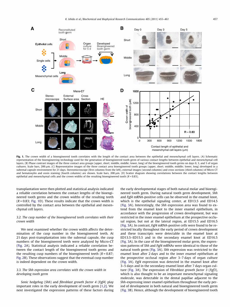
Day 0 Day 3 Day 5A BReconstituted tooth germ
ShortOrgan
g
E ith li l ll Developed
Short
Organculturefor 5 d
Epithelial cells DevelopedBioengineered tooth germ
MiddleSingle cells
Middle
LongLongMesenchymal cells
Long
C DMicro-CT 1600c o C
HE Stereo-microscope
External Surface area
CrossSection
1600
m)
Short
1200
dth
of
toot
h ( µ
m
800
Cro
wn
wid
gine
ered
Middle 400
Cbi
oeng
R 0 83
0
R = 0.83
Long 0 300 600 900 1200 1500 1800
Contact length of epithelial andmesenchymal cell layers (µm)
Fig. 1. The crown width of a bioengineered tooth correlates with the length of the contact area between the epithelial and mesenchymal cell layers. (A) Schematicrepresentation of the bioengineering technology used for the generation of bioengineered tooth germ of various contact lengths between epithelial and mesenchymal celllayers. (B) Phase contrast images of the three contact area groups (upper, short; middle, middle; lower, long) of the bioengineered tooth germs on days 0, 3, and 5 of organcultures. Scale bars, 200 lm. (C) Representative images of the three contact area bioengineered tooth groups (upper, short; middle, middle; lower, long) developed in asubrenal capsule environment for 21 days. Stereomicroscope (first columns from the left), external images (second columns) and cross sections (third columns) of Micro CTand hematoxylin and eosin staining (fourth columns) are shown. Scale bars, 200 lm. (D) Scatter diagram showing correlations between the contact lengths betweenepithelial and mesenchymal cells and the crown widths of the resulting bioengineered tooth (R = 0.83).
K. Ishida et al. / Biochemical and Biophysical Research Communications 405 (2011) 455–461 457
transplantation were then plotted and statistical analysis indicateda reliable correlation between the contact lengths of the bioengi-neered tooth germs and the crown widths of the resulting teeth(R = 0.83; Fig. 1D). These results indicate that the crown width iscontrolled by the contact area between the epithelial and mesen-chymal cell layers.
3.2. The cusp number of the bioengineered teeth correlates with theircrown width
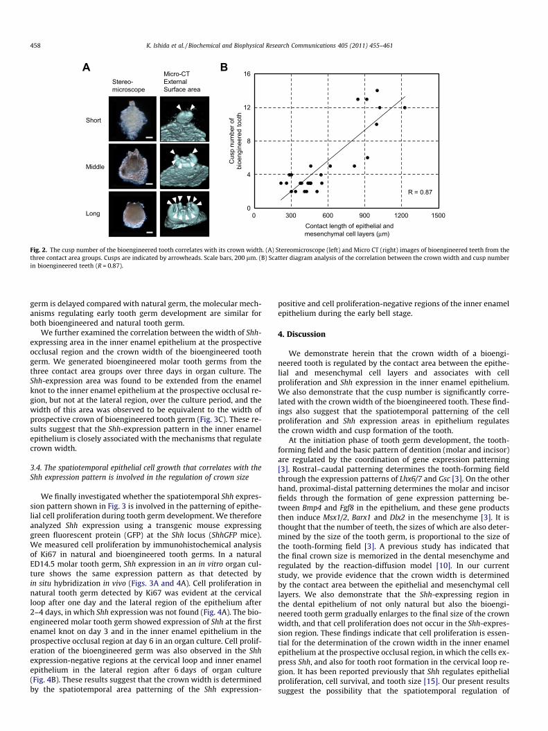
We next examined whether the crown width affects the deter-mination of the cusp number in the bioengineered teeth. At21 days post-transplantation into the subrenal capsule, the cuspnumbers of the bioengineered teeth were analyzed by Micro-CT(Fig. 2A). Statistical analysis indicated a reliable correlation be-tween the contact length of the bioengineered tooth germs andthe resulting cusp numbers of the bioengineered teeth (R = 0.87;Fig. 2B). These observations suggest that the eventual cusp numberis indeed dependent on the crown width.
3.3. The Shh expression area correlates with the crown width indeveloping tooth germ
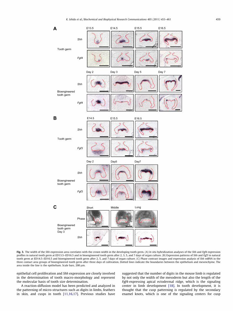
Sonic hedgehog (Shh) and fibroblast growth factor 4 (Fgf4) playimportant roles in the early development of tooth germ [1,2]. Wenext investigated the expression patterns of these factors during
the early developmental stages of both natural molar and bioengi-neered tooth germ. During natural tooth germ development, Shhand Fgf4 mRNA-positive cells can be observed in the enamel knot,which is the epithelial signaling center, at ED13.5 and ED14.5(Fig. 3A). Interestingly, the Shh expression area was found to ex-tend from the enamel knot to the inner enamel epithelium, inaccordance with the progression of crown development, but wasrestricted in the inner enamel epithelium at the prospective occlu-sal region, but not at the lateral region, at ED15.5 and ED16.5(Fig. 3A). In contrast, Fgf4 mRNA-positive cells were found to be re-stricted locally throughout the early period of crown developmentand these transcripts were detectable in the enamel knot atED13.5–ED15.5 and in the secondary enamel knot at ED16.5(Fig. 3A). In the case of the bioengineered molar germ, the expres-sion patterns of Shh and Fgf4 mRNAs were identical to those of thenatural tooth germ (Fig. 3A). Shh expression was restricted to theenamel knot after 2 days and to the inner enamel epithelium atthe prospective occlusal region after 3–7 days of organ culture(Fig. 3A). Fgf4 expression was detected in the enamel knot after3–5 days and in the secondary enamel knot after 7 days organ cul-ture (Fig. 3A). The expression of Fibroblast growth factor 3 (Fgf3),which is also thought to be an important mesenchymal signalingmolecule, was detectable in the dental papillae adjacent to theShh-expressing inner enamel epithelium throughout the early per-iod of development in both natural and bioengineered tooth germ(Fig. 3B). Hence, although the development of bioengineered tooth
A
c
B
Short
Middle
Long
Stereo-microscope
Micro-CTExternal Surface area
0
4
8
12
16
0 300 600 900 1200 1500
Cus
p nu
mbe
r of
bioe
ngin
eere
d to
oth
Contact length of epithelial and mesenchymal cell layers (µm)
R = 0.87
0 300 600 900 1200 1500
Fig. 2. The cusp number of the bioengineered tooth correlates with its crown width. (A) Stereomicroscope (left) and Micro CT (right) images of bioengineered teeth from thethree contact area groups. Cusps are indicated by arrowheads. Scale bars, 200 lm. (B) Scatter diagram analysis of the correlation between the crown width and cusp numberin bioengineered teeth (R = 0.87).
458 K. Ishida et al. / Biochemical and Biophysical Research Communications 405 (2011) 455–461
germ is delayed compared with natural germ, the molecular mech-anisms regulating early tooth germ development are similar forboth bioengineered and natural tooth germ.
We further examined the correlation between the width of Shh-expressing area in the inner enamel epithelium at the prospectiveocclusal region and the crown width of the bioengineered toothgerm. We generated bioengineered molar tooth germs from thethree contact area groups over three days in organ culture. TheShh-expression area was found to be extended from the enamelknot to the inner enamel epithelium at the prospective occlusal re-gion, but not at the lateral region, over the culture period, and thewidth of this area was observed to be equivalent to the width ofprospective crown of bioengineered tooth germ (Fig. 3C). These re-sults suggest that the Shh-expression pattern in the inner enamelepithelium is closely associated with the mechanisms that regulatecrown width.
3.4. The spatiotemporal epithelial cell growth that correlates with theShh expression pattern is involved in the regulation of crown size
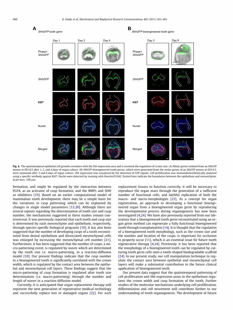
We finally investigated whether the spatiotemporal Shh expres-sion pattern shown in Fig. 3 is involved in the patterning of epithe-lial cell proliferation during tooth germ development. We thereforeanalyzed Shh expression using a transgenic mouse expressinggreen fluorescent protein (GFP) at the Shh locus (ShhGFP mice).We measured cell proliferation by immunohistochemical analysisof Ki67 in natural and bioengineered tooth germs. In a naturalED14.5 molar tooth germ, Shh expression in an in vitro organ cul-ture shows the same expression pattern as that detected byin situ hybridization in vivo (Figs. 3A and 4A). Cell proliferation innatural tooth germ detected by Ki67 was evident at the cervicalloop after one day and the lateral region of the epithelium after2–4 days, in which Shh expression was not found (Fig. 4A). The bio-engineered molar tooth germ showed expression of Shh at the firstenamel knot on day 3 and in the inner enamel epithelium in theprospective occlusal region at day 6 in an organ culture. Cell prolif-eration of the bioengineered germ was also observed in the Shhexpression-negative regions at the cervical loop and inner enamelepithelium in the lateral region after 6 days of organ culture(Fig. 4B). These results suggest that the crown width is determinedby the spatiotemporal area patterning of the Shh expression-
positive and cell proliferation-negative regions of the inner enamelepithelium during the early bell stage.
4. Discussion
We demonstrate herein that the crown width of a bioengi-neered tooth is regulated by the contact area between the epithe-lial and mesenchymal cell layers and associates with cellproliferation and Shh expression in the inner enamel epithelium.We also demonstrate that the cusp number is significantly corre-lated with the crown width of the bioengineered tooth. These find-ings also suggest that the spatiotemporal patterning of the cellproliferation and Shh expression areas in epithelium regulatesthe crown width and cusp formation of the tooth.
At the initiation phase of tooth germ development, the tooth-forming field and the basic pattern of dentition (molar and incisor)are regulated by the coordination of gene expression patterning[3]. Rostral–caudal patterning determines the tooth-forming fieldthrough the expression patterns of Lhx6/7 and Gsc [3]. On the otherhand, proximal-distal patterning determines the molar and incisorfields through the formation of gene expression patterning be-tween Bmp4 and Fgf8 in the epithelium, and these gene productsthen induce Msx1/2, Barx1 and Dlx2 in the mesenchyme [3]. It isthought that the number of teeth, the sizes of which are also deter-mined by the size of the tooth germ, is proportional to the size ofthe tooth-forming field [3]. A previous study has indicated thatthe final crown size is memorized in the dental mesenchyme andregulated by the reaction-diffusion model [10]. In our currentstudy, we provide evidence that the crown width is determinedby the contact area between the epithelial and mesenchymal celllayers. We also demonstrate that the Shh-expressing region inthe dental epithelium of not only natural but also the bioengi-neered tooth germ gradually enlarges to the final size of the crownwidth, and that cell proliferation does not occur in the Shh-expres-sion region. These findings indicate that cell proliferation is essen-tial for the determination of the crown width in the inner enamelepithelium at the prospective occlusal region, in which the cells ex-press Shh, and also for tooth root formation in the cervical loop re-gion. It has been reported previously that Shh regulates epithelialproliferation, cell survival, and tooth size [15]. Our present resultssuggest the possibility that the spatiotemporal regulation of
A
Shh
Fgf4
E14.5
Day 3
B
C
Bioengineeredtooth germDay 3
E15.5 E16.5
Day 2 Day 5
E13.5
E14.5 E15.5 E16.5
Day5Day 2 Day7
Day 7
Bioengineered tooth germ
Tooth germ
Short Middle Long
Shh
Fgf4
Shh
Fgf3
Bioengineeredtooth germ
Tooth germ
Shh
Fgf3
Shh
Phase
Fig. 3. The width of the Shh expression area correlates with the crown width in the developing tooth germ. (A) In situ hybridization analyses of the Shh and Fgf4 expressionprofiles in natural tooth germ at ED13.5–ED16.5 and in bioengineered tooth germ after 2, 3, 5, and 7 days of organ culture. (B) Expression patterns of Shh and Fgf3 in naturaltooth germ at ED14.5–ED16.5 and bioengineered tooth germ after 2, 5, and 7 days of organ culture. (C) Phase contrast images and expression analysis of Shh mRNA in thethree contact area groups of bioengineered tooth germ after three days of cultivation. Dotted lines indicate the boundaries between the epithelium and mesenchyme. Thearea inside the line is the epithelium. Scale bars, 200 lm.
K. Ishida et al. / Biochemical and Biophysical Research Communications 405 (2011) 455–461 459
epithelial cell proliferation and Shh expression are closely involvedin the determination of tooth macro-morphology and representthe molecular basis of tooth size determination.
A reaction-diffusion model has been predicted and analyzed inthe patterning of micro-structures such as digits in limbs, feathersin skin, and cusps in tooth [11,16,17]. Previous studies have
suggested that the number of digits in the mouse limb is regulatedby not only the width of the mesoderm but also the length of theFgf4-expressing apical ectodermal ridge, which is the signalingcenter in limb development [18]. In tooth development, it isthought that the cusp patterning is regulated by the secondaryenamel knots, which is one of the signaling centers for cusp
BAShhGFP bioengineered tooth germShhGFP tooth germ
Day 1 3 yaD4 yaD2 yaD Day 6
Phase+ Phase+ShhGFP ShhGFP
ShhGFP ShhGFP
Ki67 Ki67Ki67 Ki67
Nuclei Nuclei
Merged Merged
Fig. 4. The spatiotemporal epithelial cell growth correlates with the Shh expression area and is involved the regulation of crown size. (A) Molar germs isolated from an ShhGFPmouse at ED14.5 after 1, 2, and 4 days of organ culture. (B) ShhGFP bioengineered tooth germs, which were generated from the molar germs of an ShhGFP mouse at ED14.5,were examined after 3 and 6 days of organ culture. Shh expression was visualized by the detection of GFP signals. Cell proliferation was immunohistochemically analyzedusing a specific antibody against Ki67. Nuclei were detected by staining with Hoechst33342. Dotted lines indicate the boundaries between the epithelium and mesenchyme.Scale bars, 100 lm.
460 K. Ishida et al. / Biochemical and Biophysical Research Communications 405 (2011) 455–461
formation, and might be regulated by the interaction betweenFGF4, as an activator of cusp formation, and the BMPs and SHHas inhibitors [19]. Based on an earlier computational model ofmammalian tooth development, there may be a simple basis forthe variations in cusp patterning which can be explained bychanges in single model parameters [12,20]. Although there areseveral reports regarding the determination of tooth size and cuspnumber, the mechanisms suggested in these studies remain con-troversial. It was previously reported that each tooth and cusp sizeis determined by each mesenchyme and epithelium, respectively,through species-specific biological programs [10]. It has also beensuggested that the number of developing cusps of a tooth reconsti-tuted from dental epithelium and dissociated mesenchymal cellswas enlarged by increasing the mesenchymal cell number [21].Furthermore, it has been suggested that the number of cusps, a mi-cro-patterning event, is regulated by waves which are determinedby the tooth size i.e. macro-patterning, in a reaction-diffusionmodel [10]. Our present findings indicate that the cusp numberin a bioengineered tooth is significantly correlated with the crownwidth, which is regulated by the contact area between the epithe-lial and mesenchymal cell layers. These findings suggest that themicro-patterning of cusp formation is regulated after tooth sizedetermination (i.e. macro-patterning) through the number andlength of waves in a reaction-diffusion model.
Currently, it is anticipated that organ replacement therapy willrepresent the next generation of regenerative medical technologyand successfully replace lost or damaged organs [22]. For such
replacement tissues to function correctly, it will be necessary toreproduce the organ mass through the generation of a sufficientnumber of functional cells, and faithful replication of both themacro- and micro-morphologies [23]. As a concept for organregeneration, an approach to developing a functional bioengi-neered organ from a bioengineered organ germ by reproducingthe developmental process during organogenesis has now beeninvestigated [4,24]. We have also previously reported from our lab-oratory that a bioengineered tooth germ reconstituted using an or-gan germ method can regenerate a fully functional bioengineeredtooth through transplantation [14]. It is thought that the regulationof a bioengineered tooth morphology, such as the crown size andthe number and location of the cusps, is important for occlusionto properly occur [11], which is an essential issue for future toothregenerative therapy [4,24]. Previously, it has been reported thatthe morphology of a bioengineered tooth can be regulated by cul-turing tooth germ cells onto a tooth-shaped biodegradable scaffold[24]. In our present study, our cell manipulation technique to reg-ulate the contact area between epithelial and mesenchymal celllayers will make a substantial contribution to the future clinicalapplication of bioengineered teeth.
Our present data suggest that the spatiotemporal patterning ofcell proliferation and Shh expression areas in the epithelium regu-lates the crown width and cusp formation of the tooth. Furtherstudies of the molecular mechanisms underlying cell proliferation,differentiation and cell movement will contribute further to ourunderstanding of tooth organogenesis. The development of future

K. Ishida et al. / Biochemical and Biophysical Research Communications 405 (2011) 455–461 461
technologies to more precisely regulate the morphology of bioen-gineered teeth will be required to realize tooth regenerativetherapy.
Acknowledgments
This work was partially supported by Health and LabourSciences Research Grants from the Ministry of Health, Labour,and Welfare (No. 21040101), a Grant-in-Aid for Scientific Researchin Priority Areas (No. 50339131), a Grant-in-Aid for ScientificResearch (A) from Ministry of Education, Culture, Sports andTechnology, Japan (all to T.T.).
References
[1] J. Pispa, I. Thesleff, Mechanisms of ectodermal organogenesis, Dev. Biol. 262(2003) 195–205.
[2] I. Thesleff, Epithelial–mesenchymal signalling regulating tooth morphogenesis,J. Cell Sci. 116 (2003) 1647–1648.
[3] A. Tucker, P. Sharpe, The cutting-edge of mammalian development; how theembryo makes teeth, Nat. Rev. Genet. 5 (2004) 499–508.
[4] P.T. Sharpe, C.S. Young, Test-tube teeth, Sci. Am. 293 (2005) 34–41.[5] S.F. Gilbert, Developmental Biology, ninth ed., Sinauer, Massachusetts, 2010.[6] A. Gritli-Linde, M. Bei, R. Maas, X.M. Zhang, A. Linde, A.P. McMahon, Shh
signaling within the dental epithelium is necessary for cell proliferation,growth and polarization, Development 129 (2002) 5323–5337.
[7] M.I. Cho, P.R. Garant, Development and general structure of the periodontium,Periodontology 2000 (24) (2000) 9–27.
[8] K.D. Kavanagh, A.R. Evans, J. Jernvall, Predicting evolutionary patterns ofmammalian teeth from development, Nature 449 (2007) 427–432.
[9] Z. Zhang, Y. Lan, Y. Chai, R. Jiang, Antagonistic actions of Msx1 and Osr2 patternmammalian teeth into a single row, Science 323 (2009) 1232–1234.
[10] J. Cai, S.W. Cho, J.Y. Kim, M.J. Lee, Y.G. Cha, H.S. Jung, Patterning the size andnumber of tooth and its cusps, Dev. Biol. 304 (2007) 499–507.
[11] J. Jernvall, I. Thesleff, Reiterative signaling and patterning during mammaliantooth morphogenesis, Mech. Dev. 92 (2000) 19–29.
[12] I. Salazar-Ciudad, J. Jernvall, A gene network model accounting fordevelopment and evolution of mammalian teeth, Proc. Natl. Acad. Sci. USA99 (2002) 8116–8120.
[13] K. Nakao, R. Morita, Y. Saji, K. Ishida, Y. Tomita, M. Ogawa, M. Saitoh, Y.Tomooka, T. Tsuji, The development of a bioengineered organ germ method,Nat. Methods 4 (2007) 227–230.
[14] E. Ikeda, R. Morita, K. Nakao, K. Ishida, T. Nakamura, T. Takano-Yamamoto, M.Ogawa, M. Mizuno, S. Kasugai, T. Tsuji, Fully functional bioengineered toothreplacement as an organ replacement therapy, Proc. Natl. Acad. Sci. USA 106(2009) 13475–13480.
[15] H.R. Dassule, P. Lewis, M. Bei, R. Maas, A.P. McMahon, Sonic hedgehogregulates growth and morphogenesis of the tooth, Development 127 (2000)4775–4785.
[16] S. Kondo, A mechanistic model for morphogenesis and regeneration of limbsand imaginal discs, Mech. Dev. 39 (1992) 161–170.
[17] H.S. Jung, P.H. Francis-West, R.B. Widelitz, T.X. Jiang, S. Ting-Berreth, C. Tickle,L. Wolpert, C.M. Chuong, Local inhibitory action of BMPs and theirrelationships with activators in feather formation: implications for periodicpatterning, Dev. Biol. 196 (1998) 11–23.
[18] Y. Litingtung, R.D. Dahn, Y. Li, J.F. Fallon, C. Chiang, Shh and Gli3 aredispensable for limb skeleton formation but regulate digit number andidentity, Nature 418 (2002) 979–983.
[19] J. Jernvall, Linking development with generation of novelty in mammalianteeth, Proc. Natl. Acad. Sci. USA 97 (2000) 2641–2645.
[20] I. Salazar-Ciudad, J. Jernvall, A computational model of teeth and thedevelopmental origins of morphological variation, Nature 464 (2010) 583–586.
[21] B. Hu, A. Nadiri, S. Kuchler-Bopp, F. Perrin-Schmitt, H. Peters, H. Lesot, Tissueengineering of tooth crown, root, and periodontium, Tissue Eng. 12 (2006)2069–2075.
[22] R.I. Lechler, M. Sykes, A.W. Thomson, L.A. Turka, Organ transplantation – Howmuch of the promise has been realized?, Nat Med. 11 (2005) 605–613.
[23] M.C. Raff, Size control: the regulation of cell numbers in animal development,Cell 86 (1996) 173–175.
[24] E. Ikeda, T. Tsuji, Growing bioengineered teeth from single cells: potential fordental regenerative medicine, Expert. Opin. Biol. Ther. 8 (2008) 735–744.


![Materials Science & Engineering C - WordPress.com€¦ · neurons, fat cells, osteoblasts and odontoblast, and can produce bone and dentin in the right environment [3]. The first](https://img.pdfslide.us/doc/110x75/5ece1192c9f8163d2d78ef27/materials-science-engineering-c-neurons-fat-cells-osteoblasts-and-odontoblast.jpg)


![Biochemical and Biophysical Research Communications · Type I collagen is mainly expressed in odontoblast [7]. How-ever, little is known regarding the expression of minor fibrillar](https://img.pdfslide.us/doc/110x75/5e88b90270b5d44b3918b8db/biochemical-and-biophysical-research-type-i-collagen-is-mainly-expressed-in-odontoblast.jpg)