Embed Size (px)
Citation preview
Biocatalytic Methods for the Hydroxylation of
Non-Activated Carbon Centres
Suzanne Jill Aitken
A thesis submitted for the degree of
Doctor of Philosophy
September 2000
Acknowledgements
I would like to thank Prof. Sabine Flitsch for her advice, encouragement and support
throughout my PhD. Thanks are also due to Prof Nick Turner for useful input and
suggestions. I am indebted to Dr. Gideon Grogan whose involvement from the start of
the project has shaped both my knowledge and the direction of the project. I cannot
thank him enough for teaching me the new techniques required for the work reported in
addition to carrying out some of the initial biological screening. Special thanks are also
due to Drs. James Dowden and Gideon Grogan for their time in proofreading this thesis.
I am very grateful to those who provided valuable analytical services, namely John
Millar and Wesley Kerr for NMR spectroscopy and Alan Taylor and Harry MacKenzie
for mass spectrometry. Dr. David Reed has also been invaluable - first for running 360
MHz spectra and later for fully training me and putting up with my questions.
It would not be possible to name everyone who has helped make the lab a nice place to
be but thanks to all past and present members of the SLF/NJT groups for contributing to
it and particularly to Dave, Anita, Stuart, Alison, Adam, Sharon, Shagufta, Alexis, Josie
and Garnet. Thanks are also due to everyone outside of work who helped me to remain
sane against the odds and to keep things in perspective, particularly Marie-Claire, Ian,
Suzie, Lou, Mark and Rach.
Finally I have to thank those closest to me - James for everything; Carolyn - the best
landlady, sister and friend I could ever ask for and finally Mum and Dad for their
constant support. I would not be here if it were not for you. Thank you.
Eff
Abbreviations
Ac acetyl
ADH alcohol dehydrogenase
APCI atmospheric pressure chemical ionisation
ATCC American Type Culture Collection
ax axial
Bn benzyl
bp boiling point
br broad
Cbz benzyloxycarbonyl
d doublet
DCC dicyclohexylcarbodiimide
DCM dichloromethane
DCU dicyclohexylurea
d.e. diastereomeric excess
DEPT distortionless enhancement through polarisation transfer
DHP 3 ,4-dihydro-2H-pyran
DMAP 4-dimethylaminopyridine
e.e. enantiomeric excess
El electron impact
eq equatorial
ES electrospray
Et ethyl
FAB fast atom bombardment
GC gas chromatography
GC-MS gas chromatography mass spectrometry
hfc 3 -(heptafluoropropylhydroxymethylene)-(+)-camphorate
HPLC high performance liquid chromatography
Iv
m multiplet
Me methyl
min minutes
mol % molar percentage
MOM methoxymethyl
mp melting point
MTPA ix-methoxy-a-trifluorophenyl acetic acid
m/z mass to charge ratio
NADH nicotinamide adenine dinucleotide, reduced form
NADPH nicotinamide adenine dinucleotide phosphate, reduced
NCIMB National Collection of Industrial and Marine Bacteria
NMR nuclear magnetic resonance
nOe nuclear Overhauser effect
op optical purity
PCP pentachiorophenol
Ph phenyl
ppm parts per million
PTSA p-toluenesulfonic acid
s singlet
spp. species
t triplet
THF tetrahydrofuran
THP tetrahydropyran
V
2
3
Contents
Introduction 1
1.1 Biohydroxylations 1 1.2 Cytochrome P-450 monooxygenases 3 1.3 Practicalities of biohydroxylatjons 5 1.4 Beauverja bassiana ATCC 7159 7 1.5 Genus Rhodococcus 17 1.6 Summary 26 1.7 Aims 27 Biotransformations using Beauveria bassiana ATCC 715 973 29 2.1 Aims 29 2.2 Methods 31 2.3 Results 32 2.4 Discussion 41 2.5 Conclusions 47 B iotrans formations with Rhodococcus sp. NCIMB 9784 49 3.1 Aims
49 3.2 Growth of organism 50 3.3 Methods 50 3.4 Oxidation of non-camphor like substrates si 3.5 Metabolism of camphor-like substrates 59 3.6 Conclusions 61 Biotransformatjons with Rhodococcus rhodochrous NCIMB 9703 63 4.1 Aims
63 4.2 Growth of organism 63 4.3 Methods 64 4.4 Initial screening 65 4.5 Synthesis of substrates 65
VI
5
6
7
8
9
10
4.6 Biooxidations of tetrahydropyranosyl derivatives 66
4.7 Biooxidation of tetrahydrofuranosyl derivatives 76
4.8 Stereoselectivity of hydroxylation 81
4.9 Biooxidations of carbocyclic ethers 89
4.10 Biooxidations of nitrogen heterocycles 92
4.11 Biooxidations of carbocyclic nucleoside precursor 93
4.12 Nature of the hydroxylating enzyme 96
4.13 Optimisation of transformation 98
4.14 Conclusions 99
Experimental 100
5.1 Apparatus and chemicals 100
5.2 Beauveria bassiana ATCC 7159 102
5.3 Rhodococcus sp. NCIMB 9784 108
5.4 Rhodococcus rhodochrous NCIMB 9703 110
Bibliography 132
Appendix I 139
Appendix II 150
Appendix III 156
Appendix IV 165
vii
1. Introduction
I Introduction
1.1 Biohydroxylations
Biocatalysis is widely accepted as a viable alternative to traditional chemical methods
offering good chemo-, regio and stereoselectivity. The source of this selectivity is the
chiral nature of the biocatalyst that, as a protein, is made up of naturally occurring L-
amino acids; the protein structure interacts with the substrate molecule, placing the
substrate in a particular orientation thus conferring selectivity on the transformation.
Biocatalysis is primarily utilised in the pharmaceutical and specialised organic sector
due to the need to produce compounds in homochiral form and the benign conditions in
which biocatalysts operate. An increasing number of processes combine biocatalysis
and chemical methods into one synthetic process, with resolution by hydrolysis being
the most common.'
Greater understanding of enzyme function at the genetic level leading to the genomic
based identification of novel catalytic activity and subsequent improvement of catalytic
selectivity and efficiency through directed evolution of biocatalysts are thus likely to
remain growth areas and increase the value and utility of biocatalysts .2,3
Of particular interest and importance are the biocatalysts involved in hydroxylation.
Under the control of a biocatalyst, hydroxylations that are still chemo-, regio and
stereoselective can occur at positions remote from existing functionality. Highly
selective hydroxylations of this type are difficult to carry out chemically. Research for
chemical methods for non-activated carbon hydroxylation have concentrated on
biomimetic systems. The most comprehensive to date have been the 'Gif systems
investigated by Professor D. H. R. Barton4 and the biomimetic systems studied by
I. Introduction
Professor R. Breslow.5 Although these systems go some way in terms of mimicking the
selectivity of biological systems, they certainly do not provide a general method.
Biocatalytic reactions therefore remain the more successful and widespread method of
selective hydroxylation.
As indicated, one of the key advantages of utilising biocatalysis is the selectivity
imparted on the transformation by the biocatalyst. There are numerous cases, such as
the hydroxylation of camphor by Pseudomonas putida (discussed in section 1.2), where
the biocatalyst is extremely selective in the transformation of its natural substrate. More
importantly, since the exclusive use of natural substrates would distinctly limit the
methodology, it is encouraging that hydroxylation of non-natural substrates can also be
carried out with good selectivity.
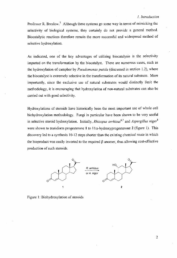
Hydroxylations of steroids have historically been the most important use of whole cell
biohydroxylation methodology. Fungi in particular have been shown to be very useful
in selective steroid hydroxylation. Initially, Rhizopus arrhizus6'7 and Aspergillus niger8
were shown to transform progesterone 1 t 1 la-hydroxyprogesterone 2 (figure 1). This
discovery led to a synthesis 10-12 steps shorter than the existing chemical route in which
the bioproduct was easily inverted to the required 13 anomer, thus allowing cost-effective
production of such steroids.
R. arrhizus
orAniger Oj
2 I
Figure 1: Biohydroxylation of steroids
2
I. Introduction
Although this initial literature is now dated, microbial transformation remains the
method of choice for steroid hydroxylation. Many years of research have resulted in the
knowledge that virtually any carbon centre in the steroid nucleus can now be
hydroxylated selectively.9
Terpene hydroxylation products are of particular interest to the fragrance industry and as
intermediates for the pharmaceutical industry. As for steroids, controlled chemical
hydroxylation is difficult due to the large number of potential sites of hydroxylation and
successful production of such compounds is mainly restricted to microbial methods.
The indisputable success of biohydroxylation methodology in naturally occurring
compounds, such as steroids and terpenes, has provided the basis for recent research that
has concentrated on the microbial hydroxylation of non-natural compounds.
1.2 Cytochrome P-450 monooxygenases
Monooxygenases are a class of oxidoreductases that catalyse the insertion of one atom
from molecular oxygen into the substrate, while the second atom is reduced to water.
An important group of monooxygenases are the cytochrome P-450 monooxygenases.
These haem-dependent monooxygenases carry out the NAD(P)H dependent oxidation of
a diverse range of compounds. Cytochrome P-450 monooxygenases are extremely
common in nature and are found in virtually all organisms.
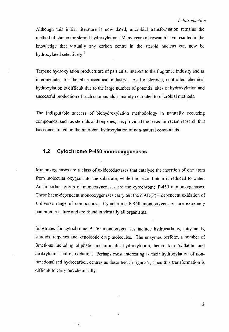
Substrates for cytochrome P-450 monooxygenases include hydrocarbons, fatty acids,
steroids, terpenes and xenobiotic drug molecules. The enzymes perform a number of
functions including aliphatic and aromatic hydroxylation, heteroatom oxidation and
dealkylation and epoxidation. Perhaps most interesting is their hydroxylation of non-
functionalised hydrocarbon centres as described in figure 2, since this transformation is
difficult to carry out chemically.
3
1. Introduction
H H cytochrome H OH
R1 R2 P-450 R1 R2
Figure 2: Hydroxylation of non-activated carbon centres by cytochrome P-450
monooxygenases
Mammalian cytochrome P-450 monooxygenases are particularly abundant in
detoxification organs such as the liver, where they play an important role in the
detoxification and excretion of xenobiotic molecules. They often catalyse initial
hydroxylation of lipophilic compounds introducing them into breakdown pathways or
making them more hydrophilic so facilitating excretion from the body. Perhaps due to
this function and the need to accept a range of compounds, mammalian cytochrome
P-450 monooxygenases tend to display wide substrate specificity that contrasts with
their more specific prokaryotic counterparts.
Since cytochrome P-450 monooxygenases in the liver are important in drug metabolism
and excretion, much of the research on the mammalian enzymes has concentrated on the
identification of inhibitors that could increase the lifetime of the drug molecule in the
body.'° Unfortunately, drug molecules themselves may inhibit cytochrome P-450
monooxygenases leading to potentially harmful accumulation of other lipophilic
compounds in the body.'
Eukaryotic cytochrome P-450 monooxygenases from fungal sources have proved very
difficult to study due to instability of the protein and low expression levels and there
remain few examples.'2
Unlike their eukaryotic counterparts, prokaryotic cytochrome P-450 monooxygenases
tend to display very strict substrate specificities that are generally confined to the natural
substrate and its analogues. Since many monooxygenases are membrane bound, there
are few examples of stable, isolated cytochrome P-450 monooxygenases, a problem that
I. Introduction
has slowed the progress in research in human cytochrome P-450 monooxygenases. An
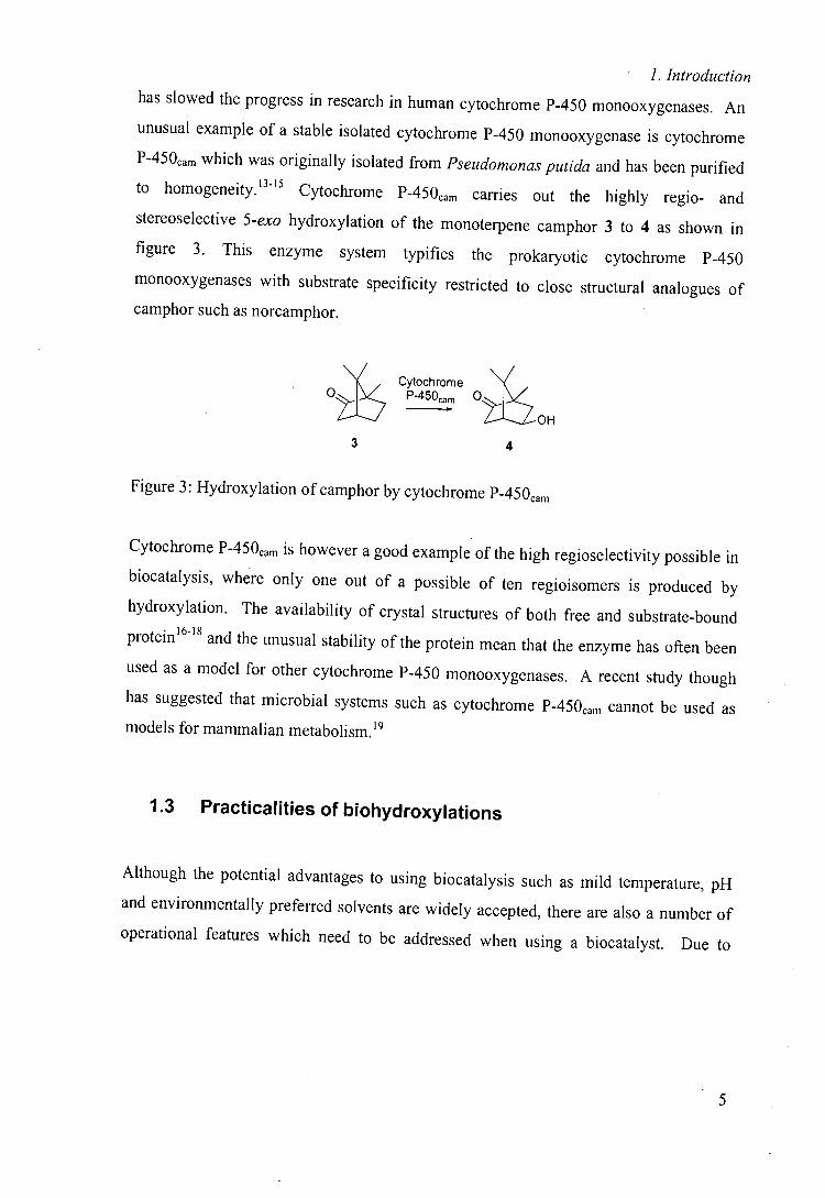
unusual example of a stable isolated cytochrome P-450 monooxygenase is cytochrome
P450cam which was originally isolated from Pseudomonas putida and has been purified to homogeneity. 1315 Cytochrome P450cam carries out the highly regio- and stereoselective 5-exo hydroxylation of the monoterpene camphor 3 to 4 as shown in
figure 3. This enzyme system typifies the prokaryotic cytochrome P-450
monooxygenases with substrate specificity restricted to close structural analogues of
camphor such as norcamphor.
0 7
Cytochrome P45O m
OH
3 4
Figure 3: Hydroxylation of camphor by cytochrome P450cam
Cytochrome 450cam is however a good example of the high regioselectivity possible in
biocatalysis, where only one out of a possible of ten regioisomers is produced by
hydroxylation. The availability of crystal structures of both free and substrate-bound
protein 16-18 and the unusual stability of the protein mean that the enzyme has often been
used as a model for other cytochrome P-450 monooxygenases. A recent study though
has suggested that microbial systems such as cytochrome P450caj,, cannot be used as
models for mammalian metabolism. 19
1.3 Practicalities of biohydroxylations
Although the potential advantages to using biocatalysis such as mild temperature, pH
and environmentally preferred solvents are widely accepted, there are also a number of
operational features which need to be addressed when using a biocatalyst. Due to
1. Introduction
difficulties in isolating many oxidoreductases and their subsequent instability, many
biohydroxylations are carried out utilising whole-cell methodology.
Aside from increased stability of the catalyst, the added advantage of whole-cell
methodology is that any required co-factors and co-proteins will be provided and
recycled by the organism. This is extremely important for biohydroxylations since
cytochrome P-450 monooxygenases require not only a supply of oxygen but also a
source of electrons, namely NADH or NADPH, and generally two co-proteins to deliver
these electrons to the enzyme. Among the disadvantages are that other enzymes in the
cell can cause undesired side-reactions. Additionally the larger volume of aqueous
media that is required can make the product isolation laborious.
On the other hand, the use of isolated enzymes as biocatalysts can be advantageous since
the work-up is likely to be simpler, often with higher concentration tolerance compared
to whole cell systems and competing side reactions are much less likely. The method is
however severely compromised since isolation and purification of enzymes can be very
difficult, and co-proteins also need to be produced and co-factors supplied.
Although examples of isolated enzyme catalysis do exist, they are certainly not common
and due to these difficulties are likely to remain so.
Whole cell biohydroxylations are widespread in the literature mainly due to the robust
nature of many such biocatalysts. Because the use of such biocatalysts require little
specialist knowledge or equipment, it is likely that whole cell methodologies will remain
the method of choice.
M.
1. Introduction
1.4 Beauveria bassiana ATCC 7159
Beauveria bassiana ATCC 7159 is a member of a small family of fungi and is one of the
most frequently used whole-cell biocatalysts. It is thought to contain a number of
hydroxylase enzymes20 and it is their function which has been most studied. Due to its
widespread use as a biocatalyst, the literature is too wide ranging to carry out a
comprehensive review here, but a comprehensive review of the biocatalytic reactions of
Beauveria spp. has recently been published .21
Among the biocatalytic reactions catalysed by Beauveria bassiana are many that are
typically carried out by cytochrome P-450 monooxygenases such as aliphatic and
aromatic hydroxylation; sulfoxidation and heteroatom dealkylation reactions. A number
of other whole cell transformations such as double bond reduction 22 and Baeyer-Villiger
oxidation 23 have also been reported.
The bulk of the literature of Beauveria bassiana has centred on the action of its
hydroxylase activity; some key areas of which will be discussed here. It should be noted
when examining the literature that the species under investigation has been redesignated
twice and that Sporotrichium sulfurescens and Beauveria sulfurescens both refer to the
strain under study but that the ATCC strain number is constant throughout.
1.4.1 Non-hydroxylating transformations
In the only reports of sulfoxidation by the organism, Holland and co-workers have
described a combination of biocatalytic and chemical routes to reach all the
stereoisomers of some amino acid sulfoxides, affording a superior route to the existing
resolution-based methods. 24,25
7
1. Introduction
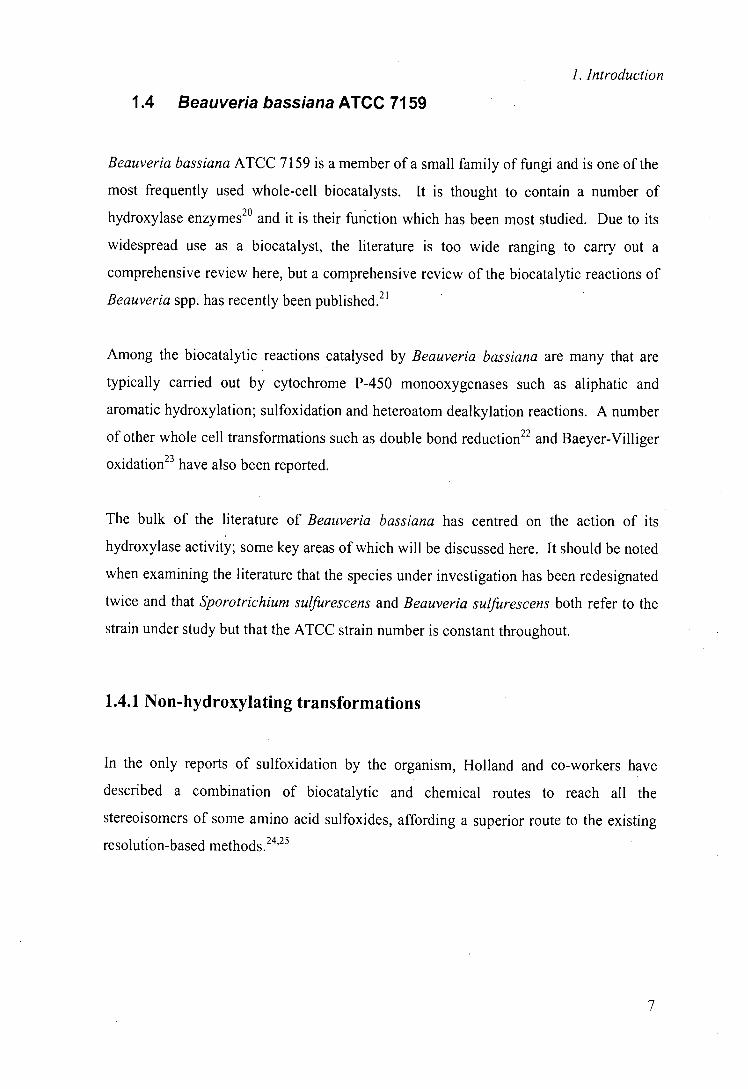
Heteroatom dealkylations by Beauveria bassiana also appear in the literature. 13-lactam
5 not only undergoes hydroxylation of an ethyl side chain 6 but also yields a significant
amount of debenzylated, non-hydroxylated product 7 (figure 4).
OH
0 )—N"C~Ph B. bassiana
FCPh + H
5
6(10%) 7(20%)
Figure 4: Incubation of J3-lactam derivative 5 with Beauveria bassiana
This compound was the only one in a series of five structurally related compounds,
which underwent N-debenzylation that is thought to occur via benzylic hydroxylation
followed by hydrolysis of the resultant aminoacetal.26 Another compound in the series
8, which did not show N-debenzylation, did undergo O-demethylation in a very high
yield via hydroxylation of the methyl group to a hemiacetal followed again by
hydrolysis. The difference in selectivity of hydroxylase action on these compounds
indicates the large impact that seemingly very minor substrate alterations can have on
the selectivity of an enzyme.
MeO-
,)—NCH2Ph
8
1.4.2 Aryl hydroxylation
Hydroxylation of aryl carbons by Beauveria bassiana has been widely reported for
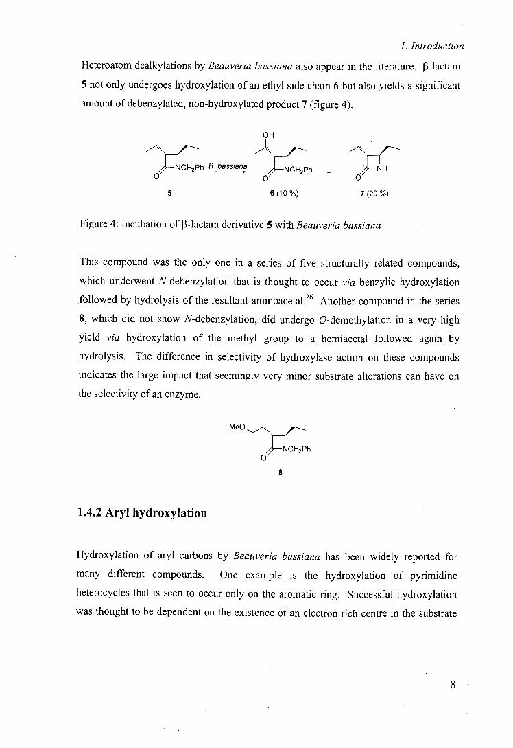
many different compounds. One example is the hydroxylation of pyrimidine
heterocycles that is seen to occur only on the aromatic ring. Successful hydroxylation
was thought to be dependent on the existence of an electron rich centre in the substrate
1. Introduction
since incubation of substrate 9 and alkyl substituted analogues was unsuccessful,
whereas pyrimidine 10 was hydroxylated successfully.27 This hydroxylase activity was
seen however to be very limited when tested with substrate analogues. Other organisms
such as Rhodococcus erthyropolis were, for these substrate types, found to be more
general hydroxylation catalysts.
We
N NJ
9 10
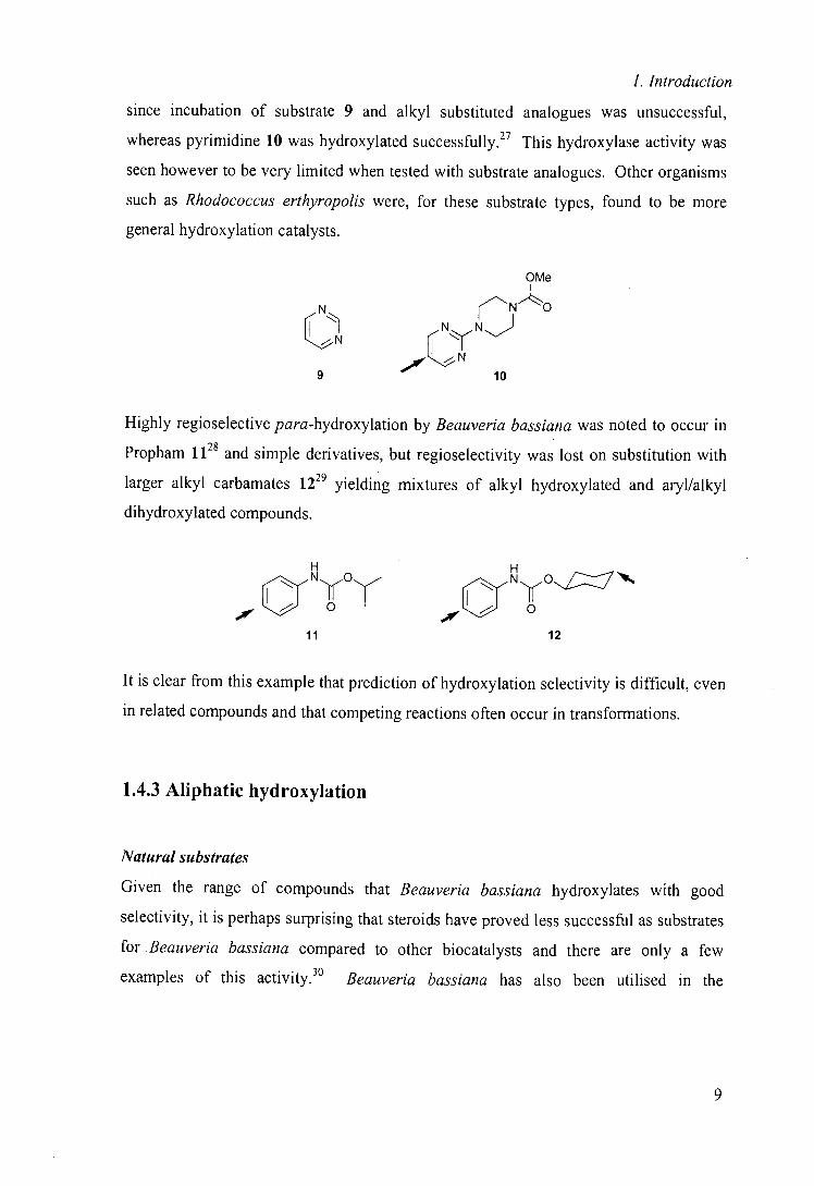
Highly regioselective para-hydroxylation by Beauveria bassiana was noted to occur in
Propham 1128 and simple derivatives, but regioselectivity was lost on substitution with
larger alkyl carbamates 1229 yielding mixtures of alkyl hydroxylated and aryl/alkyl
dihydroxylated compounds.
H NyO (
'JVK Cr 0 AVK Cr 0
11 12
It is clear from this example that prediction of hydroxylation selectivity is difficult, even
in related compounds and that competing reactions often occur in transformations.
1.4.3 Aliphatic hydroxylation
Natural substrates
Given the range of compounds that Beauveria bassiana hydroxylates with good
selectivity, it is perhaps surprising that steroids have proved less successful as substrates
for .Beauveria bassiana compared to other biocatalysts and there are only a few
examples of this activity. 30 Beauveria bassiana has also been utilised in the
1. Introduction
hydroxylation of hydrocarbons and alcohols 31 and terpenes32 but these substrate types
have attracted only limited interest.
Amide and related substrates
By far the most prolific research in Beauveria bassiana has been in the hydroxylation of
non-activated carbon centres in substrates containing a remote 'directing group'. This
work was initiated by a methodical examination of hydroxylation in a long series of
papers by Upjohn research group. The Upjohn group, in common with many others,
have concentrated on the hydroxylation of hetero- and carbocycles often containing an
electron rich substituent; typical structures being 14 and 31
R
0 N 0 o
°
o=s=o N°
'i
13 14 15
Since the initial reports by the Upjohn research group in the late 1960s, there have been
many reports of aliphatic hydroxylation by Beauveria bassiana, some of which have been very selective.
A study by Roberts, Willetts and co-workers demonstrates that an electron rich
substituent is not a prerequisite for hydroxylation by the fungus and that regioselectivity
of hydroxylation can be directed. They showed that substituting and effectively
blocking the preferred site of hydroxylation can significantly alter the regioselectivity of
hydroxylation of N-arylamines directing hydroxylation away from the aryl portion and
into the aliphatic ring (figure 5)36
Unsubstituted N-arylpiperjdine 16 was hydroxylated regioselectively to give the N-(4-
hydroxyphenyl)piperidine 17 whereas for 18, where this para position was substituted
IN
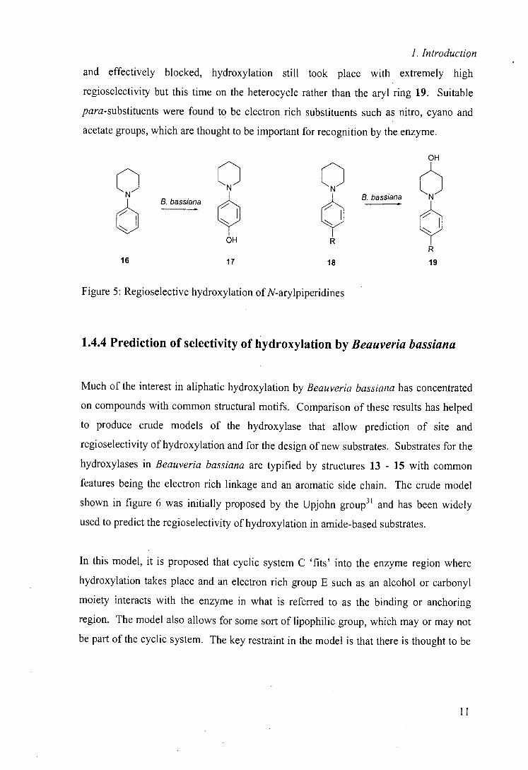
1. Introduction
and effectively blocked, hydroxylation still took place with extremely high
regioselectivity but this time on the heterocycle rather than the aryl ring 19. Suitable
para-substituents were found to be electron rich substituents such as nitro, cyano and
acetate groups, which are thought to be important for recognition by the enzyme.
Q _ Q Q B. bassiana
6 1 1
OH R
16
17
18
B. bassiana
Figure 5: Regioselective hydroxylation of N-arylpiperidines
1.4.4 Prediction of selectivity of hydroxylation by Beauveria bassiana
Much of the interest in aliphatic hydroxylation by Beauveria bassiana has concentrated
on compounds with common structural motifs. Comparison of these results has helped
to produce crude models of the hydroxylase that allow prediction of site and
regioselectivity of hydroxylation and for the design of new substrates. Substrates for the
hydroxylases in Beauveria bassiana are typified by structures 13 - 15 with common
features being the electron rich linkage and an aromatic side chain. The crude model
shown in figure 6 was initially proposed by the Upjohn group3 ' and has been widely
used to predict the regioselectivity of hydroxylation in amide-based substrates.
In this model, it is proposed that cyclic system C 'fits' into the enzyme region where
hydroxylation takes place and an electron rich group E such as an alcohol or carbonyl
moiety interacts with the enzyme in what is referred to as the binding or anchoring
region. The model also allows for some sort of lipophilic group, which may or may not
be part of the cyclic system. The key restraint in the model is that there is thought to be
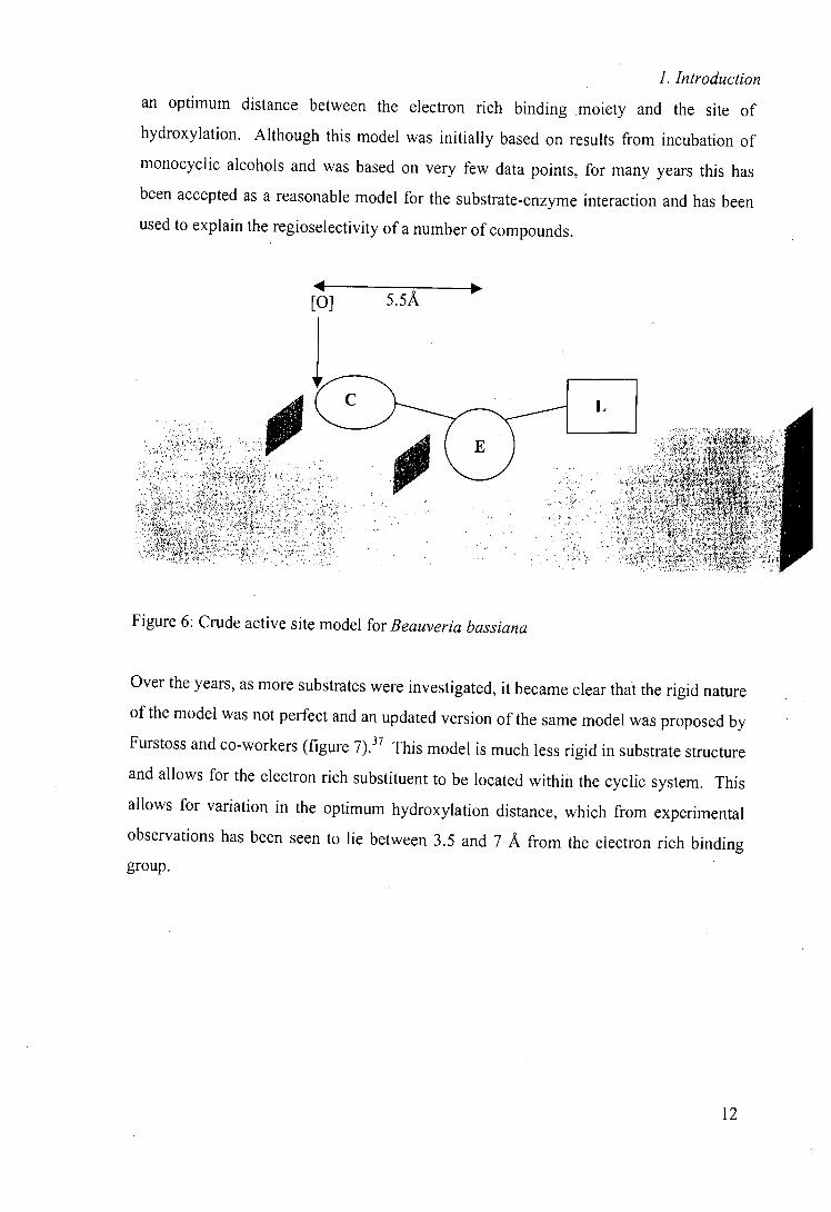
1. Introduction
an optimum distance between the electron rich binding moiety and the site of
hydroxylation. Although this model was initially based on results from incubation of
monocyclic alcohols and was based on very few data points, for many years this has
been accepted as a reasonable model for the substrate-enzyme interaction and has been
used to explain the regioselectivity of a number of compounds.
[0] 5.5A
Figure 6: Crude active site model for Beauveria bassiana
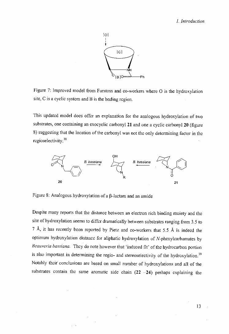
Over the years, as more substrates were investigated, it became clear that the rigid nature
of the model was not perfect and an updated version of the same model was proposed by
Furstoss and co-workers (figure 7)37 This model is much less rigid in substrate structure
and allows for the electron rich substituent to be located within the cyclic system. This
allows for variation in the optimum hydroxylation distance, which from experimental
observations has been seen to lie between 3.5 and 7 A from the electron rich binding group.
12
1. Introduction
01
Figure 7: Improved model from Furstoss and co-workers where 0 is the hydroxylation
site, C is a cyclic system and B is the bnding region.
This updated model does offer an explanation for the analogous hydroxylation of two
substrates, one containing an exocyclic carbonyl 21 and one a cyclic carbonyl 20 (figure
8) suggesting that the location of the carbonyl was not the only determining factor in the
regioselectivity.38
OH
O
B. bassiana B. bassiana
N ~— 1~~
. " ' 0
20 21
Figure 8: Analogous hydroxylation of a J3-lactam and an amide
Despite many reports that the distance between an electron rich binding moiety and the
site of hydroxylation seems to differ dramatically between substrates ranging from 3.5 to
7 A, it has recently been reported by Pietz and co-workers that 5.5 A is indeed the
optimum hydroxylation distance for aliphatic hydroxylation of N-phenylcarbamates by
Beauveria bassiana. They do note however that 'induced fit' of the hydrocarbon portion
is also important in determining the regio- and stereoselectivity of the hydroxylation.39
Notably their conclusions are based on small number of hydroxylations and all of the
substrates contain the same aromatic side chain (22 —24) perhaps explaining the
13
1. Introduction
acceptability of the simplest active site model. It is unclear whether these results are
broadly applicable.
I L H
aOyN~,O,'~ 0 N,,C
22 23
o 1
24
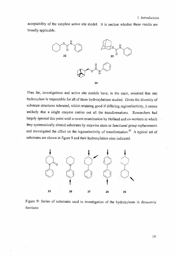
Thus far, investigations and active site models have, in the main, assumed that one
hydroxylase is responsible for all of those hydroxylations studied. Given the diversity of
substrate structures tolerated, whilst retaining good if differing regioselectivity, it seems
unlikely that a single enzyme carries out all the transformations. Researchers had
largely ignored this point until a recent examination by Holland and co-workers in which
they systematically altered substrates by stepwise atom or functional group replacements
and investigated the effect on the regioselectivity of transformation .20 A typical set of
substrates are shown in figure 9 and their hydroxylation sites indicated.
aNO C
t t 25 26 27 28 29
Figure 9: Series of substrates used in investigation of the hydroxylases in Beauveria
bassiana
II
1. Introduction
Holland suggests that this particular series of substrates show that the hydroxylation of
these compounds is not dependent on the amide since compounds 27 - 29 are
hydroxylated in similar regions to that in amide 25. para-Hydroxylation of amine 26 is
accounted for by the activation of this position to electrophilic attack which is perhaps a
surprising explanation since one of the advantages of biocatalysis is that it is not usually
dependent on activity, since non-activated groups can be transformed in the presence of
other more chemically activated groups. A number of groups of compounds were
similarly investigated with the results suggesting that, in some cases, regioselective
hydroxylation did not seem to be dependent on an electron rich directing group.
Inhibition studies were also carried out using a range of known cytochrome P-450
monooxygenase inhibitors, which in some cases where competing hydroxylation
reactions were taking place, produced a product array that was dependent on the
inhibitor concentration. This combined evidence led Holland to propose that as many as
four separate hydroxylases may exist in Beauveria bassiana. These may be responsible
for arene hydroxylation; benzylic hydroxylation; non-activated aliphatic hydroxylation
and the much reported hydroxylation of substrates with remote electron-rich directing
groups.
This study does seem to offer a more satisfactory explanation for the selectivity of
hydroxylation in such a wide range of substrates and could offer a reason for the
appearance of more than one 'type' of hydroxylation in particular substrates.
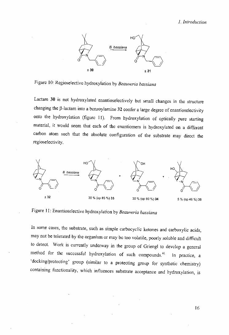
1.4.5 Enantioselectivity of hydroxylation
Although in most cases, hydroxylation by Beauveria bassiana is regioselective, it has
been noted that enantioselectivity is not always high. One notable example is the
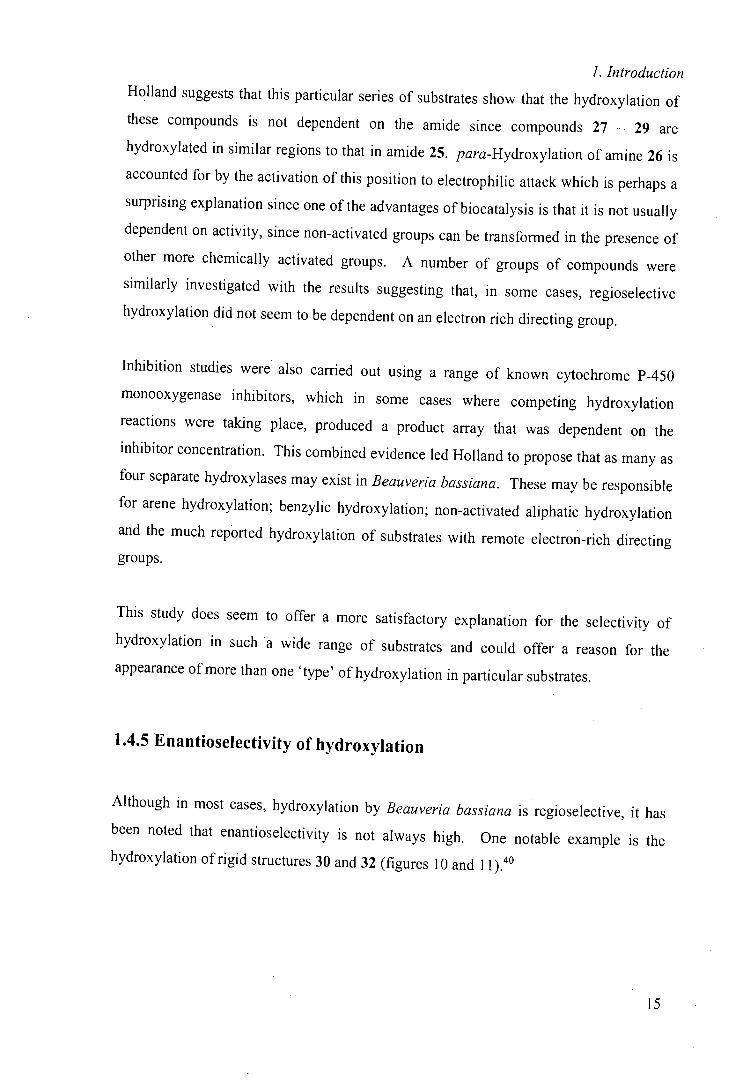
hydroxylation of rigid structures 30 and 32 (figures 10 and 11).40
15
1. Introduction
HOJ
- B. bassiana
O O __O
±30 ±31
Figure 10: Regioselective hydroxylation by Beauveria bassiana
Lactam 30 is not hydroxylated enantioselectively but small changes in the structure
changing the J3-lactam into a benzoylamine 32 confer a large degree of enantioselectivity
onto the hydroxylation (figure 11). From hydroxylation of optically pure starting
material, it would seem that each of the enantiomers is hydroxylated on a different
carbon atom such that the absolute configuration of the substrate may direct the
regioselectivity.
± 32
B. bassiana
HO OH HO
~_O ~_O
3O% (op 85 %)33 30 % (op 9O%)34 5 % (op 46%)35
Figure 11: Enantioselective hydroxylation by Beauveria bassiana
In some cases, the substrate, such as simple carbocyclic ketones and carboxylic acids,
may not be tolerated by the organism or may be too volatile, poorly soluble and difficult
to detect. Work is currently underway in the group of Griengl to develop a general
method for the successful hydroxylation of such compounds.4' In practice, a
'docking/protecting' group (similar to a protecting group for synthetic chemistry)
containing functionality, which influences substrate acceptance and hydroxylation, is
16

I. Introduction
introduced prior to and removed after transformation. This approach has been extremely
successful in some cases; the example shown in figure 12 is the b i otrans formation of
cyclopentanone 36, which is not transformed by Beauveria bassiana in its natural state.
protection NBz B. bassiana NBz Steps -
0,,,C,,,,0Bn 0,~'1L.,o0H
36 37 38 3984%ee
Figure 12: Use of a 'docking/protecting' group in biotransformation
Although this method. is primarily aimed at increasing the types of substrates accepted
by the enzyme, the example shown uses a chiral docking/protecting group which has the
further advantage that it can improve the optical purity of the product, acting much like a
chiral auxiliary.
Although these and other cases do show that stereoselective hydroxylation is possible
with Beauveria . bassiana, much work is needed to ascertain the requirements for
stereoselectivity.
As can be seen from this brief review of the literature surrounding Beauveria bassiana,
although many substrates have been studied and many selective transformations
identified, our understanding of the factors involved in this selectivity is limited.
Overall, Beauveria bassiana has shown itself to be a very versatile selective whole cell
biocatalyst.
1.5 Genus Rhodoou
Actinomycetes are widely distributed in both soil and aquatic environments and have
great metabolic diversity. The genus Rhodococcus is classified as a group of gram-
17
1. Introduction
positive bacteria belonging to the family Nocardioform Actinomycetes. Actinomycetes
have been used for industrial processes over many years and can metabolise a wide
range of compounds including simple hydrocarbons, aromatic compounds and steroids,
a characteristic that has increased the awareness of this family as potential biocatalysts.
In common with many organisms, the first step of metabolism or transformation of
many Rhodococcus strains is oxidation.
1.5.1 Degradation of aromatics
The degradation of aromatic compounds by Rhodococcus species has been well
documented. Styrene, an important industrial chemical, has been shown to be
metabolised by Rhodococcus rhodochrous NCIMB 13259,42 an organism that can also
oxidise a range of aromatic substrates including toluene.
Rhodococcus chiorophenolicus PCP-1 was isolated from a pentachiorophenol (PCP)
enriched culture inoculated from lake sediment and degrades pentachiorophenol and
other polychlorinated phenols.43 Pentachiorophenol is widely used as a wood
preservative and although it is known that it is biodegradable, PCP contamination of soil
and groundwater is a known environmental problem.
Rhodococcus spp. are generally persistent in the environment and apparently lack
catabolite repression, making them potential bioremediation catalysts. This use has been
suggested for both styrene and pentachiorophenol utilising strains and indeed the
pentachlorophenol utilising organism has already been shown to be successful in
bioremediation tests .44'45
Rhodococcus sp. strain C125 (Corynebacterium strain C125) has been shown to grow
well on a range of aromatic compounds such as o-xylene, ethylbenzene and tetralin and
contains an NAD(P)H-dependent dioxygenase activity. 46
1. Introduction
The Rhodococcus spp. demonstrate large substrate diversity even within aromatic
compounds with phenols, halogenated phenols and other substituted aromatics tolerated.
1.5.2 Degradation of hydrocarbons
There are numerous reports of Rhodococcus species which are able to transform
hydrocarbons, primarily through initial terminal hydroxylation; including substrates
ranging from propane (Rhodococcus rhodochrous ATCC 21 198)47 and octane
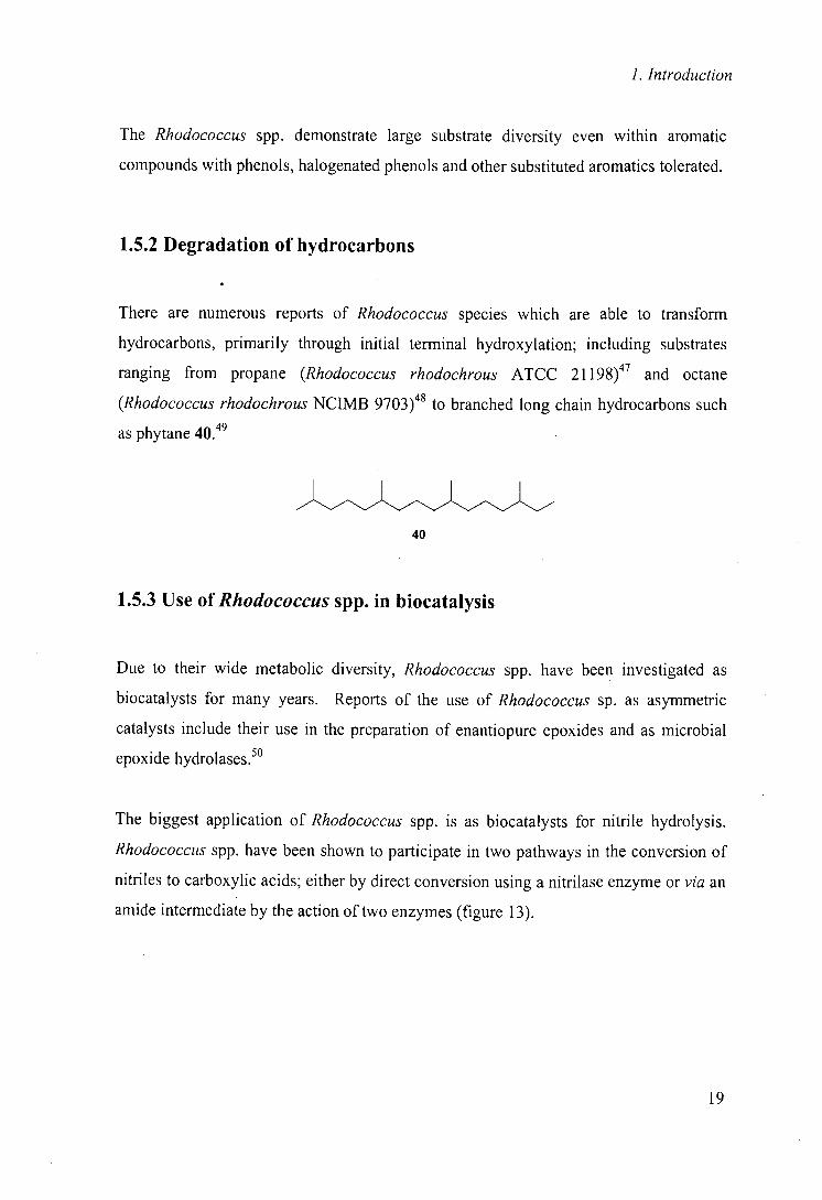
(Rhodococcus rhodochrous NCIMB 9703)48 to branched long chain hydrocarbons such
as phytane 40 49
40
1.5.3 Use of Rhodococcus spp. in biocatalysis
Due to their wide metabolic diversity, Rhodococcus spp. have been investigated as
biocatalysts for many years. Reports of the use of Rhodococcus sp. as asymmetric
catalysts include their use in the preparation of enantiopure epoxides and as microbial
epoxide hydrolases.5°
The biggest application of Rhodococcus spp. is as biocatalysts for nitrile hydrolysis.
Rhodococcus spp. have been shown to participate in two pathways in the conversion of
nitriles to carboxylic acids; either by direct conversion using a nitrilase enzyme or via an
amide intermediate by the action of two enzymes (figure 13).
Me
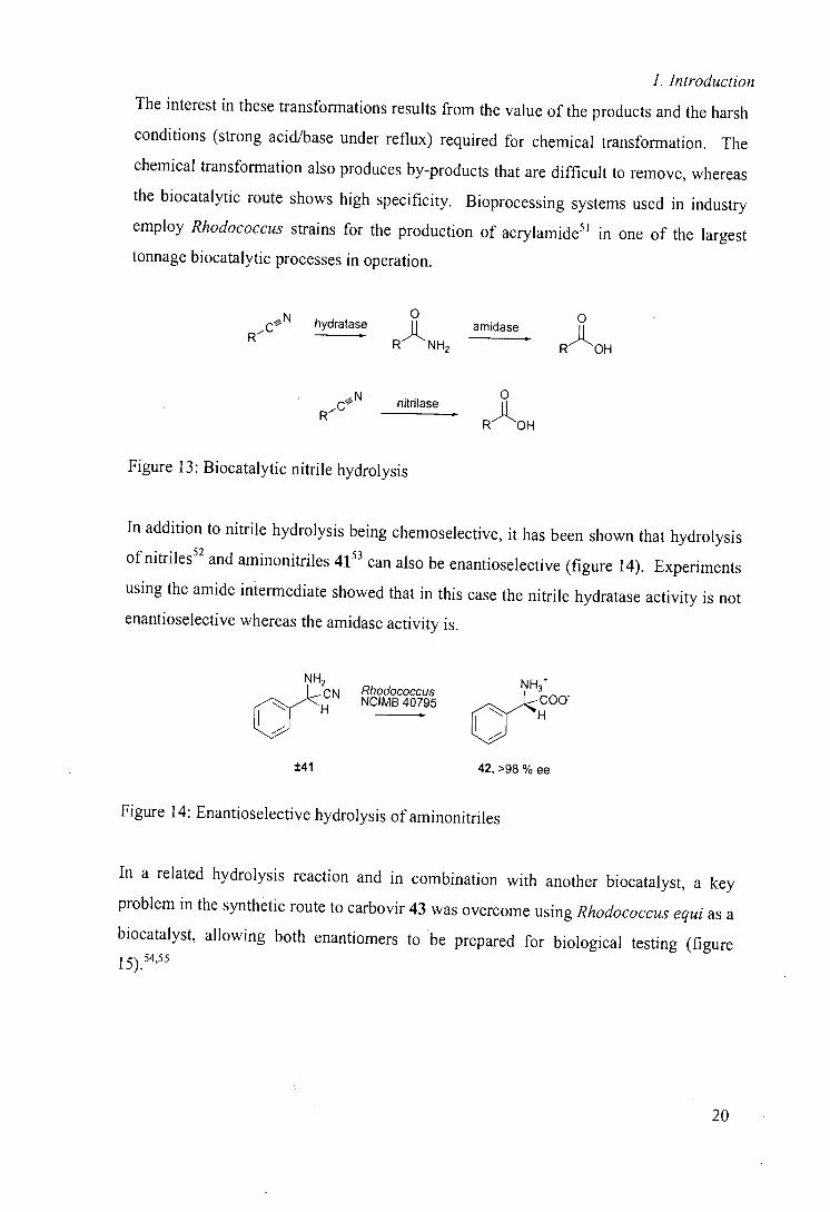
1. Introduction
The interest in these transformations results from the value of the products and the harsh
conditions (strong acid/base under reflux) required for chemical transformation. The
chemical transformation also produces by-products that are difficult to remove, whereas
the biocatalytic route shows high specificity. Bioprocessing systems used in industry
employ Rhodococcus strains for the production of acrylamide51 in one of the largest
tonnage biocatalytic processes in operation.
0 0 hydratase RNH2 amidase ROH
R
nitrilase 0
R RAOH
Figure 13: Biocatalytic nitrile hydrolysis
In addition to nitrile hydrolysis being chemoselective, it has been shown that hydrolysis
of nitriles52 and aminonitriles 4 153 can also be enantioselectjve (figure 14). Experiments
using the amide intermediate showed that in this case the nitrile hydratase activity is not
enantioselective whereas the amidase activity is.
NH2 NH3
cr JCN '0B8c,u9s5 coo
H
±41
42, >98 % ee
Figure 14: Enantioselective hydrolysis of aminonitriles
In a related hydrolysis reaction and in combination with another biocatalyst, a key
problem in the synthetic route to carbovir 43 was overcome using Rhodococcus equi as a
biocatalyst, allowing both enantiomers to . be prepared for biological testing (figure
I55 5)."
20
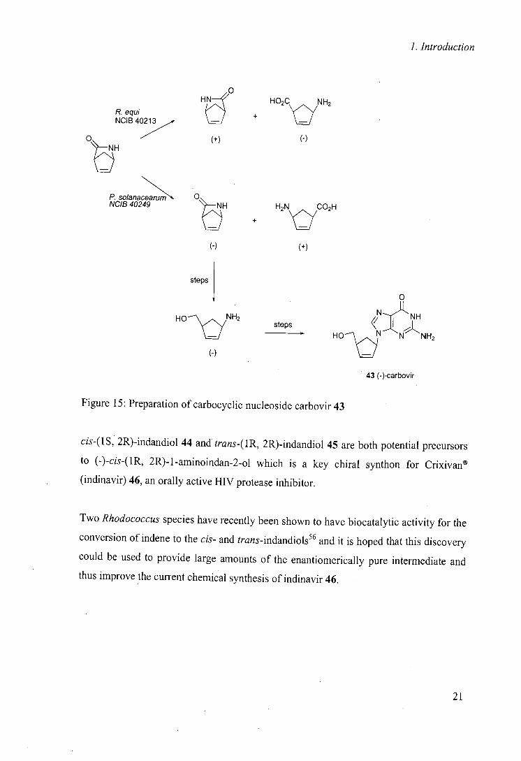
1. Introduction
HE
P. equi NCIB 40213
H02C
lc~
NH2 +
Ot7
(+) (-)
0 P. solanaceanim NCIB 40249 + H2N yCO2H
(-) (+)
steps
0
N:11 NH steps < ;j
(-)
H0\ S N N NH2
43 (-)-carbovir
Figure 15: Preparation of carbocyclic nucleoside carbovir 43
cis-(1S, 2R)-indandiol 44 and trans-(1R, 2R)-indandiol 45 are both potential precursors
to (-)-cis-(1R, 2R)-1-aminoindan-2.ol which is a key chiral synthon for Crixivan®
(indinavir) 46, an orally active HIV protease inhibitor.
Two Rhodococcus species have recently been shown to have biocatalytic activity for the
conversion of indene to the cis- and trans-indandiols56 and it is hoped that this discovery
could be used to provide large amounts of the enantiomerically pure intermediate and
thus improve the current chemical synthesis of indinavir 46.
21
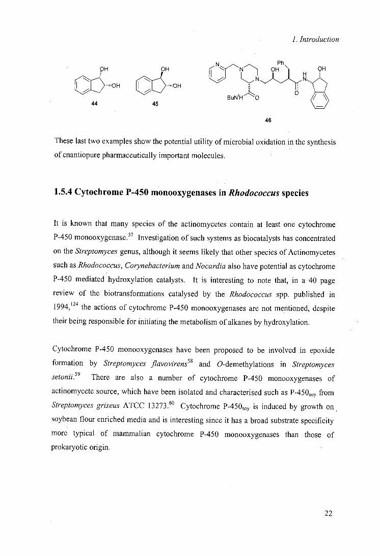
I. Introduction
N QH OH OH OH
Ph
"OH ' 'OH
BuNtHO 0
44 45
46
These last two examples show the potential utility of microbial oxidation in the synthesis
of enantiopure pharmaceutically important molecules.
1.5.4 Cytochrome P-450 monooxygenases in Rhodococcus species
It is known that many species of the actinomycetes contain at least one cytochrome
P-450 monooxygenase.57 Investigation of such systems as biocatalysts has concentrated
on the Streptomyces genus, although it seems likely that other species of Actinomycetes
such as Rhodococcus, Corynebacterium and Nocardia also have potential as cytochrome
P-450 mediated hydroxylation catalysts. It is interesting to note that, in a 40 page
review of the biotransformations catalysed by the Rhodococcus spp. published in
1994,124 the actions of cytochrome P-450 monooxygenases are not mentioned, despite
their being responsible for initiating the metabolism of alkanes by hydroxylation.
Cytochrome P-450 monooxygenases have been proposed to be involved in epoxide
formation by Streptomyces Jlavovirens58 and O-demethylations in Streptomyces
setonhi.59 There are also a number of cytochrome P-450 monooxygenases of
actinomycete source, which have been isolated and characterised such as P-450 0 . from
Streptomyces griseus ATCC 13273.60 Cytochrome P-450 0 is induced by growth on
soybean flour enriched media and is interesting since it has a broad substrate specificity
more typical of mammalian cytochrome P-450 monooxygenases than those of
prokaryotic origin.
22
1. Introduction
Although a number of Rhodococcus strains have been reported for their hydroxylating
abilities, some of these have been shown to have non-P-450 dependent hydroxylating
ability. An example of this is Rhodococcus erythropolis,27 where a molybdoenzyme is
thought to be responsible for hydroxylation of pyrimidine heterocycles as determined
through inhibition studies using known inhibitors. There are however increasing
numbers of cytochrome P-450 monooxygenases implicated in hydroxylation by
Rhodococcus spp.
A single cytochrome P-450 monooxygenase system has recently been implicated in the
degradation of herbicides such as atrazine by Rhodococcus sp. strain N186/21 and has
been suggested as a good target for bioremediation.61'62
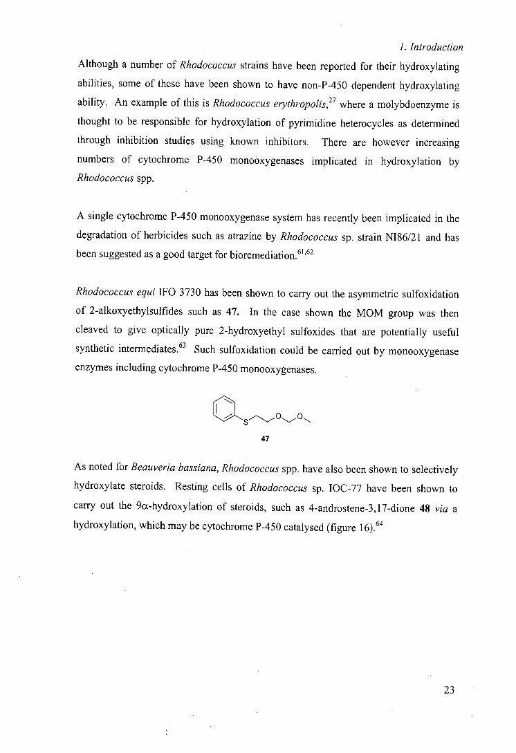
Rhodococcus equi IFO 3730 has been shown to carry out the asymmetric sulfoxidation
of 2-alkoxyethylsulfides such as 47. In the case shown the MOM group was then
cleaved to give optically pure 2-hydroxyethyl sulfoxides that are potentially useful
synthetic intermediates. 63 Such sulfoxidation could be carried out by monooxygenase
enzymes including cytochrome P-450 monooxygenases.
47
As noted for Beauveria bassiana, Rhodococcus spp. have also been shown to selectively
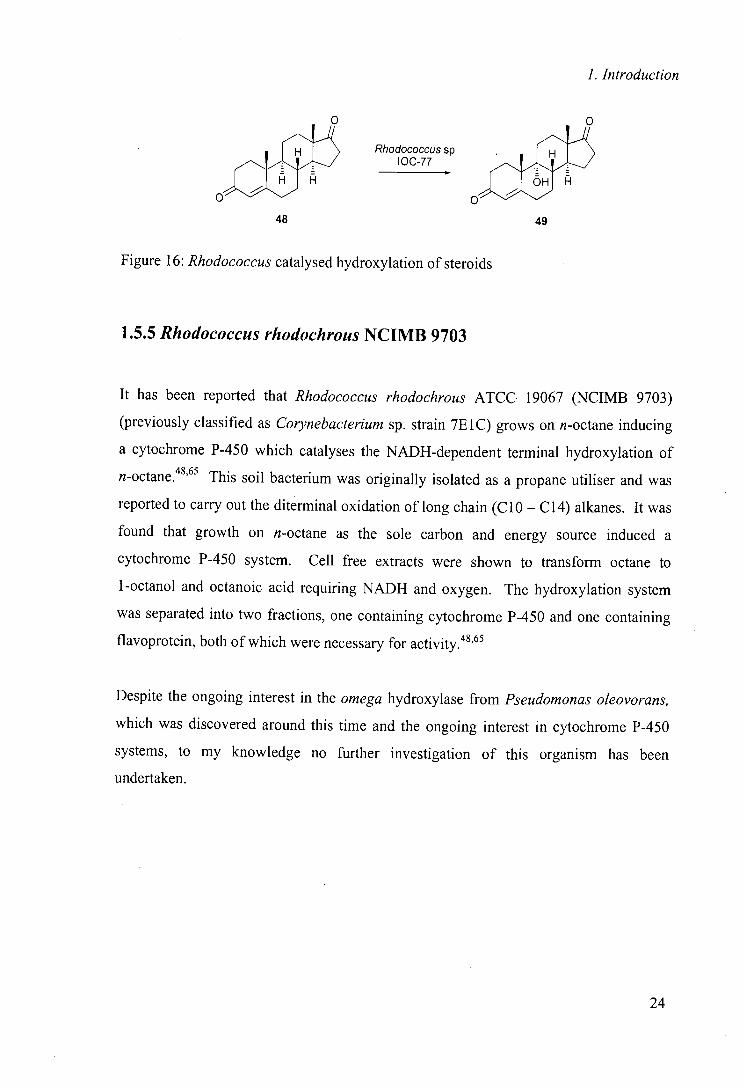
hydroxylate steroids. Resting cells of Rhodococcus sp. IOC-77 have been shown to
carry out the 9(x-hydroxylation of steroids, such as 4-androstene-3, 1 7-dione 48 via a 64 hydroxylation, which may be cytochrome P-450 catalysed (figure 16).
23
1. Introduction
Rhodococcussp
oA
48 49
Figure 16: Rhodococcus catalysed hydroxylation of steroids
1.5.5 Rhodococcus rhodochrous NUMB 9703
It has been reported that Rhodococcus rhodochrous ATCC 19067 (NCIMB 9703)
(previously classified as Corynebacterium sp. strain 7E1C) grows on n-octane inducing
a cytochrome P-450 which catalyses the NADH-dependent terminal hydroxylation of
n-octane.48'65 This soil bacterium was originally isolated as a propane utiliser and was
reported to carry out the diterminal oxidation of long chain (C 10 - C14) alkanes. It was
found that growth on n-octane as the sole carbon and energy source induced a
cytochrome P-450 system. Cell free extracts were shown to transform octane to
1-octanol and octanoic acid requiring NADH and oxygen. The hydroxylation system
was separated into two fractions, one containing cytochrome P-450 and one containing
flavoprotein, both of which were necessary for activity.48'65
Despite the ongoing interest in the omega hydroxylase from Pseudomonas oleovorans,
which was discovered around this time and the ongoing interest in cytochrome P-450
systems, to my knowledge no further investigation of this organism has been
undertaken.
24
1. Introduction
1.5.6 Rhodococcus sp. NUMB 9784
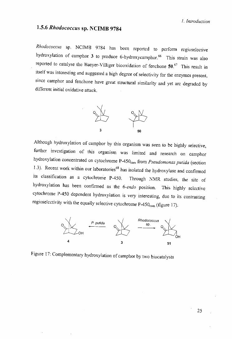
Rhodococcus sp. NCIMB 9784 has been reported to perform regioselective
hydroxylation of camphor 3 to produce 6-hydroxycamphor.66 This strain was also
reported to catalyse the Baeyer-Villiger biooxidation of fenchone 50.67 This result in
itself was interesting and suggested a high degree of selectivity for the enzymes present,
since camphor and fenchone have great structural similarity and yet are degraded by
different initial oxidative attack.
Although hydroxylation of camphor by this organism was seen to be highly selective,
further investigation of this organism was limited and research on camphor
hydroxylation concentrated on cytochrome 450cam from Pseudomonas putida (section 1.3). Recent work within our laboratories 18
has isolated the hydroxylase and confirmed
its classification as a cytochrome P-450. Through NMR studies, the site of
hydroxylation has been confirmed as the 6-endo position. This highly selective
cytochrome P-450 dependent hydroxylation is very interesting, due to its contrasting
regioselectivity with the equally selective cytochrome P450cam (figure 17).
P. fida _ SP. Rhodococcus
O
OH OH
4
3 51
Figure 17: Complementary hydroxylation of camphor by two biocatalysts
25
1. Introduction
This complementary regioselectivity makes the camphor utilising cytochrome P-450 in Rhodococcus sp., an interesting target in the search for more information regarding the
origin of selectivity of transformation by cytochrome P-450 monooxygenases.
Another cytochrome P-450 of Actinomycete source, cytochrome P-450 0 from Streptomyces griseus was also shown to catalyse the hydroxylation of camphor. It is
noted that, in this case, the regioselectivity is much lower with four products isolated.69
This lower regioselectivity of hydroxylation is similar to the regioselectivity of
hydroxylation of 450LM2 from rabbit liver where 5-exo, 3-endo, 5-endo and other
minor hydroxycamphor products were produced, showing an example of the similarity
of eukaryotic cytochrome P-450 0 to its mammalian counterparts .70
Perhaps surprisingly, with the implication of cytochrome P-450 monooxygenases in hydroxylations by Rhodococcus sp., very few have been isolated and identified. One
example is the identification of two cytochrome P-450 monooxygenases in Rhodococcus rhodochrous strain 116 which degrade aromatic ethers. These P-450's are induced by
2-ethoxyphenol and 4-methoxybenzoate respectively and the compounds inducing the
expression of each P-450 binds specifically to that enzyme, suggesting a narrow
substrate specificity.7' Cytochrome P-450,, (induced by and binder of 2-ethoxyphenol)
has since been purified to apparent homogeneity and fully characterised as a cytochrome
P-450.72
1.6 Summary
For this brief review of some of the key literature, it should be clear to the reader that
biohydroxylations are an important class of biocatalytic reaction and are likely to remain
so for the foreseeable future. The key to this importance is the high selectivity that the
enzyme catalyst imparts to the transformation and that there is, to date, no easily or
widely acceptable chemical alternative.
26
1. Introduction
One of the key questions in the field is the prediction .of the site and selectivity of
hydroxylation. This issue is being addressed particularly with the Beauveria bassiana
hydroxylase system(s) based on substrate studies, with some success; further success is
likely to depend on the isolation of the enzymes involved.
Although attempts have been made to utilise microbial hydroxylation systems as models
for mammalian metabolism, it has been shown that while there are often commonalities
between the microbial and mammalian systems, they have been found to be unreliable
and at times can be misleading.'9
The most important application of biocatalysis in pharmaceutical research is the use of
these organisms to overcome synthetic problems and allow the production of selectively
hydroxylated drug molecules. Such hydroxylated compounds are extremely important
for use in identifying mammalian metabolites and for subsequent testing as a metabolite.
Biocatalysis is also extremely important in the production of key chiral intermediates for
synthesis.
1.7 Aims
The aims of this project were two fold. Firstly we were interested in carrying out a
systematic study into the hydroxylation of a series of structurally related compounds by
the well-studied Beauveria bassiana to investigate the impact of structure modifications
on the regioselectivity of hydroxylation.
Secondly we wanted to investigate the hitherto undetermined cytochrome P-450
dependent hydroxylation potential of a number of Actinomycetes belonging to the
Rhodococcus genus. Furthermore we wanted to investigate the scope and selectivity of
any activities identified.
27
1. Introduction
Combination of this re-investigation of a previously utilised biocatalyst with' the
investigation of new biocatalytic activities was hoped to yield more information
regarding cytochrome P-450 selectivity and perhaps identify routes to useful chiral
intermediates for use in further synthesis.
2. Beauveria bassiana ATCC 7159
2 Biotransformations using Beauveria bassiana ATCC
715973
2.1 Aims
Beauveria bassiana ATCC 7159, as discussed in the introduction, is used widely as a
whole cell biocatalyst for hydroxylation. Attempts to model the substrate/active site of
the hydroxylase(s) have allowed prediction of the regioselectivity of hydroxylation with
some success. As indicated in the discussion of the active site models, the important
factor in determining the site of hydroxylation has always been assumed to be the
relative position of the electron rich moiety. Although apparently not necessary for
regioselective hydroxylation, the presence of an aromatic moiety in the substrate
molecule is noted in many good substrates for the hydroxylase(s). As an extension of
the active site models, we were interested in the importance of this aromatic side chain,
in particular on the regioselectivity of hydroxylation.
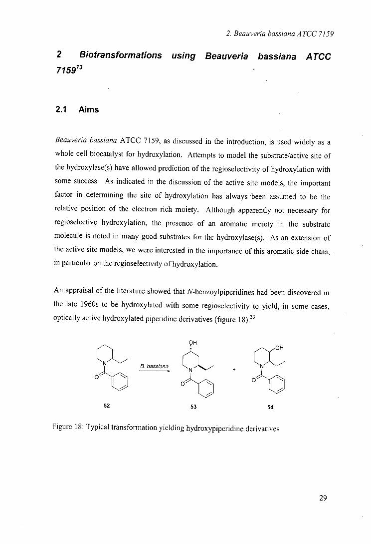
An appraisal of the literature showed that N-benzoylpiperidines had been discovered in
the late 1960s to be hydroxylated with some regioselectivity to yield, in some cases,
optically active hydroxylated piperidine derivatives (figure 1
OH
(N B. bassiana
O'D CN
52
53
54
Figure 18: Typical transformation yielding hydroxypiperidine derivatives
29
2. Beauverja bassiana ATCC 7159
It has been noted previously that seemingly minor changes in substrate structure can
have a significant effect on the selectivity of hydroxylation. We envisaged that the
importance of the aromatic group could be investigated by the examination of analogous
compounds to those previously studied retaining the N-heterocycle but incorporating a
different aromatic side chain.
Another important factor in our investigations was the utility of the hydroxylated
products, since we wanted not only to gain further information on the structural
requirements required for selectivity, but to investigate the potential for this biocatalytic
route as a means of accessing selectively hydroxylated compounds. Important to this
utility is the ease of removal of the aromatic side chain. Although the
N-benzoylpiperidjne transformations could be considered a potential route to
hydroxylated piperidines, subsequent removal of the N-benzoyl group is not trivial.
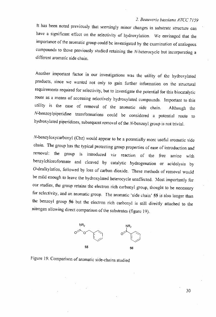
N-benzyloxycarbonyl (Cbz) would appear to be a potentially more useful aromatic side
chain. The group has the typical protecting group properties of ease of introduction and
removal: the aroun is intrndiiiw f -
benzylchloroformate and cleaved by catalytic hydrogenation or acidolysis by
O-dealky!ation, followed by loss of carbon dioxide. These methods of removal would
be mild enough to leave the hydroxylated heterocycle unaffected. Most importantly for
our studies, the group retains the electron rich carbonyl group, thought to be necessary
for selectivity, and an aromatic group. The aromatic 'side chain' 55 is also longer than the benzoyl group 56 but the electron rich carbonyl is still directly attached to the
nitrogen allowing direct comparison of the substrates (figure 19).
NR2 NR2
0 o
55 56
Figure 19: Comparison of aromatic side-chains studied
30
2. Beauveria bassiana ATCC 7159
The third point under investigation is the level of substitution around the heterocycle. In
Johnson's work, a number of alkyl substituents around the heterocycle were tolerated;
indeed some of these alkyl side chains were actually hydroxylated. We wanted to
investigate the incorporation of such substituents and their effect on the selectivity of
hydroxylation.
In summary, our aims were three-fold; namely the investigation of the effect of a longer
aromatic side chain with different properties to that previously investigated, the effect of
introducing alkyl substituents around the heterocycle and the use of an easily removed
protecting group to direct hydroxylation.
A range of N-Cbz protected alkyl piperidines were prepared from the corresponding
commercially available alkyl piperidines by reaction with benzylchloroformate.74
2.2 Methods
In common with the literature, these transformations were carried out using growing
cells. Since the hydroxylases in B. bassiana are thought to be constitutively expressed,
growth was on glucose/corn steep solids media with no attempted induction. The
substrate was added as a concentrated solution in ethanol to a final concentration of 0.1
mg/ml. This low substrate tolerance is often regarded as one of the main disadvantages
of whole-cell methodology as, in this case, conversion of 250 mg required extraction of 2.5 L of aqueous phase. This procedure was not optimised although it was based heavily
on literature methods. 34,75
31
2. Beauveria bassiana ATCC 7159
2.3 Results
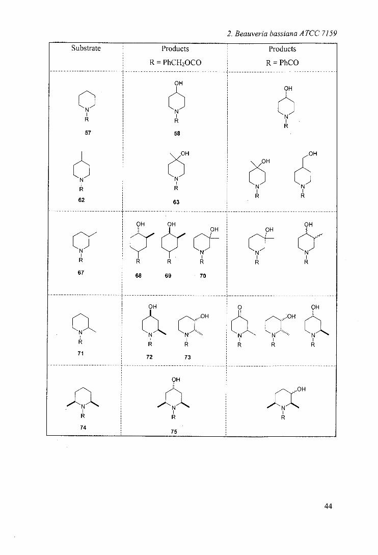
Since the initial part of this work has been reported previously, 74 little detail will be
given here, but the results are included to allow a complete comparison of results. The
results of the biohydroxylations are summarised in table 2 (page 44) along with a
comparison with results obtained using N-benzoyl analogues. 33 NMR experiments to
ascertain the relative stereochemistry have not been reported previously.
Incubation of N-Cbz-piperidine 5774
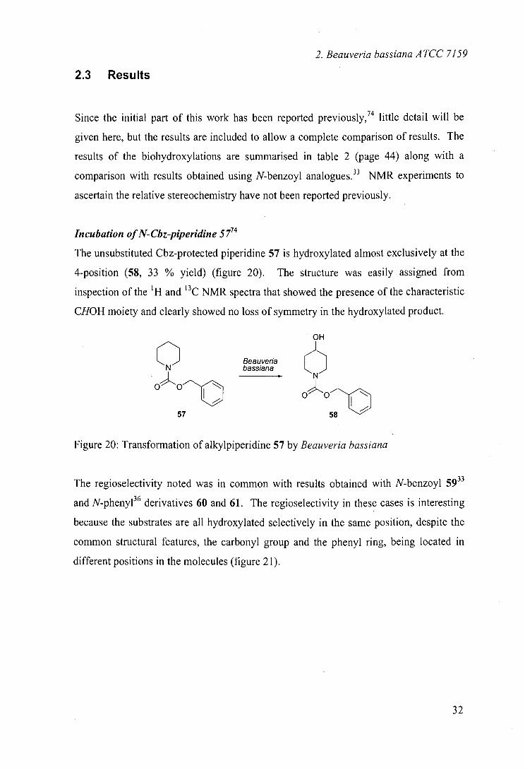
The unsubstituted Cbz-protected piperidine 57 is hydroxylated almost exclusively at the
4-position (58, 33 % yield) (figure 20). The structure was easily assigned from
inspection of the 'H and ' 3C NMR spectra that showed the presence of the characteristic
CHOH moiety and clearly showed no loss of symmetry in the hydroxylated product.
0 Beauveria N bassiana
57
Figure 20: Transformation of alkylpiperidine 57 by Beauveria bassiana
The regioselectivity noted was in common with results obtained with N-benzoyl 5933
and N-phenyl36 derivatives 60 and 61. The regioselectivity in these cases is interesting
because the substrates are all hydroxylated selectively in the same position, despite the
common structural features, the carbonyl group and the phenyl ring, being located in
different positions in the molecules (figure 21).
32
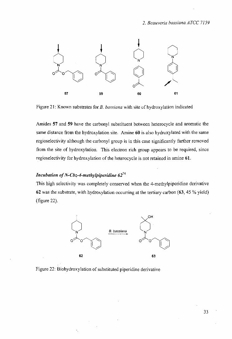
2. Beauveria bassiana ATCC 7159
QQ OOf o a a
57 59 60 61
Figure 21: Known substrates for B. bassiana with site of hydroxylation indicated
Amides 57 and 59 have the carbonyl substituent between heterocycle and aromatic the
same distance from the hydroxylation site. Amine 60 is also hydroxylated with the same
regioselectivity although the carbonyl group is in this case significantly further removed
from the site of hydroxylation. This electron rich group appears to be required, since
regioselectivity for hydroxylation of the heterocycle is not retained in amine 61.
Incubation of N-Oz-4-methylpiperidine 62 74
This high selectivity was completely conserved when the 4-methylpiperidine derivative
62 was the substrate, with hydroxylation occurring at the tertiary carbon (63, 45 % yield)
(figure 22).
OH
6 N B. bassiana N
oOm oo
62 63
Figure 22: Biohydroxylation of substituted piperidine derivative
33
2. Beauverja bassiana A TCC 7159
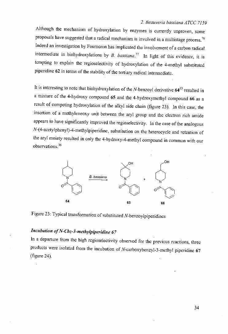
Although the mechanism of hydroxylation by enzymes is currently unproven, some
proposals have suggested that a radical mechanism is involved in a multistage process. 16
Indeed an investigation by Fourneron has implicated the involvement of a carbon radical
intermediate in biohydroxylations by B. bassiana.77 In light of this evidence, it is
tempting to explain the regioselectivity of hydroxylation of the 4-methyl substituted
piperidine 62 in terms of the stability of the tertiary radical intermediate.
It is interesting to note that biohydroxylation of the N-benzoyl derivative 64 33 resulted in
a mixture of the 4-hydroxy compound 65 and the 4-hydroxymethyl compound 66 as a
result of competing hydroxylation of the alkyl side chain (figure 23). In this case, the
insertion of a methyleneoxy unit between the aryl group and the electron rich amide
appears to have significantly improved the regioselectivity. In the case of the analogous
N-(4 acetylpheny1)4methy1pjperidine substitution on the heterocycle and retention of
the aryl moiety resulted in only the 4-hydroxy-4-methyl compound in common with our
observations. 36
6 OH OH
C N B. bassiana + 6 o O
64 65 66
Figure 23: Typical transformation of substituted N-benzoylpiperidines
Incubation of N-Cbz-3-methylpiperja'jne 67
In a departure from the high regioselectivity observed for the previous reactions, three
products were isolated from the incubation of N-carboxybenzyl-3methyl piperidine 67
(figure 24).
34
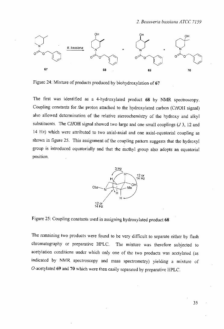
2. Beauveria bassiana ATCC 7159
OH OH OH
CN B. bassiana + ~N? +
0 0"~c oo OO
c 01-11
67 68 69 70
Figure 24: Mixture of products produced by biohydroxylation of 67
The first was identified as a 4-hydroxylated product 68 by NMR spectroscopy.
Coupling constants for the proton attached to the hydroxylated carbon (CHOH signal)
also allowed determination of the relative stereochemistry of the hydroxy and alkyl
substituents. The CHOH signal showed two large and one small couplings (J 3, 12 and
14 Hz) which were attributed to two axial-axial and one axial-equatorial coupling as
shown in figure 25. This assignment of the coupling pattern suggests that the hydroxyl
group is introduced equatorially and that the methyl group also adopts an equatorial
position.
3 Hz
12 or H/ 14 Hz
OH CbzN Me
12 or 14 Hz
Figure 25: Coupling constants used in assigning hydroxylated product 68
The remaining two products were found to be very difficult to separate either by flash
chromatography or preparative HPLC. The mixture was therefore subjected to
acetylation conditions under which only one of the two products was acetylated (as
indicated by NMR spectroscopy and mass spectrometry) yielding a mixture of
O-acetylated 69 and 70 which were then easily separated by preparative HPLC.
35
2. Beauveria bassiana ATCC 7159
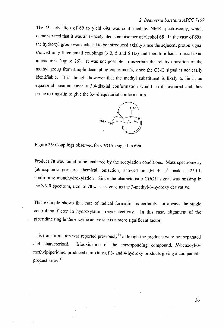
The O-acetylation of 69 to yield 69a was confirmed by NMR spectroscopy, which
demonstrated that it was an O-acetylated stereoisomer of alcohol 68. In the case of 69a,
the hydroxyl group was deduced to be introduced axially since the adjacent proton signal
showed only three small couplings (J 3, 5 and 5 Hz) and therefore had no axial-axial
interactions (figure 26). It was not possible to ascertain the relative position of the
methyl group from simple decoupling experiments, since the C3-H signal is not easily
identifiable. It is thought however that the methyl substituent is likely to lie in an
equatorial position since a 3,4-diaxial conformation would be disfavoured and thus
prone to ring-flip to give the 3,4-diequatorial conformation.
Hfl
Cbz N
Me
Figure 26: Couplings observed for CHOAc signal in 69a
Product 70 was found to be unaltered by the acetylation conditions. Mass spectrometry
(atmospheric pressure chemical ionisation) showed an (M + 1) peak at 250. 1,
confirming monohydroxylation. Since the characteristic CHOH signal was missing in
the NMR spectrum, alcohol 70 was assigned as the 3-methyl-3-hydroxy derivative.
This example shows that ease of radical formation is certainly not always the single
controlling factor in hydroxylation regioselectivity. In this case, alignment of the
piperidine ring in the enzyme active site is a more significant factor.
This transformation was reported previously 74 although the products were not separated
and characterised. Biooxidation of the corresponding compound, N-benzoyl-3-
methylpiperidine, produced a mixture of 3- and 4-hydroxy products giving a comparable
product array. 33
36
2. Beauveria bassiana ATCC 7159
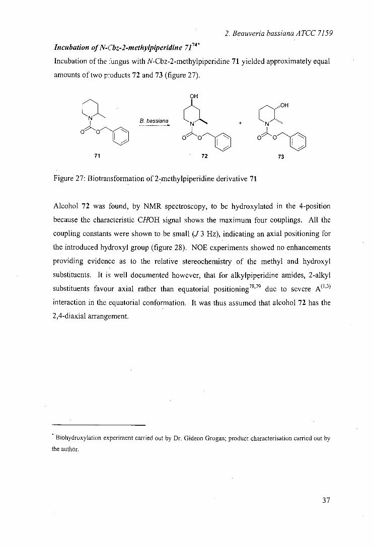
Incubation of N-Cbz-2-methylpiperidine 7174*
Incubation of the fungus with N-Cbz-2-methylpiperidine 71 yielded approximately equal
amounts of two p-oducts 72 and 73 (figure 27).
a.N B. bassiana
71
Figure 27: B iotrans formation of 2-methylpiperidine derivative 71
Alcohol 72 was found, by NMR spectroscopy, to be hydroxylated in the 4-position
because the characteristic CHOH signal shows the maximum four couplings. All the
coupling constants were shown to be small (J 3 Hz), indicating an axial positioning for
the introduced hydroxyl group (figure 28). NOE experiments showed no enhancements
providing evidence as to the relative stereochemistry of the methyl and hydroxyl
substituents. It is well documented however, that for alkylpiperidine amides, 2-alkyl
substituents favour axial rather than equatorial positioning 78,79 due to severe A"3
interaction in the equatorial conformation. It was thus assumed that alcohol 72 has the
2,4-diaxial arrangement.
Biohydroxylation experiment carried out by Dr. Gideon Grogan; product characterisation carried out by
the author.
37
2. Beauveria bassiana ATCC 7159
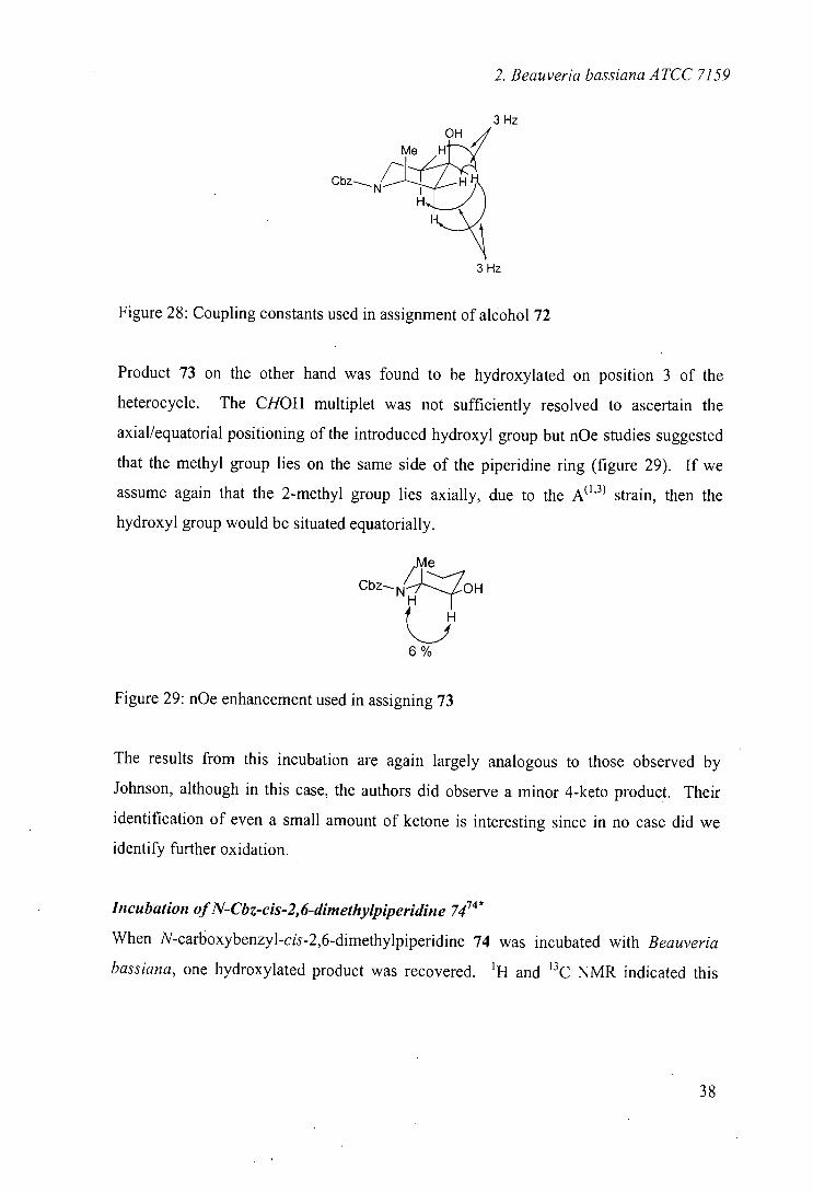
3 Hz
Cbz
3 Hz
Figure 28: Coupling constants used in assignment of alcohol 72
Product 73 on the other hand was found to be hydroxylated on position 3 of the
heterocycle. The CHOH multiplet was not sufficiently resolved to ascertain the
axial/equatorial positioning of the introduced hydroxyl group but nOe studies suggested
that the methyl group lies on the same side of the piperidine ring (figure 29). If we
assume again that the 2-methyl group lies axially, due to the A'3 strain, then the
hydroxyl group would be situated equatorially.
Me
Cbz. N ,LZoH
~j 6%
Figure 29: nOe enhancement used in assigning 73
The results from this incubation are again largely analogous to those observed by
Johnson, although in this case, the authors did observe a minor 4-keto product. Their
identification of even a small amount of ketone is interesting since in no case did we
identify further oxidation.
Incubation of N-Gbz-cis-2, 6-dimethylpiperidine 7474 *
When N-carboxybenzyl-cis-2,6-dimethylpiperjdjne 74 was incubated with Beau veria
bassiana, one hydroxylated product was recovered. 'H and 13C NMR indicated this
NEZ
2. Beauveria baiana ATCC 7159
compound to be the 4-hydroxy derivative 75 (figure 30), this pro.:uct has relative
configuration 2,6-cis-4-trans (again assuming that the 2-alkyl groups adopt an axial
positioning) (figure 31).
,JIII1%.... B. bassiana
oo
74 75
Figure 30: Conversion of 2,6-dimethylpiperidine 74
4 Hz 11 Hz
Me H 4 H Me H
C b z N2 H 11 Hz
Figure 31: Coupling constants used in assignment of 75
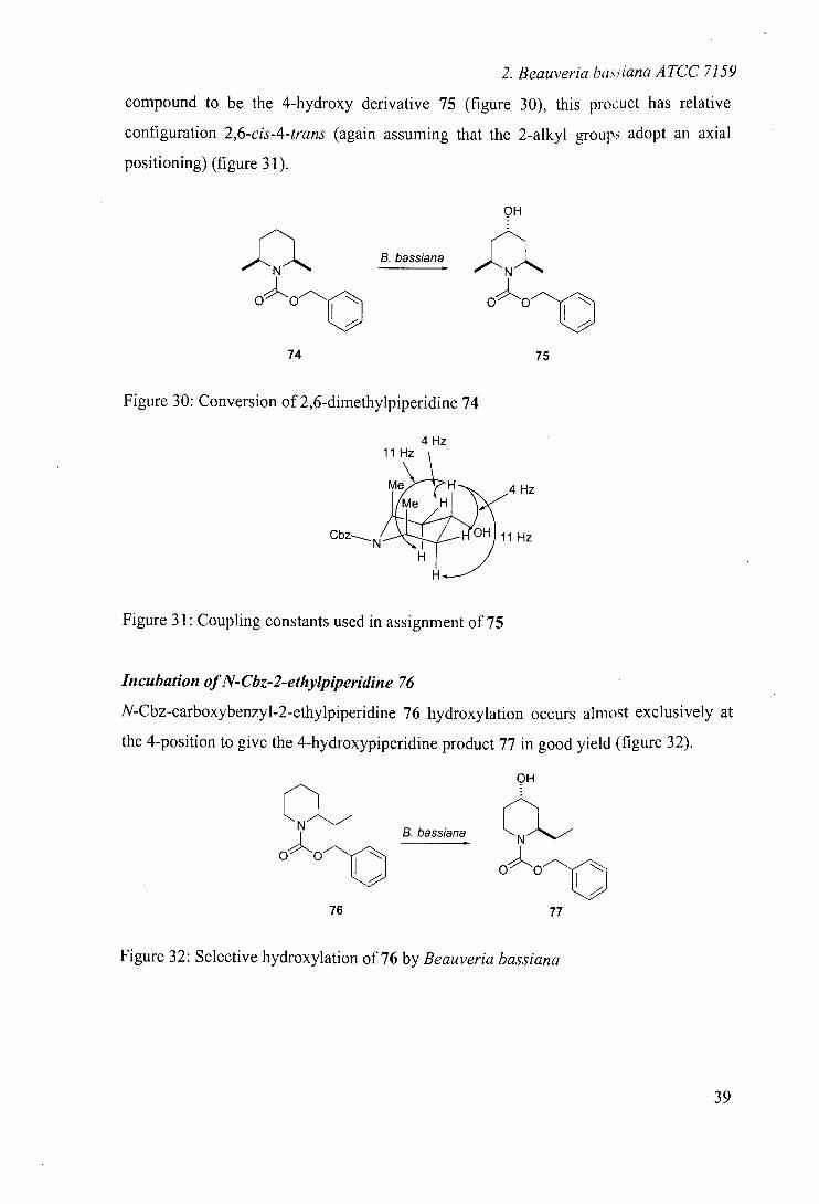
Incubation of N-Cbz-2-ethylpiperidine 76
N-Cbz-carboxybenzyl-2-ethylpiperidine 76 hydroxylation occurs almost exclusively at
the 4-position to give the 4-hydroxypiperidine product 77 in good yield (figure 32).
76
B. bassiana
Figure 32: Selective hydroxylation of 76 by Beauveria bassiana
39
2. Beauveria bassiana ATCC 7159
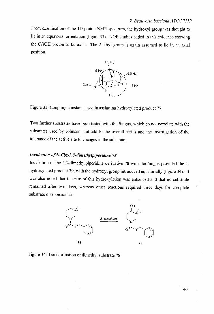
From examination of the 1D proton NMR spectrum, the hydroxyl group was thought to
lie in an equatorial orientation (figure 33). NOE studies added to this evidence showing
the CHOH proton to be axial. The 2-ethyl group is again assumed to lie in an axial
position.
4.5 Hz
11.5
-.
Hz Et H
Cbz HOH 11.5 Hz
Figure 33: Coupling constants used in assigning hydroxylated product 77
Two further substrates have been tested with the fungus, which do not correlate with the
substrates used by Johnson, but add to the overall series and the investigation of the
tolerance of the active site to changes in the substrate.
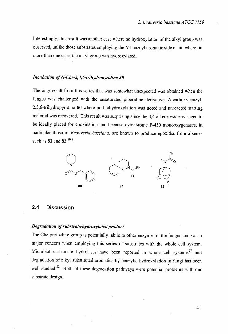
Incubation of N-Cbz-3,3-dimethylpiperidine 78
Incubation of the 3,3-dimethylpiperidine derivative 78 with the fungus provided the 4-
hydroxylated product 79, with the hydroxyl group introduced equatorially (figure 34). It
was also noted that the rate of this hydroxylation was enhanced and that no substrate
remained after two days, whereas other reactions required three days for complete
substrate disappearance.
OH
-f CN B. bassiana
Nf
op"OO
OO
78 79
Figure 34: Transformation of dimethyl substrate 78
2. Beauveria bassiana ATCC 7159
Interestingly, this result was another case where no hydroxylation of the alkyl group was
observed, unlike those substrates employing the N-benzoyl aromatic side chain where, in
more than one case, the alkyl group was hydroxylated.
Incubation of N-Cbz-2,3, 6-trihydropyridine 80
The only result from this series that was somewhat unexpected was obtained when the
fungus was challenged with the unsaturated piperidine derivative, N-carboxybenzyl-
2,3,6-trihydropyridine 80 where no biohydroxylation was noted and unreacted starting
material was recovered. This result was surprising since the 3,4-alkene was envisaged to
be ideally placed for epoxidation and because cytochrome P-450 monooxygenases, in
particular those of Beauveria bassiana, are known to produce epoxides from alkenes
such as 81 and 82.80,81
Ph
N
Ph
0 A 81 82
2.4 Discussion
Degradation of substrate/hydroxylated product
The Cbz-protecting group is potentially labile to other enzymes in the fungus and was a
major concern when employing this series of substrates with the whole cell system.
Microbial carbamate hydrolases have been reported in whole cell systems27 and
degradation of alkyl substituted aromatics by benzylic hydroxylation in fungi has been
well, studied.82 Both of these degradation pathways were potential problems with our
substrate design.
41
2, Beauveria bassiana ATCC 7159
The only case in which these problems arose was N-Cbz-2,6-dimethylpiperidine 74
where the hydroxylated product 75 was isolated in very low yield (-.40 mg from 270 mg
substrate), but a large amount of benzoic acid was also recovered (90 mg). The
isolation of benzoic acid suggests some sort of benzylic hydroxylation, either of the
starting material or of the hydroxylated product. Such a degradative mechanism would
not be possible in the N-benzoyl case and is reflected in the higher yield reported.
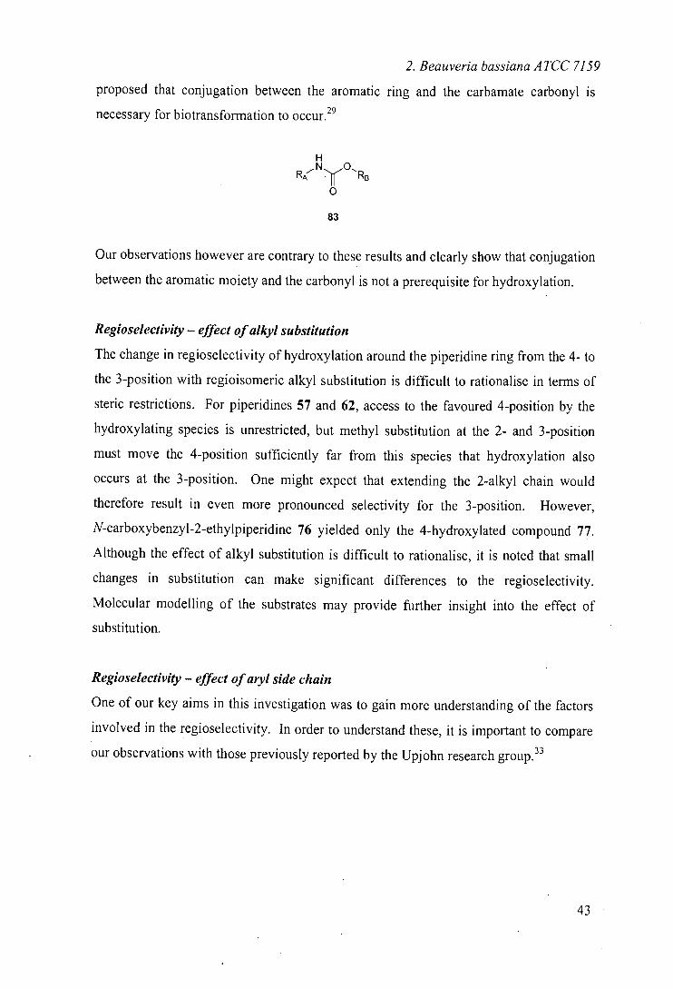
The yields of product recorded in these transformations are between 5 - 4.8 %, which is
very respectable for this type of reaction (table l).333536 Unlike many literature
examples, no starting material was recovered along with the products after three days
incubation. It is likely that the mass balance of the conversions (greater than 50 % of
substrate unaccounted for) is lost through degradative mechanisms.
Substrate Substituent Yieldt
57 None 33
62 . 4-methyl 45
67 3-methyl 24
71 2-methyl 14
74 2,6-dimethyl 5
76 2-ethyl 45
78 3,3-dimethy! 48
Table 1: Yields of biotransformations reported for substituted N-Cbz-piperidines
It has previously been reported that the inclusion of 'inverted' urethane linkers in
substrates for Beauveria bassiana led to no biotransformation whatsoever; substrates of
structure 83 where RA is aryl and RB alkyl are successfully hydroxylated whereas the
'inverted linker' where RA is alkyl and RB is aryl are not. Based on these results, it was
Combined yields taken before separation of hydroxylated products
42
2. Beauveria bassiana ATCC 7159
proposed that conjugation between the aromatic ring and the carbamate carbonyl is
necessary for biotransformation to occur. 29
RA N1O RB
83
Our observations however are contrary to these results and clearly show that conjugation
between the aromatic moiety and the carbonyl is not a prerequisite for hydroxylation.
Regioselectivity - effect of alkyl substitution
The change in regioselectivity of hydroxylation around the piperidine ring from the 4- to
the 3-position with regioisomeric alkyl substitution is difficult to rationalise in terms of
steric restrictions. For piperidines 57 and 62, access to the favoured 4-position by the
hydroxylating species is unrestricted, but methyl substitution at the 2- and 3-position
must move the 4-position sufficiently far from this species that hydroxylation also
occurs at the 3-position. One might expect that extending the 2-alkyl chain would
therefore result in even more pronounced selectivity for the 3-position. However,
N-carboxybenzyl-2-ethylpiperidine 76 yielded only the 4-hydroxylated compound 77.
Although the effect of alkyl substitution is difficult to rationalise, it is noted that small
changes in substitution can make significant differences to the regioselectivity.
Molecular modelling of the substrates may provide further insight into the effect of
substitution.
Regioselectivity - effect of aryl side chain
One of our key aims in this investigation was to gain more understanding of the factors
involved in the regioselectivity. In order to understand these, it is important to compare
our observations with those previously reported by the Upjohn research group.33
43
2. Beauveria bassiana ATCC 7159
2. Beauveria bassiana ATCC 7159
Substrate Products Products
R = PhCH2000 R = PhCO
OH OH
N Et
OH
77
Et Et N Et
76
Table 2: Results of biohydroxylations of Cbz-alkyl piperidines including a comparison
with the results obtained from the N-benzoyl analogues33
The results obtained are compared in table 2 and can be summarised:
Cbz series shows selectivity for 4- position, whereas benzoyl series
shows product mixtures in most cases.
Cbz series shows no side chain hydroxylation - common in benzoyl
series
Cbz series shows no further oxidation to ketone - noted in benzoyl series
To compare and draw tentative conclusions from these observations, we have to assume
that the same hydroxylase enzyme within the whole cell system carries out all the
biohydroxylations reported. Although there is clearly more than one active hydroxylase
in the system, the assumption is thought to be valid given the close structural similarities
of the substrates.
With this assumption in mind, it is difficult to explain the improvement in
regioselectivity realised on examination of the Cbz alkylpiperidine series solely on the
basis of the current active site model. Even in the updated models, it is assumed that the
key restraint on the site of hydroxylation is the distance between the electron rich moiety
and the hydroxylation site.
45
2. Beauveria bassiana ATCC 7159
The hydroxylase is clearly now disposed to hydroxylation in the 4-position, except
where there is a 2- or 3-methyl substituent when the selectivity is compromised,
suggesting that the active site of the Beauveria hydroxylating system is very sensitive to
relatively small changes in the nature of the protecting group. The distance from the
carbonyl group to the putative site of hydroxylation is, of course, unchanged from N-
benzoyl to N-benzyloxycarbonyl. Our results would suggest that the more critical
characteristic for selective hydroxylation is the distance from the carbonyl to the
aromatic binding pocket.
In addition, greater flexibility of the N-carboxybenzyl molecule is afforded by rotation
around the benzylic centre. This would appear to allow improved accommodation by
the active site and perhaps greater regioselectivity for some substrates.
We propose that there is a defined aromatic binding pocket within the active site of the
hydroxylase and that interaction of the substrate with the enzyme is affected sufficiently
for the hydroxylation site to be no longer controlled solely by the site of the electron rich
moiety in the substrate molecule.
Stereochemistry of hydroxylation
It has been extensively reported that, in rigid systems, the hydroxyl group is introduced
trans with respect to the amide functional group. 35,81,83,84 By the use of proton NMR
analysis, we have established that the hydroxyl group is introduced into the equatorial
position in most cases. The exceptions are the 2-methyl-4-hydroxyl product 72 where
the hydroxyl group was seen to lie in an axial position (this was shown by the lack of an
nOe enhancement between the axial C(6)H and the C(4)H and also from the coupling
constants of the CHOH signal, showing no axial-axial coupling) and the 3-methyl-4-
hydroxyl product (the acetylated derivative of which was isolated 69a) where the
CHOAc signal showed no axial-axial coupling. Our results therefore show a clear
2. Beauveria bassiana ATCC 7159 preference for hydroxylation trans to electron rich carbamate. It is unclear whether such
selectivity exists in the N-benzoyl series due to assignment difficulties.
Stereoselectivity of hydroxylation
Unusually for flexible substrates, Johnson and co-workers reported some
enantiodifferentiation by the Beauveria hydroxylating system when operating on
N-benzoylalkylpiperjdines 33 On examination of the data presented, one must be
sceptical as to the accuracy of these conclusions. The authors themselves note that their
product isolation and purification depends on crystallisation and given that in some
cases the crystallisation yields are as low as 6 % of the crude fraction, there is a
likelihood that at least some resolution could have taken place. Although not all of the
optically active products have crystallisation yields as low as this, the results should be
examined critically. The optical purity of our products has not been determined but it
was seen that in all cases, a 72 hour incubation of the substrate with the fungus resulted
in complete substrate disappearance and since the yield of hydroxylated products in
some cases was approximately 50 %, it is perhaps less likely that a resolution process is
operating. It is likely, on the basis of previous work, 85-87 that the enantioselectivity and
resulting enantiomeric excess (if relevant) will depend on many factors, including the
time course of the reaction. There could be preferential hydroxylation of one
enantiomer of the substrate at a single position as well as preferential metabolism of one
of the product molecules, both of which would affect the enantiomeric excess of the
final product. At this stage of the project and due to the expected low enantioselectivity
with Beauveria bassiana, investigation of the optical purity of these products was not undertaken.
2.5 Conclusions
This study has established that the regioselectivity of hydroxylation by Beauveria bassiana ATCC 7159, an organism much used for biotransformatjons of this type, may
47
2. Beauveria bassiana ATCC 7159
be significantly altered by changes to the linker between the putative site of
hydroxylation to the aromatic group. This finding confirmed the previously noted
observation that apparently small changes in substrate structure can significantly alter
the regioselectivity of hydroxylation. In light of the comparison between the previously
studied N-benzoylpiperidines and our N-benzyloxycarbonylpiperidines, it appears that
regioselectivity is not only determined by the distance between hydroxylation site and
carbonyl group that is constant in the molecules studied. We propose that the active site
of the hydroxylase may contain a defined aromatic binding pocket and that the distance
from aromatic group to site of hydroxylation may also be a contributing factor to the
regioselectivity. The site of hydroxylation in a series of related substrates was found to
be dependent on substitution of the heterocyclic ring and could be manipulated by
appropriate alkyl substitution around the ring.
M.
3. Rhodococcus sp. NUMB 9784
3 Biotrans formations with Rhodococcus sp. NUMB 9784
3.1 Aims
The use of Rhodococcus species in biotransformations has mainly been for nitrile
hydrolysis, indeed this use has proven the utility of Rhodococcus systems for industrial
scale biotransformation. Bioremediation tests have also shown the adaptability of
Rhodococcus species to changing environments. Although examples do exist,
Rhodococcus spp. are not widely used for oxidative biotransformations. It is known that
many Actinomycetes such as Streptomyces spp. can be useful for biohydroxylations and
our aim was to investigate the utility and scope of Actinomycetes belonging to the
family Rhodococcus for such transformations. We were particularly interested in the
hydroxylation of non-activated carbon centres by cytochrome P-450 monooxygenases.
The work described here was carried out as part of a larger investigation into the, as yet
under utilised, activity of Rhodococcus species as biohydroxylation catalysts.
Initial screening, carried out by Dr. Gideon Grogan, encompassed six Rhodococcus spp.,
which were identified from the literature as containing hydroxylating monooxygenase
(and therefore potentially cytochrome P-450) activity. The organisms were grown on
carbon sources reported to elicit their monooxygenase hydroxylation activities and their
hydroxylation activity tested against a number of substrates known to be hydroxylated
by cytochrome P-450 monooxygenases.
One of the monooxygenase systems identified from this screening was Rhodococcus sp.
NCIMB 9784. As highlighted in the introduction, this organism was reported to catalyse
the regioselective hydroxylation of (+)-camphor.66 To our knowledge, there have been
no further literature reports of the use of this organism.
49
3. Rhodococcus sp. NCIMB 9784
Our aim in studying this hydroxylating organism was to investigate its activity in terms
of substrate specificity and selectivity. Studies within our group have shown that (+)-
camphor is hydroxylated in the 6-endo position in a hydroxylation that is
complementary to the 5-exo hydroxylation carried out by cytochrome P450cam from
Pseudomonas putida. In light of this complementary stereoselectivity, we wanted to
investigate the specificity and selectivity of the Rhodococcus system for comparison
with the well-characterised cytochrome P-450cam system.
3.2 Growth of the organism
Since monooxygenase activities are often substrate-inducible,57 the organism was
initially grown on camphor in an effort to elicit the hydroxylation activity reported
previously. 66 Growth on basal salts medium augmented with ferrous sulfate, to allow
heme production, and (+)-camphor, as the sole carbon source, was found to be extremely
slow. Faster, more reliable growth was obtained using starter cultures grown on sodium
pyruvate as sole carbon source as the innoculum for larger scale growth on D-(+)-
camphor. Using pyruvate-grown starter cultures, it was found that the bacteria grew
well in both shake flasks and in a fermenter, yielding approximately 3 - 4 g L' wet cell
mass. SDS-PAGE of a cell extract showed that growth on camphor induces four major
proteins. Of these, one has a molecular weight of around 45, 000 Da, 68 which is known
to be the typical mass of cytochrome P-450 enzymes. For comparison, cells grown on
sodium pyruvate as a general carbon source were also investigated.
3.3 Methods
Biotransformations using the Rhodococcus species under investigation were carried out
using resting cells. Such transformations are carried out as whole cell systems but
unlike growing whole cells, there are no nutrients present for cell growth. The organism
50
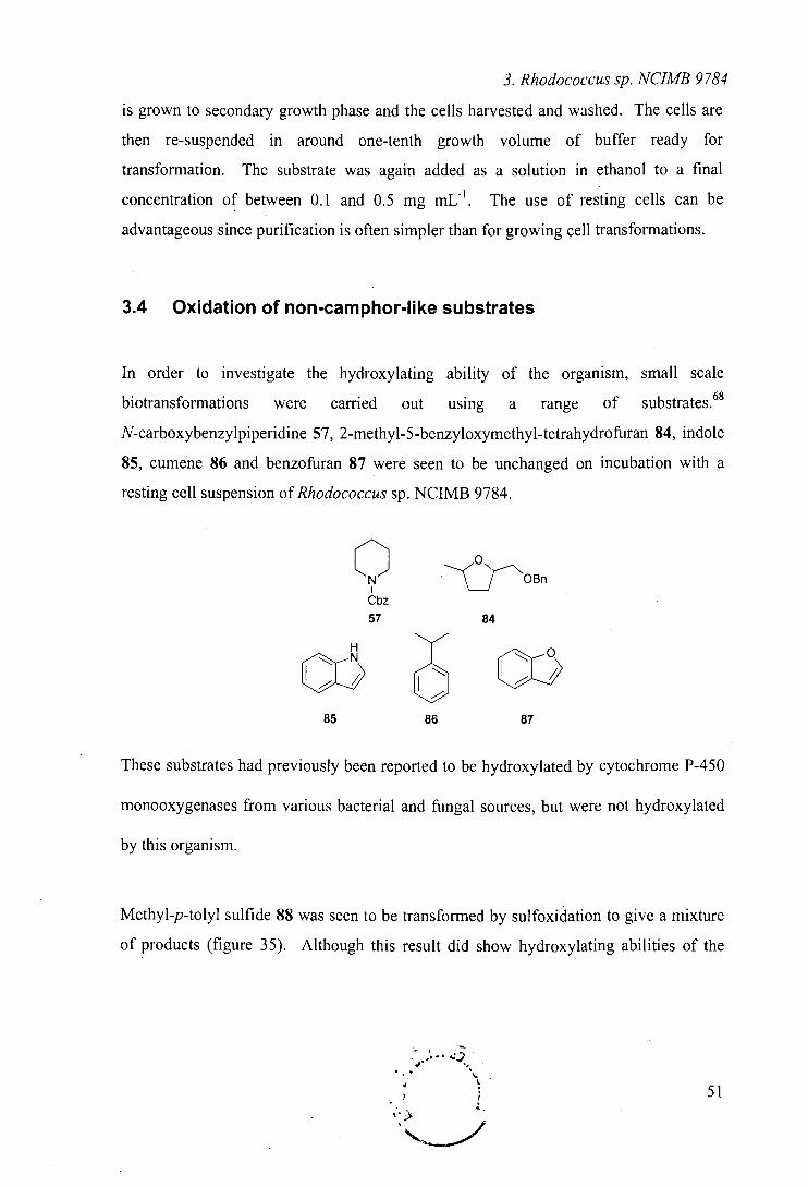
3. Rhodococcus sp. NUMB 9784
is grown to secondary growth phase and the cells harvested and washed. The cells are
then re-suspended in around one-tenth growth volume of buffer ready for
transformation. The substrate was again added as a solution in ethanol to a final
concentration of between 0.1 and 0.5 mg mU'. The use of resting cells can be
advantageous since purification is often simpler than for growing cell transformations.
3.4 Oxidation of non-camphor-like substrates
In order to investigate the hydroxylating ability of the organism, small scale
biotransformations were carried out using a range of substrates. 68
N-carboxybenzylpiperidine 57, 2-methyl-5-benzyloxymethyl-tetrahydrofuran 84, indole
85, cumene 86 and benzofuran 87 were seen to be unchanged on incubation with a
resting cell suspension of Rhodococcus sp. NCIMB 9784.
0N OBn
Cbz
57 84
H
Cox 61-11
LL? 85 86 87
These substrates had previously been reported to be hydroxylated by cytochrome P-450
monooxygenases from various bacterial and fungal sources, but were not hydroxylated
by this organism.
Methyl-p-tolyl sulfide 88 was seen to be transformed by sulfoxidation to give a mixture
of products (figure 35). Although this result did show hydroxylating abilities of the
1 51
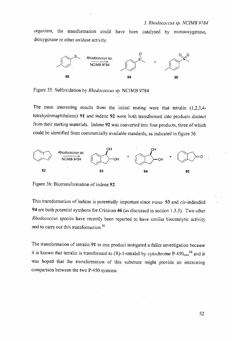
3. Rhodococcus sp. NCIMB 9784
organism, the transformation could have been catalysed by monooxygenase,
dioxygenase or other oxidase activity.
S", Rhodococcus sp. I
NCIMB 9784
88
0
+
89 90
Figure 35: Sulfoxidation by Rhodococcus sp. NCIMB 9784
The most interesting results from the initial testing were that tetralin (1,2,3,4-
tetrahydronaphthalene) 91 and indene 92 were both transformed into products distinct
from their starting materials. Indene 92 was converted into four products, three of which
could be identified from commercially available standards, as indicated in figure 36.
OH OH
CO Rhodococcussp.
EIJj; 'OH + + 14 NCIMB 9784
cf~— OH
92 93 94 95
Figure 36: Biotransformation of indene 92
This transformation of indene is potentially important since trans- 93 and cis-indandiol
94 are both potential synthons for Crixivan 46 (as discussed in section 1.5.3). Two other
Rhodococcus species have recently been reported to have similar biocatalytic activity
and to carry out this transformation. 56
The transformation of tetralin 91 to one product instigated a fuller investigation because
it is known that tetralin is transformed to (R)-1-tetralol by cytochrome P450cam88 and it
was hoped that the transformation of this substrate might provide an interesting
comparison between the two P-450 systems.
52
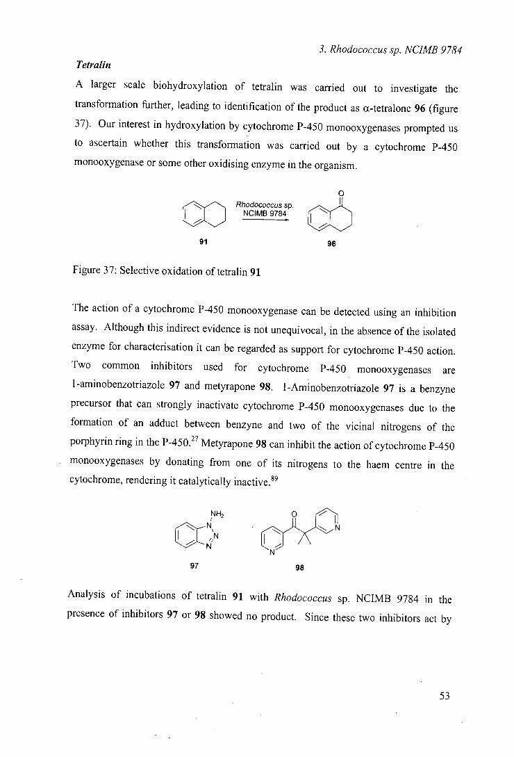
3. Rhodococcus sp. NCIMB 9784
Tetralin
A larger scale biohydroxylation of tetralin was carried out to investigate the
transformation further, leading to identification of the product as u-tetralone 96 (figure
37). Our interest in hydroxylation by cytochrome P-450 monooxygenases prompted us
to ascertain whether this transformation was carried out by a cytochrome P-450
monooxygenase or some other oxidising enzyme in the organism.
0
Rhodococcus sp Co 0 NCIMB 9784.
91 96
Figure 37: Selective oxidation of tetralin 91
The action of a cytochrome P-450 monooxygenase can be detected using an inhibition
assay. Although this indirect evidence is not unequivocal, in the absence of the isolated
enzyme for characterisation it can be regarded as support for cytochrome P-450 action.
Two common inhibitors used for cytochrome P-450 monooxygenases are
I-aminobenzotriazole 97 and metyrapone 98. l-Aminobenzotriazole 97 is a benzyne
precursor that can strongly inactivate cytochrome P-450 monooxygenases due to the
formation of an adduct between benzyne and two of the vicinal nitrogens of the
porphyrin ring in the P-450.27 Metyrapone 98 can inhibit the action of cytochrome P-450
monooxygenases by donating from one of its nitrogens to the haem centre in the
cytochrome, rendering it catalytically inactive.89
NH2 o
N\N N
N
97 98
Analysis of incubations of tetralin 91 with Rhodococcus sp. NCIMB 9784 in the
presence of inhibitors 97 or 98 showed no product. Since these two inhibitors act by
53
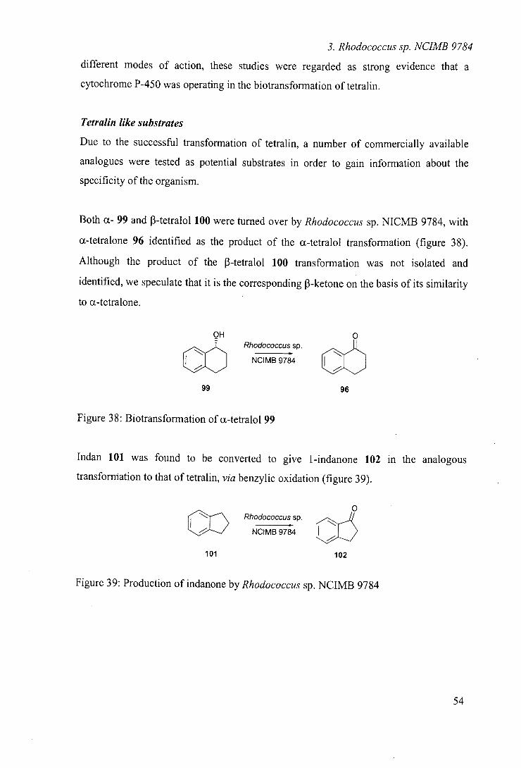
3. Rhodococcus sp. NCIMB 9784
different modes of action, these studies were regarded as strong evidence that a
cytochrome P-450 was operating in the biotransformation of tetralin.
Tetralin like substrates
Due to the successful transformation of tetralin, a number of commercially available
analogues were tested as potential substrates in order to gain information about the
specificity of the organism.
Both a- 99 and 3-tetralol 100 were turned over by Rhodococcus sp. NICMB 9784, with
a-tetralone 96 identified as the product of the a-tetralol transformation (figure 38).
Although the product of the -tetralol 100 transformation was not isolated and
identified, we speculate that it is the corresponding 3-ketone on the basis of its similarity
to a-tetralone.
OH 0 - Rhodococcus sp.
NCIMB 9784 Ec::I:ItIIl 99 96
Figure 38: Biotransformation of a-tetralol 99
Indan 101 was found to be converted to give 1-indanone 102 in the analogous
transformation to that of tetralin, via benzylic oxidation (figure 39).
0
CO Rhodococcus sp.
cIIIIiiiI NCIMB 9784
101 102
Figure 39: Production of indanone by Rhodococcus sp. NCIMB 9784
54
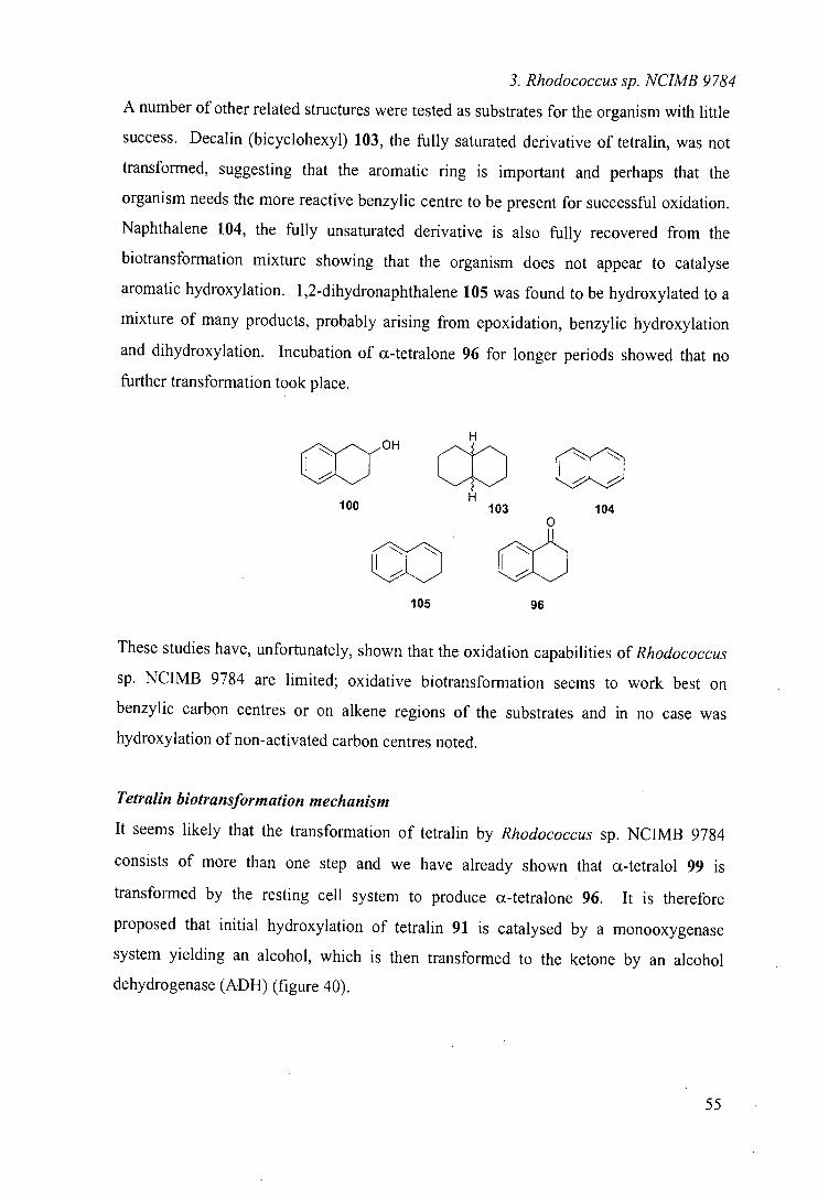
3. Rhodococcus sp. NCIMB 9784
A number of other related structures were tested as substrates for the organism with little
success. Decalin (bicyclohexyl) 103, the fully saturated derivative of tetralin, was not
transformed, suggesting that the aromatic ring is important and perhaps that the
organism needs the more reactive benzylic centre to be present for successful oxidation.
Naphthalene 104, the fully unsaturated derivative is also fully recovered from the
biotransformation mixture showing that the organism does not appear to catalyse
aromatic hydroxylation. 1 ,2-dihydronaphthalene 105 was found to be hydroxylated to a
mixture of many products, probably arising from epoxidation, benzylic hydroxylation
and dihydroxylation. Incubation of a-tetralone 96 for longer periods showed that no
further transformation took place.
OH H
Ca c O CO 100
H103 104
0
CO 105 96
These studies have, unfortunately, shown that the oxidation capabilities of Rhodococcus
sp. NCIMB 9784 are limited; oxidative biotransformation seems to work best on
benzylic carbon centres or on alkene regions of the substrates and in no case was
hydroxylation of non-activated carbon centres noted.
Tetraljn biotransformation mechanism
It seems likely that the transformation of tetralin by Rhodococcus sp. NCIMB 9784
consists of more than one step and we have already shown that a-tetralol 99 is
transformed by the resting cell system to produce x-tetralone 96. It is therefore
proposed that initial hydroxylation of tetralin 91 is catalysed by a monooxygenase
system yielding an alcohol, which is then transformed to the ketone by an alcohol
dehydrogenase (ADH) (figure 40).
55
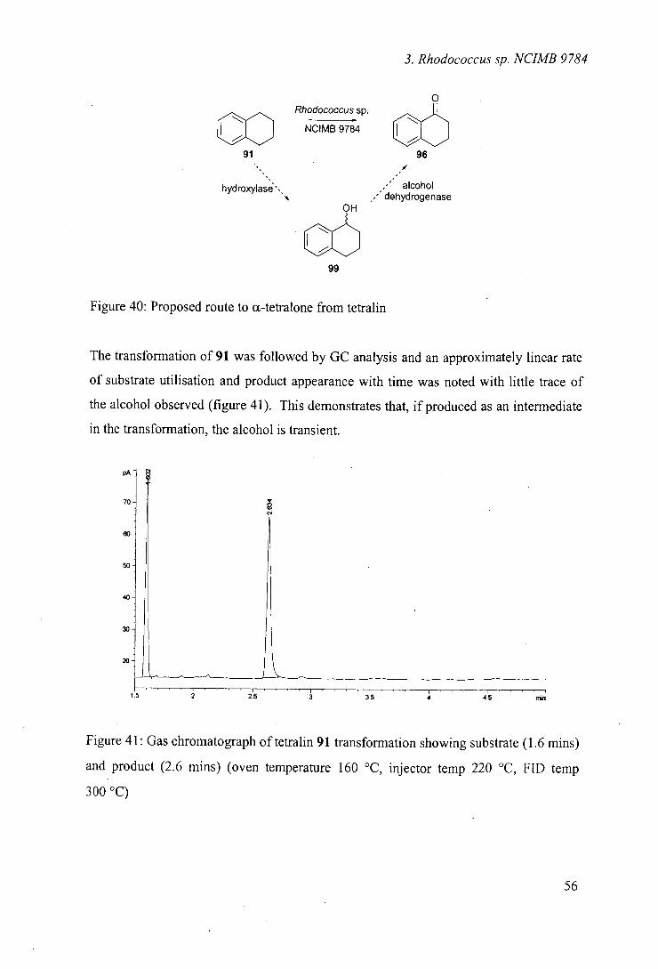
3. Rhodococcus sp. NUMB 9784
0 Rhodococcus sp.
CO 0 NCIMB 9784
91 96 I
hydrolas'. alcohol/ dehydrogenase
OH
0 99
Figure 40: Proposed route to cc-tetralone from tetralin
The transformation of 91 was followed by GC analysis and an approximately linear rate
of substrate utilisation and product appearance with time was noted with little trace of
the alcohol observed (figure 41). This demonstrates that, if produced as an intermediate
in the transformation, the alcohol is transient.
PA
70
60
50
40
30
20
Figure 41: Gas chromatograph of tetralin 91 transformation showing substrate (1.6 mins)
and product (2.6 mins) (oven temperature 160 °C, injector temp 220 °C, FID temp
300 °C)
56
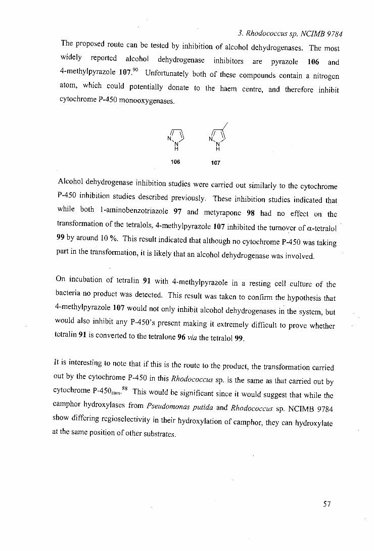
3. Rhodococcus sp. NCIMB 9784
The proposed route can be tested by inhibition of alcohol dehydrogenases. The most
widely reported alcohol dehydrogenase inhibitors are pyrazole 106 and 4-methylpyrazole 107. ° Unfortunately both of these compounds contain a nitrogen
atom, which could potentially donate to the haem centre, and therefore inhibit
cytochrome P-450 monooxygenases.
flNf
106 107
Alcohol dehydrogenase inhibition studies were carried out similarly to the cytochrome
P-450 inhibition studies described previously. These inhibition studies indicated that
while both l-aminobenzotriazole 97 and metyrapone 98 had no effect on the
transformation of the tetralols, 4-methylpyrazole 107 inhibited the turnover of ct-tetralol 99 by around 10 %. This result indicated that although no cytochrome P-450 was taking
part in the transformation, it is likely that an alcohol dehydrogenase was involved.
On incubation of tetralin 91 with 4-methylpyrazole in a resting cell culture of the
bacteria no product was detected. This result was taken to confirm the hypothesis that
4-methylpyrazole 107 would not only inhibit alcohol dehydrogenases in the system, but
would also inhibit any P-450's present making it extremely difficult to prove whether
tetralin 91 is converted to the tetralone 96 via the tetralol 99.
It is interesting to note that if this is the route to the product, the transformation carried
out by the cytochrome P-450 in this Rhodococcus sp. is the same as that carried out by cytochrome P450cam.88 This would be significant since it would suggest that while the
camphor hydroxylases from Pseudomonas putida and Rhodococcus sp. NCIMB 9784
show differing regioselectivity in their hydroxylation of camphor, they can hydroxylate
at the same position of other substrates.
57
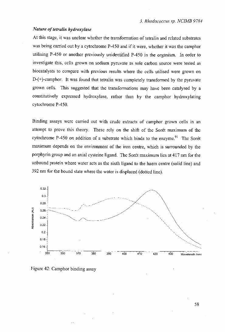
3. Rhodococcus sp. NUMB 9784
Nature of tetralin hydroxylase
At this stage, it was unclear whether the transformation of tetralin and related substrates
was being carried out by a cytochrome P-450 and if it were, whether it was the camphor
utilising P-450 or another previously unidentified P-450 in the organism. In order to
investigate this, cells grown on sodium pyruvate as sole carbon source were tested as
biocatalysts to compare with previous results where the cells utilised were grown on
D-(+)-camphor. It was found that tetralin was completely transformed by the pyruvate
grown cells. This suggested that the transformations may have been catalysed by a
constitutively expressed hydroxylase, rather than by the camphor hydroxylating
cytochrome P-450.
Binding assays were carried out with crude extracts of camphor grown cells in an
attempt to prove this theory. These rely on the shift of the Sorét maximum of the
cytochrome P-450 on addition of a substrate which binds to the enzyme. 91 The Sorét
maximum depends on the environment of the iron centre, which is surrounded by the
porphyrin group and an axial cysteine ligand. The Sorét maximum lies at 417 nm for the
unbound protein where water acts as the sixth ligand to the haem centre (solid line) and
392 nm for the bound state where the water is displaced (dotted line).
O.3
0.
0.2€
0.2€
0.24
0.2
0.2
0.18
0.16
Figure 42: Camphor binding assay
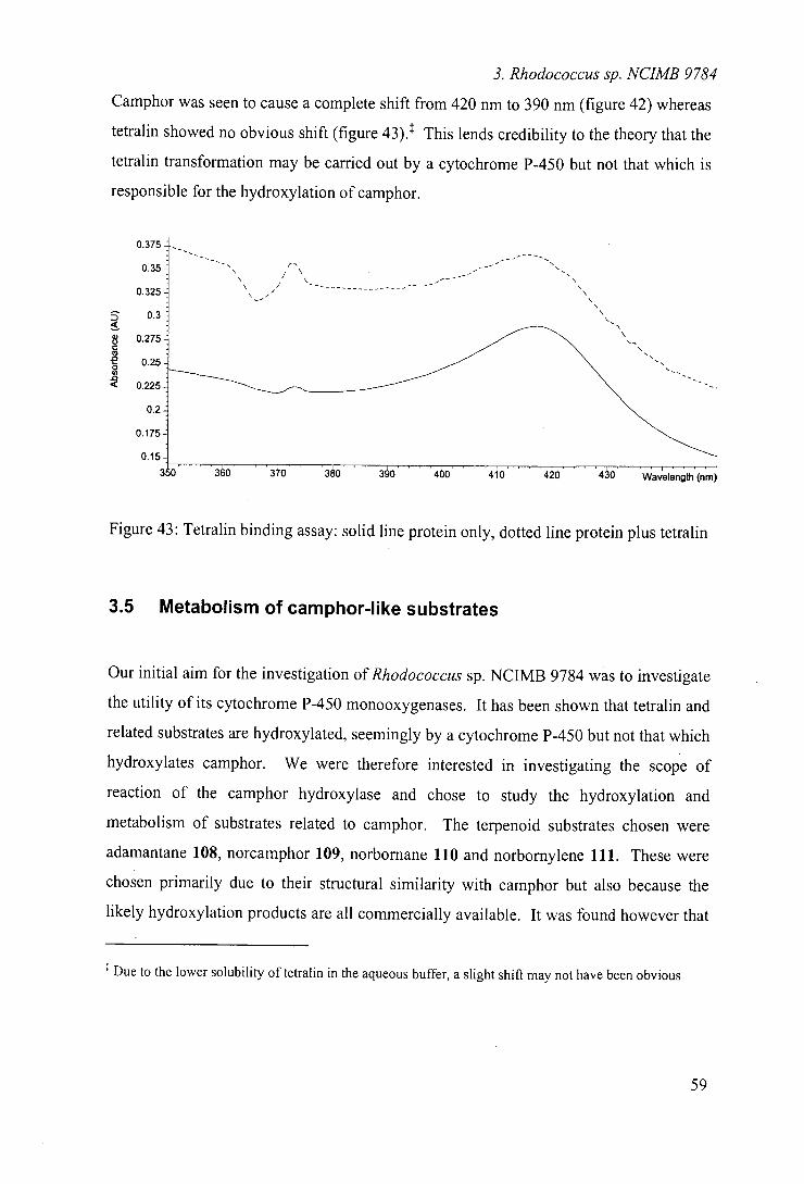
3. Rhodococcus sp. NUMB 9784
Camphor was seen to cause a complete shift from 420 nm to 390 nm (figure 42) whereas
tetralin showed no obvious shift (figure 43).1 This lends credibility to the theory that the
tetralin transformation may be carried out by a cytochrome P-450 but not that which is
responsible for the hydroxylation of camphor.
0.375
0.35
0.325
0.3
0.275
0.25
0.225
0.2
0.175
0.15
Figure 43: Tetralin binding assay: solid line protein only, dotted line protein plus tetralin
3.5 Metabolism of camphor-like substrates
Our initial aim for the investigation of Rhodococcus sp. NCIMB 9784 was to investigate
the utility of its cytochrome P-450 monooxygenases. It has been shown that tetralin and
related substrates are hydroxylated, seemingly by a cytochrome P-450 but not that which
hydroxylates camphor. We were therefore interested in investigating the scope of
reaction of the camphor hydroxylase and chose to study the hydroxylation and
metabolism of substrates related to camphor. The terpenoid substrates chosen were
adamantane 108, norcamphor 109, norbornane 110 and norbornylene 111. These were
chosen primarily due to their structural similarity with camphor but also because the
likely hydroxylation products are all commercially available. It was found however that
Due to the lower solubility of tetralin in the aqueous buffer, a slight shift may not have been obvious
59
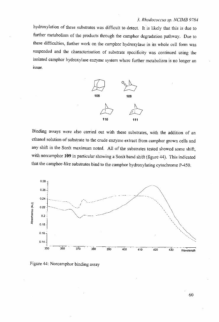
0.2
0.24
C
0. 0 In
OlE
0.16
0.14
3. Rhodococcus sp. NUMB 9784
hydroxylation of these substrates was difficult to detect. It is likely that this is due to
further metabolism of the products through the camphor degradation pathway. Due to
these difficulties, further work on the camphor hydroxylase in its whole cell form was
suspended and the characterisation of substrate specificity was continued using the
isolated camphor hydroxylase enzyme system where further metabolism is no longer an
issue.
108 109
zb_ 110 111
Binding assays were also carried out with these substrates, with the addition of an
ethanol solution of substrate to the crude enzyme extract from camphor grown cells and
any shift in the Sorét maximum noted. All of the substrates tested showed some shift,
with norcamphor 109 in particular showing a Sorét band shift (figure 44). This indicated
that the camphor-like substrates bind to the camphor hydroxylating cytochrome P-450.
Figure 44: Norcamphor binding assay
M.

3. Rhodococcus sp. NUMB 9784
Because these binding assays showed some binding to the camphor-hydroxylating
enzyme, they lend evidence to the supposed hydroxylation of camphor-like substrates by
the enzyme.
3.6 Conclusions
Our conclusions from these studies are two fold. Although no good substrates were
positively identified for the camphor utilising cytochrome P-450 that initially attracted
our attention to this organism, we believe that we have identified a second cytochrome
P-450 in the system. We have discovered that this cytochrome P-450 has narrow
substrate specificity for tetralin 91 and related substrates. We believe that the oxidation
to a ketone occurs via the alcohol in a two step procedure catalysed by a cytochrome
P-450 and an alcohol dehydrogenase. It is also noted that hydroxylation of substrates
seems to depend on the existence of a benzylic or alkene carbon within the molecule.
Thus, the outcome of the tetralin incubation was not particularly interesting in the
context of microbial hydroxylation of non-activated carbon centres. The transformation
of indene 92 to yield, amongst other products, indandiols 93 and 94 is potentially
interesting as there is known synthetic utility for such compounds in their optically pure
form.56
From our preliminary studies into the camphor utilising hydroxylase, it would seem that,
like cytochrome P 450cam, the camphor monooxygenase in Rhodococcus sp. NCIMB
9784 has narrow substrate specificity, limited to camphor-like structures. Camphor
analogues such as norcamphor 109 were found to bind to the enzyme, as indicated by
Sorét band shift assays. Although this may limit its use as a hydroxylation catalyst, the
enzyme has now been isolated and could potentially be cloned for use in a suitable host
system for hydroxylation. The enzyme could also be investigated in a manner similar to
61
3. Rhodococcus sp. NUMB 9784 cytochrome P450cam where mutagenesis experiments have widened the substrate range
such that larger molecules containing aromatic groups can now be hydroxylated by
cytochrome P450cam 92'93
62
4. Rhodococcus rhodochrous NCIMB 9703
4 Biotrans formations with Rhodococcus rhodochrous
NCIMB 9703
4.1 Aims
As indicated in chapter 3, we are interested in the potential utility of Rhodococcus
species as biohydroxylation catalysts, a property that has, as yet, not been fully
investigated. Screening of a range of such microorganisms thought to have potentially
cytochrome P-450 dependent hydroxylating abilities identified a number of organisms
that warranted further investigation. One of these, Rhodococcus sp. NCIMB 9784, is
reported in chapter 3 where we have shown that the system appears to contain at least
two cytochrome P-450 monooxygenases which both show narrow substrate specificity.
Another of the Rhodococcus species identified for further investigation was
Rhodococcus rhodochrous NCIMB 9703. As highlighted in the introduction, this
organism is reported to catalyse the terminal hydroxylation of n-octane. To our
knowledge there have been no investigations as to the utility of this organism as a
general biohydroxylation catalyst.
Our aim in this investigation was to determine the existence of cytochrome P-450
monooxygenase(s) within the organism and to investigate the scope and selectivity of
any hydroxylation catalysed.
4.2 Growth of organism
Rhodococcus rhodochrous NCIMB 9703 is reported to contain an inducible NADH
dependent hydroxylase that catalyses the conversion of n-octane to 1 -octanol.48 Growth
63
4. Rhodococcus rhodochrous NCIMB 9703
of the organism on octane was therefore attempted to induce this hydroxylating enzyme
but growth on basal salts medium augmented with ferrous sulfate and n-octane was
found to be extremely sluggish. In common with Rhodococcus sp. NCIMB 9784, it was
found that initial growth on sodium pyruvate for use as innoculum for larger scale
growth on n-octane proved a successful and reliable method for growth. Due to the
potential experimental risks when utilising a flammable additive such as octane, 94
fermenter growth was not attempted but growth in shake flask was found to yield
sufficient cell mass for biotransformation. For comparison, cells were also grown on
sodium pyruvate with no induction. Cell mass was typically greater from growth on
pyruvate (5.5 gL1) compared with growth on octane (2.5 gU').
4.3 Methods
Biotransformations using Rhodococcus rhodochrous NCIMB 9703 were carried out
using resting cell suspensions where the, cells are live but do not have the nutrients to
continue growing. As for Rhodococcus sp. NCIMB 9784, it was envisaged that the use
of resting cells might ease purification.
The organism was first grown to secondary growth phase and the cells harvested and
washed. The cells were then re-suspended in approximately one-tenth growth volume of
phosphate buffer (50 mM, pH 7.0) and substrate added as a solution in ethanol to a final
concentration of 0.5 gU'. On completion, the cells were removed by centrifugation and
the products isolated, purified by flash chromatography and identified by NMR
spectroscopy.
64
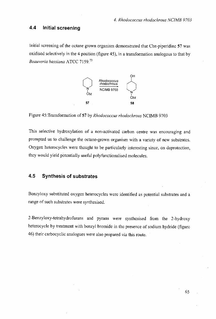
4. Rhodococcus rhodochrous NUMB 9703
4.4 Initial screening
Initial screening of the octane grown organism demonstrated that Cbz-piperidine 57 was
oxidised selectively in the 4 position (figure 45), in a transformation analogous to that by
Beauveria bassiana ATCC 7I59. 71
OH
fl Rhodococcus rhodochrous
NUMB 9703 N) Cbz I Cbz
57 58
Figure 45:Transformation of 57 by Rhodococcus rhodochrous NCIMB 9703
This selective hydroxylation of a non-activated carbon centre was encouraging and
prompted us to challenge the octane-grown organism with a variety of new substrates.
Oxygen heterocycles were thought to be particularly interesting since, on deprotection,
they would yield potentially useful polyfunctionalised molecules.
4.5 Synthesis of substrates
Benzyloxy substituted oxygen heterocycles were identified as potential substrates and a
range of such substrates were synthesised.
2-Benzyloxy-tetrahydrofurans and pyrans were synthesised from the 2-hydroxy
heterocycle by treatment with benzyl bromide in the presence of sodium hydride (figure
46) their carbocyclic analogues were also prepared via this route.
IN
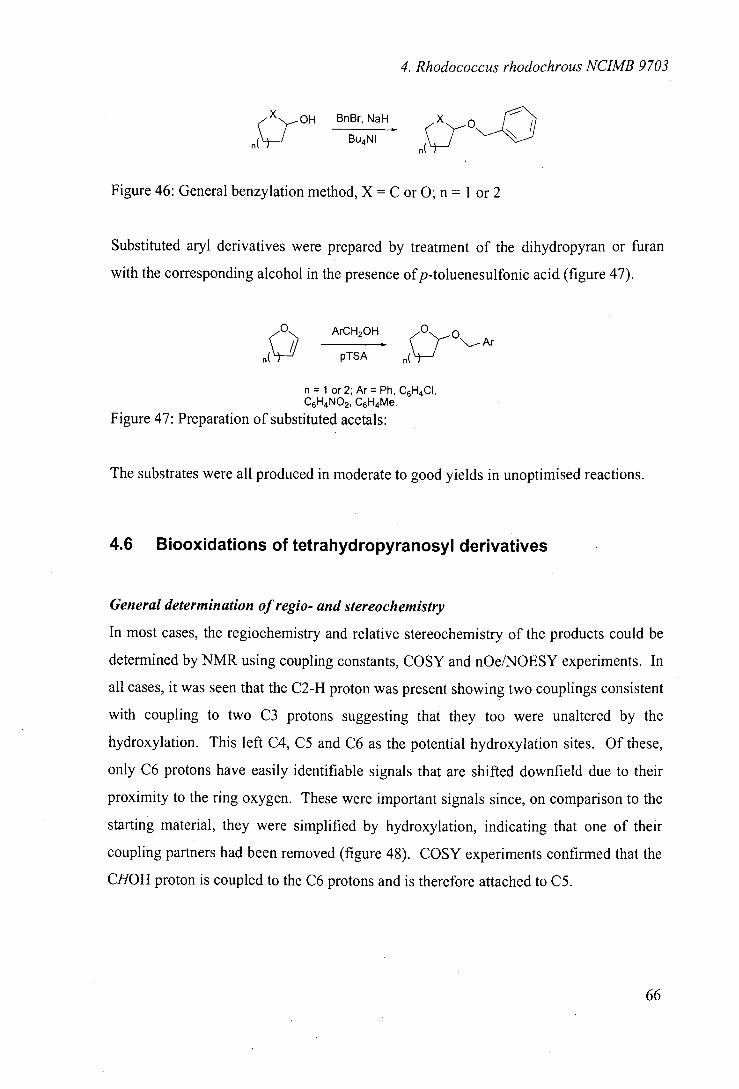
4. Rhodococcus rhodochrous NUMB 9703
~OH BnBr, NaH
BuNI r'(V
Figure 46: General benzylation method, X = C or 0; n = I or 2
Substituted aryl derivatives were prepared by treatment of the dihydropyran or furan
with the corresponding alcohol in the presence ofp-toluenesulfonic acid (figure 47).
0 Ar2 (°OA (
CHOH
pTSA
n = 1 or 2; Ar = Ph, C6H4CI, C6H4NO2, C6H4Me.
Figure 47: Preparation of substituted acetals:
The substrates were all produced in moderate to good yields in unoptimised reactions.
4.6 Biooxidations of tetrahydropyranosyl derivatives
General determination of regio- and stereochemistry
In most cases, the regiochemistry and relative stereochemistry of the products could be
determined by NMR using coupling constants, COSY and nOe/NOESY experiments. In
all cases, it was seen that the C2-H proton was present showing two couplings consistent
with coupling to two C3 protons suggesting that they too were unaltered by the
hydroxylation. This left C4, C5 and C6 as the potential hydroxylation sites. Of these,
only C6 protons have easily identifiable signals that are shifted downfield due to their
proximity to the ring oxygen. These were important signals since, on comparison to the
starting material, they were simplified by hydroxylation, indicating that one of their
coupling partners had been removed (figure 48). COSY experiments confirmed that the
CHOH proton is coupled to the C6 protons and is therefore attached to C5.
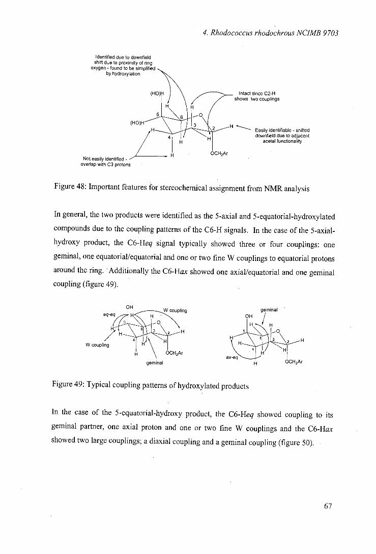
(HO
Not easily identified - overlap with C3 protons
Identified due to downfield shift due to proximity of ring
oxygen - found to be sin'-'- by hydroxylation
4. Rhodococcus rhodochrous NUMB 9703
Intact since C2-H shows two couplings
Easily identifiable - shifted downfield due to adjacent
acetal functionality
Figure 48: Important features for stereochemical assignment from NMR analysis
In general, the two products were identified as the 5-axial and 5-equatorial-hydroxylated
compounds due to the coupling patterns of the C6-H signals. In the case of the 5-axial-
hydroxy product, the C6-Heq signal typically showed three or four couplings: one
geminal, one equatorial/equatorial and one or two fine W couplings to equatorial protons
around the ring. Additionally the C6-Hax showed one axial/equatorial and one geminal
coupling (figure 49).
OH W coupling
eq-eq H
6 H 2 H
W coupling H \ HOCH2Ar
geminal
geminal OH
ax-e.4_ HCH2Ar
Figure 49: Typical coupling patterns of hydroxylated products
In the case of the S-equatorial-hydroxy product, the C6-Heq showed coupling to its
geminal partner, one axial proton and one or two fine W couplings and the C6-Hax
showed two large couplings; a diaxial coupling and a geminal coupling (figure 50).
67
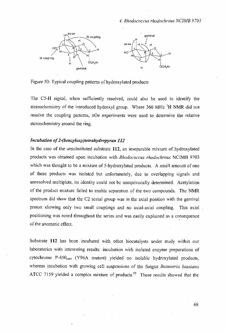
4. Rhodococcus rhodochrous NUMB 9703
H eq-ax W coupling
HO
W coupling H \ HO
CH2Ar
geminal
geminal
ax-ax H*H/ H
2 H HO
H HOCH2Ar
Figure 50: Typical coupling patterns of hydroxylated products
The CS-H signal, when sufficiently resolved, could also be used to identify the
stereochemistry of the introduced hydroxyl group. Where 360 MHz 'H NMR did not
resolve the coupling patterns, nOe experiments were used to determine the relative
stereochemistry around the ring.
Incubation of2-(benzyloxy)tetrahydropyran 112
In the case of the unsubstituted substrate 112, an inseparable mixture of hydroxylated
products was obtained upon incubation with Rhodococcus rhodochrous NCIMB 9703
which was thought to be a mixture of 5-hydroxylated products. A small amount of one
of these products was isolated but unfortunately, due to overlapping signals and
unresolved multiplets, its identity could not be unequivocally determined. Acetylation
of the product mixture failed to enable separation of the two compounds. The NMR
spectrum did show that the C2 acetal group was in the axial position with the geminal
proton showing only two small couplings and no axial-axial coupling. This axial
positioning was noted throughout the series and was easily explained as a consequence
of the anomeric effect.
Substrate 112 has been incubated with other biocatalysts under study within our
laboratories with interesting results: incubation with isolated enzyme preparations of
cytochrome P450cam (Y96A mutant) yielded no isolable hydroxylated products,
whereas incubation with growing cell suspensions of the fungus Beau veria bassiana
ATCC 7159 yielded a complex mixture of products. 95 These results showed that the
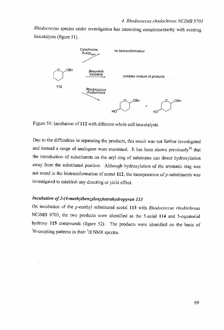
4. Rhodococcus rhodochrous NUMB 9703
Rhodococcus species under investigation has interesting complementarity with existing
biocatalysts (figure 51).
Cytochrome no biotransformation
Beauveria a bassiana complex mixture of products
112 Rhodococcus rhodochrous
OBn 0 OBn
HOU + H0'r3 Figure 51: Incubation of 112 with different whole cell biocatalysts
Due to the difficulties in separating the products, this result was not further investigated
and instead a range of analogues were examined. It has been shown previously36 that
the introduction of substituents on the aryl ring of substrates can direct hydroxylation
away from the substituted position. Although hydroxylation of the aromatic ring was
not noted in the biotransformation of acetal 112, the incorporation of p-substituents was
investigated to establish any directing or yield effect.
Incubation of 2-(4-methylbenzyloxy)tetrahydropyran 113
On incubation of the p-methyl substituted acetal 113 with Rhodococcus rhodochrous
NCIMB 9703, the two products were identified as the 5-axial 114 and 5-equatorial
hydroxy 115 compounds (figure 52). The products were identified on the basis of
'H-coupling patterns in their 'H NMR spectra.
Me
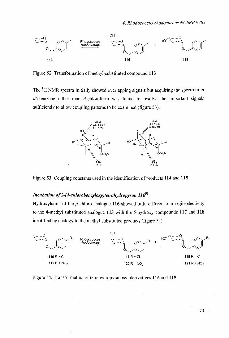
4. Rhodococcus rhodochrous NUMB 9703
OH 0
rhodochrous Rhodococcus + HO
113
114
115
Figure 52: Transformation of methyl-substituted compound 113
The 'H NMR spectra initially showed overlapping signals but acquiring the spectrum in
d6-benzene rather than d-chloroform was found to resolve the important signals
sufficiently to allow coupling patterhs to be examined (figure 53).
dddd J 0.5, 2.0. 3.4
& 11.8 Hz OH
H
H \ H
dd J 2.2&
11.8 Hz
ddd J 1.7, 4.3 & 10.7 Hz
HO— IL\_ H
H \ HOCH2Ar
dd J8.8 &
10.7 Hz
Figure 53: Coupling constants used in the identification of products 114 and 115
Incubation of 2-(4-chlorobenzyloxy)tetrahydropyran 11696
Hydroxylation of the p-chloro analogue 116 showed little difference in regioselectivity
to the 4-methyl substituted analogue 113 with the 5-hydroxy compounds 117 and 118
identified by analogy to the methyl-substituted products (figure 54).
OH
r Rhodococcus hodochrous R +
117 R = CI
120 R = NO2
116 R = CI
119 R = NO2
118 R = CI
121 R = NO2
Figure 54: Transformation of tetrahydropyranosyl derivatives 116 and 119
'1€
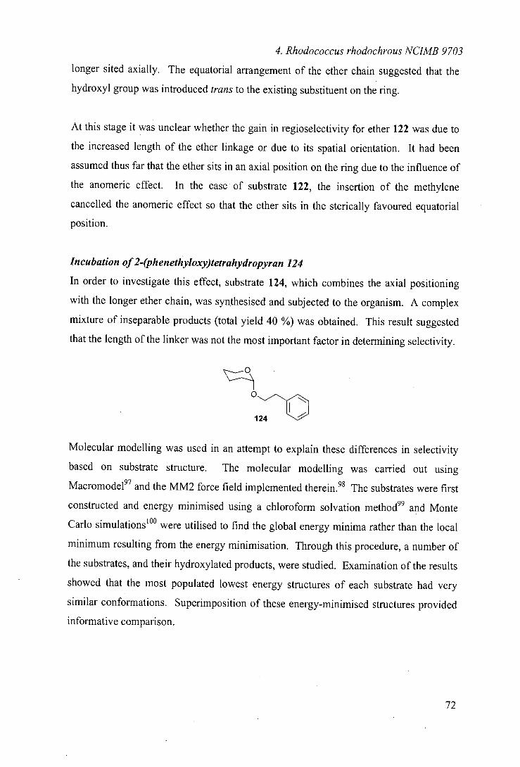
4. Rhodococcus rhodochrous NUMB 9703
longer sited axially. The equatorial arrangement of the ether chain suggested that the
hydroxyl group was introduced trans to the existing substituent on the ring.
At this stage it was unclear whether the gain in regioselectivity for ether 122 was due to
the increased length of the ether linkage or due to its spatial orientation. It had been
assumed thus far that the ether sits in an axial position on the ring due to the influence of
the anomeric effect. In the case of substrate 122, the insertion of the methylene
cancelled the anomeric effect so that the ether sits in the sterically favoured equatorial
position.
Incubation of 2-(phenethyloxy)tetrahydropyran 124
In order to investigate this effect, substrate 124, which combines the axial positioning
with the longer ether chain, was synthesised and subjected to the organism. A complex
mixture of inseparable products (total yield 40 %) was obtained. This result suggested
that the length of the linker was not the most important factor in determining selectivity.
" 0 -~) "--~O 124
Molecular modelling was used in an attempt to explain these differences in selectivity
based on substrate structure. The molecular modelling was carried out using
Macromode197 and the MM2 force field implemented therein. 98 The substrates were first
constructed and energy minimised using a chloroform solvation method" and Monte
Carlo simulations10° were utilised to find the global energy minima rather than the local
minimum resulting from the energy minimisation. Through this procedure, a number of
the substrates, and their hydroxylated products, were studied. Examination of the results
showed that the most populated lowest energy structures of each substrate had very
similar conformations. Superimposition of these energy-minimised structures provided
informative comparison.
72
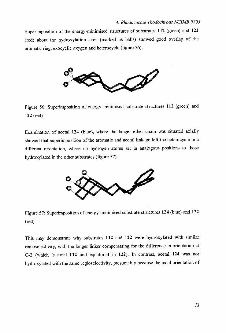
4. Rhodococcus rhodochrous NUMB 9703
Superimposition of the energy-minimised structures of substrates 112 (green) and 122
(red) about the hydroxylation sites (marked as balls) showed good overlap of the
aromatic ring, exocyclic oxygen and heterocycle (figure 56).
Figure 56: Superimposition of energy minimised substrate structures 112 (green) and
122 (red)
Examination of acetal 124 (blue), where the longer ether chain was situated axially
showed that superimposition of the aromatic and acetal linkage left the heterocycle in a
different orientation, where no hydrogen atoms sat in analogous positions to those
hydroxylated in the other substrates (figure 57).
Figure 57: Superimposition of energy minimised substrate structures 124 (blue) and 122
(red)
This may demonstrate why substrates 112 and 122 were hydroxylated with similar
reg io selectivity, with the longer linker compensating for the difference in orientation at
C-2 (which is axial 112 and equatorial in 122). In contrast, acetal 124 was not
hydroxylated with the same regioselectivity, presumably because the axial orientation of
73
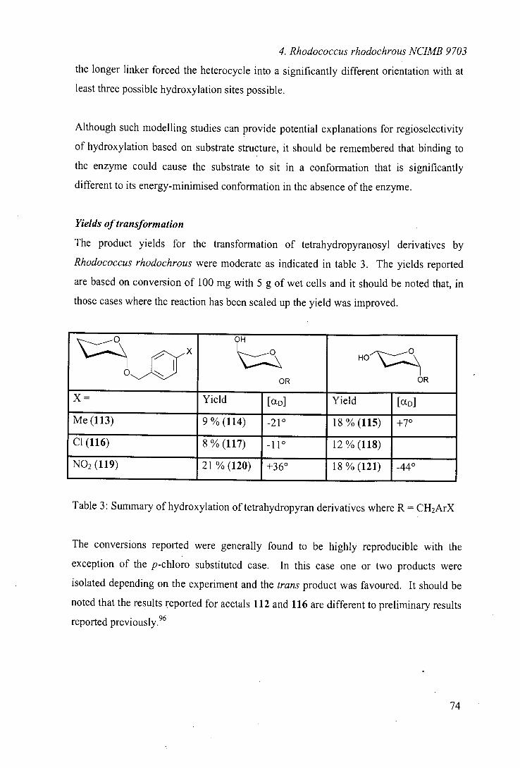
4. Rhodococcus rhodochrous NUMB 9703
the longer linker forced the heterocycle into a significantly different orientation with at
least three possible hydroxylation sites possible.
Although such modelling studies can provide potential explanations for regioselectivity
of hydroxylation based on substrate structure, it should be remembered that binding to
the enzyme could cause the substrate to sit in a conformation that is significantly
different to its energy-minimised conformation in the absence of the enzyme.
Yields of transformation
The product yields for the transformation of tetrahydropyranosyl derivatives by
Rhodococcus rhodochrous were moderate as indicated in table 3. The yields reported
are based on conversion of 100 mg with 5 g of wet cells and it should be noted that, in
those cases where the reaction has been scaled up the yield was improved.
0"~~a X
OH
OR
HO
OR
X = Yield [an] Yield [(XD]
Me (113) 9%(114) -210 18%(115) +7°
Cl (116) 8%(117) -11° 12%(118)
NO2 (119) 21 % (120) +36° 18 % (121) -44°
Table 3: Summary of hydroxylation of tetrahydropyran derivatives where R = CH2ArX
The conversions reported were generally found to be highly reproducible with the
exception of the p-chloro substituted case. In this case one or two products were
isolated depending on the experiment and the trans product was favoured. It should be
noted that the results reported for acetals 112 and 116 are different to preliminary results
reported previously. 96
74
4. Rhodococcus rhodochrous NCIMB 9703
The transformations reported were all carried out on racemic substrates and the optical
activity of the products suggested a degree of stereoselectivity in the hydroxylation
process. However, because the products described are not known compounds, the
optical rotations give no indication of optical purity. Since less than 50 % of the mass
balance is accounted for and no starting material was recovered, further breakdown of
either substrate or product is possible. It was therefore not clear whether initial
hydroxylation or further breakdown of the product was responsible for the observed
stereoselectivity.
Interestingly when the aromatic side chain was altered as in ether 122, the previously
noted increase in regioselectivity was accompanied by an increased yield. Unfortunately
the optical rotation of the product was negligible, suggesting that stereoselectivity was
lost on insertion of the methylene unit.
Further investigation of the stereoselectivity of selected transformations is discussed in
section 4.8.
Summary of biooxidation of tetrahydropyranosides
Examination of a range of closely related structures based on six-membered oxygen 2-
alkyl heterocycles has demonstrated that Rhodococcus rhodochrous NCIMB 9703 can
be a regioselective hydroxylation catalyst. It was observed that, irrespective of aromatic
substituents, the enzyme has a preference for hydroxylation at C5, yielding mixtures of
axial and equatorial substituted hydroxy products. Small structure modifications were
seen to be potentially important in determining selectivity, leading, in some cases, to
improved selectivity. The hydroxylations also showed a degree of stereoselectivity
because racemic compounds were converted to optically active hydroxylated products.
75
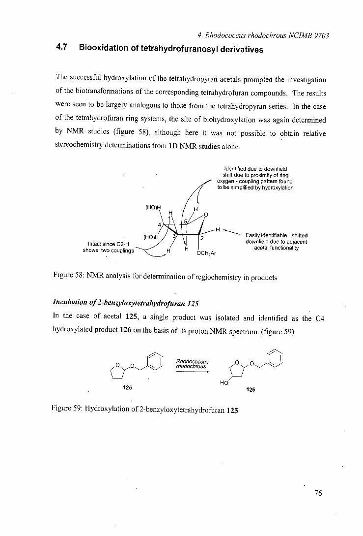
4. Rhodococcus rhodochrous NUMB 9703
4.7 Biooxiciation of tetrahydrofuranosyl derivatives
The successful hydroxylation of the tetrahydropyran acetals prompted the investigation
of the biotransformations of the corresponding tetrahydrofuran compounds. The results
were seen to be largely analogous to those from the tetrahydropyran series. In the case
of the tetrahydrofuran ring systems, the site of biohydroxylation was again determined
by NMR studies (figure 58), although here it was not possible to obtain relative
stereochemistry determinations from 1D NMR studies alone.
Identified due to downfield shift due to proximity of ring
oxygen - coupling pattern found to be simplified by hydroxylation
(HO)H
shows o couplings
H Easily identifiable - shifted
Intact since C2-H downfield due to adjacent
H H OCH2Ar
acetal functionality
Figure 58: NMR analysis for determination of regiochemistry in products
Incubation of 2-benzyloxytetrahydrofuran 125
In the case of acetal 125, a single product was isolated and identified as the C4
hydroxylated product 126 on the basis of its proton NMR spectrum. (figure 59)
Rhodococcus rhodochrous
HO
125 126
Figure 59: Hydroxylation of 2-benzyloxytetrahydrofuran 125
76
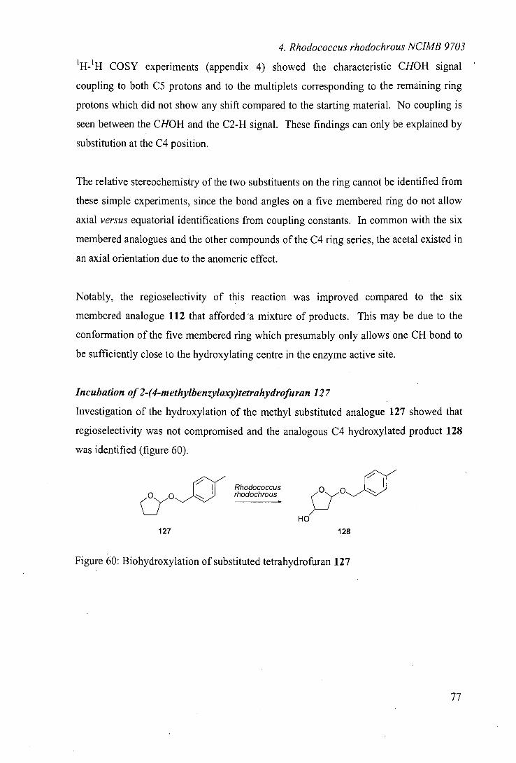
4. Rhodococcus rhodochrous NUMB 9703
'H-'H COSY experiments (appendix 4) showed the characteristic CHOH signal
coupling to both C5 protons and to the multiplets corresponding to the remaining ring
protons which did not show any shift compared to the starting material. No coupling is
seen between the CHOH and the C2-H signal. These findings can only be explained by
substitution at the C4 position.
The relative stereochemistry of the two substituents on the ring cannot be identified from
these simple experiments, since the bond angles on a five membered ring do not allow
axial versus equatorial identifications from coupling constants. In common with the six
membered analogues and the other compounds of the C4 ring series, the acetal existed in
an axial orientation due to the anomeric effect.
Notably, the regioselectivity of this reaction was improved compared to the six
membered analogue 112 that afforded 'a mixture of products. This may be due to the
conformation of the five membered ring which presumably only allows one CH bond to
be sufficiently close to the hydroxylating centre in the enzyme active site.
Incubation of 2-(4-methylbenzyloxy)tetrahydrofuran 127
Investigation of the hydroxylation of the methyl substituted analogue 127 showed that
regioselectivity was not compromised and the analogous C4 hydroxylated product 128
was identified (figure 60).
rhodochrous Rhodococcus
127 128
Figure 60: Biohydroxylation of substituted tetrahydrofuran 127
77
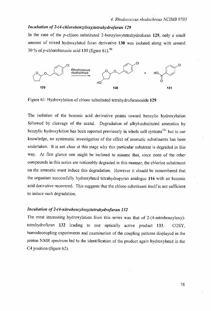
4. Rhodococcus rhodochrous NUMB 9703
Incubation of 2-(4-chlorobenzyloxy)tetrahydrofuran 129
In the case of the p-chloro substituted 2-benzyloxytetrahydrofuran 129, only a small
amount of mixed hydroxylated furan derivative 130 was isolated along with around
30 % ofp-chlorobenzoic acid 131 (figure 61).96
CI
Rhodococcus
C1
rhodochrous + HO -C, HO
129 130 131
Figure 61: Hydroxylation of chioro substituted tetrahydrofuranoside 129
The isolation of the benzoic acid derivative points toward benzylic hydroxylation
followed by cleavage of the acetal. Degradation of alkyl-substituted aromatics by
benzylic hydroxylation has been reported previously in whole cell systems'01 but to our
knowledge, no systematic investigation of the effect of aromatic substituents has been
undertaken. It is not clear at this stage why this particular substrate is degraded in this
way. At first glance one might be inclined to assume that, since none of the other
compounds in this series are noticeably degraded in this manner, the chlorine substituent
on the aromatic must induce this degradation. However it should be I remembered that
the organism successfully hydroxylated tetrahydropyran analogue 116 with no benzoic
acid derivative recovered. This suggests that the chloro substituent itself is not sufficient
to induce such degradation.
Incubation of 2-(4-nitrobenzyloxy)tetrahydrofuran 132
The most interesting hydroxylation from this series was that of 2-(4-nitrobenzyloxy)-
tetrahydrofuran 132 leading to one optically active product 133. COSY,
homodecoupling experiments and examination of the coupling patterns displayed in the
proton NMR spectrum led to the identification of the product again hydroxylated in the
C4 position (figure 62).
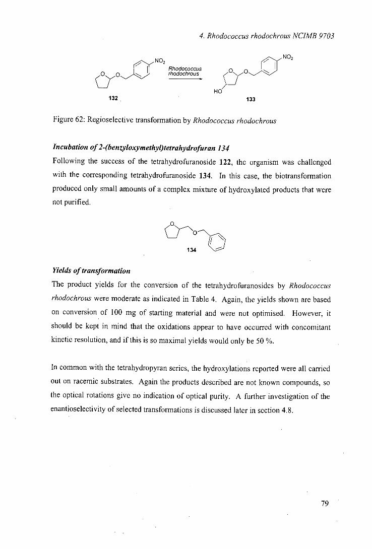
4. Rhodococcus rhodochrous NUMB 9703
NO2
_ O
NO2
Rhodococcus rhodochrous
HO' 132 133
Figure 62: Regioselective transformation by Rhodococcus rhodochrous
Incubation of2-(benzyloxymethyl)tetrahydrofuran 134
Following the success of the tetrahydrofuranoside 122, the organism was challenged
with the corresponding tetrahydrofuranoside 134. In this case, the biotransformation
produced only small amounts of a complex mixture of hydroxylated products that were
not purified.
cj"*'~ 0--'01 134
Yields of transformation
The product yields for the conversion of the tetrahydro furanos ides by Rhodococcus
rhodochrous were moderate as indicated in Table 4. Again, the yields shown are based
on conversion of 100 mg of starting material and were not optimised. However, it
should be kept in mind that the oxidations appear to have occurred with concomitant
kinetic resolution, and if this is so maximal yields would only be 50 %.
In common with the tetrahydropyran series, the hydroxylations reported were all carried
out on racemic substrates. Again the products described are not known compounds, so
the optical rotations give no indication of optical purity. A further investigation of the
enantioselectivity of selected transformations is discussed later in section 4.8.
WE
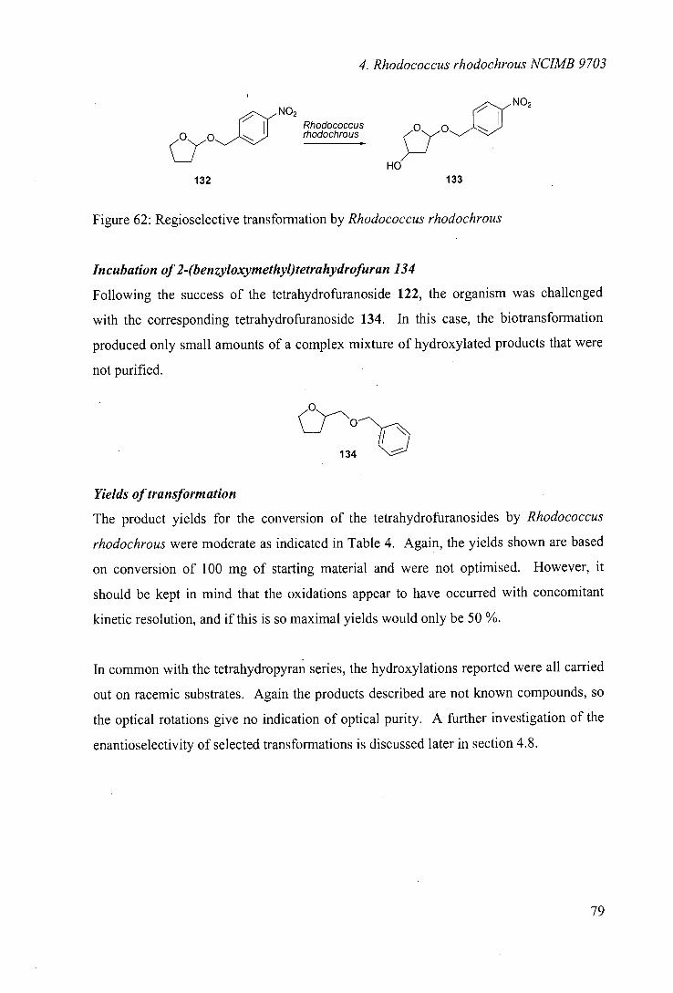
4. Rhodococcus rhodochrous NUMB 9703
NO2
Rhodococcus rhodochrous
O O HO
132 133
Figure 62: Regioselective transformation by Rhodococcus rhodochrous
Incubation of2-(benzyloxymethyl)tetrahydrofuran 134
Following the success of the tetrahydrofuranoside 122, the organism was challenged
with the corresponding tetrahydrofuranoside 134. In this case, the biotransformation
produced only small amounts of a complex mixture of hydroxylated products that were
not purified.
Yields of transformation
The product yields for the conversion of the tetrahydrofuranosides by Rhodococcus
rhodochrous were moderate as indicated in Table 4. Again, the yields shown are based
on conversion of 100 mg of starting material and were not optimised. However, it
should be kept in mind that the oxidations appear to have occurred with concomitant
kinetic resolution, and if this is so maximal yields would only be 50 %.
In common with the tetrahydropyran series, the hydroxylations reported were all carried
out on racemic substrates. Again the products described are not known compounds, so
the optical rotations give no indication of optical purity. A further investigation of the
enantioselectivity of selected transformations is discussed later in section 4.8.
79
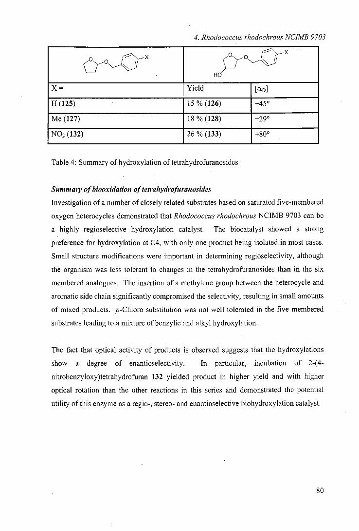
4. Rhodococcus rhodochrous NUMB 9703
o
HO
x
X = Yield [aD]
H(125) 15%(126) 450
Me (127) 18%(128) +290
NO2 (132) 26%(133) +800
Table 4: Summary of hydroxylation of tetrahydrofuranosides
Summary of biooxidation of tetrahydrofuranosides
Investigation of a number of closely related substrates based on saturated five-membered
oxygen heterocycles demonstrated that Rhodococcus rhodochrous NCIMB 9703 can be
a highly regioselective hydroxylation catalyst. The biocatalyst showed a strong
preference for hydroxylation at C4, with only one product being isolated in most cases.
Small structure modifications were important in determining regioselectivity, although
the organism was less tolerant to changes in the tetrahydrofuranosides than in the six
membered analogues. The insertion of a methylene group between the heterocycle and
aromatic side chain significantly compromised the selectivity, resulting in small amounts
of mixed products. p-Chloro substitution was not well tolerated in the five membered
substrates leading to a mixture of benzylic and alkyl hydroxylation.
The fact that optical activity of products is observed suggests that the hydroxylations
show a degree of enantioselectivity. In particular, incubation of 2-(4-
nitrobenzyloxy)tetrahydrofuran 132 yielded product in higher yield and with higher
optical rotation than the other reactions in this series and demonstrated the potential
utility of this enzyme as a regio-, stereo- and enantioselective biohydroxylation catalyst.
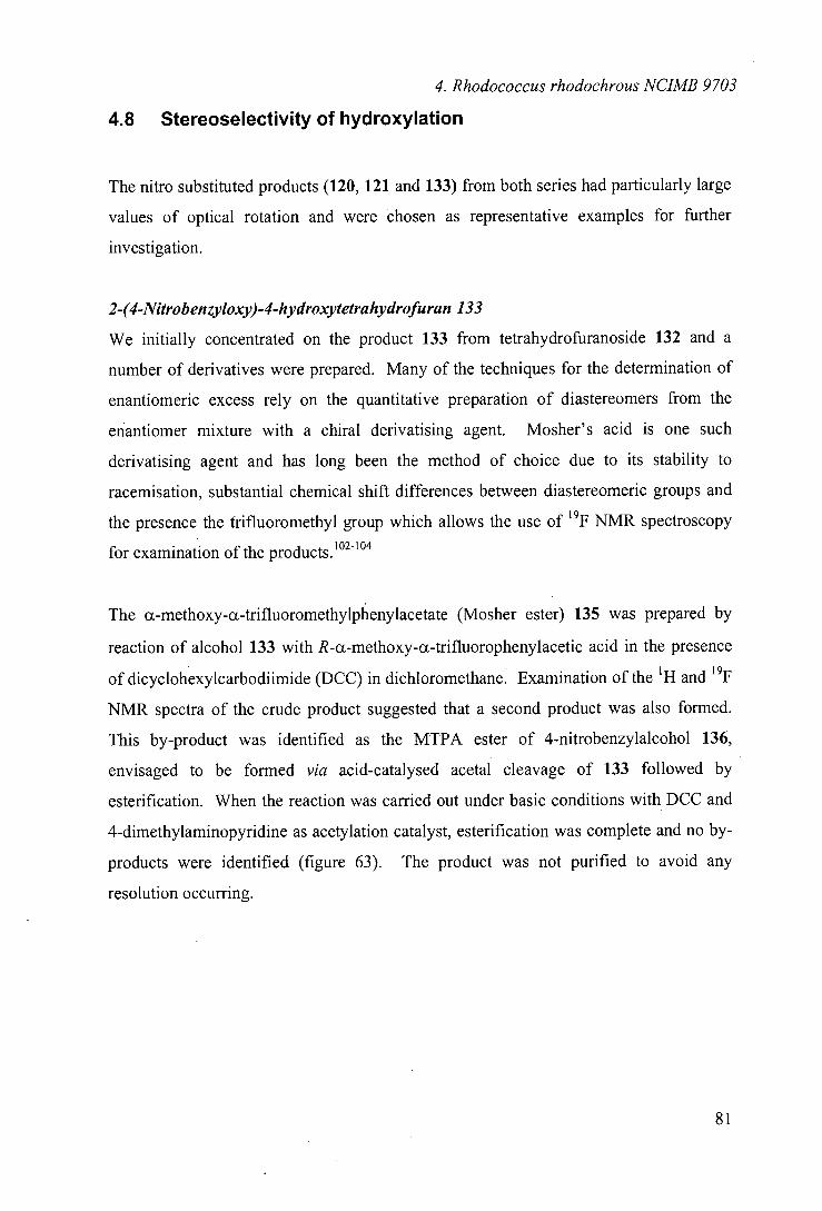
4. Rhodococcus rhodochrous NUMB 9703
4.8 Stereoselectivity of hydroxylation
The nitro substituted products (120, 121 and 133) from both series had particularly large
values of optical rotation and were chosen as representative examples for further
investigation.
2-(4-Nitrobenzyloxy)-4-hydroxytetrahydrofuran 133
We initially concentrated on the product 133 from tetrahydrofuranoside 132 and a
number of derivatives were prepared. Many of the techniques for the determination of
enantiomeric excess rely on the quantitative preparation of diastereomers from the
enantiomer mixture with a chiral derivatising agent. Mosher's acid is one such
derivatising agent and has long been the method of choice due to its stability to
racemisation, substantial chemical shift differences between diastereomeric groups and
the presence the trifluoromethyl group which allows the use of 19F NMR spectroscopy
for examination of the products. 102-104
The ct-methoxy-a-trifluoromethylphenylacetate (Mosher ester) 135 was prepared by
reaction of alcohol 133 with R-a-methoxy-c-trifluorophenylacetic acid in the presence
of dicyclohexylcarbodiimide (DCC) in dichioromethane. Examination of the 'H and '9F
NMR spectra of the crude product suggested that a second product was also formed.
This by-product was identified as the MTPA ester of 4-nitrobenzylalcohol 136,
envisaged to be formed via acid-catalysed acetal cleavage of 133 followed by
esterification. When the reaction was carried out under basic conditions with DCC and
4-dimethylaminopyridine as acetylation catalyst, esterification was complete and no by-
products were identified (figure 63). The product was not purified to avoid any
resolution occurring.
[31
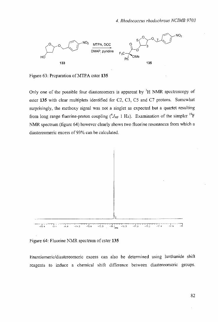
4. Rhodococcus rhodochrous NUMB 9703
~NO, 0
5/ MTPA, DCC 0
DMAP, pyridine 3CZ 0
HO P zWe
133 135
Figure 63: Preparation of MTPA ester 135
Only one of the possible four diastereomers is apparent by 'H NMR spectroscopy of
ester 135 with clear multiplets identified for C2, C3, C5 and C7 protons. Somewhat
surprisingly, the methoxy signal was not a singlet as expected but a quartet resulting
from long range fluorine-proton coupling (4JHF 1 Hz). Examination of the simpler '9F
NMR spectrum (figure 64) however clearly shows two fluorine resonances from which a
diastereomeric excess of 93% can be calculated.
0 -13.5 -14.0 -74,5 -75,0 '-15.5 -76.0 -16.5 -71.0 -17.5 - 7 1.0 -78.5 -70 PPM
Figure 64: Fluorine NMR spectrum of ester 135
Enantiomeric/diastereomeric excess can also be determined using lanthanide shift
reagents to induce a chemical shift difference between diastereomeric groups.
EX
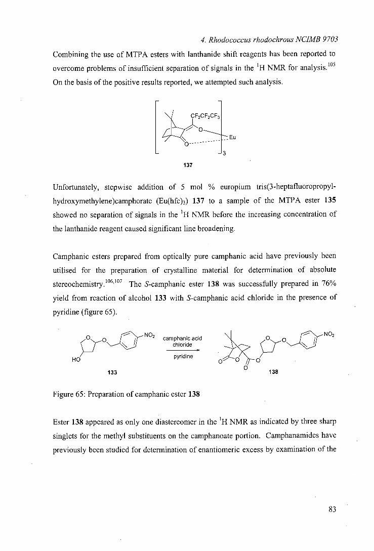
4. Rhodococcus rhodochrous NCIMB 9703
Combining the use of MTPA esters with lanthanide shift reagents has been reported to
overcome problems of insufficient separation of signals in the 'H NMR for analysis.105
On the basis of the positive results reported, we attempted such analysis.
[EU
137
Unfortunately, stepwise addition of 5 mol % europium tris(3-heptafluoropropyl-
hydroxymethylene)camphorate (Eu(hfc)3) 137 to a sample of the MTPA ester 135
showed no separation of signals in the 'H NMR before the increasing concentration of
the lanthanide reagent caused significant line broadening.
Camphanic esters prepared from optically pure camphanic acid have previously been
utilised for the preparation of crystalline material for determination of absolute
stereochemistry. 101,107 The S-camphanic ester 138 was successfully prepared in 76%
yield from reaction of alcohol 133 with S-camphanic acid chloride in the presence of
pyridine (figure 65).
NO2 camphanic acid
chloride
HO pyridine
0 NO2
0ho 133
0 138
Figure 65: Preparation of camphanic ester 138
Ester 138 appeared as only one diastereomer in the 'H NMR as indicated by three sharp
singlets for the methyl substituents on the camphanoate portion. Camphanamides have
previously been studied for determination of enantiomeric excess by examination of the
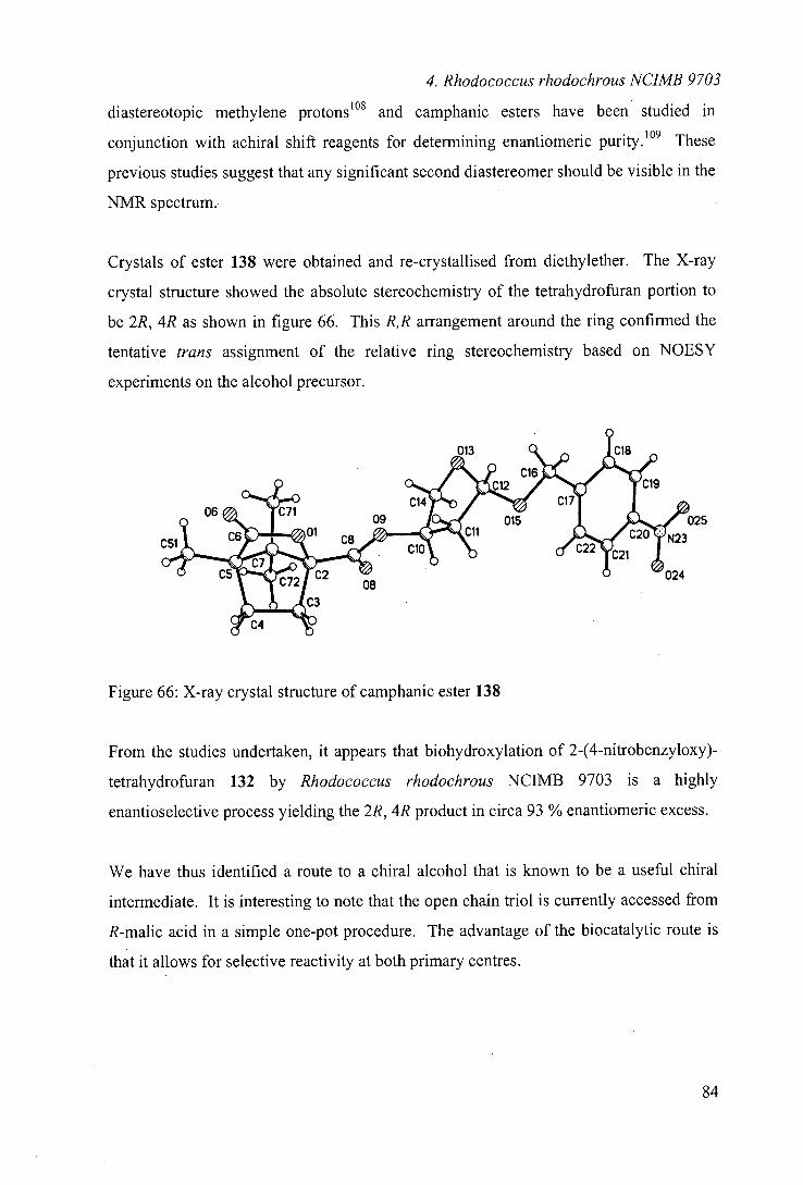
4. Rhodococcus rhodochrous NUMB 9703
diastereotopic methylene protons108 and camphanic esters have been studied in
conjunction with achiral shift reagents for determining enantiomeric purity.109 These
previous studies suggest that any significant second diastereomer should be visible in the
NMR spectrum.
Crystals of ester 138 were obtained and re-crystallised from diethylether. The X-ray
crystal structure showed the absolute stereochemistry of the tetrahydrofuran portion to
be 2R, 4R as shown in figure 66. This R,R arrangement around the ring confirmed the
tentative trans assignment of the relative ring stereochemistry based on NOESY
experiments on the alcohol precursor.
25
Figure 66: X-ray crystal structure of camphanic ester 138
From the studies undertaken, it appears that biohydroxylation of 2-(4-nitrobenzyloxy)-
tetrahydrofuran 132 by Rhodococcus rhodochrous NCIMB 9703 is a highly
enantioselective process yielding the 2R, 4R product in circa 93 % enantiomeric excess.
We have thus identified a route to a chiral alcohol that is known to be a useful chiral
intermediate. It is interesting to note that the open chain triol is currently accessed from
R-malic acid in a simple one-pot procedure. The advantage of the biocatalytic route is
that it allows for selective reactivity at both primary centres.
ON
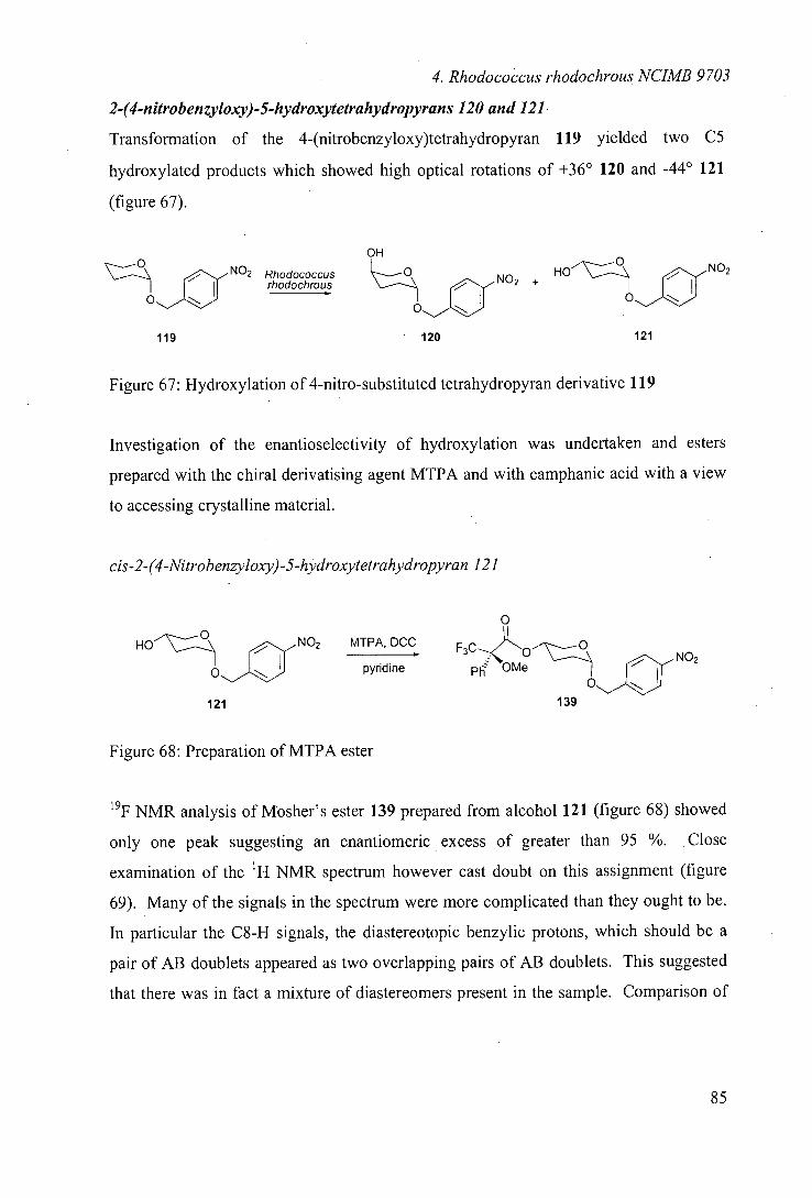
4. Rhodococcus rhodochrous NUMB 9703
2-(4-nitrobenzyloxy)-5-hydroxytetrahydropyrans 120 and 121
Transformation of the 4-(nitrobenzyloxy)tetrahydropyran 119 yielded two C5
hydroxylated products which showed high optical rotations of +36° 120 and -44° 121
(figure 67).
OH 0
Rhodococcus +HO 0
NO2 rhodochrous
119 120 121
Figure 67: Hydroxylation of 4-nitro-substituted tetrahydropyran derivative 119
Investigation of the enantioselectivity of hydroxylation was undertaken and esters
prepared with the chiral derivatising agent MTPA and with camphanic acid with a view
to accessing crystalline material.
cis-2-(4-Nitrobenzyloxy)-5-hydroxytetrahydropyran 121
0 0
HO 0NO2 MTPA, DCC F3C0
pyridine NO2
0 OMe
1 121 39
Figure 68: Preparation of MTPA ester
19F NMR analysis of Mosher's ester 139 prepared from alcohol 121 (figure 68) showed
only one peak suggesting an enantiomeric excess of greater than 95 %. Close
examination of the 'H NMR spectrum however cast doubt on this assignment (figure
69). Many of the signals in the spectrum were more complicated than they ought to be.
In particular the C8-H signals, the diastereotopic benzylic protons, which should be a
pair of AB doublets appeared as two overlapping pairs of AB doublets. This suggested
that there was in fact a mixture of diastereomers present in the sample. Comparison of
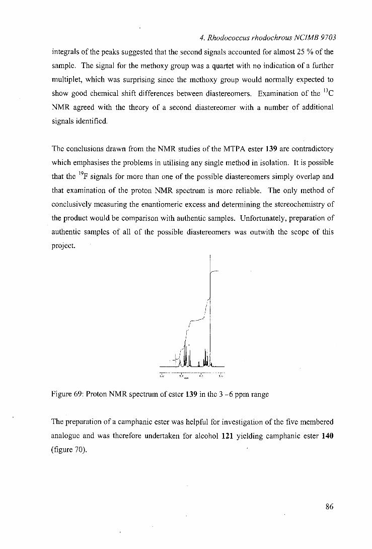
4. Rhodococcus rhodochrous NUMB 9703
integrals of the peaks suggested that the second signals accounted for almost 25 % of the
sample. The signal for the methoxy group was a quartet with no indication of a further
multiplet, which was surprising since the methoxy group would normally expected to
show good chemical shift differences between diastereomers. Examination of the 13C
NMR agreed with the theory of a second diastereomer with a number of additional
signals identified.
The conclusions drawn from the NMR studies of the MTPA ester 139 are contradictory
which emphasises the problems in utilising any single method in isolation. It is possible
that the 19F signals for more than one of the possible diastereomers simply overlap and
that examination of the proton NMR spectrum is more reliable. The only method of
conclusively measuring the enantiomeric excess and determining the stereochemistry of
the product would be comparison with authentic samples. Unfortunately, preparation of
authentic samples of all of the possible diastereomers was outwith the scope of this
project.
Figure 69: Proton NMR spectrum of ester 139 in the 3 —6 ppm range
The preparation of a camphanic ester was helpful for investigation of the five membered
analogue and was therefore undertaken for alcohol 121 yielding camphanic ester 140
(figure 70).
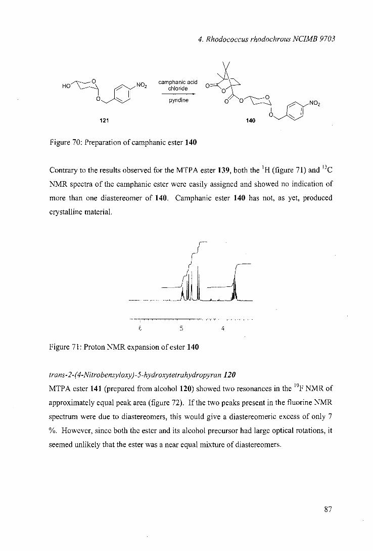
4. Rhodococcus rhodochrous NUMB 9703
0 camphanic acid HO NO2 chloride
0"_"a pyridine
121
NO2
Figure 70: Preparation of camphanic ester 140
Contrary to the results observed for the MTPA ester 139, both the 'H (figure 71) and '3C
NMR spectra of the camphanic ester were easily assigned and showed no indication of
more than one diastereomer of 140. Camphanic ester 140 has not, as yet, produced
crystalline material.
6 5 4
Figure 71: Proton NMR expansion of ester 140
trans-2-(4-Nitrobenzyloxy) -5-hydroxytetrahydropyran 120
MTPA ester 141 (prepared from alcohol 120) showed two resonances in the 19F NMR of
approximately equal peak area (figure 72). If the two peaks present in the fluorine NMR
spectrum were due to diastereomers, this would give a diastereomeric excess of only 7
%. However, since both the ester and its alcohol precursor had large optical rotations, it
seemed unlikely that the ester was a near equal mixture of diastereomers.
4. Rhodococcus rhodochrous NUMB 9703
I I - I 45 -50 55 -60 -65 70 -05 -80 -85 -90 -95 _200 105
Figure 72: Fluorine NMR spectrum of ester 141
Examination of the 'H NMR spectrum clearly showed a mixture of compounds in the
sample, in particular the methoxy and C6-Heq and C6-Hax signals clearly showed
multiplets for different molecules and other signals in the spectrum were much broader
than would be expected. Carbon-13 NMR spectroscopy also showed the existence of
more than one compound with secondary peaks identified at similar chemical shift to the
main peak.
We suggest that the product is not a mixture of diastereomers but that, on formation of
the bulky ester from the axial hydroxyl group, the diaxial ring strain is increased
sufficiently that the energy of the system can overcome the anomeric effect and cause
the ring to flip to produce the more favourable diequatorial arrangement around the
tetrahydropyran ring as shown in figure 73.
OR
- RO9OBfl
OBn
Figure 73: Possible ring flipping to produce a mixture of conformers
M.
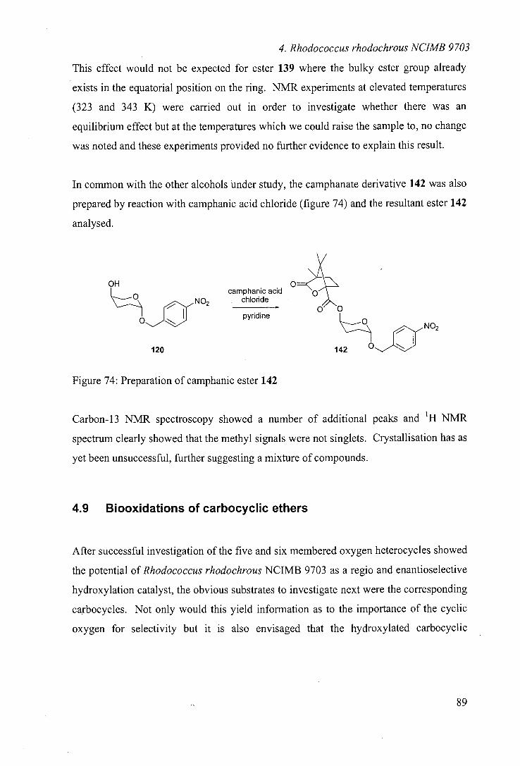
4. Rhodococcus rhodochrous NUMB 9703
This effect would not be expected for ester 139 where the bulky ester group already
exists in the equatorial position on the ring. NMR experiments at elevated temperatures
(323 and 343 K) were carried out in order to investigate whether there was an
equilibrium effect but at the temperatures which we could raise the sample to, no change
was noted and these experiments provided no further evidence to explain this result.
In common with the other alcohols under study, the camphanate derivative 142 was also
prepared by reaction with camphanic acid chloride (figure 74) and the resultant ester 142
analysed.
OH
NO2 0'—ja camphanic acid chloride
pyridine NO2
120
142
Figure 74: Preparation of camphanic ester 142
Carbon-13 NMR spectroscopy showed a number of additional peaks and 'H NMR
spectrum clearly showed that the methyl signals were not singlets. Crystallisation has as
yet been unsuccessful, further suggesting a mixture of compounds.
4.9 Biooxidations of carbocyclic ethers
After successful investigation of the five and six membered oxygen heterocycles showed
the potential of Rhodococcus rhodochrous NCIMB 9703 as a regio and enantioselective
hydroxylation catalyst, the obvious substrates to investigate next were the corresponding
carbocycles. Not only would this yield information as to the importance of the cyclic
oxygen for selectivity but it is also envisaged that the hydroxylated carbocyclic
89
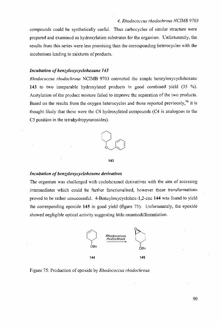
4. Rhodococcus rhodochrous NCIMB 9703
compounds could be synthetically useful. Thus carbocycles of similar structure were
prepared and examined as hydroxylation substrates for the organism. Unfortunately, the.
results from this series were less promising than the corresponding heterocycles with the
incubations leading to mixtures of products.
Incubation of benzyloxycyclohexane 143
Rhodococcus rhodochrous NCIMB 9703 converted the simple benzyloxycyclohexane
143 to two inseparable hydroxylated products in good combined yield (35 %).
Acetylation of the product mixture failed to improve the separation of the two products.
Based on the results from the oxygen heterocycles and those reported previously,96 it is
thought likely that these were the C4 hydroxylated compounds (C4 is analogous to the
CS position in the tetrahydropyranosides).
143
Incubation of benzyloxycyclohexene derivatives
The organism was challenged with cyclohexenol derivatives with the aim of accessing
intermediates which could be further functionalised, however these transformations
proved to be rather unsuccessful. 4-Benzyloxycyclohex-1,2-ene 144 was found to yield
the corresponding epoxide 145 in good yield (figure 75). Unfortunately, the epoxide
showed negligible optical activity suggesting little enantiodifferentiation.
Rhodococcus rhodochrous
OBn OBn
144 145
Figure 75: Production of epoxide by Rhodococcus rhodochrous
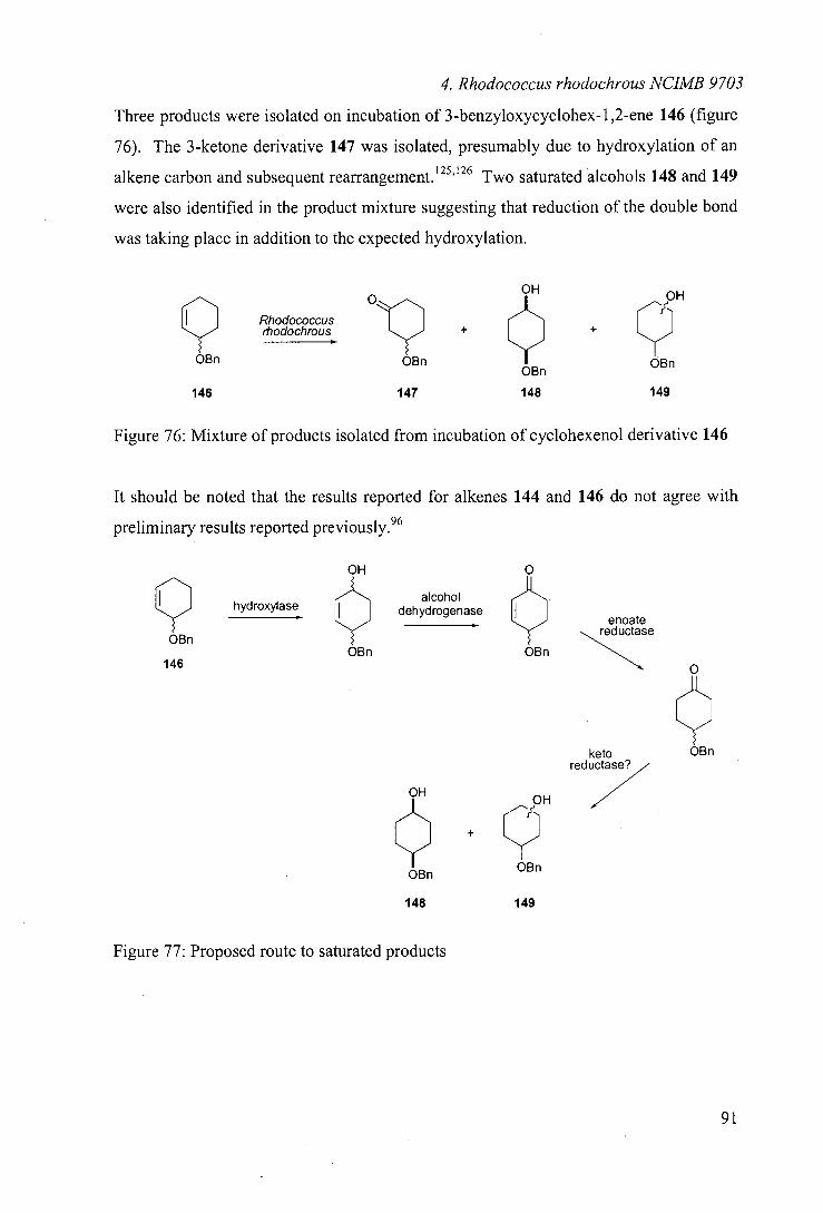
4. Rhodococcus rhodochrous NUMB 9703
Three products were isolated on incubation of 3-benzyloxycyclohex- 1 ,2-ene 146 (figure
76). The 3-ketone derivative 147 was isolated, presumably due to hydroxylation of an
alkene carbon and subsequent rearrangement. 125,126 Two saturated alcohols 148 and 149
were also identified in the product mixture suggesting that reduction of the double bond
was taking place in addition to the expected hydroxylation.
OH OH
9 occus rhodochrous + + 9 OBn OBn OBn Bn
146 147 148 149
Figure 76: Mixture of products isolated from incubation of cyclohexenol derivative 146
It should be noted that the results reported for alkenes 144 and 146 do not agree with
preliminary results reported previously. 96
OH 0
alcohol
enoate
9 hydroxylase dehydrogenase IIIIIIII
redLitase
OBn OBn OBn
146
keto
O
redu>
H OH
/
OBn Bn
148 149
OBn
Figure 77: Proposed route to saturated products
91
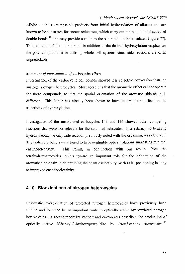
4. Rhodococcus rhodochrous NUMB 9703
Allylic alcohols are possible products from initial hydroxylation of alkenes and are
known to be substrates for enoate reductases, which carry out the reduction of activated
double bonds"° and may provide a route to the saturated alcohols isolated (figure 77).
This reduction of the double bond in addition to the desired hydroxylation emphasises
the potential problems in utilising whole cell systems since side reactions are often
unpredictable.
Summary of biooxidation of carbo cyclic ethers
Investigation of the carbocyclic compounds showed less selective conversion than the
analogous oxygen heterocycles. Most notable is that the anomeric effect cannot operate
for these compounds so that the spatial orientation of the aromatic side-chain is
different. This factor has already been shown to have an important effect on the
selectivity of hydroxylation.
Investigation of the unsaturated carbocycles 144 and 146 showed other competing
reactions that were not relevant for the saturated substrates. Interestingly no benzylic
hydroxylation, the only side reaction previously noted with the organism, was observed.
The isolated products were found to have negligible optical rotations suggesting minimal
enantioselectivity. This result, in conjunction with our results from the
tetrahydropyranosides, points toward an important role for the orientation of the
aromatic side-chain in determining the enantioselectivity, with axial positioning leading
to improved enantioselectivity.
4.10 Biooxidations of nitrogen heterocycles
Enzymatic hydroxylation of protected nitrogen heterocycles have previously been
studied and found to be an important route to optically active hydroxylated nitrogen
heterocycles. A recent report by Witholt and co-workers described the production of
optically active N-benzyl-3-hydroxypyrrolidine by Pseudomonas oleovorans.' 1 '
92
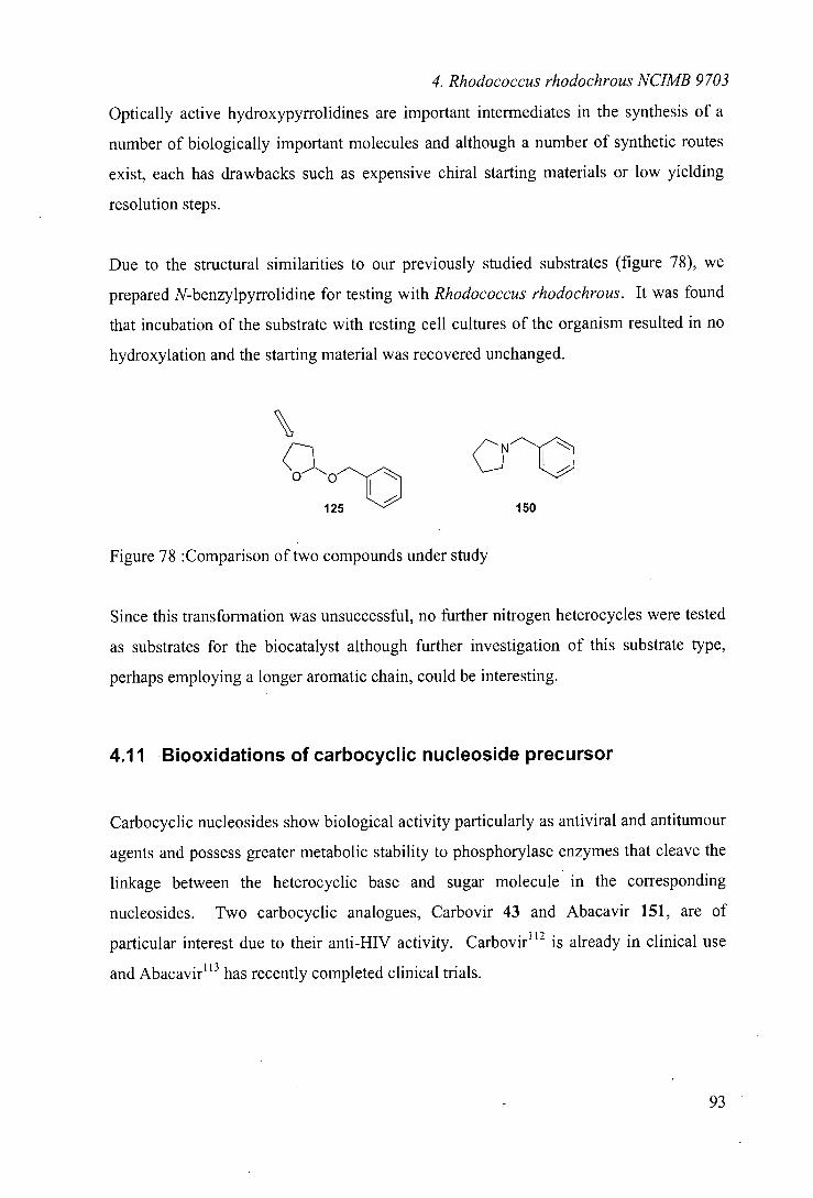
4. Rhodococcus rhodochrous NUMB 9703
Optically active hydroxypyrrolidines are important intermediates in the synthesis of a
number of biologically important molecules and although a number of synthetic routes
exist, each has drawbacks such as expensive chiral starting materials or low yielding
resolution steps.
Due to the structural similarities to our previously studied substrates (figure 78), we
prepared N-benzylpyrrolidine for testing with Rhodococcus rhodochrous. It was found
that incubation of the substrate with resting cell cultures of the organism resulted in no
hydroxylation and the starting material was recovered unchanged.
Qo cr 125 150
Figure 78 :Comparison of two compounds under study
Since this transformation was unsuccessful, no further nitrogen heterocycles were tested
as substrates for the biocatalyst although further investigation of this substrate type,
perhaps employing a longer aromatic chain, could be interesting.
4.11 Biooxidations of carbocyclic nucleoside precursor
Carbocyclic nucleosides show biological activity particularly as antiviral and antitumour
agents and possess greater metabolic stability to phosphorylase enzymes that cleave the
linkage between the heterocyclic base and sugar molecule in the corresponding
nucleosides. Two carbocyclic analogues, Carbovir 43 and Abacavir 151, are of
particular interest due to their anti-HIV activity. Carbovir"2 is already in clinical use
and Abacavir"3 has recently completed clinical trials.
93
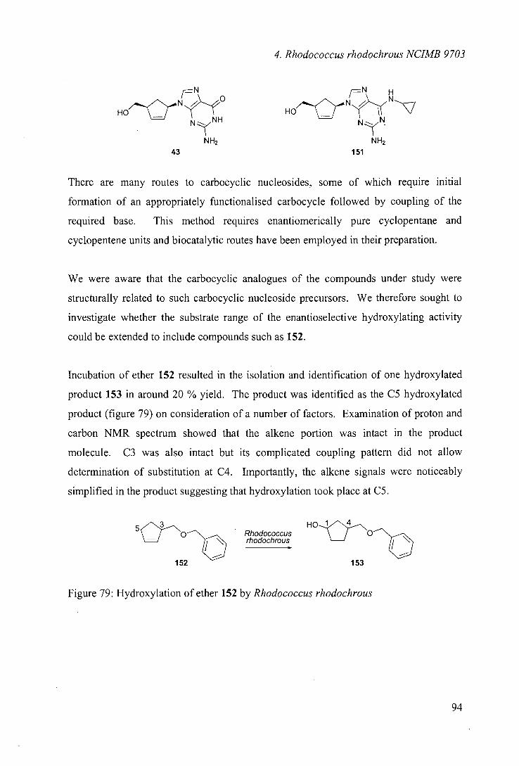
4. Rhodococcus rhodochrous NUMB 9703
_N 1rN H
HO N N NH N2N.
NH2 NH2 43
151
There are many routes to carbocyclic nucleosides, some of which require initial
formation of an appropriately functionalised carbocycle followed by coupling of the
required base. This method requires enantiomerically pure cyclopentane and
cyclopentene units and biocatalytic routes have been employed in their preparation.
We were aware that the carbocyclic analogues of the compounds under study were
structurally related to such carbocyclic nucleoside precursors. We therefore sought to
investigate whether the substrate range of the enantioselective hydroxylating activity
could be extended to include compounds such as 152.
Incubation of ether 152 resulted in the isolation and identification of one hydroxylated
product 153 in around 20 % yield. The product was identified as the C5 hydroxylated
product (figure 79) on consideration of a number of factors. Examination of proton and
carbon NMR spectrum showed that the alkene portion was intact in the product
molecule. C3 was also intact but its complicated coupling pattern did not allow
determination of substitution at C4. Importantly, the alkene signals were noticeably
simplified in the product suggesting that hydroxylation took place at C5.
5
HO 'o--\, Rhodococcus
rhodochrous
152 153
Figure 79: Hydroxylation of ether 152 by Rhodococcus rhodochrous
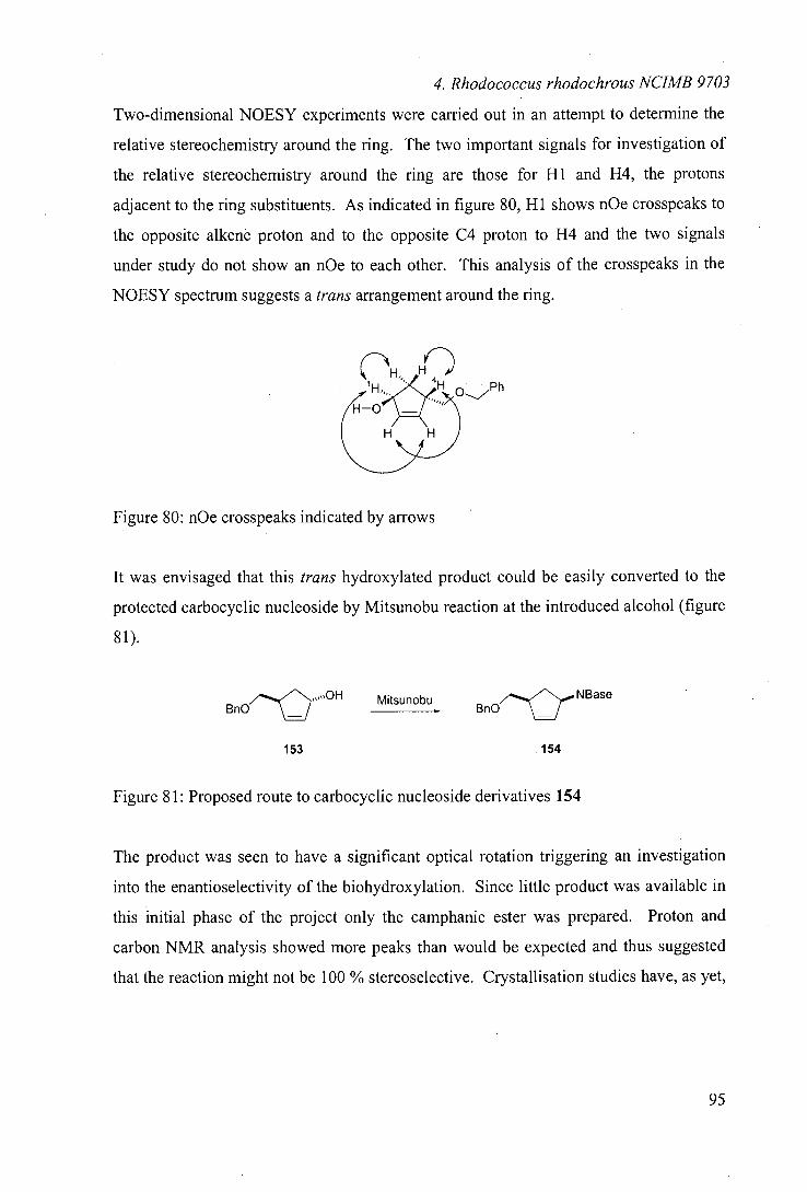
4. Rhodococcus rhodochrous NUMB 9703
Two-dimensional NOESY experiments were carried out in an attempt to determine the
relative stereochemistry around the ring. The two important signals for investigation of
the relative stereochemistry around the ring are those for Hi and H4, the protons
adjacent to the ring substituents. As indicated in figure 80, Hi shows nOe crosspeaks to
the opposite alkenè proton and to the opposite C4 proton to H4 and the two signals
under study do not show an nOe to each other. This analysis of the crosspeaks in the
NOESY spectrum suggests a trans arrangement around the ring.
Figure 80: nOe crosspeaks indicated by arrows
It was envisaged that this trans hydroxylated product could be easily converted to the
protected carbocyclic nucleoside by Mitsunobu reaction at the introduced alcohol (figure
81).
BnO Mjtsijnobu
153
154
Figure 81: Proposed route to carbocyclic nucleoside derivatives 154
The product was seen to have a significant optical rotation triggering an investigation
into the enantioselectivity of the biohydroxylation. Since little product was available in
this initial phase of the project only the camphanic ester was prepared. Proton and
carbon NMR analysis showed more peaks than would be expected and thus suggested
that the reaction might not be 100 % stereoselective. Crystallisation studies have, as yet,
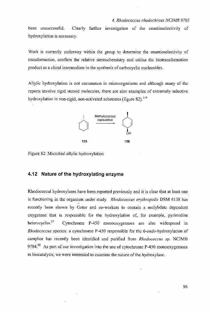
4. Rhodococcus rhodochrous NUMB 9703
been unsuccessful. Clearly further investigation of the enantioselectivity of
hydroxylation is necessary.
Work is currently underway within the group to determine the enantioselectivity of
transformation, confirm the relative stereochemistry and utilise the biotransformation
product as a chiral intermediate in the synthesis of carbocyclic nucleosides.
Allylic hydroxylation is not uncommon in microorganisms and although many of the
reports involve rigid steroid molecules, there are also examples of extremely selective
hydroxylation in non-rigid, non-activated substrates (figure 82).1 14
Met hylococcus capsulatus
OH
155 156
Figure 82: Microbial allylic hydroxylation
4.12 Nature of the hydroxylating enzyme
Rhodococcal hydroxylases have been reported previously and it is clear that at least one
is functioning in the organism under study. Rhodococcus erythropolis DSM 6138 has
recently been shown by Gotor and co-workers to contain a molybdate dependent
oxygenase that is responsible for the hydroxylation of, for example, pyrimidine
heterocycles. 27 Cytochrome P-450 monooxygenases are also widespread in
Rhodococcus species: a cytochrome P-450 responsible for the 6-endo-hydroxylation of
camphor has recently been identified and purified from Rhodococcus sp. NCIMB
9784.68 As part of our investigation into the use of cytochrome P-450 monooxygenases
in biocatalysis, we were interested to examine the nature of the hydroxylase.
4. Rhodococcus rhodochrous NUMB 9703
Alkane hydroxylators have previously been shown to hydroxylate non-natural
substrates. Another octane utilising enzyme, Pseudomonas óleovorans is known to
contain a cytoplasmic membrane monooxygenase, which carries out the 00-
hydroxylation of octane. A recent publication by Witholt et a!" has shown the utility
of the alkane hydroxylase system by preparing optically active hydroxylated N-
benzylpyrrolidine (as described in section 4.8).
Initial literature reports suggested that terminal hydroxylation of n-octane by
Rhodococcus rhodochrous NCIMB 9703 is catalysed by an inducible cytochrome
P-450.48'65 In this work we hoped to utilise this cytochrome P-450 as a biohydroxylation
catalyst. The reactions reported here were carried out using cells grown on octane as the
sole carbon source, but cells grown on sodium pyruvate were also tested in an attempt to
establish the nature of the hydroxylase. Comparable yields and optical activities of
products were observed utilising both octane and pyruvate grown cells, suggesting that
these reactions do not utilise the octane-hydroxylating enzyme, or that there is an
unknown limiting factor in operation. Such a limiting factor could render the expression
level of the protein insignificant and could be NADH supply, substrate diffusion or
oxygen transfer into the cell.
Inhibition studies using 1 -aminobenzotriazole and metyrapone showed that
hydroxylation was inhibited by both of these compounds. Because these cytochrome
P-450 inhibitors inhibit the enzyme by different mechanisms,27'89 these studies were
taken to be evidence that a cytochrome P-450 system was operating in these
transformations.
Thus far, these studies have yielded only limited information about the hydroxylase
under study but it is clear that the hydroxylations reported are cytochrome P-450
catalysed. As in chapter 3, it is unclear whether the hydroxylase operating is the induced
cytochrome P-450 or another constitutively expressed enzyme.
97
4. Rhodococcus rhodochrous NUMB 9703
4.13 Optimisation of transformations
Although some studies have been undertaken to improve the biotransformation, there are
many avenues for optimisation of the whole cell system as a hydroxylation catalyst. The
effect of substrate concentration has been investigated and the optimum concentration
found to be around 0.5 gU', with higher concentrations of up to 0.6 and 0.7 gU'
tolerated for some substrates. The exact concentration tolerance seems to be substrate
dependent and would require further investigation for larger scale preparations.
Another extremely important variable is cell concentration. In some studies, the authors
report extensive investigation to optimise this parameter."' In our case, the growth of
the organism was the slowest stage in the testing of substrates and we therefore sought to
minimise the cell mass used for each reaction. We found that the use of 5 g wet cell
mass for transformation of 100 mg substrate (200 mL volume) was the minimum cell
mass that did not increase the required time unnecessarily.
The time of transformation is an important factor, both in terms of practicality and
selectivity. In the hydroxylations described, optically active products were isolated in
yields of up to 40 %. The mass balance of the reaction is thought to be lost through
degradation pathways from the substrate or the hydroxylated product. The enantiomeric
excess of the isolated product is likely to be dependent on a number of factors including
further degradation of the desired product. In future, periodic sampling of aliquots
should provide data to optimise this.
Co-metabolism of a co-substrate such as glucose could also be advantageous since this
provides energy for the reaction and the recycling of co-factors that could increase the
rate sufficiently to impact on the yield or selectivity of hydroxylation.
Work is currently underway on the optimisation of conditions for the preparation of
carbocyclic nucleoside precursors.
M.
4. Rhodococcus rhodochrous NUMB 9703
4.14 Conclusions
This study has established that Rhodococcus rhodochrous NCIMB 9703, an organism
not previously reported for biotransformations of non-natural substrates, can be used to
regioselectively and enantioselectively hydroxylate a range of oxygen heterocycles. It
was also noted that the regio- and enantioselectivity of reaction was significantly altered
by changes to the p-substituent on the benzyloxy group or by extension of the ether
linkage. It is envisaged that this biocatalyst could be used to produce hydroxylated,
polyfunctionalised chiral intermediates for use in asymmetric synthesis.
2-Benzyloxytetrahydropyrans were substrates for resting cell suspensions of the bacteria
yielding predominantly 5-hydroxylated products in combined yields of up to 40 %.
Corresponding tetrahydrofuranosides were also hydroxylated, mainly to 4-hydroxy
compounds in yields of up to 26 %. Most interestingly, 2-(4-nitrobenzyloxy)-
tetrahydrofuran 125 was transformed exclusively to the 4-hydroxylated product 130 with
an optical rotation of +80° in 93 % enantiomeric excess and the 2-(4-nitrobenzyloxy)-
tetrahydropyran 115 was hydroxylated exclusively in the 5-position giving two products
with optical rotation +36° and -44°.
The transformation of a carbocyclic nucleoside precursor 152 was investigated with the
desired trans product identified as the only product. The hydroxylation was found to be
enantioselective in addition to its high regiospecificity. This particularly positive result
is currently under further study within the group.
5. Experimental
5 Experimental
5.1 Apparatus and chemicals
All chemicals were purchased from the Aldrich Chemical Company, Poole, Dorset U.K.
or Acros Organics, Loughborough, U.K.. 'H and '3C NMR analysis was performed on
Bruker AC250 (250 MHz and 63MHz respectively) and WH-360 (360 MHz and 90
MHz) spectrometers. 19F NMR analysis was performed on a Bruker AC250
spectrometer at 235 MHz. Unless otherwise specified, spectra were recorded in
deuterochloroform at ambient temperature. Chemical shifts are quoted. in parts per
million (ppm) and were referenced internally ('H, CHC13, 7.27 ppm; 13CHC13, 77.0
ppm). Carbon multiplicity was established by DEPT (distortionless enhancement by
polarisation transfer).
Infra-red spectra were recorded on a Perkin-Elmer FT-JR spectrometer (Paragon 1000)
as thin films with frequencies (v) measured in wavenumbers (cm'). Mass spectra were
run using electron impact (El) on Finnigan 4500 or 4600 instruments. Thin layer
chromatography was performed on glass sheets coated with silica gel Merck 60E-254
(0.24 mm, Art. 5715). Components were detected by UV (254 nm) and visualised by
treating the plate with ammonium molybdate solution and heating. Wet flash
chromatography was carried out on silica gel (Merck 9385, Kieselgel 60).
Optical rotations [aD] were measured on an Optical Activity AA-1000 polarimeter with
a cell path length of 1 dm at a temperature of 25 °C and concentrations (c) quoted in
g/100 mL in methanol (sodium 589 nm detection). Unless otherwise stated, the
compounds reported have been prepared in racemic form.
5. Experimental
Gas chromatography (GC) was carried out using a Hewlett-Packard 6890 series GC
system with flame ionisation detection or mass selective detection (HP 5973) employing
a HP-5MS 5% phenyl methyl siloxane capillary column (30.Om x 320tm x 0.25 jtm).
The HPLC analysis was carried out on a Waters 486 system employing Millennium
software using a Sphericlone® 5 t ODS-2, 250 x 4.6 mm column (Phenomenex).
Preparative scale HPLC was carried out on an equivalent column with an internal
diameter of 10.0 mm at a flow-rate of 4.5 mL/min.
101
5. Experimental
5.2 Beauveria bassiana ATCC 7159
5.2.1 Maintenance and growth of microorganism
Beauveria bassiana ATCC 7159 was obtained from the American Type Culture
Collection, U.S.A.. The organism was maintained on malt extract agar plates at room
temperature and these were subcultured at regular intervals. A loop of fungus was used
to inoculate 60 mL of sterile medium containing 7.5 g U' corn steep solids and 10 g U'
glucose in distilled water adjusted to pH 4.85 in a 250 mL Erlenmeyer flask. After three
days growth on an orbital shaker at 200 r.p.m. at 25° C, the culture was used to inoculate
600 mL of the same medium in a 2 L Erlenmeyer flask. This culture was again grown
for three days.
5.2.2 General biotransformation procedure
A solution of 66 mg substrate in 1 mL ethanol was prepared and added to a three day old
660 mL culture of Beauveria bassiana ATCC 7159. After a further three days
incubation as above, the fungal cells were removed from the broth by centrifugation and
the supernatant extracted into ethyl acetate. Purification of metabolites was routinely
carried out using wet flash chromatography with petroleum ether/ethyl acetate gradients
as eluent.
102
5. Experimental
5.2.3 Results of biotransformations
Typical numbering scheme used for substituted piperidines under study
Biohydroxylation of N-carboxybenzylpiperidine 5774 produced hydroxylated product 58
that was isolated as a colourless oil (100 mg, 33 %). This piperidinol has been reported
and characterised previously.' 15,116
Biohydroxylation of N-carboxybenzyl-4-methylpiperidine 62 74
N-carboxybenzyl-4-methyl-4-hydroxy-piperidine 63 was isolated as a pale yellow solid
(130 mg, 45 %); mp 76 °C; oH (250 MHz) 1.24 (3H, s, CH3); 1.45 - 1.56 (5H, in,
C(3)H2, C(5)H2 & C(4)OH), 3.23 - 3.34 (2H, dt, C(2)Hax & C(6)Hax, J 7 & 13), 3.80
(2H, br. d, C(2)Heq & C(6)Heq, 1 13), 5.11 (2H, s, CH2Ph), 7.25 - 7.36 (5H, in,
aromatic); 0c (63 MHz) 30.0 (q, CH3), 38.2 (t, C3 & CS), 40.2 (t, C2 & C6), 66.9 (t,
CH2Ph), 67.7 (s, C4), 127.7 (d, aromatic), 127.8 (d, aromatic), 128.3 (d, aromatic), 136.7
(s), 155.1 (s); m/z (El) required 249.13649, found 249.13637; 249 (44 %, Mt), 158 (14,
M - PhCH2), 142 (16, M - PhCH20), 91(100, PhCH2).
Biohydroxylation of N-carboxybenzyl-3-methylpiperidine 67
Three products were isolated from this biohydroxylation, two of which were found to be•
difficult to separate and were thus separated by subjecting the mixture to acetylation
conditions (10 mol equivalents acetic anhydride, 5 mol equivalents pyridine and
catalytic dimethylaminopyridine) under which conditions one of the products was
103
5. Experimental
acetylated 69 and one was left untouched 70. The products were then separated by
preparative HPLC
N-carboxybenzyl-trans-3-methyl-4-hydroxypiperidine 68 was isolated as a colourless oil
(10 mg, 3 %); oH (360 MHz) 0.99 (3H, d, CH3, J 6.5), 1.43 - 1.59 (3H, br. m) & 1.91
(1H, br. dd, J 3.& 13) (C(3)H, C(4)OH or C(5)H2), 2.53 (1H, br. s, C(2)Hax or
C(6)Hax), 2.90 (1H, ddd, C(4)H, J 3, 12 & 14), 3.30 (1H, br. m, C(2)Hax or C(6)Hax),
4.05 - 4.13 (2H, br. m, C(2)Heq & C(6)Heq), 5.11 (1H, s, CHAHBPh), 5.12 (1H, s,
CHAHBPh), 7.28 - 7.36 (5H, m, aromatic); m/z (El) required 249.13649, found
249.13616; 249 (8 %, Mt), 158 (11, M - PhCH2), 142 (6, M - PhCH20), 91(100,
PhCH2).
N-carboxybenzyl-cis-3-methyl-4-hydroxypiperidine was isolated as its acetylated
derivative 69a as a colourless oil (5 mg); oH (360 MHz): 0.88 (3H, d, CH3, J 7). 1.67 -
1.93 (3H, br. m, C(3)H & C(5)H2), 2.07 (3H, s, acetyl CH3), 3.00 - 3.15 (1H, br. m,
C(2)Hax or C(6)Hax), 3.25 - 3.28 (1H, br. rn, C(2)Hax or C(6)Hax), 3.70 (1H, br. 5,
C(2)Heq), 3.75 (1H, ddd, C(6)Heq,J4.5, 4.5 & 13.5), 5.01 (1H, ddd, C(4)Heq,J3, 3 &
5), 5.12 (2H, d, CH2Ph, J2), 7.28 - 7.39 (5H, m, aromatic); m/z (El) required 29 1.14706,
found 291.14768; 291 (3 %. M), 200 (11, M - PhCH2), 91(100, PhCH2).
N-carboxybenzyl-3-methyl-3-hydroxypiperidine 70 was isolated as a colourless oil (10
mg, 3 %); 0H (360 MHz) 1.21 (3H, s, CH3), 1.49 - 1.79 (5H, m, C(3)OH, C(4)H2 &
C(5)H2). 2.96 - 3.03 (2H, br. m, C(2)Hax & C(6)Hax), 3.68 (1H, br.s, C(2)Heq), 3.84
(1H, ddd, C(6)Heq, J4.5, 4.5 & 13), 5.13 (2H, s, CH2Ph), 7.28 - 7.35 (5H, m, aromatic);
m/z (El) required 249.13649, found 249.13725; 249 (5 %, Mt), 114 (26, M -
PhCH2000), 91(100, PhCH2).
104

5. Experimental
Biohydroxylalion of N-carboxybenzyl-2-methylpiperidine 71
N-carboxybenzyl-cis-2-methyl-4-hydroxy-piperidine 72 was isolated as a colourless oil
(20 mg, 7%); oH (250 MHz) 1.35 (3H, d, CH3, J 7),1.46- 1.91 (5H, m, C(3)H2, C(4)OH
& C(5)H2), 3.34 (1H, ddd, C(6)Hax, J 3.5 & 12.5), 3.90 (1H, ddd, C(6)Heq, J 3.5, 4 &
12.5), 4.16 (1H, q, C(4)H, J3), 4.31 -4.42 (1H, m, C(2)Heq), 5.12 (2H, s, CH2Ph), 7.25
- 7.36 (5H, m, aromatic); 6c (90 MHz) 19.1 (q, CH3), 32.2, 33.5 (t, C3 & C5), 36.4 (t,
C6), 45.7 (d, C2), 64.7 (d, C4), 66.8 (t, CH2Ph), 127.7 (d, aromatic), 127.8 (d, aromatic),
128.4 (d, aromatic), 136.9 (s), 155.2 (s); m/z (El) required 249.13649, found
249.136337, (FAB) 234 (4 %, M - CH3), 158 (15, M - PhCH2), 142 (31, M - PhCH20),
114 (2, M - PhCH2CO2), 91(100, PhCH2).
Figure 83: nOe enhancements and coupling constants used in assigning relative
stereochemistry in product 72
N-carboxybenzyl-cis-2-methyl-3-hydroxy-piperidine 73 was isolated as a colourless oil
(20 mg, 7 %); 011 (360 MHz) 1.12 (3H, d, CH3, J7), 1.43 - 1.78 (SH, m, C(3)OH, C(4)H2
& C(5)H2), 2.79 (1H, ddd, C(6)Hax, J3, 13 & 13.5), 3.77 (1H, m, C(3)H), 3.95 (1H, br
d, C(6)Heq, J 12.5), 4.51 (1H, q, C(2)Heq, J6.5), 5.12 (2H, s, CH2Ph), 7.25 - 7.38 (5H,
m, aromatic); Oc (90 MHz) 9.2 (q, CH3), 24.0 (t, C4 or CS), 27. 1, (t, C4 or CS), 37.6 (t,
C6), 51.0 (d, C2), 67.0 (t, CH2Ph), 69.0 (d, 0), 127.7 (d, aromatic), 127.9 (d, aromatic),
128.4 (d, aromatic), 136.7 (s), 155.3 (s); m/z (El) required 249.13649, found 249.13637,
(APCi) 250 (100 %, MH), 158 (7, M - PhCH2).
105
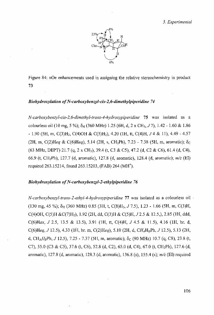
5. Experimental
Figure 84: nOe enhancements used in assigning the relative stereochemistry in product
73
Biohydroxylation of N-carboxybenzyl-cis-2, 6-dimethylpiperidine 74
N-carboxybenzyl-cis-2, 6-dimethyl-trans-4-hydroxypiperidine 75 was isolated as a
colourless oil (10 mg, 5 %); oH (360 MHz) 1.25 (6H, d, 2 x CH3, J7), 1.42- 1.60 & 1.86
- 1.90 (5H, in, C(3)H2, C(4)OH & C(5)H2), 4.20 (1H, tt, C(4)H, J4 & 11), 4.49 -4.57
(2H, in, C(2)Heq & C(6)Heq), 5.14 (2H, s, CH2Ph), 7.23 - 7.38 (5H, in, aromatic); 0c
(63 MHz, DEPT) 21.7 (q, 2 x CH3), 39.4 (t, C3 & CS), 47.2 (d, C2 & C6), 61.4 (d, C4),
66.9 (t, CH2Ph), 127.7 (d, aromatic), 127.8 (d, aromatic), 128.4 (d, aromatic); m/z (El)
required 263.152 14, found 263.15203, (FAB) 264 (MH).
Biohydroxylation of N-carboxybenzyl-2-ethylpiperidine 76
N-carboxybenzyl-trans-2-ethyl-4-hydroxypiperidine 77 was isolated as a colourless oil
(130 mg, 45 %); OH (360 MHz) 0.85 (3H, t, C(8)H3, J7.5), 1.23 - 1.66 (5H, in, C(3)H,
C(4)OH, C(5)H &C(7)H2), 1.92 (2H, dd, C(3)H & C(5)H,J2.5 & 12.5,), 2.85 (1H, ddd,
C(6)Hax, J 2.5, 13.5 & 13.5), 3.91 (1H, tt, C(4)H, J 4.5 & 11.5), 4.16 (1H, br. d,
C(6)Heq, J 12.5), 4.33 (1H, br. in, C(2)Heq), 5.10 (2H, d, CHAHBPh, J 12.5), 5.13 (2H,
d, CHAHBPh, J 12.5), 7.25 - 7.37 (5H, in, aromatic); Oc (90 MHz) 10.7 (q, C8), 23.8 (t,
C7), 35.0 (C3 & C5), 37.6 (t, C6), 52.8 (d, C2), 65.0 (d, C4), 67.0 (t, CH2Ph), 127.6 (d,
aromatic), 127.8 (d, aromatic), 128.3 (d, aromatic), 136.8 (s), 155.4 (s); m/z (El) required
106
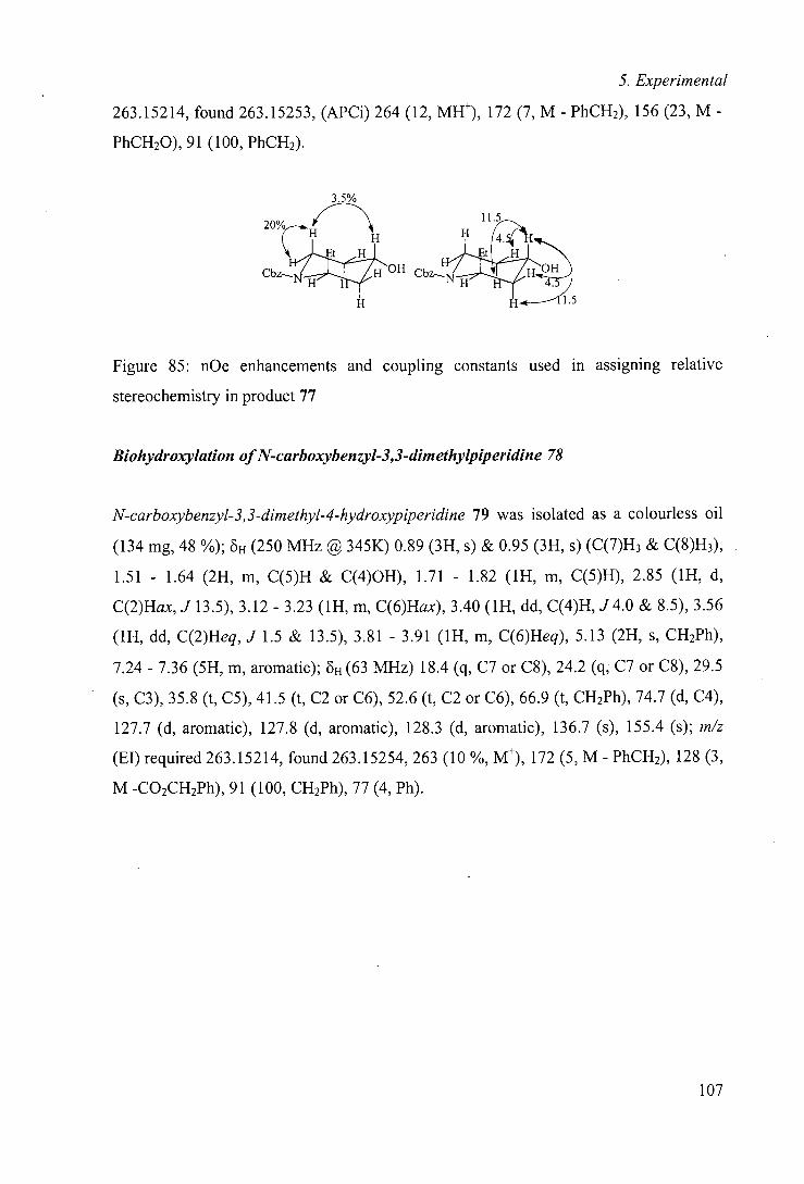
5. Experimental
263.15214, found 263.15253, (APCi) 264 (12, MH), 172 (7, M - PhCH2), 156 (23, M -
PhCH20), 91(100, PhCH2).
3.5%
20%
'•: H H
. H C Et H Et
Cbz_ I1 0H CbZ4I 0H
Figure 85: nOe enhancements and coupling constants used in assigning relative
stereochemistry in product 77
Biohydroxylation of N-carboxybenzyl-3,3-dimethylpiperidine 78
N-carboxybenzyl-3,3-dimethyl-4-hydroxypiperidine 79 was isolated as a colourless oil
(134 mg, 48 %); 8H (250 MHz @ 345K) 0.89 (3H, s) & 0.95 (3H, s) (C(7)H3 & C(8)H3),
1.51 - 1.64 (2H, m, C(5)H & C(4)OH), 1.71 - 1.82 (1H, m, C(5)H), 2.85 (1H, d,
C(2)Hax, J 13.5), 3.12 - 3.23 (1H, m, C(6)Hax), 3.40 (1H, dd, C(4)H, J4.0 & 8.5), 3.56
(1H, dd, C(2)Heq, J 1.5 & 13.5), 3.81 - 3.91 (1H, m, C(6)Heq), 5.13 (2H, s, CH2Ph),
7.24 - 7.36 (5H, m, aromatic); 6H (63 MHz) 18.4 (q, C7 or C8), 24.2 (q, C7 or C8), 29.5
(s, 0), 35.8 (t, CS), 41.5 (t, C2 or C6), 52.6 (t, C2 or C6), 66.9 (t, CH2Ph), 74.7 (d, C4),
127.7 (d, aromatic), 127.8 (d, aromatic), 128.3 (d, aromatic), 136.7 (s), 155.4 (s); m/z
(El) required 263.15214, found 263.15254, 263 (10 %, M), 172 (5, M - PhCH2), 128 (3,
M -0O2CH2Ph), 91(100, CH2Ph), 77 (4, Ph).
107
5. Experimental
5.3 Rhodococcus sp. NCIMB 9784
5.3.1 Maintenance and growth of microorganism
Rhodococcus sp. NCIMB 9784 was obtained from the National Collection of Industrial
and Marine Bacteria, Aberdeen, U.K.. The organism was maintained on agar plates at
4 °C and these were subcultured at regular intervals. A loop of bacteria was used to
inoculate 100 mL of sterile basal salts medium§ with 0.5 g U' ferrous sulfate
heptahydrate and 4 g U' sodium pyruvate added through sterile filters, in a 250 mL
Erlenmeyer flask. After three days growth on an orbital shaker at 250 r.p.m. at 30 °C,
five such cultures were used to inoculate 8 L sterile basal salts media in a 10 L fermenter
with 0.05 g U' ferrous sulfate heptahydrate and 1 g L' (+)-camphor added. The culture
was grown for three days at 30 °C with aeration and stirring (200 rpm), with additional
(+)-camphor (1 g L') added after two days growth. The cells were harvested by
centrifugation (11K r.p.m. for 30 minutes) and washed with phosphate buffer (50 MM,
pH 7.4) to remove excess camphor.
5.3.2 General biotransformation procedure
Resting cell transformations were carried out by resuspending cell mass in one-tenth
growth volume of phosphate buffer (50 mM, pH 7.0), the substrate added as a solution
in ethanol to 0.5 mg mL-1 and the suspension incubated at 30 °C with shaking (250
§ Solution A: ammonium chloride 2.0 g U'; potassium dihydrogen phosphate 3.1 g U', dipotassium
hydrogen phosphate 8.2 g U', yeast extract 0.1 g U', tryptone 0.1 g L 1 ; Solution B: magnesium sulfate
0.05 g U', manganese sulfate 0.05 g U', calcium chloride 0.01 g U', ammonium molybdate 0.01 g U'.
Solutions autoclaved separately and mixed when cool.
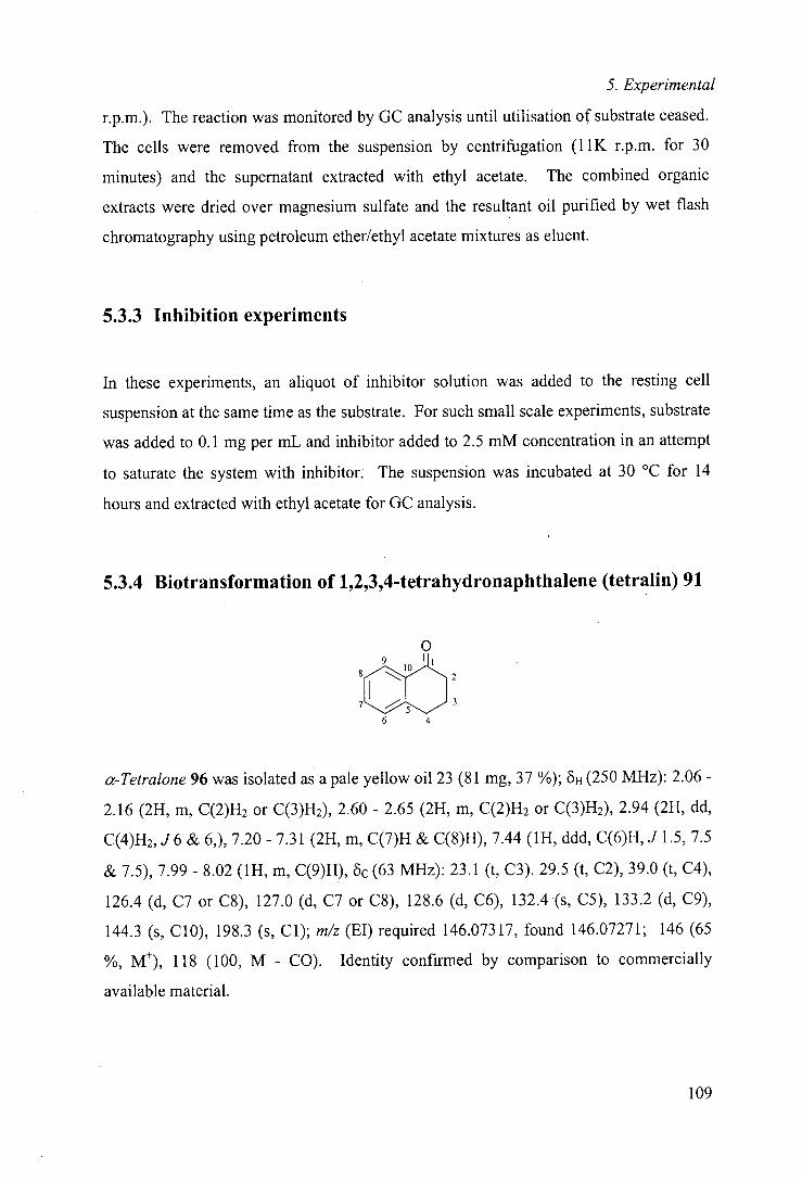
5. Experimental
r.p.m.). The reaction was monitored by GC analysis until utilisation of substrate ceased.
The cells were removed from the suspension by centrifugation (1 1K r.p.m. for 30
minutes) and the supernatant extracted with ethyl acetate. The combined organic
extracts were dried over magnesium sulfate and the resultant oil purified by wet flash
chromatography using petroleum ether/ethyl acetate mixtures as eluent.
5.3.3 Inhibition experiments
In these experiments, an aliquot of inhibitor solution was added to the resting cell
suspension at the same time as the substrate. For such small scale experiments, substrate
was added to 0.1 mg per mL and inhibitor added to 2.5 mM concentration in an attempt
to saturate the system with inhibitor. The suspension was incubated at 30 °C for 14
hours and extracted with ethyl acetate for GC analysis.
5.3.4 Biotransformation of 1,2,3,4-tetrahydronaphthalene (tetralin) 91
0
8 3
3
6 4
cz-Tetralone 96 was isolated as a pale yellow oil 23 (81 mg, 37 %); 6H (250 MHz): 2.06 -
2.16 (21-1, m, C(2)H2 or C(3)H2), 2.60 - 2.65 (2H, m, C(2)H2 or C(3)H2), 2.94 (21-1, dd,
C(4)H2, J6 & 6,), 7.20 - 7.31 (21-1, m, C(7)H & C(8)H), 7.44 (1H, ddd, C(6)H, J 1. 5, 7.5
& 7.5), 7.99-8.02 (1H, m, C(9)H), 8c(63 MHz): 23.1 (t, C3). 29.5 (t, C2), 39.0 (t, C4),
126.4 (d, C7 or C8), 127.0 (d, C7 or C8), 128.6 (d, C6), 132.4 (s, C5), 133.2 (d, C9),
144.3 (s, CIO), 198.3 (s, Cl); m/z (El) required 146.07317, found 146.07271; 146 (65
M), 118 (100, M - CO). Identity confirmed by comparison to commercially
available material.
109
5. Experimental
5.4 Rhodococcus rhodochrous NCIMB 9703
5.4.1 Substrate synthesis
General benzylation procedure (A)
Alcohol (1 mot eq.) and tetrabutylammonium iodide (trace) were added to a solution of
sodium hydride (60 % suspension in mineral oil; 1.33 mot eq.) and freshly distilled
benzyl bromide (1.67 mot eq.) in anhydrous THF under an atmosphere of argon at 0 C.
On complete utilisation of the alcohol, the reaction mixture was diluted with diethyl
ether and washed with saturated sodium hydrogen carbonate, saturated brine and water.
The organic extracts were dried over anhydrous MgSO4, filtered, concentrated under
reduced pressure and purified by flash chromatography.
General preparation of 2-oxy-substituted oxygen heterocycles (B)
The dihydroheterocycle (1.5 mot eq.) was added to a stirred solution of alcohol (1 mot
eq.) and 4-toluenesulfonic acid (4 mot %) in anhydrous dichloromethane under an
atmosphere of argon. On complete consumption of the alcohol, the reaction mixture was
diluted with diethylether and washed with saturated brine, saturated sodium hydrogen
carbonate and water. The organic extracts were then dried over anhydrous MgSO4,
filtered, concentrated under reduced pressure and purified by flash chromatography.
General esterijication procedure using acid chloride (C)
Acid chloride (1.2 mot eq.) and pyridine (2 mot eq.) were added to a stirred solution of
the alcohol (1 mot eq.) in anhydrous dichloromethane under an atmosphere of argon.
Dimethylaminopyridine (trace) was also added if reaction was sluggish. On complete
consumption of alcohol, the reaction mixture was diluted with dichloromethane and
washed with citric acid (5 % aqueous solution), saturated sodium hydrogen carbonate
solution, saturated brine and water. The organic extracts were dried over anhydrous
MgSO4, filtered, concentrated under reduced pressure and purified by flash
chromatography.
110
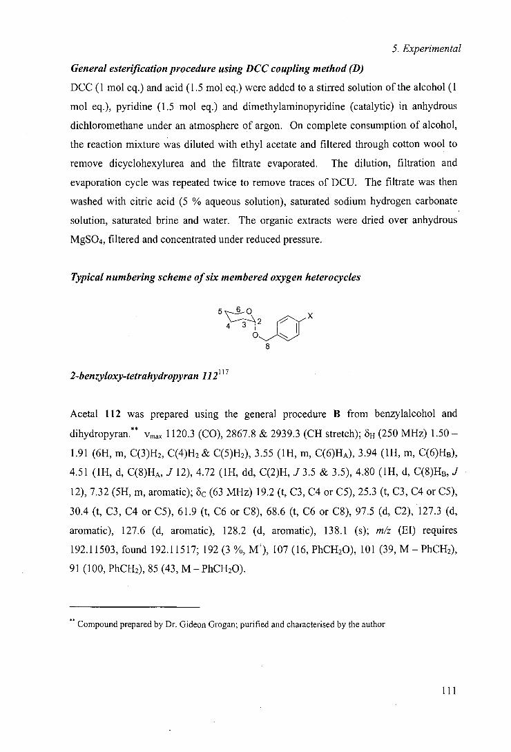
5. Experimental
General esterification procedure using DCC coupling method (D)
DCC (1 mot eq.) and acid (1.5 mot eq.) were added to a stirred solution of the alcohol (1
mot eq.), pyridine (1.5 mot eq.) and dimethylaminopyridine (catalytic) in anhydrous
dichloromethane under an atmosphere of argon. On complete consumption of alcohol,
the reaction mixture was diluted with ethyl acetate and filtered through cotton wool to
remove dicyclohexylurea and the filtrate evaporated. The dilution, filtration and
evaporation cycle was repeated twice to remove traces of DCU. The filtrate was then
washed with citric acid (5 % aqueous solution), saturated sodium hydrogen carbonate
solution, saturated brine and water. The organic extracts were dried over anhydrous
MgSO4, filtered and concentrated under reduced pressure.
Typical numbering scheme of six membered oxygen heterocycles
6
tLZIr x 8
2-benzyloxy-tetrahydropyran 112117
Acetal 112 was prepared using the general procedure B from benzylalcohol and
dihydropyran.** Vmax 1120.3 (CO), 2867.8 & 2939.3 (CH stretch); 6H (250 MHz) 1.50 -
1.91 (6H, m, C(3)H2, C(4)H2 & C(5)H2), 3.55 (1H, m, C(6)HA), 3.94 (1H, m, C(6)1-10,
4.51 (1H, d, C(8)HA, J 12), 4.72 (1H, dd, C(2)H, J 3.5 & 3.5), 4.80 (1H, d, C(8)HB, J
12), 7.32 (5H, m, aromatic); 8c (63 MHz) 19.2 (t, C3, C4 or CS), 25.3 (t, C3, C4 or CS),
30.4 (t, C3, C4 or CS), 61.9 (t, C6 or C8), 68.6 (t, C6 or C8), 97.5 (d, C2), 127.3 (d,
aromatic), 127.6 (d, aromatic), 128.2 (d, aromatic), 138.1 (s); m/z (El) requires
192.11503, found 192.11517; 192(3%, M), 107 (16, PhCH20), 101 (39, M—PhCH2),
91(100, PhCH2), 85 (43, M - PhCH20).
Compound prepared by Dr. Gideon Grogan; purified and characterised by the author
111
5. Experimental
2-(4-methylbenzyloxy)-tetrahydropyran 113
Acetal 113 was prepared following the general procedure from 4-methylbenzylalcohol
and dihydropyran.** Vmax 1118.7 (CO), 2869.3 & 2490.5 (CH stretch); S (250 MHz)
1.51 - 1.91 (6H, m, C(3)H2, C(4)H2 & C(5)H2), 2.36 (3H, s, Me), 3.56 (1H, m, C(6)HA),
3.95 (1H, m, C(6)HB), 4.49 (1H, d, C(8)HA, J 12), 4.72 (1H, dd, C(2)H, J 3.5 & 3.5),
4.76 (1H, d, C(8)HB, J 12), 7.17 (2H, d, aromatic, J 8), 7.28 (2H, d, aromatic, J 8); öc
(63 MHz) 19.2 (t), 21.0 (q, Me), 25.3 (t), 30.4 (t), 61.9 (t, C6 or C8), 68.5 (t, C6 or C8),
97.3 (d, C2), 127.8 (d, aromatic), 128.8 (d, aromatic), 135.1 (s), 136.9 (s); m/z (El)
requires 206.13068 found 206.11321; 206 (2 %, M), 105 (100, CH3C6H4CH2), 91(16,
PhCH2), 85 (29, M - CH3C6H4CH20).
2-(4-chlorobenzyloxy)-tetrahydropyran 116
Acetal 116 prepared following the general method B from dihydropyran and 4-
chlorobenzylalcohol.** Vmax 1089.5 (CO), 2880.9 & 2978.1 (CH); 6H (250 MHz) 1.49 -
1.87 (6H, m, C(3)H2, C(4)H2 & C(5)H2), 3.53 (1H, m, C(6)HA), 3.88 (1H, m, C(6)HB),
4.45 (1H, d, C(8)HA, J 12), 4.68 (1H, dd, C(2)H, J 3 & 3), 4.73 (1H, d, C(8)HB, J 12),
7.27 (4H, m, aromatic); öc (63 MHz) 19.1 (t, C3, C4 or CS), 25.3 (t, C3,C4 or CS), 30.3
(t, C3, C4 or CS), 62.0 (t, C6 or C8), 67.8 (t, C6 or C8), 97.6 (d, C2), 128.3 (d,
aromatic), 128.9 (d, aromatic), 133.0 (s), 136.6 (s); m/z (El) requires 226.07606 and
228.073 11, found 226.07604 and 228.07357; 228 (2%, 37C1 M), 226 (7, 35C1, M), 127
(47, 37C1C6H4CH2), 125 (100, 35C1C6H4CH2), 101 (27, M - C1C6H4CH2). Although this
compound has been described previously in the literature, its characterisation has not
been reported.
112
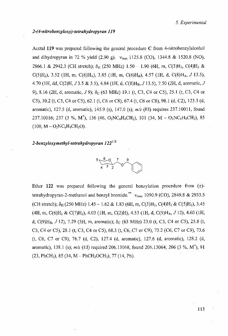
5. Experimental
2-(4-nitrobenzyloxy)-tetrahydropyran 119
Acetal 119 was prepared following the general procedure C from 4-nitrobenzylalcohol
and dihydropyran in 72 % yield (2.90 g). Vmax 1125.8 (CO), 1344.8 & 1520.8 (NO),
2866.1 & 2942.3 (CH stretch); 6H (250 MHz) 1.50 - 1.90 (6H, m, C(3)H2, C(4)H2 &
C(5)H2), 3.52 (1H, m, C(6)HA), 3.85 (1H, m, C(6)HB), 4.57 (1H, d, C(8)HA, J 13.5),
4.70 (1H, dd, C(2)H, J3.5 & 3.5), 4.84 (1H, d, C(8)HB, J 13.5), 7.50 (2H, d, aromatic, J
9), 8.16 (2H, d, aromatic, J 9); 6c (63 MHz) 19.1 (t, C3, C4 or C5), 25.1 (t, C3, C4 or
C5), 30.2 (t, C3, C4 or CS), 62.1 (t, C6 or C8), 67.4 (t, C6 or C8), 98.1 (d, C2), 123.3 (d,
aromatic), 127.5 (d, aromatic), 145.9 (s), 147.0 (s); m/z (El) requires 237.10011, found
237.10016; 237 (3 %, M), 136 (46, 02NC6H4CH2), 101 (34, M - 02NC6H4CH2), 85
(100, M - 02NC6H4CH2O).
2-benzyloxymethyl-tetrahydropyran 122118
5 00 7 9
4 2
Ether 122 was prepared following the general benzylation procedure from (±)-
tetrahydropyran-2-methanol and benzyl bromide.** Vmax 1090.9 (CO), 2849.8 & 2933.5
(CH stretch); oH (250 MHz) 1.45 - 1.62 & 1.83 (6H, m, C(3)H2, C(4)H2 & C(5)H2), 3.45
(4H, m, C(6)H2 & C(7)H2), 4.03 (1H, m, C(2)H), 4.53 (1H, d, C(9)HA, J 12), 4.60 (1H,
d, C(9)H,3, J 12), 7.29 (5H, m, aromatic); oc (63 MHz) 23.0 (t, C3, C4 or CS), 25.8 (t,
C3, C4 or CS), 28.1 (t, C3, C4 or CS), 68.3 (t, C6, C7 or C9), 73.2 (C6, C7 or C9), 73.6
(t, C6, C7 or C9), 76.7 (d, C2), 127.4 (d, aromatic), 127.6 (d, aromatic), 128.2 (d,
aromatic), 138.1 (s); m/z (El) required 206.13068, found 206.13064; 206 (3 %, M), 91
(23, PhCH2), 85 (34, M - PhCH20CH2), 77 (14, Ph).
113
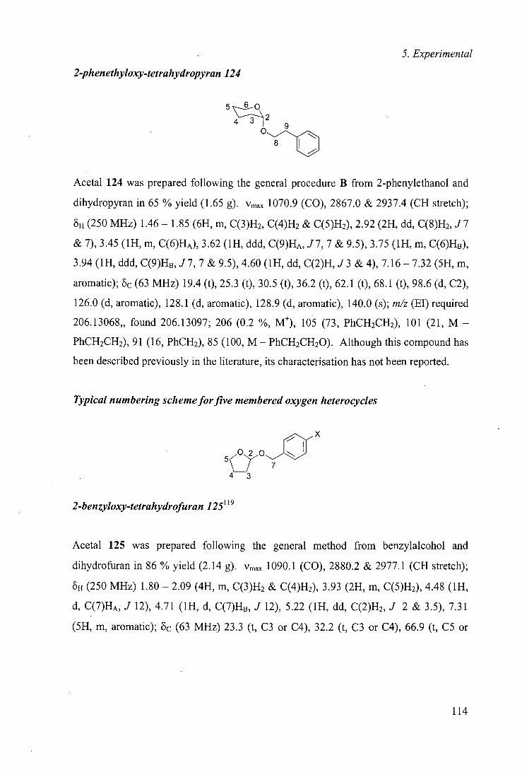
5. Experimental
2-ph enethyloxy-tetrahydropyran 124
6
o c Acetal 124 was prepared following the general procedure B from 2-phenylethanol and
dihydropyran in 65 % yield (1.65 g). Vmax 1070.9 (CO), 2867.0 & 2937.4 (CH stretch);
H (250 MHz) 1.46— 1.85 (6H, in, C(3)H2, C(4)H2 & C(5)H2), 2.92 (2H, dd, C(8)H2, J7
&7), 3.45 (1H, in, C(6)HA), 3.62 (1H, ddd, C(9)HA, J7, 7 & 9.5), 3.75 (1H, m, C(6)HB),
3.94 (1H, ddd, C(9)HB,J7, 7 & 9.5), 4.60 (1H, dd, C(2)H,J3 & 4), 7.16-7.32 (5H, m,
aromatic); 8c (63 MHz) 19.4 (t), 25.3 (t), 30.5 (t), 36.2 (t), 62.1(t), 68.1 (t), 98.6 (d, C2),
126.0 (d, aromatic), 128.1 (d, aromatic), 128.9 (d, aromatic), 140.0 (s); m/z (El) required
206.13068,, found 206.13097; 206 (0.2 %, M), 105 (73, PhCH2CH2), 101 (21, M -
PhCH2CH2), 91(16, PhCH2), 85 (100, M - PhCH2CH20). Although this compound has
been described previously in the literature, its characterisation has not been reported.
Typical numbering scheme for five membered oxygen heterocycles
,,,,a 7
43
2-benzyloxy-tetrahydrofuran 125119
Acetal 125 was prepared following the general method from benzylalcohol and
dihydrofuran in 86 % yield (2.14 g). Vmax 1090.1 (CO), 2880.2 & 2977.1 (CH stretch);
H (250 MHz) 1.80 - 2.09 (4H, m, C(3)H2 & C(4)H2)5 3.93 (2H, in, C(5)H2), 4.48 (1H,
d, C(7)HA5 J 12)5 4.71 (1H, d, C(7)HB5 J 12), 5.22 (1H, dd, C(2)H2, J 2 & 3.5), 7.31
(5H, m, aromatic); & (63 MHz) 23.3 (t, C3 or C4), 32.2 (t, C3 or C4), 66.9 (t, C5 or
114
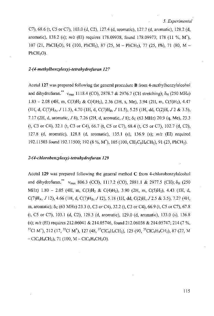
5. Experimental
C7), 68.6 (t, CS or C7), 103.0 (d, C2), 127.4 (d, aromatic), 127.7 (d, aromatic), 128.2 (d,
aromatic), 138.2 (s); m/z (El) requires 178.09938, found 178.09973; 178 (11 %, M),
107 (21, PhCH20), 91(100, PhCH2), 87 (25, M - PhCH2), 77 (25, Ph), 71(80, M -
PhCH2O).
2-(4-methylbenzyloxy)-tetrahydrofuran 127
Acetal 127 was prepared following the general procedure B from 4-rnethylbenzylalcohol
and dihydroftlran.** Vmax 1118.4 (CO), 2878.7 & 2976.7 (CH stretching); 6H (250 MHz)
1.83 - 2.08 (4H, m, C(3)H2 & C(4)H2), 2.36 (3H, s, Me), 3.94 (2H, m, C(5)H2), 4.47
(1H, d, C(7)HA, J 11.5), 4.70 (1H, d, C(7)HB, J 11.5), 5.25 (1H, dd, C(2)H, J2 & 3.5),
7.17 (2H, d, aromatic, J8), 7.26 (2H, d, aromatic, J8); oc (63 MHz) 20.9 (q, Me), 23.3
(t, C3 or C4), 32.1 (t, C3 or C4), 66.7 (t, CS or C7), 68.4 (t, CS or C7), 102.7 (d, C2),
127.8 (d, aromatic), 128.8 (d, aromatic), 135.1 (s), 136.9 (s); m/z (El) required
192.11503 found 192.11500; 192 (8 %, M), 105 (100, CH3C6H4CH2), 91(23, PhCH2).
2-(4-chlorobenzyloxy)-tetrahydrofuran 129
Acetal 129 was prepared following the general method C from 4-chlorobenzylalcohol
and dihydrofuran.** Vmax 806.3 (CC1), 1117.2 (CO), 2881.1 & 2977.5 (CH); 6H (250
MHz) 1.80 - 2.05 (4H, m, C(3)H2 & C(4)H2), 3.90 (2H, m, C(5)H2), 4.43 (1H, d,
C(7)HA, J 12), 4.66 (1H, d, C(7)HB, J 12), 5.18 (1H, dd, C(2)H, J 2.5 & 3.5), 7.27 (4H,
m, aromatic); öc (63 MHz) 23.3 (t, C3 or C4), 32.2 (t, C3 or C4), 66.9 (t, CS or C7), 67.8
(t, CS or C7), 103.1 (d, C2), 128.3 (d, aromatic), 129.0 (d, aromatic), 133.0 (s), 136.8
(s); m/z (El) requires 212.06041 & 2 14.05746, found 212.06038 & 214.05747; 214 (7 %,
37C1 Mt), 212 (17, 35C1 M), 127(48, 37C1C6H4CH2), 125 (90, 35C1C6H4CH2), 87 (27, M
- C1C6H4CH2), 71(100, M - C1C6H4CH20).
115
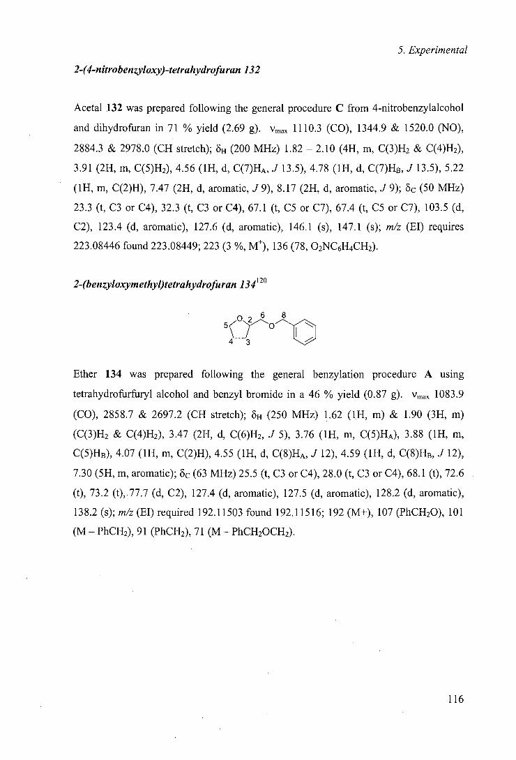
5. Experimental
2-(4-nitrobenzyloxy)-tetrahydrofuran 132
Acetal 132 was prepared following the general procedure C from 4-nitrobenzylalcohol
and dihydrofuran in 71 % yield (2.69 g). Vmax 1110.3 (CO), 1344.9 & 1520.0 (NO),
2884.3 & 2978.0 (CH stretch); oH (200 MHz) 1.82 - 2.10 (4H, m, C(3)H2 & C(4)H2),
3.91 (2H, m, C(5)H2), 4.56 (1H, d, C(7)HA, J 13.5), 4.78 (1H, d, C(7)H8, J 13.5), 5.22
(1H, m, C(2)H), 7.47 (2H, d, aromatic, J 9), 8.17 (2H, d, aromatic, J 9); 8c (50 MHz)
23.3 (t, C3 or C4), 32.3 (t, C3 or C4), 67.1 (t, C5 or C7), 67.4 (t, C5 or C7), 103.5 (d,
C2), 123.4 (d, aromatic), 127.6 (d, aromatic), 146.1 (s), 147.1 (s); m/z (El) requires
223.08446 found 223.08449; 223 (3 %, Mt), 136 (78, 02NC6H4CH2).
2-(benzyloxymethyl)tetrahydrofuran 134120
026 8
43
Ether 134 was prepared following the general benzylation procedure A using
tetrahydrofurfuryl alcohol and benzyl bromide in a 46 % yield (0.87 g). Vmax 1083.9
(CO), 2858.7 & 2697.2 (CH stretch); 0H (250 MHz) 1.62 (1H, m) & 1.90 (3H, m)
(C(3)H2 & C(4)H2), 3.47 (2H, d, C(6)H2, J 5), 3.76 (1H, m, G(5)HA), 3.88 (1H, m,
G(5)HB), 4.07 (1H, m, C(2)H), 4.55 (1H, d, G(8)HA, J 12), 4.59 (1H, d, G(8)HB, J 12),
7.30 (5H, m, aromatic); oc (63 MHz) 25.5 (t, C3 or C4), 28.0 (t, C3 or C4), 68.1 (t), 72.6
(t), 73.2 (t),77.7 (d, C2), 127.4 (d, aromatic), 127.5 (d, aromatic), 128.2 (d, aromatic),
138.2 (s); m/z (El) required 192.11503 found 192.11516; 192 (M+), 107 (PhCH20), 101
(M - PhCH2), 91 (PhCH2), 71 (M - PhGH20GH2).
116
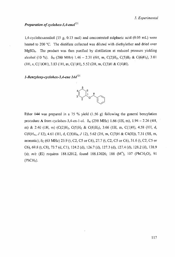
5. Experimental
Preparation of cyclohex-3,4-enol' 21
1,4-cyclohexanediol (15 g, 0.13 mol) and concentrated sulphuric acid (0.05 mL) were
heated to 200 °C. The distillate collected was diluted with diethylether and dried over
MgSO4. The product was then purified by distillation at reduced pressure yielding
alcohol (10 %). 5H (200 MHz) 1.46 - 2.31 (6H, m, C(2)H2, C(5)H2 & C(6)H2), 3.01
(1H, s, C(l)OH), 3.83 (1H, m, C(l)H), 5.52 (2H, m, C(3)H & C(4)H).
1-Benzyloxy-cyclohex-3,4-ene 144121
5
U1
Ether 144 was prepared in a 75 % yield (1.56 g) following the general benzylation
procedure A from cyclohex-3,4-en-1-ol. 6H (250 MHz) 1.66 (1H, m), 1.94 - 2.26 (4H,
m) & 2.40 (1H, m) (C(2)H2, C(5)H2 & C(6)H2), 3.66 (1H, m, C(1)H), 4.58 (1H, d,
C(8)HA, J 12), 4.61 (1H, d, C(8)H,3, J 12), 5.62 (2H, m, C(3)H & C4(H)), 7.31 (5H, m,
aromatic); oc (63 MHz) 23.9 (t, C2, CS or C6), 27.7 (t, C2, CS or C6), 31.6 (t, C2, CS or
C6), 69.8 (t, C8), 73.7 (d, Cl), 124.2 (d), 126.7 (d), 127.3 (d), 127.4 (d), 128.2 (d), 138.9
(s); m/z (El) requires 188.12012, found 188.12026; 188 (M), 107 (PhCH20), 91
(PhCH2).
117
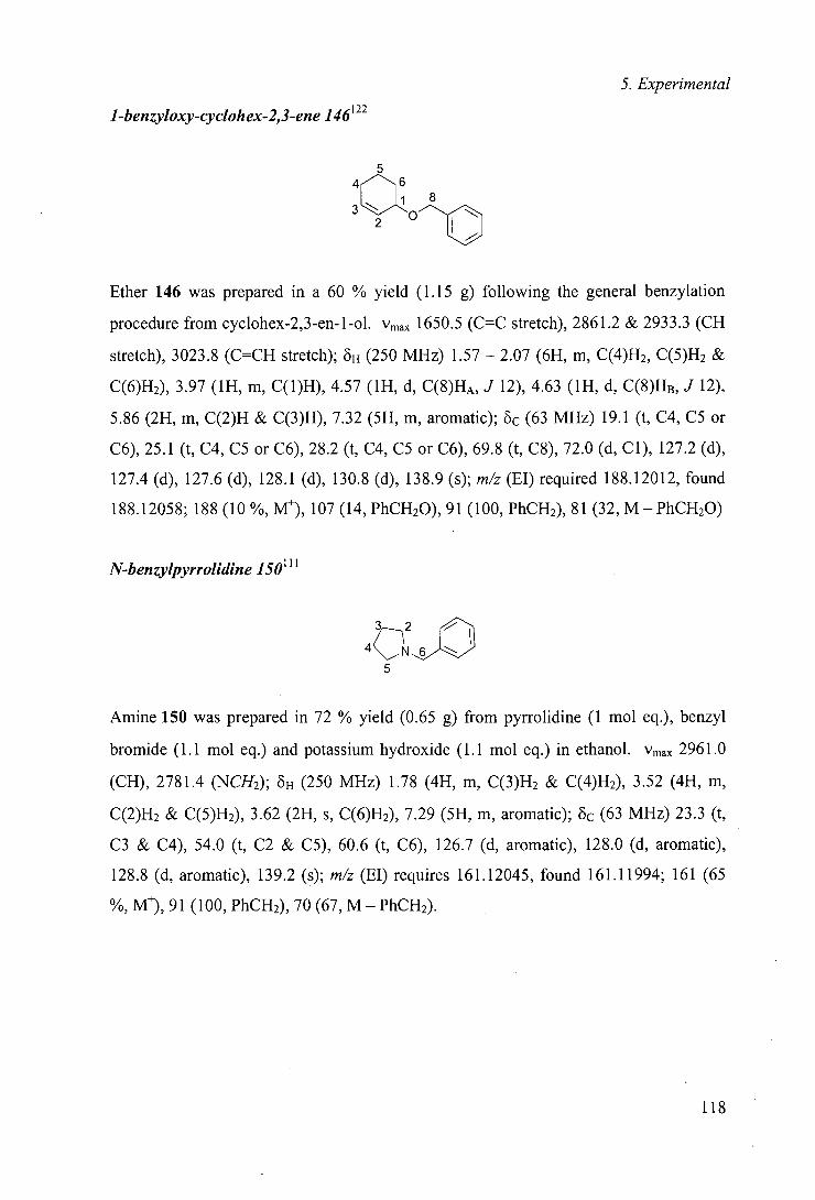
5. Experimental
1-benzyloxy-cyclohex-2,3-ene 146122
5 4 C~j 8
3 2
Ether 146 was prepared in a 60 % yield (1.15 g) following the general benzylation
procedure from cyclohex-2,3-en-1-ol. Vmax 1650.5 (C=C stretch), 2861.2 & 2933.3 (CH
stretch), 3023.8 (C=CH stretch); 6H (250 MHz) 1.57 - 2.07 (6H, m, C(4)H2, C(5)H2 &
C(6)H2), 3.97 (1H, m, C(1)H), 4.57 (1H, d, C(8)HA, J 12), 4.63 (1H, d, C(8)HB, J 12),
5.86 (2H, m, C(2)H & C(3)H), 7.32 (5H, m, aromatic); 8c (63 MHz) 19.1 (t, C4, C5 or
C6), 25.1 (t, C4, CS or C6), 28.2 (t, C4, C5 or C6), 69.8 (t, C8), 72.0 (d, Cl), 127.2 (d),
127.4 (d), 127.6 (d), 128.1 (d), 130.8 (d), 138.9 (s); m/z (El) required 188.12012, found
188.12058; 188(10%,M), 107(14,PhCH20),91 (100,PhCH2),81 (32,M—PhCH20)
N-benzylpyrrolidine 150"
Amine 150 was prepared in 72 % yield (0.65 g) from pyrrolidine (1 mol eq.), benzyl
bromide (1.1 mol eq.) and potassium hydroxide (1.1 mol eq.) in ethanol. Vmax 2961.0
(CH), 2781.4 (NCH2); ö,j (250 MHz) 1.78 (4H, m, C(3)H2 & C(4)H2), 3.52 (4H, m,
C(2)H2 & C(5)H2), 3.62 (2H, s, C(6)H2), 7.29 (5H, m, aromatic); 6c (63 MHz) 23.3 (t,
C3 & C4), 54.0 (t, C2 & C5), 60.6 (t, C6), 126.7 (d, aromatic), 128.0 (d, aromatic),
128.8 (d, aromatic), 139.2 (s); m/z (El) requires 161.12045, found 161.11994; 161 (65
%, M), 91(100, PhCH2), 70 (67, M - PhCH2).
118
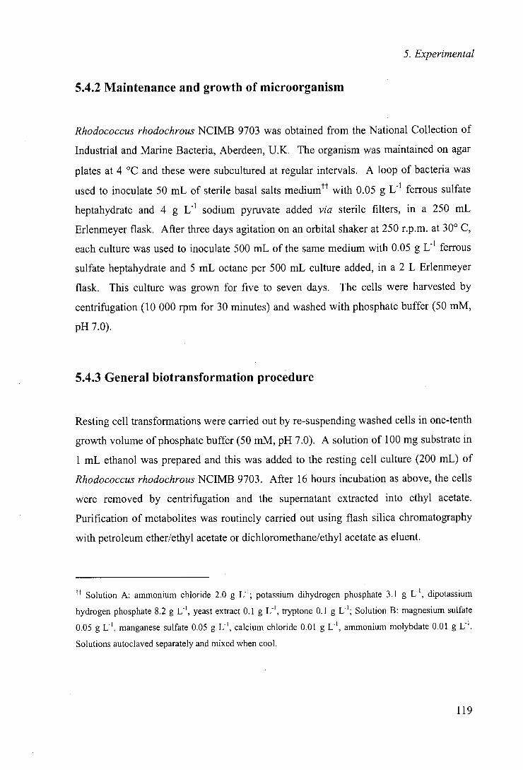
5. Experimental
5.4.2 Maintenance and growth of microorganism
Rhodococcus rhodochrous NCIMB 9703 was obtained from the National Collection of
Industrial and Marine Bacteria, Aberdeen, U.K. The organism was maintained on agar
plates at 4 °C and these were subcultured at regular intervals. A loop of bacteria was
used to inoculate 50 mL of sterile basal salts mediumtt with 0.05 g U' ferrous sulfate
heptahydrate and 4 g U' sodium pyruvate added via sterile filters, in a 250 mL
Erlenmeyer flask. After three days agitation on an orbital shaker at 250 r.p.m. at 30° C,
each culture was used to inoculate 500 mL of the same medium with 0.05 g U' ferrous
sulfate heptahydrate and 5 mL octane per 500 mL culture added, in a 2 L Erlenmeyer
flask. This culture was grown for five to seven days. The cells were harvested by
centrifugation (10 000 rpm for 30 minutes) and washed with phosphate buffer (50 mM,
pH 7.0).
5.4.3 General biotransformation procedure
Resting cell transformations were carried out by re-suspending washed cells in one-tenth
growth volume of phosphate buffer (50 mM, pH 7.0). A solution of 100 mg substrate in
1 mL ethanol was prepared and this was added to the resting cell culture (200 mL) of
Rhodococcus rhodochrous NCIMB 9703. After 16 hours incubation as above, the cells
were removed by centrifugation and the supernatant extracted into ethyl acetate.
Purification of metabolites was routinely carried out using flash silica chromatography
with petroleum ether/ethyl acetate or dichloromethane/ethyl acetate as eluent.
tt Solution A: ammonium chloride 2.0 g U'; potassium dihydrogen phosphate 3.1 g U', dipotassium
hydrogen phosphate 8.2 g U', yeast extract 0.1 g U', tryptone 0.1 g L'; Solution B: magnesium sulfate
0.05 g U', manganese sulfate 0.05 g U', calcium chloride 0.01 g U', ammonium molybdate 0.01 g U'.
Solutions autoclaved separately and mixed when cool.
119
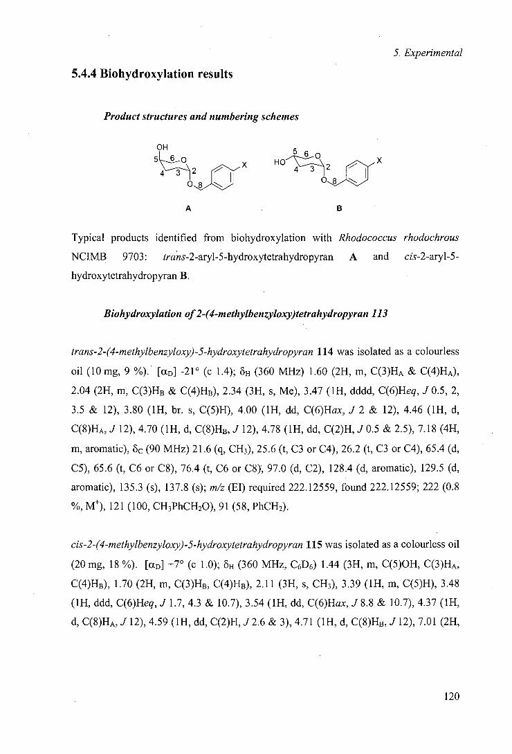
5. Experimental
5.4.4 Biohydroxylation results
Product structures and numbering schemes
OH 5 6o
4 3 0
_,a HO 4 3 0
A
B
Typical products identified from biohydroxylation with Rhodococcus rhodochrous
NCIMB 9703: trans-2-aryl-5-hydroxytetrahydropyran A and cis-2-aryl-5-
hydroxytetrahydropyran B.
Biohydroxylation of2-(4-methylbenzyloxy)tetrahydropyran 113
trans-2-(4-methylbenzyloxy)-5-hydroxytetrahydropyran 114 was isolated as a colourless
Oil (10 mg, 9 %). [aD] -21° (c 1.4); 8H (360 MHz) 1.60 (2H, in, C(3)HA & C(4)HA),
2.04 (2H, in, C(3)HB & C(4)HB), 2.34 (3H, s, Me), 3.47 (1H, dddd, C(6)Heq, J 0.5, 2,
3.5 & 12), 3.80 (1H, hr. s, C(5)H), 4.00 (1H, dd, C(6)Hax, J 2 & 12), 4.46 (1H, d,
C(8)HA, J 12), 4.70 (1H, d, C(8)HB5 J 12)5 4.78 (1H, dd, C(2)H, .10.5 & 2.5), 7.18 (4H,
in, aromatic), 6c (90 MHz) 21.6 (q, CH3), 25.6 (t, C3 or C4), 26.2 (t, C3 or C4), 65.4 (d,
C5), 65.6 (t, C6 or C8), 76.4 (t, C6 or C8), 97.0 (d, C2), 128.4 (d, aromatic), 129.5 (d,
aromatic), 135.3 (s), 137.8 (s); m/z (El) required 222.12559, found 222.12559; 222 (0.8
%, M), 121 (100, CH3PhCH20), 91(58, PhCH2).
cis-2-(4-methylbenzyloxy)-5-hydroxytetrahydropyran 115 was isolated as a colourless oil
(20 mg, 18 %). [aD] +7° (c 1.0); oH (360 MHz, C6D6) 1.44 (3H, in, C(5)OH, C(3)HA,
C(4)HB), 1.70 (2H, in, C(3)HB, C(4)HB), 2.11 (3H, s, CH3), 3.39 (1H, in, C(5)H), 3.48
(1H, ddd, C(6)Heq,.1 1.7, 4.3 & 10.7), 3.54 (1H, dd, C(6)Hax, J 8.8 & 10.7), 4.37 (1H,
d, C(8)HA5 .112), 4.59 (1H, dd, C(2)H, .1 2.6 & 3), 4.71 (1H, d, C(8)HB, J 12), 7.01 (2H5
120
5. Experimental
d, aromatic, J 8), 7.25 (2H, d, aromatic, J 8); öc (93 MHz) 21.6 (q, CH3), 28.6 (t, C3 or
C4), 30.1 (t, C3 or C4), 66.0 (d, C5), 66.5 (t, C6 or C8), 69.3 (t, C6 or C8), 96.7 (d, C2),
128.5 (d, aromatic), 129.5 (d, aromatic), 135.2 (s, C12), 137.9 (s, C9); m/z (El) required
222.12559, found 222.12634; 222(8%, M), 121 (4, M - OH), 118 (12, M—CH3PhCH),
105 (45, CH3PhCH2).
Biohydroxylation of 2-(4-chlorobenzyloxy)tetrahydropyran 116
trans-2-(4-chlorobenzyloxy)-5-hydroxytetrahydropyran 117 was isolated as a colourless
Oil (9 Mg, 8%). {a0] —11 (c 1.6); 6H(2SO MHz) 1.60 (2H, m, C(3)HA & C(4)HA), 2.04
(2H, m, C(3)HB & C(4)HB), 3.48 (1H, m, C(6)Heq), 3.81 (1H, br. m, C(5)H), 3.97 (1H,
dd, C(6)Hax, J 2 & 12), 4.47 (1H, d, C(8)HA, J 12), 4.70 (1H, d, C(8)HB, J 12), 4.77
(1H, dd, C(2)H, J 2.5 & 2.5), 7.25 - 7.33 (4H, m, aromatic); 6c (63 MHz) 25.0 (t, C3 or
C4), 25.6 (t, C3 or C4), 64.8 (d, CS), 65.1 (t, C6 or C8), 68.1 (t, C6 or C8), 96.6 (d, C2),
128.4 (d, aromatic), 129.0 (d, aromatic), 133.3 (s), 136.3 (s); m/z (El) required
242.07097 and 244.06802, found 242.07065 and 244.06782; 244 (0.1 %, M 37C1), 242
(0.5 %, M "Cl), 127 (52, 37C1C6H4CH2), 125 (100, 35C1C6H4CH2), 91(15, PhCH2).
cis-2-(4-chlorobenzyloxy)-5-hydroxytetrahydropyran 118 was isolated as a colourless oil
(13 mg, 12%). 6H(360 MHz) 1.71 - 1.95 (5H, m, C(3)H2, C(4)H2 & C(5)OH), 3.67 (2H,
hr. m, C(6)H2), 3.79 (1H, br. m, C(5)H), 4.50 (1H, d, C(8)HA, J 12.2), 4.72 (1H, m,
C(2)H), 4.77 (1H, d, C(8)HB, J 12.2), 7.28 - 7.38 (4H, m, aromatic); 6c (63 MHz) 27.9
(t, C3 & C4), 65.3 (d, CS), 66.0 (t, C6 or C8), 68.0 (t, C6 or C8), 96.3 (d, C2), 128.4 (d,
aromatic), 129.0 (d, aromatic), 133.3 (s), 136.2 (s); m/z 242 (M).
Biohydroxylation of2-(4-nitrobenzyloxy)tetrahydropyran 119
trans-2- (4-nitrobenzyloxy)-5-hydroxytetrahydropyran 120 was isolated a colourless oil
(22 mg, 21 %). [aD] +36 (c 1.15); 8H (360 MHz) 1.70 (2H, m, C(3)HA & C(4)HA), 1.70
121

5. Experimental
(1H, br. s, C(5)OH), 2.13 (2H, m, C(3)HB & C(4)HB), 3.53 (1H, m, C(6)Heq), 3.99 (1H,
dd, C(6)Hax, J2.2 & 11.8), 4.65 (1H, d, C8-HA, J 13.5), 4.85 (1H, m, C(2)H), 4.89 (1H,
d, C(8)HB, J 13.5), 7.55 (2H, d, aromatic, J 8.8), 8.24 (2H, d, aromatic, J 8.8); 8c (90
MHz) 25.5 (t, C3 or C4), 26.2 (t, C3 or C4), 65.2 (d, C5), 65.7 (t, C6 or C8), 68.3 (t, C6
or C8), 97.7 (d, C2), 124.1 (d, aromatic), 128.2 (d, aromatic), 146.1 (s), 147.8 (s); m/z
(El) required 253.09502, found 253.09509; 253 (1%, M), 236 (4, M - OH), 153 (92,
02NC6H4CH20H), 152 (44, 02NC6H4CH20).
cis-2-(4-nitrobenzyloxy)-5-hydroxytetrahydropyran 121 was isolated as a pale yellow oil
(19 mg, 18 %). [aD] -44 (c 0.7); oH (360 MHz) 1.88 (5H, m, C(3)H2, C(4)H2 &
C(5)OH), 3.63 (1H, dd, C(6)Hax, J 8 & 11), 3.70 (1H, ddd, C(6)Heq, J 1.2, 4 & 11),
3.80 (1H, m, C(5)H), 4.63 (1H, d, C(8)HA, J 13.4), 4.76 (1H, dd, C(2)H, J 3.1 & 3.3),
4.90 (1H, d, C(8)HB, J 13.4), 7.55 (2H, m, aromatic), 8.24 (2H, m, aromatic); oc (63
MHz) 27.8 (t, C3 or C4), 29.5 (t, C3 or C4), 65.2 (d, CS), 66.0 (t, C6 or C8), 67.6 (t, C6
or C8), 96.8 (d, C2), 123.5 (d, aromatic), 127.7 (d, aromatic), 145.5 (s), 147.7 (s); m/z
(El) required 253.09502, found 253.09501; 253 (0.1 %, M), 153 (21,
02NC6H4CH20H).
Biohydroxylation of2-(benzyloxymethyl)tetrahydropyran 122
07 9
4 3 HO o
trans-2-(benzylmethyloxy)-5-hydroxytetrahydropyran 123 was isolated as a colourless
oil (38 mg, 35 %). [aD] +2.9 (c 1.0); 8H (360 MHz) 1.38 - 1.72 (4H, m) & 2.12 (1H, m)
(C(5)OH, C(3)H2 & C(4)H2), 3.13 (1H, dd, C(6)Hax, J 10.4 & 10.6), 3.39 - 3.50 (3H,
overlapping m, C(7)H2 & C(2)H), 3.70 (1H, m, C(5)H), 4.03 (1H, ddd, C(6)Heq, J 2.2,
4.8 & 10.6), 4.53 (1H, d, C(8)HA, J 12.2), 4.58 (1H, d, C(8)HB, J 12.2), 7.29 (5H, m,
aromatic); Oc (90 MHz) 27.0 (t, C3 or C4), 32.4 (t, C3 or C4), 66.2 (d, C2), 72.6 (t, C6,
122
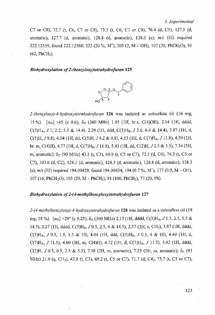
5. Experimental
C7 or C8), 72.7 (t, C6, C7 or C8), 73.3 (t, C6, C7 or C8), 76.4 (d, C5), 127.5 (d,
aromatic), 127.7 (d, aromatic), 128.8 (d, aromatic), 138.0 (s); m/z (El) required
222.12559, found 222.12568; 222 (20%, M), 205 (2, M - OH), 107 (30, PhCH20), 91
(62, PhCH2).
Biohydroxylation of2-(benzyloxy)tetrahydrofuran 125
5 0 2 0~1-'10 ~_Z 7
HO
2-(benzyloxy)-4-hydroxytetrahydrofuran 126 was isolated as colourless oil (16 mg,
15 %). [aD] +45 (c 0.6); oH (360 MHz) 1.95 (1H, br.s, C(4)OH), 2.14 (1H, dddd,
C(3)HA, J 1, 2.2, 5.5 & 14.4). 2.29 (1H, ddd, C(3)HB, J 2.6, 6.4 & 14.4), 3.87 (1H, d,
C(5)H, J9.8), 4.04 (1H, dd, C(5)H, J4.2 & 9.8), 4.53 (1H, d, C(7)HA, J 11.8), 4.59 (1H,
br. m, C(4)H), 4.77 (1H, d, C(7)HB, J 11.8), 5.43 (1H, dd, C(2)H, J2.5 & 5.5), 7.34 (5H,
m, aromatic); oc (90 MHz) 43.3 (t, 0), 69.9 (t, CS or C7), 72.1 (d, C4), 74.3 (t, CS or
C7), 103.6 (d, C2), 128.1 (d, aromatic), 128.3 (d, aromatic), 128.8 (d, aromatic), 138.3
(s); m/z (El) required 194.09429, found 194.09434; 194 (0.7 %, M), 177 (0.9, M - OH),
107 (16, PhCH20), 103 (20, M - PhCH2), 91(100, PhCH2), 77 (20, Ph).
Biohydroxylation of2-(4-methylbenzyloxy)tetrahydrofuran 127
2-(4-methylbenzyloxy)-4-hydroxytetrahydrofuran 128 was isolated as a colourless oil (19
Mg, 18 %). [CCD] +290 (c 0.25); 0H (360 MHz) 2.13 (1H, dddd, C(3)HA, J 1.5, 2.5, 5.5 &
14.5), 2.27 (1H, dddd, C(3)HB, J 0.5, 2.5, 6 & 14.5), 2.37 (3H, s, CH3), 3.87 (1H, dddd,
C(5)HA, J 0.5, 1.5, 1.5 & 10), 4.04 (1H, ddd, C(5)HB, J 0.5, 4 & 10), 4.49 (1H, d,
C(7)HA, J 11.5), 4.60 (1H, m, C(4)H), 4.72 (1H, d, C(7)HB, J 11.5), 5.42 (1H, dddd,
C(2)H, J 0.5, 0.5, 2.5 & 5.5), 7.18 (2H, m, aromatic), 7.25 (2H, m, aromatic); oc (93
MHz) 21.0 (q, CH3), 42.8 (t, 0), 69.2 (t, CS or C7), 71.7 (d, C4), 73.7 (t, CS or C7),
123
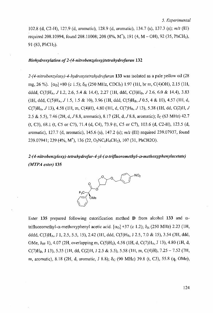
5. Experimental
102.8 (d, C2-H), 127.9 (d, aromatic), 128.9 (d, aromatic), 134.7 (s), 137.3 (s); m/z (El)
required 208.10994, found 208.11008; 208 (8%, M), 191 (4, M - OH), 92 (35, PhCH3),
91(83, PhCH2).
Biohydroxylation of 2-(4-nitrobenzyloxy)tetrahydrofuran 132
2-(4-nitrobenzyloxy)-4-hydroxytetrahydrofuran 133 was isolated as a pale yellow oil (28
mg, 26 %). [(XD] +80 (c 1.5); oH (250 MHz, CDC13) 1.97 (1H, br in, C(4)OH), 2.15 (1H,
dddd, C(3)HA, J 1.2, 2.6, 5.4 & 14.4), 2.27 (1H, ddd, C(3)HB, J 2.6, 6.0 & 14.4), 3.83
(1H, ddd, C(5)HA, J 1.5, 1.5 & 10), 3.96 (1H, ddd, C(5)HB, J0.5, 4 & 10), 4.57 (1H, d,
C(7)HA, J 13), 4.58 (1H, in, C(4)H), 4.80 (1H, d, C(7)HB, J 13), 5.38 (1H, dd, C(2)H, J
2.5 & 5.5), 7.46 (2H, d, J 8.8, aromatic), 8.17 (2H, d, J 8.8, aromatic); 6c (63 MHz) 42.7
(t, 0), 68.1 (t, CS or C7), 71.4 (d, C4), 73.9 (t, CS or C7), 103.6 (d, C2-H), 123.5 (d,
aromatic), 127.7 (d, aromatic), 145.6 (s), 147.2 (s); m/z (El) required 239.07937, found
239.07941; 239(4%, M), 136 (22, 02NC6H4CH2), 107 (31, PhCH20).
2-(4-nitrobenzyloxy)-tetrahydrofur-4-yl-(ci-trifluoromethyl- ci-methoxyphenylacetate)
(MTPA ester) 135
o 7-0N__(1
NO2
F3C—< /== OMe
Ester 135 prepared following esterification method D from alcohol 133 and a-
trifluoromethyl-a-methoxyphenyl acetic acid. [(XD] +37 (c 1.2); oH (250 MHz) 2.23 (1H,
dddd, C(3)HA, J 1, 2.5, 5.5, 15), 2.42 (1H, ddd, C(3)HB, J 2.5, 7.0 & 15), 3.54 (3H, ddd,
OMe, JHF 1), 4.07 (2H, overlapping in, C(5)H2), 4.58 (1H, d, C(7)HA, J 13), 4.80 (1H, d,
C(7)Hs, J 13), 5.35 (1H, dd, C(2)H, J 2.5 & 5.5), 5.58 (1H, in, C(4)H), 7.25 - 7.52 (7H,
in, aromatic), 8.18 (2H, d, aromatic, J 8.8); Oc (90 MHz) 39.8 (t, 0), 55.8 (q, OMe),
124
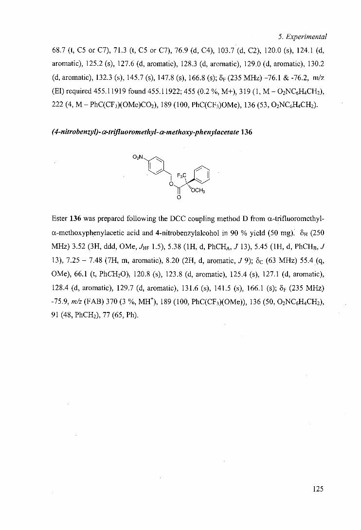
5. Experimental
68.7 (t, CS or C7), 71.3 (t, C5 or C7), 76.9 (d, C4), 103.7 (d, C2), 120.0 (s), 124.1 (d,
aromatic), 125.2 (s), 127.6 (d, aromatic), 128.3 (d, aromatic), 129.0 (d, aromatic), 130.2
(d, aromatic), 132.3 (s), 145.7 (s), 147.8 (s), 166.8 (s); 6F (235 MHz) —76.1 & -76.2, mlz
(El) required 455.11919 found 455.11922; 455(0.2%, M+), 319 (1, M—O2NC6H4CH2),
222 (4, M - PhC(CF3)(OMe)CO2), 189 (100, PhC(CF3)OMe), 136 (53, 02NC6H4CH2).
(4-nitrobenzyl)- tz-tri:fluoromethyl- a-methoxy-phenylacetate 136
02N
F3C)3 OCH3
0
Ester 136 was prepared following the DCC coupling method D from a-trifluoromethyl-
c-methoxypheny1acetic acid and 4-nitrobenzylalcohol in 90 % yield (50 mg). E- (250
MHz) 3.52 (3H, ddd, OMe, JHF 1.5), 5.38 (1H, d, PhCHA, J 13), 5.45 (1H, d, PhCHB, J
13), 7.25 - 7.48 (7H, m, aromatic), 8.20 (2H, d, aromatic, J 9); 5c (63 MHz) 55.4 (q,
OMe), 66.1 (t, PhCH20), 120.8 (s), 123.8 (d, aromatic), 125.4 (s), 127.1 (d, aromatic),
128.4 (d, aromatic), 129.7 (d, aromatic), 131.6 (s), 141.5 (s), 166.1 (s); 6F (235 MHz)
-75.9, m/z (FAB) 370 (3 %, MH), 189 (100, PhC(CF3)(OMe)), 136 (50, 02NC6H4CH2),
91(48, PhCH2), 77 (65, Ph).
125
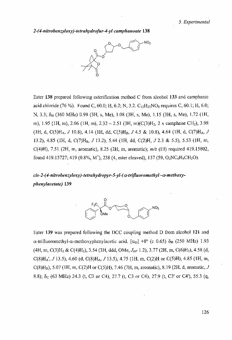
5. Experimental
2-(4-nitrobenzyloxy)-tetrahydrofur-4-yl camphanoate 138
o
M 0
Ester 138 prepared following esterification method C from alcohol 133 and camphanic
acid chloride (76 %). Found C, 60.0; H, 6.2; N, 3.2. C12H25N08 requires C, 60.1; H, 6.0;
N, 3.3; 8H (360 MHz) 0.99 (3H, s, Me), 1.08 (3H, s, Me), 1.15 (3H, s, Me), 1.72 (1H,
m), 1.95 (1H, m), 2.06 (1H, m), 2.32 - 2.51 (3H, m)(C(3)H2, 2 x camphane CH2), 3.98
(1H, d, C(5)HA, J 10.8), 4.14 (1H, dd, C(5)HB, J4.5 & 10.8), 4.64 (1H, d, C(7)HA, J
13.2), 4.85 (1H, d, C(7)HB, .1 13.2), 5.44 (1H, dd, C(2)H, J 2.3 & 5.5), 5.53 (1H, m,
C(4)H), 7.51 (2H, m, aromatic), 8.25 (2H, m, aromatic); m/z (El) required 419.15802,
found 419.15727; 419 (0.8%, M), 238 (4, ester cleaved), 137 (59, O2NC6H4CH2O).
cis-2-(4-nitrobenzyloxy)-tetrahydropyr-5-yl-(a-trifluorom ethyl —a-methoxy-
phenylacetate) 139
0 F3C II
('°H No2
ii
We 0
Ester 139 was prepared following the DCC coupling method D from alcohol 121 and
a-trifluoromethyl-a-methoxyphenylacetic acid. [CCD] +8° (c 0.65) 6H (250 MHz) 1.93
(4H, m, C(3)H2 & C(4)H2), 3.54 (3H, ddd, OMe, JHF 1.2), 3.77 (2H, m, C(6)H2), 4.58 (d,
C(8)HA', J 13.5), 4.60 (d, C(8)HA, J 13.5), 4.75 (1H, m, C(2)H or C(5)H), 4.85 (1H, m,
C(8)HB), 5.07 (1H, m, C(2)H or C(5)H), 7.46 (7H, m, aromatic), 8.19 (2H, d, aromatic, J
8.8); oc (63 MHz) 24.3 (t, C3 or C4), 27.7 (t, C3 or C4), 27.9 (t, C3' or C4'), 55.3 (q,
126
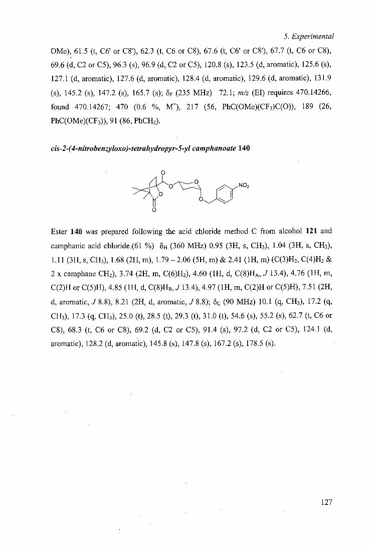
5. Experimental
OMe), 61.5 (t, C6' or C8'), 62.3 (t, C6 or C8), 67.6 (t, C6' or C8'), 67.7 (t, C6 or C8),
69.6 (d, C2 or CS), 96.3 (s), 96.9 (d, C2 or C5), 120.8 (s), 123.5 (d, aromatic), 125.6 (s),
127.1 (d, aromatic), 127.6 (d, aromatic), 128.4 (d, aromatic), 129.6 (d, aromatic), 131.9
(s), 145.2 (s), 147.2 (s), 165.7 (s); 8F (235 MHz) —72.1; m/z (El) requires 470.14266,
found 470.14267; 470 (0.6 %, M), 217 (56, PhC(OMe)(CF3)C(0)), 189 (26,
PhC(OMe)(CF3)), 91(86, PhCH2).
cis-2-(4-nitrobenzyloxo)-tetrahydropyr-5-yl camphanoate 140
0
>o
NO
0 0
Ester 140 was prepared following the acid chloride method C from alcohol 121 and
camphanic acid chloride.(61 %) oH (360 MHz) 0.95 (3H, s, CH3), 1.04 (3H, s, CH3),
1.11 (3H, s, CH3), 1.68 (2H, m), 1.79 —2.06 (5H, m) & 2.41 (1H, m) (C(3)H2, C(4)H2 &
2 x camphane CH2), 3.74 (2H, m, C(6)H2), 4.60 (IH, d, C(8)HA, J 13.4), 4.76 (1H, m,
C(2)H or C(5)H), 4.85 (1H, d, C(8)HB, J 13.4), 4.97 (1H, m, C(2)H or C(5)H), 7.51 (2H,
d, aromatic, J 8.8), 8.21 (2H, d, aromatic, J 8.8); Oc (90 MHz) 10.1 (q, CH3), 17.2 (q,
CH3), 17.3 (q, CH3), 25.0 (t), 28.5 (t), 29.3 (t), 31.0 (t), 54.6 (s), 55.2 (s), 62.7 (t, C6 or
C8), 68.3 (t, C6 or C8), 69.2 (d, C2 or CS), 91.4 (s), 97.2 (d, C2 or CS), 124.1 (d,
aromatic), 128.2 (d, aromatic), 145.8 (s), 147.8 (s), 167.2 (s), 178.5 (s).
127
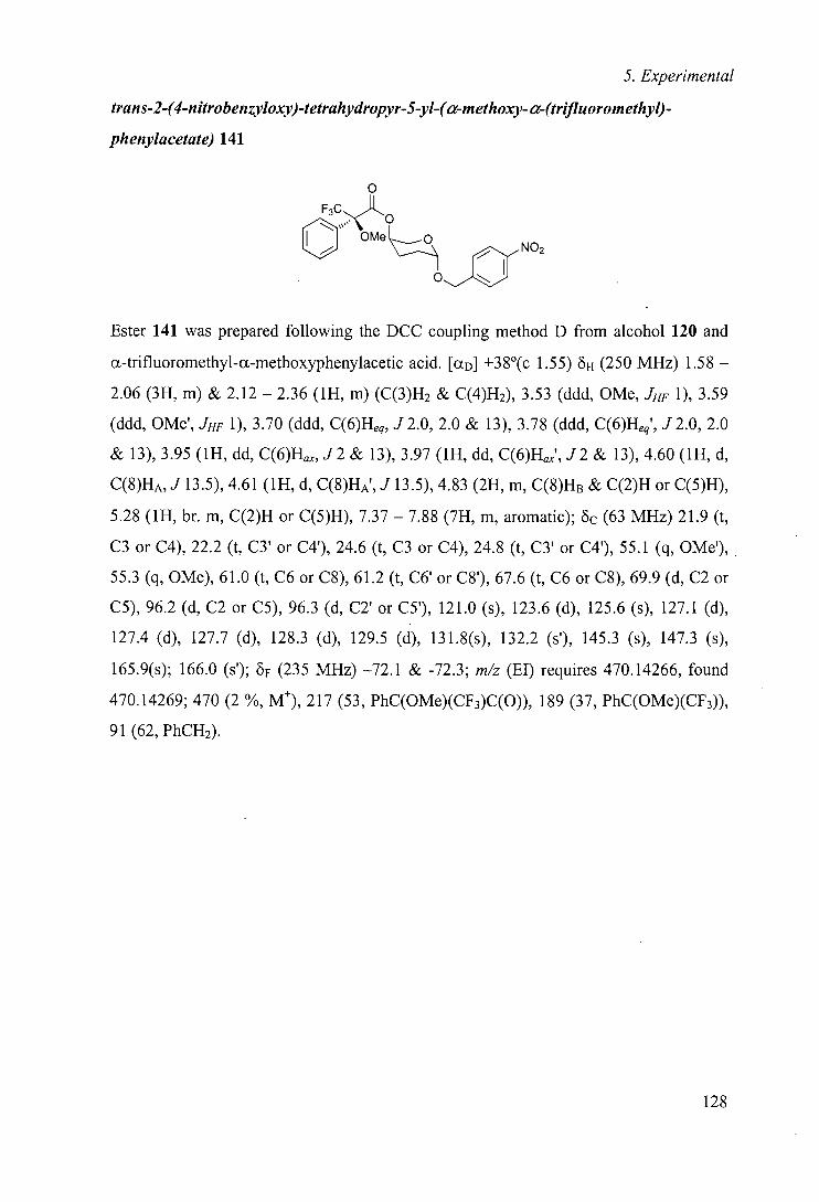
5. Experimental
trans-2-(4-nitrobenzyloxy)-tetrahydropyr-5-yl-(a-methoxy- a-(trifluorom ethyl)-
phenylacetate) 141
0
We NO2
Ester 141 was prepared following the DCC coupling method D from alcohol 120 and
a-trifluoromethyl-ct-methoxyphenylacetic acid. [cD] +38°(c 1.55) oH (250 MHz) 1.58 -
2.06 (3H, m) & 2.12 - 2.36 (1H, m) (C(3)H2 & C(4)H2), 3.53 (ddd, OMe, JHF 1), 3.59
(ddd, OMe', JHF 1), 3.70 (ddd, C(6)Heq, J2.0, 2.0 & 13), 3.78 (ddd, C(6)Heq', J2.0, 2.0
& 13), 3.95 (1H, dd, C(6)H, J2 & 13), 3.97 (1H, dd, C(6)H', J2 & 13), 4.60 (1H, d,
C(8)HA, J 13.5), 4.61 (1H, d, C(8)HA', J 13.5), 4.83 (2H, m, C(8)HB & C(2)H or C(5)H),
5.28 (1H, br. m, C(2)H or C(5)H), 7.37 - 7.88 (7H, m, aromatic); Oc (63 MHz) 21.9 (t,
C3 or C4), 22.2 (t, C3' or C4'), 24.6 (t, C3 or C4), 24.8 (t, C3' or C4'), 55.1 (q, OMe'),
55.3 (q, OMe), 61.0 (t, C6 or C8), 61.2 (t, C6' or C8'), 67.6 (t, C6 or C8), 69.9 (d, C2 or
CS), 96.2 (d, C2 or CS), 96.3 (d, C2' or CS'), 121.0 (s), 123.6 (d), 125.6 (s), 127.1 (d),
127.4 (d), 127.7 (d), 128.3 (d), 129.5 (d), 131.8(s), 132.2 (s'), 145.3 (s), 147.3 (s),
165.9(s); 166.0 (s'); 8F (235 MHz) —72.1 & -72.3; m/z (El) requires 470.14266, found
470.14269; 470 (2 %, M), 217 (53, PhC(OMe)(CF3)C(0)), 189 (37, PhC(OMe)(CF3)),
91(62, PhCH2).
128
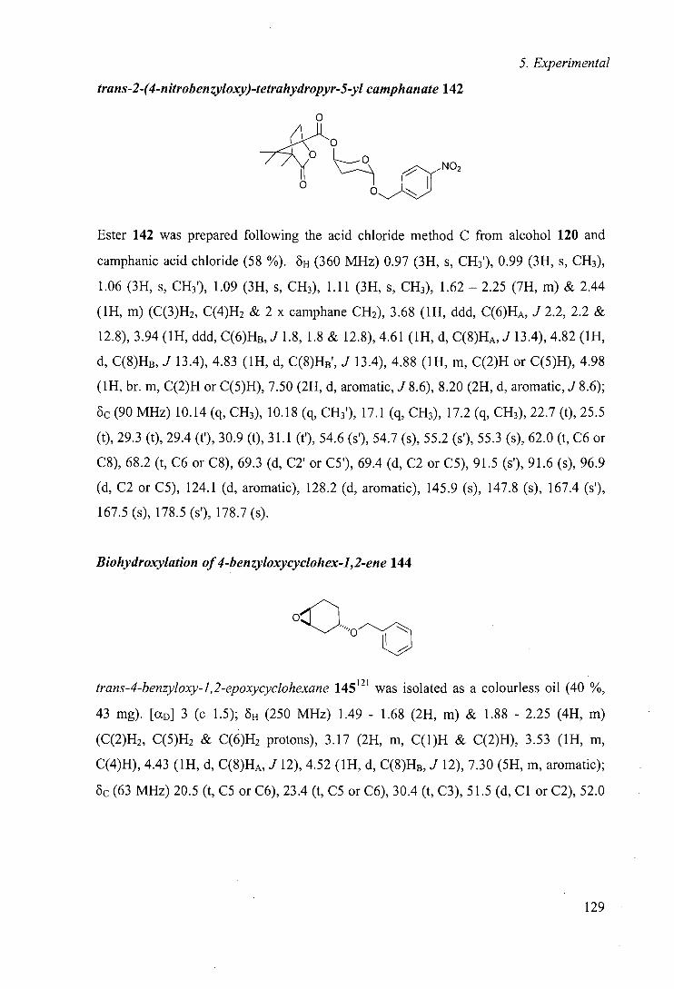
5. Experimental
trans-2-(4-nitrobenzyloxy)-tetrahydropyr-5-y1 camphanate 142
0
O O o NO2
Ester 142 was prepared following the acid chloride method C from alcohol 120 and
camphanic acid chloride (58 %). oH (360 MHz) 0.97 (3H, s, CH3 ), 0.99 (3H, s, CH3),
1.06 (3H, s, CH3'), 1.09 (3H, s, CH3), 1.11 (3H, s, CH3), 1.62 —2.25 (7H, m) & 2.44
(1H, m) (C(3)H2, C(4)H2 & 2 x camphane CH2), 3.68 (1H, ddd, C(6)HA, J 2.2, 2.2 &
12.8), 3.94 (1H, ddd, C(6)HB, J 1. 8, 1.8 & 12.8), 4.61 (1H, d, C(8)HA, J 13.4), 4.82 (1H,
d, C(8)HB, J 13.4), 4.83 (1H, d, C(8)HB', J 13.4), 4.88 (1H, m, C(2)H or C(5)H), 4.98
(1H, br. m, C(2)H or C(5)H), 7.50 (2H, d, aromatic, J 8.6), 8.20 (2H, d, aromatic, J 8.6);
0c (90 MHz) 10.14 (q, CH3), 10.18 (q, CH31), 17.1 (q, CH3), 17.2 (q, CH3), 22.7 (t), 25.5
(t), 29.3 (t), 29.4 (t'), 30.9 (t), 31.1 (t'), 54.6 (s'), 54.7 (s), 55.2 (s'), 55.3 (s), 62.0 (t, C6 or
C8), 68.2 (t, C6 or C8), 69.3 (d, C2' or CS'), 69.4 (d, C2 or CS), 91.5 (s'), 91.6 (s), 96.9
(d, C2 or CS), 124.1 (d, aromatic), 128.2 (d, aromatic), 145.9 (s), 147.8 (s), 167.4 (s'),
167.5 (s), 178.5 (s'), 178.7 (s).
Biohydroxylation of4-benzyloxycyclohex-1,2-ene 144
oC trans-4-benzyloxy-1,2-epoxycyclohexane 145121 was isolated as a colourless oil (40 %,
43 mg). [aD] 3 (c 1.5); OH (250 MHz) 1.49 - 1.68 (2H, m) & 1.88 - 2.25 (4H, m)
(C(2)H2, C(5)H2 & C(6)H2 protons), 3.17 (2H, m, C(l)H & C(2)H), 3.53 (1H, m,
C(4)H), 4.43 (1H, d, C(8)HA, J 12), 4.52 (1H, d, C(8)HB, J 12), 7.30 (5H, m, aromatic);
Oc (63 MHz) 20.5 (t, CS or C6), 23.4 (t, CS or C6), 30.4 (t, 0), 51.5 (d, Cl or C2), 52.0
129
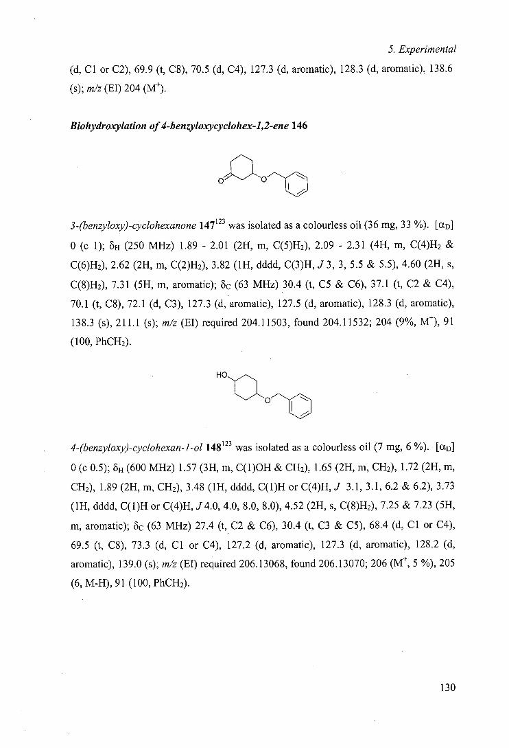
5. Experimental
(d, Cl or C2), 69.9 (t, C8), 70.5 (d, C4), 127.3 (d, aromatic), 128.3 (d, aromatic), 138.6
(s); m/z (El) 204 (M).
Biohydroxylation of 4-benzy1oxycyc1ohex-1,2-ene 146
oOm 3-(benzyloxy)-cyclohexanone 147123 was isolated as a colourless oil (36 mg, 33 %). [cLD]
0 (c 1); oH (250 MHz) 1.89 - 2.01 (2H, m, C(5)H2), 2.09 - 2.31 (4H, m, C(4)H2 &
C(6)H2), 2.62 (2H, m, C(2)H2), 3.82 (1H, dddd, C(3)H, J 3, 3, 5.5 & 5.5), 4.60 (2H, s,
C(8)H2), 7.31 (5H, m, aromatic); 0c (63 MHz) 30.4 (t, CS & C6), 37.1 (t, C2 & C4),
70.1 (t, C8), 72.1 (d, 0), 127.3 (d, aromatic), 127.5 (d, aromatic), 128.3 (d, aromatic),
138.3 (s), 211.1 (s); m/z (El) required 204.11503, found 204.11532; 204 (9%, M), 91
(100, PhCH2).
HO ,aO,-,,C
4-(benzyloxy)-cyclohexan-1-ol 148123 was isolated as a colourless oil (7 mg, 6 %). [aD]
0 (c 0.5); 6H (600 MHz) 1.57 (3H, m, C(1)OH & CH2), 1.65 (2H, m, CH2), 1.72 (2H, m,
CH2), 1.89 (2H, m, CH2), 3.48 (1H, dddd, C(1)H or C(4)H, J 3.1, 3.1, 6.2 & 6.2), 3.73
(1H, dddd, C(1)H or C(4)H, J4.0, 4.0, 8.0, 8.0), 4.52 (2H, s, C(8)H2), 7.25 & 7.23 (5H,
m, aromatic); oc (63 MHz) 27.4 (t, C2 & C6), 30.4 (t, C3 & C5), 68.4 (d, Cl or C4),
69.5 (t, C8), 73.3 (d, Cl or C4), 127.2 (d, aromatic), 127.3 (d, aromatic), 128.2 (d,
aromatic), 139.0 (s); m/z (El) required 206.13068, found 206.13070; 206 (Mt, 5 %), 205
(6, M-H), 91(100, PhCH2).
130
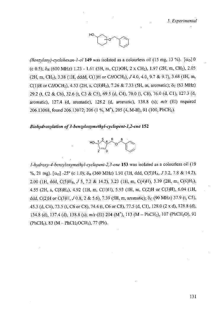
5. Experimental
HO,,C
(Benzyloxy)-cyclohexan-1-ol 149 was isolated as a colourless oil (15 mg, 13 %). [(XD] 0
(c 0.5); 8H (600 MHz) 1.23 - 1.61 (5H, m, C(1)OH, 2 x CH2), 1.97 (2H, m, CH2), 2.05
(2H, m, CH2), 3.38 (1H, dddd, C(1)H or CHOCH2), J4.0,4.0, 9.7 & 9.7), 3.68 (1H, m,
C(1)H or CHOCH2), 4.53 (2H, s, C(8)H2), 7.26 & 7.33 (5H, m, aromatic); öc (63 MHz)
29.2 (t, C2 & C6), 32.6 (t, C3 & C5), 69.5 (d, C4), 70.0 (t, C8), 76.0 (d, Cl), 127.3 (d,
aromatic), 127.4 (d, aromatic), 128.2 (d, aromatic), 138.8 (s); m/z (El) required
206.13068, found 206.13072; 206 (1 %, Mt), 205 (4, M-H), 91(100, PhCH2).
Biohydroxylation of3-benzyloxymethyl-cyclopent-1,2-ene 152
5 6 8 -!~~O--- 01 --,
1-hydroxy-4-benzyloxymethyl-cyclopent-2,3-ene 153 was isolated as a colourless oil (19
%, 21 mg). [aD] -25° (c 1.0); oH (360 MHz) 1.91 (1H, ddd, C(5)HA, J3.2, 7.8 & 14.2),
2.00 (1H, ddd, C(5)HB, J 5, 7.2 & 14.2), 3.23 (1H, m, C(4)H), 3.39 (2H, m, C(6)H2),
4.55 (2H, s, C(8)H2), 4.92 (1H, m, C(1)H), 5.93 (1H, m, C(2)H or C(3)H), 6.04 (1H,
ddd, C(2)H or C(3)H, J 0.8, 2 & 5.6), 7.39 (5H, m, aromatic); Oc (90 MHz) 37.9 (t, CS),
45.3 (d, C4), 73.5 (t, C6 or C8), 74.4 (t, C6 or C8), 77.5 (d, Cl), 128.0 (2 x d), 128.8 (d),
134.8 (d), 137.4 (d), 138.8 (s); m/z (El) 204 (M), 113 (M—PhCH2), 107 (PhCH20), 91
(PhCH2), 83 (M - PhCH20CH2), 77 (Ph).
131
Bibliography
6 References
1 S. M. Roberts and N. M. Williamson, Curr. Org. Chem., 1997, 1, 1.
2 F. H. Arnold, Acc. Chem. Res., 1998, 31, 125.
3 M. T. Reetz, A. Zonta, K. Schimossek, K. Liebeton, and K. E. Jaeger, Angew.
Chem., mt. Ed. Engl., 1997, 36, 2830.
4 D. H. R. Barton, F. Hally, N. Ozbalik, M. Schmitt, E. Young, and G. Balaroine,
J. Am. Chem. Soc., 1989, 111, 7144.
5 T. Yang and R. Breslow, Angew. Chem., mt. Ed. Engl., 2000, 39, 2692.
6 D. H. Peterson and H. C. Murray, I Am. Chem. Soc., 1952, 74, 1871.
7 D. H. Peterson, H. C. Murray, S. H. Eppstein, L. M. Reineke, A. Weintraub, P.
D. Meister, and H. M. Leigh, J. Am. Chem. Soc., 1952, 74, 5933.
8 J. Fried, R. W. Thoma, and J. R. Gerke, J. Am. Chem. Soc., 1952, 74, 3692.
9 H. G. Davies, R. H. Green, D. R. Kelly, and S. M. Roberts, in
'Biotransformations in Preparative Organic Chemistry', London, 1989.
10 C. L. Smith, E. M. Hampton, J. A. Pederson, L. R. Pennington, and D. W.
Bourne, Pharmacotherapy, 1994, 14, 471.
11 M. Murray, mt. J. Mo!. Medicine, 1999, 3, 227.
12 H. J. M. van den Brink, R. F. M. van Gorcom, C. van den Hondel, and P. J. Punt,
Fungal Genetics and Biology, 1998, 23, 1.
13 B. P. Unger, I. C. Gunsalus, and S. G. Sligar, J. Biol. Chem., 1986, 261, 1158.
14 M. Katagiri, B. N. Ganguli, and I. C. Gunsalus, J. Biol. Chem., 1968, 243, 3543.
15 I. C. Gunsalus and S. G. Sugar, Adv. Enzymol. Relat. Areas Mo!. Biol., 1978, 47,
1.
16 T. L. Poulos, B. C. Finzel, and A. J. Howard, J. Mol. Biol., 1987, 195, 687.
17 T. L. Poulos, B. C. Finzel, and A. J. Howard, Biochemistry, 1986, 25, 5314.
18 T. L. Poulos, B. C. Finzel, I. C. Gunsalus, G. C. Wagner, and J. Kraut, J. Biol.
Chem., 1985, 260, 16122.
19 S. G. Jezequel, J. Mo!. Catal. B-Enzymatic, 1998, 5, 371.
132
Bibliography
20 H. L. Holland, T. A. Morris, P. J. Nava, and M. Zabic, Tetrahedron, 1999, 55,
7441.
21 G. J. Grogan and H. L. Holland, J. Mo!. Catal. B: Enzymatic, 2000, 9, 1.
22 A. Kergomard, M. F. Renard, and H. Veschambre, J. Org. Chem., 1982, 47, 792.
23 C. Fuganti, J. Minut, G. Pedrocchi-Fantoni, and S. Servi, J. Mo!. Catal. B:
Enzymatic, 1998, 4, 47.
24 H. L. Holland and F. M. Brown, Tetrahedron: Asymmetry, 1998, 9, 535.
25 H. L. Holland, P. R. Andreana, and F. M. Brown, Tetrahedron: Asymmetry,
1999, 10, 2833.
26 A. Archelas, J. D. Fourneron, and R. Furstoss, Tetrahedron Lett., 1988, 29, 6611.
27 V. Gotor, M. Quiros, R. Liz, J. Frigola, and R. Fernandez, Tetrahedron, 1997,
53, 6421.
28 B. Vigne, A. Archelas, and R. Furstoss, Tetrahedron, 1986, 42, 2451.
29 B. Vigne, A. Archelas, and R. Furstoss, Tetrahedron, 1991, 47, 1447.
30 J. Ilavsky and H. L. Herzog, in 'US Patent US 30139', 1961.
31 G. S. Fonken, M. E. Herr, H. C. Murray, and L. M. Reineke, I Am. Chem. Soc.,
1967, 89, 672.
32 V. Lamare, J. D. Fourneron, R. Furstoss, C. Ehret, and B. Corbier, Tetrahedron
Lett., 1987, 28, 6269.
33 R. A. Johnson, H. C. Murray, L. M. Reineke, and G. S. Fonkeñ, I Org. Chem.,
1969, 34, 2279.
34 R. A. Johnson, E. H. Milton, H. C. Murray, and G. S. Fonken, J. Org. Chem.,
1968, 33, 3187.
35 C. R. Davis, R. A. Johnson, J. I. Ciadella, W. F. Liggett, S. A. Mizsak, and V. P.
Marshall, I Org. Chem., 1997, 62, 2244.
36 N. Floyd, F. Munyemana, S. M. Roberts, and A. J. Willetts, I Chem. Soc.,
Perkin Trans. 1, 1993, 881.
37 R. Furstoss, A. Archelas, J. D. Fourneron, and B. Vigne, 'Enzymes as Catalysts
in Organic Chemistry', ed. P. Schneider, 1986.
133
Bibliography
38 R. Furstoss, A. Archelas, B. Waegell, J. L. Petit, and L. Deveze, Tetrahedron
Lett., 1980, 21, 451.
39 S. Pietz, D. Wolker, and G. Haufe, Tetrahedron, 1997, 53, 17067.
40 A. Archelas, J. D. Fourneron, and R. Furstoss, J. Org. Chem., 1988, 53, 1797.
41 G. Braunegg, A. deRaadt, S. Feichtenhofer, H. Griengl, I. Kopper, A. Lehmann,
and H. J. Weber, Angew. Chem., mt. Ed. Engl., 1999, 38, 2763.
42 A. M. Warhurst, K. F. Clarke, R. A. Hull, R. A. Holt, and C. A. Fewson, App!.
Environ. Microbiol., 1994, 60, 1137.
43 J. H. A. Apajahti and M. S. Salkinoja-Salonen, App!. Microbiol. Biotechnol.,
1986, 25, 62.
44 R. Valo and M. Salkinoja-Salonen, App!. Microbiol. Biotechnol., 1986, 25, 68.
45 R. J. Valo, M. M. Haggblom, and M. S. Salkinoja-Salonen, Water Research,
1990, 24, 253.
46 J. Sikkema and J. A. M. d. Bont, App!. Environ. Microbiol., 1993, 59, 567.
47 G. J. MacMichael and L. R. Brown, App!. Environ. Microbiol., 1987, 53, 65.
48 G. Cardini and P. Jurtshuk, J. Biol. Chem., 1970, 245, 2789.
49 K. Nakajima, A. Sato, Y. Takahara, and T. lida, Agric. Biol. Chem., 1985, 49,
1993.
50 R. Hechtberger, G. Wirnsberger, M. Mischitz, N. Kiempier, and K. Faber,
Tetrahedron: Asymmetry, 1993, 4, 1161.
51 M. Kobayashi, T. Nagasawa, and H. Yamada, Trends Biotechnol., 1992, 10, 402.
52 M. A. Cohen, J. S. Parratt, and N. J. Turner, Tetrahedron: Asymmetry, 1992, 3,
1543.
53 T. M. Beard and M. I. Page, Antonie Van Leeuwenhoek International J. Gen.
Mo!. Microbiol., 1998, 74, 99.
54 C. T. Evans, S. M. Roberts, K. A. Shoberu, and A. G. Sutherland, J. Chem. Soc.,
Perkin Trans. 1, 1992, 589.
55 S. J. C. Taylor, R. McCague, R. Wisdom, C. Lee, K. Dickson, G. Ruecroft, F.
O'Brien, J. Littlechild, J. Bevan, S. M. Roberts, and C. T. Evans, Tetrahedron:
Asymmetry, 1993, 4, 117.
134
Bibliography
56 M. Chartrain, B. Jackey, C. Taylor, V. Sandford, K. Gbewonyo, L. Lister, L.
Dimichele, C. Hirsch, B. Heimbuch, C. Maxwell, D. Pascoe, B. Buckland, and
R. Greasham, I Fermentation Bioeng., 1998, 86, 550.
57 D. P. O'Keefe and P. A. Harder, Mol. Microbiol., 1991, 5, 2099.
58 J. B. Sutherland, J. P. Freeman, A. L. Selby, P. P. Fu, D. W. Miller, and C. E.
Cerniglia, Arch. Microbiol., 1990, 154, 260.
59 J. B. Sutherland, Appi. Environ. Microbiol., 1986, 52, 98.
60 M. K. Trower, F. S. Sariaslani, and D. P. O'Keefe,I Bacteriol., 1989, 171, 1781.
61 I. Nagy, F. Compernolle, K. Ghys, J. Vanderleyden, and R. d. Mot, Appl,
Environ. Microbiol., 1995, 61, 2056.
62 I. Nagy, G. Schoofs, F. Compernolle, P. Proost, J. Vanderleyden, and R. d. Mot,
J. Bacteriol., 1995, 177, 676.
63 H. Ohta, S. Matsumoto, Y. Okanoto, and T. Sugai, Chem. Lett., 1989, 625.
64 B. Angelova, S. Mutafov, T. Avramora, and L. Boyadjieva, Process Biochem.,
1996, 31, 179.
65 G. Cardini and P. Jurtshuk, J. Biol. Chem., 1968, 243.
66 P. J. Chapman, J. F. Kuo, and I. C. Gunsalus, Fed. Proc., 1963, 22, 296.
67 P. J. Chapman, G. Meerman, and I. C. Gunsalus, Biochem. Biophys. Res.
Commun., 1965, 20,104.
68 G. Grogan, S. L. Flitsch, and N. J. Turner, Unpublished results.
69 F. S. Sariaslani, L. R. McGee, M. K. Trower, and F. G. Kitson, Biochem.
Biophys. Res. Commun., 1990, 170, 456.
70 M. K. Trower, F. S. Sariaslani, and F. G. Kitson, Biochem. Biophys. Res.
Commun., 1988, 157, 1417.
71 U. Karison, D. F. Dwyer, S. W. Hooper, E. R. B. Moore, K. N. Timmis, and L.
D. Eltis, I Bacteriol., 1993, 175, 1467.
72 L. D. Eltis, U. Karison, and K. N. Timmis, Eur. I Biochem., 1993, 213, 211.
73 S. J. Aitken, G. Grogan, C. S. Y. Chow, N. J. Turner, and S. L. Flitsch, I Chem.
Soc., Perkin Trans 1, 1998, 3365.
135
Bibliography
74 S. J. Aitken, 'Cytochrome P-450 Monooxygenases: Their Application in
Biocatalysis', MSc (by Research), University of Edinburgh, 1997.
75 J. D. Fourneron, A. Archelas, B. Vigne, and R. Furstoss, Tetrahedron, 1987, 43,
2273.
76 J. T. Groves, G. A. McClusky, R. E. White, and M. J. Coon, Biochem. Biophys.
Res. Commun., 1978, 81, 154.
77 J. D. Fourneron, A. Archelas, and R. Furstoss, J. Org. Chem., 1989, 54, 2478.
78 Y. L. Chow, C. J. Colon, and J. N. S. Tam, Can. J. Chem, 1968, 46, 2822.
79 R. A. Johnson, J. Org. Chem., 1968, 33, 3627.
80 W. Carruthers, J. D. Prail, S. M. Roberts, and A. J. Willetts, J. Chem. Soc.,
Perkin Trans. 1, 1990, 2854.
81 R. A. Johnson, M. E. Herr, H. C. Murray, C. G. Chidester, and F. Han, I Org.
Chem., 1992, 57, 7209.
82 H. L. Holland, M. Conn, P. C. Chenchaiah, and F. M. Brown, Tetrahedron Lett.,
1988, 29, 6393.
83 M. E. Herr, R. A. Johnson, H. C. Murray, L. M. Reineke, and G. S. Fonken, I
Org. Chem., 1968, 33, 3201.
84 C. R. Davis, R. A. Johnson, J. I. Ciadella, W. F. Liggett, S. A. Mizsak, F. Han,
and V. P. Marshall, I Org. Chem., 1997, 62, 2252.
85 A. de Raadt, H. Griengl, M. Petsch, P. Plachato, N. Schoo, H. Weber, G.
Braunegg, I. Kopper, M. Kreiner, and A. Zeiser, Tetrahedron: Asymmetry, 1996,
7,473.
86 A. de Raadt, H. Griengl, M. Petsch, P. Plachato, N. Schoo, H. Weber, G.
Braunegg, I. Kopper, M. Kreiner, and A. Zeiser, Tetrahedron: Asymmetry, 1996,
7,491.
87 A. de Raadt, H. Griengl, M. Petsch, P. Plachato, N. Schoo, H. Weber, G.
Braunegg, I. Kopper, M. Kreiner, A. Zeiser, and K. Kieslich, Tetrahedron:
Asymmetry, 1996, 7,467.
88 D. A. Grayson, Y. B. Tewari, M. P. Mayhew, V. L. Vilker, and R. N. Goldberg,
Arch. Biochem. Biophys., 1996, 332, 239.
136
Bibliography
89 T. L. Poulos and A. J. Howard, Biochemistry, 1987, 26, 8165.
90 K. Gill, J. F. Menez, D. Lucas, and R. A. Dietrich, Alcoholism: Clinical and
Experimental Research, 1992, 16, 910.
91 M. Sharrock, E. Munck, P. G. Brunner, V. Marshall, J. B. Lipscomb, and I. C.
Gunsalus, Biochemistry, 1973, 12, 258.
92 S. M. Fowler, P. A. England, A. C. G. Westlake, D. Rouch, D. P. Nickerson, C.
Blunt, D. Braybrook, S. West, L. L. Wong, and S. L. Flitsch, J. Chem. Soc.,
Chem. Commun., 1994, 2761.
93 J. P. Jones, E. J. O'Hare, and L. L. Wong, J. Chem. Soc., Chem. Commun., 2000,
247.
94 A. Schmid, A. Kollmer, B. Sonnleitner, and B. Witholt, Bioprocess Eng., 1999,
20, 91.
95 S. L. Flitsch, S. J. Aitken, C. S. Y. Chow, G. Grogan, and A. Staines, Bioorg.
Chem., 1999, 27, 81.
96 C. S.-Y. Chow, 'Selective hydroxylations catalysed by cytochrome P-450
monooxygenases', PhD Thesis, University of Edinburgh, 1998.
97 F. Mohamadi, N. G. J. Richards, W. C. Guids, R. Liskamp, M. Lipton, C.
Caufield, G. Chang, T. Hendrickson, and W. C. Still, J. Comput. Chem., 1990,
11,440.
98 N. L. Allinger,J. Am. Chem. Soc., 1977, 99, 8127.
99 W. C. Still, A. Tempezyk, R. C. Hawley, and T. Hendrickson, J. Am. Chem. Soc.,
1990, 112, 6127.
100 G. Chang, W. C. Guida, and W. C. Still, J. Am. Chem. Soc., 1989, 111, 4379.
101 H. L. Holland, M. Kindermann, S. Kumaresan, and T. Stefanac, Tetrahedron:
Asymmetry, 1993, 4, 1353.
102 J. A. Dale, D. L. Dull, and H. S. Mosher, J. Org. Chem., 1969, 34, 2543.
103 J. A. Dale and H. S. Mosher, J. Am. Chem. Soc., 1973, 95, 512.
104 G. R. Sullivan, J. A. Dale, and H. S. Mosher, J. Org. Chem., 1973, 38, 2143.
105 F. Yasuhara, S. Yamaguchi, M. Takeda, T. Abe, and S. Miyano, Bull. Chem.
Soc. Jpn., 1991, 64, 3390.
137
Bibliography
106 K. Mori, A. Horinaka, and M. Kido, Liebigs Ann. Chem., 1994, 817.
107 S. Yao, M. Robertson, F. Reichel, R. G. Hazel!, and K. A. Jorgensen, J. Org.
Chem., 1999, 64, 6677.
108 D. Parker, R. J. Taylor, G. Ferguson, and A. Tonge, Tetrahedron, 1986, 42, 617.
109 H. Gerlach and B. Zagalak, J. Chem. Soc., Chem. Commun., 1973, 274.
110 P. Darrigo, G. Pedrochi-Fantoni, and S. Servi, Adv. Appi. Microbiol.,' 1997, 44,
81.
111 Z. Li, H. J. Feiten, J. B. van Beilen, W. Duetz, and B. Witholt, Tetrahedron:
Asymmetry, 1999, 10, 1323.
112 E. L. White, N. B. Barker, L. J. Macy, S. C. Shaddix, C. McCaleb, J. A. S. III, R.
Vince, and W. M. Shannon, Biochem. Biophys. Res. Commun., 1989, 161, 393.
113 H. F. Olivo and J. Yu, J. Chem. Soc., Perkin Trans. 1, 1998, 391.
114 D. J. Leak, S. L. R. Ellison, C. Murricane, and P. B. Baker, Biocatalysis, 1988, 1,
197.
115 C. S. Cooper, P. L. Klock, D. T. W. Chu, D. J. Hardy, R. N. Swanson, and J. J.
Plattner, .1 Med. Chem., 1992, 35, 1392.
116 L. Alig, A. Edenhoffer, P. Hadvary, M. Huerzeler, D. Knopp, N. Muller, B.
Steiner, A. Trzeciak, and T. Weller, J. Med. Chem., 1992, 35, 4393.
117 S. Hoyer and P. Laszlo, Synthesis, 1986, 656.
118 S. Koto, N Morishima, R. Kawahara, K. Ishikawa, and S. Zen, 'Bull. Chem. Soc.
Jpn., 1982, 55, 1092.
119 H. Kurokawa, T. Ishigami, K. Morimoto, and A. Siegimori, Bull. Chem. Soc.
Jpn., 1979, 52, 959.
120 J. F. Garst and C. D. Smith, J. Am. Chem. Soc., 1976, 98, 1526.
121 J. Schulz and D. Gani, J. Chem. Soc., Perkin Trans. 1, 1997, 657.
122 F. Calvani, P. Crotti, C. Gardelli, and M. Pineschi, Tetrahedron, 1994, 50,
12999.
123 M. Chini, P. Crotti, L. A. Flippin, and F. Macchia, J. Org. Chem., 1990, 55,
4265.
124 A. M. Warhurst and C. A. Fewson, Grit. Rev. Biotechnol., 1994, 14, 29.
138
Bibliography
125 J. Ooyama and K. Yamada, Antonie Van Leeuwenhoek International J. Gen.
Mol. Microbiol., 1965, 31, 45.
126 S. H. El-Sharkawy, W. Yang, L. Dostal and J. P. N. Rosazza, App. Environ.
Microbiol, 1992, 58, 2116.
139
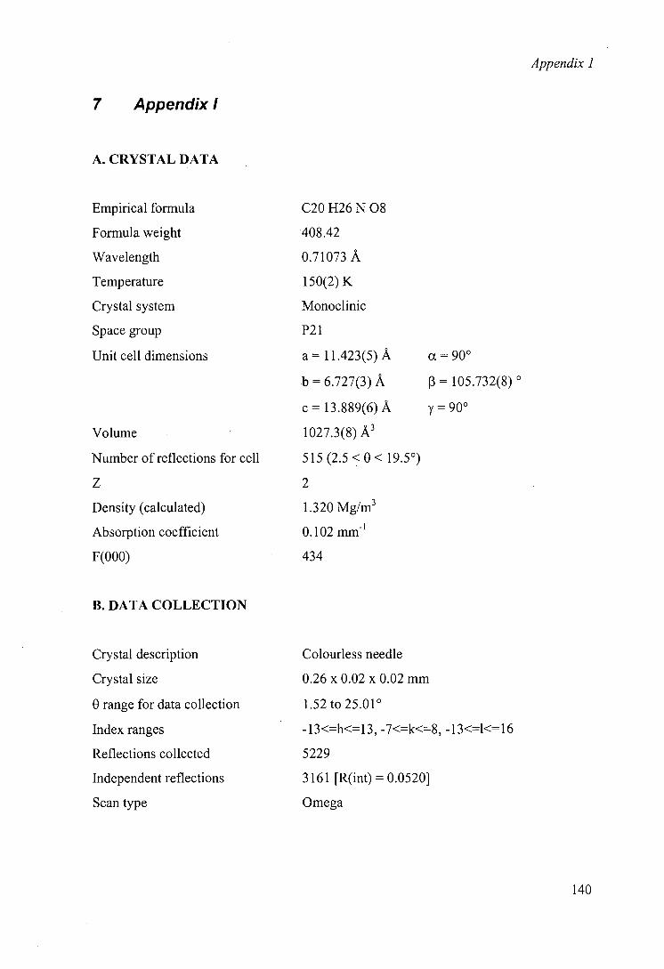
Appendix 1
7 Appendix!
A. CRYSTAL DATA
Empirical formula C20 H26 N 08
Formula weight 408.42
Wavelength 0.71073 A
Temperature 150(2) K
Crystal system Monoclinic
Space group P21
Unit cell dimensions a= 11.423(5) A
= 6.727(3) A
c= 13.889(6)A
Volume 1027.3(8) A3
Number of reflections for cell 515 (2.5 <0< 19.50)
Z 2
Density (calculated) 1.320 Mg/M3
Absorption coefficient 0.102 mm-
F(O00) 434
= 105.732(8)
= 90°
B. DATA COLLECTION
Crystal description
Crystal size
0 range for data collection
Index ranges
Reflections collected
Independent reflections
Scan type
Colourless needle
0.26 x 0.02 x 0.02 mm
1.52 to 25.01°
-13<=h<=13, -7<=k<=8, -13<=l<=16
5229
3161 [R(int) = 0.0520]
Omega
140
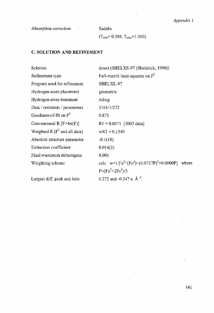
Appendix 1
Absorption correction Sadabs
(Tmin 0.588, Tmax 1.000)
C. SOLUTION AND REFINEMENT
Solution direct (SHELXS-97 (Sheidrick, 1990))
Refinement type Full-matrix least-squares on F2
Program used for refinement SHELXL-97
Hydrogen atom placement geometric
Hydrogen atom treatment riding
Data / restraints / parameters 3161/1/272
Goodness-of-fit on F2 0.871
Conventional R [F>4c(F)] RI = 0.0571 [2003 data]
Weighted R (F2 and all data) wR2 = 0.1340
Absolute structure parameter -0.1(18)
Extinction coefficient 0.014(3)
Final maximum delta/sigma 0.001
Weighting scheme calc w=1/[\s2"(Fo2)+(0.0737P)2+0.0000P] where
P=(Fo2+2Fc2)/3
Largest diff. peak and hole 0.272 and -0.247 e. A
141
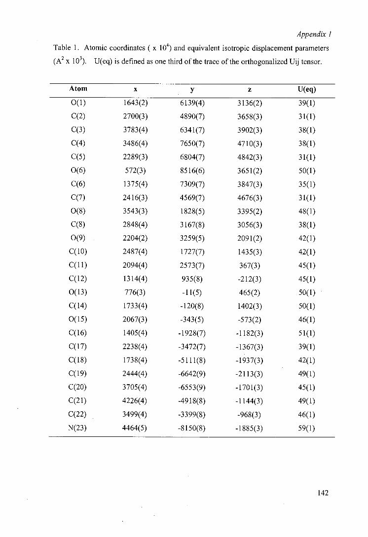
Appendix 1
Table 1. Atomic coordinates ( x 10) and equivalent isotropic displacement parameters
(A2 x 10). U(eq) is defined as one third of the trace of the orthogonalized Uij tensor.
Atom x y z U(eq)
0(1) 1643(2) 6139(4) 3 136(2) 39(1)
2700(3) 4890(7) 3658(3) 31(1)
3783(4) 6341(7) 3902(3) 38(1)
3486(4) 7650(7) 4710(3) 38(1)
2289(3) 6804(7) 4842(3) 31(1)
0(6) 572(3) 8516(6) 3651(2) 50(1)
1375(4) 7309(7) 3847(3) 35(1)
2416(3) 4569(7) 4676(3) 31(1)
0(8) 3543(3) 1828(5) 3395(2) 48(1)
2848(4) 3167(8) 3056(3) 38(1)
0(9) 2204(2) 3259(5) 2091(2) 42(1)
2487(4) 1727(7) 1435(3) 42(1)
2094(4) 2573(7) 367(3) 45(1)
1314(4) 935(8) -212(3) 45(1)
0(13) 776(3) -11(5) 465(2) 50(1)
C(14) 1733(4) -120(8) 1402(3) 50(1)
0(15) 2067(3) -343(5) -573(2) 46(1)
1405(4) -1928(7) -1182(3) 51(1)
2238(4) -3472(7) -1367(3) 39(1)
1738(4) -5111(8) -1937(3) 42(1)
2444(4) -6642(9) -2113(3) 49(1)
3705(4) -6553(9) -1701(3) 45(1)
4226(4) -4918(8) -1144(3) 49(1)
3499(4) -3399(8) -968(3) 46(1)
N(23) 4464(5) -8150(8) -1885(3) 59(1)
142
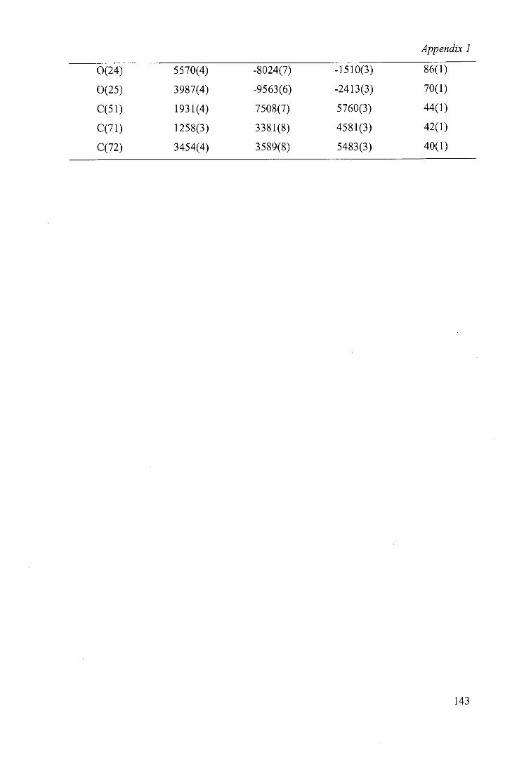
Appendix 1
0(24) 5570(4) -8024(7) -1510(3) 86(1)
0(25) 3987(4) -9563(6) -2413(3) 70(1)
C(51) 1931(4) 7508(7) 5760(3) 44(1)
1258(3) 3381(8) 4581(3) 42(1)
3454(4) 3589(8) 5483(3) 40(1)
143
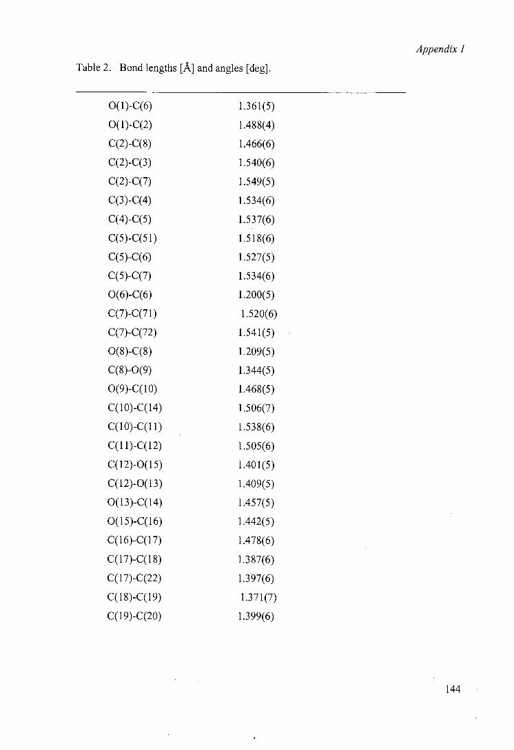
Appendix 1
Table 2. Bond lengths [A] and angles [deg].
0(l)-C(6) 1.361(5)
0(l)-C(2) 1.488(4)
C(2)-C(8) 1.466(6)
C(2)-C(3) 1.540(6)
C(2)-C(7) 1.549(5)
C(3)-C(4) 1.534(6)
C(4)-C(5) 1.537(6)
C(5)-C(51) 1.518(6)
C(5)-C(6) 1.527(5)
C(5)-C(7) 1.534(6)
0(6)-C(6) 1.200(5)
C(7)-C(71) 1.520(6)
C(7)-C(72) 1.541(5)
0(8)-C(8) 1.209(5)
C(8)-0(9) 1.344(5)
0(9)-C(10) 1.468(5)
C(10)-C(14) 1.506(7)
C(10)-C(11) 1.538(6)
C(1 1)-C(12) 1.505(6)
C(12)-0(15) 1.401(5)
C(12)-0(13) 1.409(5)
0(13)-C(14) 1.457(5)
0(1 5)-C( 16) 1.442(5)
C(16)-C(17) 1.478(6)
C(17)-C(18) 1.387(6)
C(17)-C(22) 1.397(6)
C(18)-C(19) 1.371(7)
C(19)-C(20) 1.399(6)
144
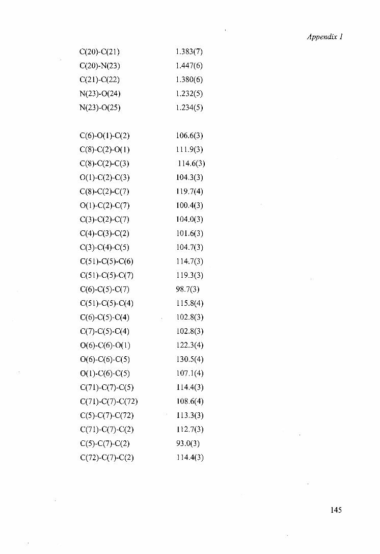
Appendix 1
C(20)-C(21) 1.383(7)
C(20)-N(23) 1.447(6)
C(2 I)-C(22) 1.380(6)
N(23)-0(24) 1.232(5)
N(23)-0(25) 1.234(5)
C(6)-O(1)-C(2) 106.6(3)
C(8)-C(2)-O(1) 111.9(3)
C(8)-C(2)-C(3) 114.6(3)
0(1)-C(2)-C(3) 104.3(3)
C(8)-C(2)-C(7) 119.7(4)
0(1)-C(2)-C(7) 100.4(3)
C(3)-C(2)-C(7) 104.0(3)
C(4)-C(3)-C(2) 101.6(3)
C(3)-C(4)-C(5) 104.7(3)
C(5 1)-C(5)-C(6) 114.7(3)
C(51)-C(5)-C(7) 119.3(3)
C(6)-C(5)-C(7) 98.7(3)
C(51)-C(5)-C(4) 115.8(4)
C(6)-C(5)-C(4) 102.8(3)
C(7)-C(5)-C(4) 102.8(3)
0(6)-C(6)-O(1) 122.3(4)
0(6)-C(6)-C(5) 130.5(4)
0(1)-C(6)-C(5) 107.1(4)
C(71)-C(7)-C(5) 114.4(3)
C(71)-C(7)-C(72) 108.6(4)
C(5)-C(7)-C(72) 113.3(3)
C(71)-C(7)-C(2) 112.7(3)
C(5)-C(7)-C(2) 93.0(3)
C(72)-C(7)-C(2) 114.4(3)
145
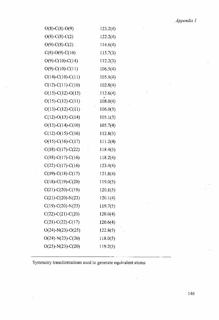
Appendix 1
0(8)-C(8)-0(9) 123.2(4)
0(8)-C(8)-C(2) 122.2(4)
0(9)-C(8)-C(2) 114.6(4)
C(8)-O(9)-C(10) 115.7(3)
0(9)-C(10)-C(14) 112.2(3)
0(9)-C(10)-C(1 1) 106.5(4)
C(14)-C(10)-C(11) 105.1(4)
C(12)-C(1 1)-C(1O) 102.8(4)
O(15)-C(12)-O(13) 112.6(4)
O(15)-C(12)-C(1 1) 108.0(4)
0(13)-C(12)-C(11) 106.0(3)
C(12)-0(13)-C(14) 105.1(3)
0(13)-C(14)-C(10) 105.7(4)
C(12)-O(15)-C(16) 112.8(3)
O(15)-C(16)-C(17) 111.2(4)
C(18)-C(17)-C(22) 118.4(5)
C(18)-C(11)-C(16) 118.2(4)
C(22)-C( 1 7)-C( 16) 123.4(4)
C(19)-C(18)-C(17) 121.8(4)
C(18)-C(19)-C(20) 119.0(5)
C(21)-C(20)-C(19) 120.1(5)
C(2 I)-C(20)-N(23) 120.1(4)
C(19)-C(20)-N(23) 119.7(5)
C(22)-C(2 l)-C(20) 120.0(4)
C(2 1 )-C(22)-C( 17) 120.6(4)
0(24)-N(23)-0(25) 122.8(5)
0(24)-N(23)-C(20) 118.0(5)
0(25)-N(23)-C(20) 119.2(5)
Symmetry transformations used to generate equivalent atoms
146
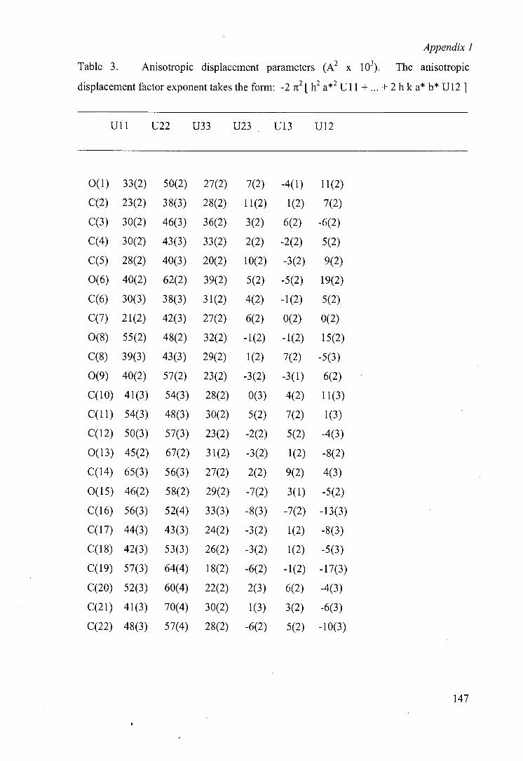
Appendix 1
Table 3. Anisotropic displacement parameters (A2 x 10). The anisotropic
displacement factor exponent takes the form: -2 it2 [ h 2 a*2 Ui 1 +... + 2 h k a* b* U12]
Ull U22 U33 U23 U13 U12
0(1) 33(2) 50(2) 27(2) 7(2) -4(1) 11(2)
23(2) 38(3) 28(2) 11(2) 1(2) 7(2)
30(2) 46(3) 36(2) 3(2) 6(2) -6(2)
30(2) 43(3) 33(2) 2(2) -2(2) 5(2)
28(2) 40(3) 20(2) 10(2) -3(2) 9(2)
0(6) 40(2) 62(2) 39(2) 5(2) -5(2) 19(2)
30(3) 38(3) 3 1(2) 4(2) -1(2) 5(2)
21(2) 42(3) 27(2) 6(2) 0(2) 0(2)
0(8) 55(2) 48(2) 32(2) -1(2) -1(2) 15(2)
39(3) 43(3) 29(2) 1(2) 7(2) -5(3)
0(9) 40(2) 57(2) 23(2) -3(2) -3(1) 6(2)
41(3) 54(3) 28(2) 0(3) 4(2) 11(3)
54(3) 48(3) 30(2) 5(2) 7(2) 1(3)
50(3) 57(3) 23(2) -2(2) 5(2) -4(3)
0(13) 45(2) 67(2) 31(2) -3(2) 1(2) -8(2)
C(14) 65(3) 56(3) 27(2) 2(2) 9(2) 4(3)
0(15) 46(2) 58(2) 29(2) -7(2) 3(1) -5(2)
56(3) 52(4) 33(3) -8(3) -7(2) -13(3)
44(3) 43(3) 24(2) -3(2) 1(2) -8(3)
42(3) 53(3) 26(2) -3(2) 1(2) -5(3)
57(3) 64(4) 18(2) -6(2) -1(2) -17(3)
52(3) 60(4) 22(2) 2(3) 6(2) -4(3)
41(3) 70(4) 30(2) 1(3) 3(2) -6(3)
48(3) 57(4) 28(2) -6(2) 5(2) -10(3)
147
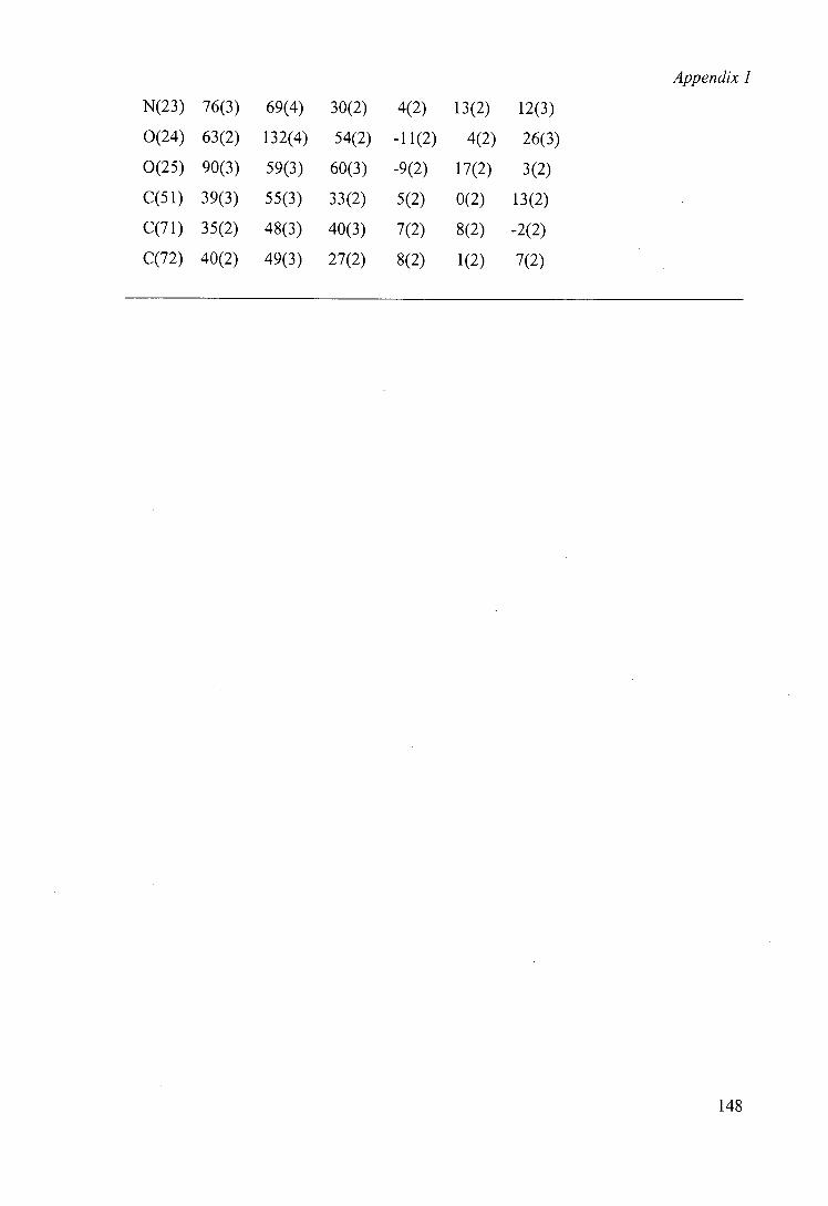
Appendix 1
N(23) 76(3) 69(4) 30(2) 4(2) 13(2) 12(3)
0(24) 63(2) 132(4) 54(2) -11(2) 4(2) 26(3)
0(25) 90(3) 59(3) 60(3) -9(2) 17(2) 3(2)
C(51) 39(3) 55(3) 33(2) 5(2) 0(2) 13(2)
35(2) 48(3) 40(3) 7(2) 8(2) -2(2)
40(2) 49(3) 27(2) 8(2) 1(2) 7(2)
om
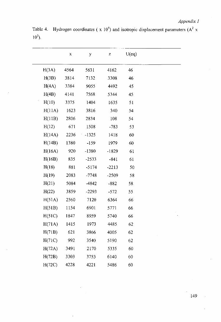
Appendix 1
Table 4. Hydrogen coordinates ( x 10) and isotropic displacement parameters (A2 x
1O).
X y z U(eq)
H(3A) 4564 5631 4162 46
H(313) 3814 7132 3308 46
H(4A) 3384 9055 4492 45
H(4B) 4141 7568 5344 45
H(10) 3375 1404 1635 51
H(11A) 1623 3816 340 54
H(11B) 2806 2834 108 54
H(12) 671 1508 -783 53
H(14A) 2236 -1325 1418 60
H(14B) 1380 -159 1979 60
H(1 6A) 920 -1380 -1829 61
H(1 6B) 835 -2533 -841 61
881 -5174 -2213 50
2083 -7748 -2509 58
5084 -4842 -882 58
3859 -2293 -572 55
H(51A) 2560 7120 6364 66
H(51B) 1154 6901 5771 66
H(51C) 1847 8959 5740 66
H(71A) 1415 1973 4485 62
H(71B) 621 3866 4005 62
H(71C) 992 3540 5190 62
H(72A) 3491 2170 5335 60
H(7213) 3303 3753 6140 60
H(72C) 4228 4221 5486 60
149
Appendix II
Biohydroxylations of Cbz-protected alkyl substituted piperidines by Beauveria bassiana ATCC 7159
Suzanne J. Aitken, Gideon Grogan, Cathy S.-Y. Chow, Nicholas J. Turner * and Sabine L. Flitsch *
Department of Chemistry, Centre for Protein Technology, University of Edinburgh, King's Buildings, West Mains Road, Edinburgh, UK EH9 3JJ
Received (in Cambridge) 24th July 1998, Accepted 2nd September 1998
N-Benzyloxycarbonyl (Cbz) protected piperidines are hydroxylated with greater regioselectivity than the corresponding N-benzoyl analogues when incubated with the fungus Beauveria bassiana ATCC 7159. Cbz-protected piperidines 1-3, 5-7, have been biotransformed by growing cell suspensions of this fungus to yield predominantly 4-hydroxylated products in up to 48% yield. The regiospecificity of hydroxylation was only compromised significantly with N-Cbz-3-methylpiperidine 3 and N-Cbz-2-methylpiperidine 4 where hydroxylation occurred in both the 3 and 4 positions.
Introduction The hydroxylation of non-activated carbon centres remains a challenge in organic synthesis. In most cases, a biocatalytic route is favoured, most notably in the industrial conversion of steroids by fungi.' Such biocatalytic methods are preferred for reasons of both regio- and stereoselectivity and the technique of fungal biohydroxylation has now been applied to the produc-tion of a wide range of hydroxylated derivatives of xenobiotic compounds. Most notable of these is the use of the entomo-pathogen Beauveria bassiana to selectively hydroxylate a wide range of organic compounds including N-containing hetero-cycles.' Pioneering work by the group of Fonken and Johnson at the Upjohn company established that the biohydroxylation of N-heterocycles was much improved by their conjugation to an aromatic moiety linked to the heterocycle via a bridge with an electron rich centre.' Detailed studies revealed that Sporo-trichum suiflirescens ATCC 7159, now redesignated Beauveria bassiana, would selectively hydroxylate N-benzoylalkylpiper-idines to give optically active hydroxypiperidines.4 Hydroxy-piperidines are constituents of many pharmacologically and biochemically important natural products including pseudo-conhydrine 5 and esters of 1-methylpiperidin-3-o16 which have cholinergic or anticholinergic properties.
A particular feature of biohydroxylations reactions is the possible formation of a large number of reaction products given that the reaction is not generally directed by a particular functional group. For example, N-benzoylpiperidine studied by Johnson can potentially lead to six different hydroxylation products, and the number increases dramatically with substitu-tion within the piperidine ring. It is therefore of great interest to understand how to direct regio- and stereoselectivity of hydroxylation to give the desired alcohol. Changes in selectivity can either be achieved by using a different biocatalyst or by temporarily changing the structural characteristics of the sub-strate using protecting groups.' Here we show that the selectiv-ity of biohydroxylation by Beauveria bassiana can be modified by using benzyloxycarbonyl (Cbz) instead of benzoyl protected piperidines. The Cbz-group has the additional advantage that it is more amenable to facile removal than a benzoyl group.' A number of Cbz-protected alkylpiperidines 1-8 were therefore synthesised and incubated with Beauveria bassiana ATCC 7159 and the results of biotransformation compared with those obtained with the corresponding N-benzoyl analogues.
R,h W
ooC ooc
I R =H
2 R'H,R4 =Me
3 RH,R3 =Me
4 R0 =H,R2=Me
5 R"H,R2,R6 Me
6 R0 =H,R2 =Et
7 RH,R3,R3 =Me
Results and discussion Cbz-protected alkylpiperidines 1-8 were synthesised from the unprotected piperidines by treatment with benzyl chloroform-ate in the presence of potassium carbonate. The results of the biohydroxylations are summarised in Table 1, including a com-parison with those obtained using the N-benzoyl analogues, as reported previously' The unsubstituted Cbz-protected piperi-dine 1 is hydroxylated almost exclusively at the 4-position to give 9 (33% yield), in common with results obtained with N-benzoyl 4 and N-phenyl 9 derivatives. The structure was easily assigned from inspection of the '3C NMR spectrum which clearly showed no loss of symmetry in the hydroxylated product. This selectivity was completely conserved when the 4-methylpiperidine derivative 2 was the substrate, with hydroxy-lation occurring at the tertiary carbon to give 10 (45% yield). This is not surprising as the radical mechanism by which hydroxylation by Beauveria bassiana is proposed to occur" would favour this product due to the stability of the tertiary radical intermediate. It is interesting to note that biohydroxyl-ation of the N-benzoyl derivative' resulted in a 1:2 mixture of the 4-hydroxy compound and the 4-hydroxymethyl compound as a result of competing hydroxylation of the C-4 methyl group.
J Chem. Soc., Perkin Trans. 1, 1998, 3365-3370 3365
150
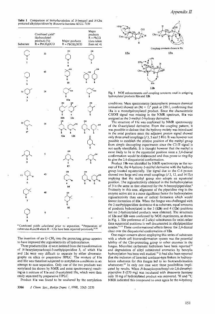
Appendix II
Table I Comparison of biohydroxylation of N-benzoyl and N-Cbz protected alkylpiperidines by Beauveria bassiana ATCC 7159
Major Combined yield products Hydroxylated R = PhCO products (%) Major products (data taken
Substrate R = PhCH2000 R = PhCH2000 from ref. 4)
C) 33
011 OH
A CC 1 It
9
6 A
OH
It
OH
10 It 2
24 (''JH
I I A It
3 ha OH OH
A A lib lic
14 OH
011
95
cx, CL c N off
0 A A A A 12a 12b 0
A QH
OH
K
5 A A 13
) a Oil OH 45 Oil
K
N Cç, LNEk 6 A A A
14
OH
7
A 15
A - 8
Combined yields calculated prior to separation. Preparation of substrates 4 and 8 where R = Cbz have been reported previously. 21'22
The insertion of an O—CH, into the protecting group appears to have improved the regioselectivity of hydroxylation.
Three products (ha—c) were isolated from the transformation of N-benzyloxycarbonyl-3-methylpiperidine 3, of which ha and He were very difficult to separate by either chromato-graphy on silica or preparative HPLC. The mixture of ha and lie was therefore subjected to acetylation conditions in an attempt to ease separation. Only one of the two products was acetylated (as shown by NMR and mass spectrometry) result-ing in a mixture of ha and O-acetylated lie, which were then easily separated by preparative HPLC.
Product ha was found to be unaltered by the acetylation
3366 J Chem. Soc., Perkin Trans. 1, 1998, 3365-3370
3
C C
H 3 H16
16/ 4/ H OH
CbZ HH CbzN H
Coupling constants shown in Hz
12a 12a
12b
Fig. I NOE enhancements and coupling constants used in assigning hydroxylated products 12a and 12b.
conditions. Mass spectrometry (atmospheric pressure chemical ionisation) showed an (M + 1) peak at 250. 1, confirming that ha is a monohydroxylated product. Since the characteristic CHOH signal was missing in the NMR spectrum, ha was assigned as the 3-methyl-3-hydroxy derivative,
The structure of He was confirmed by NMR spectroscopy of the O-acetylated derivative. From the coupling pattern, it was possible to deduce that the hydroxy moiety was introduced in the axial position since the adjacent proton signal showed only three small couplings (J 3, 5 and 5 Hz). It was however not possible to establish the relative position of the methyl group from simple decoupling experiments since the C3-H signal is not easily identifiable. It is thought however that the methyl is most likely to lie in the equatorial position since a 3,4-diaxial conformation would be disfavoured and thus prone to ring-flip to give the 3,4-diequatorial conformation.
Product lib was identified by NMR spectroscopy as the iso-mer of lie, the 4-hydroxy-3-methyl derivative with the hydroxy group located equatorially. The signal due to the C-4 proton showed two large and one small couplings (J 3, 12, and 14 Hz) implying that the methyl group also adopts an equatorial position. The regioselectivity obtained in the biohydroxylation of 3 is the same as that observed for the N-benzoylpiperidine.4 Evidently in this case, alignment of the piperidine ring in the enzyme active site is a more significant factor for hydroxylation regioselectivity than ease of radical formation which would favour formation of 11 a. When the fungus was challenged with the 2-methylpiperidine derivative 4 as substrate, equal amounts of products hydroxylated in the 3 (12b) and 4 (12a) positions but no 2-hydroxylated products were obtained. The structures of 12a and 12b were confirmed by NOE experiments, as shown in Fig. 1. The preference of 2-alkyl substituents for axial rather than equatorial positions is well documented in alkylpiperidine amides ."2 These conformational effects favour the 2,4-diaxial chair over the diequatorial conformation of 12a.
One major concern about employing this series of substrates with a whole cell biotransformation system was the potential lability of the Cbz-protecting group to other enzymes in the fungus. Microbial carbamate hydrolases have been reported '3 and degradation of alkyl substituted aromatics by benzylic hydroxylation has been well studied.'4 It had also been reported that the inclusion of inverted urethane-type linkers in hydroxy-lation substrates for this fungus led to no biotransformation whatsoever.'5 In only one case were these possibilities impli-cated by results. When N-benzyloxycarbonyl-cis-2,6-dimethyl-piperidine 5 (270 mg) was incubated with Beauveria bassiana only 10 mg of hydroxylated product was recovered. 'H and '3C NMR indicated this compound to once again be the 4-hydroxy
151
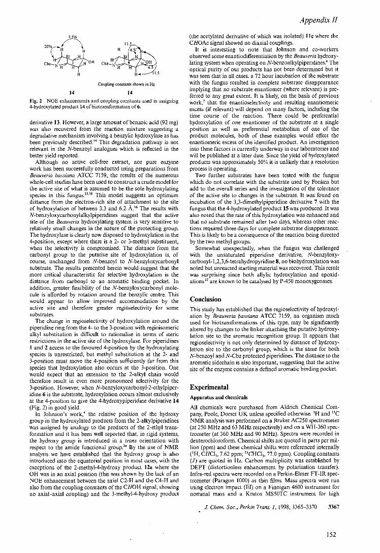
Appendix II
20% H H 4.5
W OH CbziZH H 0t4
H
H l.5
Coupling constants shown in Hz
14 14
Fig. 2 NOE enhancements and coupling constants used in assigning 4-hydroxylated product 14 of biotransformation of 6.
derivative 13. However, a large amount of benzoic acid (92 mg) was also recovered from the reaction mixture suggesting a degradative mechanism involving a benzylic hydroxylase as has been previously described. 14 This degradation pathway is not relevant in the N-benzoyl analogues which is reflected in the better yield reported.
Although no active cell-free extract, nor pure enzyme work has been successfully conducted using preparations from Beau veria bassiana ATCC 7159, the results of the numerous whole-cell studies have been used to construct a crude model for the active site of what is assumed to be the sole hydroxylating species in this fungus.t5t6 This model suggests an optimum distance from the electron-rich site of attachment to the site of hydroxylation of between 3.3 and 6.2 A.t6 The results with N-benzyloxycarbonylalkylpiperidines suggest that the active site of the Beau veria hydroxylating system is very sensitive to relatively small changes in the nature of the protecting group. The hydroxylase is clearly now disposed to hydroxylation in the 4-position, except where there is a 2- or 3-methyl substituent, when the selectivity is compromised. The distance from the carbonyl group to the putative site of hydroxylation is, of course, unchanged from N-benzoyl to N-benzyloxycarbonyl substrate. The results presented herein would suggest that the more critical characteristic for selective hydroxylation is the distance from carbonyl to an aromatic binding pocket. In addition, greater flexibility of the N-benzyloxycarbonyl mole-cule is afforded by rotation around the benzylic centre. This would appear to allow improved accommodation by the active site and therefore greater regioselectivity for some substrates.
The change in regioselectivity of hydroxylation around the piperidine ring from the 4- to the 3-position with regioisomeric alkyl substitution is difficult to rationalise in terms of steric restrictions in the active site of the hydroxylase. For piperidines 1 and -2 access to the favoured 4-position by the hydroxylating species is unrestricted, but methyl substitution at the 2- and 3-position must move the 4-position sufficiently far from this species that hydroxylation also occurs at the 3-position. One would expect that an extension to the 2-alkyl chain would therefore result in even more pronounced selectivity for the 3-position. However, when N-benzyloxycarbonyl-2-ethylpiper-idine 6 is the substrate, hydroxylation occurs almost exclusively at the 4-position to give the 4-hydroxypiperidine derivative 14 (Fig. 2) in good yield.
In Johnson's work,' the relative position of the hydroxy group in the hydroxylated products from the 2-alkylpiperidines was assigned by analogy to the products of the 2-ethyl trans-formation and it has been well reported that, in rigid systems, the hydroxy group is introduced in a trans orientation with respect to the amide functional group.' By the use of NMR analysis we have established that the hydroxy group is also introduced into the equatorial position in most cases, with the exceptions of the 2-methyl-4-hydroxy product 12a where the OH was in an axial position (this was shown by the lack of an NOE enhancement between the axial C2-H and the C4-H and also from the coupling constants of the CHOH signal, showing no axial—axial coupling) and the 3-methyl-4-hydroxy product
(the acetylated derivative of which was isolated) 11 where the CHOAc signal showed no diaxial couplings.
It is interesting to note that Johnson and co-workers observed some enantiodifferentiation by the Beau veria hydroxy-lating system when operating on N-benzoalkylpiperidines.4 The optical purity of our products has not been determined but it was seen that in all cases, a 72 hour incubation of the substrate with the fungus resulted in complete substrate disappearance implying that no substrate enantiomer (where relevant) is pre-ferred to any great extent. It is likely, on the basis of previous work,' that the enantioselectivity and resulting enantiomeric excess (if relevant) will depend on many factors, including the time course of the reaction. There could be preferential hydroxylation of one enantiomer of the substrate at a single position as well as preferential metabolism of one of the product molecules, both of these examples would effect the enantiomeric excess of the identified product. An investigation into these factors is currently underway in our laboratories and will be published at a later date. Since the yield of hydroxylated products was approximately 50% it is unlikely that .a resolution process is operating.
Two further substrates have been tested with the fungus which do not correlate with the substrate used by Fonken but add to the overall series and the investigation of the tolerance of the active site to changes in the substrate. It was found on incubation of the 3,3-dimethylpiperidine derivative 7 with the fungus that the 4-hydroxylated product 15 was produced. It was also noted that the rate of this hydroxylation was enhanced and that no substrate remained after two days, whereas other reac-tions required three days for complete substrate disappearance. This is likely to be a consequence of the reaction being directed by the two methyl groups.
Somewhat unexpectedly, when the fungus was challenged with the unsaturated piperidine derivative, N-benzyloxy-carbonyl- 1 ,2,3,6-tetrahydropyridine 8, no biohydroxylation was noted but unreacted starting material was recovered. This result was surprising since both allylic hydroxylation and epoxid-ations'7 are known to be catalysed by P-450 monoxygenases.
Conclusion This study has established that the regioselectivity of hydroxyl-ation by Beauveria bassiana ATCC 7159, an organism much used for biotransformations of this type, may be significantly altered by changes to the linker attaching the putative hydroxy-lation site to the aromatic recognition group. It appears that regioselectivity is not only determined by distance of hydroxy-lation site to the carbonyl group, which is the same for both N-benzoyl and N-Cbz protected piperidines. The distance to the aromatic sidechain is also important, suggesting that the active site of the enzyme contains a defined aromatic binding pocket.
Experimental
Apparatus and chemicals
All chemicals were purchased from Aldrich Chemical Com-pany, Poole, Dorset UK unless specified otherwise. tH and tsC
NMR analysis was performed on a Bruker AC250 spectrometer (at 250 MHz and 63 MHz respectively) and on a WH-360 spec-trometer (at 360 MHz and 90 MHz). Spectra were recorded in deuterochloroform. Chemical shifts are quoted in parts per mil-lion (ppm) and these chemical shifts were referenced internally (t H, CHC13, 7.62 ppm; '3CHCI3, 77.0 ppm). Coupling constants (J) are quoted in Hz. Carbon multiplicity was established by DEPT (distortionless enhancement by polarisation transfer). Infra-red spectra were recorded on a Perkin-Elmer FT-IR spec-trometer (Paragon 1000) as thin films. Mass spectra were run using electron impact (El) on a Finnigan 4600 instrument for nominal mass and a Kratos MS50TC instrument for high
I Chem. Soc., Perkin Trans. 1, 1998, 3365-3370 3367
152
Appendix II
resolution spectra. Thin layer chromatography was performed on glass sheets coated with silica gel Merck 60E-254 (0.24 mm, Art. 5715). Components were detected by UV (254 nm) and visualised by treating the plate with ammonium molybdate solution and heating. Wet flash chromatography was carried out on silica gel (Merck 9385, Kieselgel 60). Gas chrom-atography was carried out using a Hewlett-Packard 6890 series GC system (employing a HP-5 5% phenyl methyl siloxane capil-lary column (30.0 m x 320 I.tm x 0.25 pm)). Oven temperature gradient from 200'C (2 mm) ramp to 250'C (20°C min'; 10 mm); inlet temperature 200°C and detector temperature 300 °C. The HPLC system used was a Waters 486 controller using Millenium software using Phenomenex ODS-2 5 ii column (25 cm x 4.6 mm) using CH3CN-0.1% NH4OAc solution as eluent.
General method for the preparation of Cbz-protected alkylpiperidines
The substituted piperidine (0.010 mol) and benzyl chlorofor-mate (0.012 mol) were added to a stirred solution of potassium carbonate (0.030 mol) in tetrahydrofuran (50 cm') under an atmosphere of argon. After stirring for 1 h, water (10 cm') was added and the solution was stirred for I h. Water (150 cm') was then added and the mixture extracted with ethyl acetate (3 x 150 cm'). The combined organic extracts were washed with saturated sodium carbonate solution (160 cm') and brine (2 x 120 cm') and dried over anhydrous MgSO4. After filtration, the solvent was removed in vacuo and the residue purified by wet flash chromatography using petroleum ether-ethyl acetate mixture as eluent.
N-Benzyloxycarbonylpiperidine 1.18 Carbamate (1) was iso-lated as a colourless oil (2.04 g, 93%); Vmx 1701 cm; oH 1.46-1.63 (6H, m, C(3)H21 C(4)H2 and C(5)H2), 3.41-3.46 (4H, m, C(2)H2 and C(6)H2), 5.12 (2H, s, CH2Ph), 7.25-7.38 (5H, m, aromatic); Oc 24.1 (t, C3 and CS), 25.4 (t, C4), 44.6 (t, C2 and C6), 66.6 (t, CH2Ph), 127.6 (d, aromatic), 127.6 (d, aromatic), 128.2 (d, aromatic), 136.8 (s, aromatic), 155.1 (s, C=O); m/z (El) required 219.12593, found 219.12558, 219 (14%, M), 128 (12, M - CH2Ph), 91 (100, PhCH2), 84 (10, M - CO2CH2Ph).
N-Benzytoxycarbonyl4-methylpiperidine 2. Carbamate (2) was isolated as a colourless oil (2.08 g, 89%); Vms. 1701 cm'; OH (250 MHz) 0.93 (3H, d, J 6.5, CH3), 1.03-1.18 (1H'm ' m, C(4)H), 1.46-1.61 (4H, m, C(3)H2 and C(5)H2), 2.75 (2H, ddd, J2.5, 13 and 13, C(2)Hax and C(6)Hax), 4.13 (2H, br d, J 13, C(2)Heq and C(6)Heq), 5.11 (2H, s, CH2Ph), 7.24-7.36 (5H, m, aromatic); Jc 63 MHz) 21.7 (q, CH3), 30.8 (d, C4), 33.8 (t, C3 and CS), 44.1 (t, C2 and C6), 66.8 (d, CH2Ph), 127.7 (d, aromatic), 127.8 (d, aromatic), 128.3 (d, aromatic), 136.9 (s, aromatic), 155.2 (s, C=O); m/z (El) required 233.14158, found 233.14262, 233 (81%, Mt), 218 (4, M - CH,), 142 (78, M - CH2Ph), 126 (70, M - OCH2Ph), 98 (73, M - CO2CH2Ph), 91(62, CH2Ph), 77(13, Ph).
N-Benzyloxycarbonyl-3-methylpiperidine 3. Carbamate (3) was isolated as a colourless oil (2.14 g, 92%); Vmx 1701 cm'; 0H (250 MHz) 0.87 (3H, d, J6.5, CH3), 1.01-1.13 (1 H, in, C(3)H), 1.39-1.81 (4H, m, C(4)H2 and C(5)H2), 2.42 (1H, dd, J 11 and 13, C(2)Hax), 2.71-2.81 (1H, m, C(6)Hax), 4.00-4.08 (2H, m, C(2)Heq and C(6)Heq), 5.12 (2H, s, CH2Ph), 7.25-7.37 (SH, m, aromatic); O (63 MHz) 18.7 (q, CH,), 25.7 (d, C3), 30.8 (t, C4 or CS), 32.7 (t, C4 or CS), 44.2 (t, C2 or C6), 51.1 (t, C2 or C6), 66.7 (t, CH2Ph), 127.6 (d, aromatic), 127.7 (d, aromatic), 128.3 (d, aromatic), 136.9 (s, aromatic), 155.1 (s, C=O); m/z (El) required 233.14158, found 233.14109, 233 (35%, Mt), 142 (46, M - CH2Ph), 126 (26, M - OCH2Ph), 98 (27, M - CO2CH2Ph), 91(100, CH2Ph).
3368 .1 Chem. Soc., Perkin Trans. 1, 1998, 3365-3370
N-Benzyloxycarbonyl-cis-2,6-dimethylpiperidine 5•19 Carb-amate (5) was isolated as a colourless oil (2.19 g, 89%); 1693 CM-'; 6H (250 MHz) 1.20 (6H, d, J7, 2 X CH3), 1.41-1.77 (6H, m, C(3)H21 C(4)H2 and C(5)H2), 4.32-4.38 (2H, hr m, C(2)H and C(6)H), 5.13 (2H, s, CH2Ph), 7.25-7.36 (5H, in, aromatic); 6c 13.5 (t, C4), 20.7 (q, C7 and C8), 29.8 (t, C3 and CS), 45.9 (d, C2 and C6), 66.6 (t, CH2Ph), 127.5 (d, aromatic), 127.6 (d, aromatic), 128.3 (d, aromatic), 137.0 (s, CH2Ph), 155.5 (s, C=O); m/z (El) required 247.157229, found 247.15832, 247 (4%, M), 32 (22, M - CH,), 156 (13, M - CH2Ph), 91(100, PhCH2).
N-Benzyloxycarbonyl-2-ethylpiperidine 6. Carbamate (6) was isolated as a colourless oil (2.28 g, 92%); Vmx 1693 cm'; OH (250 MHz) 0.83 (3H, t, J 7.5, C(8)H3), 1.34-1.80 (8H, in, C(3)H2, C(4)H21 C(5)H2 and C(7)H2), 2.75-2.86 (lH, m, C(6)Hax), 4.02-4.07 (1H, hr m, C(2)Heq or C(6)Heq), 4.16-4.23 (1H, br m, C(2)Heq or C(6)Heq), 5.17 (2H, s, CH2Ph), 7.25-7.37 (5H, m, aromatic); 6c (63 MHz) 10.6 (q, C8), 18.8 (t, C7), 22.4 (t, C3, C4 or CS), 25.5 (t, C3, C4 or CS), 27.9 (t, C3, C4 or CS), 38.9 (t, C6), 52.2 (d, C2), 66.7 (t, CH2Ph), 127.6 (d, aromatic), 127.6 (d, aromatic), 128.3 (d, aromatic), 137.0 (s, aromatic), 155.6 (s, C=O); mhz (El) required 247.15723, found 247.15706, 247 (6%, Mt), 218(87%, M - CH2CH3), 156 (9, M - CH2Ph), 140 (51 M - OCH2Ph), 112 (6, M - CO2CH2Ph), 91(67, CH2Ph).
N-Benzyloxycarbonyl-3,3-dimethylpiperidine 7. Carbamate (7) was isolated as a colourless oil (2.20 g, 89%); Vms, 1701 cm; OH 0.89 (6H, s, C(7)H3 and C(8)H3), 1.32-1.37 (2H, m, C(4)H2 or C(5)H2), 1.55-1.60 (2H, m, C(4)H2 or C(5)H2), 3.13 (2H, s, C(2)H2), 3.40 (2H, dd, J 5.5 and 5.5, C(6)H2), 5.12 (2H, s, CH2Ph), 7.25-7.36 (5H, m, aromatic); 6c 25.8 (q, C7 and C8), 30.8 (s, C3), 37.6 (t, C4), 44.3 (t, C2 or C6), 55.3 (t, C2 or C6), 66.7 (t, CH2Ph), 127.6 (d, aromatic), 127.7 (d, aromatic), 128.3 (d, aromatic), 137.0 (s, aromatic), 155.5 (s, C=O); mhz (El) required 247.15723, found 247.15813, 247 (36%, MF), 156 (42, M - CH2Ph), 140 (20, M - OCH2Ph), 112 (17, M - CO2-CH2Ph), 91(63, PhCH2).
Maintenance and growth of microorganism
Beauveria bassiana ATCC 7159 was obtained from the Ameri-can Type Culture Collection, USA. The organism was main-tained on malt extract agar plates at room temperature and these were subcultured at regular intervals. A loop of fungus was used to inoculate 60 ern' of sterile medium containing 7.5 g dm 3 corn steep solids and 10 g dm-' glucose in distilled water adjusted to pH 4.85 in a 250 cm' Erlenmeyer flask. After three days growth on an orbital shaker at 200 rpm at 25 °C, a culture was used to inoculate 600 cm' of the same medium in a 2 dm' Erlenmeyer flask. This culture was again grown for three days.
General biotransformation procedure
A solution of 66 mg substrate in 1 cm' ethanol was prepared and this was added to the three day old culture of Beauveria bassiana ATCC 7159. After a further three days incubation as above, the fungal cells were removed from the broth by centri-fugation and the supernatant extracted into ethyl acetate. Purification of metabolites was routinely carried out using flash silica chromatography with petroleum ether-ethyl acetate gradients as eluent.
Biohydroxylation of N-benzyloxycarbonylpiperidine 1 pro-duced hydroxylated product (9) which was isolated as a colour-less oil (100 mg, 33%). The product 9 was identical to previously reported material as judged by NMR spectroscopy.2°
Biohydroxylation of N-benzyloxycarbonyl-4-methylpiperidine 2. Hydroxylated product (10) was isolated as a pale yellow solid (130 mg, 45%); mp 76 °C; OH (250 MHz) 1.24 (3H, s, CH3), 1.45-1.56 (SH, m, C(3)H21 C(5)H2 and C(4)OH), 3.34-3.23
153
Appendix II
(2H, dt, J 7 and 13, C(2)Hax and C(6)Hax), 3.80 (2H, br d, J 13, C(2)Heq and C(6)Heq), 5.11 (2H, s, CH2Ph), 7.25-7.36 (5H, m, aromatic); ôc (63 MHz) 30.0 (q, CH3), 38.2 (t, C3 and C5), 40.2 (t, C2 and C6), 66.9 (t, CH2Ph), 67.7 (s, C4), 127.7 (d, aromatic), 127.8 (d, aromatic), 128.3 (d, aromatic), 136.7 (s, aromatic), 155.1 (s, C=O); m/z (El) required 249.13649, found 249.13637, 249 (44%, Mt), 158 (14, M - PhCH2), 142 (16, M - PhCH2)1 91(100, PhCH2).
Biohydroxylation of N-benzyloxycarbonyl-3-methylpiperidine Three products ha-c were isolated from this biohydroxyl-
ation, two of which (ha and lic) were found to be very closely related and thus difficult to separate. These two products were separated by subjecting the mixture to acetylation condi-tions (10 mol equiv. acetic anhydride, 5 mol equiv. pyridine and catalytic dimethylaminopyridine) under which conditions one of the products was acetylated 1 ic and one was left untouched ha. The products were then separated by preparative HPLC.
Product ha was isolated as a colourless oil (10 mg, 3%); oH (360 MHz) 1.21 (3H, s, CH01 1.49-1.79 (SH, m, C(3)OH, C(4)H2 and C(5)H2), 2.96-3.03 (2H, br m, C(2)Hax and C(6)Hax), 3.68 (1H, br s, C(2)Heq), 3.84 (1H, ddd, J 4.51 4.5 and 13, C(6)Heq), 5.13 (2H, s, CH2Ph), 7.28-7.35 (5H, m, aromatic); m/z (El) required 249.13649, found 249.13725, 249 (5%, M), 114(26, M - PhCHOCO), 91(100, PhCH2).
Hydroxylated product lib was isolated as a colourless oil (10 mg, 3%); 0H (360 MHz) 0.99 (3H, d, J6.5, CH3), 1.43-1.59 (3M, br m, three of C(3)H, C(4)OH and C(5)H2), 1.91 (lH, br dd, J 3.5 and 13, C(3)H, C(4)OH or C(5)H), 2.53(1 H, br s, C(2)Hax or C(6)Hax), 2.90 (1H, ddd, J 3,12 and 14, C(4)H), 3.30 (1H, br m, C(2)Hax or C(6)Hax), 4.05-4.13 (2H, br m, C(2)Heq and C(6)Heq), 5.11-5.12 (2H, d, J 2, CH2Ph), 7.28-7.36 (5H, m, aromatic); m/z (El) required 249.13649, found 249.13616, 249 (8%, Mt), 158 (11, M - PhCH2)1 142 (M - PhCH20), 91 (PhCH2).
The O-acetylated product of lic was isolated as a colourless oil (5 mg); O j (360 MHz) 0.88 (3H, d, J 12.5, CM01 1.67-1.93 (3H, br m, C(3)H and C(5)H2), 2.07 (3H, s, CH3CO20.00-3.15 (lH, br m, C(2)Hax or C(6)Hax), 3.25-3.28 (1H, br m, C(2)Hax or C(6)Hax), 3.70 (1H, br s, C(2)Heq or C(6)Heq), 3.75 (1H, ddd, J4.5, 4.5 and 13.5, C(2)Heq or C(6)Heq), 5.01 (1H, ddd, J 3, 3 and 5, C(4)Heq), 5.12 (2H, d, J 2, CH2Ph), 7.28-7.39 (SM, m, aromatic); m/z (El) required 291.14706, found 291.14768; 291 (3%, M), 200(11, M - PhCH2), 91(100, PhCH2).
Biohydroxylation of N-Benzyloxycarbonyl-2-methylpiperidine Hydroxylated product 12a was isolated as a colourless oil
(20 mg, 7%); 0H (250 MHz) 1.35 (3H, d, J 7, CH01 1.46-1.91 (SH, m, C(3)H2, C(4)OH and C(5)H2), 3.34 (1H, ddd, J 51 12.5 and 13.5, C(6)Hax), 3.90 (1H, ddd, J 3.5, 4 and 12.51 C(6)Heq), 4.16 (lH, q, J 3, CHOH), 4.31-4.42 (1H, m, C(2)Heq), 5.12 (2H, s, CH2Ph), 7.25-7.36 (SM, m, aromatic); O (90 MHz) 19.1 (q, CH3), 32.2 (t, C3 or CS), 33.5 (t, C3 or CS), 36.4 (t, C6), 45.7 (d, C2), 64.7 (d, CHOH), 66.8 (t, CH2Ph), 127.7 (d, aromatic), 127.8 (d, aromatic), 128.4 (d, aromatic), 136.9 (s, aromatic), 155.2 (s, C=O); m/z (El) required 249.13649, found 249.136337, (FAB) 234 (4%, M - CH3), 158 (15, M - PhCH2), 142 (31, M - PhCH20), 114 (2, M - PhCH2CO2)1 91(100, PhCH2).
Hydroxylated product 12b was isolated as a colourless oil (20 mg, 7%); OH (360 MHz) 1.12 (3H, d, J 7, CH01 1.43-1.78 (SM, m, C(3)OH, C(4)H2 and C(5)H2), 2.79 (1H, ddd, J 3, 13 and 13.5, C(6)Hax), 3.74-3.80 (1H, m, CHOH), 3.95 (lH, br d, J 12.5, C(6)Heq), 4.51 (lH, q, J 6.5, C(2)H), 5.12 (2H, s, CH2Ph), 7.25-7.38 (5H, m, aromatic); O (90 MHz) 9.2 (q, CH3), 24.0 (t, CS or C4), 27.1 (t, CS or C4), 37.6 (t, C6), 51.0 (d, C2), 67.0 (t, CH2Ph), 69.0 (d, CHOH), 127.7 (d, aromatic), 127.9 (d, aromatic), 128.4 (d, aromatic), 136.7 (s, aromatic), 155.3 (s, C=O); mlz (El) required 249.13649, found 249.13637,
(APCIt) 250 (100%, M + 1), 217 (3, M - OHCH3), 158 (7, M - PhCH2).
Biohydroxylation of N-benzyloxycarbonyl-cis-2,6-dimethyl-piperidine 5. Hydroxylated product 13 was isolated as a colour-less oil (10 mg, 5%); OH (360 MHz) 1.25 (6H, d, J7, 2 x CH01 1.42-1.60 and 1.86-1.90 (5H, m, C(3)H21 C(4)OH and C(5)H2), 4.20 (lH, tt, J 11 and 4, CHOH), 4.49-4.57 (2H, m, C(2)Heq and C(6)Heq), 5.14 (2H, s, CH2Ph), 7.23-7.38 (5H, m, aromatic); O (63 MHz, DEPT) 21.7 (q, 2 x CH3), 39.4 (t, C3 and CS), 47.2 (d, C2 and C6), 61.4 (d, CHOH), 66.9 (t, CH2Ph), 127.7 (d, aromatic), 127.8 (d, aromatic), 128.4 (d, aromatic); m/z (El) required 263.15214, found 263.15203, (FAB) 264 (M+ 1).
Biohydroxylation of N-benzyloxycarbonyl-2-ethylpiperidine 6. Hydroxylated product 14 was isolated as a colourless oil (130 mg, 45%); OH (360 MHz) 0.85 (3H, t, J 7.5, C(8)H3), 1.23-1.66 (5H, m, C(3)H, C(4)OH, C(5)H and C(7)H2), 1.92 (2H, dd, J2.5 and 12.5, C(3)H and C(S), 2.85 (lH, ddd, J2.5, 12.5 and 12.5, C(6)Hax), 3.91 (lH, tt, J4.S and 11. 5, CHOH), 4.16 (1H, br d, J 12.5, C(6)Heq), 4.33 (1H, br m, C(2)Heq), 5.12 (2H, d, J2, CH2Ph), 7.25-7.37 (SH, m, aromatic);O (90 MHz) 10.7 (q, C8), 23.8 (t, C7), 35.0 (C3 and CS), 37.6 (t, C6), 52.8 (d, C2), 65.0 (t, CHOH), 67.0 (t, CH2Ph), 127.6 (d, aromatic), 127.8 (d, aromatic), 128.3 (d, aromatic), 136.8 (s, aromatic), 155.4 (s, C=O); m/z (El) required 263.15214, found 263.15253, (APCi) 264 (12%, M + 1), 220 (M - CH3CH2), 172 (7, M - PhCH2), 156 (23, M - PhCH20)1 91(100, PhCH2).
Biohydroxylation of N-benzyloxycarbonyl-3,3-dimethyl-piperidine 7. Hydroxylated product 15 was isolated as a colour-less oil (134 mg, 48%); OH (250 MHz; 345 K) 0.89 (3H, s, C(7)H3 or C(8)H3), 0.95 (3H, s, C(7)H3 or C(8)H3), 1.51-1.64 (2H, m, C(5)H and C(4)OH), 1.71-1.82 (lH, m, C(5)H), 2.85 (lH, d, J 13.5, C(2)Hax), 3.12-3.23 (lH, m, C(6)Hax), 3.40 (lH, dd, J 8.5 and 4.0, CHOH), 3.56 (1H, dd, J 1.5 and 13.5, C(2)Heq), 3.81-3.91 (lH, m, C(6)Heq), 5.13 (2H, s, CH2Ph), 7.24-7.36 (5H, m, aromatic); OH (63 MHz) 18.4 (q, C7 or C8), 24.2 (q, C7 or C8), 29.5 (s, 0), 35.8 (t, C5), 41.5 (t, C2 or C6), 52.6 (t, C2 or C6), 66.9 (t, CH2Ph), 74.7 (d, CHOH), 127.7 (d, aromatic), 127.8 (d, aromatic), 128.3 (d, aromatic), 136.7 (s, aromatic), 155.4 (s, C=O); m/z required 263.15214, found 263.15254, 263 (10%, M), 172 (5, M - PhCH2), 128 (3, CO2CH2Ph), 91(100, CH2Ph), 77 (4, Ph).
Acknowledgements We are grateful to the BBSRC and Zylepsis Ltd. for funding (to S. J. A. and G. G. respectively). We would also like to acknowledge the assistance of Mr J. Millar and Dr D. Reed with NMR studies.
t APCI = Atmospheric pressure chemical ionisation.
References I E. W. Weller, M. Droste, J. Eberle, H. J. Halfmann and A. Weber,
App!. Microbiol. BiotechnoL, 1987, 27, 252. 2 R. A. Johnson, M. E. Herr, H. C. Murray, L. M. Reineke and G. S.
Fonken, J Org. Chem., 1968, 33, 3195; R. Furstoss, A. Archelas and B. Waegell, Tetrahedron Let!., 1981, 22, 445; W Carruthers, J. D. Prail, S. M. Roberts and A. J. Wil!etts, .1. Chem. Soc., Perkin Trans. 1, 1990, 2854; C. F. Palmer, B. Webb, S. Broad, S. Casson, R. McCague, A. J. Willetts and S. M. Roberts, Bioorg. Med. Chem. Lett., 1997, 7, 1299.
3 G. S. Fonken and R. A. Johnson, in Chemical Oxidations with Microorganisms, VCH, Germany, 1972.
4 R. A. Johnson, H. C. Murray, L. M. Reineke and G. S. Fonken, J Org. Chem., 1969, 34, 2279.
5 K. Tadano, Y. limura and T. Suami, J Carbohydr. Chem., 1985, 4, 129.
.1 Chem. Soc., Perkin Trans. 1, 1998, 3365-3370 3369
154

6 B. Ringdahl, U. F. W Ohnsorge and J. C. Craig, J Chem. Soc., Perkin Trans. 2, 1981, 697.
7 A. de Raat, H. Griengi, M. Petsch, P. Plachota, N. Schoo, H. Weber, G. Braunegg, I. Kopper, M. Kreiner, A. Zeiser and K. Kieslich, Tetrahedron.' Asymmetry, 1996, 7, 467; A. de Raat, H. Griengi,
Petsch, P. Plachota, N. Schoo, H. Weber, G. Braunegg, 1. Kopper, M. Kreiner and A. Zeiser, Tetrahedron: Asymmetry, 1996, 7, 473; A. de Raat, H. Griengi, M. Petsch, P. Plachota,
Schoo, H. Weber, G. Braunegg, I. Kopper, M. Kreiner and A. Zeiser, Tetrahedron.' Asymmetry, 1996,7,491.
8 D. D. Keith, J. A. Tortora, K. Ineichen and W Leimgruber, Tetrahedron, 1975, 31, 2633; D. A. 'Evans and J. A. ElIman, J Am, Chem. Soc., 1989, 111, 1063; K. E. Harding, T. H. Marman and D.-H. Nam, Tetrahedron Lett., 1988, 29, 1627.
9 N. Floyd, F Munyemana, S. M. Roberts and A. J. Willetts, J Chem. Soc., Perkin Trans. 1, 1993, 881.
10 J. D. Fourneron, A. Archelas and R. Furstoss, .1 Org. Chem., 1989, 54, 2478.
II R. A. Johnson, .1 Org. Chem., 1968, 33, 3627. 12 V. L. Chow, C. J. Colon and J. N. S. Tam, Can. J. Chem., 1968, 46,
2821. 13 V. Gotor, M. Quiros, L. Ramon, J. Frigola and R. Fernandez,
Tetrahedron, 1997, 53, 6421.
Appendix II
14 H. L. Holland, M. Conn, P. Chinna Chenchaiah and F. M. Brown, Tetrahedron Lett., 1988, 29, 6393.
15 B. Vigne, A. Archelas and R. Furstoss, Tetrahedron, 1991, 47, 1447. 16 R. Furstoss, A. Archelas, J. D. Fourneron and B. Vigne, in
Enzymes as Catalysts in Organic Chemistry, NATO ASI Series, ed. P. Schneider, 1986, 178, 361.
17 R. A. Johnson, M. E. Herr, H. C. Murray, C. G. Chidester and F. Han, .1 Org Chem., 1992, 57, 7209.
18 A. C. Pierce and M. M. Joullie, J. Org Chem., 1963, 28, 658; G. Barcelo, J.-P. Senet, G. Sennyey, J. Bensoam and A. Loffet, Synthesis, 1986, 627.
19 K. E. Harding and T. H. Marman, .1 Org Chem., 1984, 49, 2838. 20 C. S. Cooper, P. L. Klock, D. T. W. Chu, D. J. Hardy and R. N.
Swanson, J Med. Chem., 1992, 35, 1392; L. Alig, A. Edenhofer, P. Hadvhry, M. Hürzeler, D. Knopp, M. Muller, B. Steiner, A. Trzeciak and T. Weller, J Med. Chem., 1992, 35, 4393.
21 A. M. Morello and A. D. Ward, Aust. J Chem., 1995, 48, 445. 22 S. V. D'Andrea, E. T. Michalson, J. P. Freeman, C. G. Chidester and
J. Szmuszkovicz, J Org Chem., 1991, 56, 3133.
Paper 8/05800H
3370 J Chem. Soc., Perkin Trans. 1, 1998, 3365-3370
155
Bioorganic Chemistry 27, 81-90 (1999) (K;
Article ID bioo. 1999.1135, available online at http://www.idealibrary.com on 101 E
Biohydroxylation Reactions Catalyzed by Enzymes and
Whole-Cell Systems
Sabine L. Flitsch,1 Suzanne J. Aitken, Cathy S.-Y. Chow, Gideon Grogan, and Adam Staines
Centre for Protein Technology, University of Edinburgh, Edinburgh, United Kingdom
Received August 24, 1998
The biohydroxylation of a number of cyclic substrates (3-24) containing aromatic side chains was used to compare substrate specificity and selectivity of hydroxylation using microbial enzymes and whole-cell biocatalysts. In general, the regioselectivity of reaction was remarkably similar between the different catalysts in that little aromatic or benzylic, but significant aliphatic hydroxylation was observed. However, a more detailed investigation of isolated products showed complementary substrate specificity, functional group compatibility, and regioselectivity of hydroxylation. Substrate specificity and regioselectivity could be further modulated by small changes to the nature of the aromatic side chain, which appears to play an important role in substrate recognition. © 1999 Academic Press
The hydroxylation of nonfunctionalized hydrocarbon centers using biological sys-tems is recognized as an important biotransformation (1). This reaction has traditionally been achieved using whole-cell systems, but as more isolated enzyme systems and their three-dimensional structure are now becoming available, enzymes can be used for catalysis and are particularly important for studying substrate recognition and regio- and stereo selectivity of hydroxylation. The regioselectivity of hydroxylation is of particular importance to these biotransformations, because substrates often have a number of potential hydroxylation sites and thus a very high degree of selectivity is required. In our studies, we have looked at a particular class of cyclic substrates bearing aromatic side chains (3-24) and compared the selectivity of hydroxylation in defined enzymatic systems with that of whole-cell systems.
BIOHYDROXYLATIONS USING ISOLATED ENZYMES
The best studied of the cell-free hydroxylase systems is the cytochrome P450cam monooxygenase from Pseudomonas putida which catalyzes the hydroxylation of camphor 1 to 5-exo-hydroxy camphor 2 (Scheme 1). This enzyme was first reported in the 1960s by Gunsalus and colleagues and has been widely used as a model system because it is easily obtained as a soluble protein from heterologous expression (2)
To whom correspondence should be addressed at Department of Chemistry, King's Buildings, West Mains Rd., Edinburgh EH9 3JJ, United Kingdom. Fax: (0)131-6504737. E-mail: [email protected].
O 81 0045-2068/99 $30.00
Copyright © 1999 by Academic Press
All rights of reproduction in any form reserved.
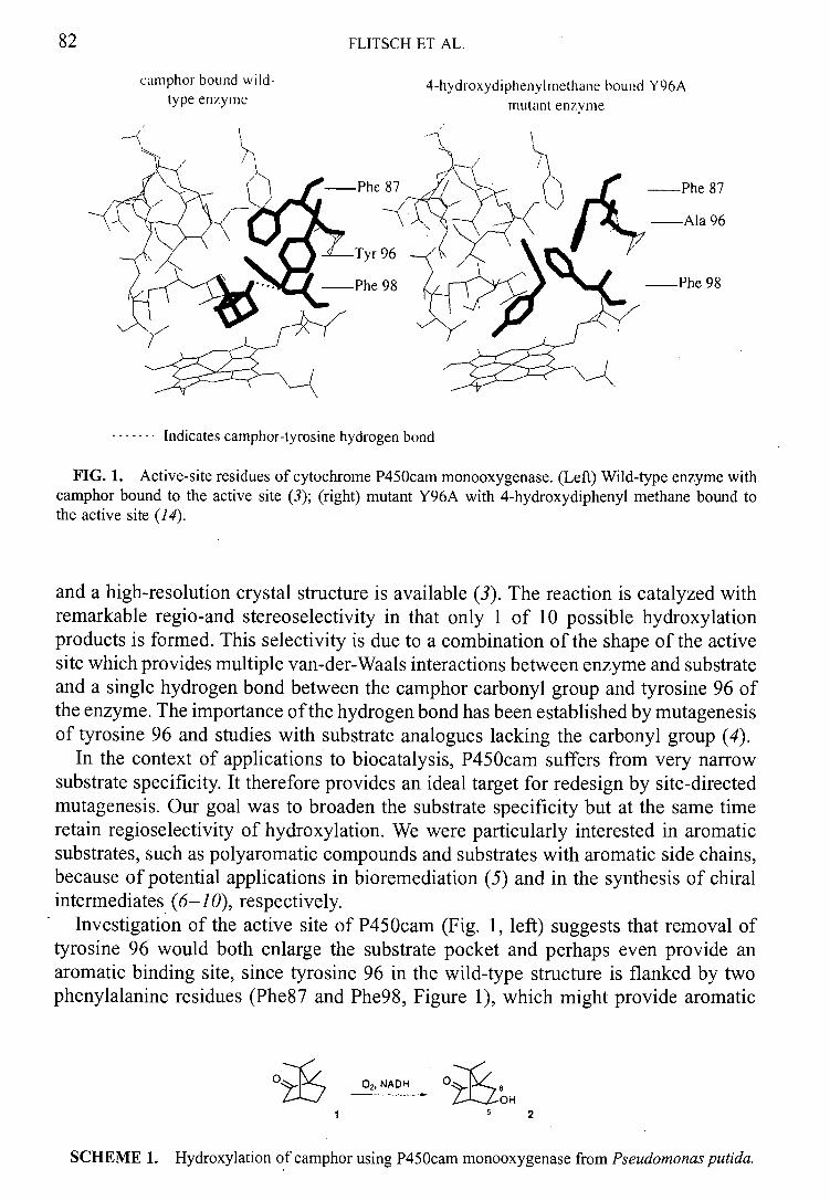
82 FLITSCH ET AL.
camphor bound wild 4-hydroxydipheny1methaiie bound Y96A type enzyme mutant enzyme
/
-Phe 87 -
Tyr 96
Phe 98
'___Phe 87
—Ala 96
—Phe 98
Indicates camphor-tyrosine hydrogen bond
FIG. 1. Active-site residues of cytochrome P450cam monooxygenase. (Left) Wild-type enzyme with camphor bound to the active site (3); (right) mutant Y96A with 4-hydroxydiphenyl methane bound to the active site (14).
and a high-resolution crystal structure is available (3). The reaction is catalyzed with remarkable regio-and stereos electivity in that only 1 of 10 possible hydroxylation products is formed. This selectivity is due to a combination of the shape of the active site which provides multiple van-der-Waals interactions between enzyme and substrate and a single hydrogen bond between the camphor carbonyl group and tyrosine 96 of the enzyme. The importance of the hydrogen bond has been established by mutagenesis of tyrosine 96 and studies with substrate analogues lacking the carbonyl group (4).
In the context of applications to biocatalysis, P450cam suffers from very narrow substrate specificity. It therefore provides an ideal target for redesign by site-directed mutagenesis. Our goal was to broaden the substrate specificity but at the same time retain regioselectivity of hydroxylation. We were particularly interested in aromatic substrates, such as polyaromatic compounds and substrates with aromatic side chains, because of potential applications in bioremediation (5) and in the synthesis of chiral intermediates (6-10), respectively.
Investigation of the active site of P450cam (Fig. 1, left) suggests that removal of tyrosine 96 would both enlarge the substrate pocket and perhaps even provide an aromatic binding site, since tyrosine 96 in the wild-type structure is flanked by two phenylalanine residues (Phe87 and Phe98, Figure 1), which might provide aromatic
Of7 02,NADH
Z-LLOH 15 2
SCHEME 1. Hydroxylation of camphor using P450cam monooxygenase from Pseudomonas putida.
CATALYZATION OF BIOHYDROXYLATION REACTIONS 83
stacking interactions. With this in mind, a number of single-site mutants at position 96 were generated, of which the alanine mutant Y96A was the most interesting and hence was studied in most detail (11,12).
First, simple aromatic compounds such as naphthalene and diphenylmethane (3) were tested as substrates. The hydroxylation of naphthalene was much more efficient with the Y96A mutant than with wild-type enzyme (13), and diphenylmethane 3 was almost exclusively converted to 4-hydroxy diphenylmethane (Table 1) by Y96A, whereas no hydroxylation was observed with wild-type enzyme (11). No benzylic hydroxylation was observed, which emphasizes that the mode of binding of the substrate to the enzyme active site can be more important for selectivity than C—H bond strength.
Thus, the type of substrate binding we observed with the Y96A mutant (Table 1) suggested to us that removal of the tyrosine aromatic side chain had indeed created a binding pocket for aromatic rings as predicted, where the substrate now takes the place of the former protein tyrosine side chain. To verify this, we conducted crystallographic studies on the Y96A mutant. We have recently obtained the structure of the mutant Y96A with 4-hydroxy diphenylmethane bound to the active site to 1.9 A resolution in collaboration with Malcolm Walkinshaw at Edinburgh University (14). These preliminary studies have revealed that, perhaps not surprisingly, some significant rearrangement of active-site residues occurs. Comparison of the wild-type with the Y96A mutant shows that most active-site residues remain in the same place, but very importantly that the phenylalanine 87 side chain has moved into the original site of tyrosine 96 (Fig. 1, right). Thus, the mutation has increased the size of the substrate binding pocket which now appears to be lined at the bottom, away from the active site, with aromatic side chains (Phe 87 and 98). Thus, rather than, as predicted, providing an aromatic pocket at the previous site of tyrosine 96, Phe 98 appears to be stacking against aromatic side chains of the substrate, resulting in regio selectivity of hydroxylation. We are currently investigating cocrystals of Y96A with other substrates to see if this change of active residue orientation is generally observed.
Further evidence for this aromatic stacking interaction comes from studies of the mutant Y96A with substrates 4-9 (Table 1), where no aromatic hydroxylation was observed, but good regioselectivity of hydroxylation of the aliphatic part of the molecule occurred. However, this can also be explained by favorable van-der-Waals interactions of the aliphatic part of the substrate with aliphatic protein side chains (Val 244, Leu 247, Val 295) closer to the active site.
In general, substrates 3-9 in Table 1 demonstrate that the substrate range for hydroxylation has been greatly expanded by the single mutation of tyrosine 96, in particular since none of these compounds bind to wild-type enzyme. The aromatic side chain that is necessary for good binding to the enzyme does not need to be part of the desired substrate structure, but can be a protecting group as in 5-9, which can be cleaved off after hydroxylation. This allows for the enzyme to catalyze the hydroxylation of functionalized cyclohexane (5,6,9) and piperidine (7,8) derivatives, which themselves are not substrates.
Another advantage for using aromatic protecting groups in these biotransformations is that they can be used as a further tool to manipulate regio selectivity of hydroxylation.
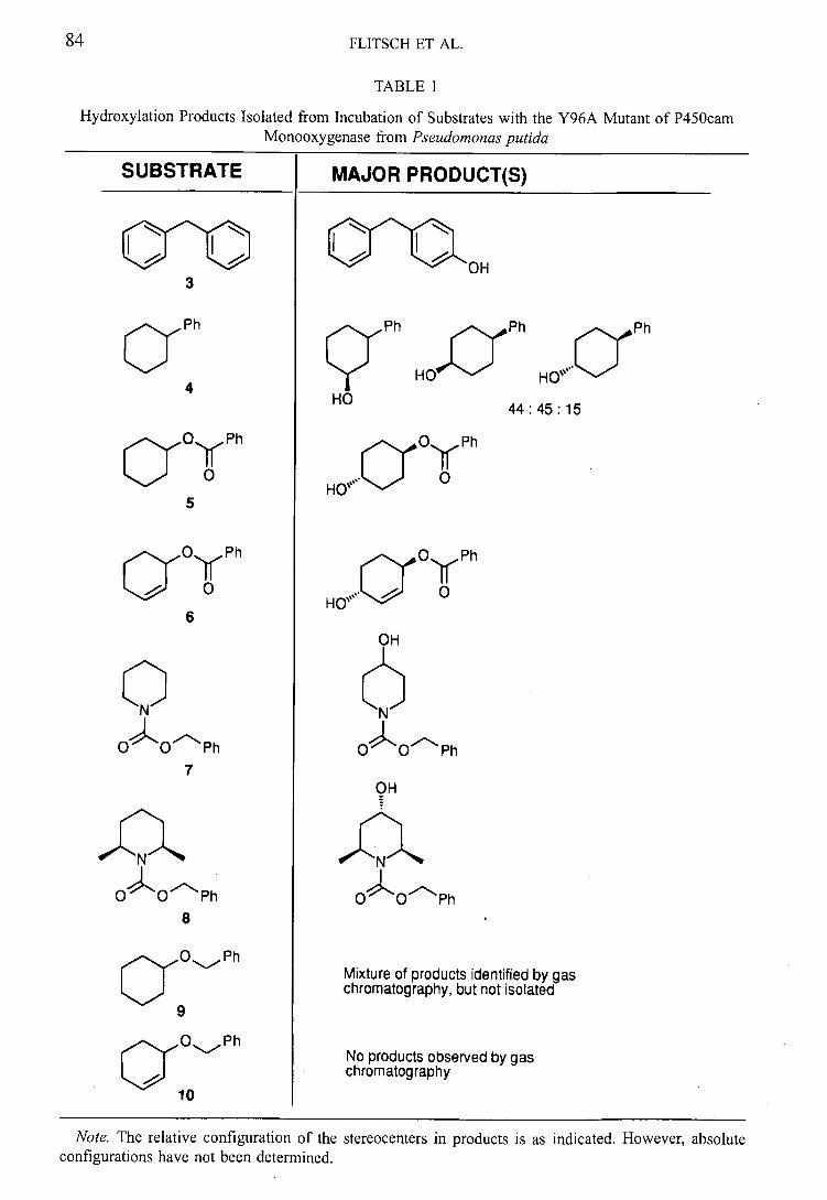
FLITSCH ET AL.
TABLE 1
Hydroxylation Products Isolated from Incubation of Substrates with the Y96A Mutant of P450cam Monooxygenase from Pseudomonas putida
SUBSTRATE I MAJOR PRODUCT(S)
Ph Ph q Ph
HO)300 HO'"
Ph
HO 44: 45:15
OyPh
a0YPh
HO' ° 5
a0YPh (::::yOyPh
0 HO''
0
6 OH
No N
0 Ph OO Ph
OH
OOPh OOPh
Ph Mixture of products identified by gas chromatography, but not isolated
Ph No products observed by gas chromatography
10
Note. The relative configuration of the stereocenters in products is as indicated. However, absolute configurations have not been determined.
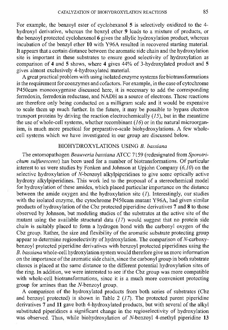
CATALYZATION OF BIOHYDROXYLATION REACTIONS 85
For example, the benzoyl ester of cyclohexanol 5 is selectively oxidized to the 4-hydroxyl derivative, whereas the benzyl ether 9 leads to a mixture of products, or the benzoyl protected cyclohexenol 6 gives the allylic hydroxylation product, whereas incubation of the benzyl ether 10 with Y96A resulted in recovered starting material. It appears that a certain distance between the aromatic side chain and the hydroxylation site is important in these substrates to ensure good selectivity of hydroxylation as comparison of 4 and 5 shows, where 4 gives 44% of 3-hydroxylated product and 5 gives almost exclusively 4-hydroxylated material.
A great practical problem with using isolated enzyme systems for b iotrans formations is the requirement for coenzymes and cofactors. For example, in the case of cytochrome P450cam monooxygenase discussed here, it is necessary to add the corresponding ferredoxin, ferredoxin reductase, and NADH as a source of electrons. These reactions are therefore only being conducted on a milligram scale and it would be expensive to scale them up much further. In the future, it may be possible to bypass electron transport proteins by driving the reaction electrochemically (15), but in the meantime the use of whole-cell systems, whether recombinant (16) or in the natural microorgan-ism, is much more practical for preparative-scale biohydroxylations. A few whole-cell systems which we have investigated in our group are discussed below.
BIOHYDROXYLATIONS USING B. bassiana
The entomopathogen Beauveria bassiana ATCC 7159 (redesignated from Sporotri-chum sulfurescens) has been used for a number of biotransformations. Of particular interest to us were studies by Fonken and Johnson at Upjohn Company (6,10) on the selective hydroxylation of N-benzoyl alkylpiperidines to give some optically active hydroxy alkylpiperidines. This work led to the proposal of a stereochemical model for hydroxylation of these amides, which placed particular importance on the distance between the amide oxygen and the hydroxylation site (1). Interestingly, our studies with the isolated enzyme, the cytochrome P450cam mutant Y96A, had given similar products of hydroxylation of the Cbz protected piperidine derivatives 7 and 8 to those observed by Johnson, but modeling studies of the substrates at the active site of the mutant using the available structural data (17) would suggest that no protein side chain is suitably placed to form a hydrogen bond with the carbonyl oxygen of the Cbz group. Rather, the size and flexibility of the aromatic substrate protecting group appear to determine regioselectivity of hydroxylation. The comparison of N-carboxy-benzoyl protected piperidine derivatives with benzoyl protected piperidines using the B. bassiana whole-cell hydroxylation system would therefore give us more information on the importance of the aromatic side chain, since the carbonyl group in both substrate classes is placed at the same distance to the different potential hydroxylation sites of the ring. In addition, we were interested to see if the Cbz group was more compatible with whole-cell b iotrans formations, since it is a much more convenient protecting group for amines than the N-benzoyl group.
A comparison of the hydroxylated products from both series of substrates (Cbz and benzoyl protected) is shown in Table 2 (17). The protected parent piperidine derivatives 7 and 11 gave both 4-hydroxylated products, but with several of the alkyl substituted piperidines a significant change in the regio selectivity of hydroxylation was observed. Thus, while biohydroxylation of N-benzoyl 4-methyl piperidine 13
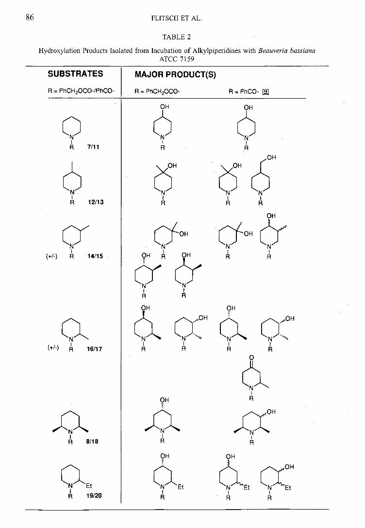
MAJOR PRODUCT(S)
R = PhCH2000- R=PhCO- 11
OH
(N
CJOH
OH OH
aN C :0H
FLITSCH ET AL.
TABLE 2
Hydroxylation Products Isolated from Incubation of Alkylpiperidines with Beauveria bassiana ATCC 7159
SUBSTRATES
R = PhCH2000-/PhCO-
0
R 7/11
6
R 12/13
01", (+1-) 14/15
aN
(+1-)R 16/17
1'14 R 8/18
OH R
OH
;N~ N I I R R
OH OH
OH
aEt Et
OH
I I R R R
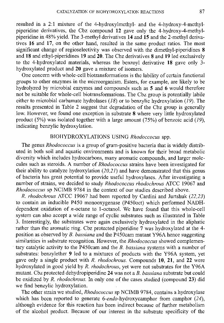
CATALYZATION OF BIOHYDROXYLATION REACTIONS 87
resulted in a • 2:1 mixture of the 4-hydroxylmethyl- and the 4-hydroxy-4-methyl-piperidine derivatives, the Cbz compound 12 gave only the 4-hydroxy-4-methyl-piperidine in 48% yield. The 3-methyl derivatives 14 and 15 and the 2-methyl deriva-tives 16 and 17, on the other hand, resulted in the same product ratios. The most significant change of regioselectivity was observed with the dimethyl-piperidines 8 and 18 and ethyl-piperidines 19 and 20. The Cbz derivatives 8 and 19 led exclusively to the 4-hydroxylated materials, whereas the benzoyl derivative 18 gave only 3-hydroxylated product and 20 gave a mixture of isomers.
One concern with whole-cell biotransformations is the lability of certain functional groups to other enzymes in the microorganism. Esters, for example, are likely to be hydrolyzed by microbial enzymes and compounds such as 5 and 6 would therefore not be suitable for whole-cell biotransformations. The Cbz group is potentially labile either to microbial carbamate hydrolases (18) or to benzylic hydroxylation (19). The results presented in Table 2 suggest that degradation of the Cbz group is generally low. However, we found one exception in substrate 8 where very little hydroxylated product (5%) was isolated together with a large amount (75%) of benzoic acid (19), indicating benzylic hydroxylation.
BIOHYDROXYLATIONS USING Rhodoccocus spp.
The genus Rhodococcus is a group of gram-positive bacteria that is widely distrib-uted in both soil and aquatic environments and is known for their broad metabolic diversity which includes hydrocarbons, many aromatic compounds, and larger mole-cules such as steroids. A number of Rhodococcus strains have been investigated for their ability to catalyze hydroxylation (20,21) and have demonstrated that this genus of bacteria has great potential to provide useful hydroxylases. After investigating a number of strains, we decided to study Rhodococcus rhodochrous ATCC 19067 and Rhodococcus sp NCIMB 9784 in the context of our studies described above.
R. rhodochrous ATCC 19067 had been reported by Cardini and Jurtshuk (22,23) to contain an inducible P450 monooxygenase (P450oct) which performed NADH-dependent oxidation of n-octane to 1-octanol. We have found that this whole-cell system can also accept a wide range of cyclic substrates such as illustrated in Table 3. Interestingly, the substrates were again exclusively hydroxylated in the aliphatic rather than the aromatic ring. Cbz protected piperidine 7 was hydroxylated at the 4-position as observed by B. bassiana and the P450cam mutant Y96A hence suggesting similarities in substrate recognition. However, the Rhodococcus showed complemen-tary catalytic activity to the P450cam and the B. bassiana systems with a number of substrates: benzylether 9 led to a mixtures of products with the Y96A system, yet gave only a single product with R. rhodochrous. Compounds 10, 21, and 22 were hydroxylated in good yield by R. rhodochrous, yet were not substrates for the Y96A mutant. Cbz protected dehydropiperidine 24 was not a B. bassiana substrate but could be oxidized by R. rhodochrous. In only one of the cases studied (compound 23) did we find benzylic hydroxylation.
The other strain we studied, Rhodococcus sp NCIMB 9784, contains a hydroxylase which has been reported to generate 6-endo-hydroxycamphor from camphor (24), although evidence for this reaction has been indirect because of further metabolism of the alcohol product. Because of our interest in the substrate specificity of the
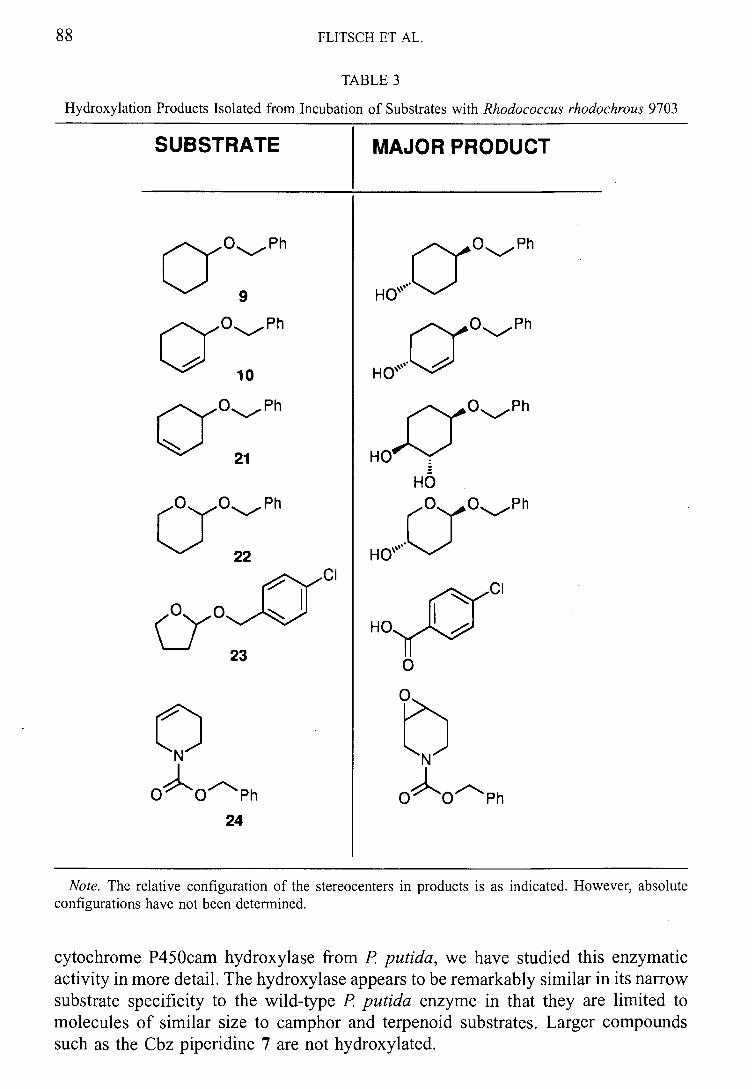
FLITSCH ET AL.
TABLE 3
Hydroxylation Products Isolated from Incubation of Substrates with Rhodococcus rhodochrous 9703
SUBSTRATE
MAJOR PRODUCT
Ph
Hef T
ON--,~
0,1:::~
O.Ph
H O"
Cr0 1-11-*, Ph
21
CJOPh 22
HO
r Oyo#Ph
H
24
Note. The relative configuration of the stereocenters in products is as indicated. However, absolute configurations have not been determined.
cytochrome P450cam hydroxylase from P putida, we have studied this enzymatic activity in more detail. The hydroxylase appears to be remarkably similar in its narrow substrate specificity to the wild-type P putida enzyme in that they are limited to molecules of similar size to camphor and terpenoid substrates. Larger compounds such as the Cbz piperidine 7 are not hydroxylated.
CATALYZATION OF BIOHYDROXYLATION REACTIONS 89
This hydroxylase will therefore not so much be of interest as a new complementary hydroxylation system, but rather be useful for comparison to the P450cam from P putida in order to allow us to further understand factors underlying regio selectivity of hydroxylation. Thus, we have been able to purify a cytochrome P450 monooxygen-ase from Rhodococcus species NCIMB 9784 which accepts camphor as a substrate. The first 16 N-terminal amino acids have been determined by sequencing and little homology was found in this short sequence to the cytochrome P450cam monooxygen-ase of putida. Studies toward the cloning and further characterization of this enzyme are currently in progress in our laboratories.
CONCLUSIONS
Comparison of the substrate specificity of the different monooxygenases from microbial sources studied here suggests some surprising similarities in selectivity of hydroxylation, in that the 3- and 4-positions of the aliphatic rings are the most likely to be hydroxylated. However, the substrate selectivity and the regio selectivity of hydroxylation can be fine-tuned either by change of substrate structure (e.g., using the Cbz instead of benzoyl-derivate) or by change of biocatalyst. With cell-free and purified enzyme systems becoming more and more available, we can start to rationalize regio selectivity of hydroxylation and redesign the active site of hydroxylases to suit the particular requirements of reaction selectivity.
ACKNOWLEDGMENTS
The studies with P450cam monooxygenase were conducted in collaboration with Dr. Luet-Lok Wong at the University of Oxford and crystallographic studies with Professor Malcolm Walkinshaw at the University of Edinburgh. This work was funded by the BBSRC and the Edinburgh Centre for Protein Technology.
REFERENCES
Holland, H. L. (1998) in Biotransformations (D. R. Kelly, Ed.), pp. 475-533, Wiley—VCH, Weinheim. Koga, H., Rauchfuss, B., and Gunsalus, I. C. (1985) Biochem. Biophys. Res. Commun. 130,412-417. Poulos, T. L., Finzel, B. C., and Howard, A. J. (1987) J. Mol. Biol. 195, 697-700. Atkins, W. M., and Sugar, S. G. (1988) 1 Biol. Chem. 263, 18842-18849. Kulisch, G. P., and Wilker, V. L. (1991). Biotechnol. Prog. 7, 93-98. Johnson, R. A., Herr, M. E., Murray, H. C., Reineke, L. M., and Fonken, G. S. (1968) 1 Org. Chem. 33, 3195. Furstoss, R., Archelas, R., and Waegell, B. (1981) Tetrahedron Lett. 22, 445. Carruthers, W., Prail, J. D., Roberts, S. M., and Willetts, A. J. (1990) J. Chem. Soc. Perkin Trans. I, 2854. Palmer, C. F., Webb, B., Broad, S., Casson, S., McCague, R., Willetts, A. J., and Roberts, S. M. (1997) Bioorg. Med. Chem. Lett. 7, 1299. Johnson, R. A., Murray, H. C., Reineke, L. M., and Fonken, G. S. (1969) 1. Org. Chem. 34, 2279. Fowler, S., England, P. A., Westlake, A. C. G., Rouch, D. R., Nickerson, D. P., Blunt, C., Braybrook, D., West, S., Wong, L.-L., and Flitsch, S. L. (1994) 1 Chem. Soc. Chem. Commun., 2761-2762. Wong, L.-L. (1998) Curr. Opin. Chem. Biol. 2, 263-268. England, P. A., Harford-Cross, C. F., Stevenson, J.-A., Rouch, D., and Wong, L.-L. (1998) FEBS Lett. 424, 271-274. Staines, A., Taylor, P., Flitsch, S. L., and Walkinshaw, M. Unpublished results. Estabrook, R. W., Faulkner, K. M., Shet, M. S., and Fisher, C. W. (1996) Methods Enzymol. 272,44-51. Sibbesen, 0., De Voss, J. J., and Ortiz de Montellano, P. R. (1996)1 Biol. Chem. 271, 22462-22469.
FLITSCH ET AL.
17. Aitken, S. J., Grogan, G., Chow, C. S.-Y., Turner, N. J., and Flitsch, S. L. (1998) J. Chem. Soc. Perkin Trans. 1 20, 3365-3370.
18, Gotor, V., Quiros, M., Liz, R., Frifola, J., and Fernandez, R. (1997) Tetrahedron 53, 6421-6432. Holland, H. L., Conn, M., Chiima Chenchaiah, P., and Brown, F. M. (1988) Tetrahedron Lett. 29, 6393. Banci, L., Bertini, I., Eltis, L. D., and Pierattelli, R. (1993) Biophys. J. 65, 806-813. Nagy, I., Compernolle, F., Ghys, K., Vanderleyden, J., and De Mot, R. (1995) App!. Environ. Microbiol., 2056-2060. Cardini, G., and Jurtshuk, P. (1968) J. Rio!. Chem. 243, 6070-6072. Cardini, G., and Jurtshuk, P. (1970) J. Biol. Chem. 245, 2789-2796. Chapman, P. J., Meerman, G., Gunsalus, I. C., Srinivasan, R., and Rinehart, K. L. (1966) 1 Am. Chem. Soc. 88, 618-619.
—1
Ui
nam
Appendix IV
00 00 —J —J m "1
ul il W () N) N) t
Q Ui CD Ui Ui Ui Ui Ui Ui (J Ui u Ui
90-1 0 a w ai t , Am 2 01. 519
'H-'H COSY of alcohol 120
Appendix IV
01 0.) 0.) NJ NJ 10 • • • • • • • 10 01 01 Q 01 01
CCO1Z (fl W) CCflHZ'J =Coc)CCcncCCmCz CC.HCCC>Cr.OZO1H-1,-HD-? CCIZO CO1O1flI-1 flWC1-.j Z.-jCQ-- Z C C,— or'1rCOO-(nnoosthwrJ tt<>C
CC = z:.cx ZN<>C'-flC C CWCt CDZC C CH(DJ I
CC C '1 0 fl H C S CI C
'H-'H COSY of alcohol 121
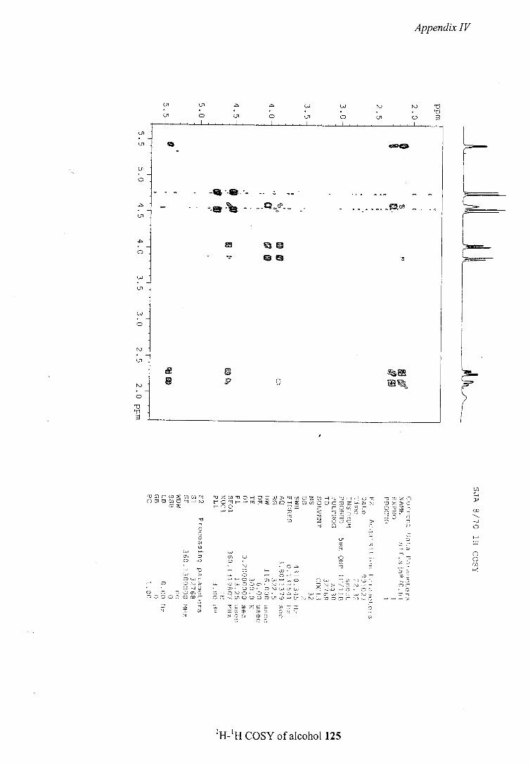
Appendix IV
1 1
01 CD 01 Cn CD
- -•e- -- -
F.
N)Cnciis OZNN)DHN)N) NN)ZHN)N)HN)- N)SiZfl OWN)N)JHN) tC - iG1OuisioCcsz-NN) N)XN)
N)0CN2 N) < N)N)H15 flN)N)-
N N) N) N) 0CC > C 0
H C - C)
N 2 • B
C) N) = • N) c(- -
N)-)2 N) N
N) N)N) N)N)N). fl -)0--N)). N))) C) N) N) N) 0N)- N)
0 N) N) 10 L, 1) Ij N)N)N)N)LP.N)-U-N)N) = N) 1 a
N N5() (DC) C) N
'H-'H COSY of alcohol 125
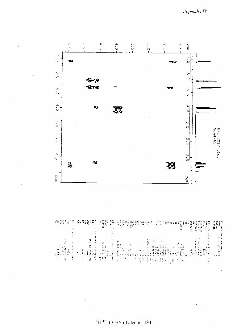
11
Appendix IV
C)' 01 01 0) N) NJ • • • Ui C' 01 01 01 C) Cii C) Toi
orwzwzcn ',Crtntcc' ti1Ci,zi -Cl nnnnnnoc -ooa -occLaHcc>-,iioz HOt-3O'i '2ifl ii C - - cc-- C ---- -v ---- --- -------------- 5. - NJ CO = ZNiCN-(X -00 = t rOtfl2 Qv
CC)'- C CO
i-
00NJt=-.. 00i)0._ C==0-00=0tC0oo0.===o00vi;J.,,.C_...r
'H-'H COSY of alcohol 133
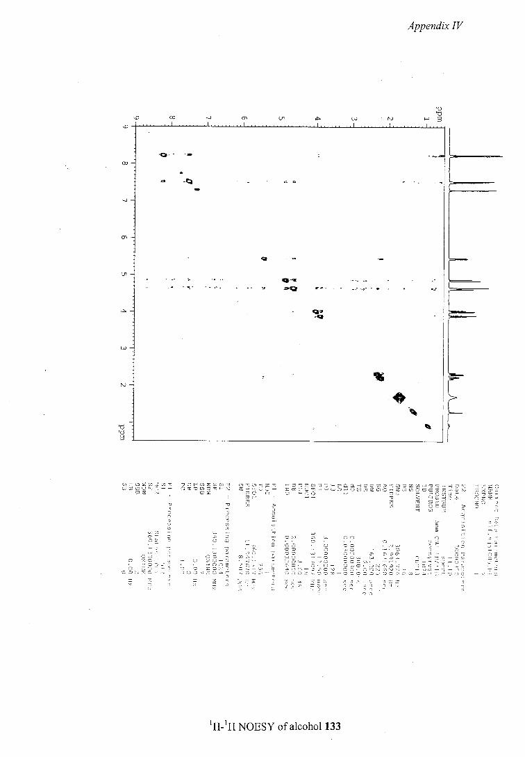
'.0
U,
N)
Appendix IV
0 'C
U, w N)
'UHZ 0wc')•- .--
S
S .Li - . . . • • 0
C Li S Li 5 00 -Li 0 0 2) 32 32 -t C' - 0 Li 2
-' C S -• CO 2 - Li Li - - C S C Li Lii -. Li -.i S Cr 0 2) - = Ci = = 120- 5 - S 220-i • -._-- - LiS. -.30 005Cr.
-C2r5 • -055 CC2..Li S CO. 0.0- 5_.0. .323 -5012 SC- -D.
css.sc-._ - S0Sc503. -.12G._c--; SZI)S12001O-00O05CO0-0C.-1OLiCc-Or0O -:5--C
1H-1H NOESY of alcohol 133
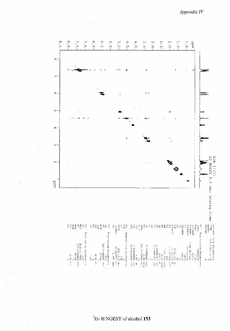
Appendix IV
F-j
U' Ui D Ui Q U' U' U' U' Ui U' Ui U' Ui U'
- -
'H-'H NOESY of alcohol 153