Embed Size (px)
Citation preview
H O S T E D B Y Contents lists available at ScienceDirect
www.elsevier.com/locate/jpa
Journal of Pharmaceutical Analysis
Journal of Pharmaceutical Analysis 2015;5(2):75–84
http://dx.doi.org/10.102095-1779 & 2015 Xi’NC-ND license (http:/
Peer review under r
nCorresponding autE-mail address: tar
www.sciencedirect.com
REVIEW PAPER
Bioautography and its scope in the field ofnatural product chemistry
Saikat Dewanjeea, Moumita Gangopadhyayb, Niloy Bhattacharyaa,Ritu Khanraa, Tarun K. Duaa,n
aAdvanced Pharmacognosy Research Laboratory, Department of Pharmaceutical Technology, Jadavpur University,188 Raja S C Mullick Road, Kolkata 700032, IndiabBiophysics Division, Saha Institute of Nuclear Physics, Kolkata 700064, India
Received 8 April 2014; revised 1 June 2014; accepted 19 June 2014Available online 26 June 2014
KEYWORDS
Bioactivity;Bioassay;Bioautography;Detection principle;Thin layerchromatography
16/j.jpha.2014.06.00an Jiaotong Univers/creativecommons.o
esponsibility of Xi’
hor. Tel.: þ91 33 [email protected]
Abstract Medicinal plants, vegetables and fruits are the sources of huge number of bioactive lead/scaffolds with therapeutic and nutraceutical importance. Bioautography is a means of target-directedisolation of active molecules on chromatogram. Organic solvents employed in chromatographic separationprocess can be completely removed before biological detection because these solvents cause inactivationof enzymes and/or death of living organisms. They offer a rapid and easy identification of bioactive lead/scaffolds in complex matrices of plant extracts. Bioautography is a technique to isolate hit(s)/lead(s) byemploying a suitable chromatographic process followed by a biological detection system. This reviewcritically describes the methodologies to identify antimicrobial, antioxidant, enzyme inhibitor lead/scaffolds by employing bioautography. A significant number of examples have been incorporated toauthenticate the methodologies.& 2015 Xi’an Jiaotong University. Production and hosting by Elsevier B.V. All rights reserved. This is an open
access article under the CC BY-NC-ND license (http://creativecommons.org/licenses/by-nc-nd/4.0/).
1. Introduction
Planar chromatographic analysis hyphenated with the biologicaldetection method is termed as bioautography [1]. It is an effectiveand inexpensive technique for the phytochemical analysis of plantextracts to identify bioactive lead/scaffolds. It can thus beperformed both in highly developed laboratories as well as insmall research laboratories which have minimum access tosophisticated equipments [2]. Despite having sophisticated on-
2ity. Production and hosting by Elsevierg/licenses/by-nc-nd/4.0/).
an Jiaotong University.
572043; fax: þ91 33 28371078.(T.K. Dua).
line high-performance liquid chromatography coupled bioassays,bioautography offers a simple, rapid and inexpensive method forthe chemical and biological screening of complex plant extracts,with subsequent bioassay-guided isolation [3]. In 1946, Goodalland Levi [4] introduced paper chromatography (PC)-based bioau-tography for the first time to estimate the purity of penicillin.In 1961, Fisher and Lautner [5] and Nicolaus et al. [6] introducedthin layer chromatography (TLC)-based bioautography. The firstreview on bioautography was written by Betina in 1973 [7].Generally, planar chromatographic, viz. TLC and PC are used forbioautography, but the detection method can be successfullyimproved by the application of advanced chromatographic tools,namely, high performance thin layer chromatography (HPTLC),
r B.V. All rights reserved. This is an open access article under the CC BY-

S. Dewanjee et al.76
over-pressured layer chromatography (OPLC), and planar electrochromatography (PLC). The major applications of bioautographyare the fast screening of a large number of samples for bioactivity,namely, antibacterial, antifungal, antioxidant, enzyme inhibition,etc. and in the target-directed isolation of active compounds [8–11].In this review, the techniques and application of bioautography arediscussed in details with suitable examples.
2. Detection of anti-microbial agents by bioautography
PC and TLC have become tools in the screening of antimicrobialagents through bioautography. Three bioautographic methods,namely, (i) agar diffusion or contact bioautography, (ii) directTLC bioautographic detection and (iii) immersion or agar overlaybioautography are used to detect antimicrobial agents in a mixtureof compounds [12,13].
2.1. Agar diffusion or contact bioautography
In contact bioautography, antimicrobial agents diffuse from adeveloped TLC plate or paper to an inoculated agar plate [14–16].The chromatogram is placed face down onto the inoculated agarlayer for a specific period to enable diffusion. Then the chromato-gram is removed and the agar layer is incubated. The zones ofinhibition on the agar surface, corresponding to the spots inchromatographic plates, are indicative of the antimicrobial sub-stances. An overall view of contact bioautography has beendepicted in Fig. 1. Incubation time for the growth ranges between16 and 24 h but it can be reduced to 5–6 h by spraying with 2,6-dichlorophenol-indophenol or 2,3,5-tetrazoliumchloride [17]. Thedisadvantages of contact bioautography are difficulties in obtainingcomplete contact between the agar and the plate and adherence ofthe adsorbent to the agar surface. Another problem may arise dueto the differential diffusion of components, especially water-insoluble, from the chromatogram to the agar plate. To overcomethese difficulties, Wagman and Bailey [12] introduced Chrom-AR
Chromatogramwas placed face
downforallowing diffusion into agar
media
Agar media
Fig. 1 Schematic overview
and silicic acid/glass fiber sheets for bioautography of antimicro-bial compounds. The principle of the method was the same andantimicrobials had to be transferred from the chromatographicplates to agar causing their loss and dilution. Another special caseis bioautographic detection of 6-aminopenicilanic acid which is avery weak antibiotic and must be converted through phenylacetylation to benzyl penicillin by spraying chromatographic platesor paper with acetyl chloride in mild alkaline condition beforebioautography [18]. This is a technique familiar to the microbiol-ogists in search for antibiotics from microorganisms, and differentprocedures have been used to improve its performance [19].Sphaerococcenol A, a bromoditerpene antibiotic, was isolated bycontact bioautography [20]. Three carboxylic polyether antibiotics,namely, monensin, lasalocid and salinomycin, were isolated usingcontact bioautography by VanderKop et al. [21]. Eight differentantifungal agents were isolated from the bark of Bridelia retusausing contact bioautography [22]. The development of HPTLC hasimproved resolution and sensitivity over TLC [23]. Application ofHPTLC also reduced the consumption of time and solvents.Through HPTLC-contact bioautography, Ramirez et al. [24]showed multiple antibiotic residues in cow’s milk. TLC contactbioautographic assay was introduced by Shahverdi et al. [25] forthe detection of antibiotic resistance reversal agents.
2.2. Direct TLC bioautographic detection
In direct TLC bioautography, the developed TLC plate is sprayedwith or dipped into a fungal or bacterial suspension (Fig. 2).A suspension of test bacteria or fungi is used for the spraying ordipping purpose. An inoculam of absorbance of 0.84 at 560 nmwas suggested for bacteria like Staphylococcus aureus [26], whileusing a suspension of 106 CFU/mL could be employed for bothbacteria and fungi [27]. The bioautogram is then incubated at25 1C for 48 h under humid condition. For visualization ofmicrobial growth, tetrazolium salts are used. These salts areconverted by the dehydrogenases of living microorganisms tointensely colored formazen [28]. These salts are sprayed onto the
Incubation
Zone of inhibition identify antimicrobial lead/scaffolds
Inoculat
ionwith
microb
ial
strain
s
of contact bioautography.

Spraying with microbial strains Dipping of
chromatogram in microbial strains
Incubation under humid condition for 48 h
or
Spraying with tetrazolium salt
White zones of inhibition against purple background indicate antimicrobial leads
Fig. 2 Schematic diagram indicates the direct bioautographic process.
Bioautography and its scopes 77
bioautogram and are reincubated at 25 1C for 24 h [28] or at 37 1Cfor 3–4 h [29,30]. Clear white zones against a purple backgroundon the TLC plate indicate antimicrobial activity of the sample [31].Okusa et al. [32] incorporated a new medium for direct TLCbioautography which is fluid enough to disperse microorganismsand viscous enough to adhere to the TLC plates; according to thema mixture of Muller–Hinton (MH) broth and MH agar in the ratioof 90:10 fulfils this requirement. Homans et al. [33] found thatdirect spraying of the thin-layer chromatograms with a sporesuspension of the test fungus in glucose–mineral salts medium wasthe easiest technique for the detection of fungitoxic substance, andit also gave the most reliable results [33]. Direct bioautographicmethods have been described for spore-producing fungi such asAspergillus, Penicillium and Cladosporium [33] and also forbacteria [34,35]. Bacillus subtilis, S. aureus and Escherichia coliare used to identify the active compounds. p-Iodonitrotetrazoliumviolet is the most suitable detection reagent [35]. TLC bioauto-graphy showed Pseudomonas cepacia B37W produces an anti-fungal compound pyrronitrin which acts against potato’s dry rootinvasion in nanogram level at the wound sight [36]. A cyclopep-tide antibiotic named condaline-A has been isolated from the barkof Condalia buxifolia along with the other antibiotics adoutine-Y/,scutianine-B and scutianine-C, and has been established usingdirect bioautography [37]. Separation of erythromycin from itsbase to identify systemic concentration of erythromycin wasestablished through direct TLC and PC bioautography by Ester-brook et al. [38]. A simple bioautographic technique has been usedin laboratories for many years to detect fungitoxic substancesaccording to Weltzine et al. [39], and modified by Dekhuijzenet al. [40]. In this technique, chromatograms are developed onWhatman no. 3 mm paper with propanol–water (85:15) and afterdrying are sprayed with a conidial suspension of Glomerellacingulata. After incubation clearly visible inhibition zones indicatethe presence of fungitoxic compounds. TLC-direct bioautographyis also useful for the rapid chemical and biological screening of
plant extracts [41]. Once an activity has been located at the TLCplate, the sample can be analyzed by liquid chromatography-massspectrometry to establish whether known or new compounds and/or substance classes are involved. This screening strategy concernsrapid detection of antibacterial and antifungal compounds [42–51].Choma et al. [52–54] in his review on TLC-bioautographyindicated plenty of examples of analysis on bacteria and fungifor the detection of antimicrobial activity of antimicrobial agentthrough bioautography and bioluminicence [52–56]. The BioLu-minizer™ system (commercialized by Camag, Muttenz, Switzer-land), which is a general detection system for bioactive substances,uses Vibrio fischeri, a non-pathogenic gram negative marinebacterium. This is an intrinsic bioluminescence, emitting a green-ish light as a product of cellular respiration at a critical cellulardensity. Luciferase, the bioluminescence catalyst, is expressed andcatalyzes an oxidation reaction that releases excess energy in theform of light [57]. After migration, the TLC plate is coated withthe bioluminescent bacteria which produce dark spots on aluminescent background with toxic or bioactive compounds [58].This method is suitable for the detection of toxins and chemicaladulterants in food, beverages, cosmetics, waste water and drink-ing water in picogram quantities. Du and Li [59] used thisluciferase assay for evaluation of estrogenic activity of Mucunasempervirens extracts. The bioautographic method can beextended for use in 2D-TLC experiments [60]. In 2D-TLC directbioautography, TLC plates are developed once with a polarsolvent, turned 901, and then developed again with a non-polarsolvent system. The TLC plates are then dried and sprayed with anutrient broth seeded with microbial suspension, and the microbialculture is grown directly on the silica gel surface of the TLC plate.The advantage of this method is that migration in each of the twodimensions can be brought about with solvents of very differentpolarities. 2D-TLC direct bioautography has been found to beuseful for the detection of inhibitors of the plant pathogens,viz. Colletotrichum acutatum, Colletotrichum fragariae and

S. Dewanjee et al.78
Colletotrichum gloeosporioides [60]. Antifungal activity of chro-menes from Peperomia serpens (Sw.) Loudon (Piperaceae) wasestablished by the direct bioautographic assay technique againstCladosporium cladosporioides and Cladosporium sphaerosper-mum [61]. Tyiháka et al. [62] introduced a new system ofbioautography known as bio-arena where they combined OPLCwith bioautography. Separation in an OPLC system results inbetter-defined spots than in conventional TLC and HPTLCseparations. A number of applications have been reported, includ-ing the investigation of chelidonium alkaloids [63].
2.3. Immersion or agar overlay bioautography
Agar overlay is a combination of contact and direct bioautography.In this method, the chromatogram is covered with a molten, seededagar medium. After solidification, incubation and staining (usuallywith tetrazolium dye), the inhibition or growth bands are visua-lized (Fig. 3) [64–66]. For Gram-negative bacteria, an agarsolution containing the red-colored bacterium Serracia marcescenscan be employed. The red-colored gel is incubated overnight atroom temperature and inhibition zones appear as white or paleyellow areas on a red background [67]. With other, colorless,microorganisms, zones of microbial growth inhibition are visua-lized with the aid of a dehydrogenase activity-detecting reagent(tetrazolium salt). Metabolically active microorganisms convert thetetrazolium salt (MTT) into the corresponding intensely coloredformazan (Fig. 4).
1.Solidification2.Incubation3.Staining with tetrazolium salt
Fig. 3 Schematic diagram of immersion or agar overlaybioautography.
Fig. 4 Reduction of tetrazolium salts.
The agar overlay assay has been used for yeasts such asCandida albicans and can also be applied to bacteria such asB. subtilis, E. coli, Pseudomonas aeruginosa and S. aureus [48,68].The antibacterial activity of isoflavonoid and sesquiterpenoidphytoalexins has been evaluated by an overlay method usingPseudomonas syringae pv. phaseolicola, with 2,3,5-triphenyl-tetrazolium chloride (TZC) as visualizating reagent. Addition ofglycerol to the overlay nutrient medium, as a carbon source,facilitated the reduction of TZC to pink colored formazans bybacterial dehydrogenases [69]. Antimicrobial carotenoids havebeen characterized in Bixa orellana seed extracts [70]. Theantibacterial activity was detected by overlaying TLC plates withagar containing S. aureus and then spraying with tetrazolium salt(INT) [70]. Cytrynska et al. [71] characterized eight antimicrobialpeptides from Galleria mellonella larvae immune hemolymphusing tricine SDS-PAGE bioautography. Several antimicrobialcompounds were isolated from Tylosema esculentum husks,cotyledons, and tubers by employing agar overlay bioautographyusing S. aureus, E. coli, B. subtilis, P. aeruginosa and C. albicansas test microorganisms [72].
3. Detection of antioxidant agents by bioautography
3.1. Bioautography using DPPH as detection reagent
The stable 2,2-diphenyl-1-picrylhydrazyl radical (DPPH) has anabsorption maximum at 517 nm, which decreases upon reductionthrough reaction with a radical scavenger [73]. The correspondingcolor change can thus be observed in a TLC bioassay [74]. Thereaction has been depicted in Fig. 5. The developed chromatogramis sprayed with a solution of 0.2% DPPH in methanol/ethanol. Theplate is examined in daylight after 30 min. Free-radical scavengersappear as cream/yellow spots against a purple background. Theintensity of the yellow color can be measured with a chromameter.Rossi et al. [75] identified the radical scavenging molecules inessential oil from Croton lechleri and successfully compared the
Fig. 5 Reaction of DPPH radical with radical scavengers.
Fig. 6 Reaction of ABTS radical with radical scavengers.
Bioautography and its scopes 79
antioxidant potential with thyme oil. Four antioxidant compounds,viz. rosmarinic acid, luteolin, apigenin, and chrysoeriol, were theisolated fruit of Perilla frutescens var. acuta by bioautography onTLC using DPPH as a detection reagent [76].
3.2. Bioautography using ABTS as detection reagent
2,2-Azino-bis(3-ethylbenzthiazoline-6-sulfonic) acid (ABTS) radi-cal scavenging method can be extended to TLC plates [77]. Thereaction involved is shown in Fig. 6. Layers stained with ABTSshow a green background and radical scavengers give colorless or
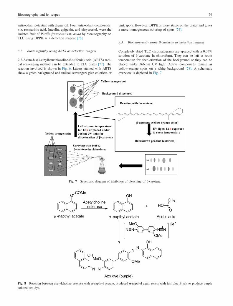
Spraying with 0.05%β-carotene in chloroform
Yellow orange stain
Yellow orang
Background
Left at room temperaturefor 12 h or placed under 366nm UV light fordiscoloration of β-carotene
Fig. 7 Schematic diagram of inhib
Fig. 8 Reaction between acetylcholine esterase with α-napthyl acetate, prcolored azo dye.
pink spots. However, DPPH is more stable on the plates and givesa more homogeneous coloring of spots [74].
3.3. Bioautography using β-carotene as detection reagent
Completely dried TLC chromatograms are sprayed with a 0.05%solution of β-carotene in chloroform. They can be left at roomtemperature for decolorization of the background or they can beplaced under 366 nm UV light. Active compounds remain asyellow-orange spots on a white background [78]. A schematicoverview is depicted in Fig. 7.
e spot
discolored
β-carotene (yellow orange color)
Breakdown product (colorless)
UV-light/ 12 h exposure to room temperature
Reaction with β-carotene:
ition of bleaching of β-carotene.
oduced α-napthol again reacts with fast blue B salt to produce purple

Fig. 9 Ellman reaction.
Fig. 10 Diazotization reaction for the detection of α and β-glucosidase inhibition.
S. Dewanjee et al.80
3.4. Inhibition of bleaching of β-carotene inducedby auto-oxidation of linoleic acid
Dry TLC plates are sprayed with a mixture of linoleic acid inethanol and β-carotene in chloroform. After exposing the plate tosunlight, antioxidant activity is shown by the presence of orangespots on a white background [79].
Fig. 11 Reaction to detect xanthine oxidase inhibitors.
4. Enzyme inhibition
Enzymes are important molecular targets for lead discovery inprimary screening assays. The use of a TLC support to screen for

Fig. 12 Schematic diagram of the HPTLC-YES process.
Bioautography and its scopes 81
potential plant-derived enzyme inhibitors is a rapid method whichis relatively free of disturbances due to the solvent.
4.1. Bioautographic detection of acetyl cholinesterase (AchE)inhibitor
4.1.1. Detection by diazotizationThe basic principle of this method is that the enzyme convertsα-naphthyl acetate (substrate) into α-naphthol. α-Naphthol reactswith fast blue B salt (chromogenic agent) to make a purple coloredbackground on the TLC plates, while AchE inhibitors producewhite spots. Enzyme inhibitors block the formation of α-naphtholand hence no purple coloration is produced (Fig. 8). Samples wereapplied to silica gel TLC plate and migrated in a suitable solvent.After complete removal of solvents, the AchE solution is sprayed.The enzyme is allowed to incubate for 20 min at 37 1C and is thensprayed with a mixture of 1-naphthyl acetate and fast blue B salt.After 1–2 min, AchE inhibitors show white spots on a purplebackground [80]. Detection limits for AchE inhibitors remainwithin the nanogram level [9]. This bioautographic method offersa rapid identification of a large number of AchE inhibitors.
Another variant of the bioassay [81,82] uses β-naphthyl acetateand enzyme solution in the TLC elution solvent and incubated withenzyme for 10 min. This method supposedly gives a deeper violetbackground color and higher sensitivity of detection. However,β-naphthyl acetate is considerably more expensive than α-naphthylacetate. To overcome the cost of the experiment, Yang et al. [83] uses4-methoxy phenyl acetate as substrate for AchE and a mixed solutionof K3(FeCN)6 and FeCl3 � 6H2O as chromogenic agent. In thismethod, the spot appeared as a light yellow spot and the other partsare aquamarine blue. The consumption of the enzyme is satisfactory.A microplate assay has been developed for the diazotization method[84]. This gives a quantitative result (such as IC50 values), but TLCassays, which are useful as qualitative methods, are in general moresensitive than microplate assays. However, there was a goodcorrelation between the TLC bioautographic assay and the solutionassay. Queiroz et al. [85] showed that dichloromethane extract of theaerial parts of Blumea gariepina (Asteraceae) was found to be active
against AchE. Seven new AchE inhibitors were isolated from thisplant through bioautography. The original detection method bydiazotization has been employed to screen Amaryllidaceae speciesfor AChE inhibition [86].
4.1.2. Detection by the Ellman reactionThe Ellman method for the colorimetric determination of AchEactivity was first described in 1961 [87]. Acetylthiocholine (ATCI)is cleaved by AchE to form thiocholine which reacts with 5,5-dithiobis-(2-nitrobenzoic acid) (DTNB) to give the yellow 5-thio-2-nitrobenzoate anion (Fig. 9). It could be adapted for the TLCscreening of AchE inhibitors [88]. In this method, a solution ofDTNB and ATCI is sprayed on the chromatogram (after completeremoval of solvents), followed by spraying with AchE. A paleyellow background forms within about 5 min. AchE inhibitorsappear as white spots. The Ellman reaction is more difficult tovisualize than the diazotization method. False positive effect ispossible with the presence of aldehydes [89]. An indole alkaloidfrom Tabernaemontana australis showed considerable AchEinhibitory activity and it is evaluated through the Ellman reactionon the TLC plate [90].
4.2. α and β-glucosidase inhibition
Glucosidase inhibitors are potentially useful as antidiabetic, anti-obesity, antiviral, antiadhesive, antibacterial or antimetastaticagents [91]. Screening methods for glucosidase inhibitors includespectrophotometry with o- or p-nitrophenyl-β-D-glucopyranosideas substrate [92,93] or an agar plate method [94]. Both of thesehave limitations. With p-nitrophenyl-β-D-glucopyranoside, gluco-sidases cleave the sugar, to develop a yellow background due tothe formation of p-nitrophenol. However, this is only observed onthe TLC plate at pH 7.5 and the zone of inhibition remains poorlyvisible. No yellow background color is observed in the cor-responding test with p-nitrophenyl-β-D-glucopyranoside [95].In TLC-bioautography, the chromatogram is sprayed with enzymesolution in appropriate buffer. An alternative is a test which

S. Dewanjee et al.82
involves the cleavage of 2-naphthyl-β-D-glucopyranoside or2-naphthyl-α-D-glucopyranoside by respective α- or β-glucosidaseinhibitors [96]. The β-naphthol which is formed reacts with fastblue B salt, to give a purple colored diazo dye (Fig. 10) [96]. Forthe detection of the active enzyme, the solutions of 2-naphthyl-α-D-glucopyranoside (for α-glucosidase) or 2-naphthyl-β-D-glucopyr-anoside (for β-glucosidase) in ethanol and fast blue Bsalt indistilled water are prepared. The naphthyl-glucopyranoside solu-tion and the fast blue B salt solution are mixed at a ratio of 1:1 (forα-glucosidase) or 1:4 (for β-glucosidase). The mixture is sprayedonto the plate to give a purple coloration within 2–5 min. A TLCassay has previously been established for the detection ofβ-glucosidase inhibitors. It is based on the hydrolysis of esculininto esculetin which reacts with FeCl3 to provide a brown complex[65,97].
4.3. Xanthine oxidase inhibition
The enzyme xanthine oxidase (XO) catalyzes the oxidation ofhypoxanthine and xanthine to uric acid and producing O2
� andH2O2. The inhibition of XO diminishes oxidative stress andexhibits prophylactic role on inflammation, arteriosclerosis, can-cer, aging, etc. [65]. To identify XO inhibitors on TLC plates, theenzyme is suspended in agar and distributed on the TLC plate(direct measurement of enzyme activity on the TLC plate is notpossible). After solidification, the plate is immersed into a solutionof xanthine at 38 1C for 20 min in the dark. Enzymatic oxidationof xanthine produces O2
� which reduces the pale yellowtetrazolium salt (NBT) to a formazan and hence a purple back-ground is obtained on the plate (Fig. 11). Allopurinol, an inhibitorof XO, is detected as a white spot on the purple background. Thismethod could determine the nanogram level of XO inhibitor.A modification of the test, in which O2
� is generated chemically,allows the distinction between pure inhibitors of XO, such asallopurinol and radical scavengers [98].
5. Detection of estrogenic compounds
Estrogen receptor agonists include natural and synthetic hormones,phytoestrogens and chemicals such as metabolites of alkyl-phenolextoxylate, bisphenol A, parabens, benzophenons and phthalates[99]. Muller et al. [1] first observed that an estrogenic compoundcan be bioautographed using production capacity of β-galactosidase enzyme from yeast cell. This process is also knownas the HPTLC-YES (yeast estrogen screen) procedure [1]. This is areporter gene assay which had been developed for the assessmentof the estrogenic potency of individual substances. Here 4-methylumbelliferyl β-D-galactopyranoside (MUG) is used as a fluoro-genic substrate on which the enzyme β-galactosidase works andconverts it into 4-methyl umbelliferone (Fig. 12). Chlorophenolred β-D-galactopyranoside a chromogenic substrate can also beused in place of MUG but the sensitivity of the test was the highestin the case of MUG.
6. Conclusion
In spite of wide employment of sophisticated chromatographictechniques coupled with on-line bioassays, bioautography is stillproving its worth as a simple and inexpensive tool for simulta-neous chemico-biological screening of natural sources. In other
word, it offers the simplest mean of bioassay guided leaddiscovery from natural products. For the natural product theseparation process is not easy, and if separated the amount isvery less in maximum cases, so it is necessary to develop a processwhich can detect lead in a small amount and biological activity canalso be measured successively. Considering these problems, wecan say that bioautographic detection technique would create anew era in separation science.
References
[1] M.B. Muller, C. Dausend, C. Weins, et al., A new bioautographicscreening method for the detection of estrogenic compounds, Chro-matographia 60 (2004) 207–211.
[2] A. Marston, M. Maillard, K. Hostettmann, A TLC bioautographicmethod for the detection of α- and β-glucosidase inhibitors in plantextracts, GIT Lab. J 1 (1997) 36–39.
[3] K. Hostettmann, C. Terreaux, A. Marston, et al., The role of planarchromatography in the rapid screening and isolation of bioactivecompounds from medicinal plants, J. Planar Chromatogr. 10 (1997)251–258.
[4] R.R. Goodall, A.A. Levi, A microchromatographic method for thedetection and approximate determination of the different penicillins ina mixture, Nature 158 (1946) 675–676.
[5] R. Fisher, H. Lautner, Zum papierchromatographischen Nachweisvon Penicillinpräparaten, Arch. Der. Pharm. 294 (1961) 1–7.
[6] B.J.R. Nicolaus, C. Coronelli, A. Binaghi, Microbiological determi-nation of antibiotics by thin layer chromatograms, Farmaco. Ed. Prat.16 (1961) 349–370.
[7] V. Betina, Bioautography in paper and thin-layer chromatography andits scope in the antibiotic field, J. Chromatogr. 78 (1973) 41–51.
[8] B. Heinemann, A.J. Howard, Z.J. Hollister, Application of paperchromatograms to the study of inducers of λ bacteriophage inEscherichia coli, Appl. Microbiol. 15 (1967) 723–725.
[9] I.M. Choma, E.M. Grzelak, Bioautography detection in thin-layerchromatography, J. Chromatogr. A 1218 (2011) 2684–2691.
[10] P.J. Houghton, Use of small scale bioassays in the discovery of noveldrugs from natural sources, Phytother. Res. 14 (2000) 419–423.
[11] L. Bohlin, J.G. Bruhn, Bioassay Methods in Natural Product Researchand Drug Development, Kluwer Academic Publishers, Dordrecht,1999, p. 67.
[12] G.H. Wagman, J.V. Bailey, Use of silicic acid–glass fiber sheets forbioautography of antimicrobial substances, J. Chromatogr. 41 (1969)263–264.
[13] J.L. Rios, M.C. Recio, A. Villar, Screening methods for naturalproducts with antimicrobial activity: a review of the literature,J. Ethnopharmacol. 23 (1988) 127–149.
[14] J. Sherma, Planar chromatography, Anal. Chem. 80 (2008)4253–4267.
[15] E. Meyers, D.A. Smith, Bioautography of antibiotic spread-layerchromatograms, J. Chromatogr. 14 (1964) 129–132.
[16] N. Narasimhachari, S. Ramachandran, A simple bioatuographictechnique for identifying biologically active material on thin-layerchromatograms, J. Chromatogr. 27 (1967) 494.
[17] A.A. Shahat, G. El-Barouty, R.A. Hassan, et al., Chemical composi-tion and antimicrobial activities of the essential oil from the seeds ofEnterolobium contortisiliquum (Leguminosae),J. Environ. Sci. Health B 43 (2008) 519–525.
[18] V. Betina, L. Pilatova, A paper chromatography method for thedetermination of suitable pH values for the extraction of antibiotics,Csl. Mikrobiol. 3 (1958) 202–204.
[19] R.M.C. Dawson, D.C. Elliott, W.H. Elliott, et al., Data for Biochem-ical Research, 2nd edn., Oxford University Press, Oxford, 1969.
[20] S. Caccamese, O. Cascio, A. Compagnini, Isolation of an antimicro-bial bromoditerpene from a marine alga aided by improved bioauto-graphy, J. Chromatogr. A 478 (1989) 255–258.

Bioautography and its scopes 83
[21] P.A. VanderKop, J.D. MacNeil, Separation and detection of mon-ensin, lasalocid and salinomycin by thin-layer chromatography/bioautography, J. Chromatogr. 508 (1990) 386–390.
[22] L. Jayasinghe, B.M.M. Kumarihamy, K.H.R.N. Jayarathna, et al.,Antifungal constituents of the stem bark of Bridelia retusa, J.Phytochem. 62 (2003) 637–647.
[23] J.C. Touchstone, M.F. Dobbins, Practice of Thin Layer Chromato-graphy, 2nd edn., Wiley, New York, 1983.
[24] A. Ramirez, R. Gutiérrez, G. Diaz, et al., High-performance thin-layerchromatography-bioautography for multiple antibiotic residues incow’s milk, J. Chromatogr. B. Anal. Technol. Biomed. Life Sci.784 (2003) 315–322.
[25] A.R. Shahverdi, F. Abdolpour, H.R. Monsef-Esfahani, et al., A TLCbioautographic assay for the detectionof nitrofurantoin resistancereversal compound, J. Chromatogr. B. Anal. Technol. Biomed. LifeSci. 850 (2007) 528–530.
[26] J.J.M. Meyer, F. Dilika, Antibacterial activity of Helichrysumpedunculatum used in circumcision rites, J. Ethnopharmacol. 53(1996) 51–54.
[27] G. Schmourlo, R.R. Mendonca-Filho, C.S. Alviano, et al., Screeningof antifungal agents using ethanol precipitation and bioautography ofmedicinal and food plants, J. Ethnopharmacol. 96 (2004) 563–568.
[28] M.T.G. Silva, S.M. Simas, T.G.F.M. Batista, et al., Studies onantimicrobial activity, in vitro, of Physalis angulata L. (Solanaceae)fraction and physalin B bringing out the importance of assaydetermination, Mem. Instit. Oswaldo Cruz 100 (2005) 779–782.
[29] F. Dilika, A.J. Afolayan, J.J.M. Meyer, Comparative antibacterialactivity of two Helichrysum species used in male circumcision inSouth Africa, S. Afr. J. Bot. 63 (1997) 158–159.
[30] D.K.B. Runyoro, M.I.N. Matee, O.D. Ngassapa, et al., Screening ofTanzanian medicinal plants for anti-candida activity, BMC Comple-ment. Altern. Med. 6 (2006) 1–10, http://dx.doi.org/10.1186/1472-6882-6-11.
[31] K. Das, R.K.S. Tiwari, D.K. Shrivastava, Techniques for evaluationof medicinal plant products as antimicrobial agent: current methodsand future trends, J. Med. Plants Res. 4 (2010) 104–111.
[32] P.N. Okusa, C. Stevigny, M. Devleeschouwer, et al., Optimization ofthe culture medium used for direct TLC-bioautography. Applicationto the detection of antimicrobial compounds from Cordia gilletii DeWild (Boraginaceae), J. Planar Chromatogr. 23 (2010) 245–249.
[33] A.L. Homans, A. Fuchs, Direct bioautography on thin-layer chroma-tograms as a method for detecting fungitoxic substances,J. Chromatogr. 51 (1970) 327–329.
[34] M.O. Hamburger, G.A. Cordell, A direct bioautographic TLC assayfor compound possessing antibacterial activity, J. Nat. Prod. 50(1987) 19–22.
[35] M.S. Blois, Antioxidant determinations by the use of a stable freeradical, Nature 181 (1958) 1199–1200.
[36] K.D. Burkhead, D.A. Schisler, P. Slininger, Pyrrolnitrin productionby biological control agent Pseudomonas cepacia B37w in cultureand in colonized wounds of potatoes, J. Appl. Environ. Microbiol. 60(1994) 2031–2039.
[37] A.F. Morel, C.A. Araujo, U.F. da Silva, et al., Antibacterialcyclopeptide alkaloids from the bark of Condalia buxifolia,Phytochemistry 61 (2002) 561–566.
[38] S.M. Esterbrook, J.A. Hersey, Bioautography of erythromycin and itsesters, J. Chromatogr. 121 (1976) 390–394.
[39] H.C. Weltzien, Ein biologischer Test für fungizide Substanzen aufdem Paper chromatogram, Naturwiss 45 (1958) 288–289.
[40] H.M. Dekhuijzen, The transformation in plants of sodium dimethyl-dithiocarbamate into other fungitoxic compounds, Staat. Gent. 26(1961) 1542–1543.
[41] J.D. Paxton, Assays for antifungal activity, in: K. Hostettmann (Ed.),Methods in Plant Biochemistry—Assays for Bioactivity, vol. 6,Academic Press, London, 1991.
[42] K. Hostettmann, A. Marston, Studies in Natural Products Chemistry,Elsevier, Amsterdam, 1990.
[43] K. Hostettmann, O. Potterat, Strategy for the isolation and analysis ofantifungal, molluscicidal and larvicidal agents from tropical plants,ACS Symp. Ser. 658 (1997) 14–26.
[44] E. Meyers, R.C. Erickson, Bioautography of antibiotics on thin layerchromatograms, J. Chromatogr. 26 (1967) 531–532.
[45] P.B. Hamilton, C.E. Cook, Some techniques for bioautography ofantimicrobial substances on thin-layer chromatograms, J. Chromatogr.35 (1968) 295–296.
[46] N. Islam, S.A. Parveen, N. Nakazawa, et al., Bioautography with thefungus Valsa ceratosperma in the search for antimycotic agents,Pharm. Biol. 41 (2003) 637–640.
[47] J. Billow, T.J. Speaker, Bioautography of antibiotic compounds: asimplification and improvement, J. Chromatogr. 67 (1972) 191–192.
[48] L. Rahalison, M. Hamburger, K. Hostettmann, et al., A bioauto-graphic agar overlay method for the detection of antifungal com-pounds from higher plants, Phytochem. Anal. 2 (1991) 199–203.
[49] L. Rocha, A. Marston, O. Potterat, et al., Antibacterial phlorogluci-nols and flavonoids from Hypericum brasiliense, J. Phytochem. 40(1995) 1447–1452.
[50] S. Rodriguez, J.L. Wolfender, E. Hakizamungu, et al., An antifungalnaphthoquinone, xanthones and secoiridoids from Swertia calycina,Planta Med. 61 (1995) 362–364.
[51] C. Terreaux, M. Maillard, K. Hostettmann, et al., Analysis of thefungicidal constituents from the bark of Ocotea usambarensis Engl.(Lauraceae), Phytochem. Anal. 5 (1994) 233–238.
[52] I.M. Choma, A. Choma, K. Staszczuk, Direct bioautography – thinlayer chromatography of flumequine and doxycycline in milk,J. Planar Chromatogr. 15 (2002) 187–191.
[53] I.M. Choma, I. Komaniecka, Matrix solid-phase dispersion combinedwith thin-layer chromatography-direct bioautography for determina-tion of enrofloxacin and ciprofloxacin residues in milk, J. Liq.Chromatogr. Relat. Technol. 28 (2005) 2467–2478.
[54] I.M. Choma, C. Kowalski, R. Lodkowski, et al., TLC-DB as analternative to the HPLC method in the determination of cefacetrilresidues in cow’s milk, J. Liq. Chromatogr. Relat. Technol. 31 (2008)1903–1912.
[55] I.M. Choma, Thin-layer chromatography—direct bioautography offlumequine residues in milk, J. Liq. Chromatogr. Relat. Technol. 29(2006) 2083–2093.
[56] I.M. Choma, Screening of enrofloxacin and ciprofloxacin residues inmilk by HPLC and by TLC with direct bioautography, J. PlanarChromatogr. 19 (2006) 104–108.
[57] G. Eberz, H.G. Rast, K. Burger, et al., Bioactivity screening bychromatography—bioluminescence coupling, Chromatographia 43(1996) 5–9.
[58] C. Weins, H. Jork, Toxicological evaluation of harmful substances byin situ enzymatic and biological detection in high-performance thin-layer chromatography, J. Chromatogr. A 750 (1996) 403–407.
[59] Q. Du, B. Li, Identification of antioxidant compounds of Mucunasempervirens by high-speed counter-current chromatographic separa-tion—DPPH radical scavenging detection and their oestrogenicactivity, Food Chem. 131 (2012) 1181–1186.
[60] D.E. Wedge, D.G. Nagle, A new 2D-TLC bioautography method forthe discovery of novel antifungal agents to control plant pathogens, J.Nat. Prod. 63 (2000) 1050–1054.
[61] R.O.S. Kitamura, P. Romoff, M.C.M. Young, et al., Chromenes fromPeperomia serpens (Sw.) Loudon (Piperaceae), Phytochemistry 67(2006) 2398–2402.
[62] E. Tyiháka, E. Mincsovics, Á.M. Móricza, Over pressured layerchromatography: from the pressurized ultramicro chamber to BioAr-ena system, J. Chromatogr. A 1232 (2012) 3–18.
[63] A. Sarkozi, A.M. Moricz, P.G. Ott, et al., Investigation of chelido-nium alkaloids by use of a complex bioautographic system, J. PlanarChromatogr. 19 (2006) 267–272.
[64] J.B. Harborne, Phytochemical Methods, Chapman and Hall Ltd.,London, 1973, pp. 49–188.
[65] A. Marston, Thin-layer chromatography with biological detection inphytochemistry, J. Chromatogr. A 1218 (2011) 2676–2683.

S. Dewanjee et al.84
[66] J.B. Harborne, Phenolic compounds, in: E. Heftmann (Ed.), Chro-matography, 5th edn., Elsevier, Amsterdam, 1992.
[67] L. Williams, O. Bergersen, Towards an integrated platform forcombinatorial library synthesis and screening, J. Planar Chromatogr.14 (2001) 318–321.
[68] G. Saxena, S. Farmer, G.H.N. Towers, et al., Use of specific dyes inthe detection of antimicrobial compounds from crude plant extractsusing a thin layer chromatography agar overlay technique, Phyto-chem. Anal. 6 (1995) 125–129.
[69] A.J. Slusarenko, A.C. Longland, I.M. Whitehead, A convenientsensitive and rapid assay for antibacterial activity of phytoalexins,Bot. Helv. 99 (1989) 203–207.
[70] V. Galindo-Cuspinera, S.A. Rankin, Bioautography and chemicalcharacterization of antimicrobial compound(s) in commercial water-soluble annatto extracts, J. Agric. Food Chem. 53 (2005) 2524–2529.
[71] M. Cytrynska, P. Mak, A. Zdybicka-Barabas, et al., Purification andcharacterization of eight peptides from Galleria mellonella immunehemolymph, Peptides 28 (2007) 533–546.
[72] O. Mazimba, R.R.T. Majinda, C. Modibedi, et al., Tylosemaesculentum extractives and their bioactivity, Bioorg. Med. Chem.19 (2011) 5225–5230.
[73] M. Olech, Ł. Komsta, R. Nowak, et al., Investigation of antiradicalactivity of plant material by thin-layer chromatography with imageprocessing, Food Chem. 132 (1) (2012) 549–553.
[74] T. Takao, F. Kitatani, N. Watanabe, et al., Simple screening methodfor antioxidants and isolation of several antioxidant produced bymarine bacteria from fish and shellfish, Biosci. Biotechnol. Biochem.58 (1994) 1780–1783.
[75] D. Rossi, A. Guerrini, S. Maietti, et al., Chemical fingerprinting andbioactivity of Amazonian Ecuador Croton lechleri Müll. Arg.(Euphorbiaceae) stem bark essential oil: a new functional foodingredient? Food Chem. 126 (2011) 837–848.
[76] L. Gu, T. Wua, Z. Wang, TLC bioautography-guided isolation ofantioxidants from fruit of Perilla frutescens var. acuta, LWT – FoodSci. Technol. 42 (2009) 131–136.
[77] N.J. Miller, C.A. Rice-Evans, Factors influencing the antioxidantactivity determined by the ABTS radical cation assay, Free. Radic.Res. 26 (1997) 195–199.
[78] D.E. Pratt, E.E. Miller, A flavonoid anti-oxidant in Spanish pea nuts,Am. J. Oil Chem. Soc. 61 (1984) 1064–1067.
[79] C.C. Whittern, E.E. Miller, D.E. Pratt, Cotton seed flavonoids as lipidantioxidants, J. Am. Oil Chem. 61 (1984) 1075–1078.
[80] Z. Yang, X. Zhang, D. Duan, et al., Modified TLC bioautographicmethod for screening acetylcholinesterase inhibitors from plantextracts, J. Sep. Sci. 32 (2009) 3257–3259.
[81] T. Mroczek, Highly efficient, selective and sensitive molecular screeningof acetylcholinesterase inhibitors of natural origin by solid-phaseextraction-liquid chromatography/electrospray ionisation-octopole-orthogonal acceleration time-of-flight-mass spectrometry and novelthin-layer chromatography-based bioautography, J. Chromatogr. A1216 (2009) 2519–2528.
[82] T. Mroczek, J. Mazurek, Pressurized liquid extraction and antic-holinesterase activity-based thin-layer chromatography with bioauto-graphy of Amaryllidaceae alkaloids, Anal. Chim. Acta 633 (2009)188–196.
[83] Z.D. Yang, Z.W. Song, J. Ren, et al., Improved thin-layer chromato-graphy bioautographic assay for the detection of actylcholinesteraseinhibitors in plants, Phytochem. Anal. 22 (2011) 509–515.
[84] A. Marston, J. Kissling, K. Hostettmann, A rapid TLC bioautographicmethod for the detection of acetylcholinesterase and butyrylcholines-terase inhibitors in plants, Phytochem. Anal. 13 (2002) 51–54.
[85] E.F. Queiroz, J.R. Ioset, K. Ndjoko, et al., On-line identification ofthe bioactive compounds from Blumea gariepina by HPLC-UV-MSand HPLC-UV-NMR, combined with HPLC-micro-fractionation,Phytochem. Anal. 16 (2005) 166–174.
[86] J. Kissling, J.R. Ioset, A. Marston, et al., Bio-guided isolation ofcholinesterase inhibitors from the bulbs of Crinum x powellii,Phytother. Res. 19 (2005) 984–987.
[87] G.L. Ellman, K.D. Courtney, V. Andres, et al., A new and rapidcolorimetric determination of acetylcholinesterase activity, Biochem.Pharmacol. 7 (1961) 88–95.
[88] I.K. Rhee, M. van de Meent, K. Ingkaninan, et al., Screening foracetylcholinesterase inhibitors from Amaryllidaceae using silica gelthin-layer chromatography in combination with bioactivity staining, J.Chromatogr. A 915 (2001) 217–223.
[89] I.K. Rhee, M. van de Meent, K. Ingkaninan, et al., Qualitativedetermination of false-positive effects in the acetylcholinesteraseassay using thin layer chromatography, Phytochem. Anal. 14 (2003)127–131.
[90] M.T. Andrade, J.A. Lima, A.C. Pinto, et al., Indole alkaloids fromTabernaemontana australis (Müell. Arg) Miers that inhibit acetyl-cholinesterase enzyme, Bioorg. Med. Chem. 13 (2005) 4092–4095.
[91] A. Mehta, N. Zitzmann, P.M. Rudd, et al., Alpha-glucosidaseinhibitors as potential broad based anti-viral agent, FEBS Lett. 430(1998) 17–22.
[92] O.S. Kwon, S.H. Park, B.S. Yun, et al., Cyclo (D-Pro-L-Val), aspecific beta-glucosidase inhibitor produced by Aspergillus sp.F70609, J. Antibiot. 54 (2001) 179–181.
[93] M.S. Ali, M. Jahangir, S.S. Hussan, et al., Inhibition of alpha-glucosidase by oleanolic acid and its synthetic derivatives, Phyto-chemistry 60 (2002) 295–299.
[94] H. Kurihara, M. Sasaki, M. Hatano, A new screening method forglucosidase inhibitors and its application to algal extracts, Fish. Sci.60 (1994) 759–761.
[95] C.A. Simoes-Pires, B. Hmicha, A. Marston, et al., A TLC bioauto-graphic method for the detection of α- and β-glucosidase inhibitors inplant extracts, Phytochem. Anal. 20 (2009) 511–515.
[96] M.O. Salazar, R.L.E. Furlan, A rapid TLC autographic method for thedetection of glucosidase inhibitors, Phytochem. Anal. 18 (2007) 209–212.
[97] B.M. Lund, G.D. Lyon, Detection of inhibitors of Erwinia carotovoraand E. herbicola on thin-layer chromatograms, J. Chromatogr. 110(1975) 193–196.
[98] I.A. Ramallo, S.A. Zacchino, R.L.E. Furlan, A rapid TLC autographicmethod for the detection of xanthine oxidase inhibitors and super-oxide scavengers, Phytochem. Anal. 17 (2006) 15–19.
[99] E.J. Routledge, J.P. Sumpter, Structural features of alkylphenolicchemicals associated with estrogenic activity, J. Biol. Chem. 272(1997) 3280–3288.


![[Hanyu] AAdvanced Spoken Chinese Textbook Sinolingua](https://img.pdfslide.us/doc/110x75/577cd9db1a28ab9e78a44843/hanyu-aadvanced-spoken-chinese-textbook-sinolingua.jpg)