Embed Size (px)
Citation preview
1
Bio-effects and safety of low intensity, low frequency ultrasonic exposure
Farzaneh Ahmadi1 Ian V. McLoughlin1†
Sunita Chauhan2 Gail ter-Haar3
1 School of Computer Engineering, Nanyang Technological University, N4-02b-52, Nanyang Avenue, Singapore
(639798). 2 School of Mechanical & Aerospace Engineering , Nanyang Technological University, N3-02b-41, Nanyang Avenue,
Singapore. 3 Joint Physics Department, Royal Marsden Hospital: Institute of Cancer Research, Sutton, Surrey SM2 5PT, UK.
Key Words: Hazard, bio-effects, low frequency ultrasound, sonophoresis, mechanical index, thermal index.
Acknowledgement: The authors would like to acknowledge the following: Mr. Baris Polat from the Department of Chemical Engineering, Massachusetts Institute of Technology for his precious advice on the safety issues covered in this work. Professor Christy Holland from the Department of Biomedical Engineering, University of Cincinnati, for her kind clarification on the Apfel-Holland theory. Our late colleague Dr. Arash Pooya who passed away during this work. His contribution to this research is highly appreciated.
Conflict of Interest: The authors have no conflict of interest.
†Corresponding Author: E-mail: [email protected], Tel: +65 6790 6207
2
Abstract Low frequency (LF) ultrasound (20-100 kHz) has a diverse set of industrial and medical applications. In fact, high power industrial applications of ultrasound mainly occupy this frequency range. This range is also used for various therapeutic medical applications including sonophoresis (ultrasonic transdermal drug delivery), dentistry, eye surgery, body contouring, the breaking of kidney stones and eliminating blood clots. While emerging LF applications such as ultrasonic drug delivery continue to be developed and undergo translation for human use, significant gaps exist in the coverage of safety standards for this frequency range. Accordingly, the need to understand the biological effects of LF ultrasound is becoming more important. This paper presents a broad overview of bio-effects and safety of LF ultrasound as an aid to minimize and control the risk of these effects. Its particular focus is at low intensities where bio-effects are initially observed. To generate a clear perspective of hazards in LF exposure, the mechanisms of bio-effects and the main differences in action at low and high frequencies are investigated and a survey of harmful effects of LF ultrasound at low intensities is presented. Mechanical and thermal indices are widely used in high frequency diagnostic applications as a means of indicating safety of ultrasonic exposure. The direct application of these indices at low frequencies needs careful investigation. In this work, using numerical simulations based on the mathematical and physical rationale behind the indices at high frequencies, it is observed that while thermal index (TI) can be used directly in the LF range, mechanical index (MI) becomes less reliable at lower frequencies. Accordingly, an improved formulation for the MI is proposed for frequencies below 500 kHz.
1. Introduction In April 1917, Paul Langevin, was testing a 150 kHz, 1 kW, ultrasonic transducer, underwater in a
lake, when he observed the immediate death of fish swimming near to the acoustic beam (Duck, 2008)1. This was probably the first recorded observation of adverse biological effects of ultrasound, but was by no means the last.
The human body can be exposed to different regions of the ultrasonic frequency range in medical or industrial applications. These include high frequency (1-20 MHz) exposure in medical diagnostic ultrasound, (Duck, 2007), “medium” frequencies (0.7-3 MHz) in therapeutic ultrasound, (Duck et al., 1998 ) and “low” frequencies (20-200 kHz) in industrial and therapeutic applications. The frequency range of 200 to 700 kHz (intermediate frequency ultrasound) has historically found fewer applications (Polat et al., 2011b).
The interest of the current research lies in the low frequency (LF) range (20-100 kHz) within which numerous therapeutic medical applications reside. These include sonophoresis (transdermal drug delivery) (Mitragotri, 2005), dentistry (Walmsley et al., 1992), eye surgery (Takahashi, 2005), body contouring (Cooter et al., 2001) and sono-thrombolysis (eliminating blood clots) (Siegel and Luo, 2008). In fact, most high power industrial applications of ultrasound also occupy this frequency range (Shoh, 1975). The relatively long wavelengths mean that the LF ultrasonic wave is influenced by larger obstacles than is high frequency ultrasound in its passage through the propagation medium. This results in lower spatial resolution and the LF range is thus less useful for diagnostic medical applications.
1 This effect was later identified to be the result of cavitation (Nyborg, 2001).
3
Low frequency therapeutic ultrasound can broadly be divided into "high" and "low" intensity. Low intensity treatments generally use an unfocused ultrasonic beam. The spatial and temporal power densities in these applications are in the range 0.125 to 3 Wcm-2 (Curra and Crum, 2003). High intensity applications apply strongly focused ultrasound beams and have peak power densities ranging from about 5 Wcm-2 to several thousand Wcm-2. Table 1 summarizes the intensity levels and frequency ranges of the most significant LF therapeutic applications.
Existing industrial and domestic applications of LF ultrasound cover a wide range of uses including cleaning, drilling, welding, ranging and navigation and numerous domestic products (including remote controls and pest repellers). Table 2 summarizes the frequency range and intensity of major industrial applications within the kHz ultrasonic range.
Despite the vast range of applications, there are significant gaps in the coverage of safety standards for low frequency therapeutic ultrasound (see Section 5). In industrial applications also, to the best of our knowledge, there is currently no relevant standard supporting contact exposure for low frequency ultrasound (Duck, 2007). Accordingly, the need to understand the biological effects of LF ultrasound is becoming more important.
Ultrasonic transducers used to expose the human body to LF ultrasound, may be piezoelectric, resistive, magnetostrictive or electrostatic (Safari and Akdoğan, 2008). Piezoelectricity has been the dominant technology for ultrasonic transducers for many years (Oralkan et al., 2002). Recent advances in Microelectromechanical systems (MEMS) design have enabled the fabrication of a relatively new generation of electrostatic transducers. These devices are called capacitive micromachined ultrasonic transducers (CMUTs) and use changes in capacitance to convert electrical energy to ultrasound. The emergence of CMUTs has provided several advantages over piezo-electric transducers (Ergun et al., 2003). As CMUTs are micromachined, it is easier to construct two or three dimensional arrays using CMUT transducers. The inclusion of large numbers of CMUTs to form a transducer array provides a larger bandwidth than for more conventional transducer technologies. In addition, high frequencies of operation are more easily achieved using CMUTs due to their smaller dimensions (Oralkan et al., 2002).
Human exposure to ultrasound may be through direct contact, or be airborne. The time of exposure can vary greatly, from a few seconds in contact exposure for transdermal drug delivery (Becker et al., 2005; Brown et al., 2006; Katz et al., 2004; Spierings et al., 2008), to several hours and days for industrial airborne exposures (Acton, 1974, 1983; Acton and Carson, 1967; Acton and Hill, 1977).
An air/tissue interface provides a highly reflecting boundary, which may bounce back up to 99% of incident ultrasound energy (Zagzebski, 1996). Consequently, airborne exposure has only limited penetration into human body. When there is the need for greater ultrasound penetration to provide information (imaging) or generate therapeutic effects in medical applications, a contact exposure technique is usually used. The air gap between the transducer and skin is eliminated using a coupling medium with the probe being placed in direct contact with the body.
Coupling media can play a significant role in mediating the bio-effects due to LF ultrasound. The viscosity, surface tension, density, acoustic impedance, and other properties of the coupling medium can greatly influence the extent and location of effects generated. Based on application requirements, the coupling media for LF ultrasound may be aqueous formulations or high viscosity gels (see the discussion in Section 3.2.1). In all cases, the acoustic impedance of the coupling solution is matched to that of the tissue in contact with the transducer, to maximize ultrasonic energy transfer to the body.
4
Contact exposure can be either noninvasive (when the exposure is through the intact skin surface) or invasive where a macroscopic mechanical change is induced directly in the tissues using ultrasonic vibrations from a driven tool, e.g. for cutting tissues in ultrasonic surgery (O’Daly et al., 2008). In invasive applications, the ultrasonic transducer is connected to a vibrating tip which acts like a micro-drill causing mechanical or direct ‘jackhammer’ effects in the tissues in contact with the tip (Vajpayee et al., 2005). The bio-effects of invasive applications of LF ultrasound are not relevant to this review because of the high intensities used1.
This paper is concerned with the interaction, hazards and safety of low frequency (20-100 kHz), low intensity ultrasound. Ultrasound propagation inside the body at low intensities can be described using a linear model. With the increase of intensity, nonlinear effects start to emerge which may themselves generate bio-effects (Miller, 1987). Accordingly, the study of safe levels of exposure must consider the generation of bio-effects in the region between these intensity thresholds. Section 2 of this work discusses the thermal and non-thermal effects of LF exposure and provides a mathematical description of the role of frequency in generating these effects. Using the analysis of this section, Section 3 compares the bio-effects of ultrasound at high and low frequencies. A survey of LF bio-effects for both contact and airborne exposure is provided in Section 4 and a discussion of the current status of standards in this range is summarized in Section 5, emphasizing the need for LF exposure safety studies. Section 6 looks at the concept of the safety indices (mechanical and thermal indices) used for diagnostic ultrasound to investigate whether these indices can be used directly for LF exposure. Using numerical simulations from the Apfel and Holland theory (Holland and Apfel, 1989), an alternative formulation is proposed for the mechanical index in the LF range.
2. Frequency dependence of ultrasonic bio-effects
In understanding the bio-effects of low frequency ultrasound, it is essential to analyze the influence of frequency on the interactions between an ultrasonic wave and biological tissues. This interaction can be the result of the following phenomena: thermal and cavitational effects, and other “mechanical” effects including acoustic micro-streaming and radiation forces. The role of frequency in generating these phenomena will be investigated.
2.1 Thermal effects
Ultrasound energy is attenuated when a wave propagates through a medium. Attenuation is the result of absorption and scattering effects. Absorption in tissue leads to the transformation of sound energy into heat, and is the main component of attenuation (Ensminger and Stulen, 2008). In fact, scattering has shown to contribute little to attenuation in most soft tissues (Shung 1993). If a sinusoidal ultrasound wave with frequency and pressure amplitude travels through tissue, the time harmonic solution of the wave equation becomes:
(1)
1 Readers are referred to several studies on the potential hazards and safety of invasive applications of LF ultrasound (Hafez, 2006; O’Daly et al., 2008) in ultrasonic surgery (O’Daly et al., 2008), cataract surgery (Takahashi, 2005), dentistry (Arabaci et al., 2007; Trenter and Walmsley, 2003; Walmsley, 1988b) and ultrasonic thrombolysis (Siegel and Luo, 2008). For non-invasive applications, bio-effects of high intensity LF ultrasound in lithotripsy (Coleman and Saunders, 1993; Miller, 2007) and lipoplasty have also been discussed (Cooter et al., 2001).
5
where is the sound pressure value at a point having distance with the source of applied pressure (the transducer). The absorption coefficient is a characteristic of biological tissue and its dependence on frequency (Hill and ter-Haar, 1988) is:
(2)
where is the reference absorption coefficient of the particular tissue (Wells, 1977). Absorption of ultrasound in soft tissues can be the result of two phenomena: classical absorption (as a result of viscosity) and relaxation (as a result of the existence of a time-lag between the application of an ultrasonic wave to a tissue particle and oscillation of that particle in response to the wave). When the inhomogeneities of a lossy medium such as tissue are small compared to the wavelength, classical absorption is dominant and 2 in (2). For many biological tissues, as in the general case of soft tissues, relaxation is predominant and has a value slightly greater than 1 (Shung, 2008). As observed here, in both cases of classical and relaxation absorption, demonstrates an increasing dependence on frequency. In fact, it has been experimentally proven and well documented that in the frequency range of LF ultrasound up to the low megahertz region1, the absorption coefficient of biological soft tissues increases monotonically with frequency (Shung, 2008). This point will be revisited in Section 3.
2.2 Cavitation
Acoustic Cavitation is the term used to describe the influence of an ultrasonic wave on the formation, growth, oscillation and collapse of bubbles (gaseous cavities) in a liquid medium. A sound wave propagating through a liquid, consists of expansion (negative-pressure) and compression (positive-pressure) half cycles. If the pressure in the sound wave is high enough, in the expansion half-cycles, the distance between liquid molecules can exceed the critical molecular distance necessary to hold the liquid intact. Consequently the liquid may break down and bubbles (gas-filled cavities) will be created (Brennen, 1995).
At lower pressures where the de novo formation of bubbles is not possible, the ultrasonic pressure wave can affect the population of pre-existing bubbles in the liquid medium. At these pressures, a number of forces are applied to a pre-existing bubble by the acoustic field. If the bubble radius is comparable in size to the wavelength, the bubble will become trapped in the spatial distribution of compression and expansion of the acoustic field and undergo periodic variations in size. For relatively small pressure amplitudes, the interactions of this bubble-liquid system can be described as a linear oscillator having a natural resonance frequency (Leighton, 1998). Consequently for an acoustic wave with frequency propagating in a liquid medium which includes different bubble sizes, bubbles with a natural resonance frequency matching will undergo maximum oscillations. For water under normal atmospheric pressure, the linear resonant radius ( ) of spherical air bubbles which will resonate at frequency can be approximated as: .
; 0.01 (3)
where is in kHz and is in . In deriving (3), the surface tension of the liquid is neglected, rendering this formulation less valid for high frequency ultrasound (Wu and Nyborg, 2006).
The oscillation of bubbles in response to the acoustic wave is called stable cavitation. Stable cavitation occurs at low to moderate pressure amplitudes (less than ~1 MPa with an inverse dependence on frequency (Carvell, 2011)). A radially oscillating bubble inside a liquid medium can 1 For frequencies up to 15 MHz (Shung, 2008).
6
grow in an acoustic pressure field as a result of rectified diffusion. In this process, during the expansion half cycle, because of the pressure decrease inside the bubble, some of the liquid surrounding the bubble can diffuse through the boundary layer and turn into vapour inside the bubble. In the subsequent contraction half cycle, as a result of the increase of pressure inside the bubble, some of this vapour will condense and diffuse out. This diffusion is “rectified” because the surface area of a bubble in the expanded state is much greater than in the contracted state providing a greater area for diffusion of the gas into the bubble than for diffusion out of it. Rectified diffusion favours bubble growth over subsequent oscillations as a result of the increase of gas inside the bubble (Polat et al., 2011b). If this process proceeds quickly, the bubble experiences rapid growth over a few cycles followed by a collapse during the positive half cycle (due to the inertia of the spherically converging liquid). This phenomenon is called inertial or transient cavitation (Wells, 1977). In terms of bio-effects, depending on the position, the collapsing bubble will either generate a shock wave in the bulk of the liquid or a micro-jet near a boundary (Crum, 1999). During bubble collapse, high temperatures (several thousand degrees Kelvin) occurring at the location of the bubble may cause free radicals formation (Wells, 1977).
For a free spherical bubble in a given liquid the onset of transient cavitation depends on the acoustic pressure amplitude, frequency, and the size of the bubble. There is an optimum range for the initial size of a bubble which can undergo inertial cavitation at the threshold pressure amplitude of a wave with frequency ( ). In general this optimum size is of the order of /3 where is the radius of the bubbles resonating at frequency ( ) as in (3) (Crum, 1999). For inertial cavitation, the lower the frequency of the wave, the more time the bubble has to grow by rectified diffusion in the expansion half cycle, and consequently a more violent collapse during the following compression half cycle results. Accordingly it is reasonable to expect that the influence of transient cavitation will be more significant at lower ultrasonic frequencies (Odegaard et al., 2005).
Having discussed the effect of frequency and bubble size, it has to be noted that inertial cavitation is a threshold phenomenon and is characterized by a specific value below which the negative pressure amplitude is insufficient for the implosion of gas bubbles. The threshold values for transient cavitation of the optimum sized spherical air bubbles in fresh water and blood is calculated in Section 6.2. As will be observed in that section, the thresholds for inertial cavitation follows a frequency dependent pattern with continuous wave ultrasonic exposure in the 20–500 kHz1 range which can be approximated by: A (4)
with is in MegaPascal (MPa) and in MHz. Using the numerical simulations of Section 6.2 for this frequency range, and are shown to be positive and n is approximately 0.6 for water and 0.68 for blood. Similar experimental results with a monotonous increase in threshold with frequency, have been reported by Urick for continuous exposure of aerated water at room temperature in this frequency range (Urick, 1983).
2.3 Other effects
Ultrasound propagation in biological tissue is fundamentally a nonlinear process. This nonlinearity becomes more dominant with the increase of intensity where secondary phenomena, which are not
1 A similar monotonous increase exists for frequencies up to 15 MHz (Shung, 2008).
7
predicted from a purely linear propagation model, begin to emerge (NCRP, 1983; Nyborg, 1978b). These effects include cavitation, radiation forces, and micro-streaming (Nyborg, 1978a, Khokhlova et al., 2006). Except for cavitation, the magnitudes of the other mechanisms are often insignificant compared to thermal effects. These phenomena however, become important when heating is minimized (Miller, 2007).
An ultrasonic beam generates a radiation force which tends to push any object in the path of the wave in the direction of propagation so that the particles in the medium are driven to regions of maximum pressure amplitude. The temporal average of radiation force is independent of frequency and is directly proportional to the local temporal-average intensity of the wave (O'Brien, 2007). Consequently radiation forces are expected to be insignificant at low intensities. For low intensity LF application in sonophoresis, this effect has been shown to be very small (Polat et al., 2011b; Simonin, 1995).
Periodic radial oscillations of gas bubbles in a liquid medium (stable cavitation), create complex steady state streaming patterns within the liquid in the vicinity of the oscillating bubble surface. This phenomenon is termed acoustic micro-streaming. The shear produced by the micro-streams passing over cell boundaries may affect the neighbouring tissue structures and rupture cell or blood vessel walls (Pahade et al., 2010).
With the decrease of frequency, the size of resonant bubbles, and consequently the volume of liquid which is forced into motion by their oscillation, increases. As a result, the effect of micro-streaming may become more important at low frequencies (Hafez, 2006). However, these effects seem to be insignificant at low intensities, since acoustic micro-streaming depends on the oscillations of pre-existing nuclei in the liquid medium. For biological tissue, no bubbles have been observed in vivo except in digestive and respiratory tracts (Hafez, 2006; Miller, 1987). For the LF range, the size of resonant bubbles is much larger than the diameter of the capillaries (the linear resonant radius at 20 kHz is about 150 compared to a typical diameter of 5 for capillaries (Baskurt, 2007)). It seems improbable that at low intensities bubble resonances and micro-streaming can occur in blood inside capillaries. For blood vessels, wall rupture may require high pressure amplitudes. In fact the disruption of blood flow and rupture of blood cells has been observed during acoustic streaming, resulting from the high intensity levels used in dentistry and above (Laird and Walmsley, 1991; Walmsley et al., 1987).
Analytical review of the role of frequency in thermal and cavitational interactions raises fundamental questions about the differences in the mechanisms for bio-effects between low and high frequency ultrasound. Section 3 elaborates on this.
3. Comparison of biological effects in LF and HF ultrasound Among the large range of parameters which contribute to the bio-effects of ultrasound, frequency
plays a major role in the differences seen in biological effects between LF and HF ultrasonic exposures1. From a physical point of view, these differences are not only the result of frequency dependent wave parameters, but also of the frequency dependence of the characteristics of the coupling and propagation media (absorption coefficient, penetration depth, linear resonant radius of cavitation bubbles, and cavitation threshold).
1 Airborne ultrasound is highly damped in air with the increase of frequency (Crocker, 1998). Consequently, airborne bio-effects more correspond to low frequencies in industrial ultrasound. As a result comparison of high and low frequency bio-effects is more meaningful for contact ultrasonic exposure.
8
3.1 Thermal effects
The significance of thermal effects rises with increasing frequency. Thermal effects depend on the absorption coefficient of the tissue. As discussed in Section 2.1 this parameter increases monotonically with frequency for soft tissues. The temperature induced in tissue is directly proportional to the absorption coefficient (Nyborg, 2001). Thermal and cavitational thresholds are not generally independent. However, at medium intensities (where the temperature rises are significant, but cavitational effects do not predominate (Wells, 1977)), thermal effects are expected to increase with frequency becoming most significant at Megahertz frequencies1.
Bio-effects occur with thermal exposures equivalent to 41-45°C for at least 5 minutes (Kenneth et al., 2007). At these temperatures, and with controlled exposure times, thermal effects can have therapeutic consequences such as an increase in the blood flow or reduction in muscle spasm (Speed, 2001). Significant increase in temperature at the transducer surface and sustained exposure to high temperatures can have many harmful effects in the skin and underlying tissues including burns, epidermal detachment and necrosis of tissues. Accordingly, to control and minimize thermal effects, transducer surface temperature is controlled by periodic replacement of the coupling medium as normally advised in LF applications including sonophoresis (Polat et al., 2011b; Tezel et al., 2002).
3.2 Cavitational effects
The most important parameters for acoustic cavitation are: the acoustic frequency, intensity, exposure time and the availability of the cavitation nuclei. While the decrease in frequency lowers the threshold for transient cavitation (Section 2.2), it also affects the cavitation nuclei size and consequently the location of cavitation phenomena. To provide a comprehensive investigation of the effect of frequency on the location of cavitation phenomena, cavitational effects in the coupling medium, in the skin and in soft tissue are compared here for LF and HF ultrasound.
3.2.1 Cavitation in the coupling medium
Contact based ultrasound exposure requires a coupling medium, to enable waves to penetrate the body. The impedance of the coupling medium should be similar to that of the skin in order to minimize reflection. Coupling media are typically aqueous formulations, often gels. Gels have higher viscosity than aqueous solutions and accordingly are more resistant to cavitation induction. The amount of transient cavitation generated in the coupling medium has been shown to increase with decreasing ultrasound frequency in the range of 41–445 kHz (Ueda et al., 2009). The main explanation for this increase is the monotonous decrease of cavitation threshold at lower frequencies (as described in Section 2.2).
Oscillating bubbles are subject to several forces in an ultrasound field. When the radius of the bubble is sufficiently small, (such that it is does not travel to the surface of the liquid by the upward buoyant force), the primary Bjerknes force can cause translational bubble motion in the acoustic field (Leighton and et al., 1990). This force pushes bubbles smaller than the resonant size (at the applied frequency of the wave), towards pressure antinodes, and bubbles larger than the resonant size towards pressure nodes (Leighton and et al., 1990). The effects of the primary Bjerknes force becomes more important at the longer wavelengths of LF ultrasound (Polat et al., 2011b). The distance between
1 Lower than 15 MHz (Shung, 2008).
9
nodes and antinodes in a pressure field is a quarter wavelength λ. For longitudinal ultrasonic waves, the transducer face is a pressure node. The distance between the ultrasound transducer (pressure node) and the skin for LF ultrasound is normally less than λ/4, corresponding to the first pressure anti-node (λ/4=1.9 cm in water at 20 kHz, compared to 0.19 cm at 2 MHz). Therefore primary Bjerknes forces will push any small bubbles produced between the transducer and the skin toward the surface of the skin. This will increase the population of bubbles in the vicinity of the skin in the coupling medium. With the increase of intensity the asymmetrical explosion of these bubbles may cause micro-jets which can destroy the outmost layer of the skin and generate bio-effects (Tezel and Mitragotri, 2003).
Cavitation may occur in the coupling medium for both low frequency and high frequency ultrasound. However, the effects of such cavitation is more intense at low frequencies. This intense cavitation effect in coupling media at low frequencies has been observed in sonophoresis applications and described as a major difference between low and high frequency ultrasonic exposures in a recent review of ultrasound-mediated transdermal drug delivery by Polat et al. (2011b). Tezel and Mitragotri (2003) used an aqueous coupling medium and placed aluminum foil in the position of the skin. A large amount of pitting, attributed to the effect of micro-jets in the aqueous coupling hitting the foil, was observed on the aluminum foil surface. Another study by Yamashita et al. (1997) showed electron microscopy images of the surface of the skin after LF exposure indicating that ultrasound had induced large openings in the outermost layer of the skin possibly due to micro-jets in the coupling medium impinging on the skin surface (see Table 4 in Section 4). These findings, along with theoretical analysis and experiments to determine cavitation location, suggest that cavitation in the coupling medium near the skin surface is more intense at low frequencies than high, and is considered to be the primary cause of bio-effects (local destruction of skin surface) in LF sonophoresis applications (Tang et al., 2002; Tezel and Mitragotri, 2003; Tezel et al., 2002; Wolloch and Kost, 2010, Polat et al., 2011b).
Therefore, for the safety of LF ultrasonic exposures, the use of exposure protocols that suppress cavitation in the coupling medium is advised in order to eliminate unwanted cavitation effects which may damage the skin. These include the degassing of the coupling medium, placing the ultrasound transducer in contact with the skin (to minimize the effect of primary Bjerknes forces), or using high viscosity coupling media (e.g. castor oil (Tang et al., 2002)).
3.2.2 Cavitation inside the skin
A critical difference between cavitation induced bio-effect mechanisms for high and low frequencies is the location of cavitation in the skin (Polat et al., 2011b). Human skin is comprised of different layers, including the stratum corneum (the outer most layer which is in contact with the coupling medium), epidermis, and dermis (Baroli, 2010; Cevc and Vierl, 2010). Cavitation is unlikely to occur in the tangled structure of cells in the stratum corneum and is most likely in the crevices of the skin (such as hair follicle shafts, sweat glands ducts, etc). These become filled with the coupling medium applied to the skin, and may be the sites where bubbles form, grow and attain resonant size prior to transient collapse (Simonin, 1995).
For low frequency ultrasound, the size of potential voids in the tightly packed lipid bilayers of the stratum corneum are much smaller than the linear resonant bubble radius ( being 150 at 20 kHz for water while the typical diameter of a void is about 5 (Scheuplein, 1967)). Therefore, cavitation within the skin is not a likely mechanism for effects produced by low frequency ultrasound. This has been investigated by Tang et al. in a comprehensive series of experiments on the selective suppression
10
of cavitation inside and outside the skin for low frequency sonophoresis (Tang et al., 2002). Tang et al. demonstrated that eliminating cavitation (while preserving other thermal and non-thermal effects) suppressed any changes in skin permeability. Cavitation was thus thought to be the main cause of structural changes in the skin for low frequency sonophoresis. Cavitation elimination was accomplished by increasing the ambient pressure on the skin sample. The cavitation threshold in a liquid increases with rising ambient pressure (Leighton, 1997). Increasing the ambient pressure can thus control the onset of cavitation activity in a medium, while non-cavitational effects of ultrasound, including thermal effects, radiation forces, and acoustic streaming are not affected by this change.
Tang et al. also used a highly viscous liquid, (castor oil) in place of water based coupling medium. This completely suppressed cavitation effects outside the skin and eliminated any changes to the skin structure. It is important to note that the acoustic impedances of water and castor oil are similar to that of the skin, and therefore, the efficiency of energy transfer between the coupling medium and the skin is similar for both substances. Accordingly, it was concluded that cavitation inside the skin is not significant for low intensity, low frequency exposures (Tang et al., 2002) (see the details in Section 4, Table 4). This is completely different from high frequency ultrasound where the size of the skin crevices is comparable with the linear resonant bubble radius and cavitation within the skin may be significant for frequencies in the range 1-16 MHz (Bommannan et al., 1992; Mitragotri et al., 1995b).
3.2.3 Cavitation inside the body
While cavitation inside and outside the skin has been widely investigated for sonophoresis applications (Lavon and Kost, 2004; Mitragotri, 2005; Mitragotri and Kost, 2004; Ogura et al., 2008; Pahade et al., 2010), cavitation in the tissue layers beneath the skin and subcutaneous tissues at low intensity LF exposures has received less attention. However, the penetration depth of ultrasound (being inversely proportional to the tissue attenuation coefficient) increases at lower frequencies. Therefore a study of possible harmful effects of LF ultrasound should consider subcutaneous tissues and organs.
The threshold for onset of cavitation depends on the presence of cavitation nuclei of appropriate size. For human blood, bubbles have not been observed to occur naturally in vivo except in the digestive and respiratory tracts (Hitchcock, 2010; Miller, 1987), although, as seen in some pathological conditions such as decompression sickness, pre-existing nuclei may exist in the blood stream (Harvey et al., 1944; Hitchcock, 2010). The induction of cavitation bubbles at the acoustic pressures used in medical therapies, even shockwave lithotripsy, has proved very difficult in blood vessels, especially in regions distant from gas or solid interfaces (Ivey et al., 1995). Thus, given the absence of cavitation nuclei in blood, cavitation at low acoustic pressures in vivo depends on the injection of stabilized bubbles (Hitchcock, 2010).
A survey of bio-effects induced by LF exposure is provided in Section 4.2. This reviews the literature involving cavitational and thermal bio-effects of LF ultrasound in different parts of the body.
4. Survey of Health Hazards from LF ultrasound exposure The following survey of hazards covers airborne and contact exposure for both medical and
industrial ultrasound applications. Low frequency medical ultrasound applications mostly involve contact exposure whereas industrial applications may involve both contact and airborne exposures.
11
4.1 Airborne LF ultrasound exposure
Since the air/tissue interface acts as a highly reflective boundary for ultrasound, the impact of airborne ultrasound on the human body is mainly confined to external body organs such as the ear and the eye (EHDC, 1991; IPCS, 1982; Dalecki, 2004). Low frequency airborne ultrasound has been extensively studied (Acton, 1974, 1983; Acton and Carson, 1967; Acton and Hill, 1977; Allen et al., 1948; Hill and ter-Haar, 1982; Howard et al., 2005; Repacholi, 1981). Among these, two major reviews by the World Health organization (1982) and Environmental Health Directorate of Canada (1991), have provided the rationale for the derivation of the safety limits of LF airborne exposure by Environmental Health Directorate of Canada (1991). These are considered to provide a general consensus among the standard organizations on exposure limits of this type1. The following subsection includes a summary of the most significant findings of these two reviews. Airborne exposure has been defined as providing three categories of hazard: heating and cavitation, hazards for the auditory system and subjective hazards. A summary of these effects is provided in Table 3.
Neppiras (1980) demonstrated that airborne ultrasound will not lead to cavitation in the human body for sound pressure levels (SPL)2 less than 190 dB. Accordingly, at lower SPLs, the effect of heating is likely to be more important. Parrack (1966) has calculated that SPLs above 180 dB (equivalent of 100 Wcm-2) could be lethal to humans as a result of heating. In LF ultrasound at SPLs greater than approximately 155 dB, acute harmful thermal effects can occur in humans exposed to airborne ultrasound (Allen et al., 1948). The temperature of human body can be raised rapidly to damaging levels at SPLs above 155 dB (EHDC, 1991), while slight heating of the skin is observed at sound pressure levels of 140-150dB (Acton, 1974; Parrack, 1966).
Airborne ultrasound has been recognized as a possible cause of biological effects in the human auditory system (HAS). The pinna acts as an efficient impedance matching device for high frequency airborne sound, and therefore easily absorbs ultrasonic frequencies. The application of the resulting radiation forces to the ear structure may lead to temporary threshold shift (TTS) in sound perception at high sound pressure levels (Acton, 1974; IPCS, 1982). In addition, studies have shown that ultrasound is able to cause hearing effects at high SPLs as a result of being modulated down to the audible frequency range in the middle ear causing generation of audible sub-harmonics (Dalecki, 2004). The adverse effects of industrial airborne ultrasonic exposure in the HAS occur at lower sound pressure levels than those which give rise to heating (Lawton, 2001). Pressure levels lower than 110 dB have not been demonstrated to cause hearing loss (Acton and Hill, 1977; Grigoreva, 1966; Parrack, 1966). Other physiological effects of airborne ultrasound are likely to occur only at SPLs greater than or equal to those which would lead to TTS (EHDC, 1991).
Exposure to ultrasonic radiation, when sufficiently intense, appears to result in a syndrome involving nausea, headache, vomiting, pain, disturbance of coordination, dizziness, and fatigue in some people. These effects are subjective and have mostly been recognized as being a result of the reaction of the central nervous system to ultrasound in response to HAS excitation (EHDC, 1991). The type of symptom and the degree of severity appear to vary depending upon the actual spectrum of the
1 The only exception to the general agreement is the safety limits of US Occupational and Safety Administration (OSHA) which from 2004 has complied with the recommendations of the American Conference of Government Industrial Hygienists (ACGIH, 2001) to increase the exposure limits of Environmental Health Directorate of Canada under certain conditions (Howard et al., 2005). 2 The conversion from SPL in dB to intensity, I, in Wcm-2 is given by 10 / in air. Thus, a SPL of 160 dB has an intensity of 1 Wcm-2 (EHDC, 1991).
12
ultrasonic radiation and the susceptibility of the exposed individual, and in particular their hearing perception for ultrasonic frequencies (Table 3).
4.2 Contact LF ultrasound exposure
In order to minimize or control the bio-effects of LF contact exposures, the focus should be on the threshold intensities at which bio-effects appear. Table 4 summarizes the most significant literature reports, indicating the hazards of low intensity applications of LF exposure. As observed in this table, the literature on the safety of this type of exposure is sparse and is mainly concerned with sonophoresis applications (Mitragotri, 1996, 2005; Mitragotri and Kost, 2004; Lavon and Kost, 2004; Machet and Boucaud, 2002; Ogura et al., 2008; Pahade et al., 2010; Smith, 2007).
The work of Nagel et al. (1999) on human blood, suggests that continuous exposure with LF ultrasound at a pressure level of 20 kPa (180 dB) even for one minute can be lethal, resulting in the total destruction of blood cells. This experiment was conducted in vitro.
To evaluate the safety of LF ultrasound for blood–brain barrier disruption, Schneider et al. (2006) tested continuous contact exposures to rat skull at 20 kHz for 20 minutes. They concluded that while no changes were detected in rat brains at intensities below 0.2 Wcm-2, intensities of 0.5 and 1.1 Wcm-2 resulted in accumulation of water in the intracellular and/or extracellular spaces of the brain. They reported significant neuron loss and death of brain cells for continuous exposures of 2.6 Wcm-2 for 20 minutes.
Some body tissues, most notably adult lung and intestine, contain pre-existing gas bodies, and are therefore most vulnerable to cavitation effects (AIUM, 2000). The threshold for intestinal damage in the mouse using lithotripter shock waves is estimated to be between 1.4 and 3.5 MPa peak negative pressure (Miller and Thomas, 1995). For shockwave pulses of 10 duration at a pulse repetition rate of 100 Hz at center frequencies above 0.7 MHz, the thresholds for intestinal hemorrhage remained in the MPa range (Dalecki et al., 1995).
O’Brien et al. (1994, 1996) performed a series of investigations of lung damage resulting from exposure to continuous ultrasound at 30 kHz. They derived a threshold pressure level of 145 kPa for damage at all exposure durations of 5, 10, 20 minutes. At this level, severe damage of lung tissue and blood entering the chest cavity was seen in mice in vivo. The data from the mouse model is not directly applicable to the human because of the differences in lung structure. However, extrapolation of the threshold findings from animal models (mice, rabbit and pig) suggested that the threshold for human lung hemorrhage damage might be ~500 kPa for 10 minutes continuous wave exposure to 30 kHz (O'Brien and Zachary, 1994).
The effect of contact exposure on the skin has been widely studied. Tang et al. (2002) focused on eliminating cavitation in the skin at 20 kHz ultrasound in pig skin in vitro. Using a high viscosity fluid (castor oil) as the coupling medium, he managed to suppress cavitation in the coupling medium for a maximum intensity level of 1.6 Wcm-2 (the cavitation threshold in castor oil). Based on this work, cavitational effects can be completely suppressed inside and outside the skin using castor oil for up to 2 hours in pulsed mode with a maximum intensity level of 1.6 Wcm-2 at 20 kHz (Table 4).
The fact that cavitation does not occur at low frequencies in the stratum corneum has been explained in Section 3.2.2, however, more experiments may be required to answer safety concerns about eliminating cavitation effects in deeper sub-cutaneous tissue layers. Boucaud et al. (2001b) used an exposure of 20 kHz continuous mode at 2.5 Wcm-2 for 10 minutes on rats in vivo and reported
13
that the use of a plastic film (4 thickness) between the aqueous cavitation medium and the rat skin seemed to prevent any changes in the outer-most layer of the skin in vivo. However, his findings are impressive since deep lesions and necrosis were observed in the muscle cells 24 hours after exposure. These delayed bio-effects were thought to be of non-thermal origin. Boucaud’s work may have a twofold significance. Firstly continuous exposure to intensity of 2.5 Wcm-2 for 10 minutes may be hazardous for rats in vivo even when cavitation effects outside the skin are eliminated. Secondly, the bio-effects of ultrasound can be observed after a time delay. Accordingly, an important requirement when investigating the safety of LF contact exposures is the assessment of delayed bio-effects.
Tang et al. (2002) suggested that cavitation in the skin can be avoided by the use of viscous coupling media provided the intensities used are below the cavitation threshold in that media. The upper limit of safe exposure in this case may be addressed by derivation of mechanical indices for LF ultrasound contact exposures as discussed in Section 6.2.
If instead of eliminating cavitation effects at the skin, controlled cavitation in the coupling medium is required (e.g. as in sonophoresis applications), the question turns to thresholds for cavitation in the coupling medium which are safe for human skin, (i.e. produce reversible changes). Using an aqueous coupling medium, the threshold to produce observable lesions in human skin were determined by Boucaud et al. to be 2.5 Wcm-2 for human skin at 20 kHz in vitro (1 hour exposure to pulsed and 10 minutes to continuous wave) (Boucaud et al., 1999). This can be considered a threshold for contact exposure to human skin (in vitro) when cavitation in the coupling medium is permitted.
In addition Tezel et al. (2001), using water based coupling medium, determined the threshold value below which no detectable change was observed in pig skin in vitro for continuous exposure at frequencies in the range 19.6 to 93.4 kHz. The threshold increased with frequency from about 0.11 Wcm-2 at 19.6 kHz to more than 2 Wcm-2 at 93.4 kHz. These values provide an estimate of safe exposure limits for LF contact ultrasound applied to the human skin when no cavitation is wanted in the water-based coupling mediums. It is important to note that in the animal models used to study human skin (mainly for drug delivery applications), the experimental results in pig skin provide the most relevant data for human skin (Testa, 2001).
Another important finding is that of Mitragotri and Tezel et al. who indicated that when increasing the pressure beyond the threshold for the coupling medium (which is frequency dependent as mentioned above), the effect of ultrasound on skin strongly depends on the total energy density of ultrasound calculated as: E=ultrasound intensity net exposure time (Mitragotri et al., 2000a; Tezel et al., 2001, Boucaud et al., 2002). With increasing intensity, this dependence continues until another threshold, the decoupling threshold, is reached at which the transducer is isolated from the coupling medium as the result of the high cavitation bubble density on its surface. Beyond this level, changes in the structure of skin do not accumulate with the increase of intensity (Boucaud et al., 1999; Mitragotri et al., 1996; Yamashita et al., 1997). At 20 kHz, with pulsed mode and more than 15 minutes exposure time, the decoupling occurs at intensities above 15 Wcm-2 for pig skin in vitro which is far in excess of the possibly hazardous limit of 2.5 Wcm-2 advised by Boucaud (1999, 2001b, 2002).
For thermal effects of LF ultrasonic contact exposure, Boucaud et al. (1999) have suggested a safe temperature limit of 42°C for the skin. For human skin in vitro (and also rat skin in vivo), at 20 kHz the skin surface temperature reached this limit for 2.3 Wcm-2 for continuous exposure (10 minutes) and 2.4 Wcm-2 for pulsed exposure (1 hour, 10% duty cycle). At the same exposure frequency and time,
14
intensities of 7 Wcm-2 in continuous mode and 12.3 in pulsed mode caused second degree burns in human skin in vitro (Boucaud et al., 1999).
5. Standards for low frequency ultrasound safety
There is an emerging need to create safety standards for LF ultrasound, particularly in relation to determining safe limits for low intensity, low frequency ultrasonic exposure. Industrial applications of LF ultrasound do not involve deliberate human body contact exposure. Therefore existing industrial standards tend to focus on minimizing risks of airborne exposure and there is currently no standard covering ultrasonic contact exposure for industrial applications (Duck, 2007). Diagnostic medical ultrasound applications, by contrast, are predominantly contact methods and several standards exist to ensure the safety in high frequency diagnostic ultrasound (e.g. IEC, 2001, 2005 ).
5.1 Standardization of medical LF ultrasound
For high intensity LF applications of dental scaling and surgical applications, no safety standard currently exists to control the bio-effects of ultrasound exposure (O’Dalya et al., 2008; Pereira et al., 2010). Other measurement and safety standards from the IEC in this frequency range as listed in Table 5.
IEC standards for safety and essential performance of all medical electrical equipment are gathered in the 60601 series. IEC 60601 part 2, concerns medical ultrasonic applications. Currently there is no separate standard for emerging LF applications such as sonoporation products (Mitragotri, 2005). IEC 60601 part 2-5 refers to particular requirements for the safety of ultrasonic physiotherapy equipment (IEC 2001a). Physiotherapy devices normally use frequencies of 750 kHz- 3 MHz (Kenneth et al., 2007). This standard provides two safety limits. The first is to limit the temperature of the front face of the transducer to a maximum of 41˚C. The second sets an upper limit of 3 Wcm-2 for the "effective intensity" of ultrasound1. The effects of LF ultrasound on human skin have been agreed to arise from cavitation effects (Mitragotri and Kost, 2004). The maximum effective intensity limit of IEC 60601-2-5 is 3 Wcm-2 (IEC, 2001a) which is higher than the experimental limit of 2.5 Wcm-2 proposed by Boucaud for LF ultrasound at 20 kHz in vitro (Boucaud et al., 2001b). Accordingly, the IEC 60601-2-5 standard may not completely cover the safety requirements of the LF frequency range of interest.
Guidance for safe limits of exposure in medical diagnostic ultrasound, comes from mechanical and thermal indices. The safe limits of these indices defined by the most significant medical diagnostic standards are summarized in Table 6. Extension of these indices to LF ultrasound requires further investigation as discussed in Section 6. In diagnostic applications, the mechanical index (MI) has a threshold of 0.3 and 0.7 in IEC 60601 (IEC, 2001, 2005). The limit of TI < 0.4 in IEC 60601 provides the safest theoretical threshold below which no thermal effect should be observed at high frequencies. Another limit for the thermal index is recommended by the British Medical Ultrasound Society (BMUS) as TI = 0.7 (Table 6). In the BMUS recommendations, for values of TI >0.7, the safe exposure time limit is reduced with increasing thermal index (BMUS, 2009).
1 Effective intensity is the ratio of acoustic output power to effective radiating area (IEC, 2001a).
15
5.2 Current standards of industrial exposure
Contact ultrasound at high intensities during industrial applications is a potential route for exposure of the body to high power ultrasound and may cause severe damage. A detailed review of medical and non-medical standards of ultrasound has been written by Duck (2007) and indicates that there are currently no protection standards for industrial contact exposure of ultrasound.
There are three major guidelines on limits of airborne ultrasound exposure, prepared by the International Nonionising Radiation Committee of the International Radiation Protection Association (IRPA, 1984), Environmental Health Directorate of Canada (EHDC, 1991), and the American Conference of Government Industrial Hygienists (ACGIH, 2001). Among these, the most conservative limits have been proposed by Environmental Health Directorate of Canada (EHDC, 1991). The Canadian guidelines (EHDC, 1991) set the same limits for occupational exposure as IRPA (IRPA, 1984) (Table 7). These levels are agreed by the International Commission on Radiological Units and Measurement (ICRU) for total exposure durations of 8 hours per day (Duck, 2007). An increase in these limits is permitted by ICRU for shorter exposure times. For example, an increase in the intensity limit of +9 dB from 110 to 119 dB, is tolerated for exposure periods of less than 1 hour (Duck, 2007). The Canadian guidelines provide no allowance for increasing thresholds for shorter exposure times. The stated rationale for this position is that subjective effects can occur almost immediately. Where ear protection may be used, a rise in the upper limit of thresholds as high as 137 dB is permitted1. Table 7 summarizes the occupational and public exposure limits of the IRPA and Canadian guidelines. While Canadian guidelines recommend the same exposure limits for occupational exposure and exposure to the public, IRPA has recommended lower thresholds for public exposure (70 dB at center frequency of 20 kHz and 100 dB for 25, 31.5, 40, 50, 63, 80, 100 kHz ) (IRPA, 1984).
6. Safety limits for low frequency contact ultrasound The safe level of exposure for low intensity, low frequency ultrasound should be described by
exposure parameters for which no detectable changes or controlled bio-effects in the biological tissues result from the ultrasonic exposure. This level of safety is required in many applications including ultrasonic speech (Ahmadi and McLoughlin, 2009; Dyball, 2010) and sonophoresis (Mitragotri, 2005). Using this concept, the current standards of contact exposure in diagnostic medical ultrasound may be referred to. Thermal and mechanical indices are used in IEC 60601-2-37 (IEC, 2001, 2005) for diagnostic ultrasound to alert the user to the bio-effects of HF exposures. This section investigates whether the same concept is directly applicable at low frequencies.
6.1 Analysis of Thermal Index for LF ultrasound
Thermal effects were shown in Section 2 to be frequency dependent. The thermal index (TI) is proportional to the acoustic power being transferred to the tissue.
(5)
1 137 dB was introduced as the absolute hazard level SPL for airborne ultrasound in the Canadian guidelines and total linear measured SPL exposure to other parts of the body excluding the ear must never exceed this value (EHDC, 1991).
16
is the acoustic power required to achieve an increase in temperature of 1°C and is considered to be a tissue characteristic. With the decrease of frequency, the amount of power required to produce 1°C temperature rise will increase, and the acoustic power transferred to the tissue ( ) will decrease monotonically (O’Brien and Ellis, 1999). This indicates that when nonlinear effects are insignificant, for the same value of sound pressure amplitude, the thermal index of kHz ultrasound is lower than its counterpart in the MHz range. Thus at a given intensity, the TI in the MHz frequency range will be higher than the low frequency TI.
(6)
Applying the threshold to will consequently lead to . It can be concluded that since thermal effects increase with rising frequency, compliance with the safe TI limits derived for diagnostic ultrasound at higher frequencies retains safety for thermal effects from low frequency ultrasound.
6.2 Analysis of Mechanical Index for LF ultrasound
The Mechanical Index (MI) was initially introduced by Holland and Apfel and Holland (1991) as a means of quantifying the likelihood of biological effects from transient cavitation as:
(7)
where is the peak rarefactional pressure in MPa and is the frequency in MHz. This concept has been widely applied in the frequency range 0.5–15 MHz (Church, 2005), but the question remains whether the formulation of MI in (7) provides an appropriate index at low frequencies (below 500 kHz). This will be investigated in this section.
The formulation of MI in (7) is based on the solution of an analytic model developed by Apfel and Holland (Apfel, 1986; Holland and Apfel, 1989) which describes the motion of a gas bubble in a liquid medium during sound propagation. The theory considers a stable bubble with initial radius , in a state of stable equilibrium inside a liquid medium with ambient pressure , density , viscosity , and surface tension , initially at temperature . During the negative portion of the acoustic pressure field in the medium, the bubble can lose its stability and grow rapidly. This occurs if the pressure amplitude is lower than the Blake threshold. The Blake pressure threshold is given by: 1
(8)
where is the ambient pressure and 2 / ( is the surface tension of the liquid) (Leighton, 1997). The Blake threshold describes a static pressure change and does not explain the behaviour of the bubble in a time varying acoustic field. If the bubble experiences transient cavitation as the result of an acoustic wave with frequency and pressure amplitude , the collapse will result a maximum temperature of inside the bubble. Using the theory of Holland and Apfel (1989), the variation of pressure amplitude with frequency and initial bubble radius can be written as:
17
13 11 3 1
/ 0.46 4 2 2 1 / 3
(9)
where is the normalized acoustic pressure / , ( is the ambient pressure), is the normalized Blake threshold ( / ), is the ratio of specific heats of the gas inside the bubble, and is given as: 2 1 (10)
Several different theoretical assumptions are used to solve the model described by (9). The main assumptions are: a) expansion and collapse of the bubble occur during a single acoustic cycle, b) The bubble experiences an adiabatic expansion and c) the maximum internal collapse temperature is
5000K in the gas trapped inside the bubble (Holland and Apfel, 1989). The temperature of 5000K is considered to be high enough to produce potentially highly destructive free radicals during the collapse.
Replacing the characteristic parameters of the liquid, equation (9) simply describes the variation of threshold pressure with frequency and initial bubble radius . To derive the pressure threshold in the frequency range 20-500 kHz, equation (9) is solved using the Newton-Raphson method, for an air bubble with initial radius ( ) in the range of 1–100 ( 1.4 for air bubbles). To maintain consistency with the work of Apfel and Holland (Apfel and Holland, 1991), two biological fluids of water and blood are considered, with the following values for density, surface tension, and viscosity, respectively: water: 1000 kg/ , 72 / , and 0.001 Pa.s and blood: 1059 kg/ ,
56 / , and 0.005 Pa.s. The equilibrium hydrostatic pressure for both liquids was taken as 0.101325 MPa 1 atm, initial temperature 300K and maximum collapse temperature 5000K. Figure 1 demonstrates the variation of pressure threshold with initial bubble radius for
the frequency range of 20-500 kHz in water. Next, the threshold for inertial cavitation is derived as a function of frequency in a fashion similar to
that used by Holland and Apfel (Apfel and Holland, 1991; Church, 2005). As observed in Figure 1, for each frequency point in the range 20-500 kHz, there exists a bubble size requiring minimum acoustic pressure to undergo transient cavitation at that frequency. The minimum acoustic pressure associated with this bubble size is the cavitation threshold at frequency . Repeating this procedure for each frequency in the range 20-500 kHz, gives the variation of threshold pressure with frequency . Figure 2 demonstrates the variation of cavitation threshold for water and blood in the 20-500 kHz range.
Fitting the calculated data in Figure 2, to the power law with in MPa and in MHz gives
t A (11)
where A= 0.10, B= 0.216, n= 0.6 for water and A= 0.11, B= 0.26, n= 0.68 for blood with the sum of least square errors being 9 10 , 9 10 respectively. These fitting results are plotted in Figure 2. The water threshold data is also fitted to since this is the basis of the definition of mechanical index (Apfel and Holland, 1991), which results in B= 0.28, n= 0.25 with the sum of least
18
square errors being 0.0019. Fitting the data to / results in B= 0.38 and the least square errors of 0.06. This means while use of / as the basis for the formulation of MI in (7) can still be considered valid in the LF range, it is less accurate in this range. With the decrease in frequency, the limit of the pressure threshold does not approach zero (as predicted by / ) but, approaches the Blake threshold in a static under-pressure condition (Leighton, 1997). This is consistent with the observation of Church (2005) indicating that a value of MI that is safe in terms of the likelihood of a cavitation-induced adverse biological effect at high frequency may not be equally safe at low frequencies.
Figure 3a demonstrates the variation of the MI using the classic definition (7) over the frequency range of 20 kHz to 3 MHz. As observed in this figure for the frequency range of 20-500 kHz, the MI based on the classic formulation deviates significantly from the curve for high frequencies. Having observed the discrepancy in the definition of MI with calculated data, a modified index for the threshold of inertial cavitation in the LF range can be proposed.
It is known that when the frequency approaches zero, the pressure threshold approaches the Blake threshold (Leighton, 1997). With the decrease of frequency, the linear resonant radius increases based on (3) and the size of the optimal cavitation nuclei ( being in the order of /3) also increases (Crum, 1999). With the increase of in (8), the Blake threshold approaches . Accordingly, in fitting the calculated data to A , when approaches zero, " " should have a value close to . This is consistent with the best fit of A to the calculated threshold data over the frequency range of 20-500 kHz. With mean values of A = 0.105, B = 0.238, n = 0.64 for water and blood, " " can be safely replaced with (which was 0.101325 MPa in this model). Consequently, the modified index can be written as:
(12)
Where , are in MPa and is in MHz. Figure 3(a,b) shows the variation of the classic definition of MI derived by (7) and the proposed low frequency index for the frequency ranges 20 kHz-3 MHz and 20 kHz-15 MHz respectively. As observed in these figures, the modified index provides less deviation in the LF range. In addition the mathematical basis of this index ( A ) better describes the calculated data in Figure 2. Accordingly the formula in (12) provides a more accurate index for describing cavitation in the frequency range of 20-500 kHz.
7. Conclusion Low frequency (20-100 kHz) ultrasound has a wide range of therapeutic and industrial applications.
In addition, novel emerging usages are being introduced in this range, including transdermal drug delivery (Mitragotri, 2005), blood brain barrier disruption (Schneider and Gerriets, 2006) and ultrasonic speech (Ahmadi and McLoughlin, 2009) which subject the human body to this type of exposure. The gaps in the existing medical and industrial standards for LF exposure and the introduction of new trends in this frequency range increase the importance of safety requirements and study of hazards especially for low intensity LF exposure.
Bio-effects mechanisms are generally inter-dependent. At low intensities, thermal effects are less significant for LF exposures than for high frequency applications. This analysis is valid where non-linear effects of ultrasound are limited. Micro-streaming and radiation forces have been found to have minor effects for low intensity LF exposure. However, cavitational effects may cause additional thermal effects which have not been included in the above analysis. Accordingly, to provide safety, and
19
minimize thermal effects especially when cavitation is permitted in the coupling medium, the temperature should be controlled with periodic replacement of the coupling medium as normally advised in LF applications such as sonophoresis (Polat et al., 2011b; Tezel et al., 2002).
Low frequency sonophoresis is currently a major application of low intensity LF exposure. Using theoretical and experimental analysis of LF and HF sonophoresis applications, it was shown that cavitation outside the skin and inside the coupling medium is most significant in generating bio-effects in the skin during contact LF exposures. Meanwhile cavitation inside the skin and especially in the outermost layer of the skin was found to be insignificant at low frequencies with the low intensity levels used in sonophoresis. Accordingly for safety, when minimizing cavitational bio-effects in the skin in contact with the transducer is desired, cavitation in the coupling medium should be controlled.
It should be noted that LF ultrasound has a lower cavitation threshold and greater penetration than HF exposure. Accordingly the possibility of cavitation at low intensities in other parts of the body beneath the skin may require further experimental investigation. A survey of the bio-effects of LF low intensity exposure has been conducted to review the current experimental data concerning the hazards and safe limits of LF exposure for several parts of the body. For the brain (which is subject to exposure in blood-brain barrier disruption for drug delivery) and the lungs, further research is required to determine hazard thresholds for low intensity LF exposure.
An important aim of this work is to propose a quantitative approach to safety for LF ultrasound. Accordingly, the thermal and mechanical indices used in high frequency ultrasound were investigated for their use in LF range applications. Using numerical simulations based upon the theory of Holland and Apfel (1989), a modification in the definition of mechanical index has been proposed, to more accurately describe cavitation thresholds for low frequency ultrasound. The thermal index formulation was found to be valid for both LF and HF ultrasound.
20
References ACGIH, 2001. Documentation of the threshold limit values for
physical agents, seventh ed.,In: American Conference of Governmental Industrial Hygienists 2001.
Acton, W.I., 1968. A criterion for the prediction of auditory and subjective effects due to airborne noise from ultrasonic sources, The Annals of Occupational Hygiene. 11, 227-234.
Acton, W.I., 1974. The effects of industrial airborne ultrasound on humans, Ultrasonics. 12 (3), 124-128.
Acton, W.I., 1983. Exposure to industrial ultrasound: hazards, appraisal and control, Journal of the Society of Occupational Medicine. 33, 107-113.
Acton, W.I. and Carson, M.B., 1967. Auditory and Subjective Effects of Airborne Noise from Industrial Ultrasonic Sources, British Journal of Industrial Medicine. 24, 297-304.
Acton, W.I. and Hill, C.R., 1977. Hazards of Industrial Ultrasound, Protection. 14 (10), 12-17.
Ahmadi, F. and McLoughlin, I.V., 2009. The use of low frequency ultrasonics in speech processing, Recent Advances in Signal Processing. Itech Book Publishers, Austria.
Allen, C.H., Frings, H. and Rudnick, I., 1948. Some biological effects of intense high frequency airborne sound, Journal of Acoustical Society of America. 20, 62-65.
American Institute of Ultrasound in Medicine (AIUM), 2000. Section 4: Bioeffects in tissues with gas bodies, Journal of ultrasound in medicine. 19 (2), 97-108.
Apfel, R.E. and Holland, C.K., 1991. Gauging the likelihood of cavitation from short-pulse, low-duty cycle diagnostic ultrasound, Ultrasound in Medicine & Biology. 17 (2), 179-185.
Apfel, R.E., 1986. Possibility of Microcavitation from Diagnostic Ultrasound, Ultrasonics, Ferroelectrics and Frequency Control, IEEE Transactions on. 33 (2), 139-142.
Arabaci, T., Çiçek, Y. and Çanakçi, C.F., 2007. Sonic and ultrasonic scalers in periodontal treatment: a review, International Journal of Dental Hygiene. 5 (1), 2-12.
Baroli, B., 2010. Penetration of nanoparticles and nanomaterials in the skin: Fiction or reality?, Journal of Pharmaceutical Sciences. 99 (1), 21-50.
Baskurt, O.K., 2007. Handbook of Hemorheology and Hemodynamics, IOS Press, Netherlands.
Becker, B.M., Helfrich, S., Baker, E., Lovgren, K., Minugh, P.A. and Machan, J.T., 2005. Ultrasound with topical anesthetic rapidly decreases pain of intravenous cannulation, Academic Emergency Medicine. 12 (4), 289-295.
Bommannan, D., Menon, G.K., Okuyama, H., Elias, P.M. and Guy, R.H., 1992. Sonophoresis. II. Examination of the mechanism(s) of ultrasound-enhanced transdermal drug delivery, Pharmaceutical Research. 9 (8), 1043-1047.
Boucaud, A., Garrigue, M.A., Machet, L., Vaillanta, L. and Patat, F., 2002. Effect of sonication parameters on transdermal delivery of insulin to hairless rats, Journal of Controlled Release. 81 (1-2), 113-119.
Boucaud, A., Machet, L., Garrigue, M.A., Vaillant, L. and Patat, F., 2001a. A practical use of low frequency ultrasound for rapid and reproducible transdermal delivery of insulin, In: IEEE Ultrasonics Symposium 2001, vol.2, 1327-1330.
Boucaud, A., Montharu, J., Machet, L., Arbeille, B., Machet, M., Patat, F. and Vaillant, L., 2001b. Clinical, histologic and electron microscopy study of skin exposure to low frequency ultrasound, Anatomical Record. 264 (1), 114–119.
Boucaud, A., Montharu, J., Lebertre, M., Patat, F., Vaillant, L. and Machet, L., 1999. Biological effects of low frequency ultrasound on the skin,In: Proceedings IEEE Ultrasonics Symposium 1999, vol.2, 1389-1392.
Boucaud, A., Tessier, L., Machet, L., Vaillant, L. and Patat, F., 2000. Transdermal delivery of insulin using low frequency ultrasound,In: IEEE Ultrasonics Symposium 2000, 1453-1456 vol.2.
Brennen, C.E., 1995. Cavitation and bubble dynamics, Oxford University Press, United Kingdom.
British Medical Ultrasound Society (BMUS), 2009. Guidelines for the safe use of diagnostic ultrasound equipment, available online: http://www.bmus.org/policies-guides/BMUS-Safety-Guidelines-2009-revision-FINAL-Nov-2009.pdf
Brown, M.B., Martin, G.P., Jones, S.A. and Akomeah, F.K., 2006. Dermal and transdermal drug delivery systems: Current and future prospects, Drug Delivery: Journal of Delivery and Targeting of Therapeutic Agents. 13 (3), 175-187.
Carvell, K.J. and Bigelow, T.A., 2011. Dependence of optimal seed bubble size on pressure amplitude at therapeutic pressure levels, Ultrasonics. 51 (2), 115-122.
Cevc, G. and Vierl, U., 2010. Nanotechnology and the transdermal route. A state of the art review and critical appraisal, Journal of Controlled Release. 141 (3), 277-299.
Church, C.C., 2005. Frequency, pulse length, and the mechanical index, Acoustics Research Letters Online. 6 (3), 162-168.
Coleman, A.J. and Saunders, J.E., 1993. A review of the physical properties and biological effects of the high amplitude acoustic fields used in extracorporeal lithotripsy Ultrasonics. 31 (2), 75-89.
Cooter, R., Babidge, W., Mutimer, K., Wickham, P., Robinson, D., Kiroff, 2001. Ultrasound-assisted lipoplasty, ANZ Journal of Surgery. 71 (5), 309–317.
Crocker, M.J., 1998. Handbook of acoustics, Wiley Interscience, United States.
21
Crum, L., 1999. Sonochemistry and Sonoluminescence, Springer, Netherlands.
Curra, F.P. and Crum, L.A., 2003. Therapeutic ultrasound: Surgery and drug delivery, Acoustical Science and Technology. 24 (6), 343-348.
Dalecki, D., 2004. Mechanical Bioeffects of Ultrasound, Annual Review of Biomedical Engineering. 6, 229-248.
Dalecki, D., Raeman, C.H., Child, S.Z. and Carstensen, E.L., 1995. Intestinal hemorrhage from exposure to pulsed ultrasound, Ultrasound in medicine & biology. 21 (8), 1067-1072.
Danner, P.A., Ackerman, E. and Frings, H.W., 1954. Heating in haired and hairless mice in high-intensity sound fields from 6 - 22 kHz, Journal of Acoustical Society of America. 26, 731-739.
Dobroserdov, V.K., 1967. The effect of low frequency ultrasonic and high frequency sound waves on workers, Hygiene and Sanitation. 32, 176-181.
Duck, F.A., 2007. Medical and non-medical protection standards for ultrasound and infrasound, Progress in Biophysics and Molecular Biology 93, 176-191.
Duck, F.A., 2008. Hazards, risks and safety of diagnostic ultrasound, Medical Engineering & Physics. 30, 1338-1348.
Duck, F.A., Baker, A.C. and Starritt, H.C., 1998 Ultrasound in medicine, CRC Press, United States.
Dyball, H., 2010. Talking ultrasound, IET Electronic Letters. 46 (6), 383.
Ensminger, D. and Stulen, F.B., 2008. Ultrasonics: Data, Equations and Their Practical Uses, CRC Press, United States.
Environmental Health Directorate of Canada (EHDC), 1991. Guidelines for the safe use of ultrasound: part II-industrial and commercial applications. Safety Code 24.
Ergun, A.S., Yaralioglu, G.G. and Khuri-Yakub, B.T., 2003. Capacitive Micromachined Ultrasonic Transducers: Theory and Technology, Journal of Aerospace Engineering, 16 (2).
Esenaliev, R., 2006. Ultrasound-based treatment methods for therapeutic treatment of skin and subcutaneous tissues, US Patent App. 11/542,057.
Fang, J.Y., Fang, C.L., Sung, K.C. and Chen, H.Y., 1999. Effect of low frequency ultrasound on the in vitro percutaneous absorption of clobetasol 17-propionate, International Journal of Pharmaceutics. 191, 33–42.
FDA, 2008. Information for Manufacturers Seeking Marketing Clearance of Diagnostic Ultrasound Systems and Transducers, US Department of Health and Human Services, Center for Devices and Radiological Health, USA.
Fine, I., Packer, M. and Hoffman, R., 2002. New phacoemulsification technologies, Journal of Cataract & Refractive Surgery 28 (6), 1054-60.
Grigoreva, V.M., 1966. Effect of ultrasonic vibrations on personnel working with ultrasonic equipment, Soviet Physics-Acoustics. 2, 426-427.
Grzesik, J. and Pluta, E., 1980. Noise and airborne ultrasound exposure in the industrial environment,In: Proceedings of the 3rd International Congress on Noise as a Public Health Problem.
Grzesik, J. and Pluta, E., 1983. High frequency hearing risk of operators of industrial ultrasonic devices, International Archives of Occupational and Environmental Health. 53, 77-78.
Hafez, Z.T., 2006. The role of microstreaming in ultrasound-enhanced thrombolysis, MSc. Thesis, University of Illinois.
Harvey, E.N., Barnes, D.K., McElroy, W.D., Whiteley, A.H., Pease, D.C. and Cooper, K.W., 1944. Bubble formation in animals. I. Physical factors, Journal of Cellular and Comparative Physiology. 24 (1), 1-22.
Hill, C.R. and ter-Haar, G., 1988. Nonionizing radiation protection. Ultrasound., WHO Regional Publications, European Series City, pp. 245-91.
Hill, C.R. and ter-Haar, G., 1982. Ultrasound, in: Sues, M.J. (Ed.), Non-Ionizing Radiation Protection , European Series World Health Organization. WHO Regional Publications, Regional Office for Europe, Copenhagen, Denmark, pp. 199-228.
Hitchcock, K.E., 2010. Ultrasound-enhanced drug delivery in a perfused ex vivo artery model, PhD Thesis, University of Cincinnati.
Hodges, R.P., 2011. Underwater Acoustics: Analysis, Design and Performance of Sonar, John Wiley and Sons, United States.
Holland, C.K. and Apfel, R.E., 1989. An improved theory for the prediction of microcavitation thresholds, Ultrasonics, Ferroelectrics and Frequency Control, IEEE Transactions on. 36 (2), 204-208.
Howard, C.Q., Hansen, C.H. and Zander, A.C., 2005. A Review of Current Ultrasound Exposure Limits, The Journal of Occupational Health and Safety of Australia and New Zealand. 21 (3), 253-257.
IEC, 1997. IEC 60601 part 2-36: Particular requirements for the safety of equipment for extracorporeally induced lithotripsy, International Electrotechnical Commission, Switzerland.
IEC, 2001, 2005. IEC 60601 part 2-37: Medical electrical equipment - particular requirements for the safety of ultrasonic medical diagnostic and monitoring equipment (Amendment 1 2005), International Electrotechnical Commission, Switzerland.
IEC, 2001a. IEC 60601 part 2–5: Medical Electrical Equipment: Particular Requirements for the Safety of Ultrasound Physiotherapy Equipment, International Electrotechnical Commission, Switzerland.
IPCS, 1982. International programme on chemical safety: environmental health criteria 22: ultrasound, Switzerland.
IRPA, 1984. Interim guidelines on limits of human exposure to airborne ultrasound. International non-ionizing radiation
22
committee of the international radiation protection association, Health Physics. 46, 969-974.
Ivey, J.A., Gardner, E.A., Fowlkes, J.B., Rubin, J.M. and Carson, P.L., 1995. Acoustic generation of intra-arterial contrast boluses, Ultrasound in medicine & biology. 21 (6), 757-767.
Katz, N.P., Shapiro, D.E., Herrmann, T.E., Kost, J. and Custer, L.M., 2004. Rapid Onset of Cutaneous Anesthesia with EMLA Cream after Pretreatment with a New Ultrasound-Emitting Device, Anesthesia and Analgesia. 98 (2), 371-376.
Kenneth, A., Knight, L. and Draper, D.O., 2007. Therapeutic modalities: the art and the science, Lippincott Williams & Wilkins, United States.
Khokhlova, V.A., Bailey, MR., Reed, JA., Cunitz, BW., Kaczkowski, PJ., Crum, LA., 2006. Effects of nonlinear propagation, cavitation, and boiling in lesion formation by high intensity focused ultrasound in a gel phantom, Journal of Acoustical Society of America. 119, 1834–1848.
Kost, J., Mitragotri, S., Gabbay, R., Pishko, M. and Langer, R., 2000. Transdermal extraction of glucose and other analytes using ultrasound, Nature Medicine. 6, 347–350.
Laird, W.R.E. and Walmsley, A.D., 1991. Ultrasound in dentistry. Part 1--biophysical interactions, Journal of Dentistry. 19 (1), 14-17.
Lavon, I. and Kost, J., 2004. Review - Ultrasound and transdermal drug delivery Drug Discovery Today. 9 (15), 670-676
Lawton, B.W., 2001. Damage to human hearing by airborne sound of very high frequency or ultrasonic frequency, Health & Safety Executive, United Kingdom.
Leighton, T., 1998. An introduction to acoustic cavitation, in: Duck F.A. et al. (Eds.), Ultrasound in Medicine. Institute of Physics, Bristol, pp. 199-223.
Leighton, T.G., 1997. The acoustic bubble, Academic Press, United Kingdom.
Leighton, T.G. and et al., 1990. Primary Bjerknes forces, European Journal of Physics. 11 (1), 47.
Lida, K. and Luo, H., 2006. Noninvasive low-frequency ultrasound energy causes vasodilation in humans, Journal of the American College of Cardiology. 48 (3), 532-537.
Machet, L. and Boucaud, A., 2002. Phonophoresis: efficiency, mechanisms and skin tolerance International Journal of Pharmaceutics. 243 (1-2), 1-15
Miller, D.L., 1987. A review of the ultrasonic bioeffects of microsonation, gas-body activation, and related cavitation-like phenomena, Ultrasound in medicine & biology. 13 (8), 443-470.
Miller, D.L., 2007. Overview of experimental studies of biological effects of medical ultrasound caused by gas body activation and inertial cavitation, Progress in Biophysics and Molecular Biology. 93 (1-3), 314-330.
Miller, D.L. and Thomas, R.M., 1995. Thresholds for hemorrhages in mouse skin and intestine induced by
lithotripter shock waves, Ultrasound in medicine & biology. 21 (2), 249-257.
Mitragotri, S., 1996. Ultrasound-mediated transdermal drug delivery : mechanisms and applications, PhD Thesis, Massachusetts Institute of Technology.
Mitragotri, S., 2005. Healing sound: the use of ultrasound in drug delivery and other therapeutic applications, Nature Reviews Drug Discovery 4,255-260.
Mitragotri, S., Blankschtein, D. and Langer, R., 1995a. Ultrasound-mediated transdermal protein delivery, Science, New Series. 269 (5225), 850–853.
Mitragotri, S., Edwards, D.A., Blankschtein, D. and Langer, R., 1995b. A mechanistic study of ultrasonically-enhanced transdermal drug delivery, Journal of Pharmaceutical Sciences. 84 (6), 697-706.
Mitragotri, S., Blankschtein, D. and Langer, R., 1996. Transdermal Drug Delivery Using Low-Frequency Sonophoresis, Pharmaceutical Research. 13 (3), 411-420.
Mitragotri, S., Farrell, J., Tang, H., Terahara, T., Kost, J. and Langer, R., 2000a. Determination of threshold energy dose for ultrasound-induced transdermal drug transport, Journal of Controlled Release. 63 (1-2), 41-52.
Mitragotri, S., Ray, D., Farrell, J., Tang, H., Yu, B., Kost, J., Blankschtein, D. and Langer, R., 2000b. Synergistic effect of low-frequency ultrasound and sodium lauryl sulfate on transdermal transport, Journal of Pharmaceutical Sciences 89, 892-900.
Mitragotri, S. and Kost, J., 2004. Low-frequency sonophoresis: A review Advanced Drug Delivery Reviews. 56 (5), 589-601.
Mittal, P.K., 2010. Oscillations, Waves and Acoustics, I. K. International, India.
Nagel, M. and Nagel, J.H., 1999. Bioeffects and safety of low frequency ultrasound,In: Proceedings of the First Joint BMES/EMBS Conference 1999.
NCRP, 1983. Biological Effects of Ultrasound. Mechanisms and Clinical Implications, Report No. 74. National Council on Radiation Protection and Measurements, Maryland, United States.
Neppiras, E.A., September 1980. Acoustic cavitation thresholds and cyclic processes, Ultrasonics. (201-209).
Nesser, H., Karia, D., Tkalec, W. and Pandian, N., 2002. Therapeutic ultrasound in cardiology, Herz. 27 (3), 269 -78.
Nyborg, W.L., 1978a. Physical Mechanisms for Biological Effects of Ultrasound, US. Government Printing Office, Washington, D.C.
Nyborg, W.L., 1978b. Physical principles of ultrasound. ,Chapt. I, in: Fry, F.J. (Ed.), Ultrasound, Its Application in Medicine and Biology. Elsevier, pp. 1-75.
Nyborg, W.L., 2001. Biological effects of ultrasound: Development of safety guidelines. Part II: General review, Ultrasound in Medicine & Biology. 27 (3), 301-333
O'Brien, J.W.D., 2007. Ultrasound-biophysics mechanisms, Progress in Biophysics and Molecular Biology. 93 (1-3), 212-255.
23
O'Brien, W.D. and Zachary, J.F., 1994. Comparison of mouse and rabbit lung damage exposure to 30 kHz ultrasound, Ultrasound in Medecine and Biology. 20 (3), 299-307.
O'Brien, W.D. and Zachary, J.F., 1996. Rabbit and pig lung damage comparison from exposure to continuous wave 30-kHz ultrasound, Ultrasound in Medecine and Biology. 22 (3), 345-353.
O’Brien, W.D. and Ellis, D.S., 1999. Evaluation of the unscanned soft-tissue thermal Index, IEEE Transactions on Ultrasonics, Ferroelectrics, and Frequency Control. 46 (6), 1459-1476.
O’Daly, B.J., Morris, E., Gavin, G.P., O’Byrne, J.M. and McGuinness, G.B., 2008. High-power low-frequency ultrasound: A review of tissue dissection and ablation in medicine and surgery, Journal of Materials Processing Technology. 200 (1-3), 38-58
Odegaard, S., Gilja, O.H. and Gregersen, H., 2005. Basic and new aspects of gastrointestinal ultrasonography, World Scientific, Singapore.
Ogura, M., Paliwal, S. and Mitragotri, S., 2008. Low-frequency sonophoresis: current status and future prospects, Drug Delivery Reviews. 60 (10), 1218-23.
Oralkan, O., Ergun, A.S., Johnson, J.A., Karaman, M., 2002. Capacitive micromachined ultrasonic transducers: next-generation arrays for acoustic imaging?, Ultrasonics, Ferroelectrics and Frequency Control, IEEE Transactions on. 49 (11), 1596-1610.
Pahade, A., V.M.Jadhav and V.J.Kadam, 2010. Sonophoresis: an overview, International Journal of Pharmaceutical Sciences Review and Research. 3 (2), 24-33.
Parrack, H.O., 1966. Effect of air-borne ultrasound on humans, International Audiology. 5, 294-308.
Pereira, A.H.A., Tirapelli, C. and Rodolpho, L.A., 2010. Ultrasonic Dental Scaler Performance Assessment with an Innovative Cavitometer, American Journal of Applied Sciences. 7 (3), 290-300.
Pettersson, H., Schulthess, G.K.v., Allison, D.J., Carty, H., Resnick, D., 1998 The encyclopaedia of medical imaging, Taylor & Francis, United Kingdom.
Plotino, G., Pameijer, C., Grande, N. and Somma, F., 2007. Ultrasonics in endodontics: a review of the literature, Journal of Endodontics. 33, 81-95.
Polat, B.E., Figueroa, P.L., Blankschtein, D. and Langer, R., 2011a. Transport pathways and enhancement mechanisms within localized and non-localized transport regions in skin treated with low-frequency sonophoresis and sodium lauryl sulfate, Journal of Pharmaceutical Sciences. 100 (2), 512-529.
Polat, B.E., Hart, D., Langer, R. and Blankschtein, D., 2011b. Ultrasound-mediated transdermal drug delivery: Mechanisms, scope, and emerging trends, Journal of Controlled Release. 152 (3), 330-348.
Repacholi, M.H., 1981. Ultrasound: Characteristics and Biological Action, National Research Council of Canada, Ottawa, Canada.
Reynolds, J.E., 2005. Marine mammal research: conservation beyond crisis, JHU Press, United States.
Safari, A. and Akdoğan, E.K., 2008. Piezoelectric and Acoustic Materials for Transducer Applications, Springer, United Kingdom.
Scheuplein, R.J., 1967. Mechanism of percutaneous absorption II.Transient diffusion and the relative importance of various routes of skin penetration, J. Invest. Dermatol. 81, 79-88.
Schneider, F. and Gerriets, F.T., 2006. Brain edema and intracerebral necrosis caused by transcranial low-frequency 20-kHz ultrasound: a safety study in rats, Stroke 37 (5), 1301-1306.
Shoh, A., 1975. Industrial Applications of Ultrasound - A Review. I. High-Power Ultrasound, IEEE Transactions on Sonics and Ultrasonics. 22 (2), 60-71.
Shung K.K., Thieme, G.A., 1993. Ultrasonic Scattering by biological tissues, CRC Press, Unites States.
Shung, K.K. 2008. Ultrasound and Tissue Interaction, p.2933-2941. in: Wnek, G.E., Bowlin, G.L. (Eds.), Encyclopedia of Biomaterials and Biomedical Engineering. Informa Healthcare, United Kingdom.
Shupak, A. and Arieli, R., 1999. The effect of low-frequency ultrasound on immersed pig lungs, Ultrasound in Medecine and Biology. 25 (9), 1439-1443.
Siegel, R.J. and Luo, H., 2008. Ultrasound thrombolysis, Ultrasonics. 48 (4), 312-20.
Simonin, J.P., 1995. On the mechanisms of in vitro and in vivo phonophoresis, Journal of Controlled Release. 33 (1), 125-141.
Singer, A.J., Homan, C.S., Church, A.L. and McClain, S.A., 1998. Low frequency sonophoresis: pathologic and thermal effects in dogs, Academin Emergency Medicine. 5 (1).
Skillern, C.P., 1965. Human response to measured sound pressure levels from ultrasonic devices, Industrial Hygiene. 26, 132-136.
Smith, N.B., 2007. International Journal of Nanomedicine, Perspectives on transdermal ultrasound mediated drug delivery. 2 (4), 585-94.
Speed, C.A., 2001. Therapeutic ultrasound in soft tissu lesions, Rheumatology. 40, 1331-1336.
Spierings, E.L.H., Brevard, J.A. and Katz, N.P., 2008. Two-minute skin anesthesia through ultrasound pretreatment and iontophoretic delivery of a topical anesthetic: A feasibility study, Pain Medicine. 9 (1), 55-59.
Tachibana, K., 1992. Transdermal Delivery of Insulin to Alloxan-Diabetic Rabbits by Ultrasound Exposure Pharmaceutical Research 9(7), 952-954.
Takahashi, H., 2005. Free radical development in phacoemulsification cataract surgery., Journal of Nippon Medical School. 72 (1), 4-12.
24
Tang, H., Wang, C.J., Blankschtein, D. and Langer, R., 2002. An investigation of the role of cavitation in low-frequency ultrasound-mediated transdermal drug transport, Pharmaceutical Research. 19 (8), 1160-1169.
Testa, B., 2001. Pharmacokinetic optimization in drug research: biological, physicochemical, and computational strategies, p162, Helvetica Chimica Acta, Switzerland
Tezel, A. and Mitragotri, S., 2003. Interactions of Inertial Cavitation Bubbles with Stratum Corneum Lipid Bilayers during Low-Frequency Sonophoresis, Biophysical journal. 85 (6), 3502-3512.
Tezel, A., Sens, A. and Mitragotri, S., 2002. Investigations of the role of cavitation in low-frequency sonophoresis using acoustic spectroscopy, Journal of Pharmaceutical Sciences. 91 (2), 444-453.
Tezel, A., Sens, A., Tuchscherer, J. and Mitragotri, S., 2001. Frequency Dependence of Sonophoresis, Journal of Pharmaceutical Research. 18 (12).
Topaz, M., Motiei, M., Assia, E., Meyerstein, D., Meyerstein, N. and Gedanken, A., 2002. Acoustic cavitation in phacoemulsification: chemical effects, modes of action and cavitation index, Ultrasound in Medecine and Biology. 28, 775―784.
Trenter, S.C. and Walmsley, A.D., 2003. Ultrasonic dental scaler: associated hazards, Journal of Clinical Periodontology 30 (2), 95–101.
Ueda, H., Mutoh, M., Seki, T., Kobayashi, D. and Morimoto, Y., 2009. Acoustic cavitation as an enhancing mechanism of low-frequency sonophoresis for transdermal drug delivery, Biological and Pharmaceutical Bulletin. 32 (5), 916-920.
Urick, R.J., 1983. Principles of underwater sound, McGraw-Hill, United States.
Vajpayee, R.B., Sharma, N. and Pandey, S.K., 2005. Phacoemulsification Surgery, Jaypee Brothers Publishers, India.
Walmsley, A.D., 1988a. Applications of ultrasound in dentistry, Ultrasound in Medecine and Biology. 14 (1), 7–14.
Walmsley, A.D., 1988b. Potential hazards of the dental ultrasonic descaler, Ultrasound in Medecine and Biology. 14 (1), 15-20.
Walmsley, A.D., Laird, W.R.E. and Williams, A.R., 1987. Intra-vascular thrombosis associated with dental ultrasound, Journal of Oral Pathology & Medicine. 16 (5), 256-259.
Walmsley, A.D., Lairda, W.R.E. and Lumleya, P.J., 1992. Ultrasound in dentistry. Part 2 - periodontology and endodontics Journal of Dentistry. 20 (1), 11-17
Wells, P.N.T., 1977. Ultrasonics in medicine and biology, Physics in Medicine and Biology. 22 (4), 629-669.
WHO, 1982. Ultrasound, Environmental Health Criteria 22, World Health Organization/International Radiation Protection Association, Switzerland.
Wolloch, L. and Kost, J., 2010. The importance of microjet vs shock wave formation in sonophoresis, Journal of Controlled Release. 148 (2), 204-211.
Wu, J. and Nyborg, W.L.M., 2006. Emerging therapeutic ultrasound, World Scientific, Singapore.
Yamashita, N., Tachibana, K., Ogawa, K. and Tsujita, N., 1997. Scanning electron microscopy evaluation of the skin surface after ultrasound exposure, The Anatomical Record. 247 (455–461).
Zagzebski, J. A., 1996. Essentials of ultrasound physics, Mosby, United States.
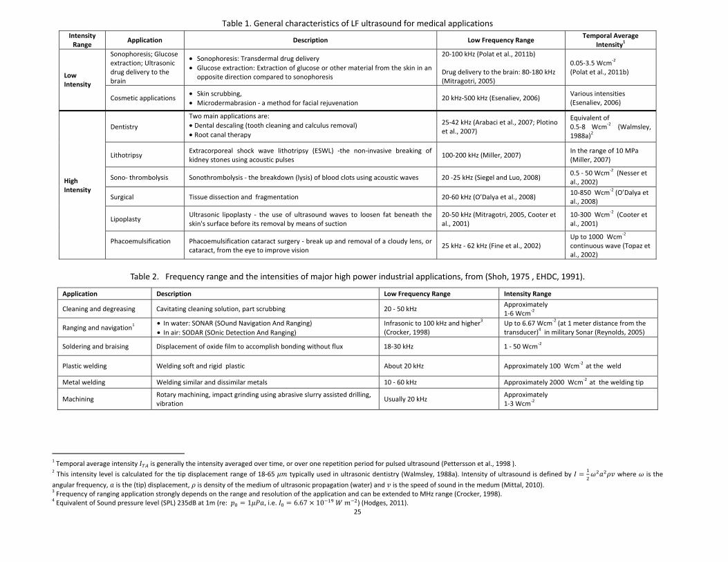
25
Table 1. General characteristics of LF ultrasound for medical applications Intensity
Range Application Description Low Frequency Range
Temporal Average Intensity1
Low Intensity
Sonophoresis; Glucose extraction; Ultrasonic drug delivery to the brain
• Sonophoresis: Transdermal drug delivery • Glucose extraction: Extraction of glucose or other material from the skin in an
opposite direction compared to sonophoresis
20-100 kHz (Polat et al., 2011b) Drug delivery to the brain: 80-180 kHz (Mitragotri, 2005)
0.05-3.5 Wcm-2
(Polat et al., 2011b)
Cosmetic applications • Skin scrubbing, • Microdermabrasion - a method for facial rejuvenation
20 kHz-500 kHz (Esenaliev, 2006) Various intensities (Esenaliev, 2006)
High Intensity
Dentistry
Two main applications are:• Dental descaling (tooth cleaning and calculus removal) • Root canal therapy
25-42 kHz (Arabaci et al., 2007; Plotino et al., 2007)
Equivalent of 0.5-8 Wcm-2 (Walmsley, 1988a)2
Lithotripsy Extracorporeal shock wave lithotripsy (ESWL) -the non-invasive breaking of kidney stones using acoustic pulses
100-200 kHz (Miller, 2007) In the range of 10 MPa (Miller, 2007)
Sono- thrombolysis Sonothrombolysis - the breakdown (lysis) of blood clots using acoustic waves 20 -25 kHz (Siegel and Luo, 2008) 0.5 - 50 Wcm-2 (Nesser et al., 2002)
Surgical Tissue dissection and fragmentation 20-60 kHz (O’Dalya et al., 2008) 10-850 Wcm-2 (O’Dalya et al., 2008)
Lipoplasty Ultrasonic lipoplasty - the use of ultrasound waves to loosen fat beneath the skin's surface before its removal by means of suction
20-50 kHz (Mitragotri, 2005, Cooter et al., 2001)
10-300 Wcm-2 (Cooter et al., 2001)
Phacoemulsification
Phacoemulsification cataract surgery - break up and removal of a cloudy lens, or cataract, from the eye to improve vision
25 kHz - 62 kHz (Fine et al., 2002) Up to 1000 Wcm-2
continuous wave (Topaz et al., 2002)
Table 2. Frequency range and the intensities of major high power industrial applications, from (Shoh, 1975 , EHDC, 1991).
Application Description Low Frequency Range Intensity Range
Cleaning and degreasing Cavitating cleaning solution, part scrubbing 20 - 50 kHz Approximately 1-6 Wcm-2
Ranging and navigation1 • In water: SONAR (SOund Navigation And Ranging) • In air: SODAR (SOnic Detection And Ranging)
Infrasonic to 100 kHz and higher3 (Crocker, 1998)
Up to 6.67 Wcm-2 (at 1 meter distance from the transducer)4 in military Sonar (Reynolds, 2005)
Soldering and braising Displacement of oxide film to accomplish bonding without flux 18-30 kHz 1 - 50 Wcm-2
Plastic welding Welding soft and rigid plastic About 20 kHz Approximately 100 Wcm-2 at the weld
Metal welding Welding similar and dissimilar metals 10 - 60 kHz Approximately 2000 Wcm-2 at the welding tip
Machining Rotary machining, impact grinding using abrasive slurry assisted drilling, vibration
Usually 20 kHz Approximately 1-3 Wcm-2
1 Temporal average intensity is generally the intensity averaged over time, or over one repetition period for pulsed ultrasound (Pettersson et al., 1998 ). 2 This intensity level is calculated for the tip displacement range of 18-65 typically used in ultrasonic dentistry (Walmsley, 1988a). Intensity of ultrasound is defined by where is the
angular frequency, is the (tip) displacement, is density of the medium of ultrasonic propagation (water) and is the speed of sound in the medum (Mittal, 2010). 3 Frequency of ranging application strongly depends on the range and resolution of the application and can be extended to MHz range (Crocker, 1998). 4 Equivalent of Sound pressure level (SPL) 235dB at 1m (re: 1 , i.e. 6.67 10 ) (Hodges, 2011).
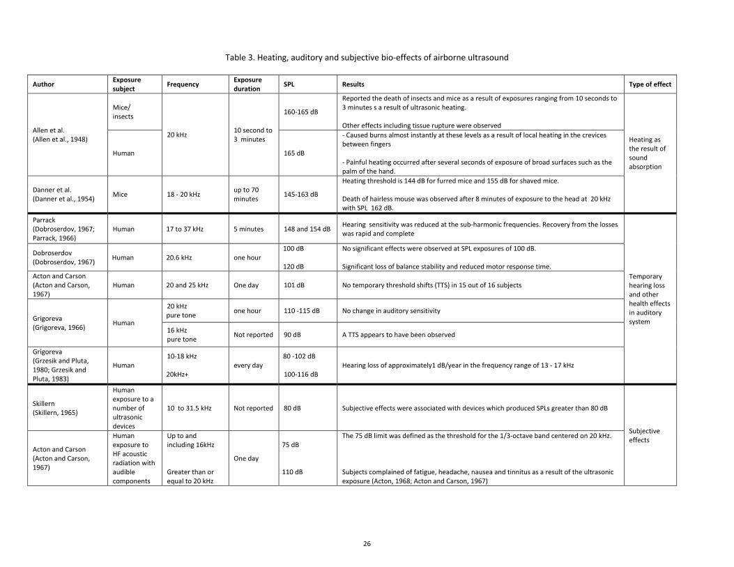
26
Table 3. Heating, auditory and subjective bio-effects of airborne ultrasound
Author Exposure subject
Frequency Exposure duration
SPL Results Type of effect
Allen et al. (Allen et al., 1948)
Mice/ insects
20 kHz 10 second to 3 minutes
160-165 dB
Reported the death of insects and mice as a result of exposures ranging from 10 seconds to 3 minutes s a result of ultrasonic heating. Other effects including tissue rupture were observed
Heating as the result of sound absorption
Human 165 dB
- Caused burns almost instantly at these levels as a result of local heating in the crevices between fingers - Painful heating occurred after several seconds of exposure of broad surfaces such as the palm of the hand.
Danner et al. (Danner et al., 1954)
Mice 18 - 20 kHz up to 70 minutes
145-163 dB
Heating threshold is 144 dB for furred mice and 155 dB for shaved mice. Death of hairless mouse was observed after 8 minutes of exposure to the head at 20 kHz with SPL 162 dB.
Parrack (Dobroserdov, 1967; Parrack, 1966)
Human 17 to 37 kHz 5 minutes 148 and 154 dB Hearing sensitivity was reduced at the sub-harmonic frequencies. Recovery from the losses was rapid and complete
Temporary hearing loss and other health effects in auditory system
Dobroserdov (Dobroserdov, 1967)
Human
20.6 kHz one hour 100 dB 120 dB
No significant effects were observed at SPL exposures of 100 dB. Significant loss of balance stability and reduced motor response time.
Acton and Carson (Acton and Carson, 1967)
Human 20 and 25 kHz One day 101 dB No temporary threshold shifts (TTS) in 15 out of 16 subjects
Grigoreva (Grigoreva, 1966)
Human
20 kHz pure tone
one hour 110 -115 dB No change in auditory sensitivity
16 kHz pure tone
Not reported 90 dB A TTS appears to have been observed
Grigoreva (Grzesik and Pluta, 1980; Grzesik and Pluta, 1983)
Human 10-18 kHz 20kHz+
every day 80 -102 dB 100-116 dB
Hearing loss of approximately1 dB/year in the frequency range of 13 - 17 kHz
Skillern (Skillern, 1965)
Human exposure to a number of ultrasonic devices
10 to 31.5 kHz Not reported 80 dB Subjective effects were associated with devices which produced SPLs greater than 80 dB
Subjective effects
Acton and Carson (Acton and Carson, 1967)
Human exposure to HF acoustic radiation with audible components
Up to and including 16kHz Greater than or equal to 20 kHz
One day
75 dB
110 dB
The 75 dB limit was defined as the threshold for the 1/3-octave band centered on 20 kHz. Subjects complained of fatigue, headache, nausea and tinnitus as a result of the ultrasonic exposure (Acton, 1968; Acton and Carson, 1967)
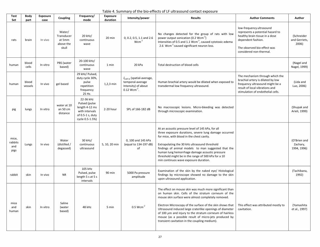
27
Table 4. Summary of the bio-effects of LF ultrasound contact exposure Test Set
Body part
Exposure case
Coupling Frequency/
mode Exposure duration
Intensity/power Results Author Comments Author
rats brain In vivo
Water/ Transducer
at 5mm above the
skull
20 kHz/ continuous
wave
20 min
0, 0.2, 0.5, 1.1 and 2.6 Wcm-2
No changes detected for the group of rats with low power output sonication (0.2 Wcm-2) Intensities of 0.5 and 1.1 Wcm-2, caused cytotoxic edema 2.6 Wcm-2 caused significant neuron loss.
low-frequency ultrasound represents a potential hazard to healthy brain tissue in a dose dependent fashion. The observed bio-effect was considered non-thermal.
(Schneider and Gerriets,
2006)
human blood cells
In vitro PBS (water
based)
20-100 kHz/ continuous
wave 1 min 20 kPa Total destruction of blood cells
(Nagel and Nagel, 1999)
human blood
vessels In vivo gel based
29 kHz/ Pulsed, duty cycle 30%,
pulse repetition frequency
25 Hz.
1,2,3 min
(spatial-average, temporal-average Intensity) of about 0.12 Wcm-2.
Human brachial artery would be dilated when exposed to transdermal low frequency ultrasound.
The mechanism through which the brachial artery is dilated by low frequency ultrasound might be a result of local vibrations and stimulation of endothelial cells.
(Lida and Luo, 2006)
pig lungs In vitro water at 10
an 50 cm distance
22-36 kHz Pulsed (pulse
length 4-12 ms with intervals
of 0.5-1 s, duty cycle 0.5-1.5%)
2-20 hour SPL of 166-182 dB No macroscopic lesions. Micro-bleeding was detected through microscopic examination.
(Shupak and Arieli, 1999)
mice, rabbits
and pigs
Lungs In vivo Water
(distilled / degassed)
30 kHz/ continuous ultrasound
5, 10, 20 min 0, 100 and 145 kPa
(equal to 134-197 dB) of
At an acoustic pressure level of 145 kPa, for all three exposure durations, severe lung damage occurred for mice, with blood in the chest cavity. Extrapolating the 30 kHz ultrasound threshold findings of animal models to man suggested that the human lung hemorrhage damage acoustic pressure threshold might be in the range of 500 kPa for a 10 min continuos wave exposure duration.
(O'Brien and Zachary,
1994, 1996)
rabbit skin In vivo NR
105 kHz Pulsed, pulse
length 5 s at 5 s intervals
90 min
5000 Pa pressure amplitude
Examination of the skin by the naked eye/ Histological findings by microscope showed no damage to the skin upon ultrasound application.
(Tachibana, 1992)
mice and
human skin In vitro
Saline (water based)
48 kHz 5 min 0.5 Wcm-2
The effect on mouse skin was much more significant than on human skin. Cells of the stratum corneum of the mouse skin surface were almost completely removed. Electron Microscopy of the surface of the skin shows that Ultrasound induced large craterlike openings of diameter of 100 and injury to the stratum corneum of hairless mouse (as a possible result of micro-jets produced by transient cavitation in the coupling medium).
This effect was attributed mostly to cavitation.
(Yamashita et al., 1997)
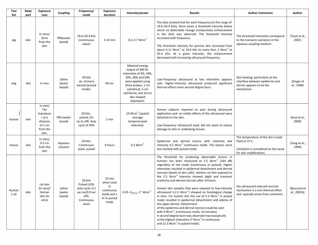
28
Test Set
Body part
Exposure case
Coupling Frequency/
mode Exposure duration
Intensity/power Results Author Comments Author
pig skin
In vitro/ 3mm
from the skin
PBS(water based)
19.6–93.4 kHz (continuous
wave) 2-14 min 0.2–2.7 Wcm-2
The data showed that for each frequency (in the range of 19.6–93.4 kHz), there exists a threshold intensity below which no detectable change (conductivity enhancement in the skin) was observed. The threshold intensity increased with frequency. The threshold intensity for porcine skin increased from about 0.11 Wcm-2 at 19.6 kHz to more than 2 Wcm-2 at 93.4 kHz. At a given intensity, the enhancement decreased with increasing ultrasound frequency.
The threshold intensities correspond to the transient cavitation in the aqueous coupling medium.
(Tezel et al., 2001)
dog skin In vivo Saline (water based)
20 kHz .6s of every
second (pulsed mode).
60 sec
Maximal energy output of 400 W.
intensities of 4%, 10%, 20%, 30% and 50% were applied using three probes, 1-cm
cylindrical, 5-cm cylindrical, and 10-cm
disc-shaped (diameter).
Low-frequency ultrasound at low intensities appears safe. Higher-intensity ultrasound produced significant thermal effects even second degree burn.
Skin heating, particularly at the interface between epidermis and dermis appears to be the mechanism.
(Singer et al., 1998)
human skin
In vivo/ The
transducer at a
distance of 1 cm
from the skin.
PBS (water based)
20 kHz , pulsed, (5s
on,5s off) duty cycle of 50%
2 min
10 Wcm-2 (spatial average
temporal peak intensity).
Human subjects reported no pain during ultrasound application and no visible effects of the ultrasound were detected on the skin. Low-frequency ultrasound used, did not seem to induce damage to skin or underlying tissues.
(Kost et al.,
2000)
mouse skin
In vitro/ 0.5 cm
from the skin
Aqueous solution
20 kHz, Continuous
wave, pulsed 4 hours 0.2 Wcm-2
Epidermal and dermal lesions with relatively low intensity 0.2 Wcm-2 continuous mode. The lesions were less marked with pulsed mode
The temperature of the skin is kept fixed at 37 C. Cavitation is considered as the cause for skin modifications.
(Fang et al., 1999)
Human/ rat
skin
rat skin (In vivo)/ Human skin (In vitro)
Saline (water based)
20 kHz Pulsed (10%
duty cycle: 0.1 sec on/0.9 sec
off); Continuous
wave
10 min when used
in continuous mode and 1 hr in pulsed
mode
0.25 < <7 Wcm-2
The threshold for producing observable lesions in humans has been measured as 2.5 Wcm-2 (164 dB) regardless of the mode (continuous or pulsed). Higher intensities resulted in epidermal detachment and dermal necrosis (death of skin cells). Hairless rat skin exposed to the 2.5 Wcm-2 intensity showed slight and transient erythema and dermal necrosis after 24 hours Human skin samples that were exposed to low-intensity ultrasound (<2.5 Wcm-2) showed no histological change in vitro. For human skin the use of 5.2 Wcm-2 in pulsed mode resulted in epidermal detachment and edema of the upper dermis. Detachment of the epidermis and dermal necrosis could be seen with 4 Wcm-2, (continuous mode, 10 minutes). A second-degree burn was observed macroscopically at the highest intensities (7 Wcm-2 in continuous and 12.3 Wcm-2 in pulsed mode).
the ultrasound-induced necrosis mechanism is a non-thermal effect and typically arises from cavitation.
(Boucaud et al., 2001b)
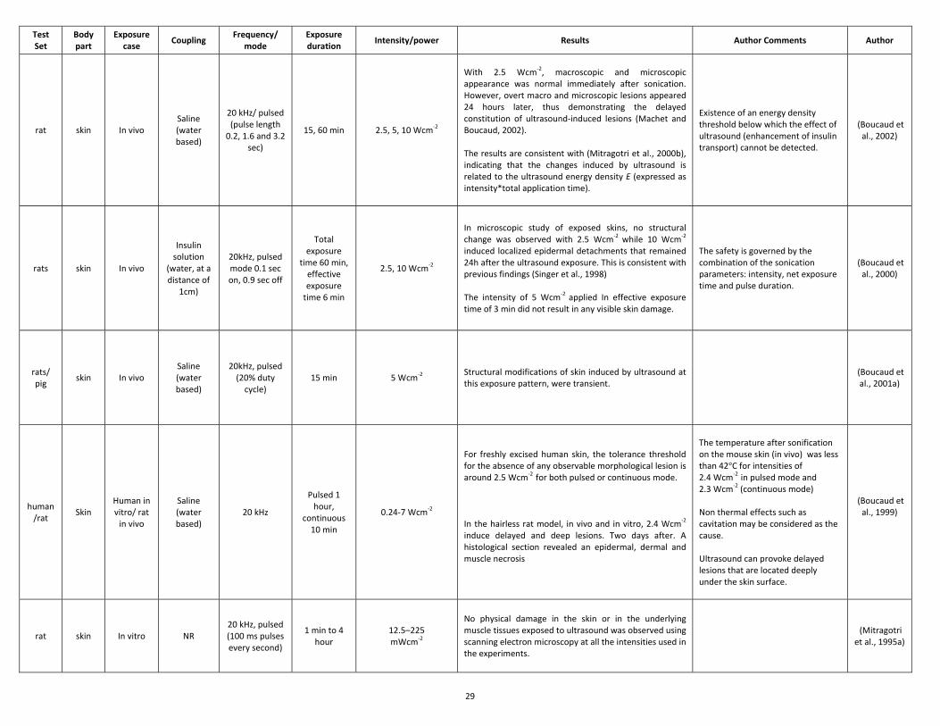
29
Test Set
Body part
Exposure case
Coupling Frequency/
mode Exposure duration
Intensity/power Results Author Comments Author
rat skin In vivo Saline (water based)
20 kHz/ pulsed (pulse length
0.2, 1.6 and 3.2 sec)
15, 60 min 2.5, 5, 10 Wcm-2
With 2.5 Wcm-2, macroscopic and microscopic appearance was normal immediately after sonication. However, overt macro and microscopic lesions appeared 24 hours later, thus demonstrating the delayed constitution of ultrasound-induced lesions (Machet and Boucaud, 2002). The results are consistent with (Mitragotri et al., 2000b), indicating that the changes induced by ultrasound is related to the ultrasound energy density E (expressed as intensity*total application time).
Existence of an energy density threshold below which the effect of ultrasound (enhancement of insulin transport) cannot be detected.
(Boucaud et al., 2002)
rats skin In vivo
Insulin solution
(water, at a distance of
1cm)
20kHz, pulsed mode 0.1 sec on, 0.9 sec off
Total exposure
time 60 min, effective exposure
time 6 min
2.5, 10 Wcm-2
In microscopic study of exposed skins, no structural change was observed with 2.5 Wcm-2 while 10 Wcm-2
induced localized epidermal detachments that remained 24h after the ultrasound exposure. This is consistent with previous findings (Singer et al., 1998) The intensity of 5 Wcm-2 applied In effective exposure time of 3 min did not result in any visible skin damage.
The safety is governed by the combination of the sonication parameters: intensity, net exposure time and pulse duration.
(Boucaud et al., 2000)
rats/ pig
skin In vivo Saline (water based)
20kHz, pulsed (20% duty
cycle) 15 min 5 Wcm-2
Structural modifications of skin induced by ultrasound at this exposure pattern, were transient.
(Boucaud et al., 2001a)
human/rat
Skin Human in vitro/ rat
in vivo
Saline (water based)
20 kHz
Pulsed 1 hour,
continuous 10 min
0.24-7 Wcm-2
For freshly excised human skin, the tolerance threshold for the absence of any observable morphological lesion is around 2.5 Wcm-2 for both pulsed or continuous mode. In the hairless rat model, in vivo and in vitro, 2.4 Wcm-2
induce delayed and deep lesions. Two days after. A histological section revealed an epidermal, dermal and muscle necrosis
The temperature after sonification on the mouse skin (in vivo) was less than 42°C for intensities of 2.4 Wcm-2 in pulsed mode and 2.3 Wcm-2 (continuous mode) Non thermal effects such as cavitation may be considered as the cause. Ultrasound can provoke delayed lesions that are located deeply under the skin surface.
(Boucaud et al., 1999)
rat skin In vitro NR 20 kHz, pulsed (100 ms pulses every second)
1 min to 4 hour
12.5–225 mWcm-2
No physical damage in the skin or in the underlying muscle tissues exposed to ultrasound was observed using scanning electron microscopy at all the intensities used in the experiments.
(Mitragotri
et al., 1995a)
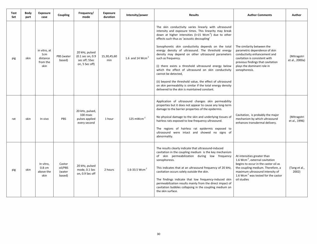
30
Test Set
Body part
Exposure case
Coupling Frequency/
mode Exposure duration
Intensity/power Results Author Comments Author
pig skin
in vitro, at 1cm
distance from the
skin
PBS (water based)
20 kHz, pulsed (0.1 sec on, 0.9
sec off; 5Sec on, 5 Sec off)
15,30,45,60 min
1.6 and 14 Wcm-2
The skin conductivity varies linearly with ultrasound intensity and exposure times. This linearity may break down at higher intensities (I>15 Wcm-2) due to other effects such thus as ‘acoustic decoupling’ Sonophoretic skin conductivity depends on the total energy density of ultrasound. The threshold energy density may depend on other ultrasound parameters such as frequency. (i) there exists a threshold ultrasound energy below which the effect of ultrasound on skin conductivity cannot be detected, (ii) beyond the threshold value, the effect of ultrasound on skin permeability is similar if the total energy density delivered to the skin is maintained constant.
The similarity between the parametric dependence of skin conductivity enhancement and cavitation is consistent with previous findings that cavitation plays the dominant role in sonophoresis.
(Mitragotri et al., 2000a)
rat skin In vivo PBS
20 kHz, pulsed, 100 msec
pulses applied every second
1 hour 125 mWcm-2
Application of ultrasound changes skin permeability properties but it does not appear to cause any long-term damage to the barrier properties of the epidermis. No physical damage to the skin and underlying tissues of hairless rats exposed to low-frequency ultrasound. The regions of hairless rat epidermis exposed to ultrasound were intact and showed no signs of abnormality.
Cavitation, is probably the major mechanism by which ultrasound enhances transdermal delivery.
(Mitragotri et al., 1996)
pig skin
In vitro, 0.8 cm
above the skin
Castor oil/PBS (water based)
20 kHz, pulsed mode, 0.1 Sec on, 0.9 Sec off
2 hours 1.6-33.5 Wcm-2
The results clearly indicate that ultrasound-induced cavitation in the coupling medium is the key mechanism of skin permeabilization during low frequency sonophoresis. This indicates that at an ultrasound frequency of 20 kHz, cavitation occurs solely outside the skin. The findings indicate that low frequency-induced skin permeabilization results mainly from the direct impact of cavitation bubbles collapsing in the coupling medium on the skin surface.
At intensities greater than 1.6 Wcm-2, external cavitation begins to occur in the castor oil as the coupling medium. Therefore, a maximum ultrasound intensity of 1.6 Wcm-2 was tested for the castor oil studies
(Tang et al., 2002)
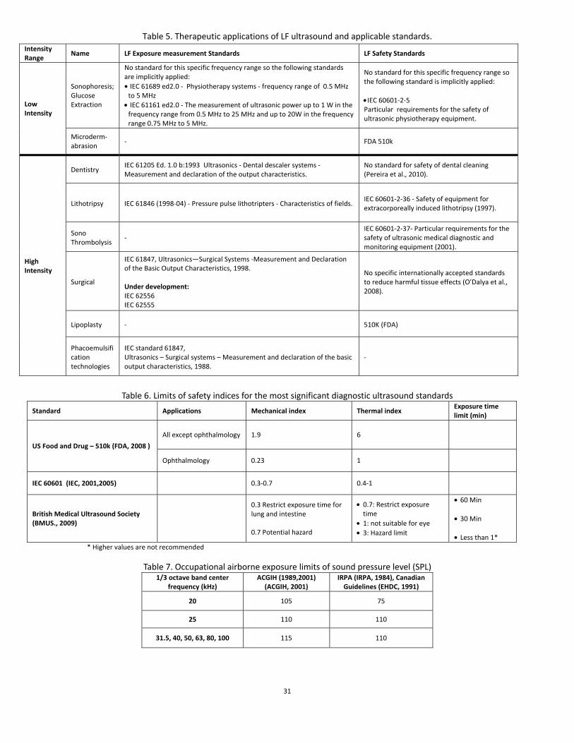
31
Table 5. Therapeutic applications of LF ultrasound and applicable standards. Intensity Range
Name LF Exposure measurement Standards LF Safety Standards
Low Intensity
Sonophoresis; Glucose Extraction
No standard for this specific frequency range so the following standards are implicitly applied: • IEC 61689 ed2.0 - Physiotherapy systems - frequency range of 0.5 MHz
to 5 MHz • IEC 61161 ed2.0 - The measurement of ultrasonic power up to 1 W in the
frequency range from 0.5 MHz to 25 MHz and up to 20W in the frequency range 0.75 MHz to 5 MHz.
No standard for this specific frequency range so the following standard is implicitly applied: • IEC 60601-2-5 Particular requirements for the safety of ultrasonic physiotherapy equipment.
Microderm-abrasion
- FDA 510k
High Intensity
Dentistry IEC 61205 Ed. 1.0 b:1993 Ultrasonics - Dental descaler systems - Measurement and declaration of the output characteristics.
No standard for safety of dental cleaning (Pereira et al., 2010).
Lithotripsy IEC 61846 (1998-04) - Pressure pulse lithotripters - Characteristics of fields. IEC 60601-2-36 - Safety of equipment for extracorporeally induced lithotripsy (1997).
Sono Thrombolysis
- IEC 60601-2-37- Particular requirements for the safety of ultrasonic medical diagnostic and monitoring equipment (2001).
Surgical
IEC 61847, Ultrasonics—Surgical Systems -Measurement and Declaration of the Basic Output Characteristics, 1998. Under development: IEC 62556 IEC 62555
No specific internationally accepted standards to reduce harmful tissue effects (O’Dalya et al., 2008).
Lipoplasty - 510K (FDA)
Phacoemulsification technologies
IEC standard 61847, Ultrasonics – Surgical systems – Measurement and declaration of the basic output characteristics, 1988.
-
Table 6. Limits of safety indices for the most significant diagnostic ultrasound standards
Standard Applications Mechanical index Thermal index Exposure time limit (min)
US Food and Drug – 510k (FDA, 2008 )
All except ophthalmology 1.9 6
Ophthalmology 0.23 1
IEC 60601 (IEC, 2001,2005) 0.3-0.7 0.4-1
British Medical Ultrasound Society (BMUS., 2009)
0.3 Restrict exposure time for lung and intestine 0.7 Potential hazard
• 0.7: Restrict exposure time
• 1: not suitable for eye • 3: Hazard limit
• 60 Min • 30 Min • Less than 1*
* Higher values are not recommended
Table 7. Occupational airborne exposure limits of sound pressure level (SPL) 1/3 octave band center
frequency (kHz) ACGIH (1989,2001)
(ACGIH, 2001) IRPA (IRPA, 1984), Canadian
Guidelines (EHDC, 1991)
20 105 75
25 110 110
31.5, 40, 50, 63, 80, 100 115 110
32
Fig. 1. Cavitation threshold of water as a function of initial nucleus radius of air bubbles for four ultrasonic frequencies of 20, 100, 300 and 500 kHz. The assumptions of the calculations are those of (Apfel and Holland, 1991; 1989) : initial bubble temperature of 300K, growth of bubbles in a single cycle of ultrasound and adiabatic collapse at a temperature of 5000K.
Fig. 2. Variation of the minimum cavitation threshold for optimum sized bubbles, in water and in blood for frequency range of 20-500 kHz using the theory of Holland and Apfel (Holland and Apfel, 1989) and assuming all nuclei sizes are present. The square and circle marks, show the calculated values of threshold. Solid lines represent least square fit of the calculated data to power law A with A=0.10, B=0.216, n=0.6 for water and A=0.11, B=0.26, n=0.68 for the blood. Dashed lines represent the least square fit to / with B= 0.38 for water and B=0.41 for blood.
Fig. 3. Variation of the mechanical index based on the classic ,equation (7), and modified definition, equation (12), for: a) LF and therapeutic ultrasonic range of 20 kHz to 3 MHz in water. b) Frequency range of 20 kHz -15 MHz in water and blood.
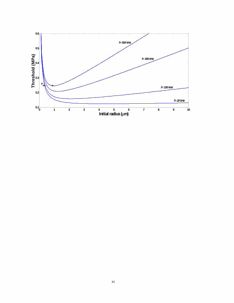
33
0 1 2 3 4 5 6 7 8 9 100.1
0.2
0.3
0.4
0.5
0.6
Initial radius (μm)
Thre
shol
d (M
Pa)
Popt
f= 20 kHz
f= 100 kHz
f= 300 kHz
f= 500 kHz
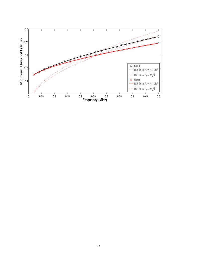
34
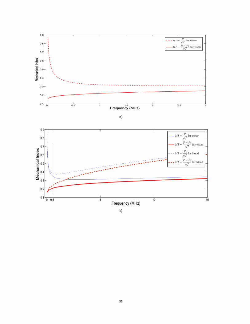
35
a)
b)