Embed Size (px)
Citation preview
5/21/2021
1
Bio 1102 Lecture 5 Chapter 8: Heart & Blood Vessels
• Functions of Circulatory System:
– To carry oxygen from lungs to all cells, tissues, and organs of body
– To carry nutrients from digestive system to all cells, tissues and organs of body
• The heart is the organ that pumps blood through the body
• Heart is located between the lungs
• Three layers of the heart: – Pericardium: thin outer layer; protects & anchors
– Myocardium: thick middle layer; cardiac muscles
– Endocardium: thin inner layer; endothelial lining
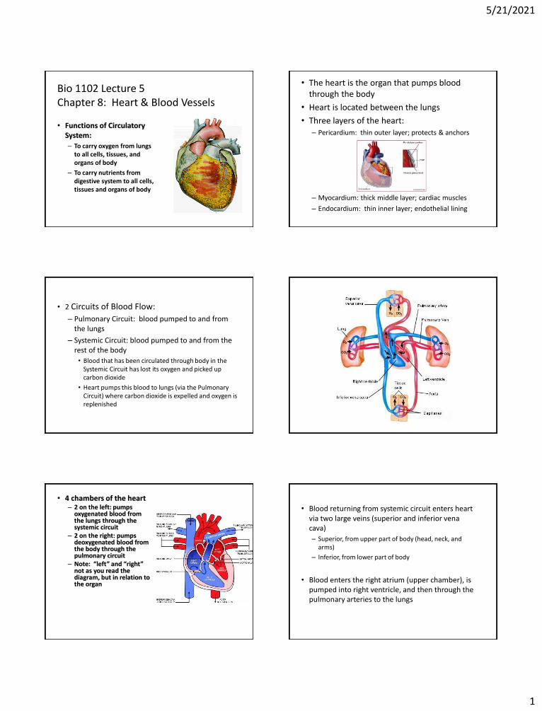
• 2 Circuits of Blood Flow:
– Pulmonary Circuit: blood pumped to and from the lungs
– Systemic Circuit: blood pumped to and from the rest of the body
• Blood that has been circulated through body in the Systemic Circuit has lost its oxygen and picked up carbon dioxide
• Heart pumps this blood to lungs (via the Pulmonary Circuit) where carbon dioxide is expelled and oxygen is replenished
• 4 chambers of the heart – 2 on the left: pumps
oxygenated blood from the lungs through the systemic circuit
– 2 on the right: pumps deoxygenated blood from the body through the pulmonary circuit
– Note: “left” and “right” not as you read the diagram, but in relation to the organ
• Blood returning from systemic circuit enters heart via two large veins (superior and inferior vena cava)
– Superior, from upper part of body (head, neck, and arms)
– Inferior, from lower part of body
• Blood enters the right atrium (upper chamber), is pumped into right ventricle, and then through the pulmonary arteries to the lungs
5/21/2021
2
• Blood is oxygenated in lungs
• Blood returns to left atrium of heart via pulmonary veins
– Systemic System begins
• Blood enters left ventricle
– Thicker-walled than right ventricle
– Pumps blood to rest of body, via the aorta • Largest artery in body
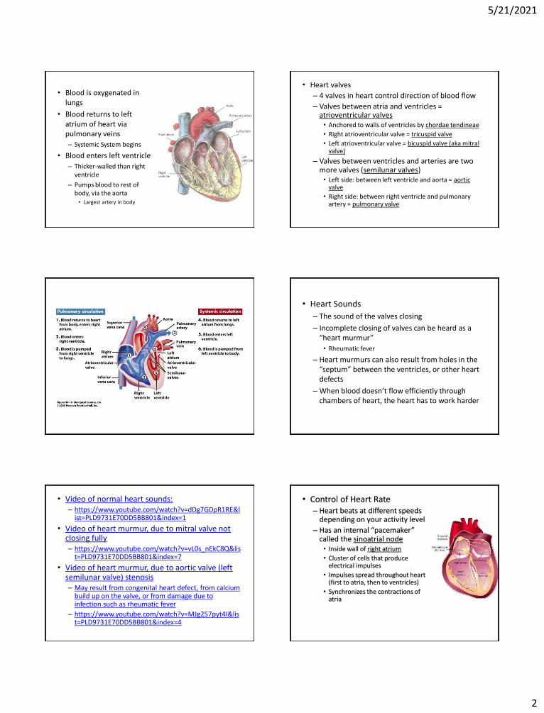
• Heart valves
– 4 valves in heart control direction of blood flow
– Valves between atria and ventricles = atrioventricular valves • Anchored to walls of ventricles by chordae tendineae
• Right atrioventricular valve = tricuspid valve
• Left atrioventricular valve = bicuspid valve (aka mitral valve)
– Valves between ventricles and arteries are two more valves (semilunar valves) • Left side: between left ventricle and aorta = aortic
valve
• Right side: between right ventricle and pulmonary artery = pulmonary valve
• Heart Sounds
– The sound of the valves closing
– Incomplete closing of valves can be heard as a “heart murmur”
• Rheumatic fever
– Heart murmurs can also result from holes in the “septum” between the ventricles, or other heart defects
– When blood doesn’t flow efficiently through chambers of heart, the heart has to work harder
• Video of normal heart sounds: – https://www.youtube.com/watch?v=dDg7GDpR1RE&l
ist=PLD9731E70DD5BB801&index=1
• Video of heart murmur, due to mitral valve not closing fully – https://www.youtube.com/watch?v=vL0s_nEkC8Q&lis
t=PLD9731E70DD5BB801&index=7
• Video of heart murmur, due to aortic valve (left semilunar valve) stenosis – May result from congenital heart defect, from calcium
build up on the valve, or from damage due to infection such as rheumatic fever
– https://www.youtube.com/watch?v=MJg257pyt4I&list=PLD9731E70DD5BB801&index=4
• Control of Heart Rate – Heart beats at different speeds
depending on your activity level
– Has an internal “pacemaker” called the sinoatrial node • Inside wall of right atrium
• Cluster of cells that produce electrical impulses
• Impulses spread throughout heart (first to atria, then to ventricles)
• Synchronizes the contractions of atria

5/21/2021
3
– Signal then travels to another cluster of special cells, the atrioventricular node • Signal then is distributed to the
ventricles by the atrioventricular bundles (in the septum) and then the Purkinje bundles (see figure 8.13)
– Without some regulation, SA node would cause heart to beat 100 times per minute at rest – too fast
– Heart control center of brain (medulla oblongata) slows down the SA node
• On average, impulses from brain slow heart rate to about 70 beats per minute (at rest)
• When active, impulses from brain are reduced, so heart rate speeds up
• Other nerves and hormones (e.g. adrenaline) can also work to increase heart rate (up to about 180 beats per minute)
• Heart beat rate is regulated to meet the needs of the body’s cells for oxygen – Maximum heart beat rate = 220-your age – Generally don’t want to exceed 85% of your maximum
during strenuous exercise – Tachycardia = when your heart beats too fast
• Heart Attacks
– Most common type: myocardial infarction • Caused by blood clots in
arteries supplying blood to the heart
• Often associated with arteries narrowed by plaque
• Lack of flow to heart starves muscles of oxygen and nutrients, killing cells
– The damaged area is called an infarct
• What causes plaque formation?
– Poor diet
– Smoking
– Lack of exercise
– Heredity
– Stress
• Severity of the heart attack depends on how much of the heart muscle is damaged
• Warnings of Heart Attack
– May occur without warning
– Chest pain (angina) may precede heart attack for several weeks • Caused by reduced blood flow to heart
• Pain in center of chest, spreading to throat, jaw, back, and arms
• Occurs when person is active, under stress, or exposed to carbon monoxide
• Taking an aspirin during a heart attack can reduce damage
– Aspirin reduces clotting
• Other causes of heart attack
– Loss of control of heart muscle by sinoatrial (SA) node
• Heart muscles thus beat independently, reducing flow of blood through heart (= fibrillation)
• Heart stops beating = cardiac arrest
• Heart beat may be restored by applying strong electrical current to chest (defibrillation), or by cardiopulmonary resuscitation (CPR) – Applying pressure to breastbone, massaging the heart
5/21/2021
4
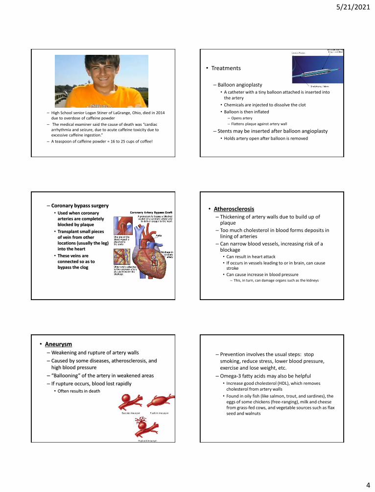
– High School senior Logan Stiner of LaGrange, Ohio, died in 2014 due to overdose of caffeine powder
– The medical examiner said the cause of death was “cardiac arrhythmia and seizure, due to acute caffeine toxicity due to excessive caffeine ingestion.”
– A teaspoon of caffeine powder = 16 to 25 cups of coffee!
• Treatments
– Balloon angioplasty
• A catheter with a tiny balloon attached is inserted into the artery
• Chemicals are injected to dissolve the clot
• Balloon is then inflated – Opens artery
– Flattens plaque against artery wall
– Stents may be inserted after balloon angioplasty
• Holds artery open after balloon is removed
– Coronary bypass surgery
• Used when coronary arteries are completely blocked by plaque
• Transplant small pieces of vein from other locations (usually the leg) into the heart
• These veins are connected so as to bypass the clog
• Atherosclerosis – Thickening of artery walls due to build up of
plaque
– Too much cholesterol in blood forms deposits in lining of arteries
– Can narrow blood vessels, increasing risk of a blockage • Can result in heart attack
• If occurs in vessels leading to or in brain, can cause stroke
• Can cause increase in blood pressure – This, in turn, can damage organs such as the kidneys
• Aneurysm
– Weakening and rupture of artery walls
– Caused by some diseases, atherosclerosis, and high blood pressure
– “Ballooning” of the artery in weakened areas
– If rupture occurs, blood lost rapidly
• Often results in death
– Prevention involves the usual steps: stop smoking, reduce stress, lower blood pressure, exercise and lose weight, etc.
– Omega-3 fatty acids may also be helpful
• Increase good cholesterol (HDL), which removes cholesterol from artery walls
• Found in oily fish (like salmon, trout, and sardines), the eggs of some chickens (free-ranging), milk and cheese from grass-fed cows, and vegetable sources such as flax seed and walnuts
5/21/2021
5
Blood Vessels
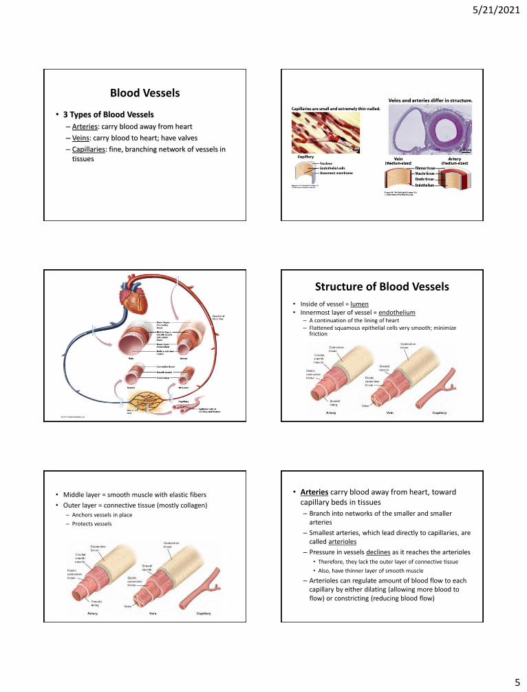
• 3 Types of Blood Vessels
– Arteries: carry blood away from heart
– Veins: carry blood to heart; have valves
– Capillaries: fine, branching network of vessels in tissues
Structure of Blood Vessels
• Inside of vessel = lumen • Innermost layer of vessel = endothelium
– A continuation of the lining of heart – Flattened squamous epithelial cells very smooth; minimize
friction
• Middle layer = smooth muscle with elastic fibers
• Outer layer = connective tissue (mostly collagen)
– Anchors vessels in place
– Protects vessels
• Arteries carry blood away from heart, toward capillary beds in tissues
– Branch into networks of the smaller and smaller arteries
– Smallest arteries, which lead directly to capillaries, are called arterioles
– Pressure in vessels declines as it reaches the arterioles
• Therefore, they lack the outer layer of connective tissue
• Also, have thinner layer of smooth muscle
– Arterioles can regulate amount of blood flow to each capillary by either dilating (allowing more blood to flow) or constricting (reducing blood flow)
5/21/2021
6
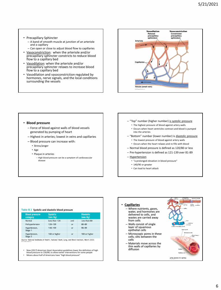
• Precapillary Sphincter – A band of smooth muscle at junction of an arteriole
and a capillary – Can open or close to adjust blood flow to capillaries
• Vasoconstriction: when the arteriole and/or precapillary sphincter constricts to reduce blood flow to a capillary bed
• Vasodilation: when the arteriole and/or precapillary sphincter relaxes to increase blood flow to a capillary bed
• Vasodilation and vasoconstriction regulated by hormones, nerve signals, and the local conditions surrounding the vessels
• Blood pressure
– Force of blood against walls of blood vessels generated by pumping of heart
– Highest in arteries; lowest in veins and capillaries
– Blood pressure can increase with:
• Stress/anger
• Age
• Plaque in arteries – High blood pressure can be a symptom of cardiovascular
disease
– “Top” number (higher number) is systolic pressure
• The highest pressure of blood against artery walls
• Occurs when heart ventricles contract and blood is pumped into the arteries
– “Bottom” number (lower number) is diastolic pressure
• The lowest pressure of blood against artery walls
• Occurs when the heart relaxes and re-fills with blood
– Normal blood pressure is defined as 120/80 or less
– Pre-hypertension is defined as 121-139 over 81-89
– Hypertension
• “a prolonged elevation in blood pressure”
• 140/90 or greater
• Can lead to heart attack
• New (2017) American Heart Association guidelines lower the definition of high blood pressure to 130/80, to allow earlier interventions for some people
• Means about half of Americans have “high blood pressure”
• Capillaries – Where nutrients, gases,
water, and hormones are delivered to cells, and wastes are carried away from cells
– Walls consist of single layer of squamous epithelial cells
– Microscopic pores in these cells; slits between the cells
– Materials move across the thin walls of capillaries by diffusion
5/21/2021
7
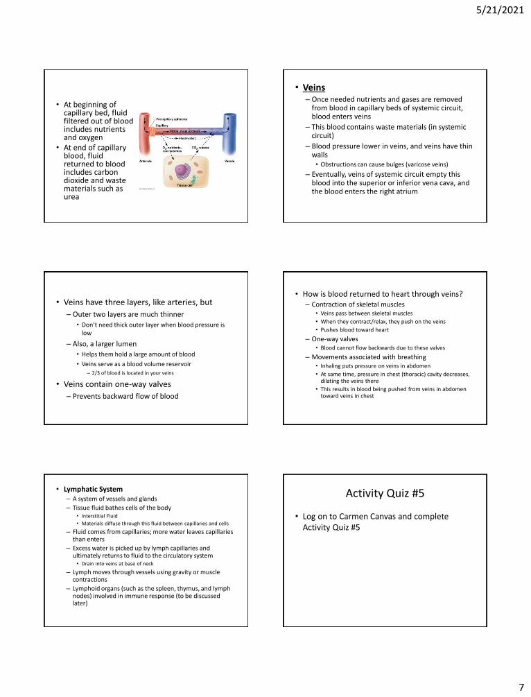
• At beginning of capillary bed, fluid filtered out of blood includes nutrients and oxygen
• At end of capillary blood, fluid returned to blood includes carbon dioxide and waste materials such as urea
• Veins – Once needed nutrients and gases are removed
from blood in capillary beds of systemic circuit, blood enters veins
– This blood contains waste materials (in systemic circuit)
– Blood pressure lower in veins, and veins have thin walls • Obstructions can cause bulges (varicose veins)
– Eventually, veins of systemic circuit empty this blood into the superior or inferior vena cava, and the blood enters the right atrium
• Veins have three layers, like arteries, but
– Outer two layers are much thinner
• Don’t need thick outer layer when blood pressure is low
– Also, a larger lumen
• Helps them hold a large amount of blood
• Veins serve as a blood volume reservoir – 2/3 of blood is located in your veins
• Veins contain one-way valves
– Prevents backward flow of blood
• How is blood returned to heart through veins? – Contraction of skeletal muscles
• Veins pass between skeletal muscles
• When they contract/relax, they push on the veins
• Pushes blood toward heart
– One-way valves • Blood cannot flow backwards due to these valves
– Movements associated with breathing • Inhaling puts pressure on veins in abdomen
• At same time, pressure in chest (thoracic) cavity decreases, dilating the veins there
• This results in blood being pushed from veins in abdomen toward veins in chest
• Lymphatic System – A system of vessels and glands
– Tissue fluid bathes cells of the body • Interstitial Fluid
• Materials diffuse through this fluid between capillaries and cells
– Fluid comes from capillaries; more water leaves capillaries than enters
– Excess water is picked up by lymph capillaries and ultimately returns to fluid to the circulatory system • Drain into veins at base of neck
– Lymph moves through vessels using gravity or muscle contractions
– Lymphoid organs (such as the spleen, thymus, and lymph nodes) involved in immune response (to be discussed later)
Activity Quiz #5
• Log on to Carmen Canvas and complete Activity Quiz #5


![Lecture 3 - Bio Molecules - Bio 105.pptx [Read-Only] 3 - Bio Mole… · 8/30/2013 15 Fatty Acids and Health Heart disease is caused by plaque collecting in the blood vessels leading](https://img.pdfslide.us/doc/110x75/5f389134930d3d0c5b65a799/lecture-3-bio-molecules-bio-105pptx-read-only-3-bio-mole-8302013-15.jpg)