Embed Size (px)
Citation preview

© 2015. Published by The Company of Biologists Ltd.
Binding partners of the kinase domains in Drosophila obscurin
and their effect on the structure of the flight muscle
Anja Katzemich1,, Ryan J. H. West1, Atsushi Fukuzawa2, Sean T. Sweeney1,
Mathias Gautel2, John Sparrow1 and Belinda Bullard1,‡
1Department of Biology, University of York, York, YO10 5DD, UK
2King’s College BHF Centre, Cardiovascular Division, London, SE1 1UL, UK Present address: Department of Biology, McGill University, Montreal, Quebec
1B1, Canada
‡Author for correspondence ([email protected])
Jour
nal o
f Cel
l Sci
ence
Acc
epte
d m
anus
crip
t
JCS Advance Online Article. Posted on 6 August 2015
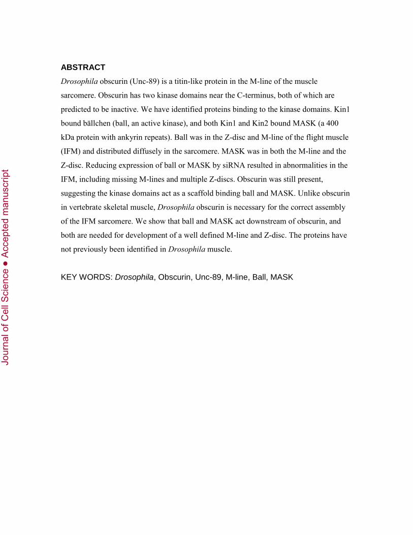
ABSTRACT
Drosophila obscurin (Unc-89) is a titin-like protein in the M-line of the muscle
sarcomere. Obscurin has two kinase domains near the C-terminus, both of which are
predicted to be inactive. We have identified proteins binding to the kinase domains. Kin1
bound bällchen (ball, an active kinase), and both Kin1 and Kin2 bound MASK (a 400
kDa protein with ankyrin repeats). Ball was in the Z-disc and M-line of the flight muscle
(IFM) and distributed diffusely in the sarcomere. MASK was in both the M-line and the
Z-disc. Reducing expression of ball or MASK by siRNA resulted in abnormalities in the
IFM, including missing M-lines and multiple Z-discs. Obscurin was still present,
suggesting the kinase domains act as a scaffold binding ball and MASK. Unlike obscurin
in vertebrate skeletal muscle, Drosophila obscurin is necessary for the correct assembly
of the IFM sarcomere. We show that ball and MASK act downstream of obscurin, and
both are needed for development of a well defined M-line and Z-disc. The proteins have
not previously been identified in Drosophila muscle.
KEY WORDS: Drosophila, Obscurin, Unc-89, M-line, Ball, MASK
Jour
nal o
f Cel
l Sci
ence
Acc
epte
d m
anus
crip
t

INTRODUCTION
A stable lattice of thick and thin filaments in striated muscle is needed to maintain the
optimum register of the filaments as the fibres contract. Thin filaments from
neighbouring sarcomeres are anchored in the Z-disc by -actinin and other cross-linking
proteins, and thick filaments are held in position by cross-links at the M-line in the
middle of the sarcomere (Agarkova and Perriard, 2005; Luther, 2009) The register of
thick filaments is also maintained by elastic links between the ends of the filaments and
the Z-disc (Horowits and Podolsky, 1987; Tskhovrebova and Trinick, 2003). Large
modular proteins of the titin family, associated with thick filaments, contribute to both
the stability and the stiffness of the sarcomere. These proteins are made up of tandem
immunoglobulin (Ig) and fibronectin-like (Fn3) domains and may have one, or
sometimes two, kinase domains near the C-terminus, and there may also be signalling
domains (Bullard et al., 2005; Labeit and Kolmerer, 1995; Linke, 2008; Russell et al.,
2002; Small et al., 2004; Young et al., 2001).
The M-line protein, obscurin, has a similar modular structure in invertebrates and
vertebrates, although the number of modules in different isoforms and the position of the
signalling domains varies. Both Unc-89 (the obscurin homologue in Caenorhabditis
elegans) and obscurin in Drosophila have SH3 and Rho-GEF signalling domains near the
N-terminus and two kinase domains near the C-terminus (Benian et al., 1996; Katzemich
et al., 2012; Small et al., 2004) In vertebrate obscurin, the signalling domains are near the
C-terminus; the isoform obscurin A has an ankyrin-binding domain instead of the two C-
terminal kinase domains in obscurin B. Both these isoforms are at the periphery of
myofibrils in the M-line region of mature skeletal fibres (Fukuzawa et al., 2008; Russell
et al., 2002; Young et al., 2001). Binding of obscurin A to ankyrins creates a link
between the sarcoplasmic reticulum (SR) and the myofibril (Bagnato et al., 2003;
Kontrogianni-Konstantopoulos et al., 2003; Lange et al., 2009). By contrast, Drosophila
obscurin is found throughout the M-line and there is no ankyrin-binding domain, so direct
binding to the SR is unlikely (Katzemich et al., 2012). However, in the nematode, loss-of
-function mutations of unc89 result in displaced ryanodine receptor and SERCA, as well
as abnormal calcium signalling (Spooner et al., 2012). This suggests a function for Unc-
Jour
nal o
f Cel
l Sci
ence
Acc
epte
d m
anus
crip
t

89 in calcium regulation involving the SR. So far, five large isoforms of obscurin have
been identified in Drosophila muscles: one expressed in the larva, and four expressed in
the pupa and adult. All these isoforms have Ig domains in the tandem Ig region, and at
least one of the kinase domains (Kin1). The indirect flight muscle (IFM) has two
isoforms: a major isoform of 475 kDa and a minor isoform that is somewhat smaller
(Katzemich et al., 2012). The two remaining isoforms are in other thoracic muscles.
Drosophila obscurin is essential for the formation of an M-line, and for the correct
assembly of thick and thin filaments in the sarcomere: lack of obscurin in the IFM results
in asymmetrical thick filaments and thin filaments of abnormal length and polarity.
Paradoxically, vertebrate obscurin is not necessary for normal sarcomere structure, since
an obscurin knockout in the mouse had no serious effect on sarcomere assembly or
maintenance (Lange et al., 2009).
The kinase domains of titin-like proteins often function as scaffolds binding other
proteins, and may or may not be active kinases (Endicott et al., 2012; Gautel, 2011a;
Mayans et al., 2013). In C. elegans Unc-89, kinase1 (PK1) is predicted to be inactive
because the ATP-binding site lacks essential residues. Unc-89 kinase 2 (PK2) may be
active, although a motif contributing to ATP-binding is atypical (Small et al., 2004). Both
Unc-89 kinase domains interact with SCPL-1 (small, C-terminal domain, phosphatase-
like 1), which is thought to be involved in muscle-specific signalling (Qadota et al.,
2008). Unc-89 PK1 also interacts with the LIM-domain protein, LIM-9; the complex of
PK1, SCPL-1 and LIM-9 links Unc-89 to integrin adhesion sites at the cell surface (Lin et
al., 2003; Warner et al., 2013; Xiong et al., 2009). In Drosophila, both obscurin Kin1 and
Kin2 are predicted to be inactive because the catalytic site lacks the catalytic aspartate
and other critical residues (Endicott et al., 2012; Mayans et al., 2013). Both kinase
domains in vertebrate obscurin B are predicted to be catalytically active, and can
apparently be auto-phosphorylated (Endicott et al., 2012; Hu and Kontrogianni-
Konstantopoulos, 2013; Mayans et al., 2013). The kinase domains are reported to interact
with membrane associated proteins: kinase 1(SK1) with the extracellular domain of a
Na+/K+-ATPase at adherens junctions, which is not a substrate, and SK2 with the cell-
adhesion molecule, N-cadherin, which is an in vitro substrate (Hu and Kontrogianni-
Konstantopoulos, 2013).
Jour
nal o
f Cel
l Sci
ence
Acc
epte
d m
anus
crip
t

The kinase domains in titin-like proteins have sequence at the C-terminus that
sterically blocks the active site (the C-terminal regulatory domain). This sequence may
inhibit an active kinase, or regulate ligand binding; it may also be part of the structure of
the kinase domain, and necessary to maintain the stability of the domain (Gautel, 2011a;
Mayans et al., 2013; von Castelmur et al., 2012). Titin-like kinases are linked to stretch-
activated signalling pathways in muscle. Mechano-sensing by the kinase can result in
changes in the C-terminal regulatory domain and transient binding of ligands to the
kinase scaffold. The precise mechanism of regulation varies in different species (Lange et
al., 2005; Mayans et al., 2013; Puchner et al., 2008; von Castelmur et al., 2012).
The aim of this study was to identify proteins binding to the two kinase domains
in Drosophila obscurin, and to determine the effect of the proteins on the assembly of an
ordered sarcomere in IFM. We show that ball (a protein kinase) binds to Kin1 and MASK
(an ankyrin repeat protein) binds to both Kin1 and Kin2. The kinase ligands are essential
for the formation of an intact M-line and Z-disc in the IFM sarcomere.
RESULTS
Kinase domains of Drosophila obscurin
The arrangement of kinase domains near the C-terminus of Drosophila obscurin is
similar to that of the kinase domains in the vertebrate and nematode proteins. Kin1 is
preceded by two IgG domains, and Kin2 by an IgG and an Fn3 domain (Fig. 1A). Both
kinase domains lack residues in the motifs that are present in an active kinase (Endicott et
al., 2012; Hanks and Hunter, 1995; Mayans et al., 2013). Fig. S1 shows an alignment of
Drosophila obscurin kinase domains with the C. elegans twitchin kinase, which is known
to be active (Lei et al., 1994) (supplementary material Fig. S1A). Kin1 has the motifs
involved in binding ATP and Mg2+: the glycine-rich loop, an invariant lysine in the ATP-
binding motif, and the DFG motif. Kin2 has the invariant lysine and the DFG motif, but
the glycine-rich loop has a single glycine. The catalytic motif in both kinases lacks the
catalytic aspartate residue. However, in Kin2, the aspartate is replaced with asparagine,
which can result in catalytic activity through compensation by other parts of the kinase
(Mayans et al., 2013). Active kinases typically have a lysine and an asparagine, which
chelates Mg2+, downstream of the catalytic aspartate; these are also missing in Kin1,
Jour
nal o
f Cel
l Sci
ence
Acc
epte
d m
anus
crip
t

though Kin2 does have the asparagine. Kin1 is predicted to be an inactive pseudokinase,
while it is possible that Kin2 may have some activity (Mayans et al., 2013). Sequence C-
terminal to Kin1 and Kin2 has little homology with the regulatory domain of titin kinase
(not shown). The sequence following Kin1 is unusually long (~209 residues), compared
with that following Kin2 (~66 residues); the function of the extra sequence is not known.
Identification of binding partners of Kin1
In order to find ligands binding to the two kinase domains, we screened an adult
Drosophila cDNA library with constructs from Kin1 or Kin2, using the yeast two-hybrid
method. For Kin1, the bait construct included the Ig domain preceding the kinase
sequence and part of a possible regulatory sequence (in the case of titin kinase, part of the
regulatory domain is needed for correct folding and stability of the kinase (Lange et al.,
2005). Residues included in this construct, Ig-Kin1a, are given in supplementary material
(Table S1). In a yeast two-hybrid screen, there were 75 positive clones. Seventeen of the
clones were strongly positive in a -galactosidase assay; plasmids from these were
isolated and the inserts sequenced. Table 1A shows a selection of the positive clones that
may be relevant to the development or function of muscle.
Two clones that grew in this screen contained prey plasmids with independent
inserts of bällchen (ball). These clones were followed up because of their repeated
identification and their apparently strong interaction with Kin1. pACT2ball1 contained
570 base pairs in the 3’ end of the coding region of the gene, and pACT2ball2
contained 429 base pairs that fully overlap with the sequence of pACT2ball1]. The ball
gene encodes a 66 kDa protein kinase. C-terminal to the kinase domain, there is a tail of
271 amino acids of unknown structure (Fig. 1B). The sequence of ball1 corresponds to a
peptide of 190 amino acids and that of ball2 to a peptide of 143 amino acids; both
peptides are in the C-terminal tail (Fig. 1B and supplementary material Table S1). Unlike
Kin1 and Kin2, ball is predicted to be an active kinase homologous with the vaccinia-
related kinases (VRKs), which are found in all invertebrates and vertebrates (Aihara et
al., 2004; Herzig et al., 2014; Lancaster et al., 2007) (supplementary material Fig. S1B).
The sequence has the glycine-rich loop, and DFG motifs needed for binding ATP and
Mg2+; in VRK, the phenyl alanine in the DFG motif is replaced by tyrosine. The lysine in
Jour
nal o
f Cel
l Sci
ence
Acc
epte
d m
anus
crip
t

the ATP-binding motif that is involved in correctly orienting phosphate groups of ATP is
present in both ball and VRK. The catalytic motif in both proteins has the conserved
aspartate residue, as well as the downstream lysine and asparagine residues.
In order to find out which domains of Ig-Kin1a interact with ball, several shorter
bait constructs were co-transformed with the construct containing the longer ball1
sequence. All the constructs tested interacted with ball, including the Ig preceding the
kinase domain, and Kin1-RD1, which had sequence C-terminal to the kinase (Fig. 1A).
Although these C-terminal sequences in titin-like kinases can inhibit enzymatic activity
or ligand binding (Mayans et al., 2013), in the case of Kin 1, the sequence following the
kinase is unusually long and a longer stretch may be needed to inhibit the interaction with
ball; alternatively, association of ball with Kin1 may not be regulated in this way. In
order to test for possible non-specific interaction between ball and the Kin1 constructs,
we co-transformed ball1 with Fn3, Kin2a and Kin2-RD2 and found that ball does not
interact with any of these. This result confirms that the interaction observed between
ball1 and the Kin1 constructs, including the Ig domain, is specific.
Ball in flight muscle and the effect of reduced expression
Ball was detected in IFM by labelling with an antibody to ball (Herzig et al., 2014). The
myofibril in Fig. 2A shows ball in the Z-disc and diffusely distributed across the
sarcomere of the wild-type IFM. In another sample of myofibrils, ball was detected in the
M-line as well as the Z-disc (Fig. 5A). The Z-disc labelling was unexpected because ball
binds to obscurin, which is in the M-line. To investigate the function of ball in the
development and structure of the IFM, we looked for ball mutants. Unfortunately, many
ball alleles are lethal in embryonic or larval stages. We therefore used siRNA targeting
ball to reduce expression. The GAL4-UAS system with the driver Mef2-GAL4, promotes
expression in all muscles. The driver line was crossed with two different UAS lines: UAS-
ball1(ex2)-IR, which targets exon 2 towards the 3’ end (line 108630), and UAS-
ball2(ex1-2)-IR, which targets exon 1 and exon 2 close to the 5’ end (line 48980). The
targets do not overlap. Ball is encoded by only two exons. UAS-ball1(ex2)-IR flies driven
with Mef2-GAL4 at 29oC eclosed but were flightless. UAS-ball2(ex1-2)-IR flies with the
same driver did not eclose at 29oC (91% died as early pupae); at 25oC, 12% of the
Jour
nal o
f Cel
l Sci
ence
Acc
epte
d m
anus
crip
t

pupated progeny eclosed and were flightless (82 % died as early pupae). Therefore, the
UAS-ball2(ex1-2)-IR line with the Mef2-GAL4 driver may be more efficient in knocking
down ball expression. The lack of flight ability of the two UAS-ball-IR lines crossed with
the Mef2-GAL4 driver, compared with the UAS-ball-IR and Mef2-GAL4 lines alone is
shown in Fig. 2C. In this study, we mainly used Mef2-GAL4;UAS-ball1(ex2)-IR flies, in
which flight was affected but not viability.
Structure of myofibrils in ball knockdown flies
Ball was undetectable in myofibrils from the IFM of Mef2-GAL4;UAS-ball1(ex2)-
IR flies (Fig. 2A). Myofibrils were often narrower than in the wild-type and appeared
wavy. In less affected myofibrils, M-line and Z-disc were regularly spaced and the
sarcomere length was somewhat shorter than in the wild-type (supplementary material,
Fig. S2). Obscurin was still present in the M-line and kettin in the Z-disc of IFM lacking
ball (Fig. 2B). Z-discs stained with anti-kettin were frayed and diffuse at the edges,
compared to the wild-type. In these myofibrils, lack of ball had a greater effect on the Z-
disc than on the M-line. The effect of ball knockdown on the fine structure of the IFM
sarcomere was seen in electron micrographs of the Mef2-GAL4;UAS-ball1(ex2)-IR line
(Fig. 2D). In the more severe cases, rudimentary Z-discs were shifted into the region of
thick and thin filament overlap and by-passed by abnormally long filaments. The H-zone
was in the middle of the sarcomere, except where Z-discs were displaced. Thus, lack of
ball has an effect on the Z-disc, as well as on the position of the M-line and H-zone. Ball
knockdown in the Mef2-GAL4;UAS-ball2(ex1-2)-IR line resulted in fragmented Z-discs
and M-lines and ragged filaments (supplementary material, Fig. S3).
Identification of a binding partner of Kin2
As for Kin1, the Drosophila cDNA library was screened with a construct from the Kin2
region of obscurin as bait. The construct, Fn3-Kin2a, included the Fn3 domain preceding
the kinase sequence and part of a potential regulatory sequence (supplementary material
Table S1). There were 16 positive clones in the screen, all of which were positive in a -
galactosidase assay. Five of the clones had sequence of mitochondrial DNA and seven
had sequence of the empty vector. Other clones are listed in Table 1B. One clone
Jour
nal o
f Cel
l Sci
ence
Acc
epte
d m
anus
crip
t

contained 117 base pairs in the 3’end of the coding region of an 18 kb gene, mask
(CG33106). The gene encodes a 400 kDa protein MASK (Multiple Ankyrin repeats
Single KH domain), which has ankyrin repeats and a K-homology domain (KH domain
involved in RNA recognition) (Fig. 1C). The mask clone was followed up because of the
potential importance of ankyrin repeats in ligand binding. The clone corresponds to a
peptide of 39 amino acids near the non-modular C-terminus of the protein (Fig. 1C and
supplementary material Table S1).
In order to find out which domains of Fn3-Kin2a interact with MASK, bait
constructs were co-transformed with the MASK prey plasmid. Kin2, without sequence C-
terminal to the kinase domain, interacted with the MASK peptide. However, Kin2-RD2,
which had sequence C-terminal to the kinase, and the Fn3 domain alone, did not interact
with the MASK peptide (Fig. 1A). Thus, interaction with MASK requires the kinase
domain. Although the C-terminal sequence appears to inhibit MASK binding to Kin2 in
the yeast two-hybrid assay, the sequence cannot be considered regulatory without testing
peptide binding in vitro.
MASK in flight muscle and the effect of reduced expression
MASK was detected by labelling IFM with antibody to MASK (Smith et al, 2002).
MASK was present in both the M-line and the Z-disc of wild-type IFM myofibrils,
although labelling was more intense on the Z-disc than on the M-line (Fig. 3A). Existing
mutant alleles of mask are lethal at the larval stage (Smith et al., 2002). Therefore, in
order to investigate the function of MASK in IFM, we used siRNA to reduce MASK
expression. The Mef2-GAL4 driver line was crossed with two different UAS lines: UAS-
mask(ex1)-IR targeting exon 1, which encodes the N-terminal region of the gene, and
UAS-mask(ex6)-IR targeting exon 6, which encodes six of the N-terminal ankyrin repeats.
The flightless phenotype of the UAS-mask(ex1)-IR line was previously identified in a
screen for flightlessness in siRNAi mutants (Schnorrer et al., 2010). Most of the UAS-
mask(ex1)-IR flies driven by Mef2-GAL4 at 29oC did not eclose (88% died as late pupae
and 12 % eclosed but were flightless). All UAS-mask(ex6)-IR flies driven by Mef2-GAL4
at 29oC died as late pupae; whereas at 25oC, all pupated progeny eclosed but were
flightless (Fig. 3C). We used both UAS lines in this study. The late-lethal phenotype of
Jour
nal o
f Cel
l Sci
ence
Acc
epte
d m
anus
crip
t

the mask RNAi flies is consistent with a function for MASK in the muscles of the fly that
are needed for eclosure from the pupal case.
Structure of myofibrils in MASK knockdown flies
MASK was barely detectable in IFM myofibrils of Mef2-GAL4;UAS-mask(ex1)-IR flies
(Fig. 3A). The few remaining fluorescent blobs suggest the knockdown of MASK may
not generate a completely null fly. Mutant myofibrils were narrower than in wild-type
and sarcomeres were shorter (supplementary material, Fig. S2). Z-discs and M-lines were
regularly spaced, but although Z-discs appeared normal, M-lines were crooked (Fig. 3B).
The extent to which myofibrils were affected in mutant flies was variable; myofibrils
with relatively normal sarcomere structure were selected for immuno-fluorescence
because the more disrupted myofibrils did not give clear images. The sarcomere length of
myofibrils from the Mef2-GAL4;UAS-MASK(ex6)-IR line were closer to that of the wild
type (supplementary material, Fig. S2).
The range of abnormalities in sarcomere structure was seen in electron
micrographs of mutant IFMs (Fig. 4). In less affected myofibrils, Z-discs at the periphery
of the myofibril were abnormal and the H-zone was wavy (Fig. 4B). A similar effect on
the H-zone was seen in obscurin knockdown myofibrils, where it is due to asymmetric
thick filaments associated with correspondingly short or long thin filaments (Katzemich
et al., 2012). In severely affected myofibrils, Z-disc structures were stacked parallel to
each other in aggregates and all sarcomere structure was lost (Fig. 4C,D). Often relatively
normal myofibrils were present in the same fibre as myofibrils with disrupted sarcomeres
(Fig. 4D). The effect on the structure of the sarcomere in the UAS-mask(ex6)-IR RNAi
line was similar to that of UAS-mask(ex1)-IR (supplementary material Fig. S4A,B).
Although stacks of abnormal Z-discs were seen in the IFM of both ball and MASK
knockdown flies, lack of MASK had a greater effect than lack of ball. The IFMs of pupae
in MASK knockdown flies that were unable to eclose at ~96 h APF had sarcomeres of
varying length with split Z-discs and abnormal M-lines, a phenotype similar to that of
IFMs after eclosion (supplementary material Fig. S4C-E). This shows that the
abnormalities in the IFM of knockdown flies are due to developmental defects, rather
than use of the muscle after eclosion.
Jour
nal o
f Cel
l Sci
ence
Acc
epte
d m
anus
crip
t

The effect of lack of obscurin on the position of ball and MASK in IFM
Since ball and MASK bind to obscurin, which is in the M-line, we investigated the effect
of lack of obscurin on the assembly of these ligands. In the wild-type myofibrils used in
this experiment, antibody to ball bound to the Z-disc and diffusely across the sarcomere;
ball was also detected in the M-line (Fig. 5A). The M-line labelling was not seen in the
myofibrils shown in Fig. 2A, suggesting ball can be mobile within the sarcomere, and can
bind to obscurin under some conditions. In IFM myofibrils from the obscurin-knockdown
fly line, Mef2-GAL4;UAS-unc89-IR (Katzemich et al., 2012), ball was still present in the
Z-disc but misplaced, although the Z-disc itself was relatively normal, as shown by
regular labelling with antibody to kettin (Fig. 5A and Katzemich et al., 2012). Diffuse
labelling across the sarcomere was more noticeable than in the wild-type and any M-line
labelling was indistinct. The position of MASK in the obscurin-knockdown fly line was
better defined. MASK was present in the Z-disc and spotty or absent in the M-line (Fig.
5B). Therefore, the proper assembly of ball and MASK in the sarcomere requires the
presence of obscurin, although the assembly of obscurin requires neither (Figs. 2B and
3B).
Identification of ligands of Kin1 and Kin2 by tandem affinity purification
(TAP)
Ligands binding to Kin1 and Kin2 in the IFM were identified by the TAP method.
Tagged kinases were expressed in live flies and the kinases, with associated proteins,
were isolated from extracts of the thorax in a two-step affinity purification. Peptides in
tryptic digests of the kinases, plus any bound proteins, were analysed by LC-MSMS and
compared with digests of proteins purified from flies expressing the tags alone. The
analysis of peptides in the three samples that are unique to particular proteins is shown in
Table 2. The association of ball with Kin1, found in the yeast two-hybrid assay, is
confirmed in the TAP result: there are significantly more unique peptides from ball in the
Kin1 sample compared to the Kin2 and control samples. Tropomyosin-1 is associated
with Kin2 but not Kin1. MASK, which was identified as a Kin2 ligand by the yeast two-
hybrid assay, was not isolated with Kin2 in the TAP assay. MASK may be associated
Jour
nal o
f Cel
l Sci
ence
Acc
epte
d m
anus
crip
t

with a membrane fraction through the ankyrin repeats, and may not be soluble under the
conditions used to extract proteins in the thorax.
Pull-down assay with Kin2 and MASK
A C-terminal sequence of MASK was found to bind to Kin2 in the yeast two-hybrid
assay. The interaction was confirmed in a pull-down assay with extracts of thoraces from
NTAP-Kin1, NTAP-Kin2 and NTAP control flies. Extracts were incubated with a
recombinant peptide (MASK1) that included the sequence already identified as binding
to Kin2; this was flanked upstream by two domains predicted to be -helical, and
downstream by a polyQ and an -helical domain (supplementary material Table S1).
Unexpectedly, MASK1 bound to both Kin1 and Kin2, although binding to Kin1 was not
detected in the yeast two-hybrid assay (Fig. 6).
DISCUSSION
The kinase domains of Drosophila obscurin differ from those of the C. elegans
homologue, Unc89, and the vertebrate protein. Both domains are predicted to be inactive
as kinases. However, some pseudokinases can become catalytically active by substituting
missing residues with residues from neighbouring domains, or from associated ligands
(Endicott et al, 2012). Pseudokinases commonly act as scaffolds for binding proteins
involved in signal transduction, and are often tethered to other domains, including Ig and
Fn3 domains, which contribute to the binding site (Anamika et al., 2009; Boudeau et al.,
2006; Scheeff et al., 2009). Titin kinase in the M-line region of vertebrate skeletal muscle
forms part of a binding site for the autophagy and kinase scaffold proteins, Nbr1 and
SQSTM1 (Lange et al., 2005), and the ubiquitin ligase, MuRF1. In the case of MuRF1,
the site includes preceding Ig and Fn3 domains, which will also bind MuRF1 without the
kinase domain (Bogomolovas et al., 2014; Centner et al., 2001; Mrosek et al., 2007).
We have found that Kin1 in Drosophila obscurin binds ball, which has the
hallmarks of an active serine-threonine kinase. Ball differs from other kinase molecules
in having a long extension C-terminal to the kinase sequence. This extension binds to
Kin1 of obscurin, with or without the flanking Ig and regulatory domains. Since ball also
binds to the Ig domain alone, it is likely the molecule spans a region of obscurin that
Jour
nal o
f Cel
l Sci
ence
Acc
epte
d m
anus
crip
t
includes the Ig domain as well as the kinase. Binding to Kin1 with the regulatory domain
was unexpected. However, it is not clear how much of the sequence downstream of the
kinase is included in a possible regulatory domain. In pseudokinases that are part of a
larger molecule, the association of the regulatory domain with a ligand-binding site can
be altered by force applied to the whole molecule (Gräter et al., 2005; Puchner et al.,
2008; Mayans et al., 2013). The regulatory domain of Kin1, taken out of its usual context,
may not associate with the active site in the same way as it would in vivo. Alternatively,
the regulatory domain may be required to stabilise the kinase structure when the muscle
is stretched, as suggested for twitchin kinase (von Castelmur et al., 2012).
Ball is found in the Z-disc of IFM, as well as being diffusely distributed in the
sarcomere and in some samples, ball is also detected at the M-line. Ball may migrate to
bind to Kin1 in the M-line when the kinase activity of ball is needed. There are other
examples of protein migration in the muscle sarcomere. A transient translocation from
the M-line to the Z-disc and cytoplasm has been observed for titin kinase ligands in
cardiac muscle (Lange et al., 2005). In the zebra fish, the myosin chaperone, Unc-45, is
associated with myosin during myofibrillogenesis; in the adult, Unc-45 is in the Z-disc in
normal fibres and it migrates to the A-band under conditions of stress, where it
transiently associates with myosin again (Etard et al., 2008). Similarly, ball may migrate
from the Z-disc to the M-line under some conditions. While we have shown that ball is
capable of binding to Kin1 in vivo, the conditions necessary for the association are not yet
known. Ball is still present in the IFM of obscurin knockdown flies, though with a less
ordered distribution in the sarcomere, which suggests there are likely to be other binding
partners for ball.
Kin2 binds MASK, which has two regions with ankyrin repeats, and a relatively
long sequence C-terminal to a KH domain. A peptide near the end of the molecule binds
to Kin2 with or without the Fn3 domain preceding the kinase. MASK does not bind to the
Fn3 domain alone, nor does it bind to Kin2 with sequence C-terminal to the kinase. It is
not clear at present if the C-terminal sequence acts as a regulatory domain.
The dual position of MASK in both the Z-disc and the M-line of IFM is unlikely
to be due to migration of a protein of 400 kDa. The RNA coding for ankyrin-repeat
proteins undergoes extensive alternative splicing, which can alter the binding sites of the
Jour
nal o
f Cel
l Sci
ence
Acc
epte
d m
anus
crip
t

different ankyrin isoforms (Cunha and Mohler, 2009). The independence of the binding
sites for MASK in the M-line and Z-disc is confirmed by the finding that in obscurin
knockdown flies, MASK is almost eliminated from the M-line but still present in the Z-
disc. Thus, obscurin is needed for MASK to bind in the M-line, but not to the Z-disc. The
presence of obscurin in the M-line of MASK-knockdown flies is consistent with an
obscurin scaffold that binds MASK.
In addition to MASK, tropomyosin-1 was identified as a ligand associated with
Kin2 expressed in vivo. As tropomyosin is a thin filament protein, the significance of an
association with obscurin, which is at the midline of the thick filament, is not clear.
Smaller isoforms of obscurin have been detected in IFM (A.K., unpublished), and it is
possible that tropomyosin-1 could bind, outside the M-line, to a small isoform containing
a Kin2 domain.
The effect of down-regulating ball or MASK on the structure of the M-line and Z-
disc in IFM shows the importance of these proteins in the development of a regular
filament lattice. In the IFM of ball-knockdown flies, the shifted position of the H-zone
and M-line is associated with fragmented Z-discs; where the Z-disc is normal within a
sarcomere, the H-zone and M-line are at the midline. The aggregates of multiple Z-discs
in the IFM of MASK-knockdown flies dominate the sarcomere and there is no regular H-
zone. This phenotype differs from the effect of reducing the expression of obscurin in
IFM, where the H-zone and M-line are often shifted from the midline, without a
corresponding anomaly in the Z-disc. The difference may be due to the presence of ball
and MASK in the Z-disc, whereas obscurin is solely in the M-line. The high mortality
rate at the larval and pupal stages of flies when either ball or MASK is reduced differs
from the survival of flies with reduced obscurin, which is unaffected in RNAi lines.
Evidently, the crucial function of ball and MASK is in binding to the Z-disc. The
association of the proteins with obscurin in the M-line is not necessary for the
performance of most muscles, although it is essential for the development of the precise
filament lattice that is needed for the performance of the IFM. The function of obscurin in
the assembly of the IFM sarcomere is not seen in vertebrates. Knockouts of obscurin in
the mouse have no effect on the assembly of myosin filaments, Z-disc or M-line, but they
do impair the assembly of the SR (Lange et al., 2009). Whether or not invertebrate and
Jour
nal o
f Cel
l Sci
ence
Acc
epte
d m
anus
crip
t

vertebrate obscurin-like proteins are true homologues (with the same functions, rather
than just having similar patterns of domains) is at present uncertain.
Ball and MASK are reported to be involved in cell proliferation, growth, and
differentiation in Drosophila. Ball regulates the proliferation and differentiation of
germline stem cells and neuroblasts (Herzig et al., 2014; Yakulov et al., 2014). However,
it is not clear if this has any relevance to the function of ball in Drosophila muscle. The
VRK family of kinases (of which ball is a member) phosphorylate the barrier-to-
autointegration factor (BAF), which is necessary for the correct assembly of chromatin
(Gorjanacz et al., 2007; Lancaster et al., 2007; Margalit et al., 2007; Nichols et al., 2006).
Comparison of domains in the sequences of Drosophila ball (also called NHK-1) and
VRK in human, mouse, Xenopus and C. elegans shows that C. elegans and Drosophila
are the only homologues with a long C-terminal sequence extension; the other species
have a relatively short stretch of sequence following the kinase domain (Aihara et al,
2004). This suggests that ball has a different function in the invertebrates; in Drosophila,
the C-terminal sequence binds to Kin1.
MASK was first identified in the developing eye of Drosophila, where it is
required for proliferation and differentiation of photoreceptors (Smith et al., 2002).
MASK genetically interacts with corkscrew (CSW), a protein phosphatase that acts
downstream of the epidermal growth factor receptor (EGFR) in a signalling pathway
involved in myogenesis in Drosophila (Johnson Hamlet and Perkins, 2001; Smith et al.,
2002). Through these interactions, obscurin can potentially be linked to a receptor
tyrosine kinase (RTK) pathway involved in myogenesis. Obscurin and kettin are present
at an early stage in the development of pupal IFM (Katzemich et al, 2012; Weitkunat et
al., 2014). Both are large titin-like proteins with tandem Ig domains, which have a dual
function in myogenesis and in the mature muscle. Obscurin kinase domains appear to be
scaffolds for binding MASK. Ankyrin repeats act as adaptor modules, binding
cytoskeletal proteins and signalling molecules. The repeats stabilise protein networks,
often together with large structural proteins (Cunha and Mohler, 2009). Therefore, an
interaction between obscurin kinases and MASK could provide a platform for the
assembly of signalling proteins, and this could be affected by force on the obscurin
molecule. Ankyrin-repeat proteins in vertebrate skeletal muscle (MARPs) interact with
Jour
nal o
f Cel
l Sci
ence
Acc
epte
d m
anus
crip
t

the N2A elastic region of titin; in the case of Ankrd2/Arpp, expression is induced by
stretch and MARPs are thought to be involved in stretch-induced signalling pathways
(Kemp et al., 2000; Miller et al., 2003). There are two smaller homologues of MASK in
human cells: hMASK1 (Ankhd1) and hMASK2 (Ankrd17), which have the same domain
structure as Drosophila MASK. The Drosophila protein (~400 kDa) is larger than the
human proteins (~280 kDa), mainly due to a longer stretch of sequence between the
ankyrin repeat domains (Sansores-Garcia et al., 2013; Sidor et al., 2013). MASK is a
cofactor of Drosophila Yorkie (Yki) and mammalian Yes-associated protein (YAP) in
the Hippo signalling pathway, which controls tissue growth. The signalling function in
this pathway is similar in Drosophila and human MASK (Sansores-Garcia et al, 2013;
Sidor et al, 2013); however, MASK isoforms have not been found it human muscle.
There is sequence in the C. elegans genome that codes for a protein with homology to
Drosophila, mouse and human MASK (Smith et al, 2002). The protein, ankyrin repeat
and KH domain-containing protein (Uniprot R11A8) is predicted to be 287 kDa, so
similar in size to the human protein. The function is unknown.
The presence of ball and MASK in mature IFM suggests the proteins have a
signalling function in the adult fly. There is turnover of contractile proteins in Drosophila
muscles, including the IFM, throughout the life of a fly (Perkins and Tanentzapf, 2014).
The function of obscurin kinase domains as scaffolds for the assembly of signalling
proteins is likely to be important in the continual remodelling of the muscle. During
contraction, the M-line experiences shearing stress, due to unbalanced forces in the two
half sarcomeres, and the M-line is thought to act as a strain sensor (Agarkova and
Perriard, 2005; Gautel, 2011b; Shabarchin and Tsaturyan, 2010). Ball and MASK may be
recruited to the M-line in response to mechanical stress sensed by obscurin. Significantly,
mutations in human titin kinase lead to a phenotype (Z-disc streaming) similar to that of
ball and MASK knockdowns, possibly by disrupting protein turnover (Chauveau et al.,
2014), which supports the finding that Z-disc abnormalities can be an indirect
consequence of mutations in proteins associated with the M-line.
In summary, we have identified two proteins, ball and MASK that are essential
for the assembly of an ordered IFM. The pseudokinase domains of obscurin act as
scaffolds binding the proteins. This raises the possibility of investigating the regulation of
Jour
nal o
f Cel
l Sci
ence
Acc
epte
d m
anus
crip
t

signalling pathways involved in assembly and maintenance of IFM through interaction
with obscurin.
MATERIALS AND METHODS
Genetic crosses and flight tests
Wild-type flies were Oregon-R. For RNAi experiments, UAS responder lines directed
against MASK (stock numbers 33396 and 103411) and ball (stock numbers 10863 and
48980) were obtained from the Vienna Drosophila RNAi Center (Dietzl et al., 2007)
(stockcenter.vdrc.at). Males of the responder lines were crossed with virgin females of
the general muscle driver line Mef2-GAL4, at 25oC or 29oC. Adult flies were selected at 2
to 3 days after eclosion, when IFMs are fully formed. Three-day old flies were flight-
tested (Cripps et al., 1994; Katzemich et al., 2012).
Coding sequences of obscurin Kin1 and Kin2, were cloned into the pUAST-
NTAP(GS) vector (Kyriakakis et al., 2008) and transgenic flies generated by standard
microinjection procedures. Constructs and primers are listed in supplementary material,
Table S1. Three transgenic fly lines were established for the Kin1 and six for the Kin2
constructs, and one for the control empty vector. Adult male flies were crossed at 25oC
with virgin females of the Mef2-GAL4 line. Expression of the kinase constructs was
confirmed by western blots of thoraces.
Yeast two-hybrid assay
Coding sequences of obscurin Kin1 and Kin2, with preceding Ig or Fn3 domains, and
part of the regulatory sequence, were cloned into the pGBKT7-Gal4 DNA binding
domain vector (Clontech). Constructs and primers are listed in supplementary material,
Table S1. Inserts were amplified using Drosophila cDNA as template. The constructs
were transformed into the Saccharomyces cerevisiae AH109 reporter strain, using lithium
acetate (Vojtek et al., 1993). The strains containing the bait constructs were subsequently
co-transformed with a Drosophila whole adult fly cDNA library cloned in the pACT2-
Gal4 activation domain vector (Clontech). Selection for reporter gene activation HIS3
was on agar plates lacking leucine, tryptophan and histidine (SD-LWH). In the case of
the Kin1 screen, 2.5 mM 3-amino-1,2,4-triazole (3-AT) was added, in order to suppress
Jour
nal o
f Cel
l Sci
ence
Acc
epte
d m
anus
crip
t
background growth. Colonies that grew after 4-5 days at 30°C were tested for reporter
gene ADE2 activation on plates lacking leucine, tryptophan, histidine and adenine (SD-
LWHA). Colonies were further tested for activity in a ß-galactosidase filter assay.
Plasmids from positive clones were confirmed in bait-dependency tests and isolated
following the Matchmaker protocol (Clontech). Inserts were sequenced and blasted
against the Drosophila genome. For confirmation and mapping of interactions, constructs
of the potential interacting partners were cloned into pGBKT7 and pACT2 vectors, and
co-transformed into yeast strain AH109. Growth on SD-LWHA plates was assayed.
Tandem affinity purification
The method was modified from previous work (Bailey et al., 2012; Kyriakakis et al.,
2008; Veraksa et al., 2005). Thoraces were dissected from 200 flies of each of the
pUAST-NTAP(GS) stocks expressed with the Mef2-GAL4 driver: NTAP-Kin1, NTAP-
Kin2 and NTAP with the empty vector. Thoraces were stored at –20oC in 0.15 M NaCl,
50 mM TrisCl pH 7.0, 1 mM EDTA, 0.5% Triton-X100, 50% glycerol with protease
inhibitors. Pelleted thoraces were homogenised and extracted with 0.7 ml TAP buffer
(0.15 M NaCl, 50 mM Tris pH 8.0, 1 mM EDTA, 0.1% Triton X-100) with protease
inhibitors for 30 min on ice. Samples were centrifuged at 16,000 g for 30 min and the
supernatants centrifuged again to clarify. Extracts were added to 50 µl rabbit-IgG agarose
beads (Sigma) equilibrated with TAP buffer and rotated for 2-3 h at 4oC. Beads with
protein bound were centrifuged at 1,200 g, washed with TAP buffer, and equilibrated
with TEV buffer (0.15 M NaCl, 10 mM TrisCl pH 8.0, 0.5 mM EDTA 1 mM DTT, 0.1%
Triton-X100). Proteins were released from the beads by cleaving the N-terminal Protein
G tag with 12 µg/ml TEV protease in 0.4 ml TEV buffer, rotating overnight at 16oC.
Beads were removed by centrifugation and re-extracted with 100 µl TEV buffer. The
supernatants, containing TAP proteins tagged with streptavidin binding protein, were
added to 30 µl Ultralink streptavidin agarose beads (Thermo Fisher) in TEV buffer and
rotated overnight at 4oC. Beads were washed in TEV buffer and proteins eluted in 20 µl
of SDS-PAGE sample buffer. The samples were run on a 4% acrylamide SDS-gel at 120
V for 7 min. The Coomassie-stained bands at the top of the gel were cut out, in-gel
reduced, alkylated, and digested with trypsin. Peptides were analysed by LC-MSMS on
Jour
nal o
f Cel
l Sci
ence
Acc
epte
d m
anus
crip
t

an Orbitrap Velos Pro instrument (Thermo Fisher) and identified by Mascot (Matrix
science) with a Uniprot species-specific Drosophila database. Scaffold (Proteome
Software, Portland, OR) was used to filter common contaminants, and to give only those
proteins identified with a minimum of two unique peptides per protein, with a confidence
in the identification of 95% (a peptide FDR of 0.5%).
Pull-down assay of MASK binding to obscurin kinases
A C-terminal sequence of MASK (MASK1) was cloned into the pETM11 vector with an
N-terminal His-tag. Primers for insert amplification from Drosophila cDNA are listed in
supplementary material, Table S1. Expression of His-tagged MASK1 was induced in
Rosetta (DE3) pLysS cells for 24 h at 21°C. MASK1 was purified on a Ni-NTA agarose
column (GE Healthcare) in binding buffer (20 mM Tris HCl pH 8.0, 200 mM NaCl, 10
mM imidazole, 2 mM -mercaptoethanol, 0.1 mM PMSF) with protease inhibitors, and
eluted in buffer with 200 mM imidazole. MASK1 (250 µg) was mixed with TAP extracts
from 100 thoraces (prepared as above, total protein ~50 µg) in binding buffer (50 mM
NaCl, 20 mM Tris-Cl pH 7.0, 0.1% Triton-X100, 2 mM -mercaptoethanol) and left on
ice for 2 h. Streptavidin beads (30 µl) in the same buffer were added to give a reaction
volume of 400 µl. Samples were rotated for 2 h at 4oC. Beads were washed with binding
buffer and bound proteins were eluted with SDS-PAGE sample buffer and identified by
western blotting.
Confocal microscopy
IFMs from wild-type flies and RNAi lines were dissected from glycerinated half thoraces
and washed in relaxing solution (0.1 M NaCl, 20 mM Na-phosphate pH 7.2, 5 mM
MgCl2, 5 mM ATP, 5 mM EGTA) with protease inhibitors (Burkart et al., 2007;
Katzemich et al., 2012). Myofibrils in relaxing solution were labelled with primary
antibody for 1 h, washed, and labelled with secondary antibody and rhodamine-
phalloidin. Primary antibodies, rabbit anti-obscurin (Ig14-16) (Burkart et al., 2007) and
rat anti-kettin MAC 155 (Lakey et al., 1993; Lakey et al., 1990) were diluted 1:100; anti-
MASK (Smith et al., 2002) was diluted 1:1 and anti-ball #224 (Herzig et al., 2014) 1:300.
Secondary antibodies were FITC anti-rabbit and Cy3 anti-rat (Jackson ImmunoResearch,
Jour
nal o
f Cel
l Sci
ence
Acc
epte
d m
anus
crip
t

USA). Myofibrils mounted in Prolong-Gold antifade (Invitrogen) were examined with a
Zeiss LSM510 confocal microscope and images were processed with LSM software.
Electron microscopy
Half thoraces were processed as described (Farman et al., 2009; Reedy and Beall, 1993;
Reedy et al., 1989). Thoraces were incubated in relaxing solution (0.150 mM KCl, 5 M
MOPS pH 6.8, 5 mM MgCl2, 5 mM EGTA, 5 mM ATP, 5 mM NaN3) with 1% Triton-
X100 and then with 50% glycerol instead of Triton X-100. After washing in rigor
solution (40 mM KCl, 5 mM MOPS, pH 6.8, 5 mM EGTA, 5 mM MgCl2, 5 mM NaN3),
thoraces were fixed in 3% glutaraldehyde, 0.2% tannic acid in 20 mM MOPS pH 6.8, 5
mM EGTA, 5 mM MgCl2, 5 mM NaN3 for 2 h at 25oC. Samples were washed in 100 mM
Na-phosphate pH 6.0, 10 mM MgCl2. Secondary fixation, dehydration and embedding
and sectioning were as described before. Sections were examined in a FEI Technai
BioTWIN electron microscope at 120 kV.
SDS-PAGE and immunoblotting
Samples were analysed by 10% SDS-PAGE and proteins were transferred to
nitrocellulose for 1 h at 70 V. NTAP-Kin1, NTAP-Kin2 and NTAP control were detected
by incubating blots with mouse IgG (80 µg/ml, Sigma), followed by anti-mouse IgG-
HRP (diluted 1:10,000, GE-healthcare). MASK1 was detected with anti-His-HRP
(diluted 1:10.000, Sigma). Immunoblots were developed with chemiluminescent
substrate (Millipore).
Acknowledgements
We are grateful to Dr M. Simon (Stanford University, Stanford, USA ) for antibody to
MASK, to Dr A. Herzig (Max-Planck Institut fur Infektionsbiologie, Berlin) for antibody
to ball and Dr A. Veraksa (University of Massachusetts, Boston, USA) for the pUAST-
NTAP(GS) vector. We thank Drs J. Kirkpatrick and J. Krijgsveld (Proteomics Core
Facility, EMBL, Heidelberg) for mass spectroscopy and expert advice. We thank Meg
Stark (York Technology Facility) for assistance with EM, Nathan Garnham (University
of York) for assistance with cloning, and Dr K. Leonard (EMBL-EBI, Hinxton) for
Jour
nal o
f Cel
l Sci
ence
Acc
epte
d m
anus
crip
t

assistance with confocal microscopy. We are grateful to Dr O. Mayans (University of
Liverpool) for much helpful advice on the kinase domains of titin-like proteins.
Author contributions
A.K. performed the majority of the experiments. A.K. and J.S. did fly crosses and
dissections; R.J.H.W. and S.T.S. created transgenic flies; A.K. and B.B. did binding
assays. A.F. and M.G. took part in initial yeast two-hybrid experiments. A.K. and B.B.
wrote the manuscript, with contributions from other authors.
Funding
The work was supported by a European Union FP6 Network of Excellence grant,
MYORES, project number 511978 to BB.
REFERENCES
Agarkova, I. and Perriard, J. C. (2005). The M-band: an elastic web that crosslinks
thick filaments in the center of the sarcomere. Trends Cell Biol 15, 477-485.
Aihara, H., Nakagawa, T., Yasui, K., Ohta, T., Hirose, S., Dhomae, N., Takio, K.,
Kaneko, M., Takeshima, Y., Muramatsu, M. et al. (2004). Nucleosomal histone
kinase-1 phosphorylates H2A Thr 119 during mitosis in the early Drosophila embryo.
Genes Dev 18, 877-888.
Anamika, K., Abhinandan, K. R., Deshmukh, K. and Srinivasan, N. (2009).
Classification of nonenzymatic homologues of protein kinases. Comp Funct Genomics,
365637.
Bagnato, P., Barone, V., Giacomello, E., Rossi, D. and Sorrentino, V. (2003). Binding
of an ankyrin-1 isoform to obscurin suggests a molecular link between the sarcoplasmic
reticulum and myofibrils in striated muscles. J Cell Biol 160, 245-253.
Bailey, D., Urena, L., Thorne, L. and Goodfellow, I. (2012). Identification of protein
interacting partners using tandem affinity purification. J Vis Exp 60. e3643.
Benian, G. M., Tinley, T. L., Tang, X. and Borodovsky, M. (1996). The
Caenorhabditis elegans gene unc-89, required fpr muscle M-line assembly, encodes a
Jour
nal o
f Cel
l Sci
ence
Acc
epte
d m
anus
crip
t
giant modular protein composed of Ig and signal transduction domains. J Cell Biol 132,
835-848.
Bogomolovas, J., Gasch, A., Simkovic, F., Rigden, D. J., Labeit, S. and Mayans, O.
(2014). Titin kinase is an inactive pseudokinase scaffold that supports MuRF1
recruitment to the sarcomeric M-line. Open Biol 4, 140041.
Boudeau, J., Miranda-Saavedra, D., Barton, G. J. and Alessi, D. R. (2006). Emerging
roles of pseudokinases. Trends Cell Biol 16, 443-452.
Bullard, B., Burkart, C., Labeit, S. and Leonard, K. (2005). The function of elastic
proteins in the oscillatory contraction of insect flight muscle. J Muscle Res Cell Motil 26,
479-485.
Burkart, C., Qiu, F., Brendel, S., Benes, V., Haag, P., Labeit, S., Leonard, K. and
Bullard, B. (2007). Modular proteins from the Drosophila sallimus (sls) gene and their
expression in muscles with different extensibility. J Mol Biol 367, 953-969.
Centner, T., Yano, J., Kimura, E., McElhinny, A. S., Pelin, K., Witt, C. C., Bang, M.
L., Trombitas, K., Granzier, H., Gregorio, C. C. et al. (2001). Identification of muscle
specific ring finger proteins as potential regulators of the titin kinase domain. J Mol Biol
306, 717-726.
Chauveau, C., Bonnemann, C. G., Julien, C., Kho, A. L., Marks, H., Talim, B.,
Maury, P., Arne-Bes, M. C., Uro-Coste, E., Alexandrovich, A. et al. (2014). Recessive
TTN truncating mutations define novel forms of core myopathy with heart disease. Hum
Mol Genet 23, 980-991.
Cripps, R. M., Ball, E., Stark, M., Lawn, A. and Sparrow, J. C. (1994). Recovery of
dominant, autosomal flightless mutants of Drosophila melanogaster and identification of
a new gene required for normal muscle structure and function. Genetics 137, 151-164.
Cunha, S. R. and Mohler, P. J. (2009). Ankyrin protein networks in membrane
formation and stabilization. J Cell Mol Med 13, 4364-4376.
Dietzl, G., Chen, D., Schnorrer, F., Su, K. C., Barinova, Y., Fellner, M., Gasser, B.,
Kinsey, K., Oppel, S., Scheiblauer, S. et al. (2007). A genome-wide transgenic RNAi
library for conditional gene inactivation in Drosophila. Nature 448, 151-156.
Endicott, J. A., Noble, M. E. and Johnson, L. N. (2012). The structural basis for
control of eukaryotic protein kinases. Annu Rev Biochem 81, 587-613.
Jour
nal o
f Cel
l Sci
ence
Acc
epte
d m
anus
crip
t

Etard, C., Roostalu, U. and Strahle, U. (2008). Shuttling of the chaperones Unc45b and
Hsp90a between the A band and the Z line of the myofibril. J Cell Biol 180, 1163-1175.
Farman, G. P., Miller, M. S., Reedy, M. C., Soto-Adames, F. N., Vigoreaux, J. O.,
Maughan, D. W. and Irving, T. C. (2009). Phosphorylation and the N-terminal
extension of the regulatory light chain help orient and align the myosin heads in
Drosophila flight muscle. J Struct Biol 168, 240-249.
Fukuzawa, A., Lange, S., Holt, M., Vihola, A., Carmignac, V., Ferreiro, A., Udd, B.
and Gautel, M. (2008). Interactions with titin and myomesin target obscurin and
obscurin-like 1 to the M-band: implications for hereditary myopathies. J Cell Sci 121,
1841-1851.
Gautel, M. (2011a). Cytoskeletal protein kinases: titin and its relations in
mechanosensing. Pflugers Arch 462, 119-134.
Gautel, M. (2011b). The sarcomeric cytoskeleton: who picks up the strain? Curr Opin
Cell Biol 23, 39-46.
Gorjanacz, M., Klerkx, E. P., Galy, V., Santarella, R., Lopez-Iglesias, C., Askjaer, P.
and Mattaj, I. W. (2007). Caenorhabditis elegans BAF-1 and its kinase VRK-1
participate directly in post-mitotic nuclear envelope assembly. Embo J 26, 132-143.
Gräter, F., Shen, J., Jiang, H., Gautel, M. and Grubmuller, H. (2005). Mechanically
induced titin kinase activation studied by force-probe molecular dynamics simulations.
Biophys J 88, 790-804.
Hanks, S. K. and Hunter, T. (1995). Protein kinases 6. The eukaryotic protein kinase
superfamily: kinase (catalytic) domain structure and classification. Faseb J 9, 576-596.
Herzig, B., Yakulov, T. A., Klinge, K., Gunesdogan, U., Jackle, H. and Herzig, A.
(2014). Ballchen is required for self-renewal of germline stem cells in Drosophila
melanogaster. Biol Open 3, 510-521.
Horowits, R. and Podolsky, R. J. (1987). The positional stability of thick filaments in
activated skeletal muscle depends on sarcomere length: evidence for the role of titin
filaments. J Cell Biol 105, 2217-2223.
Hu, L. Y. and Kontrogianni-Konstantopoulos, A. (2013). The kinase domains of
obscurin interact with intercellular adhesion proteins. Faseb J 27, 2001-2012.
Jour
nal o
f Cel
l Sci
ence
Acc
epte
d m
anus
crip
t

Johnson Hamlet, M. R. and Perkins, L. A. (2001). Analysis of corkscrew signaling in
the Drosophila epidermal growth factor receptor pathway during myogenesis. Genetics
159, 1073-1087.
Katzemich, A., Kreiskother, N., Alexandrovich, A., Elliott, C., Schock, F., Leonard,
K., Sparrow, J. and Bullard, B. (2012). The function of the M-line protein, obscurin, in
controlling the symmetry of the sarcomere in Drosophila flight muscle. J Cell Sci 125,
3367-3379.
Kemp, T. J., Sadusky, T. J., Saltisi, F., Carey, N., Moss, J., Yang, S. Y., Sassoon, D.
A., Goldspink, G. and Coulton, G. R. (2000). Identification of Ankrd2, a novel skeletal
muscle gene coding for a stretch-responsive ankyrin-repeat protein. Genomics 66, 229-
241.
Kontrogianni-Konstantopoulos, A., Jones, E. M., Van Rossum, D. B. and Bloch, R.
J. (2003). Obscurin is a ligand for small ankyrin 1 in skeletal muscle. Mol Biol Cell 14,
1138-1148.
Kyriakakis, P., Tipping, M., Abed, L. and Veraksa, A. (2008). Tandem affinity
purification in Drosophila: the advantages of the GS-TAP system. Fly (Austin) 2, 229-
235.
Labeit, S. and Kolmerer, B. (1995). Titins: giant proteins in charge of muscle
ultrastructure and elasticity. Science 270, 293-296.
Lakey, A., Labeit, S., Gautel, M., Ferguson, C., Barlow, D. P., Leonard, K. and
Bullard, B. (1993). Kettin, a large modular protein in the Z-disc of insect muscles. Embo
J 12, 2863-2871.
Lakey, A., Ferguson, C., Labeit, S., Reedy, M., Larkins, A., Butcher, G., Leonard,
K. and Bullard, B. (1990). Identification and localization of high molecular weight
proteins in insect flight and leg muscle. Embo J 9, 3459-3467.
Lancaster, O. M., Cullen, C. F. and Ohkura, H. (2007). NHK-1 phosphorylates BAF
to allow karyosome formation in the Drosophila oocyte nucleus. J Cell Biol 179, 817-
824.
Lange, S., Ouyang, K., Meyer, G., Cui, L., Cheng, H., Lieber, R. L. and Chen, J.
(2009). Obscurin determines the architecture of the longitudinal sarcoplasmic reticulum.
J Cell Sci 122, 2640-2650.
Jour
nal o
f Cel
l Sci
ence
Acc
epte
d m
anus
crip
t

Lange, S., Xiang, F., Yakovenko, A., Vihola, A., Hackman, P., Rostkova, E.,
Kristensen, J., Brandmeier, B., Franzen, G., Hedberg, B. et al. (2005). The kinase
domain of titin controls muscle gene expression and protein turnover. Science 308, 1599-
1603.
Lei, J., Tang, X., Chambers, T. C., Pohl, J. and Benian, G. M. (1994). Protein kinase
domain of twitchin has protein kinase activity and an autoinhibitory region. J Biol Chem
269, 21078-21085.
Lin, X., Qadota, H., Moerman, D. G. and Williams, B. D. (2003). C. elegans PAT-
6/actopaxin plays a critical role in the assembly of integrin adhesion complexes in vivo.
Curr Biol 13, 922-932.
Linke, W. A. (2008). Sense and stretchability: the role of titin and titin-associated
proteins in myocardial stress-sensing and mechanical dysfunction. Cardiovasc Res 77,
637-648.
Luther, P. K. (2009). The vertebrate muscle Z-disc: sarcomere anchor for structure and
signalling. J Muscle Res Cell Motil 30, 171-185.
Margalit, A., Brachner, A., Gotzmann, J., Foisner, R. and Gruenbaum, Y. (2007).
Barrier-to-autointegration factor--a BAFfling little protein. Trends Cell Biol 17, 202-208.
Mayans, O., Benian, G. M., Simkovic, F. and Rigden, D. J. (2013). Mechanistic and
functional diversity in the mechanosensory kinases of the titin-like family. Biochem Soc
Trans 41, 1066-1071.
Miller, M. K., Bang, M. L., Witt, C. C., Labeit, D., Trombitas, C., Watanabe, K.,
Granzier, H., McElhinny, A. S., Gregorio, C. C. and Labeit, S. (2003). The muscle
ankyrin repeat proteins: CARP, ankrd2/Arpp and DARP as a family of titin filament-
based stress response molecules. J Mol Biol 333, 951-964.
Mrosek, M., Labeit, D., Witt, S., Heerklotz, H., von Castelmur, E., Labeit, S. and
Mayans, O. (2007). Molecular determinants for the recruitment of the ubiquitin-ligase
MuRF-1 onto M-line titin. Faseb J 21, 1383-1392.
Nichols, R. J., Wiebe, M. S. and Traktman, P. (2006). The vaccinia-related kinases
phosphorylate the N' terminus of BAF, regulating its interaction with DNA and its
retention in the nucleus. Mol Biol Cell 17, 2451-2464.
Jour
nal o
f Cel
l Sci
ence
Acc
epte
d m
anus
crip
t

Perkins, A. D. and Tanentzapf, G. (2014). An ongoing role for structural sarcomeric
components in maintaining Drosophila melanogaster muscle function and structure. PLoS
One 9, e99362.
Puchner, E. M., Alexandrovich, A., Kho, A. L., Hensen, U., Schafer, L. V.,
Brandmeier, B., Grater, F., Grubmuller, H., Gaub, H. E. and Gautel, M. (2008).
Mechanoenzymatics of titin kinase. Proc Natl Acad Sci U S A 105, 13385-13390.
Qadota, H., McGaha, L. A., Mercer, K. B., Stark, T. J., Ferrara, T. M. and Benian,
G. M. (2008). A novel protein phosphatase is a binding partner for the protein kinase
domains of UNC-89 (Obscurin) in Caenorhabditis elegans. Mol Biol Cell 19, 2424-2432.
Reedy, M. C. and Beall, C. (1993). Ultrastructure of developing flight muscle in
Drosophila. II. Formation of the myotendon junction. Dev Biol 160, 466-479.
Reedy, M. C., Beall, C. and Fyrberg, E. (1989). Formation of reverse rigor chevrons by
myosin heads. Nature 339, 481-483.
Russell, M. W., Raeker, M. O., Korytkowski, K. A. and Sonneman, K. J. (2002).
Identification, tissue expression and chromosomal localization of human Obscurin-
MLCK, a member of the titin and Dbl families of myosin light chain kinases. Gene 282,
237-246.
Sansores-Garcia, L., Atkins, M., Moya, I. M., Shahmoradgoli, M., Tao, C., Mills, G.
B. and Halder, G. (2013). Mask is required for the activity of the Hippo pathway
effector Yki/YAP. Curr Biol 23, 229-235.
Scheeff, E. D., Eswaran, J., Bunkoczi, G., Knapp, S. and Manning, G. (2009).
Structure of the pseudokinase VRK3 reveals a degraded catalytic site, a highly conserved
kinase fold, and a putative regulatory binding site. Structure 17, 128-138.
Schnorrer, F., Schonbauer, C., Langer, C. C., Dietzl, G., Novatchkova, M.,
Schernhuber, K., Fellner, M., Azaryan, A., Radolf, M., Stark, A. et al. (2010).
Systematic genetic analysis of muscle morphogenesis and function in Drosophila. Nature
464, 287-291.
Shabarchin, A. A. and Tsaturyan, A. K. (2010). Proposed role of the M-band in
sarcomere mechanics and mechano-sensing: a model study. Biomech Model Mechanobiol
9, 163-175.
Jour
nal o
f Cel
l Sci
ence
Acc
epte
d m
anus
crip
t
Sidor, C. M., Brain, R. and Thompson, B. J. (2013). Mask proteins are cofactors of
Yorkie/YAP in the Hippo pathway. Curr Biol 23, 223-228.
Small, T. M., Gernert, K. M., Flaherty, D. B., Mercer, K. B., Borodovsky, M. and
Benian, G. M. (2004). Three new isoforms of Caenorhabditis elegans UNC-89
containing MLCK-like protein kinase domains. J Mol Biol 342, 91-108.
Smith, R. K., Carroll, P. M., Allard, J. D. and Simon, M. A. (2002). MASK, a large
ankyrin repeat and KH domain-containing protein involved in Drosophila receptor
tyrosine kinase signaling. Development 129, 71-82.
Spooner, P. M., Bonner, J., Maricq, A. V., Benian, G. M. and Norman, K. R. (2012).
Large isoforms of UNC-89 (obscurin) are required for muscle cell architecture and
optimal calcium release in Caenorhabditis elegans. PLoS One 7, e40182.
Tskhovrebova, L. and Trinick, J. (2003). Titin: properties and family relationships. Nat
Rev Mol Cell Biol 4, 679-689.
Veraksa, A., Bauer, A. and Artavanis-Tsakonas, S. (2005). Analyzing protein
complexes in Drosophila with tandem affinity purification-mass spectrometry. Dev Dyn
232, 827-834.
Vojtek, A. B., Hollenberg, S. M. and Cooper, J. A. (1993). Mammalian Ras interacts
directly with the serine/threonine kinase Raf. Cell 74, 205-214.
von Castelmur, E., Strumpfer, J., Franke, B., Bogomolovas, J., Barbieri, S., Qadota,
H., Konarev, P. V., Svergun, D. I., Labeit, S., Benian, G. M. et al. (2012).
Identification of an N-terminal inhibitory extension as the primary mechanosensory
regulator of twitchin kinase. Proc Natl Acad Sci U S A 109, 13608-13613.
Warner, A., Xiong, G., Qadota, H., Rogalski, T., Vogl, A. W., Moerman, D. G. and
Benian, G. M. (2013). CPNA-1, a copine domain protein, is located at integrin adhesion
sites and is required for myofilament stability in Caenorhabditis elegans. Mol Biol Cell
24, 601-616.
Weitkunat, M., Kaya-Copur, A., Grill, S. W. and Schnorrer, F. (2014). Tension and
force-resistant attachment are essential for myofibrillogenesis in Drosophila flight
muscle. Curr Biol 24, 705-716.
Xiong, G., Qadota, H., Mercer, K. B., McGaha, L. A., Oberhauser, A. F. and
Benian, G. M. (2009). A LIM-9 (FHL)/SCPL-1 (SCP) complex interacts with the C-
Jour
nal o
f Cel
l Sci
ence
Acc
epte
d m
anus
crip
t

terminal protein kinase regions of UNC-89 (obscurin) in Caenorhabditis elegans muscle.
J Mol Biol 386, 976-988.
Yakulov, T., Gunesdogan, U., Jackle, H. and Herzig, A. (2014). Ballchen participates
in proliferation control and prevents the differentiation of Drosophila melanogaster
neuronal stem cells. Biol Open 3, 881-886.
Young, P., Ehler, E. and Gautel, M. (2001). Obscurin, a giant sarcomeric Rho guanine
nucleotide exchange factor protein involved in sarcomere assembly. J Cell Biol 154, 123-
136.
Jour
nal o
f Cel
l Sci
ence
Acc
epte
d m
anus
crip
t
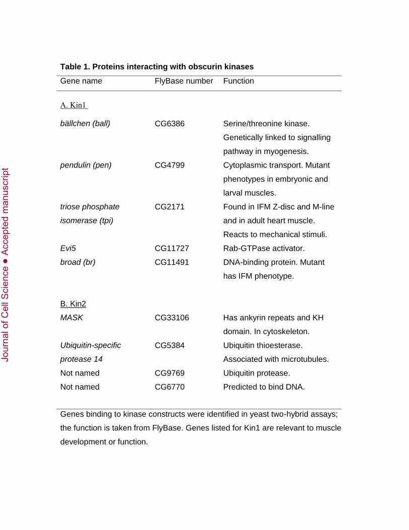
Table 1. Proteins interacting with obscurin kinases
Gene name FlyBase number Function
A. Kin1 bällchen (ball)
CG6386
Serine/threonine kinase.
Genetically linked to signalling
pathway in myogenesis.
pendulin (pen) CG4799 Cytoplasmic transport. Mutant
phenotypes in embryonic and
larval muscles.
triose phosphate
isomerase (tpi)
CG2171 Found in IFM Z-disc and M-line
and in adult heart muscle.
Reacts to mechanical stimuli.
Evi5 CG11727 Rab-GTPase activator.
broad (br)
B. Kin2
MASK
Ubiquitin-specific
protease 14
Not named
Not named
CG11491
CG33106
CG5384
CG9769
CG6770
DNA-binding protein. Mutant
has IFM phenotype.
Has ankyrin repeats and KH
domain. In cytoskeleton.
Ubiquitin thioesterase.
Associated with microtubules.
Ubiquitin protease.
Predicted to bind DNA.
Genes binding to kinase constructs were identified in yeast two-hybrid assays;
the function is taken from FlyBase. Genes listed for Kin1 are relevant to muscle
development or function.
Jour
nal o
f Cel
l Sci
ence
Acc
epte
d m
anus
crip
t

Table 2. Proteins interacting with Kin1 and Kin2 in the TAP assay
Protein FlyBase
number
Mol. Wt.
(kDa)
Unique peptides
Control Kin1 Kin2
Obscurin
(unc89)
CG33519 478 7 42 47
Ball CG6386 66 2 (2) 7 (19) 2 (2)
Tropomyosin-1 CG4898 33 0 0 5 (6)
Peptides associated with Kin1 and Kin2 and a control sample were analysed by
LC-MSMS. The number of unique peptides (as filtered by Scaffold) is shown.
The total number of compounds derived from ball or tropomyosin is given in
brackets. Proteins identified with two or more unique peptides and a probability of
correct identification of >95% (FDR <0.5%) are included.
Jour
nal o
f Cel
l Sci
ence
Acc
epte
d m
anus
crip
t

FIGURES
Fig. 1. Domains in the C-terminus of obscurin and their interaction with ball and
MASK. (A) There are two putative kinase domains (yellow), both of which have a C-
terminal sequence that may be regulatory (RD, white). Kin1 is preceded by two Ig-
domains (red) and Kin2 by an Ig-and an Fn3-domain (blue). In an initial yeast two-hybrid
screen, Ig-Kin1a (with the preceding Ig-domain and part of RD1) interacted with ball.
Interactions of other Kin1 constructs with ball are shown: all interacted with ball. In an
initial Kin2 screen, Fn3-Kin2a interacted with MASK. Interactions of other Kin2
constructs with MASK are shown: Fn3 and Kin2-RD2 did not interact with MASK. (B)
Ball is a kinase (yellow) of approximately 66 kDa, with a C-terminal non-modular tail.
Kin1 bound to a region in the C-terminal tail. (C) MASK is a protein of ~400 kDa with
multiple ankyrin repeats (green) and a C-terminal KH-domain (magenta). Kin2 interacted
with sequence in the C-terminus of MASK. The red bars below the ball and MASK
diagrams correspond to the sequence of prey clones identified in the yeast two-hybrid
assay. Residue numbers are: ball 448-599 (GenBank accession number AE014297);
MASK 3516-3555 (GenBank accession number AF425651).
Jour
nal o
f Cel
l Sci
ence
Acc
epte
d m
anus
crip
t

Fig. 2. The position of ball in the IFM and the effect of reduced expression. (A) Ball
in the IFM. Myofibrils were labelled with anti-ball (green) and with anti-kettin as a Z-
disc marker (red). In wild type myofibrils, ball is labelled at the Z-disc and, more weakly,
across the sarcomere. No ball labelling is detected in myofibrils of ball-knockdown flies.
(B) Abnormal sarcomeres in IFM of ball-knockdown flies compared with sarcomeres in
wild-type IFM. Myofibrils were labelled on actin with phalloidin (red), at the M-line with
anti-obscurin (green) and at the Z-disc with anti-kettin (magenta). In ball-knockdown
flies, labelling of actin, M-line and Z-disc is less regular than in the wild-type. Obscurin
is present in the M-line, but labelling is narrower than in the wild-type; Z-discs in this
Jour
nal o
f Cel
l Sci
ence
Acc
epte
d m
anus
crip
t
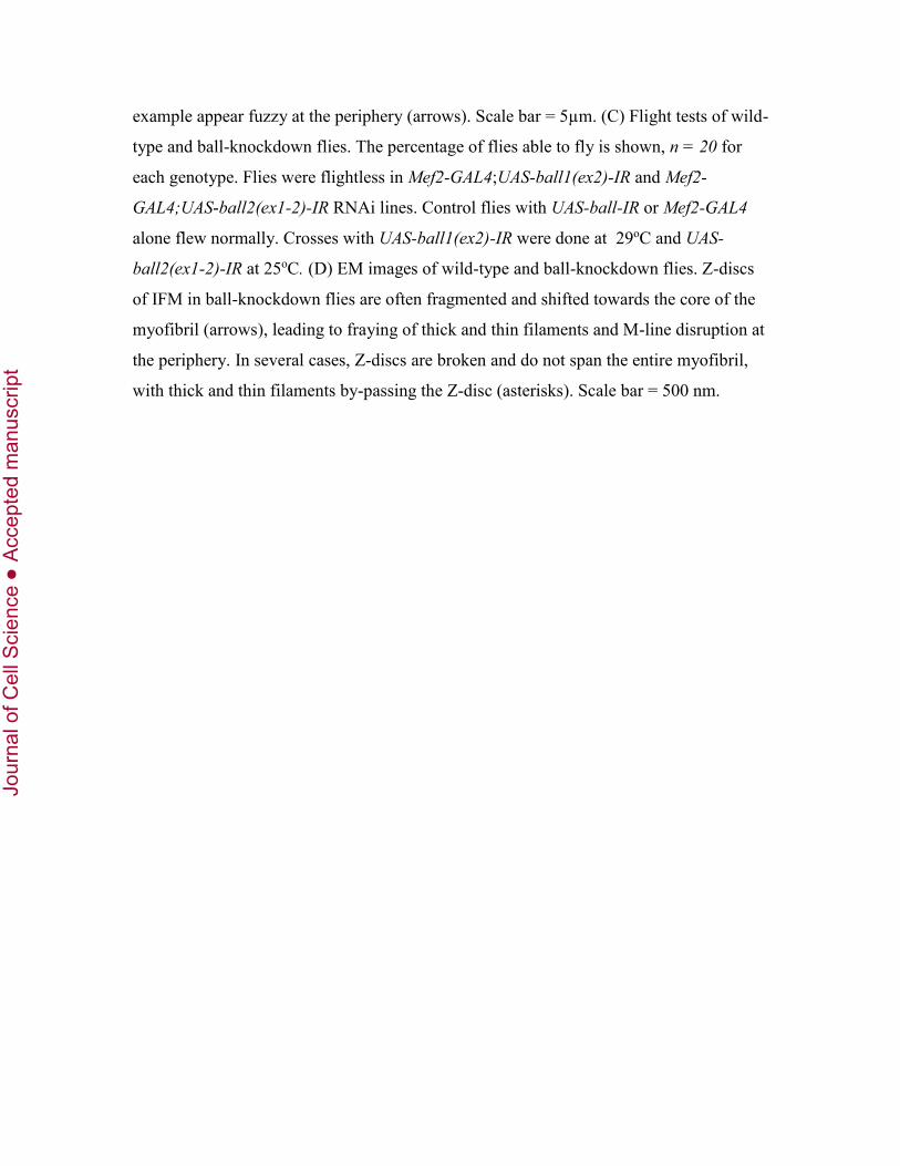
example appear fuzzy at the periphery (arrows). Scale bar = 5µm. (C) Flight tests of wild-
type and ball-knockdown flies. The percentage of flies able to fly is shown, n = 20 for
each genotype. Flies were flightless in Mef2-GAL4;UAS-ball1(ex2)-IR and Mef2-
GAL4;UAS-ball2(ex1-2)-IR RNAi lines. Control flies with UAS-ball-IR or Mef2-GAL4
alone flew normally. Crosses with UAS-ball1(ex2)-IR were done at 29oC and UAS-
ball2(ex1-2)-IR at 25oC. (D) EM images of wild-type and ball-knockdown flies. Z-discs
of IFM in ball-knockdown flies are often fragmented and shifted towards the core of the
myofibril (arrows), leading to fraying of thick and thin filaments and M-line disruption at
the periphery. In several cases, Z-discs are broken and do not span the entire myofibril,
with thick and thin filaments by-passing the Z-disc (asterisks). Scale bar = 500 nm.
Jour
nal o
f Cel
l Sci
ence
Acc
epte
d m
anus
crip
t

Fig. 3. The position of MASK in the IFM and the effect of reduced expression. (A)
MASK in the IFM. Myofibrils were labelled with phalloidin (red), anti-MASK (green),
and anti-kettin (magenta). In wild-type myofibrils, MASK is in the M-line and Z-disc,
with the Z-disc labelled more strongly by the antibody than the M-line. MASK is greatly
reduced in MASK-knockdown flies. The brightness of the Mef2-GAL4;UAS-MASK(ex1)-
IR kettin labelling has been slightly increased in order to see kettin clearly in the merged
image. (B) Abnormal sarcomeres in IFM of MASK-knockdown flies compared with
wild-type IFM. Myofibrils were labelled with phalloidin and anti-kettin, as in (A), and
anti-obscurin (green). In MASK-knockdown flies, M-lines and Z-discs are evenly spaced,
but sarcomeres are shorter. M-lines and corresponding H-zones are often bent (arrows).
MASK-knockdown flies were eclosed ‘escapers’. Scale bar = 5 µm. (C) Flight tests of
wild-type and MASK-knockdown flies. The percentage of flies able to fly is shown, n =
20 for each genotype. Flies were flightless in Mef2-GAL4;UAS-MASK(ex1)-IR and Mef2-
GAL4,UAS-MASK(ex6)-IR RNAi lines. Control flies with UAS-MASK-IR or Mef2-GAL4
alone flew normally. Crosses with UAS-mask(ex1)-IR flies were at 29oC, and UAS-
mask(ex6)-IR at 25oC.
Jour
nal o
f Cel
l Sci
ence
Acc
epte
d m
anus
crip
t

Fig. 4. Ultrastructure of IFM with reduced MASK expression. EM images of (A)
wild-type and (B-D) MASK-knockdown flies. In less affected myofibrils (B), Z-discs are
often split (arrow) and H-zones are shifted, with asymmetric thick and thin filaments. In
more severe cases (C, D), Z-discs form parallel aggregates (asterisks). Relatively normal
and more abnormal myofibrils (D) are often seen in the same fibre. Scale bar (A, B) =
500 nm. Scale bar (C, D) = 1 µm.
Jour
nal o
f Cel
l Sci
ence
Acc
epte
d m
anus
crip
t

Fig. 5. The effect of reduced obscurin expression on ball and MASK. (A) IFM
myofibrils were labelled with phalloidin (red), anti-ball (green) and anti kettin (magenta).
Wild-type myofibrils show clear labelling at the Z-disc; ball was also detected at the M-
line, in addition to some labelling across the sarcomere. In obscurin knockdown flies
(Mef2-GAL4;UAS-unc89-IR), ball is in the Z-disc, but labelling is more spread out than
in the wild-type. Ball is diffusely distributed across the sarcomere and there is no clear
M-line labelling. Arrows mark the M-line. (B) IFM myofibrils were labelled with
phalloidin and anti-kettin as for (A), and anti-MASK (green). In obscurin-knockdown
flies, MASK is present in the Z-disc, but absent or spotty in the M-lines (arrows). The
brightness of the Mef2-GAL4;UAS-unc89-IR MASK labelling has been slightly increased
in order to see remnants of MASK in the M-line of the merged image. Scale bars = 5 µm.
Jour
nal o
f Cel
l Sci
ence
Acc
epte
d m
anus
crip
t

Fig. 6. Binding of MASK to Kin1 and Kin2. Extracts of thoraces containing NTAP,
NTAP-Kin1 or NTAP-Kin2 were mixed with a MASK fragment (MASK1) and
incubated with streptavidin beads. Immunoblots: (A) and (B) Fraction bound to
streptavidin beads. (C) Supernatent from streptavidin beads. (A) was developed with
mouse IgG and anti-mouse IgG-HRP to label NTAP samples; (B) and (C) were
developed with anti-His-HRP to label MASK1. MASK1 binds to both Kin1 and Kin2,
but not to the control.
Jour
nal o
f Cel
l Sci
ence
Acc
epte
d m
anus
crip
t