Embed Size (px)
Citation preview
B I O C H E M I S T R Y
Binding of Steroids to Steroid-Specific Antibodies*
Paula E. Zimmering, Seymour Lieberman, and Bernard F. Erlanger
ABSTRACT : Antisteroid antisera of high titer have been produced when testosterone 17-hemisuccinate, testos- terone 3-O-carboxymethyloxime, and progesterone 20-0-carboxymethyloxime conjugated to bovine serum albumin (BSA) (T-l7-BSA, T-3-BSA, and P-20-BSA, respectively) were injected into unbred ewes. The antisera were absorbed with BSA to remove antibody precipitable by carrier protein and were purified sufficiently to remove all albumin and all other proteins that bind steroids. Since the antibodies are proteins specifically adapted to the binding of steroid haptens, they can possibly serve as model compounds with which to study some binding characteristics of steroids at specific binding sites, such as may be found in receptor proteins at target organs. The wide spectrum of binding affinities found in the antibodies seemed to fall into two main categories, one with high affinity and another containing relatively loosely binding sites. Purification of antibodies by specific precipitation with antigen and dissolution of the complex by a hapten analog yielded antibodies in the second category only. Chromatography on DEAE-cellulose yielded prepara- tions of which approximately 5 0 z of the protein was
S teroid-specific antibodies can be elicited in a variety of animals when immunized with steroids which have been conjugated to foreign proteins (Erlanger et al., 1957, 1959; Lieberman et al., 1959; Beiser et al., 1959). Hapten inhibition studies with rabbit antisteroid anti- sera showed varying amounts of cross-reactivity be- tween steroid hormones and steroid-specific antisera (Lieberman et a/., 1959; Beiser et al., 1959) but the details of the nature of the binding could not be de- duced by this method. The study of the cross-reactivities has now been extended and quantified. Antisteroid antisera were obtained by immunizing nonpregnant ewes with conjugates of BSA' and T-Suc, TOCMO, or POCMO. The binding of a variety of steroids to pools of partially purified preparations of these anti- bodies was studied by equilibrium dialysis.
The antibodies were found to have two main types
* From the Departments of Biochemistry, Obstetrics and Gynecology, and Microbiology, College of Physicians and Sur- geons, Columbia University, New York, New York. Receicied July 5, 1966. Supported in part by Grants AM-03491 and AM- 07572 from the National Institutes of Health, U. S. Public Health Service. Some of the steroid derivatives were prepared by Miss Olive Reynolds under Contract PH 43-64-866 with the CCNSA of the National Cancer Institute, National Institutes of Health, U. S. Public Health Service. 154
antibody. The preparations contained no albumin and no
other proteins that bind steroids, but did contain the whole spectrum of antibody binding sites present in serum. The high-affinity binding sites of the antibodies to T-17-BSA were also in part available to the cross-reacting haptens, desoxycorticosterone 21- hemisuccinate, cortisone 2l-hemisuccinate, and 1 1 - dehydrocorticosterone 21-hemisuccinate. These sites were not available to hydrocortisone 21-hemisuccinate, corticosterone 21-hemisuccinate, nor to a series of hydroxylated steroids. Antibodies to T-3-BSA had tight binding sites available only to the homologous hapten and to some extent to 116-hydroxytestosterone. All of the other steroids tested were bound only loosely to these antibodies. Average binding constants and indices of heterogeneity of binding have been calculated from the Sips equation with the use of a computer. The loose binding sites in both systems seem to be sub- jected to augmentation of further binding after a first steroid molecule has been bound. Anti-P-20-BSA antibodies also exhibited evidence for potentiation of binding at loose sites.
of binding sites, one with high affinity and the other which binds relatively loosely. At steroid concentrations below 3 x 10⁻⁶ mM/ml the antibodies showed high steroid specificity to the high-affinity binding sites. At higher concentrations of steroids, considerable cross-reactivity occurred at the low-affinity sites. The data suggested that the already bound steroid potenti- ated further steroid binding. This study was carried out because antibody proteins uniquely adapted to binding steroids with considerable steroid specificity may be suitable as models for, and may provide in- formation about, the binding characteristics of the true receptor sites of steroid hormones.
1 Abbreviations used in this work: BSA, bovine serum albu- min; T-Suc, testosterone 17-hemisuccinate; TOCMO, testos- terone 3-0-carboxymethyloxime; POCMO, progesterone 20- 0-carboxymethyloxime; E-Suc, cortisone 21-hemisuccinate; DOC-Suc, desoxycorticosterone 21-hemisuccinate; F-Suc, corti- sol 2 1 - hemisuccina te ; B-S uc, corticosterone 2 1 - hemisuccina te ; A-Suc, 1 I-dehydrocorticosterone 21-hemisuccinate; 6B-OH-T, 60-hydroxytestosterone; 14a-OH-T, 14a-hydroxytestosterone; 9a-OH-T, 9a-hydroxytestosterone; 1 18-OH-T, 1 lp-hydroxy- testosterone; Epi-F, lla,l7a,21-trihydroxy-A4-pregnene-3,20- dione; F, cortisol; T-17-BSA, conjugate of T-Suc and BSA; P-20-BSA, conjugate of POCMO and BSA; T-3-BSA, conjugate of TOCMO and BSA.
P A U L A E. Z I M M E R I N G , S E Y M O U R L I E B E R M A N , A N D B E R N A R D F. E R L A N G E R
V O L . 6, N O . 1 , J A N U A R Y 1 9 6 7
Experimental Section
Antigens. The antigens were derivatives of testos- terone and progesterone conjugated to BSA as de- scribed previously (Erlanger et al., 1957, 1959) except that they were stored at -20" without lyophilization (Zimmering et a/., 1965). T-17-BSA contained 20 steroid residues/BSA molecule ; P-20-BSA contained 26 progesterone residues/BSA molecule; T-3-BSA contained 25 testosterone residues/BSA molecule. Antigen solutions used for precipitin curves were prepared from thawed and dialyzed conjugate solutions. They were discarded after 60 days of storage at refriger- ator temperature. Precipitin curves were performed as described previously (Zimmering et al., 1965).
Antisera. Nonpregnant ewes2 were injected subcuta- neously once a week with 4 mg of antigens in 1 ml of complete Freund's adjuvant, and bleedings were taken from the jugular vein at 2-month intervals. All antisera were absorbed with BSA until tests with rabbit anti- BSA antibodies gave a slight precipitate, indicating a small excess of BSA. The antisera were then sterilized by passing them through a Seitz filter and stored at 5 '.
TABLE I : Minimum Antibody Content of Sheep Anti- steroid Antisera.
Months of Ewe Immunization
2at b 10 12 14
3.h 8 10 16 18
5 a , b 6 8
1 O j d 6 8 9
8 b , c 8
Antibody (mg/ml of
diluted serum)
4 . 2 2 . 7 3 . 1 5 . 8 7 . 2 7 . 8 4 . 2 4 . 6 3 . 8 3 . 1 2 . 0 2 . 4 5 . 0
a Anti-T-17-BSA. b Precipitin curves done at one- fifth dilution. c Anti-T-3-BSA. d Precipitin curves done at one-third dilution. e Anti-P-20-BSA.
Table I shows antibody titers obtained from the equiva- lence point of precipitin curves of antisera diluted with saline phosphate buffer3 as indicated. The curves were performed with diluted sera because of the difficulties of adequately washing the heavy precipitates obtained
2 Nonpregnant ewes were used throughout to avoid any possi- ble complications due to hormonal changes during pregnancy.
3 Unless otherwise indicated, saline phosphate buffer which was 0.01 M in phosphate, 0.15 N in NaCI, pH 7.4, was used throughout.
with undiluted sera. Since the precipitin titer is markedly affected by dilution (Zimmering et al., 1965), the anti- body content of the undiluted sera was higher than the value found by multiplying the observed titer by the dilution factor.
Purification of Antisera. Salt fractionation was carried out either by the method of Kekwick (1938) as modified by Strauss et al. (1960) or by the method of Strauss et al. (1964) using three precipitations. Purification by specific precipitation and by chromatography on DEAE- cellulose have been described previously (Zimmering et al., 1965). Fractions eluted from DEAE-cellulose columns in starting buffer (0.0175 M phosphate, pH 6.3) were pooled and used for equilibrium dialysis studies after dialysis against three changes of saline phosphate buffer. Preparations for comparative study were usually pools from five or six columns. They were stored frozen, thawed at 5" when needed, and centrifuged at low speed overnight at 5 " before use. Protein concentrations were measured by the Folin-Ciocalteu method as modified by Lowry et al. (1951). The molecular weight for y-globulin was taken as 160,000 for all calculations.
Equilibrium Dialysis. All solutions used in the binding studies were in saline phosphate buffer and were diluted to be in the range 1.75-2.40 X 10⁻⁵ mM antibody/ml. The method of equilibrium dialysis used was adapted from that described by Karush (1956). Membranes of controlled pore size (Schleicher and Schuell, no. B18) were washed by stirring in 2 0 x ethanol for 4 days, followed by distilled water washes for 10-20 days, until the washings were transparent in the ultraviolet region. This usually required about five water changes with copious rinsing of the mem- branes at each water change. Cleanliness of the mem- branes was tested by shaking a sample membrane for 24 hr in 4 ml of saline phosphate buffer and ex- amining the buffer in a Zeiss spectrophotometer at the wavelengths to be used for the analysis of hapten concentration in the equilibrium dialysis experiments. The membranes used gave equilibrium at 5 " within 20 hr in the dialysis apparatus when the haptens were tested against buffer, except for POCMO and pred- nisolone, which required 24 and 28 hr, respectively. The dialyses were accordingly run for these longer periods when these haptens were used. Membranes could be washed again and reused about three times. Each side of the dialysis cell was sealed with a small parafilm disk which fitted over the opening and was sealed to the glass by running a spatula dipped in hot paraffin along the sides. The parafilm was washed in the same way as the membranes before they were cut into disks. These disks were used only once.
Spectrophotometric Assay of Steroids. Since the ster- oids used in this study contained an a,@-unsaturated ketone chromophore group (A4-3-keto) in ring A, the solutions were analyzed by reading the absorbance at maximum absorption. The steroids and steroid derivatives were sensitive to light and gave evidence of undergoing a "dark" reaction, after exposure to light, which resulted in an alteration in the chromo- 155
B I N D I N G O F S T E R O I D S T O S T E R O I D - S P E C I F I C A N T I B O D I E S
156
P A U L A
R 1 0 C 14 E M I S T R Y
r I O
5 c X I 0
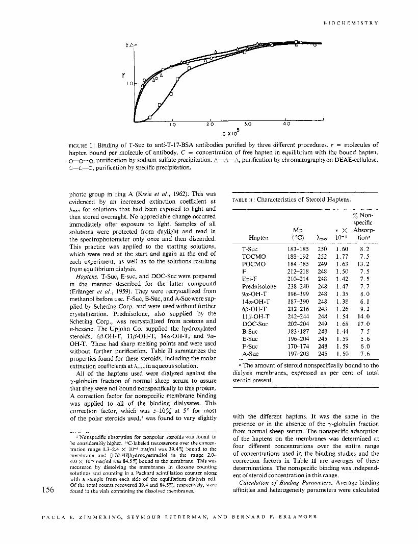
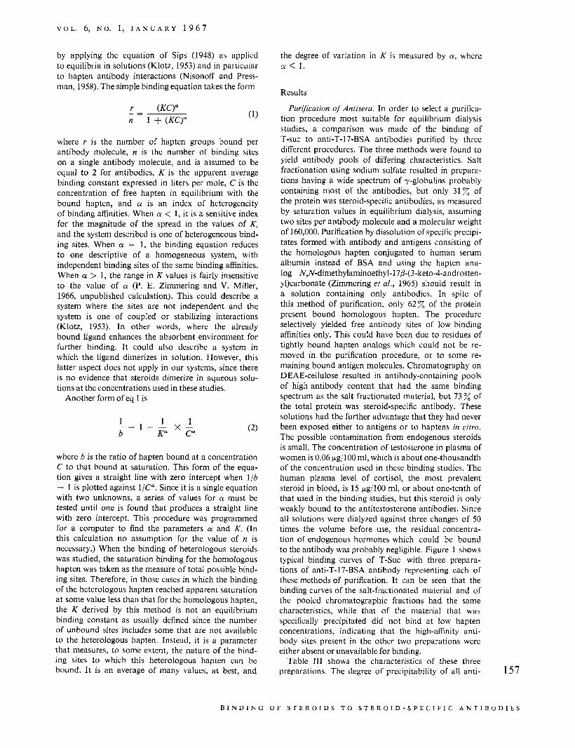
FIGURE 1: Binding of T-Suc to anti-T-17-BSA antibodies purified by three different procedures. r = molecules of hapten bound per molecule of antibody. C = concentration of free hapten in equilibrium with the bound hapten.
-
E. Z I M M E R I N G , S E Y M O U R L I E D E R M A N , A N D
0-0-0, purification by sodium sulfate precipitation. A n-n-r, purification by specific precipitation.
A
phoric group in ring A (Kwie et al., 1962). This was evidenced by an increased extinction coefficient at A,,, for solutions that had been exposed to light and then stored overnight. No appreciable change occurred immediately after exposure to light. Samples of all solutions were protected from daylight and read in the spectrophotometer only once and then discarded. This practice was applied to the starting solutions, which were read at the start and again at the end of each experiment, as well as to the solutions resulting from equilibrium dialysis.
Haptens. T-Suc, E-suc, and DOC-Suc were prepared in the manner described for the latter compound (Erlanger et al., 1959). They were recrystallized from methanol before use. F-Suc, B-Suc, and A-Suc were sup- plied by Schering Corp. and were used without further crystallization. Prednisolone, also supplied by the Schering Corp., was recrystallized from acetone and n-hexane. The Upjohn Co. supplied the hydroxylated steroids, 6P-OH-T, llP-OH-T, 14a-OH-T, and 9a- OH-T. These had sharp melting points and were used without further purification. Table I1 summarizes the properties found for these steroids, including the molar extinction coefficients at Amax in aqueous solution.
All of the haptens used were dialyzed against the y-globulin fraction of normal sheep serum to assure that they were not bound nonspecifically to this protein. A correction factor for nonspecific membrane binding was applied to all of the binding dialysates. This correction factor, which was 5-10% at 5" for most of the polar steroids used,4 was found to vary slightly
4 Nonspecific absorption for nonpolar steroids was found to be considerably higher. 1Glabeled testosterone over the concen- tration range 1.3-2.4 X 10⁻⁶ mM/ml was 39.4% bound to the membrane and [17p- ~Hlhydroxyestradiol in the range 2.0- 4.0 x 10⁻⁷ rnM/ml was 84.5 bound to the membrane. This was measured by dissolving the membranes in dioxane counting solutions and counting in a Packard scintillation counter along with a sample from each side of the equilibrium dialysis cell. Of the total counts recovered 39.4 and 84.5%, respectively, were found in the vials containing the dissolved membranes.
-A, purification by chromatography on DEAE-cellulose.
TABLE 11 : Characteristics of Steroid Haptens.
Hapten
T-SUC TOCMO POCMO F Epi-F Prednisolone 9a-OH-T 14a-OH-T 6P-OH-T 110-OH-T DOC-SUC B-SUC E-SUC F-SUC A-SUC
MP ( "C>
183-185 188-192 184-1 85 2 12-2 18 210-214 238-240 196-1 99 187-1 90 212-216 242-244 202-204 183-187 196-204 170-174 197-203
Non- specific
E X Absorp- A,,, tiona
250 1.60 8 . 2 252 1.77 7 . 5 249 1.63 13.2 248 1.50 7 . 5 248 1.42 7 . 5 248 1.47 7 . 7 248 1.35 8 . 0 248 1.38 6 . 1 243 1.26 9 .2 248 1.54 14.0 249 1.68 17.0 248 1.44 7 . 5 245 1.59 5 . 6 248 1.59 6 . 0 245 1.50 7 . 6
a The amount of steroid nonspecifically bound to the dialysis membranes, expressed as per cent of total steroid present.
with the different haptens. It was the same in the presence or in the absence of the y-globulin fraction from normal sheep serum. The nonspecific adsorption of the haptens on the membranes was determined at four different concentrations over the entire range of concentrations used in the binding studies and the correction factors in Table I1 are averages of these determinations. The nonspecific binding was independ- ent of steroid concentration in this range.
Calculation of Binding Parameters. Average binding affinities and heterogeneity parameters were calculated
B E R N A R D F. E R L A N G E R
V O L . 6, N O . 1, J A N U A R Y 1 9 6 7
by applying the equation of Sips (1948) as applied to equilibria in solutions (Klotz, 1953) and in particdar to hapten-antibody interactions (Nisonoff and Press- man, 1958). The simple binding equation takes the form
where r is the number of hapten groups bound per antibody molecule, n is the number of binding sites on a single antibody molecule, and is assumed to be equal to 2 for antibodies, K is the apparent average binding constant expressed in liters per mole, C is the concentration of free hapten in equilibrium with the bound hapten, and a is an index of heterogeneity of binding affinities. When a < 1, it is a sensitive index for the magnitude of the spread in the values of K, and the system described is one of heterogeneous bind- ing sites. When a = 1, the binding equation reduces to one descriptive of a homogeneous system, with independent binding sites of the same binding affinities. When a > 1, the range in K values is fairly insensitive to the value of a (P. E. Zimmering and V. Miller, 1966, unpublished calculation). This could describe a system where the sites are not independent and the system is one of coupled or stabilizing interactions (Klotz, 1953). In other words, where the already bound ligand enhances the absorbent environment for further binding. It could also describe a system in which the ligand dimerizes in solution. However, this latter aspect does not apply in our systems, since there is no evidence that steroids dimerize in aqueous solu- tions at the concentrations used in these studies.
Another form of eq 1 is
where b is the ratio of hapten bound a t a concentration C to that bound at saturation. This form of the equa- tion gives a straight line with zero intercept when l ib - 1 is plotted against l/C". Since it is a single equation with two unknowns, a series of values for a must be tested until one is found that produces a straight line with zero intercept. This procedure was programmed for a computer to find the parameters a and K. (In this calculation no assumption for the value of n is necessary.) When the binding of heterologous steroids was studied, the saturation binding for the homologous hapten was taken as the measure of total possible bind- ing sites. Therefore, in those cases in which the binding of the heterologous hapten reached apparent saturation at some value less than that for the homologous hapten, the K derived by this method is not an equilibrium binding constant as usually defined since the number of unbound sites includes some that are not available to the heterologous hapten. Instead, it is a parameter that measures, to some extent, the nature of the bind- ing sites to which this heterologous hapten can be bound. It is an average of many values, a t best, and
the degree of variation in K is measured by a, where a < 1 .
Results
Puri3cation of Antisera. In order to select a purifica- tion procedure most suitable for equilibrium dialysis studies, a comparison was made of the binding of T-suc to anti-T-17-BSA antibodies purified by three different procedures. The three methods were found to yield antibody pools of differing characteristics. Salt fractionation using sodium sulfate resulted in prepara- tions having a wide spectrum of y-globulins probably containing most of the antibodies, but only 31 z of the protein was steroid-specific antibodies, as measured by saturation values in equilibrium dialysis, assuming two sites per antibody molecule and a molecular weight of 160,000. Purification by dissolution of specific precipi- tates formed with antibody and antigens consisting of the homologous hapten conjugated to human serum albumin instead of BSA and using the hapten ana- log N,N-dimethylaminoethyl-17,f3-(3-keto-4-androsten- y1)carbonate (Zimmering et al., 1965) should result in a solution containing only antibodies. In spite of this method of purification, only 62z of the protein present bound homologous hapten. The procedure selectively yielded free antibody sites of low binding affinities only. This could have been due to residues of tightly bound hapten analogs which could not be re- moved in the purification procedure, or to some re- maining bound antigen molecules. Chromatography on DEAE-cellulose resulted in antibody-containing pools of high antibody content that had the same binding spectrum as the salt fractionated material, but 73 of the total protein was steroid-specific antibody. These solutions had the further advantage that they had never been exposed either to antigens or to haptens in vitro. The possible contamination from endogenous steroids is small. The concentration of testosterone in plasma of women is 0.06 pg/lOO ml, which is about one-thousandth of the concentration used in these binding studies. The human plasma level of cortisol, the most prevalent steroid in blood, is 15 pg/100 ml, or about one-tenth of that used in the binding studies, but this steroid is only weakly bound to the antitestosterone antibodies. Since all solutions were dialyzed against three changes of 50 times the volume before use, the residual concentra- tion of endogenous hormones which could be bound to the antibody was probably negligible. Figure 1 shows typical binding curves of T-Suc with three prepara- tions of anti-T-17-BSA antibody representing each of these methods of purification. It can be seen that the binding curves of the salt-fractionated material and of the pooled chromatographic fractions had the same characteristics, while that of the material that was specifically precipitated did not bind at low hapten concentrations, indicating that the high-affinity anti- body sites present in the other two preparations were either absent or unavailable for binding.
Table 111 shows the characteristics of these three preparations. The degree of precipitability of all anti- 157
D I N D 1 N G 0 F S T E R 0 1 D S T 0 S T E R 0 I D - S I' E C I F I C A N T 1 U 0 D 1 E S
B I O C H E M I S T R Y
I .o 2 .o 3.0 4.0 5 c x IO
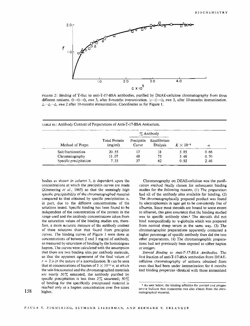
FIGURE 2 : Binding of T-Suc to anti-T-17-BSA antibodies, purified by DEAE-cellulose chromatography from three different antisera. W O - 0 , ewe 3, after 8-months immunization. o--0-0, ewe 3, after 10-months immunization. A-A-A, ewe 2 after 10-months immunization. Coordinates as for Figure 1.
TABLE 111 : Antibody Content of Preparations of' Anti-T-17-BSA Antiserum.
% Antibody
Total Protein Precipitin Equilibrium Method of Prepn (mgiml) Curve Dialysis K X 10-5 a
Salt fractionation 20.55 17 31 3.85 0 .66 Chromatography 11.07 48 73 3 46 0.70 Specific precipitation 7.35 37 62 0.92 2.46
bodies as shown in column 3, is dependent upon the concentrations at which the precipitin curves are made (Zimmering et af., 1965) so that the seemingly high specific precipitability of the chromatographed material compared to that obtained by specific precipitation is, in part, due to the different concentrations of the solutions tested. Specific binding has been found to be independent of the concentration of the protein in the range used and the antibody concentrations taken from the saturation values of the binding studies are, there- fore, a more accurate measure of the antibody content of these solutions than that found from precipitin curves. The binding curves of Figure 1 were done at concentrations of between 2 and 3 mg/ml of antibody, as measured by saturation of binding by the homologous hapten. The curves were calculated with the assumption that there are two binding sites per antibody molecule, so that the apparent agreement of the final values of r = 2 is in the nature of a normalization. It can be seen that at concentrations of hapten of 3 X M, at which the salt-fractionated and the chromatographed materials are nearly 50z saturated, the antibody purified by specific precipitation is less than 2 z saturated; 50% of binding for the specifically precipitated material is reached only at a hapten concentration over five times
158 higher.
Chromatography on DEAE-cellulose was the purifi- cation method finally chosen for subsequent binding studies for the following reasons. (1) The preparation had all of the antibody sites available for binding. (2) The chromatographically prepared product was found by electrophoresis in agar gel to be consistently free of albumin. Since most steroids are bound to some extent to albumin, this gave assurance that the binding studied was to specific antibody sites.5 The steroids did not bind nonspecifically to y-globulin which was prepared from normal sheep serum in the same way. (3) The chromatographic preparations apparently contained a higher percentage of specific antibody than did the two other preparations. (4) The chromatographic prepara- tions had not previously been exposed to either hapten or antigen.
Steroid Binding to Anti-T-17-BSA Antibodies. The first fraction of anti-T-17-BSA antibodies from DEAE- cellulose chromatography of antisera obtained from ewes that had been under immunization for 6 months had binding properties identical with those immunized
As seen below, the binding affinities for cortisol and proges- terone indicate that transcortin was also absent from the chro- matographed material.
P A U L A E. Z I M M E R I N G , S E Y M O U R L I E B E R M A N , A N D B E R N A R D F. E R L A N G E R
V O L . 6, N O . 1, J A N U A R Y 1 9 6 7
2 0
I O r
' 0 2 0 3 0 4 0
c x lo5
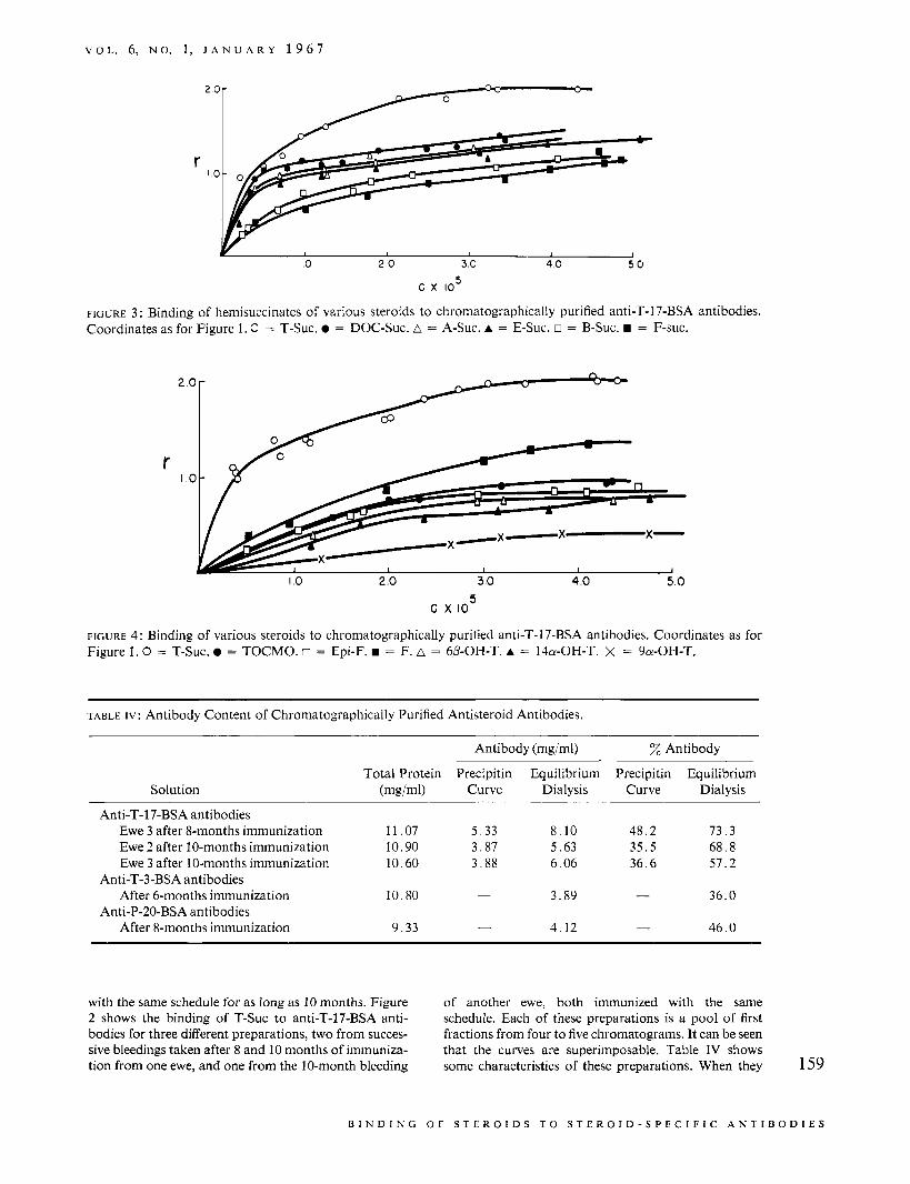
FIGURE 3 : Binding of hemisuccinates of various steroids to chromatographically purified anti-T-17-BSA antibodies. Coordinates as for Figure 1. C = T-Suc. 0 = DOC-Suc. A = A-Suc. A = E-Suc. o = B-Suc. = F-suc.
2
r I
.o
.o
I .o 2 .o 3 .O 4 .O 5.0 5 c x IO
FIGURE 4 : Binding of various steroids to chromatographically purified anti-T-17-BSA antibodies. Coordinates as for Figure 1 . 0 = T-Suc. = TOCMO. o = Epi-F. = F. A = 6P-OH-T. A = 140-OH-T. X = 9a-OH-T.
TABLE I V : Antibody Content of Chromatographically Purified Antisteroid Antibodies.
Solution
Antibody (mgiml) Antibody
Total Protein Precipitin Equilibrium Precipitin Equilibrium (mgiml) Curve Dialysis Curve Dialysis
Anti-T-17-BSA antibodies Ewe 3 after 8-months immunization 11.07 5 .33 8.10 48.2 73.3 Ewe 2 after 10-months immunization 10.90 3.87 5.63 35.5 68.8 Ewe 3 after 10-months immunization 10.60 3.88 6.06 36.6 51.2
3.89 - 36.0 After 6-months immunization 10.80 -
After 8-months immunization 9.33 4.12 - 46.0
Anti-T-3-BSA antibodies
Anti-P-20-BSA antibodies -
with the same schedule for as long as 10 months. Figure of another ewe, both immunized with the same 2 shows the binding of T-Suc to anti-T-17-BSA anti- schedule. Each of these preparations is a pool of first bodies for three different preparations, two from succes- fractions from four to five chromatograms. It can be seen sive bleedings taken after 8 and 10 months of immuniza- that the curves are superimposable. Table IV shows tion from one ewe, and one from the 10-month bleeding some characteristics of these preparations. When they 159
B I N D I N G O F S T E R O I D S T O S T E R O I D - S P E C I F I C A N T I B O D I E S
B I O C H E M I S T R Y
2 .o
r I .o
I O 2 0 3.0 4.13 5.9
c x IO5
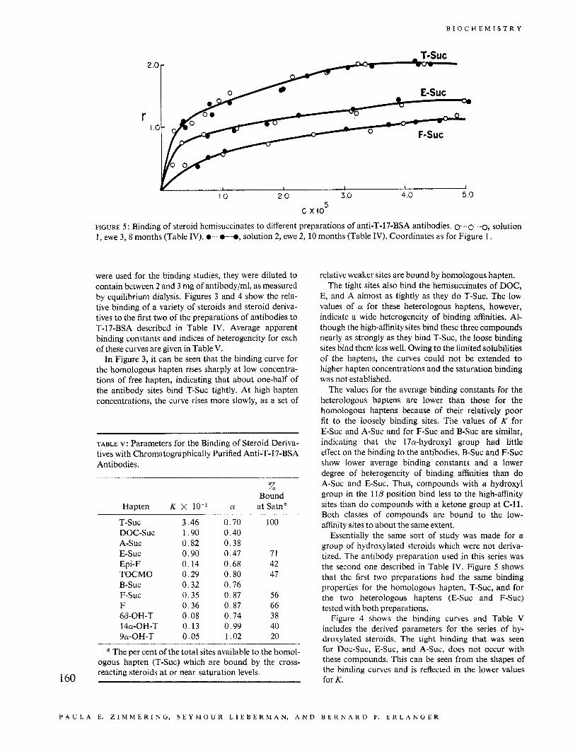
FIGURE 5 : Binding of steroid hemisuccinates to different preparations of anti-T-17-BSA antibodies. 0-0-0, solution 1, ewe 3,8 months (Table IV). O-0-0, solution 2, ewe 2, 10 months (Table IV). Coordinates as for Figure 1.
were used for the binding studies, they were diluted to contain between 2 and 3 mg of antibody/ml, as measured by equilibrium dialysis. Figures 3 and 4 show the rela- tive binding of a variety of steroids and steroid deriva- tives to the first two of the preparations of antibodies to T-17-BSA described in Table IV. Average apparent binding constants and indices of heterogeneity for each of these curves are given in Table V.
In Figure 3, it can be seen that the binding curve for the homologous hapten rises sharply at low concentra- tions of free hapten, indicating that about one-half of the antibody sites bind T-Suc tightly. At high hapten concentrations, the curve rises more slowly, as a set of
TABLE v: Parameters for the Binding of Steroid Deriva- tives with Chromatographically Purified Anti-T-17-BSA Antibodies.
% Bound
Hapten K X a at Satn"
T-SUC 3.46 0.70 100 DOC-SUC 1.90 0.40 A-SUC 0.82 0.38 E-SUC 0.90 0.47 71 Epi-F 0.14 0.68 42 TOCMO 0.29 0.80 47 B-SUC 0.32 0.76 F-SUC 0.35 0.87 56 F 0.36 0.S7 66 6P-OH-T 0.08 0.74 38 14a-OH-T 0.13 0.99 40 9a-OH-T 0.05 1.02 20
a The per cent of the total sites available to the homol- ogous hapten (T-Suc) which are bound by the cross- reacting steroids at or near saturation levels. 160
relative weaker sites are bound by homologous hapten. The tight sites also bind the hemisuccinates of DOC,
E, and A almost as tightly as they do T-Suc. The low values of a for these heterologous haptens, however, indicate a wide heterogeneity of binding affinities. Al- though the high-affinity sites bind these three compounds nearly as strongly as they bind T-Suc, the loose binding sites bind them less well. Owing to the limited solubilities of the haptens, the curves could not be extended to higher hapten concentrations and the saturation binding was not established.
The values for the average binding constants for the heterologous haptens are lower than those for the homologous haptens because of their relatively poor fit to the loosely binding sites. The values of K for E-Suc and A-Suc and for F-Suc and B-Suc are similar, indicating that the 17a-hydroxyl group had little effect on the binding to the antibodies. B-Suc and F-Suc show lower average binding constants and a lower degree of heterogeneity of binding affinities than do A-Suc and E-Suc. Thus, compounds with a hydroxyl group in the l l p position bind less to the high-affinity sites than do compounds with a ketone group at C-11. Both classes of compounds are bound to the low- affinity sites to about the same extent.
Essentially the same sort of study was made for a group of hydroxylated steroids which were not deriva- tized. The antibody preparation used in this series was the second one described in Table IV. Figure 5 shows that the first two preparations had the same binding properties for the homologous hapten, T-Suc, and for the two heterologous haptens (E-Suc and F-Suc) tested with both preparations.
Figure 4 shows the binding curves and Table V includes the derived parameters for the series of hy- droxylated steroids. The tight binding that was seen for Doc-Suc, E-Suc, and A-Suc, does not occur with these compounds. This can be seen from the shapes of the binding curves and is reflected in the lower values for K.
P A U L A E. Z I M M E R I N G , S E Y M O U R L I E B E R M A N , A N D B E K N A K D F. E R L A N G E K
V O L . 6, N O . 1, J A N U A R Y 1 9 6 7
2
r I
1.0 2.0 3.0 4.0
c X I O ~
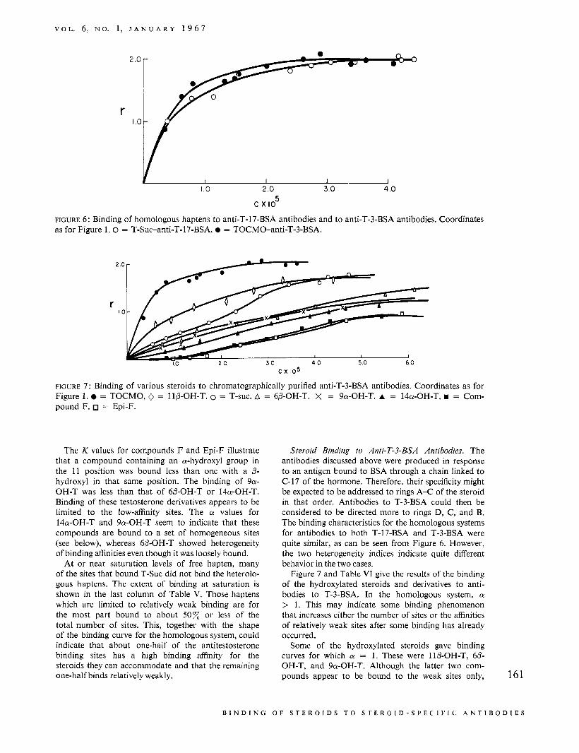
FIGURE 6: Binding of homologous haptens to anti-T-17-BSA antibodies and to anti-T-3-BSA antibodies. Coordinates as for Figure 1 . 0 = T-Suc-anti-T-17-BSA. = TOCMO-anti-T-3-BSA.
2 0
r 10
c x 105
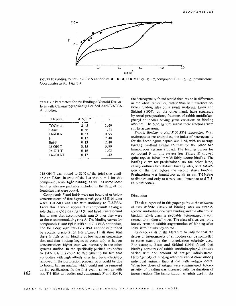
FIGURE 7 : Binding of various steroids to chromatographically purified anti-T-3-BSA antibodies. Coordinates as for Figure 1. = TOCMO, 0 = IIP-OH-T. 0 = T-suc. A = 6P-OH-T. X = 9a-OH-T. A = 14a-OH-T. = Com- pound F. = Epi-F.
The K values for compounds F and Epi-F illustrate that a compound containing an a-hydroxyl group in the 11 position was bound less than one with a P- hydroxyl in that same position. The binding of 9a- OH-T was less than that of 6P-OH-T or I4a-OH-T. Binding of these testosterone derivatives appears to be limited to the low-affinity sites. The a values for 14a-OH-T and 9a-OH-T seem to indicate that these compounds are bound to a set of homogeneous sites (see below), whereas 6P-OH-T showed heterogeneity of binding affinities even though it was loosely bound.
At or near saturation levels of free hapten, many of the sites that bound T-Suc did not bind the heterolo- gous haptens. The extent of binding at saturation is shown in the last column of Table V. Those haptens which are limited to relatively weak binding are for the most part bound to about 50z or less of the total number of sites. This, together with the shape of the binding curve for the homologous system, could indicate that about one-half of the antitestosterone binding sites has a high binding affinity for the steroids they can accommodate and that the remaining one-half binds relatively weakly.
Steroid Binding to Anti-T-3-BSA Antibodies. The antibodies discussed above were produced in response to an antigen bound to BSA through a chain linked to C-17 of the hormone. Therefore, their specificity might be expected to be addressed to rings A-C of the steroid in that order. Antibodies to T-3-BSA could then be considered to be directed more to rings D, C, and B. The binding characteristics for the homologous systems for antibodies to both T-17-BSA and T-3-BSA were quite similar, as can be seen from Figure 6. However, the two heterogeneity indices indicate quite different behavior in the two cases.
Figure 7 and Table VI give the results of the binding of the hydroxylated steroids and derivatives to anti- bodies to T-3-BSA. In the homologous system, a > 1. This may indicate some binding phenomenon that increases either the number of sites or the affinities of relatively weak sites after some binding has already occurred.
Some of the hydroxylated steroids gave binding curves for which a = 1. These were 1lP-OH-T, 6P- OH-T, and 9a-OH-T. Although the latter two com- pounds appear to be bound to the weak sites only, 161
B I N D I N G 0 F S T E R 0 I D S T 0 S T E R 0 1 D - S P E C I F I C A N T 1 B 0 D I E S
2 ' , O r
B I O C H E M I S T R Y
r I
1.0 2 .o 3.0 4.0 5.0
c x lo5
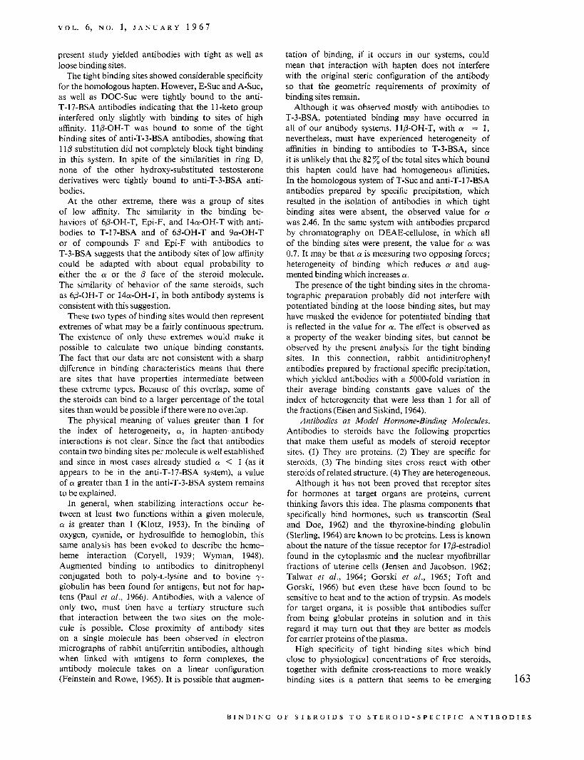
FIGURE 8 : Binding to anti-P-20-BSA antibodies. .-.-e, POCMO. 0-0-0, compound F. A-A-A, prednisolone. Coordinates as for Figure 1.
TABLE VI : Parameters for the Binding of Steroid Deriva- tives with Chromatographically Purified Anti-T-3-BSA Antibodies.
Hapten K X 10-j a
TOCMO 2.45 1.69 T-SUC 0.36 1.13 11B-OH-T 0.63 0.95 F 0.17 2.48 Epi-F 0.13 2.40 6/3-OH-T 0.15 0.99 9a-OH-T 0.16 1.03 14a-OH-T 0.17 1.42
1 ID-OH-T was bound to 82 of the total sites avail- able to T-Suc. In spite of the fact that a = 1 for this compound, some tight binding, as well as some loose binding sites are probably included in the 82% of the total sites that were bound.
Compounds F and Epi-F were not bound at or below concentrations of free hapten which gave 85 % binding when TOCMO was used with antibody to T-3-BSA. From this it would appear that compounds having a side chain at C-17 on ring D (F and Epi-F) were bound less to sites that accommodate ring D than they were to those accommodating ring A. The binding curves for compounds F and Epi-F with anti-T-3-BSA antibodies, and for T-Suc with anti-T-17 BSA antibodies purified by specific precipitation (see Figure 1) all show that there is little or no binding at low hapten concentra- tion and that binding begins to occur only at hapten concentrations higher than was necessary in the other systems studied. In the specifically purified antibodies to T-17-BSA this could be due either to the fact that antibodies with high affinity sites had been selectively removed in the purification process, or it could be due to bound hapten analog, which could not be removed during purification. In the first event, as well as with anti-T-3-BSA antibodies and compounds F and Epi-F, 162
the heterogeneity found would then reside in differences in the whole molecules, rather than in differences be- tween binding sites on a single molecule. Eisen and Siskind (1964), on the other hand, have separated by serial precipitations, fractions of rabbit antidinitro- phenyl antibodies having great variations in binding affinities. The binding sites within these fractions were still heterogeneous.
Steroid Binding to Anti-P-20-BSA Antibodies. With antiprogesterone antibodies, the index of heterogeneity for the homologous hapten was 1.58, with an average binding constant similar to that for the other two homologous systems studied. The binding curves for compound F in this system (see Figure 8) showed quite regular behavior with fairly strong binding. The binding curve for prednisolone, on the other hand, clearly outlines two distinct binding sites, with satura- tion of the first before the second starts binding. Prednisolone was bound not at all to anti-T-17-BSA antibodies and only to a very small extent to anti-T-3- BSA antibodies.
Discussion
The data reported in this paper point to the existence of two definite classes of binding sites on steroid- specific antibodies, one tight binding and the other loose binding. Each class is probably heterogeneous with respect to binding affinities. The class of sites that bind loosely seem to exhibit augmentation of binding after some steroid is already bound.
Evidence exists in the literature to indicate that the degree of heterogeneity of antibodies can be controlled to some extent by the immunization schedule used. For example, Eisen and Siskind (1964) found that binding constants of rabbit antidinitrophenyl antisera varied with the amount of antigen administered. Heterogeneity of binding affinities varied more among individual animals than it did with antigen doses. When low doses of antigen were administered, hetero- geneity of binding was increased with the duration of immunization. The immunization schedule used in the
P A U L A E. Z I M M E R I N G , S E Y M O U R L I E B E R M A N , A N D B E R N A R D F. E R L A N G E R
V O L . 6, N O . 1, J A N U A R Y 1 9 6 7
present study yielded antibodies with tight as well as loose binding sites.
The tight binding sites showed considerable specificity for the homologous hapten. However, E-Suc and A-Suc, as well as DOC-Suc were tightly bound to the anti- T-17-BSA antibodies indicating that the 11-keto group interfered only slightly with binding to sites of high affinity. 11P-OH-T was bound to some of the tight binding sites of anti-T-3-BSA antibodies, showing that 110 substitution did not completely block tight binding in this system. In spite of the similarities in ring D, none of the other hydroxy-substituted testosterone derivatives were tightly bound to anti-T-3-BSA anti- bodies.
At the other extreme, there was a group of sites of low affinity. The similarity in the binding be- haviors of 6P-OH-T, Epi-F, and 14a-OH-T with anti- bodies to T-17-BSA and of 6P-OH-T and 9a-OH-T or of compounds F and Epi-F with antibodies to T-3-BSA suggests that the antibody sites of low affinity could be adapted with about equal probability to either the a or the /3 face of the steroid molecule. The similarity of behavior of the same steroids, such as 6P-OH-T or 14a-OH-T, in both antibody systems is consistent with this suggestion.
These two types of binding sites would then represent extremes of what may be a fairly continuous spectrum. The existence of only these extremes would make it possible to calculate two unique binding constants. The fact that our data are not consistent with a sharp difference in binding characteristics means that there are sites that have properties intermediate between these extreme types. Because of this overlap, some of the steroids can bind to a larger percentage of the total sites than would be possible if there were no oveilap.
The physical meaning of values greater than 1 for the index of heterogeneity, a, in hapten-antibody interactions is not clear. Since the fact that antibodies contain two binding sites per molecule is well established and since in most cases already studied a < 1 (as it appears to be in the anti-T-17-BSA system), a value of a greater than 1 in the anti-T-3-BSA system remains to be explained.
In general, when stabilizing interactions occur be- tween at least two functions within a given molecule, a is greater than 1 (Klotz, 1953). In the binding of oxygen, cyanide, or hydrosulfide to hemoglobin, this same analysis has been evoked to describe the heme- heme interaction (Coryell, 1939; Wyman, 1948). Augmented binding to antibodies to dinitrophenyl conjugated both to poly-L-lysine and to bovine y- globulin has been found for antigens, but not for hap- tens (Paul et a/., 1966). Antibodies, with a valence of only two, must then have a tertiary structure such that interaction between the two sites on the mole- cule is possible. Close proximity of antibody sites on a single molecule has been observed in electron micrographs of rabbit antiferritin antibodies, although when linked with antigens to form complexes, the antibody molecule takes on a linear configuration (Feinstein and Rowe, 1965). It is possible that augmen-
tation of binding, if it occurs in our systems, could mean that interaction with hapten does not interfere with the original steric configuration of the antibody so that the geometric requirements of proximity of binding sites remain.
Although it was observed mostly with antibodies to T-3-BSA, potentiated binding may have occurred in all of our antibody systems. llP-OH-T, with a = 1, nevertheless, must have experienced heterogeneity of affinities in binding to antibodies to T-3-BSA, since it is unlikely that the 82 of the total sites which bound this hapten could have had homogeneous affinities. In the homologous system of T-Suc and anti-T-17-BSA antibodies prepared by specific precipitation, which resulted in the isolation of antibodies in which tight binding sites were absent, the observed value for a was 2.46. In the same system with antibodies prepared by chromatography on DEAE-cellulose, in which all of the binding sites were present, the value for a was 0.7. It may be that a is measuring two opposing forces; heterogeneity of binding which reduces a and aug- mented binding which increases a.
The presence of the tight binding sites in the chroma- tographic preparation probably did not interfere with potentiated binding at the loose binding sites, but may have masked the evidence for potentiated binding that is reflected in the value for a. The effect is observed as a property of the weaker binding sites, but cannot be observed by the present analysis for the tight binding sites. In this connection, rabbit antidinitrophenyl antibodies prepared by fractional specific precipitation, which yielded antibodies with a 5000-fold variation in their average binding constants gave values of the index of heterogeneity that were less than 1 for all of the fractions (Eisen and Siskind, 1964).
Antibodies as Model Hormone-Binding Molecules. Antibodies to steroids have the following properties that make them useful as models of steroid receptor sites. (1) They are proteins. (2) They are specific for steroids. (3) The binding sites cross react with other steroids of related structure. (4) They are heterogeneous.
Although it has not been proved that receptor sites for hormones at target organs are proteins, current thinking favors this idea. The plasma components that specifically bind hormones, such as transcortin (Seal and Doe, 1962) and the thyroxine-binding globulin (Sterling, 1964) are known to be proteins. Less is known about the nature of the tissue receptor for 170-estradiol found in the cytoplasmic and the nuclear myofibrillar fractions of uterine cells (Jensen and Jacobson, 1962; Talwar et al., 1964; Gorski et a[., 1965; Toft and Gorski, 1966) but even these have been found to be sensitive to heat and to the action of trypsin. As models for target organs, it is possible that antibodies suffer from being globular proteins in solution and in this regard it may turn out that they are better as models for carrier proteins of the plasma.
High specificity of tight binding sites which bind close to physiological concentrations of free steroids, together with definite cross-reactions to more weakly binding sites is a pattern that seems to be emerging 163
B I N D I N G O F S T E R O I D S T O S T E R O I D - S P E C I F I C A N T I B O D I E S
B I O C H E M I S T R Y
for proteins that bind hormones. Cross-reactivity of steroid binding has been found in vitro for transcortin which binds cortisol and corticosterone strongly, but also binds 17-hydroxyprogesterone and prednisolone fairly strongly and several other steroids relatively weakly (Tait and Burstein, 1964). Also the fact that innumerable synthetic compounds possess biological activity often equal to or greater than their natural occurring counterparts is proof that the in vivo receptor sites have cross-reactivity. In the case of estrogens, some nonsteroidal molecules such as stilbesterol and hex- estriol are known to have estrogenic effect and to inhibit binding of estradiol to the nuclear myofibrillar fractions of rat uterine cells (Gorski et al., 1965) and it is probable that for other steroid hormones also, the receptor sites will not be entirely specific. With the antibodies at hapten concentrations below 3 X M, the chief binding that occurred was to the tight sites, with only a few cases of cross-reactivity. Even at these concentrations, however, a small amount of cross- binding to the loose type of sites was observed.
As models for target organs, antibodies may further suffer from the fact that heterogeneity of binding sites is a characteristic found universally among antibodies. The only specific steroid-binding molecule that has so far been isolated, transcortin, has only one high- affinity site per molecule, and this appears to be homogeneous.
References
Beiser, S. M., Agate, F. J. Jr., and Lieberman, S. (1959),
Coryell, C. D. (1939), J . Phys. Chem. 43, 841. Eisen, H. N., and Siskind, G. W. (1964), Biochemistry
Erlanger, B. F., Borek, F., Beiser, S. M., and Lieber-
Science 129, 564.
3, 996.
man, S. (1957), J. Biol. Chem. 228, 712.
man, S . (1959),J. Biol. Chem. 234, 1090. Erlanger, B. F., Borek, F., Beiser, S. M., and Lieber-
Feinstein, A., and Rowe, A. J. (1965), Nature 205, 147. Gorski, J., Noteboom, W. D., and Nicolette, J. A.
Jensen, E. V., and Jacobson, H. I. (1962), Rec. Progr.
Karush, F. (1956), J . Am. Chem. SOC. 78,5519. Kekwick, R. A. (1938), Biochem. J . 32, 552. Klotz, I . M. (1953), Proteins I , 727 . Kwie, W. W., Shoulders, B. A., and Gardner, P. D.
(1962), J. Am. Chem. SOC. 84,2268. Lieberman, S., Erlanger, B. F., Beiser, S. M., and Agate,
F. J., Jr. (1959), Rec. Progr. Hormone Res. 15, 165. Lowry, 0. H., Rosebrough, N. J., Farr, A. L., and
Randall, R. J. (1951), J. Biol. Chem. 193, 265. Nisonoff, A., and Pressman, D. (1958), J . fmmunol. 80,
417. Paul, W. E., Siskind, G. W., and Benacerraf, B. (1966),
J . Exptl. Med. 123, 689. Seal, U . S., and Doe, R. P. (1962), J . Biol. Chem. 237,
3136. Sips, R. (1948), .I. Chem.Phys. 16,490. Sterling, K. (1964), Mayo Clinic Proc. 39, 586. Strauss, A. J. L., Kemp, P. G., Jr., Vannier, W. E.,
and Goodman, H. (1964), J . Immunol. 93, 24. Strauss, A. J. L. Seigel, B. C., Hsu, K. C., Burkholder,
P. M., Nostuk, W. L., and Osserman, K. E. (1960), Proc. SOC. Exptl. Biol. Med. 105, 185.
(1965), J. Cellular Comp. Physiol. 66, 91.
Hormone Res. 18, 387.
Tait, J., and Burstein, S. (1964), Hormones, 478. Talwar, G. P., Segal, S. J., Evans, A,, and Davidson,
0. W. (1964), Proc. Natl. Acad. Sci. U. S. 52, 1059. Toft, D., and Gorski, J. (1966), Proc. Natl. Acad. Sci.
U. S. 55, 1574. Wyman, J., Jr. (1948), Adoan. Protein Chem. 4,436. Zimmering, P. E., Beiser, S. M., and Erlanger, B. F.
(1965), J . Immunol. 95, 262.
164
P A U L A E. Z I M M E R I N G , S E Y M O U R L I E B E R M A N , A N D B L R N A R D F . E R L A N G E R