Embed Size (px)
Citation preview
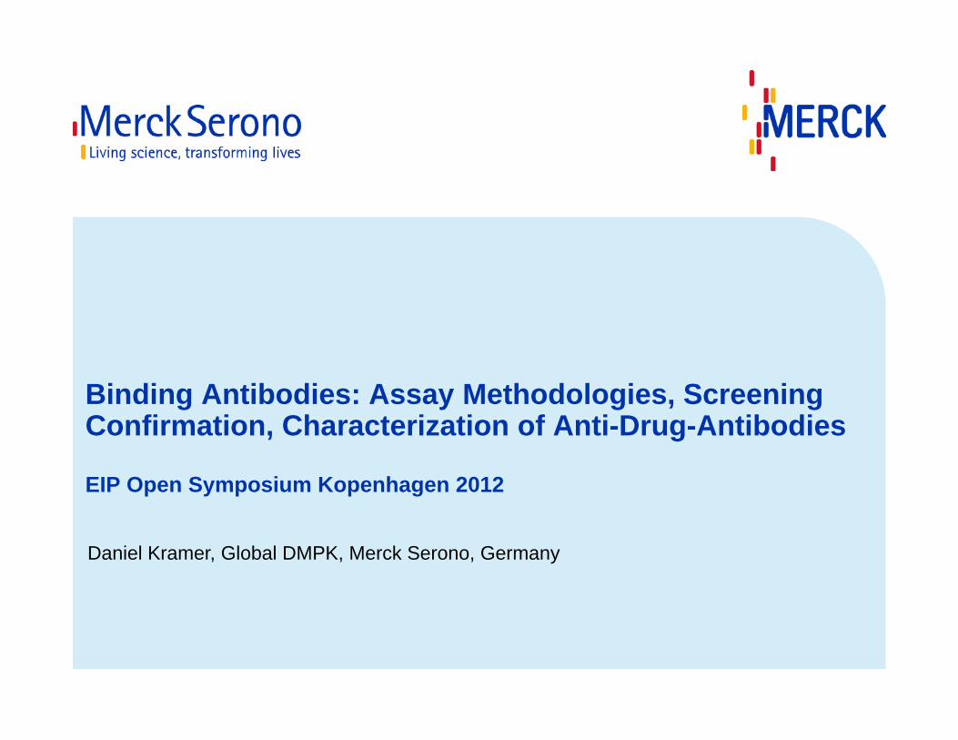
Binding Antibodies: Assay Methodologies, ScreeningConfirmation, Characterization of Anti-Drug-Antibodies
EIP Open Symposium Kopenhagen 2012
Daniel Kramer, Global DMPK, Merck Serono, Germany
2
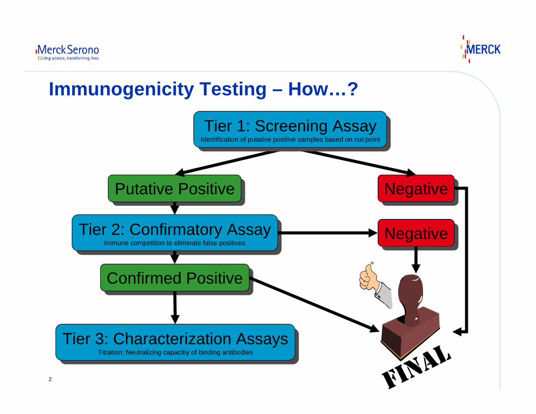
Immunogenicity Testing – How…?
Putative PositivePutative Positive NegativeNegative
Tier 2: Confirmatory AssayImmune competition to eliminate false positives
Tier 2: Confirmatory AssayImmune competition to eliminate false positives
Confirmed PositiveConfirmed Positive
Tier 3: Characterization AssaysTitration; Neutralizing capacitiy of binding antibodies
Tier 3: Characterization AssaysTitration; Neutralizing capacitiy of binding antibodies
NegativeNegative
Tier 1: Screening AssayIdentification of putative positive samples based on cut-point
Tier 1: Screening AssayIdentification of putative positive samples based on cut-point
3
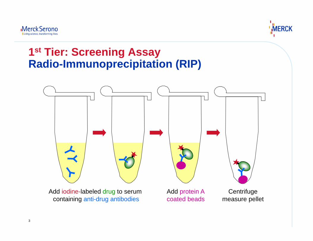
1st Tier: Screening AssayRadio-Immunoprecipitation (RIP)
Add iodine-labeled drug to serumcontaining anti-drug antibodies
Add protein Acoated beads
Centrifugemeasure pellet
4
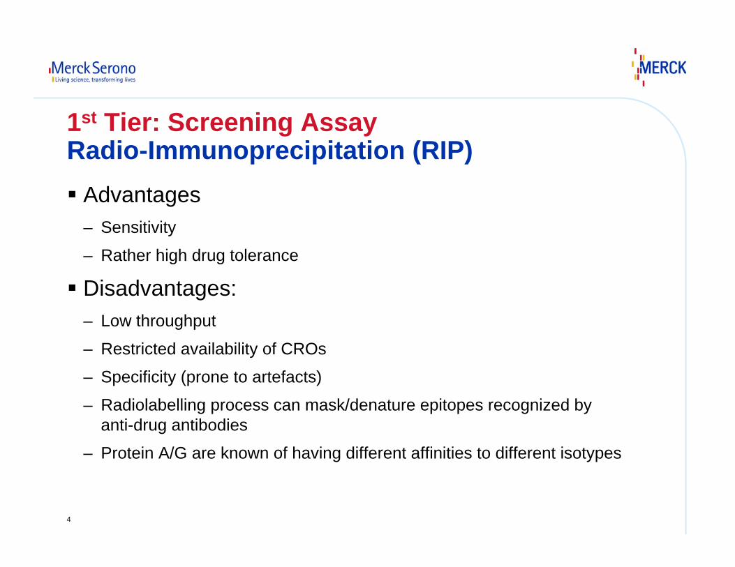
1st Tier: Screening AssayRadio-Immunoprecipitation (RIP)
Advantages– Sensitivity
– Rather high drug tolerance
Disadvantages:– Low throughput
– Restricted availability of CROs
– Specificity (prone to artefacts)
– Radiolabelling process can mask/denature epitopes recognized by anti-drug antibodies
– Protein A/G are known of having different affinities to different isotypes
5
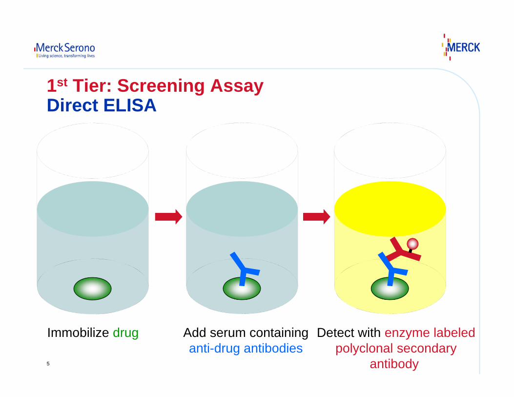
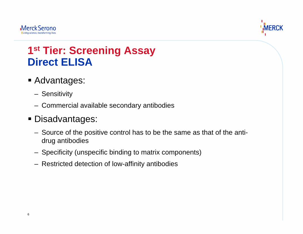
1st Tier: Screening AssayDirect ELISA
Immobilize drug Add serum containinganti-drug antibodies
Detect with enzyme labeledpolyclonal secondary
antibody
6
1st Tier: Screening AssayDirect ELISA
Advantages:– Sensitivity
– Commercial available secondary antibodies
Disadvantages:– Source of the positive control has to be the same as that of the anti-
drug antibodies
– Specificity (unspecific binding to matrix components)
– Restricted detection of low-affinity antibodies
7
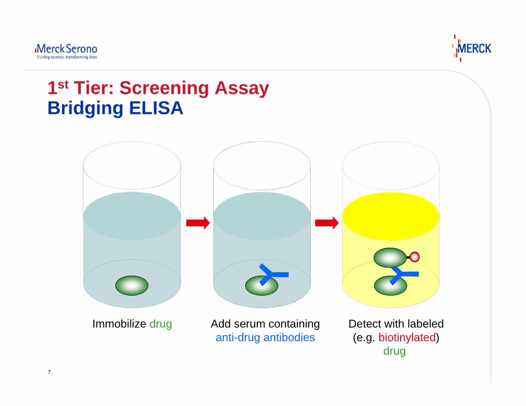
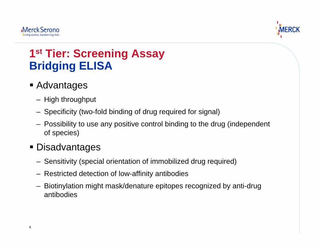
1st Tier: Screening AssayBridging ELISA
Immobilize drug Add serum containinganti-drug antibodies
Detect with labeled(e.g. biotinylated)
drug
8
1st Tier: Screening AssayBridging ELISA
Advantages– High throughput
– Specificity (two-fold binding of drug required for signal)
– Possibility to use any positive control binding to the drug (independent of species)
Disadvantages– Sensitivity (special orientation of immobilized drug required)
– Restricted detection of low-affinity antibodies
– Biotinylation might mask/denature epitopes recognized by anti-drug antibodies
9
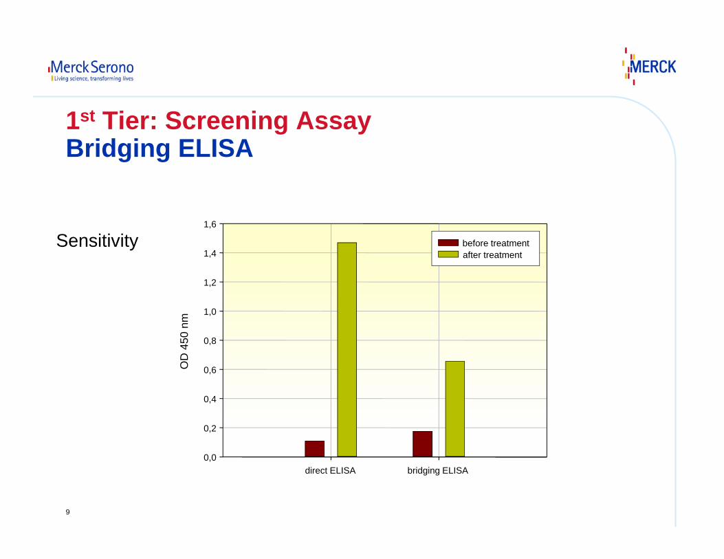
direct ELISA bridging ELISA
OD
450
nm
0,0
0,2
0,4
0,6
0,8
1,0
1,2
1,4
1,6
before treatment after treatment
1st Tier: Screening AssayBridging ELISA
Sensitivity
10
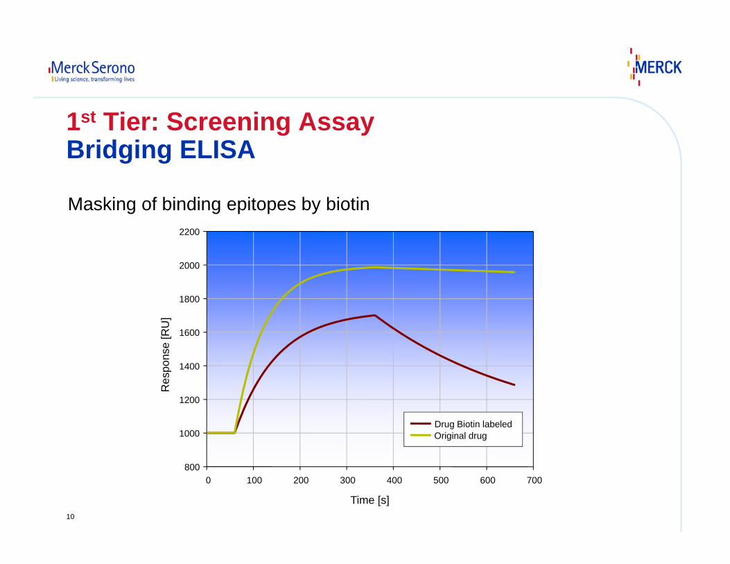
Time [s]
0 100 200 300 400 500 600 700
Res
pons
e [R
U]
800
1000
1200
1400
1600
1800
2000
2200
Drug Biotin labeled Original drug
Masking of binding epitopes by biotin
1st Tier: Screening AssayBridging ELISA
11
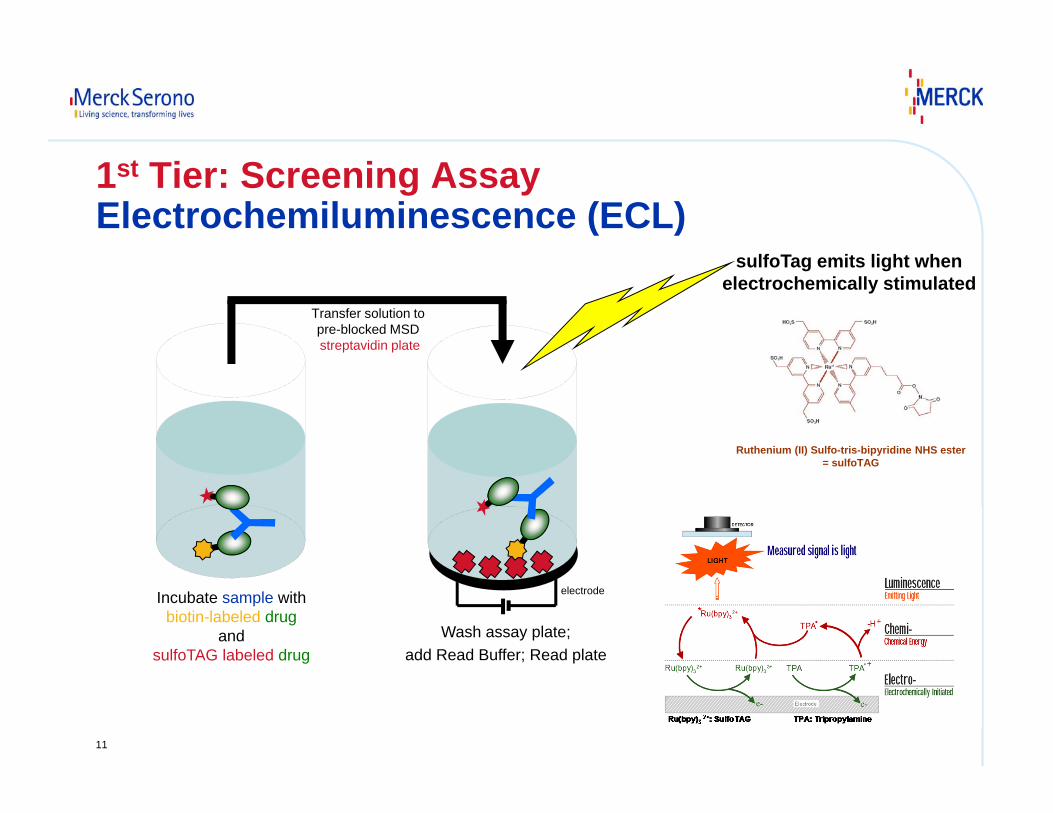
1st Tier: Screening AssayElectrochemiluminescence (ECL)
Ruthenium (II) Sulfo-tris-bipyridine NHS ester= sulfoTAG
Incubate sample withbiotin-labeled drug
andsulfoTAG labeled drug
Transfer solution to pre-blocked MSD streptavidin plate
Wash assay plate;add Read Buffer; Read plate
electrode
sulfoTag emits light whenelectrochemically stimulated
12
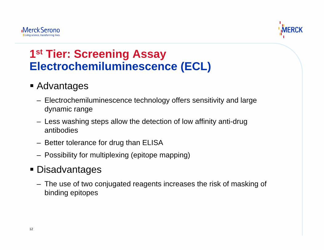
1st Tier: Screening AssayElectrochemiluminescence (ECL)
Advantages– Electrochemiluminescence technology offers sensitivity and large
dynamic range
– Less washing steps allow the detection of low affinity anti-drug antibodies
– Better tolerance for drug than ELISA
– Possibility for multiplexing (epitope mapping)
Disadvantages– The use of two conjugated reagents increases the risk of masking of
binding epitopes
13
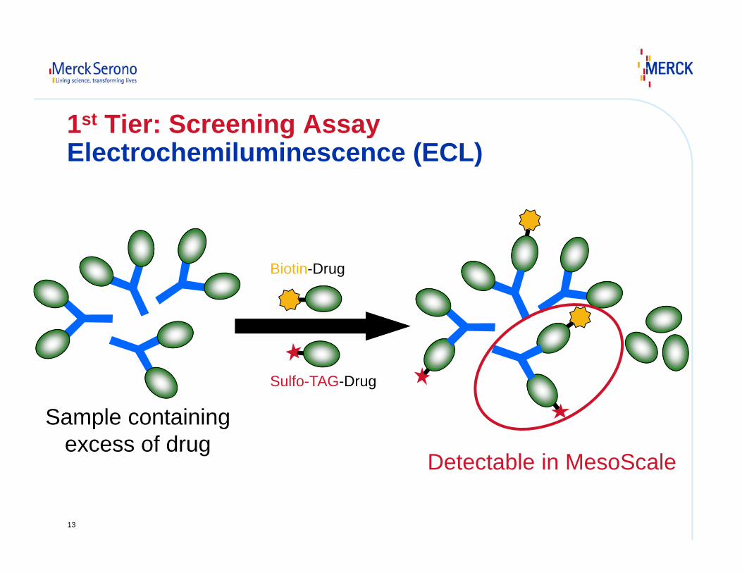
1st Tier: Screening AssayElectrochemiluminescence (ECL)
Sulfo-TAG-Drug
Biotin-Drug
Detectable in MesoScale
Sample containingexcess of drug
14
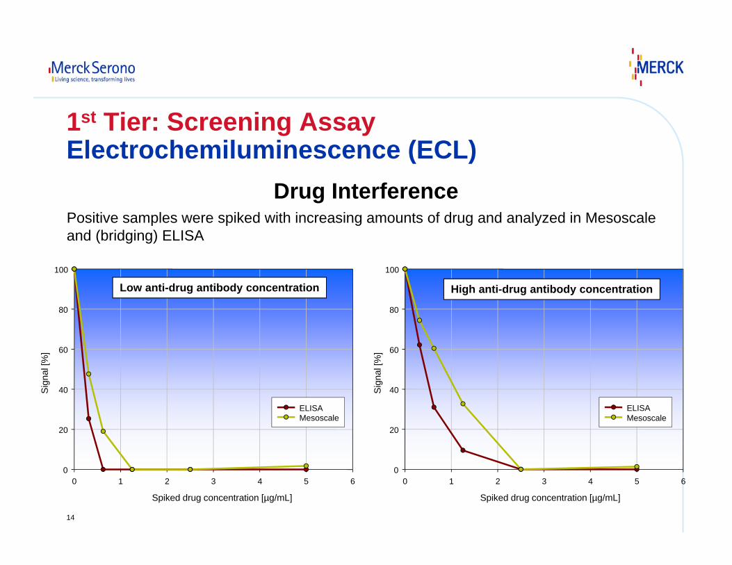
1st Tier: Screening AssayElectrochemiluminescence (ECL)
Drug InterferencePositive samples were spiked with increasing amounts of drug and analyzed in Mesoscale and (bridging) ELISA
Spiked drug concentration [µg/mL]
0 1 2 3 4 5 6
Sig
nal [
%]
0
20
40
60
80
100
ELISA Mesoscale
Low anti-drug antibody concentration
Spiked drug concentration [µg/mL]
0 1 2 3 4 5 6
Sig
nal [
%]
0
20
40
60
80
100
ELISA Mesoscale
High anti-drug antibody concentration
15
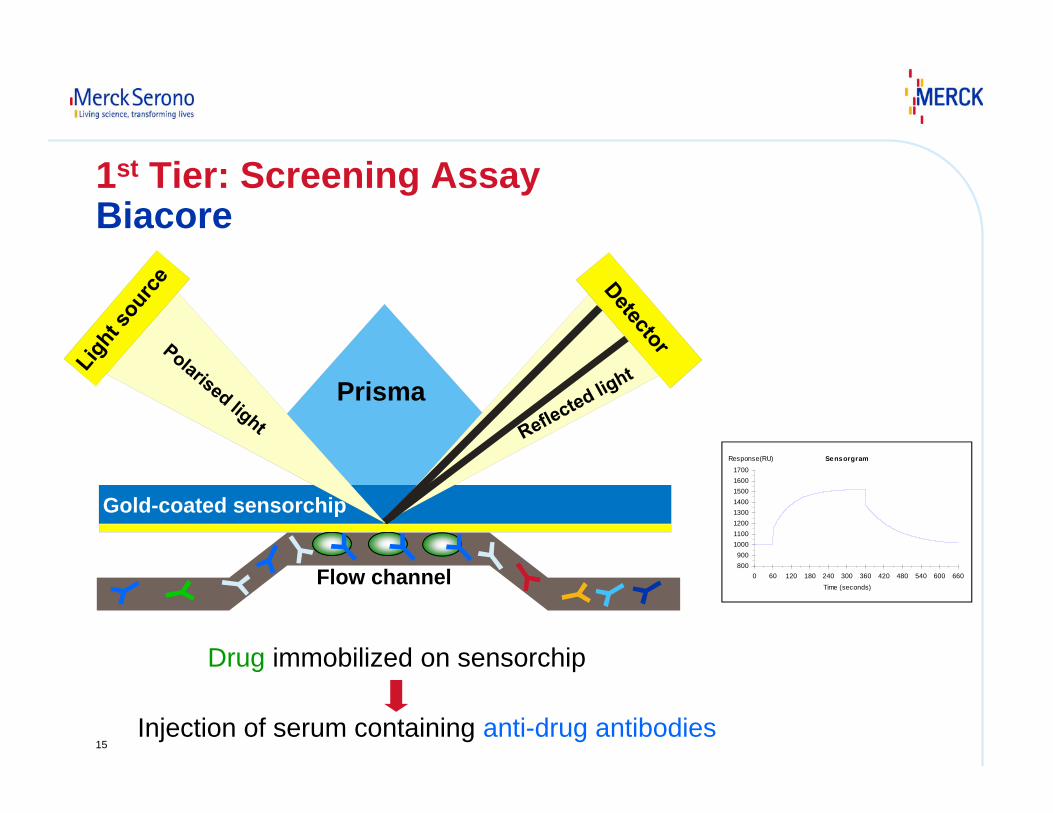
Drug immobilized on sensorchip
Injection of serum containing anti-drug antibodies
800900
10001100120013001400150016001700
0 60 120 180 240 300 360 420 480 540 600 660
Response(RU) Sensorgram
Time (seconds)
Prisma
Gold-coated sensorchip
Flow channel
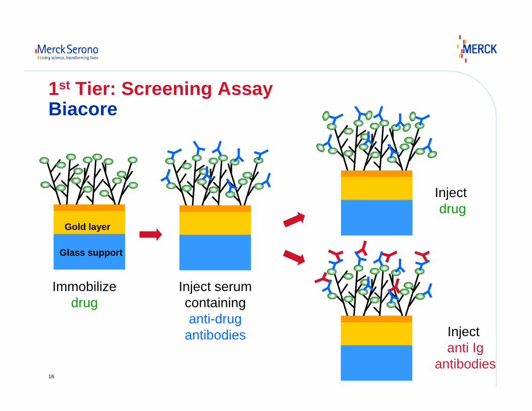
1st Tier: Screening Assay Biacore
16
Immobilizedrug
Inject serumcontaininganti-drug
antibodies
Inject drug
Glass support
Gold layer
Inject anti Ig
antibodies
1st Tier: Screening AssayBiacore
17
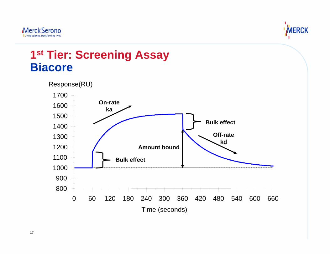
800900
10001100120013001400150016001700
0 60 120 180 240 300 360 420 480 540 600 660
Response(RU)
Time (seconds)
On-rateka
Off-ratekd
Amount bound
Bulk effect
Bulk effect
1st Tier: Screening AssayBiacore
18
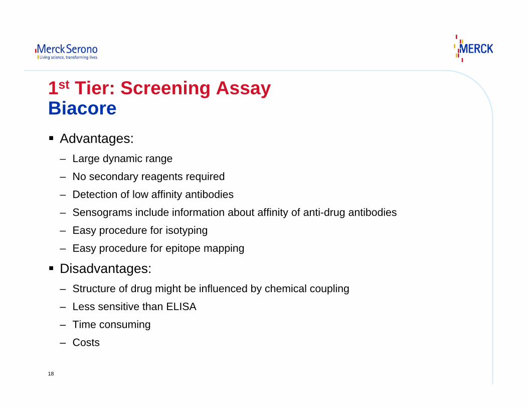
1st Tier: Screening AssayBiacore
Advantages:– Large dynamic range
– No secondary reagents required
– Detection of low affinity antibodies
– Sensograms include information about affinity of anti-drug antibodies
– Easy procedure for isotyping
– Easy procedure for epitope mapping
Disadvantages:– Structure of drug might be influenced by chemical coupling
– Less sensitive than ELISA
– Time consuming
– Costs
19
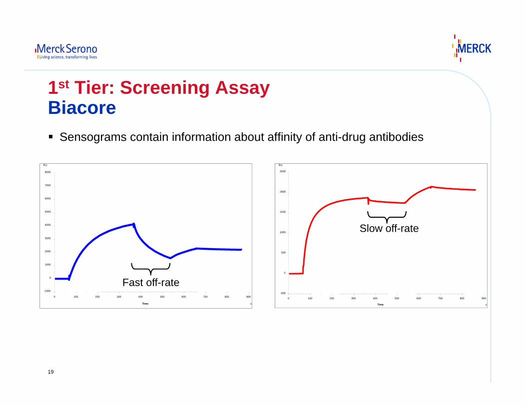
Sensograms contain information about affinity of anti-drug antibodies
-1000
0
1000
2000
3000
4000
5000
6000
7000
8000
0 100 200 300 400 500 600 700 800 900
Time s
RU
-500
0
500
1000
1500
2000
2500
0 100 200 300 400 500 600 700 800 900
Time s
RU
Fast off-rate
Slow off-rate
1st Tier: Screening AssayBiacore
20
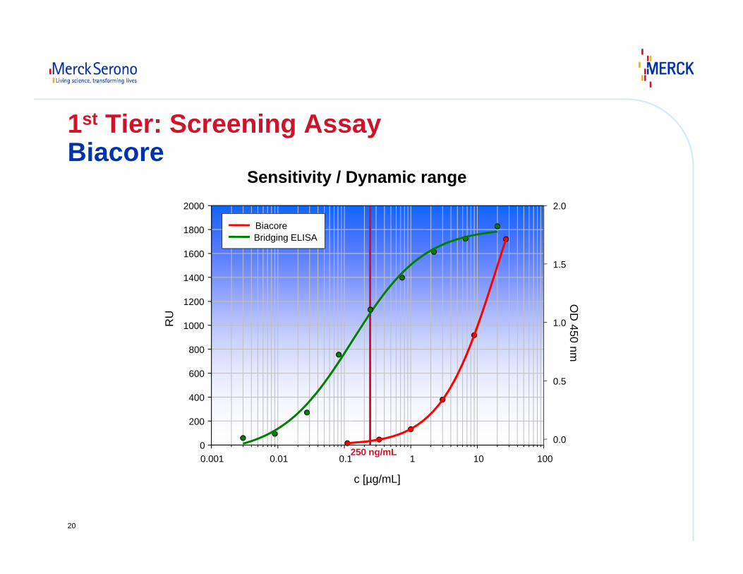
Sensitivity / Dynamic range
c [µg/mL]
0.001 0.01 0.1 1 10 100
RU
0
200
400
600
800
1000
1200
1400
1600
1800
2000
OD
450 nm
0.0
0.5
1.0
1.5
2.0
BiacoreBridging ELISA
250 ng/mL
1st Tier: Screening AssayBiacore
21
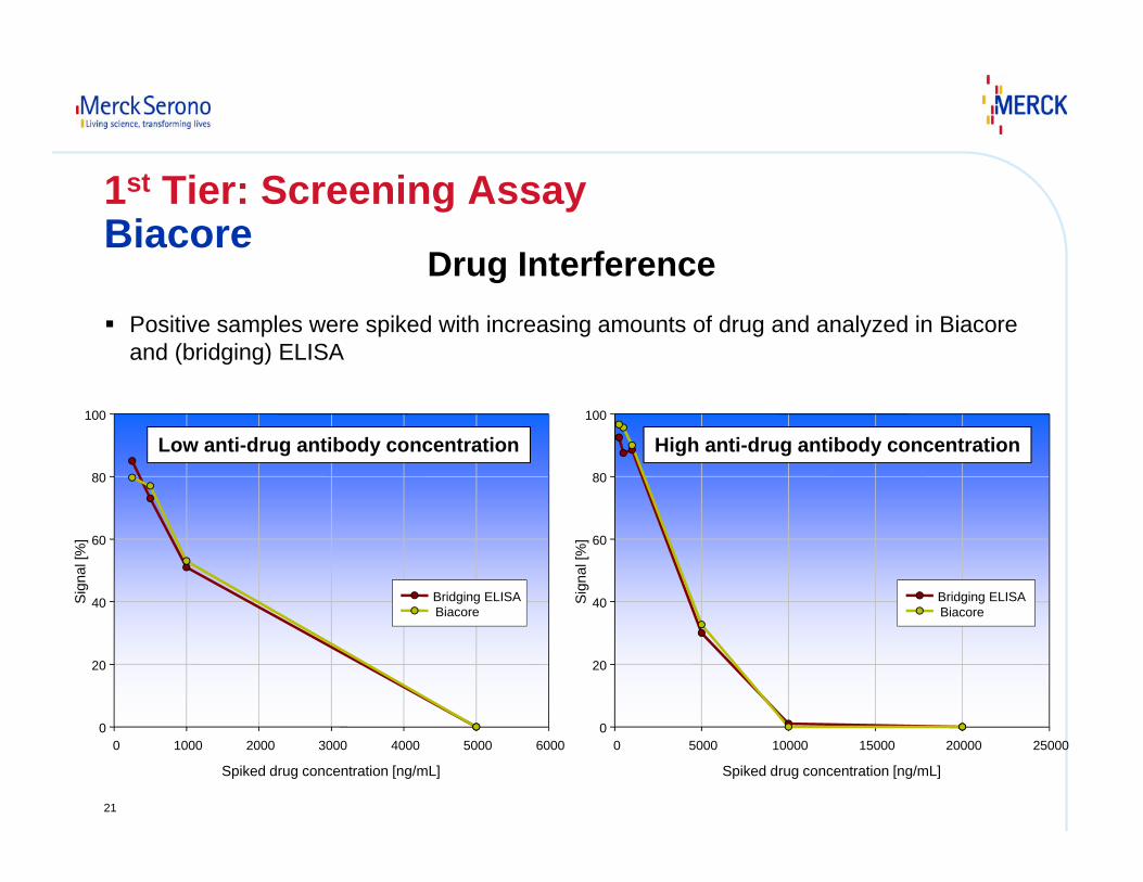
Spiked drug concentration [ng/mL]
0 1000 2000 3000 4000 5000 6000
Sig
nal [
%]
0
20
40
60
80
100
Bridging ELISABiacore
Low anti-drug antibody concentration
Spiked drug concentration [ng/mL]
0 5000 10000 15000 20000 25000
Sig
nal [
%]
0
20
40
60
80
100
Bridging ELISA Biacore
High anti-drug antibody concentration
Positive samples were spiked with increasing amounts of drug and analyzed in Biacore and (bridging) ELISA
Drug Interference
1st Tier: Screening AssayBiacore
22
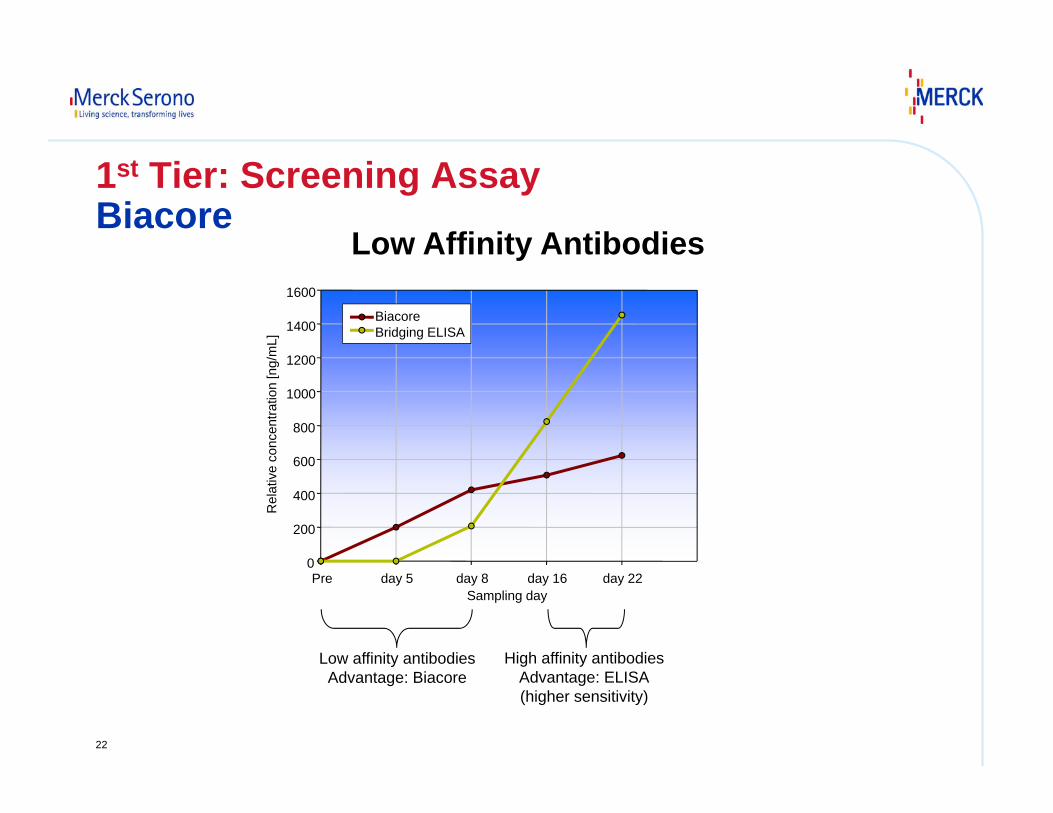
Sampling dayPre day 5 day 8 day 16 day 22
Rel
ativ
e co
ncen
tratio
n [n
g/m
L]
0
200
400
600
800
1000
1200
1400
1600
Biacore Bridging ELISA
Low affinity antibodiesAdvantage: Biacore
High affinity antibodiesAdvantage: ELISA(higher sensitivity)
Low Affinity Antibodies
1st Tier: Screening AssayBiacore
23
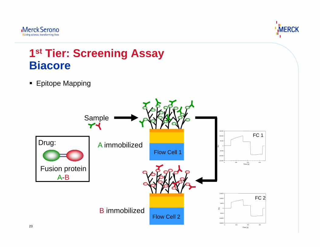
1st Tier: Screening AssayBiacore
Epitope Mapping
Flow Cell 1
Flow Cell 2
A immobilized
B immobilized
Sample
Fusion proteinA-B
Drug:
Time [s]0 200 400 600
RU
-150000
-100000
-50000
0
50000
100000
150000
FC 1
Time [s]
0 200 400 600
RU
-150000
-100000
-50000
0
50000
100000
150000
FC 2
24
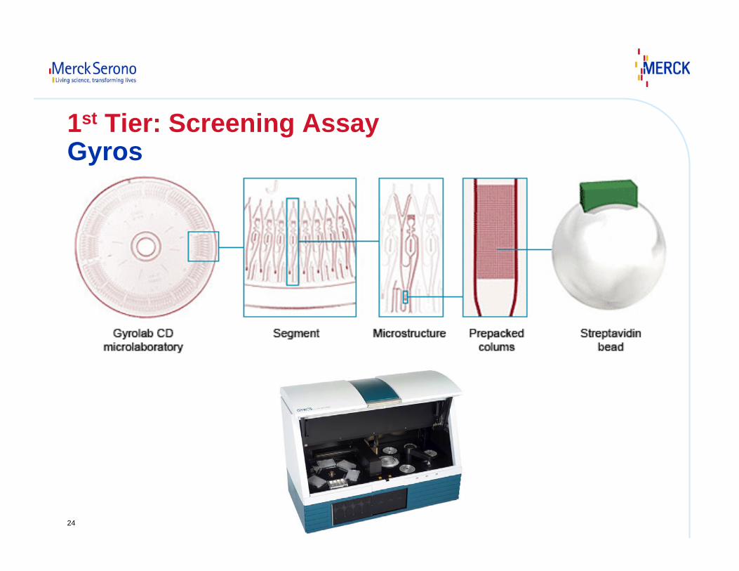
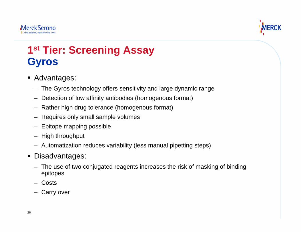
1st Tier: Screening AssayGyros
25
ALEXA-Drug
Biotin-Drug
Spin
Serum containinganti-drug antibodies
Fluorescentsignal
1st Tier: Screening AssayGyros
26
Advantages:– The Gyros technology offers sensitivity and large dynamic range– Detection of low affinity antibodies (homogenous format)– Rather high drug tolerance (homogenous format)– Requires only small sample volumes– Epitope mapping possible– High throughput– Automatization reduces variability (less manual pipetting steps)
Disadvantages:– The use of two conjugated reagents increases the risk of masking of binding
epitopes– Costs– Carry over
1st Tier: Screening AssayGyros
27
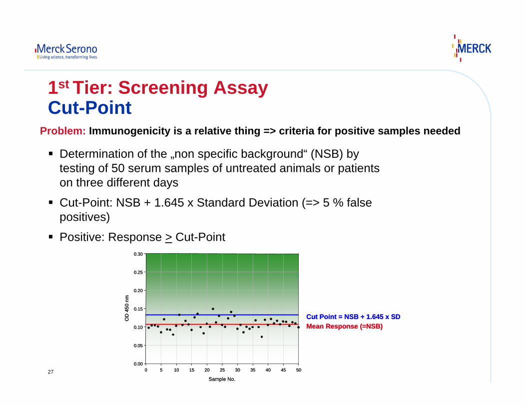
1st Tier: Screening AssayCut-Point
Determination of the „non specific background“ (NSB) by testing of 50 serum samples of untreated animals or patients on three different days
Cut-Point: NSB + 1.645 x Standard Deviation (=> 5 % false positives)
Positive: Response > Cut-Point
Sample No.
0 5 10 15 20 25 30 35 40 45 50
OD
450
nm
0.00
0.05
0.10
0.15
0.20
0.25
0.30
Cut Point = NSB + 1.645 x SDMean Response (=NSB)
Sample No.
0 5 10 15 20 25 30 35 40 45 50
OD
450
nm
0.00
0.05
0.10
0.15
0.20
0.25
0.30
Cut Point = NSB + 1.645 x SDMean Response (=NSB)
Problem: Immunogenicity is a relative thing => criteria for positive samples needed
28
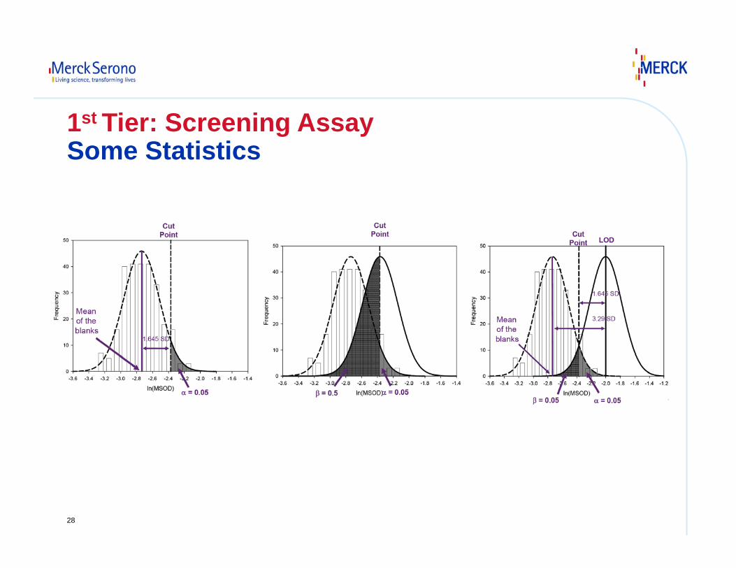
1st Tier: Screening AssaySome Statistics
29
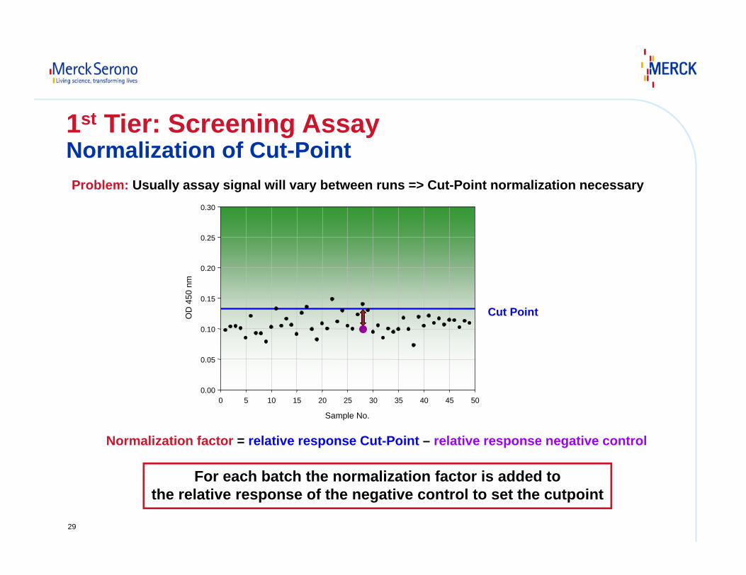
Problem: Usually assay signal will vary between runs => Cut-Point normalization necessary
Normalization factor = relative response Cut-Point – relative response negative control
For each batch the normalization factor is added tothe relative response of the negative control to set the cutpoint
1st Tier: Screening AssayNormalization of Cut-Point
Sample No.
0 5 10 15 20 25 30 35 40 45 50
OD
450
nm
0.00
0.05
0.10
0.15
0.20
0.25
0.30
Cut Point
30
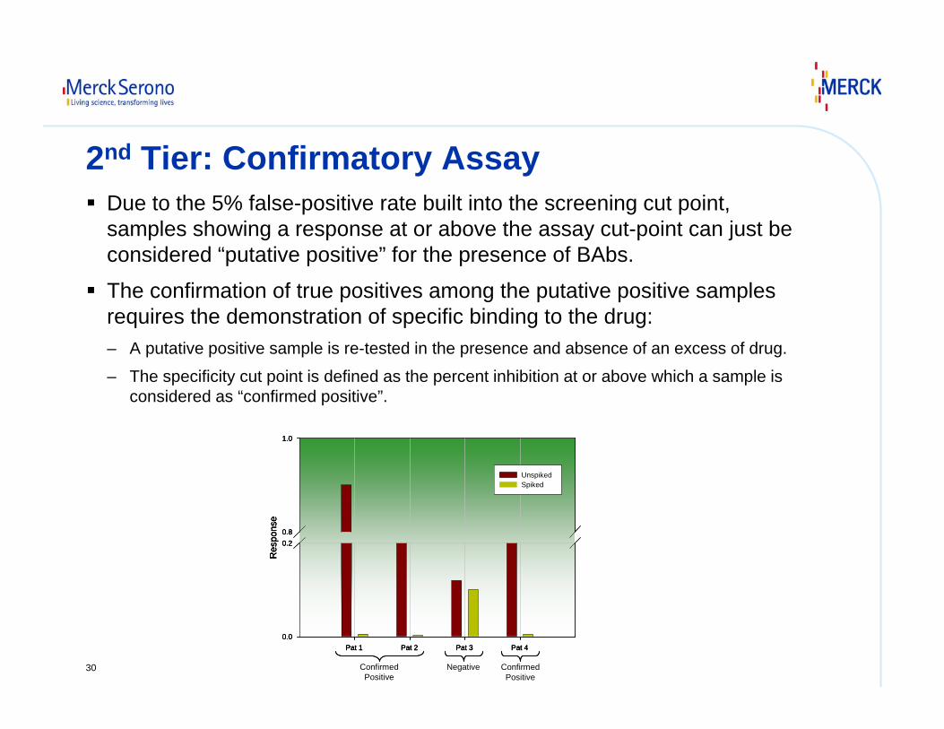
2nd Tier: Confirmatory AssayDue to the 5% false-positive rate built into the screening cut point, samples showing a response at or above the assay cut-point can just be considered “putative positive” for the presence of BAbs.
The confirmation of true positives among the putative positive samples requires the demonstration of specific binding to the drug: – A putative positive sample is re-tested in the presence and absence of an excess of drug.
– The specificity cut point is defined as the percent inhibition at or above which a sample is considered as “confirmed positive”.
Pat 1 Pat 2 Pat 3 Pat 4
Res
pons
e
0.0
0.20.8
1.0
UnspikedSpiked
Pat 1 Pat 2 Pat 3 Pat 4
Res
pons
e
0.0
0.20.8
1.0
UnspikedSpiked
Pat 1 Pat 2 Pat 3 Pat 4
Res
pons
e
0.0
0.20.8
1.0
UnspikedSpiked
ConfirmedPositive
ConfirmedPositive
Negative
31
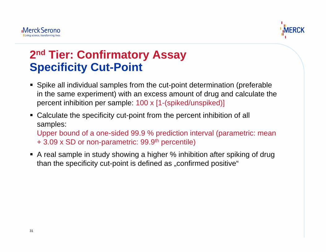
2nd Tier: Confirmatory AssaySpecificity Cut-Point
Spike all individual samples from the cut-point determination (preferable in the same experiment) with an excess amount of drug and calculate the percent inhibition per sample: 100 x [1-(spiked/unspiked)]
Calculate the specificity cut-point from the percent inhibition of all samples:Upper bound of a one-sided 99.9 % prediction interval (parametric: mean + 3.09 x SD or non-parametric: 99.9th percentile)
A real sample in study showing a higher % inhibition after spiking of drug than the specificity cut-point is defined as „confirmed positive“

32
3rd Tier: TitrationAim:– Retrieve quasi-quantitative information for confirmed positive
samples
Procedure:– Serial dilution of confirmed positive samples
– Titer = -log dilution factor of the last dilution that tests positive
Dilution
1e-5(1:100000)
1e-4(1:10000)
1e-3(1:1000)
1e-2(1:100)
1e-1(1:10)
OD
0.0
0.2
0.4
0.6
0.8
1.0
1.2
1.4
Cut Point
Dilution 1:2560 (= 3.91 x 10⁻⁴)=> Titer = 3.41
Dilution
1e-5(1:100000)
1e-4(1:10000)
1e-3(1:1000)
1e-2(1:100)
1e-1(1:10)
OD
0.0
0.2
0.4
0.6
0.8
1.0
1.2
1.4
Cut Point
Dilution 1:2560 (= 3.91 x 10⁻⁴)=> Titer = 3.41
33
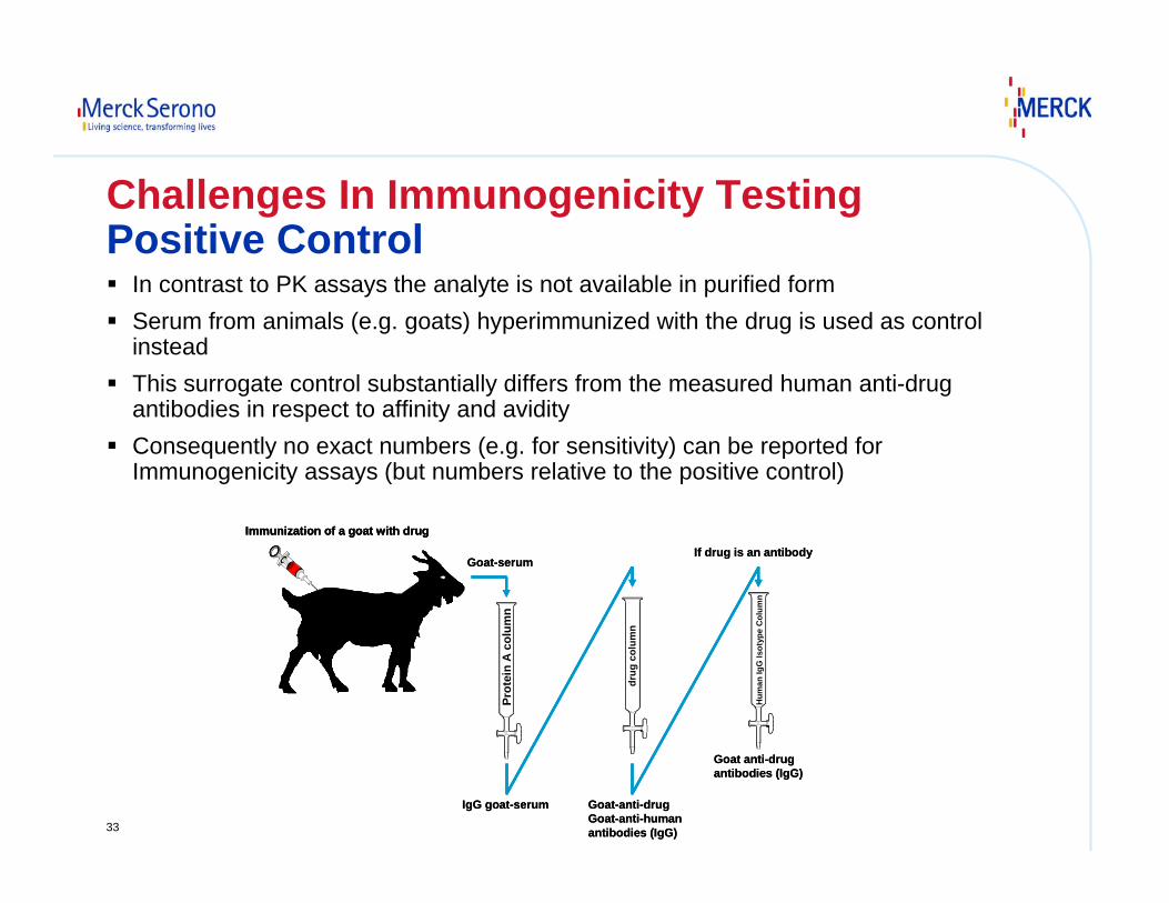
Challenges In Immunogenicity TestingPositive Control
In contrast to PK assays the analyte is not available in purified formSerum from animals (e.g. goats) hyperimmunized with the drug is used as control insteadThis surrogate control substantially differs from the measured human anti-drug antibodies in respect to affinity and avidityConsequently no exact numbers (e.g. for sensitivity) can be reported for Immunogenicity assays (but numbers relative to the positive control)
Immunization of a goat with drug
Goat-serum
IgG goat-serum Goat-anti-drugGoat-anti-humanantibodies (IgG)
Prot
ein
A co
lum
n
drug
col
umn
Hum
an Ig
GIs
otyp
eC
olum
nGoat anti-drugantibodies (IgG)
If drug is an antibody
Immunization of a goat with drugImmunization of a goat with drug
Goat-serum
IgG goat-serum Goat-anti-drugGoat-anti-humanantibodies (IgG)
Prot
ein
A co
lum
n
drug
col
umn
Hum
an Ig
GIs
otyp
eC
olum
nGoat anti-drugantibodies (IgG)
If drug is an antibody
34
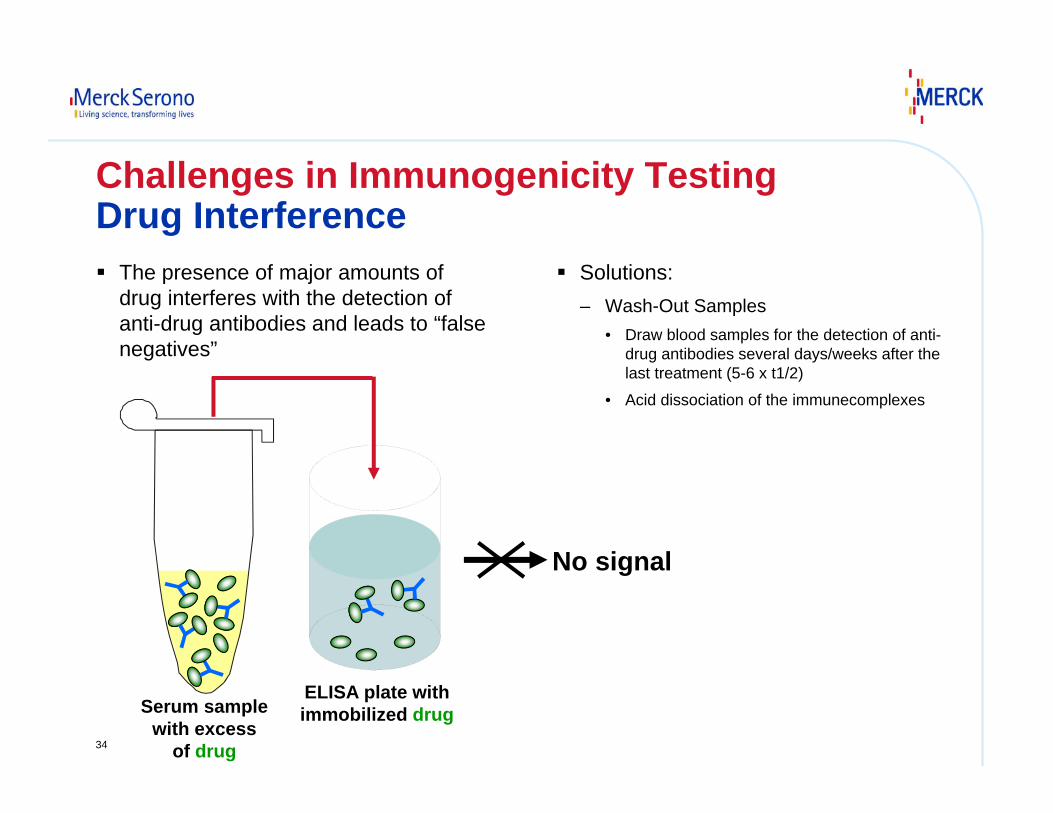
Challenges in Immunogenicity TestingDrug Interference
The presence of major amounts of drug interferes with the detection of anti-drug antibodies and leads to “false negatives”
Solutions:– Wash-Out Samples
• Draw blood samples for the detection of anti-drug antibodies several days/weeks after the last treatment (5-6 x t1/2)
• Acid dissociation of the immunecomplexes
Serum samplewith excess
of drug
ELISA plate withimmobilized drug
No signal
35
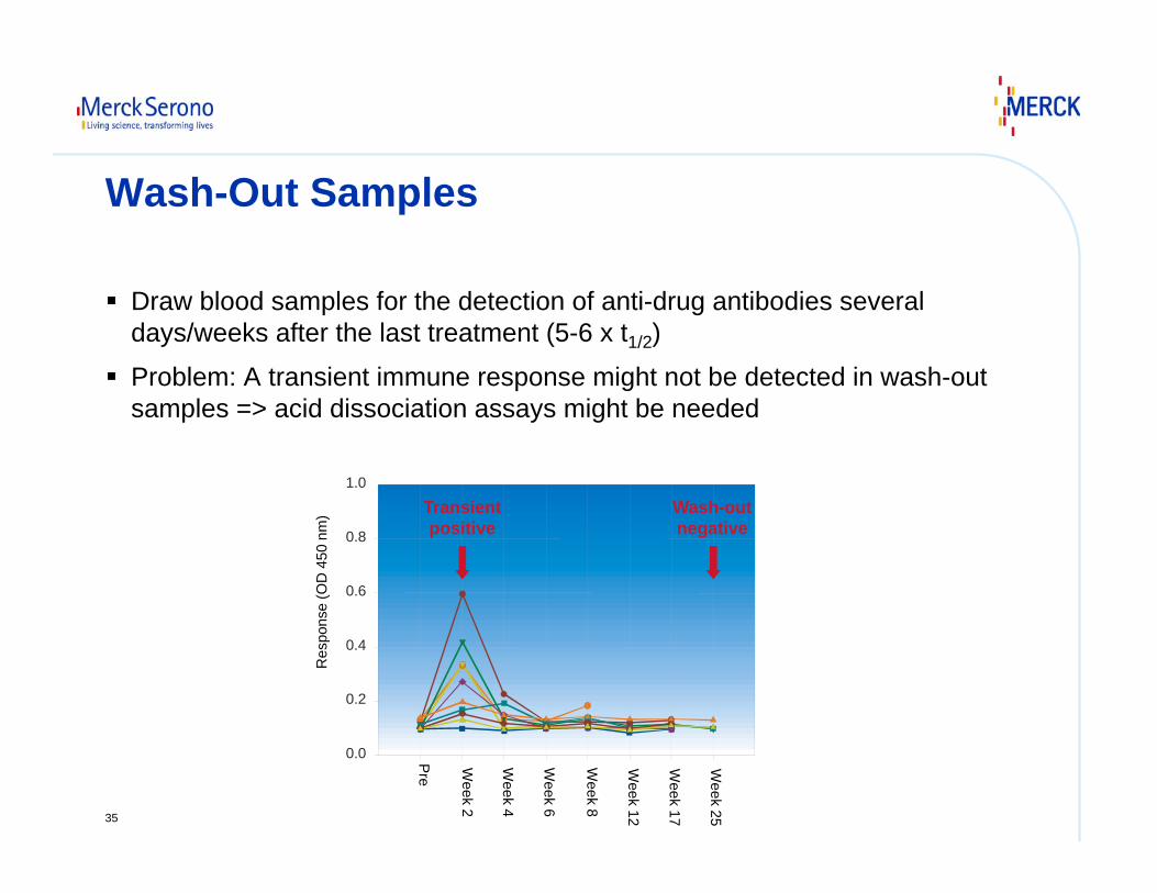
Wash-Out Samples
Draw blood samples for the detection of anti-drug antibodies several days/weeks after the last treatment (5-6 x t1/2)
Problem: A transient immune response might not be detected in wash-out samples => acid dissociation assays might be needed
0.0
0.2
0.4
0.6
0.8
1.0
Res
pons
e (O
D 4
50 n
m)
Pre
Week 2
Week 4
Week 6
Week 8
Week 12
Week 17
Week 25
Wash-outnegative
Transientpositive
36
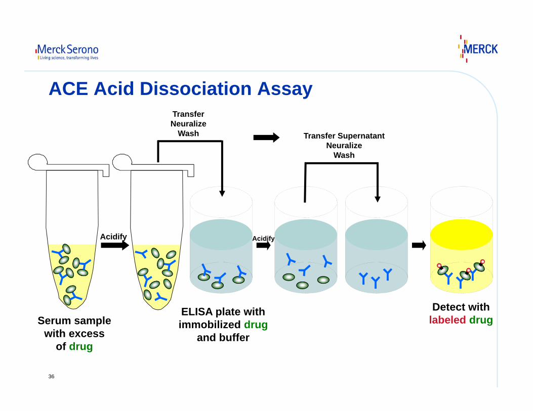
ACE Acid Dissociation Assay
Serum samplewith excess
of drug
Acidify
ELISA plate withimmobilized drug
and buffer
Detect withlabeled drug
Acidify
TransferNeuralize
Wash Transfer SupernatantNeuralize
Wash

37
THANK YOU !


![Chemical Methodologies€¦ · chromatography [HPLC] assay method validated the for determination of warfarin in solid pharmaceutical dosage forms. Isocratic phase high performance](https://img.pdfslide.us/doc/110x75/5f0d7c3e7e708231d43a9780/chemical-chromatography-hplc-assay-method-validated-the-for-determination-of-warfarin.jpg)