Embed Size (px)
Citation preview

RESEARCH PAPER
Bicontinuous Cubic Liquid Crystalline Nanoparticles for OralDelivery of Doxorubicin: Implications on Bioavailability,Therapeutic Efficacy, and Cardiotoxicity
Nitin K. Swarnakar & Kaushik Thanki & Sanyog Jain
Received: 27 July 2013 /Accepted: 20 October 2013 /Published online: 12 November 2013# Springer Science+Business Media New York 2013
ABSTRACTPurpose The present study explores the potential of bicontinouscubic liquid crystalline nanoparticles (LCNPs) for improvingtherapeutic potential of doxorubicin.Methods Phytantriol based Dox-LCNPs were prepared usinghydrotrope method, optimized for various formulationcomponents, process variables and lyophilized. Structuralelucidation of the reconstituted formulation was performed usingHR-TEM and SAXS analysis. The developed formulation wassubjected to exhaustive cell culture experiments for deliverypotential (Caco-2 cells) and efficacy (MCF-7 cells). Finally, in vivopharmacokinetics, pharmacodynamic studies in DMBA inducedbreast cancer model and cardiotoxicity were also evaluated.Results The reconstituted formulation exhibited Pn3m typecubic structure, evident by SAXS and posed stability in simulatedgastrointestinal fluids and at accelerated stability conditions for6 months. Dox-LCNPs revealed significantly higher cellcytotoxicity (16.23-fold) against MCF-7 cell lines as compared tofree drug owing to its preferential localization in the vicinity ofnucleus. Furthermore, Caco-2 cell experiments revealedformation of reversible “virtual pathways” in the cell membranefor Dox-LCNPs and hence posed significantly higher relative oralbioavailability (17.74-fold). Subsequently, Single dose of Dox-LCNPs (per oral ) led to significant reduction in % tumor burden(~42%) as compared that of ~31% observed in case ofAdriamycin® (i.v. ) when evaluated in DMBA induced breast
cancer model. Moreover, Dox induced cardiotoxicity was alsofound to be significantly lower in case of Dox-LCNPs ascompared to clinical formulations (Adriamycin® and Lipodox®).Conclusion Incorporation of Dox in the novel LCNPsdemonstrated improved antitumor efficacy and safety profile andcan be a viable option for oral chemotherapy.
KEY WORDS cardiotoxicity . doxorubicin . liquid crystallinenanoparticles (LCNPs) . oral delivery . Phytantriol
INTRODUCTION
Doxorubicin (Dox), one of the most potent anticancer drugs isprescribed for the treatment of wide range of cancers.Clinically approved Dox formulations (Adriamycin® andRubex®) are rapidly cleared from the central compartmentto non-specific tissue compartment within 5 min uponintravenous administration leading to sub-therapeutic levelsin plasma and also necessitates frequent administration (1).Direct exposure of potent drug to the tissue also leads toserious side effect such as irreversible cardiomyopathy,myelosuppression etc. Although, novel delivery strategies suchas PEGylated liposomes (Doxil, LipoDox®) have beenimplemented to enhance circulation half-life, these clinicallyavailable formulations are intended for intravenousadministration (i.v. ) only and often associated with poorpatient compliance and other clinical complications such ashand-foot syndrome apart from side effects classical to Doxand may lead to early termination of the therapy (2,3). Peroralroute is the safest route of drug administration which hashigher patient compliance, lesser complications and cost-effectiveness as compared to parental drug delivery. Oraldelivery of Dox, although fascinating, is often associated withpoor oral bioavailability (<5%) due to its inherent lowpermeability, substrate specificity to P-gp efflux pump andsusceptibility for extensive high first pass metabolism (4,5).
Electronic supplementary material The online version of this article(doi:10.1007/s11095-013-1244-8) contains supplementary material, which isavailable to authorized users.
N. K. Swarnakar : K. Thanki : S. Jain (*)Centre for Pharmaceutical Nanotechnology, Department of PharmaceuticsNational Institute of Pharmaceutical Education and Research (NIPER)Sector 67, S.A.S. Nagar, Mohali, Punjab 160062, Indiae-mail: [email protected]; [email protected]
Pharm Res (2014) 31:1219–1238DOI 10.1007/s11095-013-1244-8
Over the last few years, drug delivery via carrier basedapproaches such as either lipidic carrier or polymeric carrierand co-administration of Dox and other bioactives such asenzyme/P-gp inhibitors, quercetin, morin, cyclosporin A etc.have been implemented (6). Although, these techniques weresuccessful up to some extent, these do suffer from one or morelimitations such as poor drug loading, complexmanufacturingprocess, insufficient bioavailability and certain immunologicalor medical complications (7). At present, none of formulationswas found to be orally efficient and clinically pertinent. Ourgroup is also actively involved in development various novelformulations for oral delivery of Dox. Orally administeredDox-loaded poly(lactic-co-glycolic acid) (PLGA) nanoparticlesdemonstrated significant tumor growth inhibition in 7, 12-dimethylbenz[α]anthracene (DMBA) induced breast cancermodel in the rats which was equivalent to that of free Dox (i.v. )(5). Further, a patented Dox loaded layersomes technologiesalso exhibited significant reduction in the tumor growth ascompared to control and free Dox (i.v. ), while results werecomparable to LipoDox® (i.v. ). However, its multiple dosingwas required to demonstrate anticancer activity (8).
Over the last few years, liquid crystalline nanoparticles(LCNPs) have been identified as promising oral drug deliveryvehicle (6). The LCNPs are self-assembled structure, formedupon exposing polar lipids into polar environment in presenceof suitable surfactant. These assemblies demonstrate existenceof non-lamellar structure consisting of hydrophilic andlipophilic domains (9). The special arrangement of lipidsprovides both rigid and fluidic characteristics therebyimparting stability, sustained release profile and high drugpayload to the system (10,11). Therefore, LNCPs transformsinto various secondary vehicles (mixed micelles, cubic andhexagonal nanoparticles, vesicular carriers) in thegastrointestinal (GI) tract. In spite of their transformation,LCNPs are able to hold the drug inside the matrix and arebelieved to facilitate the absorption of drugs via variousmechanisms such as membrane fusing properties, receptormediated endocytosis, absorption transporters, etc. (10,11).Enhancement in oral bioavailability of few drugs such as suchas omapatrilat, simvastatin, silymarin etc. has been reportedvia incorporation into LCNPs (12–17).
Phytantriol, is a newer generation of lipid categorizedunder the generally recognized as safe (GRAS) substance,has been explored for the preparation of LCNPs (18–20). Incontrast to glyceride lipids, it is non-digestible lipid thatprovides superior encapsulation, improved GI stability, andsustained release behavior of encapsulated bioactives(15,17). Boyd and coworkers have demonstrated about7.0-fold higher oral bioavailability of cinnarizine byphytantriol based LNCPs (15). However, none of reporttill date has explored their potential in improving thetherapeutic efficacy and safety profile of anticancerdrugs following oral administration.
In present work, Dox loaded phytantriol based LNCPshave been prepared, extensively optimized for formulationcomponents and process parameters. Then after, formulationwas freezed dried by employing step-wise freeze drying cyclefor enhancing its storage stability. The developed formulationwas characterized by various in vitro techniques. Finally,delivery potential of Dox-LCNPs was evaluated by extensivein vitro cell culture experiments and in vivo pharmacokinetic,antitumor efficacy and cardiotoxicity studies following oraladministration and comparison with i.v. administeredmarketed formulations of Dox i.e. Adriamycin® andLipoDox®.
MATERIALS AND METHODS
Doxorubicin hydrochloride salt (>99%), 3,7,11,15-tetramethyl-1,2,3-hexadecanetriol (phytantriol), differentgrades of Pluronic® (F-108, F-127, F-68 and F-87) andcyclosporin A were generous gift from Sun PharmaAdvanced Research Company (SPARC Ltd.), Vadodara,India, DSM Nutritional Products, Inc., Germany, BASF,Germany and Panacea Biotech, Mumbai, India, respectively.Triton X-100, 12-dimethylbenz[a]anthracene (DMBA),sulforhodamine B dye (SRB), Neutral red (NR), 4′,6-diamidino-2-phenylindole (DAPI) and Minimum EssentialMedium Eagle (MEM) were purchased from Sigma, USA.6-well, 24-well and 96-well cell culture plates were procuredfromBectonDickinson, USA. Thiobarbituric acid (GR grade)was purchased from Loba Chemie, India. Malondialdehyde(MDA), lactate dehydrogenase (LDH), glutathione (GSH),superoxide dismutase (SOD) estimation kits were purchasedfrom Accurex Biomedical Pvt. Ltd, India. Creatine kinasemyocytes B (CK-MB) kit was purchased from CoralBiosystems, India. Acetone, acetonitrile and methanol(HPLC grade) were purchased from Merck, India. Ultra-pure deionized water (LaboStar™ ultrapure water Systems,Germany) was used for all the experiments. All other reagentsused were of analytical grade and purchased from localsuppliers unless mentioned.
Preparation of Dox-LCNPs
Dox-LCNPs were prepared by hydrotrope method (21).Briefly, Dox was dissolved in minimal quantity of water(250 μl) and diluted with ethanol (300 μl) containingphytantriol (100 mg). The resulting isotropic solution wasadded dropwise into the surfactant solution (10 ml) at stirring2,000 rpm for 12 h. Ethanol diffusion followed by evaporationresulted in formation of Dox-LCNPs. Further, the resultantLCNPs were subjected to probe sonication at 30% amplitudefor 10 s and the system was equilibrated by stirring at 500 rpmfor 2 h. Finally, free Dox and surfactant were removed from
1220 Swarnakar, Thanki and Jain

the formulation by dialysis (MW 15 kD, Sigma USA) againstdeionized water for 20 min at 250 rpm.
Optimization of Formulation Components and ProcessVariables
Exhaustive optimization of formulation components viz.ethanol concentration, type and concentration of stabilizerand % Dox loading and sonication time were carried out forpreparation of Dox-LCNPs. The optimum parameters wereselected on the basis of quality attributes (particle size, sizedistribution, zeta potential and encapsulation efficiency) ofprepared Dox-LCNPs. During optimization, certain otherminor experimental parameters like amount of lipid added(1% w/w with respect to dispersion), stirring speed, time andfinal volume of dispersion (10 ml) were kept constant.
Optimization of Ethanol Concentration
Various concentrations of ethanol (0.1–0.4% w/v) were triedfor the preparation of Dox-LCNPs while keeping constantconcentration of Dox (0.01% w/v) and Pluronic® F-108(0.5% w/v) with respect to dispersion volume.
Optimization of Type of Stabilizer
Surfactant screening was conducted by preparing the Dox-LCNPs in presence of 0.5% w/v concentration of varioustypes of Pluronic® surfactants (F-127, F-108, F-87 and F-68). The best stabilizer was implemented for further studies.
Optimization of Stabilizer Concentration
Various concentrations of Pluronic® F-127 (0.05–0.07% w/v with respect to dispersion volume) wereevaluated in the preparation of Dox-LCNPs. Thesuitable concentration of surfactant was utilized forfurther optimizations.
Optimization of Drug Loading
Dox-LCNPs were prepared at various drug loading 10.0,15.0, 17.5 and 20.0 (% w/w with respect to lipid) and theireffect on formulation attributes was evaluated. The optimizeddrug loading was utilized for further studies.
Optimization of Sonication Time
Probe sonication was carried out to reduce the particle size aswell as to improve the homogeneity of Dox-LCNPs.Sonication at amplitude of 30% was applied for 5, 10 and20 s and its effect on particle size, PDI and entrapmentefficiency was evaluated.
Lyophilization of Dox-LCNPs
The optimized dispersion of Dox-LCNPs was lyophilized (VirTis, Wizard 2.0, New York, USA freeze dryer) using auniversal stepwise freeze-drying cycle developed and patentedby our group (22). The type and concentration ofcryoprotectant was selected from dextrose, trehalose,mannitol and sucrose based on its capabilities to retain theoriginal quality attributes. Briefly, a volume of 0.5 ml of Dox-LCNPs along with cryoprotectant was filled in 5 ml glass vialsand subjected to freeze drying. The freeze dried formulationswere characterized for the appearance of cake, size, PDI, easeof redispersion and encapsulation efficiency afterreconstitution (23).
Characterization of Dox-LCNPs
Particle Size and Zeta Potential
Dox-LCNPs were evaluated for their mean particle size,polydispersity index (PDI) and zeta potential by using ZetaSizer (Nano ZS, Malvern Instruments, UK).
Hi-Resolution Transmission Electron Microscopy (HR-TEM)
The prepared formulations were further characterized fortheir shape and morphology by HR-TEM (FEI Tecnai G2,Fei Electron Optics, USA). Samples were stained with 1% w/vaqueous solution of phosphotungstic acid and processed as perthe standard protocols reported earlier (21). The specimenswere viewed under the microscope at an accelerating voltageof 100–200.0 kV.
Entrapment Efficiency
Dialysis method was employed to separation of free drug fromLCNPs dispersion. Briefly, formulation was filled in thedialysis membrane (15 kD, Sigma, USA) and dialyzed against50 ml water for 30 min at 500 rpm to separate unentrappeddrug. The said procedure was optimized and validated in-house for complete separation of free Dox from formulation(see Supplementary Material, Figure S1). The formulationwas then suitably diluted with ethanol and estimated(considered as direct method) using Shimadzu HPLC systemwith LC software coupled to RF-10AXL fluorescencedetector using validated method with slight modification (5).The separation was achieved using a Symmetry® C18 (4.6×150 mm, 5 μm; Waters, USA) analytical column fitted withNuceasil C18 (Macherey-Nagel, Germany) guard columnmaintained at 30°C. Doxorubicin was isolated using a mobilephase consisting of acetonitrile and acetate buffer (20 mM,pH 4.5; 24:76%v/v) at flow rate of 1.0 mL/min. The injectionvolume was 20 μl and retention time of Dox was found to be
Liquid Crystalline Nanoparticles for Oral Delivery of Doxorubicin 1221

5.2 min. The Dox was analyzed by fluorimetric measurementat λ excitation of 480 nm and λ emission of 590 nm. Drugcontent in the dialyzing media (indirect method) was alsoestimated to check the mass balance, was found to >95%w/w in all cases.
Light Microscopy/Phase Behavior
The phase behavior (isotropic and anisotropic characteristics)of the lipid dispersion under influence of varied excipients atdifferent concentrations was evaluated using a polarizingmicroscope (Nikon Eclipse LV 100, Japan) with and withoutcrossed polarizer or differential interference contrast attachedwith Q imaging, 3.3 R TV camera. Briefly, the representativemicron sized particles were prepared by dispersing 10% w/vlipid solution (200 μl, 55% v/v ethanol) in 0.5% surfactantsolution (10 ml) at stirring speed of 750 rpm for 12 h. About20 μl resultant thick dispersion was placed on a glass slide andexamined under polarizing light microscopy at a magnificationof 100× in order to study the existence of birefringence. Theisotropic and anisotropic phases were identified according toclassification established by Rosevear (24).
Small Angle X-ray Scattering (SAXS) Analysis
SAXS measurements were performed on a model SAXSessmc2 (Anton Paar GmbH, USA) equipped with Kratky block-collimation system. The SAXS system comprised with asealed-Cu tube X-ray generator (Philips, PW 1730/10)operating at 40 kV and 50 mA that generate Cu Kα radiation(wavelength 1.54 Å) which was calibrated with a silverbehenate standard. The 2D CCD detector featured 2,084×2,084 array with 24×24 μm2 pixel size at a sample-detectordistance of 311 mm was used to detect signals. Differentsamples were filled in capillary sample holder which waspre-equilibrated at 25°C for 30 min and exposed to X-raybeams for 90 min under vacuum. The temperature of thecapillary was controlled by use of a Peltier system. Thescattering files obtained after the analysis of samples werenormalized for cosmic ray, detector background and waterbackground by advanced data interpretation software (PCG).Further, the 2D images of scattering files were processed to theone-dimensional scattering function I(q), where q is the lengthof the scattering vector, defined by equation q=(4π/λ) (sin θ/2), λ being the wavelength and θ the scattering angle. Thecubic and hexagonal space groups of LCNPs were determinedby the relative positions of the Bragg peaks displayed in thescattering curves, which correspond to the reflections onplanes defined by their Miller indices (hkl). The validity ofassignedmiller indices was assessed by estimating the interceptof best fit curve obtained by plotting (h2+k2+l2)1/2 as afunction of q (Å−1). Moreover, the lattice parameter “a” wasderived from the equation a=4d/3 (h2+k2)1/2 where the
Bragg reflections are annotated using Miller indices hkl andd is the distance between the reflecting planes, defined byBragg’s law d=2π/q (17).
Stability in Different GIT Fluids
Freeze-dried formulation was evaluated for its stability insimulated gastric fluids (SGF) and simulated intestinal fluids(SIF) in order to determine the robustness of formulationunder different pH and enzymatic conditions. The SGF(pH 1.2) was composed of 0.2% NaCl, 0.7% pepsin and pHadjusted to 1.2 using 0.1 N HCl while SIF (pH 6.8) wascomposed of 0.685%w/v monobasic potassium phosphate,1% w/v pancreatin and pH adjusted to 6.8 using 1% w/vNaOH. Reconstituted Dox-LCNPs (1 ml) was added to 20 mlsimulated fluids and incubated for 2 h (in SGF) and 6 h (inSIF) in shaking water bath operated at 100 strokes per min at37°C. The stability of Dox-LCNPs were evaluated in terms ofparticle size, PDI and % entrapment efficiency. Similarly, thestability of LCNPs was also studied after incubation in SGFfor 2 h followed by re-incubation in SIF for 8 h (25).
Storage Stability
Storage stability of freeze dried formulation was tested at25°C/55% RH for 6 months as per ICH guideline (26).Stability testing at said conditions was considered as“accelerated” owing to refrigeration requirement (2–8°C) forlyophilized Dox-LCNPs. The formulations were evaluated forparticle size, PDI, zeta potential and % encapsulationefficiency after reconstitution.
In Vitro Release
The in vitro release of Dox fromDox-LCNPs was studied usingdialysis membrane method. The Dox-LCNPs equivalent to1 mg Dox were filled in dialysis bags (15 kD, Sigma USA)which was then suspended in 15 ml release medium (pH 7.4,10 mM Tris buffer) and kept in shaking water bath (37.0±1°C) operated at 100 strokes per min. Similar treatment wasgiven to free Dox for comparative purpose. A 100 μl samplewas withdrawn from releasemedia and replenished with equalvolume of fresh media. Concentration of Dox in the sampleswas measured by HPLC method (5) and the % cumulativerelease was calculated. Further, various release models werealso implemented to predict mechanism of drug release (27).
Caco-2 Cell Culture Experiments
Cell Culture
Caco-2 cells (American Type Culture Collection) were grownin tissue culture flasks (25 cm2) and maintained under 5%
1222 Swarnakar, Thanki and Jain
CO2 atmosphere at 37°C. The growth medium comprised ofMinimum Eagle’s culture medium (MEM), 20% fetal bovineserum (FBS), 100 U/ml penicillin and 100 μg/mlstreptomycin and amphotericin B (PAA, Austria). The growthmedium was changed on every alternate day until theconfluency was reached. Once confluent, cells were harvestedwith 0.25% of Trypsin–EDTA solution (Sigma, USA) andeither passaged or seeded in cell culture plates for furtherstudies.
Qualitative Cell Uptake
Harvested Caco-2 were seeded in 6 well plate at a cell densityof 3,00,000 cells/well and allowed to adhere overnight. Thecells were then exposed to free Dox or free Dox with Cys A(10 μg/ml) or Dox-LCNPs at a dose equivalent to 1.0 μg/mlof free Dox and incubated for 4 h. Following the incubationperiod, medium was removed and cells were washed twicewith the PBS, fixed with glutaraldehyde (2.5% v/v) andobserved under the confocal laser scanning microscope(CLSM) (Olympus FV1000).
Quantitative Cell Uptake
Harvested Caco-2 cells were seeded in 24 well plate at celldensity of 1×10⁵ cells/well and allowed to adhere overnight.The cell were exposed to different concentrations of Doxformulations (a dose equivalent to 0.5 and 1 μg/ml of freeDox) and incubated for 0.5, 1, 2, 3, 4 and 6 h. Afterincubation, cells were washed 5 times with Hank’s balancedsalt solution (HBSS; PAA, Austria) to remove non-internalized drug/LCNPs and then lyzed with 400 μl of0.1% v/v Triton X-100 solution in ethanol. The drugconcentration in the cell lysates was estimated by HPLC asdescribed previously (5).
MCF-7 Cell Culture Experiments
Cell Culture
MCF-7 (Human adenocarcinoma breast cancer cell line) wasprocured from National Centre for Cell Sciences, India.MCF-7 cells grown and maintained under 5% CO2 at 37°Cand 95% RH in 25 cm2 tissue culture flasks were used for cellculture experiments. Cell culture medium MEM (Sigma,USA) was supplemented with 2.2% sodium bicarbonate,1 mM sodium pyruvate, 10% fetal bovine serum (FBS), 100U/ml penicillin and 100 μg/ml streptomycin andamphotericin B (Sigma, USA). The cells were harvested by0.25% w/v trypsin-EDTA solution (Sigma, USA). onceconfluent and then sub-cultured in appropriate cell cultureplates at a specified density for subsequent studies. Briefly,cells were seeded at a density of 50,000 cells/well in 6 well and
24 well culture plates (Costars, Corning Inc.) for analyzingqualitative and quantitative uptake, respectively.
Qualitative Cell Uptake
After the cells reached the confluency, the cell culture mediumwas removed and cells were washed with HBSS for three times.Free Dox and Dox-LCNPs (equivalent to 0.5 μg/ml of freeDox) were added to each plate and incubated for 4 h. Followingthe incubation period, medium was removed and cells werewashed twice with the PBS and observed under the CLSM.
Quantitative Cell Uptake
The culture medium was replaced with fresh mediumcontaining different concentrations of Dox formulations(1 and 2 μg/ml) and incubated for 1, 2, 3, 4, 6 and 8 h. Afterincubation, cells were processed as described in “QuantitativeCell Uptake on Caco-2 Cells”.
Intracellular Trafficking
MCF-7 (1×10⁵ cells/well) were seeded in 6-well culture plateand incubated overnight for cell attachment. The attachedcells were incubated with Dox-LCNPs for 4 h (equivalent to0.5 μg/ml of free Dox). After incubation, cells were washedtwice with HBSS and re-incubated with 10 μg/ml vital dyesviz. Mitotracker® (Invitrogen-Life Technologies) and neutralred (Sigma, USA) for 15 min to label mitochondria andlysosomes, respectively. Subsequently, cells were fixed using2.5% v/v glutaraldehyde solution, washed twice andpermealized with Triton X-100. Nuclei of the permeabilizedcells were then labeled with DAPI (10 ng/ml, 30 s). Labelledcells were then visualized under CLSM and fluorescenceintensity of the dyes was measured. Various instrumentoperational parameters viz. pin hole size, electron gain,neutral density filters and background levels were set upbefore the confocal experiment and were not changedthroughout the measurements. Line analysis, box analysisand % colocalization of images were assessed using dataprocessing software of CLSM.
Cell Cytotoxicity
The cell cytotoxicity potential of free Dox and Dox-LCNPswas evaluated by seeding 1×10⁴ cells/well in 96 cell cultureplate and allowed to adhere overnight for cell attachment.The attached cells were incubated with differentconcentrations of free Dox/Dox-LCNPs for 24 h or 48 hand the cell viability was determined by SRB assay (28).Briefly, treated cells were stained with SRB (0.4% w/v in1% acetic acid, 100 μl/well). The unbound dye was washed5 times with 1% acetic acid and plates were air dried. Further,
Liquid Crystalline Nanoparticles for Oral Delivery of Doxorubicin 1223

100 μl Tris-buffer (0.01 M, pH 10.4) were added into eachwell and plates were gently shaken for 10min on a mechanicalshaker for complete dissolution of the adsorbed dye. Theoptical density (OD) was recorded using a 96 well plate reader(BioTek Instruments, Inc., USA) at 540 nm and % cellviability was calculated by following formula:
% cell viability ¼ 100� ODtest sample−ODblank
ODControl−ODblank
In Vivo Pharmacokinetics
Animals and Dosing
Female Sprague Dawley (SD) rats of 220–250 g were suppliedby the central animal facility, NIPER, India. All experimentalprotocols were duly approved by the Institutional AnimalEthics Committee, NIPER, India. The animals were housedas per standard housing conditions.
Animals were randomly distributed into two groups eachcontaining 6 animals. Free Dox and Dox-LCNPs wereadministered to the overnight fasted animals by oral gavage atan equivalent drug dose of 10 mg/kg body weight. The bloodsamples (0.15–0.2 ml) were collected from the retro-orbitalplexus under mild anesthesia into heparinized micro centrifugetubes (containing 30 μl of 1000 IU heparin/ml of blood). Plasmawas separated by centrifuging the blood samples at 5,000 rcf for5 min at 15°C. To 100 μl plasma, 50 μl of 10 μg/ml internalstandard (methyl paraben) was added followed by 500 μlmethanol. The samples were vortexed for 15 min andcentrifuged at 10,000 rpm for 15 min. The supernatants wereseparated and analyzed for drug content by validated RP-HPLC(Simadzu, Corp. Japan) using a fluorescence detector (RF-10AXL) (5).
The pharmacokinetic parameters of plasma concentration-time data were analyzed by one-compartmental model, usingKinetica software (Thermo scientific, USA). Various requiredpharmacokinetics parameters like total area under the curve(AUC)0-∞, time to reach the maximum plasma concentration(Tmax) and peak plasma concentration (Cmax) weredetermined. The relative bioavailability of formulations afteroral administration was calculated as follows:
Relative bioavailability ¼ AUC0−∞ Formulationð ÞAUC0−∞ Drug solutionð Þ � 100
Tumor Growth Inhibition
Female Sprague Dawley rats of 180–200 g were treated with7, 12-dimethylbenz[α]anthracene (DMBA) to induce breastcancer. Briefly, DMBA solution in soya bean oil was
administered orally to female rats at a dose of 45 mg/kg bodyweight at weekly interval for three consecutive weeks (5,29).Tumor bearing animals were sorted after 10 weeks of the lastdose of DMBA. The animals were randomly divided intodifferent groups (each containing 6 animals) viz. control (oraladministration of saline), Dox-LCNPs (per oral ), Adriamycin®(i.v. ), and LipoDox (i.v. ). All the formulations were given at adose equivalent to 5 mg/kg body weight of Dox (5). The tumorwidth (w) and length (l) were recorded with an electronic digitalcaliper and tumor size was calculated using the formula(l×w2/2). Tumor volume was measured up to 30 days.
Dox-Induced Cardiotoxicity
Animals of the tumor growth inhibition study were humanelysacrificed after 30 days of Dox administration and blood wascollected in heparinized appendroff tubes by cardiac puncture.The plasma was separated by centrifuging the blood samples at5,000 rcf for 5 min and stored at −20°C until analyzed.Subsequently, whole hearts were also excised and representativepart of each heart tissue was fixed in 10% (v/v) formalin solutionand processed for histopathological procedures (paraffinembedded specimen were cut into 5 μm sections and stainedwith hematoxylin and eosin) (5). Remaining heart tissues werehomogenized in 5 volume of ice cold PBS (pH 7.4) usinghomogenizer (Polytron PT 4000, Switzerland). The plasmasamples were analyzed for levels of various enzymes such asCK-MB and LDH levels while heart homogenates wereanalyzed forMDA,GSHand superoxide dismutase (SOD) levelsusing the commercially available kits following themanufacturer’s instructions.
Statistical Analysis
All in vitro and in vivo data are expressed as mean ± standarddeviation (SD) and mean ± standard error of mean (SEM),respectively. Statistical analysis was performed with SigmaStat (Version 2.03) using one-way ANOVA followed byTukey–Kramer multiple comparison test. p <0.05 wasconsidered as statistically significant difference.
RESULTS
Effect of Formulation Components and ProcessParameter on Dox-LCNPs
Optimization of Ethanol Concentration
As evident from Table I, a significant (p<0.05) decrease in theparticle size and PDI of Dox-LCNPs was observed with increasein ethanol concentration. The concentration beyond 0.3% w/wdid not induce considerable changes in particle size and PDI,
1224 Swarnakar, Thanki and Jain
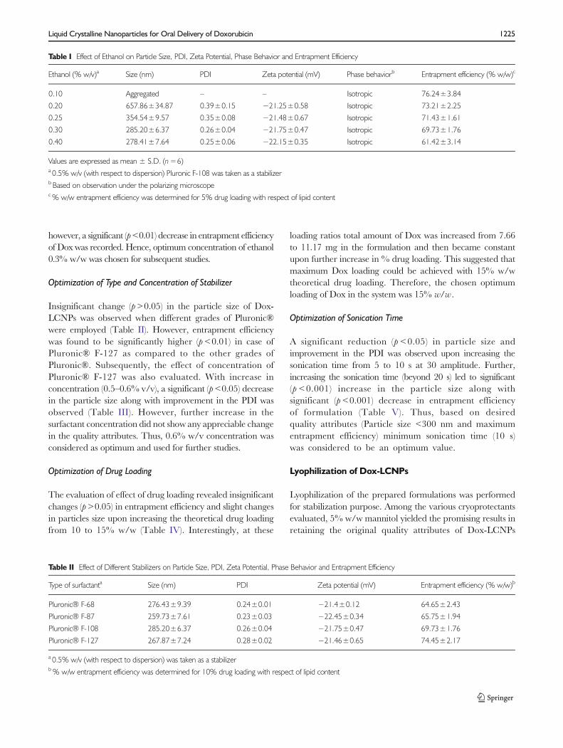
however, a significant (p<0.01) decrease in entrapment efficiencyof Dox was recorded. Hence, optimum concentration of ethanol0.3% w/w was chosen for subsequent studies.
Optimization of Type and Concentration of Stabilizer
Insignificant change (p>0.05) in the particle size of Dox-LCNPs was observed when different grades of Pluronic®were employed (Table II). However, entrapment efficiencywas found to be significantly higher (p<0.01) in case ofPluronic® F-127 as compared to the other grades ofPluronic®. Subsequently, the effect of concentration ofPluronic® F-127 was also evaluated. With increase inconcentration (0.5–0.6% v/v), a significant (p<0.05) decreasein the particle size along with improvement in the PDI wasobserved (Table III). However, further increase in thesurfactant concentration did not show any appreciable changein the quality attributes. Thus, 0.6% w/v concentration wasconsidered as optimum and used for further studies.
Optimization of Drug Loading
The evaluation of effect of drug loading revealed insignificantchanges (p>0.05) in entrapment efficiency and slight changesin particles size upon increasing the theoretical drug loadingfrom 10 to 15% w/w (Table IV). Interestingly, at these
loading ratios total amount of Dox was increased from 7.66to 11.17 mg in the formulation and then became constantupon further increase in % drug loading. This suggested thatmaximum Dox loading could be achieved with 15% w/wtheoretical drug loading. Therefore, the chosen optimumloading of Dox in the system was 15% w/w .
Optimization of Sonication Time
A significant reduction (p <0.05) in particle size andimprovement in the PDI was observed upon increasing thesonication time from 5 to 10 s at 30 amplitude. Further,increasing the sonication time (beyond 20 s) led to significant(p <0.001) increase in the particle size along withsignificant (p <0.001) decrease in entrapment efficiencyof formulation (Table V). Thus, based on desiredquality attributes (Particle size <300 nm and maximumentrapment efficiency) minimum sonication time (10 s)was considered to be an optimum value.
Lyophilization of Dox-LCNPs
Lyophilization of the prepared formulations was performedfor stabilization purpose. Among the various cryoprotectantsevaluated, 5% w/w mannitol yielded the promising results inretaining the original quality attributes of Dox-LCNPs
Table I Effect of Ethanol on Particle Size, PDI, Zeta Potential, Phase Behavior and Entrapment Efficiency
Ethanol (% w/v)a Size (nm) PDI Zeta potential (mV) Phase behaviorb Entrapment efficiency (% w/w)c
0.10 Aggregated – – Isotropic 76.24±3.84
0.20 657.86±34.87 0.39±0.15 −21.25±0.58 Isotropic 73.21±2.25
0.25 354.54±9.57 0.35±0.08 −21.48±0.67 Isotropic 71.43±1.61
0.30 285.20±6.37 0.26±0.04 −21.75±0.47 Isotropic 69.73±1.76
0.40 278.41±7.64 0.25±0.06 −22.15±0.35 Isotropic 61.42±3.14
Values are expressed as mean ± S.D. (n=6)a 0.5% w/v (with respect to dispersion) Pluronic F-108 was taken as a stabilizerb Based on observation under the polarizing microscopec% w/w entrapment efficiency was determined for 5% drug loading with respect of lipid content
Table II Effect of Different Stabilizers on Particle Size, PDI, Zeta Potential, Phase Behavior and Entrapment Efficiency
Type of surfactanta Size (nm) PDI Zeta potential (mV) Entrapment efficiency (% w/w)b
Pluronic® F-68 276.43±9.39 0.24±0.01 −21.4±0.12 64.65±2.43
Pluronic® F-87 259.73±7.61 0.23±0.03 −22.45±0.34 65.75±1.94
Pluronic® F-108 285.20±6.37 0.26±0.04 −21.75±0.47 69.73±1.76
Pluronic® F-127 267.87±7.24 0.28±0.02 −21.46±0.65 74.45±2.17
a 0.5% w/v (with respect to dispersion) was taken as a stabilizerb% w/w entrapment efficiency was determined for 10% drug loading with respect of lipid content
Liquid Crystalline Nanoparticles for Oral Delivery of Doxorubicin 1225

(detailed data are not shown). Hence, mannitol wasimplemented for further studies which resulted in toformation of intact fluffy cake. Upon reconstitution,achieved within 2 min with gentle shaking, insignificantchanges in the original quality attributes were observedwith Sf/Si ratio (ratio of the particle size after andbefore freeze drying) 1.09 (Table VI). Additionally,freeze dried Dox-LCNPs were also found to be stablefollowing 6 months of accelerated stability testing.
Characterization of Dox-LCNPs
HR-TEM
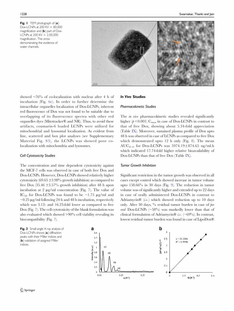
The obtained HRTEM microphotograph of Dox-LCNPsshowed discrete cubic geometry (Fig. 1a). The lipidic matrix ofphytantriol along with the water channels were clearly observedupon further increasing themagnification (Fig. 1b). The observedparticle size of Dox-LCNPs was found to be 214.12±16.25×158.37±11.45 nm (length × breadth; average of 50 particles).Notably, the observed dimensions were smaller than thatobtained from the Zeta sizer (265.87±5.54 nm).
Light Microscopy/Phase Behavior Studies
For microscopic studies, a representative micron sized Dox-liquid crystalline particulate formulation was prepared whichshowed a few aggregates in micrometer range when observed
under light microscope (see Supplementary MaterialFig. S2A, S2B). Additionally, both blank and drug loadeddispersion showed a dark background under polarized lightwhich revealed their isotropic nature (21,30). This alsosuggested incorporation of amphiphilic Dox (up to 20% w/w/with respect to lipid) into the liquid crystalline phase did notalter their phase behavior.
SAXS Studies
SAXS analysis of Dox-LCNPs showed four distinct Braggpeaks (Fig. 2a) whose relative positions were found to be inratios of √2:√4:√6:√8 which confirmed Pn3m type of cubicstructure (31). Further, the miller indices (h,k,l) = (1, 1, 0),(2, 0, 0), (2, 1, 1), (2, 2, 0) for these peak were assigned(Fig. 2b). Based on miller indices, the lattice parameters andlattice constant for Dox-LCNPs were also calculated and foundto be 127.17 Å and 11.22 Å, respectively. Similarly, presence ofPn3m type cubic structure in the plain LCNPs dispersion wasalso observed (data are not shown) as reported by other groups(15,32,33). The lattice constant for plain LCNPs were found tobe (~6.95 Å) while it was (~11.22 Å) in the case of Dox-LCNPs.
Stability in Simulated Gastrointestinal Fluids
Stability of Dox-LCNPs in GIT fluids was performed and wasfound that LCNPs could retain their initial qualities attributes(insignificant changes in particles size and a slight decrease inentrapment efficiency) in the SGF as well as in SIF condition(Table VII). Additionally, presence of high amount of druginside the carrier (65.71±2.02% w/w) even after 8 h reflecteddrug retaining capability of LCNPs upon exposure to GITenvironments.
In Vitro Release Study
An equilibrium dialysis membrane method was applied todetermine the in vitro release profile of Dox from formulation(34). Free Dox solution was also utilized as a control, acomplete release (97.13±2.89%) within 2 h was found. Butin case of Dox-LCNPs, a sustained biphasic release profile
Table III Effect of Pluronic F-127 Concentration on Particle Size, PDI, ZetaPotential and Entrapment Efficiency
Surfactant(% w/v)
Size (nm) PDI Zeta potential(mV)
Entrapmentefficiency(% w/w)a
0.50 267.87±7.24 0.28±0.02 −21.3±0.82 74.45±2.17
0.60 239.76±7.71 0.22±0.01 −21.63±0.39 76.56±2.57
0.70 241.43±8.01 0.23±0.03 −19.05±0.67 75.4±2.78
Values are expressed as mean ± S.D. (n=6)a% w/w entrapment efficiency was determined for 10% drug loading withrespect of lipid content
Table IV Effect of Drug Loading on Particle Size, PDI, Zeta Potential and Entrapment Efficiency
Drug loadinga Size (nm) PDI Zeta potential (mV) Entrapment efficiency (% w/w) Amount of Dox (mg) Phase behaviorb
10 239.76±7.71 0.22±0.01 −21.63±0.39 76.56±2.57 7.656±2.57 Isotropic
15 265.87±5.54 0.22±0.04 −20.23±0.32 74.65±1.76 11.19±2.64 Isotropic
17.5 272.45±8.35 0.24±0.04 −21.42±0.49 62.38±1.87 10.91±3.27 Isotropic
20 275.87±7.81 0.25±0.04 −18.98±0.52 55.91±2.76 11.82±5.52 Isotropic
Values are expressed as mean ± S.D. (n=6)a% w/w with respect to lipid contentb Based on observation under the polarizing microscope
1226 Swarnakar, Thanki and Jain
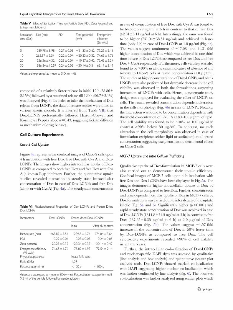
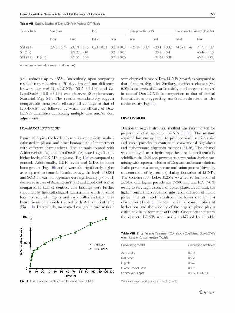
composed of a relatively faster release in initial 12 h (38.86±2.15%) followed by a sustained release till 120 h (96.7±2.1%)was observed (Fig. 3). In order to infer the mechanism of Doxrelease from LCNPs, the data of release studies were fitted invarious kinetic models. It is inferred from Table VIII thatDox-LCNPs preferentially followed Hixson-Crowell andKorsmeyer Peppas (slope n=0.43, suggesting fickian diffusionas mechanism of drug release).
Cell Culture Experiments
Caco-2 Cell Uptake
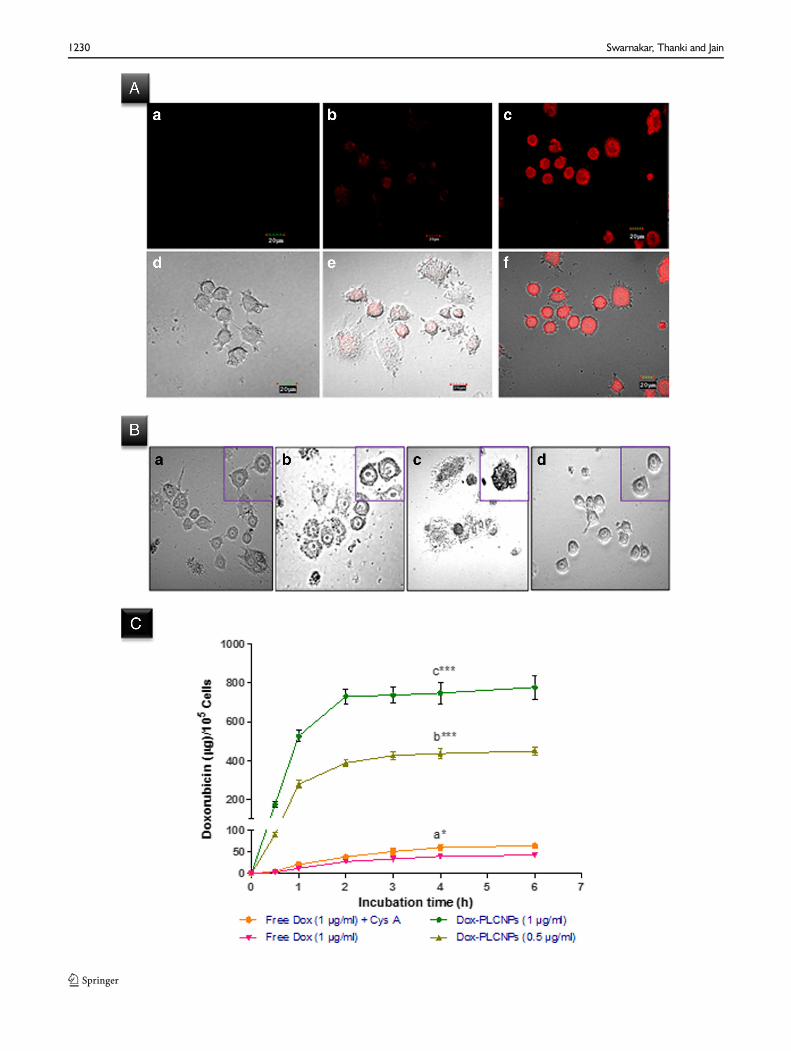
Figure 4a represents the confocal images of Caco-2 cells upon4 h incubation with free Dox, free Dox with Cys A and Dox-LCNPs. The images show higher intracellular uptake of Dox-LCNPs as compared to both free Dox and free Dox with CysA (a known P-gp inhibitor). Further, the quantitative uptakestudies revealed alteration in steady state intracellularconcentration of Dox in case of Dox-LCNPs and free Dox(alone or with Cys A) (Fig. 4a). The steady state concentration
in case of co-incubation of free Dox with Cys A was found tobe 64.63±5.79 ng/ml at 6 h in contrast to that of free Dox(42.81±3.14 ng/ml at 6 h). Interestingly, the same was foundto be higher (731.04±38.51 ng/ml) and achieved in lessertime (only 2 h) in case of Dox-LCNPs at 1.0 μg/ml (Fig. 4c).The values suggest attainment of ~17.08- and 11.31-foldhigher concentration of Dox which was achieved in one thirdtime in case of Dox-LCNPs as compared to free Dox and freeDox + CysA respectively. Furthermore, cells viability was alsofound to be >90% in all the cases indicative of absence of anytoxicity to Caco-2 cells at tested concentration (1.0 μg/ml).The studies at higher concentration of Dox-LCNPs and blankLNCPs were also performed but dramatic decrease in the cellviability was observed in both the formulations suggestinginteraction of LNCPs with cells. Hence, a systematic studydesign was employed for evaluating the effect of LNCPs oncells. The results revealed concentration dependent alterationin the cells morphology (Fig. 4b) in case of LCNPs. Notably,the interaction was found to be concentration dependent withthreshold concentration of LNCPs at 80–100 μg/ml of lipid.The cell viability was found to be ~40% at 100 μg/ml incontrast >90% below 80 μg/ml. In contrast, no suchalteration in the cell morphology was observed in case offormulation excipients (either lipid or surfactant) at all testedconcentration suggesting excipients has no detrimental effectson Caco-2 cells.
MCF-7 Uptake and Intra Cellular Trafficking
Qualitative uptake of Dox-formulation in MCF-7 cells werealso carried out to demonstrate their uptake efficiency.Confocal images of MCF-7 cells upon 4 h incubation withfree Dox andDox-LCNPs have been displayed in Fig. 5a. Theimages demonstrate higher intracellular uptake of Dox byDox-LCNPs as compared to free Dox. Further, concentrationand time dependent cellular uptake of Dox in MCF-7 cells byDox formulations was carried out to infer details of the uptakekinetic (Fig. 5a and b). Significantly higher (p<0.001) andrapid steady state concentration of Dox was achieved in caseof Dox-LCNPs (1314.8±71.5 ng/ml at 3 h) in contrast to freeDox (287.65±6.35 ng/ml at 6 h) at 2.0 μg/ml of Doxconcentration (Fig. 5b). The values suggest ~4.57-foldincrease in the concentration of Dox in 50% lesser timeby Dox-LCNPs as compared to free Dox. The cellcytotoxicity experiments revealed >90% of cell viabilityin all the cases.
Further, the intracellular co-localization of Dox-LCNPsand nuclear-specific DAPI dyes was assessed by qualitative(line analysis and box analysis) and quantitative (scatter plotanalysis) tools. Dox-LCNPs showed marked co-localizationwith DAPI suggesting higher nuclear co-localization whichwas further confirmed by line analysis (Fig. 6). The observedco-localization was further analyzed using scatter plots which
Table V Effect of Sonication Time on Particle Size, PDI, Zeta Potential andEntrapment Efficiency
Sonicationtime (Sec)
Size (nm) PDI Zeta potential(mV)
Entrapmentefficiency(% w/w)
5 289.98±8.98 0.27±0.03 −21.33±0.62 75.25±2.16
10 265.87 ±5.54 0.22±0.04 −20.23±0.32 74.65±1.76
20 256.26±4.32 0.23±0.04 −19.87±0.42 72.45±2.34
30 386.84±10.57 0.24±0.05 −20.14±0.51 65.17±3.19
Values are expressed as mean ± S.D. (n=6)
Table VI Physicochemical Properties of Dox-LCNPs and Freeze DriedDox-LCNPs
Parameters Dox-LCNPs Freeze dried Dox-LCNPs
Initial After six months
Particle size (nm) 265.87±5.54 289.5±6.74 274.84±8.64
PDI 0.22±0.04 0.23±0.03 0.24±0.05
Zeta potential −20.23±0.32 −20.34±0.37 −20.14±0.47
Entrapment efficiency(% w/w)
74.65±1.76 73.89±1.97 72.54±2.14
Physical appearance Intact fluffy cake
Ratio (Sf/Si) 1.09
Reconstitution time <100 s <100 s
Values are expressed as mean± SD (n=6); Reconstitution was performed in0.5 ml of the vehicle followed by gentle agitation
Liquid Crystalline Nanoparticles for Oral Delivery of Doxorubicin 1227
showed ∼76% of co-localization with nucleus after 4 h ofincubation (Fig. 6e). In order to further determine theintracellular organelles localization of Dox-LCNPs, inherentred fluorescence of Dox was not found to be suitable due tooverlapping of its fluorescence spectra with other redorganelles dyes (Mitotracker® and NR). Thus, to avoid theseartifacts, coumarin-6 loaded LCNPs were utilized formitochondrial and lysosomal localization. As evident fromline, scattered and box plot analyses (see SupplementaryMaterial Fig. S3), the LCNPs was showed poor co-localization with mitochondria and lysosomes.
Cell Cytotoxicity Studies
The concentration and time dependent cytotoxicity againstthe MCF-7 cells was observed in case of both free Dox andDox-LCNPs. However, Dox-LCNPs showed relatively highercytotoxicity (69.65 ±3.98% growth inhibition) as compared tofree Dox (35.46 ±3.57% growth inhibition) after 48 h uponincubation at 2 μg/ml concentration (Fig. 7). The value ofIC50 for Dox-LCNPs was found to be ~1.75 μg/ml and~0.25 μg/ml following 24 h and 48 h incubation, respectivelywhich was 3.12- and 16.23-fold lower as compared to freeDox (Fig. 7). The cell cytotoxicity of the blank formulation wasalso evaluated which showed >90% cell viability revealing itsbiocompatibility (Fig. 7).
In Vivo Studies
Pharmacokinetic Studies
The in vivo pharmacokinetic studies revealed significantlyhigher (p<0.001) Cmax in case of Dox-LCNPs in contrast tothat of free Dox, showing about 5.34-fold appreciation(Table IX). Moreover, sustained plasma profile of Dox upto48 h was observed in case of LCNPs as compared to free Doxwhich demonstrated upto 12 h only (Fig. 8). The meanAUC0–∞ for Dox-LCNPs was 5974.19±874.65 ng/ml.hwhich indicated 17.74-fold higher relative bioavailability ofDox-LCNPs than that of free Dox (Table IX).
Tumor Growth Inhibition
Significant restriction in the tumor growth was observed in allcases except control which showed increase in tumor volumeupto 158.66% in 30 days (Fig. 9). The reduction in tumorvolume was of significantly higher and extended up to 22 daysin case of orally administered Dox-LCNPs in contrast toAdriamycin® (i.v. ) which showed reduction up to 10 daysonly. After 30 days, % residual tumor burden in case of peroral Dox-LCNPs (~58%) was markedly lower than that ofclinical formulation of Adriamycin® i.v. (~69%). In contrast,lowest residual tumor burden was found in case of LipoDox®
Fig. 1 TEM photograph of (a )Dox-LCNPs at 200 KV × 80,000magnification and (b ) part of Dox-LCNPs at 200 KV × 2,00,000magnification. The arrowdemonstrating the evidence ofwater channels.
Fig. 2 Small angle X-ray analysis ofDox-LCNPs shows (a ) diffractionpeaks with their Miller indices and(b ) validation of assigned Millerindices.
1228 Swarnakar, Thanki and Jain
(i.v. ), reducing up to ~85%. Interestingly, upon comparingresidual tumor burden at 20 days, insignificant differencebetween per oral Dox-LCNPs (53.5 ±6.1%) and i.v.LipoDox® (46.8 ±8.4%) was observed (SupplementaryMaterial Fig. S4). The results cumulatively suggestcomparable therapeutic efficacy till 20 days to that ofLipoDox® (i.v. ) followed by which the efficacy of Dox-LCNPs diminishes demanding multiple dose and/or doseadjustments.
Dox-Induced Cardiotoxicity
Figure 10 depicts the levels of various cardiotoxicity markersestimated in plasma and heart homogenate after treatmentwith different formulations. The animals treated withAdriamycin® (i.v ) and LipoDox® (i.v ) posed significantlyhigher levels of CK-MB in plasma (Fig. 10a) as compared tocontrol. Additionally, LDH levels and MDA in hearthomogenates (Fig. 10b and c) were also significantly higheras compared to control. Simultaneously, the levels of GSHand SOD in heart homogenates were significantly (p<0.001)decreased in case of Adriamycin® (i.v. ) and LipoDox® (i.v. ) ascompared to that of control. The findings were furthersupported by histopathological examination, which revealedloss in structural integrity and myofibrillar architecture inheart tissue of animals treated with Adriamycin® (i.v )(Fig. 11b). Interestingly, no marked changes in cardiac tissue
were observed in case of Dox-LCNPs (per oral ) as compared tothat of control (Fig. 11c). Similarly, significant changes (p<0.05) in the levels of all cardiotoxicity markers were observedin case of Dox-LCNPs in comparison to that of clinicalformulations suggesting marked reduction in thecardiotoxicity (Fig. 10).
DISCUSSION
Dilution through hydrotrope method was implemented forpreparation of drug-loaded LCNPs (35,36). This methodrequired low energy input to produce small, uniform sizeand stable particles in contrast to conventional high-shearand high-pressure dispersion methods (21,36). The ethanolwas employed as a hydrotrope because it preferentiallysolubilizes the lipid and prevents its aggregation during pre-mixing with aqueous solution of Dox and surfactant solution.It also presumes a homogeneous nucleation process (driven byconcentration of hydrotrope) during formation of LCNPs.The concentration below 0.25% w/w led to formation ofLCNPs with higher particle size (>300 nm) and PDI (>0.3)owing to very high viscosity of lipidic phase. In contrast, thehigher concentration resulted into rapid diffusion of lipidicphase and ultimately resulted into lower entrapmentefficiencies (Table I). Hence, the initial concentration ofhydrotrope and the viscosity of the organic phase play acritical role in the formation of LCNPs. Once nucleation startsthe discrete LCNPs are usually stabilized by suitable
Table VII Stability Studies of Dox-LCNPs in Various GIT Fluids
Type of fluids Size (nm) PDI Zeta potential (mV) Entrapment efficiency (% w/w)
Initial Final Initial Final Initial Final Initial Final
SGF (2 h) 289.5±6.74 282.71±6.15 0.23±0.03 0.23±0.03 −20.34±0.37 −20.41±0.32 74.65±1.76 71.73±1.39
SIF (6 h) 271.23±7.81 0.21±0.03 −20.61±0.41 66.46±1.58
SGF (2 h)+SIF (4 h) 278.56±6.54 0.22±0.06 −21.04±0.38 65.71±2.02
Values are expressed as mean ± SD (n=6)
Fig. 3 In vitro release profile of free Dox and Dox-LCNPs.
Table VIII Drug Release Parameter (Correlation Coefficient) Dox-LCNPsAfter Fitting in Various Release Models
Curve fitting model Correlation coefficient
Zero order 0.846
First order 0.951
Higuchi 0.962
Hixon Crowell root 0.975
Korsmeyer Peppas 0.977, n=0.43
Values are expressed as mean ± S.D. (n=6)
Liquid Crystalline Nanoparticles for Oral Delivery of Doxorubicin 1229
1230 Swarnakar, Thanki and Jain

surfactants. The present study encompasses high molecularweight nonionic triblock copolymers (Pluronic® series)considering their capability to preferentially adsorb on thesurface of LCNPs and avoid unwanted mesophase structuretransitions in contrast to low molecular weight copolymerswhich have tendency to affect the interior phase of liquidcrystals (37). Among the tested surfactants from the series,maximum entrapment efficiency was observed in case ofPluronic® F-127 which could be attributed to its highlipophilic and large molecular weight resulting into lower
�Fig. 4 Uptake of formulation in Caco-2 cells (A) CLSM images of the cells after4 h incubationwith (a) freeDox (1μg/ml), (b) freeDox (1μg/ml)+CysA (10μg/ml), (c) Dox-LCNPs (equivalent to 1 μg/ml of free Dox), (d, e, f) overlay imageof corresponding fluoresces and differential interference contrast (DIC) image ofCaco-2 cells; (B) Differential interference contrast (DIC) images of cells, 20×,showing alteration in cell morphology after 3 h incubation with blank LCNPs atconcentration of (a) 50 μg/ml, (b) 100 μg/ml, (c) 200 μg/ml, (d) control cells.The inset image shows enlarge view of representative cells. (C ) showsconcentration and time dependent quantitative uptake of Dox-formulation inCaco-2 cells; ***p=0.001, *p=0.05; (a) vs. free Dox (1 μg/nl), (b) free Dox(1μg/ml)+CysA, (c) vs. Dox-LCNPs (0.5μg/ml). Each data point is representedas the mean ± SD (n=6).
Fig. 5 Uptake of Dox in MCF-7cells (A ) CLSM images of the cellsafter 4 h incubation with (a ) freeDox (1 μg/ml), (b ) Dox-LCNPs(equivalent to 1 μg/ml of free Dox),(c , d ) corresponding differentialinterference contrast (DIC) image ofMCF-7 cells treated with free Doxand Dox-LCNPs respectively; (B )shows concentration and timedependent quantitative uptake ofDox formulation in MCF-7 cells,***p=0.001, **p=0.01; (a ) vs.free Dox (1 μg/nl), (b ) free Dox(2 μg/ml), (c ) vs. Dox-LCNPs(1 μg/ml); Each data point isrepresented as the mean ± SD(n=6).
Liquid Crystalline Nanoparticles for Oral Delivery of Doxorubicin 1231

hydrophilic-lipophilic balance value (Table II) (38). Theselection of lipophilic surfactant i.e. Pluronic® F-127 alsocontrasts to the hydrophilic nature of Dox and hence assistsin maximizing the entrapment efficiency due to reducedleaching of drug during formation of LCNPs. Similar relationbetween drug solubility in surfactant and entrapmentefficiency of the carrier system has also been reported earlier(39). Further, the concentration of the surfactant was found todirectly correlate with the PDI revealing the stabilizationpotential of surfactant in the formation of LCNPs(Table III). The optimized formulation was then subjectedto effect of drug loading and saturation of drug-space inLCNPs was observed above 15% w/w (Table IV).Furthermore, effect of sonication was also employed for itsinfluence on particle size and entrapment efficiency and wasfound that upon imparting higher energy (>10 s at 30amplitude), acceleration in the kinetic energy of Dox-LNCPsincreased drastically which resulted in destabilization ofsurfactant assisted steric barrier on the LCNPs and ultimatelycaused aggregation of particles (Table V). The final
formulation was then lyophilized using universal stepwisefreeze drying cycle developed and patented by our group(22). Implementation of 5% w/v mannitol in thelyophilization of Dox-LCNPs maintained all original qualityattributes of formulations such as particle size, PDI andencapsulation efficiency and also demonstrated 6 months ofaccelerated stability (Table VI).
Morphological analysis of the developed formulation byHR-TEM revealed their cubic morphology with evidence ofwater channels within the structure which dried during samplepreparation (Fig. 1). This could be the probable reason forslightly lower particle size observed with TEM as compared tothat of Zeta Sizer (40). Additionally, the discrepancies in theparticle size could also be attributed to the differences in theoperating principle of analytical techniques, transmission ofelectron beam under the high vacuum in former case whilemeasurement of hydrodynamic volume at normal roomtemperature in later case (41). The crystallinity was furtherestablished using SAXS (42,43). The spectral pattern of Dox-LCNPs depicted four diffraction peaks with relative ratio of
Fig. 6 Intracellular co-localization of (a ) Dox-LCNPs with (b ) DAPI-stained nucleus of MCF-7 cells. Line analysis plot (c ) and box analysis (d ) shows DAPI-Doxinteraction while scatter plots (e ) shows % co-localization of Dox-LCNPs co-localizing with DAPI. (g ) Overlay image of (a , b and f) (DIC image).
Fig. 7 Concentration- and time-dependent cell viability of MCF-7cells upon treatment with Doxformulations. Table shows IC50 (μg/ml) value of Dox formulation atdifferent incubation time. Each datapoint were expressed as mean ±SD (n=6).
1232 Swarnakar, Thanki and Jain

√2:√4:√6:√8 suggestive of Pn3m type cubic structure ofdeveloped LCNPs (Fig. 2a). Furthermore, assignment ofcrystallographic space groups i.e. miller indices as (hkl) =(1, 1, 0), (2, 0, 0), (2, 1, 1), (2, 2, 0) for Dox-LCNPs wereconsidered to be valid (Fig. 2b) because points of the graphdemonstrated a linear trend line (r2 values >0.99) whichintercepts at origin (0,0). The increased value of latticeparameter (~5 Å) in Dox-LCNPs in comparison to blankLCNPs reflected that hydration of LCNPs (or widening ofthe water channels) due to presence of Dox either in theaqueous domain or at the interface of the polar head groupand apolar tail of the lipid. Such type of interactions have beenreported for both hydrophilic and lipophilic drugs (21,44,45).The observed structural information (cubic) from SAXSstudies also corroborates with the results of polarizingmicroscopy (isotropic structure; see Supplementary MaterialFig. S1).
In vitro stability studies in simulated gastrointestinal fluidsrevealed robustness of formulation in GIT fluids because ofinsignificant change (p>0.05) in particle size, PDI and zetapotential (Table VII). The entrapment efficiency of Dox in theformulation was also maintained due to characteristic featureof LCNPs which include chemical and structural stability ofphytantriol based systems (17,46), surfactant assisted stericstabilization of the nanoparticles and sustained releasecharacteristic of LCNPs limiting exposure of drug to exteriorharsh environments thus minimizing drug degradation inGIT.
In vitro drug release studies of Dox-LCNPs revealedsustained and biphasic release profile (Fig. 3). The drugrelease from Dox-LCNPs preferentially follows that Hixson-Crowell and Korsmeyer Peppas models indicating that therelease mechanism may be by diffusion, swelling of matrixfollowed by erosion of the lipid matrix. Similar type of therelease mechanism was also previously reported for lipidicsystem (25). Furthermore, presence of crystalline structure alsoimparts rigidity to the system hence making it like a matrixsystem hence these system also followed the Higuchi model(Table VIII). Broadly, it is postulated that presence of drug atthe surface along with its higher concentration in the system atinitial hour was responsible for rapid release. Further, timedependent continuous increase in diffusion path length forDox, which was tortuous in nature, might also have played arole after certain time andmight be responsible for a sustainedrelease of Dox (47).
Complementary to drug release studies, in vitro Caco-2uptake experiments further revealed significantly higheruptake of Dox-LCNPs as compared to free Dox at all testedconcentrations (Fig. 4). The uptake was even higher ascompared to that of Dox co-incubated with a known P-gpinhibitor, CysA, suggesting the involvement of other uptakemechanisms in case of Dox-LCNPs. The said observationcould be correlated with membrane modifying (fluidity orfusion) properties of LCNPs as reported earlier (21,48,49)along with other uptake mechanisms such as clathrin andcaveolae/lipid raft-mediated endocytosis (48) andtransportation across the cells by long chain fatty acidtransporters (50). Some reports also demonstratedcomplications associated with cell culture studies (49) owingto dominating membrane modifying properties of LCNPs. Toaddress this, concentration dependent alterations in themorphology was evaluated for blank LCNPs which revealedexistence of threshold concentration (~80–100 μg/ml)beyond which membrane modifying potential resulted indetrimental effects on cells (~40% cells viability at 100 μg/ml).Interestingly, the cell viability was found to be >90% in casesbelow threshold concentration (<80 μg/ml). Concomitantly, theCLSM studies revealed some morphological changes in thecell membrane (altered membrane integrity), referred to as“virtual paths” during uptake phase of LCNPs (Fig. 4b).These morphological changes were found to be reversiblein nature when measured as a function of time and normalmorphology regained within 12 h (data not shown). Thus,based on these observations a mechanism of uptake viaformation of some reversible “virtual pathways” in the cellmembrane might also possible along with above mentioneduptake mechanisms.
In vitro cell cytotoxicity potential of Dox-formulations wasalso evaluated against MCF-7 cells. Results revealed timedependent increase in the cytotoxicity in case of both freeDox and Dox-LCNPs. However, the magnitude of
Table IX Pharmacokinetic Parameters After Oral Administration of DoxFormulations
Parameters Free Dox Dox-LCNPs
Cmax (ng/ml) 35.58±4.43 190.06±30.89
Tmax (h) 4 2
AUC0-t (ng/ml*h) 322.09±34.25 4022.54±489.42
AUC0-∞ (ng/ml*h) 337.27±59.75 5974.19±874.65
Values are expressed as mean ± SEM (n=6)
Fig. 8 Plasma concentration-time profiles of free Dox and Dox-LCNPs afteroral administration to SD rats at 10 mg/kg dose. Each data points areexpressed as mean ± SEM (n=6).
Liquid Crystalline Nanoparticles for Oral Delivery of Doxorubicin 1233

cytotoxicity was significantly high (p<0.001) in case of Dox-LCNPs which showed 3.12 and 16.23-fold decrease in IC50
value following 24 h and 48 h of incubation, respectively(Fig. 7). The possible reasons for such higher cytotoxicpotential of Dox-LCNPs can be attributed to their rapid andhigher internalization as compared to free Dox. Interestingly,the observed cytotoxic potential was higher than previouslyprepared Dox-PLGA-NPs (5). This could be correlated withthe principal advantages associated with lipid based drugdelivery systems which include multiple cel lularinternalization mechanisms and relatively higher drug releasecharacteristics as compared to that of polymeric nanocarriers(51). Moreover, Dox-LCNPs demonstrated translocatingcapabilities to the site of drug action, i.e. nucleus(Figs. 5 and 6). As far as existing literature is concern,this is the first report which revealed that LCNPsprepared by phytantriol and stabilized by Pluronic® F-127 have tendency to localize near the vicinity of nucleus.Although, it has been reported that certain surfactants likePluronic® and polyvinyl alcohol or surface modificationof lipid matrix either by charge modifier like chitosan ornuclear targeting ligand, can facilitate cellular entry andnuclear transport of bioactives (5,39,52,53). However,none of the reports have shown nuclear co-localizationof the said combination. Although, detailed study iswarranted to explore the reason of nuclear colocalizationof present LNCPs.
Results of pharmacokinetic studies further exhibiteddeliverability potential of Dox-LCNPs with 17.74-foldincrease in the oral bioavailability in comparison to freeDox. This is one of the highest bioavailability achieved ascompared to other reported carrier system exploited till date(1,7,8,54). Notably the plasma levels were also maintained for48 h in case of Dox-LCNPs which could be attributed to thesuperior encapsulation and sustained release characteristic ofLCNPs. Additionally, the LCNPs and their secondarystructures are bioadhesive which increases the opportunitiesof close contact of drug loaded LNCPs with endothelial cellmembrane and could overcome “unstirred water layer”barrier (55). Subsequently, lipid-mixing, membrane-fusingand formation of “virtual pathway” may also help inenhancing bioavailability of Dox (Figs. 8 and 4b). However,one can also evaluate the potential of crystallinity of LCNPs inmaking the system eligible for intestinal epithelial M cellsuptake classically applicable to particulate carrier.
The promising pharmacokinetic profile of Dox-LCNPsinspired for evaluating its in vivo antitumor tumor efficacy inDMBA induced breast cancer model, already established inour lab (5,8,25,39,51). Tumor bearing rats treated with Dox-LCNPs showed a significant reduction (p<0.001) in tumorvolume in comparison to Adriamycin (i.v. ) (Fig. 9). Therelatively higher tumor restriction capability of Dox-LCNPscould be attributed to longer circulation half-life vis-a-vispassive targeting to tumor via enhanced permeation and
Fig. 9 In vivo antitumor efficacy ofDox-formulations in DMBA-induced tumor bearing female SDrats: (a ) time-dependent tumorprogression in animal, (b ) tumorburden in animals after 30 days, (c )photographs of representativeexcised tumors from differenttreatment groups after 30 days.Mean tumor volume was taken as100% at the start of drug treatmentand tumor progression monitoreduntil the end of the study. ***p=0.001, *p=0.05; (a ) vs. Control(vehicle treated), (b ) vs.Adriamycin® (i.v.), (c ) vs . Dox-LCNPs. Each data point isrepresented as the mean ± SEM(n=6).
1234 Swarnakar, Thanki and Jain

retention effect (EPR) as compared to that of free Dox. Theresults are in line with previous studies (5,30,39,56).Interestingly, the observed responses were found to be higherthan our previously reported polymeric formulation of Dox(5). However, the residual tumor burden after 30 days wasfound to be significantly lower (p<0.001) in case of LipoDox®(i.v. ) than Dox-LCNPs (per oral ), which could be attributed tothe inherent advantages of i.v. administered stealthnanocarriers. Of note, the results cumulatively suggestcomparable therapeutic efficacy till 20 days to that ofLipoDox® (i.v. ) followed by which the efficacy of Dox-LCNPs diminishes demanding multiple dose and/or doseadjustments.
With significant enhancement in the oral anticancerefficacy of Dox-LCNPs the obvious concern of the Doxinduced cardiotoxicity was also addressed. Mechanistically,Dox is reported to intercalate with DNA strands, inhibittopoisomerase II and generate reactive oxygen species(ROS), cumulatively leading to initiation of DNA damage
and inhibition macromolecules synthesis (57). However,biotransformation of Dox into free radicals and subsequentinterference with mitochondrial enzymes such as CoQ10,NADH dehydrogenase results into prominent weakening ofcardiac tissues which is often attributed to the Dox inducedcardiotoxicity (51,58,59). The ROS induction is oftenassociated with lipid peroxidation and reduces the levels ofessential enzyme such as superoxide dismutase (SOD) andGlutathione (GSH), which activates the a vicious cycle andfurther contributes even higher oxidative stress (60,61). Thelipid peroxidation in the cardiac tissues is also associated withmyocardial ischemic injury thereby affecting Na+/K+-ATPase activity and mitochondrial calcium activity (62).The degree of lipid peroxidation is generally measured as afunction of malondialdehyde (MDA) concentration(byproduct along with polyunsaturated fatty acids). The injuryin the heart also leaches lactate dehydrogenase (LDH; anenergy producing enzyme) and CK-MB in the plasma leadingto increased concentrations in the plasma. Therefore, all these
Fig. 10 Levels of variousbiochemical parameters in Plasma(a ) CK-MB, (b ) LDH and in hearthomogenate (c ) Lipid peroxidationproducts (MDA), (d ) GSH levelsand (e ) % SOD, after 30 days oftreatment with differentformulations. ***p<0.001, **p<0.01, *p<0.05; (a ) vs. control, (b )vs . Adriamycin® (i.v.), (c ) vs .LipoDox® (i.v.).
Liquid Crystalline Nanoparticles for Oral Delivery of Doxorubicin 1235

cardiotoxicity markers was utilized in the present studies(5,8,63). The levels of cardiac toxicity markers were found tobe significantly (p<0.05) lower in case of CK-MB, LDH andMDA and higher in case of GSH and % SOD uponadministration of Dox-LCNPs (per oral ) as compared toAdriamycin® (i.v. ) and LipoDox (i.v. ) (Fig. 10). Furthermore,Dox also down-regulates expression of a variety of cardiacmuscle-specific proteins which are directly associated withreduced contractility of cardiomyocytes and may explain thepathologic features of myofibrillar loss in cardiac tissue(57). Therefore, histopathological examination was carried outwhich further revealed excessive sarcoplasmic vacuolizationand severe disruption of fine structures (Fig. 11) in case ofAdriamycin® ( i.v. ). In contrast, fewer myocardialmorphological changes were observed in case of Dox-LCNPs which could be attributed to superior encapsulationof Dox in the LCNPs matrix. Thus, incorporation of Doxin the novel LCNPs can be a viable option for oralchemotherapy.
CONCLUSION
The employed formulation strategy has potential forimproving the oral delivery of various difficult-to-deliverdrugs. The principal features include simple method of
preparation, sustained release profile, alternate absorptionpathway, superior encapsulation of labile drugs, longercirculation half-life and preferential accumulation at targetsite based on enhanced permeation and retention effect. Thetargeting potential of the developed formulation could furtherbe improved by implementing active targeting principles toachieve therapeutic equivalence with i.v. administerednanocarriers such as LipoDox®. In addition, multiple dosekinetics and dose adjustments can also be sought for.Successful stabilization of the prepared LCNPs bylyophilization using mannitol as cryoprotectant is alsonoteworthy and studies are under way for understanding theinteractions at molecular level. The nuclear colocalizationcapabilities at intracellular level further make it potentialdelivery system for cancer therapeutics however mechanisticinsights for same needs to be explored for betterunderstanding. In nutshell, the said formulation strategy ispromising among the field of emerging novel lipid based drugdelivery system and can be explored to a greater extent forother drug delivery applications.
ACKNOWLEDGMENTS AND DISCLOSURES
Authors are thankful to Director, NIPER for providingnecessary infrastructure facilities. The work was supportedby Council of Scientific and Industrial Research (CSIR),
Fig. 11 Histopathology of hearttissue (a ) control, (b ) Adriamycin®(i.v.) the arrows depicts thevacuolization and structural changesin heart tissue (c ) LipoDox® (i.v.),(d) Dox-LCNPs.
1236 Swarnakar, Thanki and Jain

Government of India, New Delhi, India. Authors are alsothankful for the technical support rendered by Mr. RahulMahajan.
REFERENCES
1. Rahman A, Carmichael D, Harris M, Roh JK. Comparativepharmacokinetics of free doxorubicin and doxorubicin entrappedin cardiolipin liposomes. Cancer Res. 1986;46(5):2295–9.
2. Gordon KB, Tajuddin A, Guitart J, Kuzel TM, Eramo LR,VonRoenn J. Hand-foot syndrome associated with liposome-encapsulated doxorubicin therapy. Cancer. 1995;75(8):2169–73.
3. Ryberg M, Nielsen D, Skovsgaard T, Hansen J, Jensen BV,Dombernowsky P. Epirubicin cardiotoxicity: an analysis of 469 patientswith metastatic breast cancer. J Clin Oncol. 1998;16(11):3502–8.
4. Beijnen J, Van der Houwen O, Underberg W. Aspects of thedegradation kinetics of doxorubicin in aqueous solution. Int JPharm. 1986;32(2):123–31.
5. Jain AK, Swarnakar NK, Das M, Godugu C, Singh RP, Rao PR,et al . Augmented anticancer efficacy of doxorubicin-loadedpolymeric nanoparticles after oral administration in a breast cancerinduced animal model. Mol Pharm. 2011;8(4):1140–51.
6. Thanki K, Gangwal RP, Sangamwar AT, Jain S. Oral delivery ofanticancer drugs: challenges and opportunities. J Control Release.2013;170(1):15–40.
7. Kalaria DR, Sharma G, Beniwal V, Ravi Kumar MNV. Design ofbiodegradable nanoparticles for oral delivery of doxorubicin: in vivopharmacokinetics and toxicity studies in rats. PharmRes. 2009;26(3):492–501.
8. Jain S, Patil SR, Swarnakar NK, Agrawal AK. Oral delivery ofdoxorubicin using novel polyelectrolyte-stabilized liposomes(layersomes). Mol Pharm. 2012;9(9):2626–35.
9. Guo C,Wang J, Cao F, Lee RJ, Zhai G. Lyotropic liquid crystal systemsin drug delivery. Drug Discov Today. 2010;15(23–24):1032–40.
10. Barauskas J, Johnsson M, Tiberg F. Self-assembled lipidsuperstructures: beyond vesicles and liposomes. Nano Lett.2005;5(8):1615.
11. Yang D, Armitage B, Marder SR. Cubic liquid-crystallinenanoparticles. Angew Chem Int Ed Engl. 2004;43(34):4402–9.
12. Lian R, Lu Y, Qi J, Tan Y, Niu M, Guan P, et al . Silymarin glycerylmonooleate/poloxamer 407 liquid crystalline matrices: physicalcharacterization and enhanced oral bioavailability. AAPSPharmSciTech. 2011;12(4):1234–40.
13. Lai J, Chen J, Lu Y, Sun J, Hu F, Yin Z, et al. Glyceryl monooleate/poloxamer 407 cubic nanoparticles as oral drug delivery systems: I. Invitro evaluation and enhanced oral bioavailability of the poorlywater-soluble drug simvastatin. AAPS Pharm Sci Technol.2009;10(3):960–6.
14. Tamayo-Esquivel D, Ganem-Quintanar A, Martinez AL,Navarrete-Rodriguez M, Rodriguez-Romo S, Quintanar-GuerreroD. Evaluation of the enhanced oral effect of omapatrilat-monoleinnanoparticles prepared by the emulsification-diffusion method. JNanosci Nanotechnol. 2006;6(9–10):3134–8.
15. Nguyen TH, Hanley T, Porter CJ, Larson I, Boyd BJ. Phytantrioland glyceryl monooleate cubic liquid crystalline phases as sustained-release oral drug delivery systems for poorly water-soluble drugs II.In-vivo evaluation. J Pharm Pharmacol. 2010;62(7):856–65.
16. Nguyen TH, Hanley T, Porter CJ, Larson I, Boyd BJ. Phytantrioland glyceryl monooleate cubic liquid crystalline phases as sustained-release oral drug delivery systems for poorly water soluble drugs I.Phase behaviour in physiologically-relevant media. J PharmPharmacol. 2010;62(7):844–55.
17. Nguyen TH, Hanley T, Porter CJ, Boyd BJ. Nanostructured liquidcrystalline particles provide long duration sustained-release effect fora poorly water soluble drug after oral administration. J ControlRelease. 2011;153(2):180–6.
18. Final report on the safety assessment of phytantriol1. Int J Toxicol.2007;26(1):107–14.
19. Lindström M, Ljusberg-Wahren H, Larsson K, Borgström B.Aqueous lipid phases of relevance to intestinal fat digestion andabsorption. Lipids. 1981;16(10):749–54.
20. US Department of Agriculture. Glycerol monooleate processing.http://www.ams.usda.gov/AMSv1.0/getfile?dDocName=STELPRDC5057603 (accessed on 12 Sep, 2013).
21. Swarnakar NK, Jain V, Dubey V, Mishra D, Jain NK. Enhancedoromucosal delivery of progesterone via hexosomes. Pharm Res.2007;24(12):2223–30.
22. Jain S, Chauhan DS, Jain AK, Swarnakar NK, Harde H,Mahajan RR, Kumar D, Valvi PK, Das M, Datir SR, et al.,inventors. Stabilization of the nanodrug delivery systems bylyophilization using universal step-wise freeze drying cycle.India patent Indian Patent Application No. 2559/DEL/2011. 2011 6 September.
23. Jain S, Valvi PU, Swarnakar NK, Thanki K. Gelatin coated hybridlipid nanoparticles for oral delivery of amphotericin B. Mol Pharm.2012;9(9):2542–53.
24. Rosevear FB. The microscopy of the liquid crystalline neat andmiddle phases of soaps and detergents. J Am Oil Chem Soc.1954;31:628–39.
25. Jain S, Kumar D, Swarnakar NK, Thanki K. Polyelectrolytestabilized multilayered liposomes for oral delivery of paclitaxel.Biomaterials. 2012;33(28):6758–68.
26. ICH Q1A(R2): stability testing of new drug substances and productsQ1A(R2), ICH harmonized tripartite guideline; Step 4 version:February 6, 2003.
27. Costa P, Sousa Lobo JM. Modeling and comparison of dissolutionprofiles. Eur J Pharm Sci. 2001;13(2):123–33.
28. Sharma M, Agrawal SK, Sharma PR, Chadha BS, Khosla MK,Saxena AK. Cytotoxic and apoptotic activity of essential oil fromOcimumviride towards COLO 205 cells. Food Chem Toxicol.2010;48(1):336–44.
29. Upadhyay KK, Bhatt AN, Mishra AK, Dwarakanath BS, JainS, Schatz C, et al . The intracellular drug delivery and antitumor activity of doxorubicin loaded poly ([gamma]-benzyl l-glutamate)-b-hyaluronan polymersomes. Biomaterials.2010;31(10):2882–92.
30. Jain V, Swarnakar NK, Mishra PR, Verma A, Kaul A, Mishra AK,et al . Paclitaxel loaded PEGylated gleceryl monooleate basednanoparticulate carriers in chemotherapy. Biomaterials.2012;33(29):7206–20.
31. Putnam CD, Hammel M, Hura GL, Tainer JA. X-ray solutionscattering (SAXS) combined with crystallography and computation:defining accurate macromolecular structures, conformations andassemblies in solution. Q Rev Biophys. 2007;40(3):191–285.
32. Dong YD, Larson I, Hanley T, Boyd BJ. Bulk and dispersed aqueousphase behavior of phytantriol: effect of vitamin E acetate and F127polymer on liquid crystal nanostructure. Langmuir. 2006;22(23):9512–8.
33. Muller F, Salonen A,GlatterO. Phase behavior of Phytantriol/waterbicontinuous cubic Pn3m cubosomes stabilized by Laponite disc-likeparticles. J Colloid Interface Sci. 2010;342(2):392–8.
34. Wubeante YA, Garkhal K, Neeraj K. Doxorubicin-loaded (PEG)3-PLA nanopolymersomes: effect of solvents and process parameters onformulation development and in vitro study. Mol Pharm. 2011;8:466–78.
35. Spicer PT, Hayden KL, Lynch ML, Ofori-Boateng A, Burns JL.Novel process for producing cubic liquid crystalline nanoparticles(cubosomes). Langmuir. 2001;17(19):5748–56.
Liquid Crystalline Nanoparticles for Oral Delivery of Doxorubicin 1237

36. Friberg SE, Yang H, Fei L, Sadasivan S, Rasmussen DH, Aikens PA.Preparation of vesicles from hydrotrope solutions. J Dispers SciTechnol. 1998;19(1):19–30.
37. Johnsson M, Lam Y, Barauskas J, Tiberg F. Aqueous phase behaviorand dispersed nanoparticles of diglycerol monooleate/glyceroldioleate mixtures. Langmuir. 2005;21(11):5159–65.
38. Technical Bulletin, Pluronic® block copolymer NF Grades(Poloxamer NF Grades). [26 June 2013].
39. Jain AK, Swarnakar NK, Godugu C, Singh RP, Jain S. The effect ofthe oral administration of polymeric nanoparticles on the efficacy andtoxicity of tamoxifen. Biomaterials. 2011;32:503–15.
40. Jain S, Mistry MA, Swarnakar NK. Enhanced dermal delivery ofacyclovir using solid lipid nanoparticles. Drug Deliv Transl Res.2011;1(5):395–406.
41. Ito T, Sun L, Bevan MA, Crooks RM. Comparison of nanoparticlesize and electrophoretic mobility measurements using a carbon-nanotube-based coulter counter, dynamic light scattering,transmission electron microscopy, and phase analysis light scattering.Langmuir. 2004;20(16):6940–5.
42. Hyde ST. Identification of lyotropic liquid crystalline mesophases. In:Holmberg K, editor. Handbook of applied surface and colloidchemistry. J Wiley & Sons; 2001. p. 299–332.
43. Alexandridis P, OlssonU, Lindman B. A record nine different phases(four cubic, two hexagonal, and one lamellar lyotropic liquidcrystalline and two micellar solutions) in a ternary isothermal systemof an amphiphilic block copolymer and selective solvents (water andoil). Langmuir. 1998;14(10):2627–38.
44. Libster D, Aserin A, Wachtel E, Shoham G, Garti N. An HII liquidcrystal-based delivery system for cyclosporin A: physicalcharacterization. J Colloid Interface Sci. 2007;308(2):514–24.
45. Amar-Yuli I, Wachtel E, Shoshan EB, Danino D, Aserin A, Garti N.Hexosome and hexagonal phases mediated by hydration andpolymeric stabilizer. Langmuir. 2007;23(7):3637–45.
46. Lee KW, Nguyen TH, Hanley T, Boyd BJ. Nanostructure of liquidcrystalline matrix determines in vitro sustained release and in vivooral absorption kinetics for hydrophilic model drugs. Int J Pharm.2009;365(1–2):190–9.
47. Rudra A, Deepa RM, Ghosh MK, Ghosh S, Mukherjee B.Doxorubicin-loaded phosphatidylethanolamine-conjugatednanoliposomes: in vitro characterization and their accumulation inliver, kidneys, and lungs in rats. Int J Nanomedicine. 2010;5:811.
48. Zeng N, Gao X, Hu Q, Song Q, Xia H, Liu Z, et al . Lipid-basedliquid crystalline nanoparticles as oral drug delivery vehicles forpoorly water-soluble drugs: cellular interaction and in vivoabsorption. Int J Nanomedicine. 2012;7:3703–18.
49. Muir BW, Acharya DP, Kennedy DF, Mulet X, Evans RA, PereiraSM, et al . Metal-free and MRI visible theranostic lyotropic liquid
crystal nitroxide-based nanoparticles. Biomaterials. 2012;33(9):2723–33.
50. Ho SY, Storch J. Common mechanisms of monoacylglycerol andfatty acid uptake by human intestinal Caco-2 cells. Am J Physiol CellPhysiol. 2001;281(4):C1106–17.
51. Swarnakar NK, Thanki K, Jain S. Effect of co-administration ofCoQ10-loaded nanoparticles on the efficacy and cardiotoxicity ofdoxorubicin-loaded nanoparticles. RSC Adv. 2013;3:14671–85.
52. Gaymalov ZZ, Yang Z, Pisarev VM, Alakhov VY, Kabanov AV.The effect of the nonionic block copolymer pluronic P85 on geneexpression in mouse muscle and antigen-presenting cells.Biomaterials. 2009;30(6):1232–45.
53. Kabanov AV, Lemieux P, Vinogradov S, Alakhov V. Pluronic blockcopolymers: novel functional molecules for gene therapy. Adv DrugDeliv Rev. 2002;54(2):223–33.
54. Benival DM, Devarajan PV. Lipomer of doxorubicin hydrochloridefor enhanced oral bioavailability. Int J Pharm. 2012;423(2):554–61.
55. Thomson A, Schoeller C, Keelan M, Smith L, Clandinin M. Lipidabsorptions passing through the unstirred layers, brush-bordermembrane, and beyond. Can J Physiol Pharmacol. 1993;71(8):531–55.
56. Zhang Z,Ma L, Jiang S, Liu Z,Huang J, Chen L, et al . A self-assemblednanocarrier loading teniposide improves the oral delivery and drugconcentration in tumor. J Control Release. 2013;166(1):30–7.
57. Takemura G, Fujiwara H. Doxorubicin-induced cardiomyopathyfrom the cardiotoxic mechanisms to management. Prog CardiovascDis. 2007;49(5):330–52.
58. Li K, Sung RY, Huang WZ, Yang M, Pong NH, Lee SM, et al .Thrombopoietin protects against in vitro and in vivo cardiotoxicityinduced by doxorubicin. Circulation. 2006;113(18):2211–20.
59. Vasquez-Vivar J, Martasek P, Hogg N, Masters BS, Pritchard Jr KA,Kalyanaraman B. Endothelial nitric oxide synthase-dependentsuperoxide generation from adriamycin. Biochemistry. 1997;36(38):11293–7.
60. Singal PK, Deally CM, Weinberg LE. Subcellular effects ofadriamycin in the heart: a concise review. J Mol Cell Cardiol.1987;19(8):817–28.
61. Odom AL, Hatwig CA, Stanley JS, Benson AM. Biochemicaldeterminants of Adriamycin toxicity in mouse liver, heart andintestine. Biochem Pharmacol. 1992;43(4):831–6.
62. Rustenbeck I, Lenzen S. Regulation of transmembrane ion transportby reaction products of phospholipase A2. II. Effects of arachidonicacid and other fatty acids on mitochondrial Ca2+ transport. BiochimBiophys Acta. 1989;982(1):147–55.
63. Si K, Liu J, He L, Li X, Gou W, Liu C. Caulophine protectscardiomyocytes from oxidative and ischemic injury. J PharmacolSci. 2010;113(4):368–77.
1238 Swarnakar, Thanki and Jain