Embed Size (px)
Citation preview
LETTERS
Bicontinuous emulsions stabilized solely bycolloidal particles
E. M. HERZIG1,2, K. A. WHITE1,2, A. B. SCHOFIELD1, W. C. K. POON1,2 AND P. S. CLEGG1,2*1SUPA, School of Physics, University of Edinburgh, Mayfield Road, Edinburgh EH9 3JZ, UK2COSMIC, University of Edinburgh, Mayfield Road, Edinburgh EH9 3JZ, UK*e-mail: [email protected]
Published online: 4 November 2007; doi:10.1038/nmat2055
Recent large-scale computer simulations suggest that it may bepossible to create a new class of soft solids, called ‘bijels’, bystabilizing and arresting the bicontinuous interface in a binaryliquid demixing via spinodal decomposition using particles thatare neutrally wetted by both liquids1. The interfacial layer ofparticles is expected to be semi-permeable2; hence, if realized,these new materials would have many potential applications, forexample, as micro-reaction media. However, the creation of bijelsin the laboratory faces serious obstacles3. In general, fast quenchrates are necessary to bypass nucleation, so that only sampleswith limited thickness can be produced, which destroys the three-dimensionality of the putative bicontinuous network. Moreover,even a small degree of unequal wettability of the particles bythe two liquids can lead to ill-characterized, ‘lumpy’ interfaciallayers and therefore irreproducible material properties3. Here,we report a reproducible protocol for creating three-dimensionalsamples of bijel in which the interfaces are stabilized byessentially a single layer of particles. We demonstrate how totune the mean interfacial separation in these bijels, and showthat mechanically, they indeed behave as soft solids. Thesecharacteristics and their tunability will be of great value formicrofluidic applications.
Thermally induced demixing provides a route to creating variedarrangements of fluid–fluid interfaces. On a mean-field level, twokinetic pathways to demixing exist: nucleation, where droplets ofthe minority phase coarsen if they exceed a threshold size, andspinodal decomposition, where the mixed phase becomes unstableand separates into a bicontinuous arrangement of domains4.Because the miscible region is adjacent to the nucleation regionfor all compositions apart from near the critical point, spinodaldecomposition can be accessed either by quenching very fastthrough the nucleation region or directly by tuning the systemclose to the critical composition. For spinodal decomposition, apermanent bicontinuous arrangement of domains will form if theinterface can be held in place.
Here, we make use of critical 2,6-lutidine–water mixtureswhich demix on warming5 and we pin the domains using ajammed monolayer of colloidal particles that are trapped at theinterface. Trapping occurs because the colloids reduce the sharedarea between immiscible fluids, an effect that is most pronouncedfor a fluid–fluid–colloid wetting angle of 90◦ (neutral wetting)and high interfacial tension6,7. If the wetting angle differs greatlyfrom 90◦, the colloids will induce a preferred curvature in theinterface6,7 and this may also lead to an excess of particles inone of the phases. We use fluorescent silica colloids with carefully
tuned surface chemistry (see the Methods section). Although thechoice of a critical-composition mixture is a general route to three-dimensional structure formation, the colloid wetting propertiesmust be adjusted for each specific system. Our confocal microscopydata show how these bicontinuous interfacially jammed emulsiongels1,8 (bijels) can be formed and tuned reproducibly.
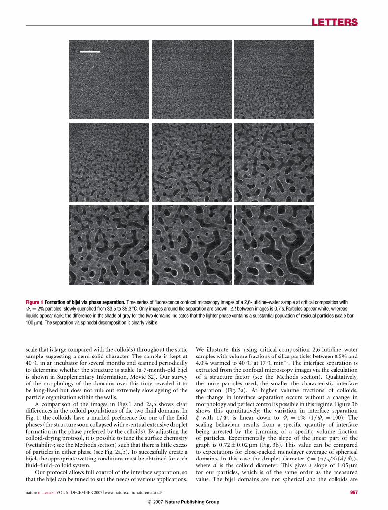
First, we show that nucleation can be bypassed by choosingmixtures with critical composition. Figure 1 shows a time series ofconfocal slices of a critical-composition 2,6-lutidine–water samplecontaining a 2% volume of silica colloids as it is warmed quasi-statically through the critical point (Tc = 34.1 ◦C). The imagesare rastered from top to bottom; a frame begins every 0.7 sand time also progresses during the scanning of each frame.The characteristic spinodal pattern is clearly visible with thedomain size growing continuously. The particles are being sweptup by the interfaces as the liquids separate. As the interfacialarea decreases the colloids become jammed together, ultimatelyarresting the phase separation as predicted1. These results are thefirst demonstration that the route to bijel formation is a slowchange in temperature at critical composition.
We are now able to create three-dimensional bijels routinely(see Supplementary Information, Movies S1,S2) using thisprotocol. Figure 2a,b presents the results of a fluorescence confocalmicroscopy study from the surface into the centre of a 1-mm-thicksample (see the Methods section). The sample was createdvia a deep quench to 40 ◦C at 17 ◦C min−1 at close-to-criticalcomposition (mole fraction of lutidine xl = 0.0644 ± 0.0001)with particle volume fraction Φv = 2% neutrally wetting silicacolloids. After the quench we find an arrested, bicontinuous patterncharacterized by a domain size ξ ≈ 40 µm independent of thedepth into the sample. Figure 2b shows vertical reconstructionsthrough the bottom, centre and top of the images in Fig. 2a.Many domains are encountered in traversing the sample along thethinnest dimension (the vertical). This is fundamentally differentto the two-dimensional structures obtained from phase-separatingpolymers9 and fast-quenched alcohol–alkane mixtures3. In thesecases, the domain size is comparable to the thickness of the sample,making it impossible to distinguish between structural stabilityand surface effects. We achieve fully three-dimensional samplesby quenching through the critical point and exploiting criticalslowing down. We demonstrate that spinodal decomposition takesplace throughout the depth of the sample (see the SupplementaryInformation Movies) resulting, in the presence of particles, in arigid, fully three-dimensional bicontinuous structure. There areclear variations in the mean curvature of the interfaces (on a length
966 nature materials VOL 6 DECEMBER 2007 www.nature.com/naturematerials
© 2007 Nature Publishing Group
LETTERS
Figure 1 Formation of bijel via phase separation. Time series of fluorescence confocal microscopy images of a 2,6-lutidine–water sample at critical composition withΦv = 2% particles, slowly quenched from 33.5 to 35.3 ◦C. Only images around the separation are shown. 1t between images is 0.7 s. Particles appear white, whereasliquids appear dark; the difference in the shade of grey for the two domains indicates that the lighter phase contains a substantial population of residual particles (scale bar100 µm). The separation via spinodal decomposition is clearly visible.
scale that is large compared with the colloids) throughout the staticsample suggesting a semi-solid character. The sample is kept at40 ◦C in an incubator for several months and scanned periodicallyto determine whether the structure is stable (a 7-month-old bijelis shown in Supplementary Information, Movie S2). Our surveyof the morphology of the domains over this time revealed it tobe long-lived but does not rule out extremely slow ageing of theparticle organization within the walls.
A comparison of the images in Figs 1 and 2a,b shows cleardifferences in the colloid populations of the two fluid domains. InFig. 1, the colloids have a marked preference for one of the fluidphases (the structure soon collapsed with eventual extensive dropletformation in the phase preferred by the colloids). By adjusting thecolloid-drying protocol, it is possible to tune the surface chemistry(wettability; see the Methods section) such that there is little excessof particles in either phase (see Fig. 2a,b). To successfully create abijel, the appropriate wetting conditions must be obtained for eachfluid–fluid–colloid system.
Our protocol allows full control of the interface separation, sothat the bijel can be tuned to suit the needs of various applications.
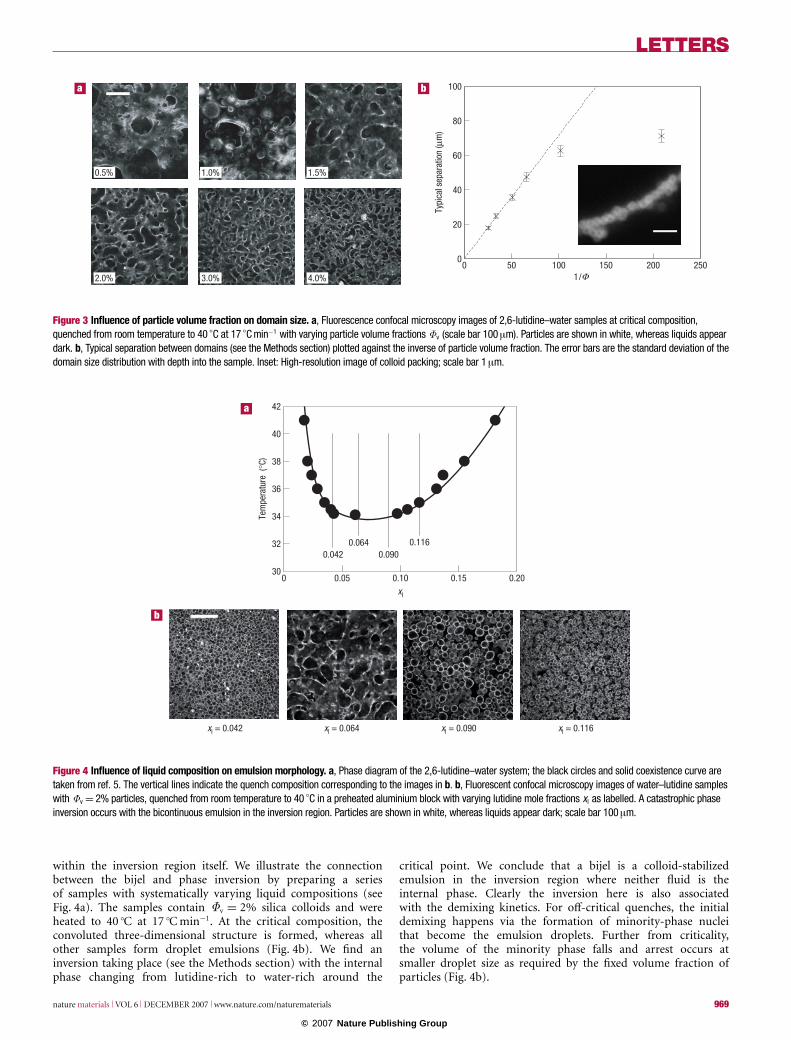
We illustrate this using critical-composition 2,6-lutidine–watersamples with volume fractions of silica particles between 0.5% and4.0% warmed to 40 ◦C at 17 ◦C min−1. The interface separation isextracted from the confocal microscopy images via the calculationof a structure factor (see the Methods section). Qualitatively,the more particles used, the smaller the characteristic interfaceseparation (Fig. 3a). At higher volume fractions of colloids,the change in interface separation occurs without a change inmorphology and perfect control is possible in this regime. Figure 3bshows this quantitatively: the variation in interface separationξ with 1/Φv is linear down to Φv = 1% (1/Φv = 100). Thescaling behaviour results from a specific quantity of interfacebeing arrested by the jamming of a specific volume fractionof particles. Experimentally the slope of the linear part of thegraph is 0.72 ± 0.02 µm (Fig. 3b). This value can be comparedto expectations for close-packed monolayer coverage of sphericaldomains. In this case the droplet diameter ξ = (π/
√3)(d/Φv),
where d is the colloid diameter. This gives a slope of 1.05 µmfor our particles, which is of the same order as the measuredvalue. The bijel domains are not spherical and the colloids are
nature materials VOL 6 DECEMBER 2007 www.nature.com/naturematerials 967
© 2007 Nature Publishing Group

LETTERS
ba
c d
100
80
60
40
20
0
G
Sink
ing
dept
h (µ
m)
0 2 4 6 8 10 12Time (s)
80 µm 160 µm
240 µm 320 µm
400 µm 480 µm
Figure 2 Structure and mechanical properties of bijels. a, Fluorescence confocal microscopy images at different depths (as indicated on each image) into the sample of2,6-lutidine–water at critical composition with Φv = 2%, quenched from room temperature to 40 ◦C at 17 ◦Cmin−1. The equal darkness of both domains shows that there arefew residual particles in either domain. b, Reconstructions along the vertical axis (thinnest dimension) for the bottom, centre and top of the images in a reaching 500 µm intothe cuvette. In both cases, the scale bar is 100 µm. Actual sample thickness is 1 mm. c, Sinking depth of a cylinder with a mass of 1.9 mg and 0.2 mm in diameter falling overtime in both a droplet emulsion (dashed line) and a bicontinuous emulsion (crosses) with compositions as above. The cylinder falls quickly through the droplet emulsion, andmuch more slowly through the bicontinuous sample with plateaux of zero sedimentation rate. d, Cylinder 6 min after being released into a bicontinuous emulsion (cuvette is1 cm wide). The wire remains supported against gravity for weeks confirming that there is a yield stress. The G (1.8 mm tall) identifies the type of glass used for the cuvette.
unlikely to be close-packed; however, as the packing fraction fallsbelow the close-packed limit, the slope decreases; therefore, thediscrepancy is not a cause for concern. The rough agreementsuggests that the interfaces may be stabilized by a monolayerof colloids: as in the computer simulations1. Confirmation isprovided by high-resolution imaging (Fig. 3b, inset). Stabilizationby a monolayer of colloids demonstrates that the key tostructure formation is the interfacial tension and not directinteractions between colloids. This shows that we have overcomethe irreproducibility of the structures shown in ref. 3, whichwere supported by thick colloidal layers. For 0.5% and 1.0%volume-fraction samples, these interfaces seem to adopt a preferred
curvature that undermines emulsion connectivity observed forall other samples. Other, more complex configurations can beobtained by also varying the warming rate (see SupplementaryInformation, Note S3).
The results we report can be connected to the known behaviourof colloid-stabilized droplet emulsions as the proportions ofdispersed and continuous phases are varied. Unlike emulsionsstabilized by amphiphilic surfactants, colloid-stabilized emulsionscan undergo a phase inversion from oil-in-water to water-in-oildue to changes in the volume fractions of fluids alone10,11.Compositions either side of inversion have been characterized6;however, it has not previously been possible to create an emulsion
968 nature materials VOL 6 DECEMBER 2007 www.nature.com/naturematerials
© 2007 Nature Publishing Group
LETTERS
0
20
40
60
80
100
Typi
cal s
epar
atio
n (μ
m)
1 /0 50 100 150 200 250
Φ
0.5% 1.0% 1.5%
2.0% 3.0% 4.0%
a b
Figure 3 Influence of particle volume fraction on domain size. a, Fluorescence confocal microscopy images of 2,6-lutidine–water samples at critical composition,quenched from room temperature to 40 ◦C at 17 ◦Cmin−1 with varying particle volume fractions Φv (scale bar 100 µm). Particles are shown in white, whereas liquids appeardark. b, Typical separation between domains (see the Methods section) plotted against the inverse of particle volume fraction. The error bars are the standard deviation of thedomain size distribution with depth into the sample. Inset: High-resolution image of colloid packing; scale bar 1 µm.
030
32
34
36
38
40
42
0.05 0.10xl
Tem
pera
ture
(°C
)
0.15 0.20
0.0420.064
0.0900.116
xl = 0.042 xl = 0.064 xl = 0.090 xl = 0.116
a
b
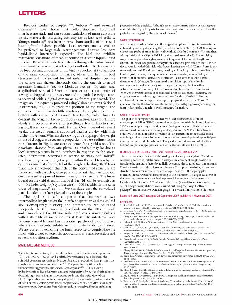
Figure 4 Influence of liquid composition on emulsion morphology. a, Phase diagram of the 2,6-lutidine–water system; the black circles and solid coexistence curve aretaken from ref. 5. The vertical lines indicate the quench composition corresponding to the images in b. b, Fluorescent confocal microscopy images of water–lutidine sampleswith Φv = 2% particles, quenched from room temperature to 40 ◦C in a preheated aluminium block with varying lutidine mole fractions x l as labelled. A catastrophic phaseinversion occurs with the bicontinuous emulsion in the inversion region. Particles are shown in white, whereas liquids appear dark; scale bar 100 µm.
within the inversion region itself. We illustrate the connectionbetween the bijel and phase inversion by preparing a seriesof samples with systematically varying liquid compositions (seeFig. 4a). The samples contain Φv = 2% silica colloids and wereheated to 40 ◦C at 17 ◦C min−1. At the critical composition, theconvoluted three-dimensional structure is formed, whereas allother samples form droplet emulsions (Fig. 4b). We find aninversion taking place (see the Methods section) with the internalphase changing from lutidine-rich to water-rich around the
critical point. We conclude that a bijel is a colloid-stabilizedemulsion in the inversion region where neither fluid is theinternal phase. Clearly the inversion here is also associatedwith the demixing kinetics. For off-critical quenches, the initialdemixing happens via the formation of minority-phase nucleithat become the emulsion droplets. Further from criticality,the volume of the minority phase falls and arrest occurs atsmaller droplet size as required by the fixed volume fraction ofparticles (Fig. 4b).
nature materials VOL 6 DECEMBER 2007 www.nature.com/naturematerials 969
© 2007 Nature Publishing Group
LETTERS
Previous studies of droplets12–14, bubbles15,16 and extendeddomains3,9,17 have shown that colloid-stabilized fluid–fluidinterfaces are static and can support variations of mean curvatureon the macroscale, indicating that they are at least semi-solid. AYoung’s modulus18 has been inferred from studies of interfacialbuckling13,14,19. Where possible, local rearrangements tend tobe preferred to large-scale rearrangements because less bareliquid–liquid interface is exposed16. The bijel, too, exhibitsmacroscale variations of mean curvature in a static liquid–liquidinterface. Because the interface extends through the entire sampleits semi-solid character makes the bijel a soft solid1. To demonstratethe load bearing properties of the bijel, we looked at two samplesof the same composition in Fig. 2a, where one had the bijelstructure and the second formed individual droplets becausethe sample was shaken vigorously during the quench to avoidstructure formation (see the Methods section). In each case,a cylindrical wire of 0.2 mm in diameter and a total mass of1.9 mg is dropped into the cuvette and the path the weight takesis recorded with a digital camera at 15 frames per second. Theimages are subsequently processed using Vision Assistant (NationalInstruments, V7.1.0) to track the position of the weight. Thedroplet emulsion provides little resistance: the weight sinks to thebottom with a speed of 960 mm s−1 (see Fig. 2c, dashed line). Incontrast, the weight in the bicontinuous emulsion sinks much moreslowly and becomes stuck after travelling a few millimetres (seeFig. 2c,d) and occasionally sinks further. Over a period of severalweeks, the weight remains supported against gravity with littlefurther movement. Whereas the slowing and stopping of the weightin the bijel suggests viscoelastic properties, the zero-sedimentationrate plateaux in Fig. 2c are clear evidence for a yield stress. Theoccasional descent from one plateau to another may be due tolocal rearrangements in the interfaces in response to the stress.Such intermittent behaviour is generic to many soft solids20.Confocal images examining the path within the bijel taken by thecylinder show that after the fall of the weight a ‘healing effect’ takesplace. The ripped-open channels of the convoluted structure arere-covered with particles, so no purely liquid interfaces are exposed,creating a self-supported tunnel through the structure. The lowerbound on the yield stress for this bijel (Fig. 2d) can be estimated asσs = (cylinder weight)/(cylinder area) = 600 Pa, which is the sameorder of magnitude18 as γ/d. We conclude that the convolutedparticle-laden interfaces give solidity to the sample.
The bijel is a soft composite that incorporates twointermediate length scales: the interface separation and the colloidsize. Consequently, elasticity and permeability can be tunedindependently. Our route using colloids on the 100s nm scaleand channels on the 10s µm scale produces a novel emulsionwith a shelf life of many months at least. The interfacial layeris semi-permeable2 and has interstitial patches of bare interfacewhere small reagents, soluble in immiscible fluids21, can meet.We are currently exploring the bijels response to counter-flowingfluids with a view to potential applications as a microreaction andsolvent-extraction medium.
MATERIALS AND METHODS
The 2,6-lutidine–water system exhibits a lower critical solution temperature(Tc = 34.1 ◦C,xl = 0.064) and a relatively symmetric phase diagram; thespinodal demixing region is easily accessible and the obtained final phases haveroughly equal volumes and densities5,22. The particles are Stober silicafluorescently tagged with fluorescein isothiocyanate23,24 (FITC) with ahydrodynamic radius of 290 nm and a polydispersity of 0.025 as obtained fromdynamic light scattering measurements. We found the wettability of theFITC-doped silica surface to strongly depend on the absorbed surface water. Toobtain neutrally wetting conditions, the particles are dried at 70 ◦C over nightunder vacuum. Deviations from this procedure strongly affect the stabilizing
properties of the particles. Although recent experiments point out new aspectsof stabilization by solid particles associated with electrostatic charge25, here theparticles are trapped by the interfacial tension7.
SAMPLE PREPARATIONA suspension of silica colloids in the single-fluid phase of 2,6-lutidine–water isobtained by initially dispersing the particles in water (MilliQ, 18 M�) using anultrasound probe (Sonics & Material), with 20 kHz for 2 min at 3–6 W and thenadding 2,6-lutidine (Sigma Aldrich, ≥99%, used as-received). The resultingsuspension is placed in a glass cuvette (Optiglass) of 1 mm pathlength. Analuminium block designed to closely fit the cuvette is preheated to 40 ◦C. Whenthe cuvette is loaded this yields the fastest heating rate of 17 ◦C min−1 and is thestandard protocol. For slower rates, heating and cooling coils surrounding theblock adjust the sample temperature, which is accurately controlled by aproportional–integral–derivative controller (Lakeshore 331) with a type-Kthermocouple (Omega). To examine the emulsion type of the dropletemulsions obtained when varying the liquid ratios, we check whethersedimentation or creaming of the emulsion droplets occurs. However, forΦv = 2% the weight of the shell makes all droplets sediment. Therefore, thedroplets were re-made using a lower volume fraction of colloids. For themechanical test on the bijel, the sample is prepared with the 17 ◦C min−1
quench, whereas the droplet counterpart is prepared by vigorously shaking thesample during the quench to avoid structure formation.
SAMPLE CHARACTERIZATIONThe quenched samples were studied with laser fluorescence confocalmicroscopy. A Nikon TE300 was used in conjunction with the Biorad Radiance2100 scanner operating an Ar-ion laser. Owing to the controlled-temperatureenvironment, we use an extra long working distance ×20 PlanFluor Nikonobjective with an adjustable correction collar. Depending on refractive indexmatching and particle volume fraction, visualization at depths of up to 800 µminto the sample could be achieved. The yield stress test was recorded with aNikon Coolpix 7 mega-pixel camera while the sample was held at 40 ◦C.
LENGTH-SCALE EXTRACTION USING FAST FOURIER TRANSFORM ANALYSISThe structures we create are static analogues of sponge mesophases26 and thescattering pattern is well known. To analyse the dominant length scales, wecalculate the structure factor by radially averaging the squared two-dimensionalFourier transform of the microscopy images. To improve statistics, we averagestructure factors for several different images. A knee in the log–log plotindicates the wavevector corresponding to the characteristic length scale. We fitthe resulting curves to a stretched exponential to extract the characteristiclength which is found at 20% decay of the curve (a small increment on the logscale). Image manipulations were carried out using the ImageJ softwarepackage27 and Interactive Data Language (ITT Visual Information Solutions).
Received 5 June 2007; accepted 28 September 2007; published 4 November 2007.
References1. Stratford, K., Adhikari, R., Pagonabarraga, I., Desplat, J.-C. & Cates, M. E. Colloidal jamming at
interfaces: A route to fluid bicontinuous gels. Science 309, 2198–2201 (2005).2. Dinsmore, A. D. et al. Colloidosomes: Selectively permeable capsules composed of colloidal particles.
Science 298, 1006–1009 (2002).3. Clegg, P. S. et al. Emulsification of partially miscible liquids using colloidal particles: Nonspherical
and extended domain structures. Langmuir 23, 5984–5994 (2007).4. Debenedetti, P. G. Metastable Liquids—Concepts and Principles (Princeton Univ. Press,
New Jersey, 1996).5. Grattoni, C. A., Dawe, R. A., Yen Seah, C. & Gray, J. D. Density, viscosity, surface tension, and
interfacial tension of 2,6-lutidine+water. J. Chem. Eng. Data 38, 516–519 (1993).6. Aveyard, R., Binks, B. P. & Clint, J. H. Emulsions stabilized solely by colloidal particles. Adv. Colloid
Interface Sci. 100–102, 503–546 (2003).7. Binks, B. P. & Horozov, T. S. Colloidal Particles At Liquid Interfaces (Cambridge Univ. Press,
Cambridge, 2006).8. Cates, M. E., Poon, W. C. K., Egelhaaf, S. U. & Clegg, P. S. European Patent Application Number
EP05761568. 4.9. Chung, H. J., Ohno, K., Fukuda, T. & Composto, R. J. Self-regulated structures in nanocomposites by
directed nanoparticle assembly. Nano Lett. 5, 1878–1882 (2005).10. Binks, B. P. Particles as surfactants—similarities and differences. Curr. Opin. Colloid Interface Sci. 7,
21–41 (2002).11. Kralchevsky, P. A., Ivanov, I. B., Ananthapadmanabhan, K. P. & Lips, A. On the thermodynamics of
particle-stabilized emulsions: Curvature effects and catastrophic phase inversion. Langmuir 21,50–63 (2005).
12. Clegg, P. S. et al. Colloid stabilized emulsions: Behaviour as the interfacial tension is reduced. J. Phys.Condens. Matter 17, S3433–S3438 (2005).
13. Xu, H., Melle, S., Golemanov, K. & Fuller, G. G. Shape and buckling transitions in solid-stabilizeddrops. Langmuir 21, 10016–10020 (2005).
14. Tsamantakis, C., Masliyah, J., Yeung, A. & Gentzis, T. Investigation of the interfacial properties ofwater-in-diluted-bitumen emulsions using micropipette techniques. J. Colloid Interface Sci. 284,176–183 (2005).
970 nature materials VOL 6 DECEMBER 2007 www.nature.com/naturematerials
© 2007 Nature Publishing Group
LETTERS
15. Subramaniam, A. B., Abkarian, M., Mahadevan, L. & Stone, H. A. Non-spherical bubbles. Nature438, 930 (2005).
16. Subramaniam, A. B., Abkarian, M., Mahadevan, L. & Stone, H. A. Mechanics of interfacial compositematerials. Langmuir 22, 10204–10208 (2006).
17. Edmond, K. V., Schofield, A. B., Marquez, M., Rothstein, J. P. & Dinsmore, A. D. Stable jets ofviscoelastic fluids and self-assembled cylindrical capsules by hydrodynamic focusing. Langmuir 22,9052–9056 (2006).
18. Vella, D., Aussillous, P. & Mahadevan, L. Elasticity of an interfacial particle raft. Europhys. Lett. 68,212–218 (2004).
19. Aveyard, R., Clint, J. H., Nees, D. & Quirke, N. Structure and collapse of particle monolayersunder lateral pressure at the octane/aqueous surfactant solution interface. Langmuir 16,8820–8828 (2000).
20. Cipelletti, L. & Ramos, L. Slow dynamics in glassy soft matter. J. Phys. Condens. Matter 17,R253–R285 (2005).
21. Luisi, P. L., Giomini, M., Pileni, M. P. & Robinson, B. H. Reverse micelles as hosts for proteins andsmall molecules. Biochim. Biophys. Acta 947, 209–246 (1988).
22. Faizullin, M. Z. & Skripov, V. P. Investigation of the (T,p,x) surface of phase separation ofxH2O+ (1−x)NC(CH3)CHCHCHC(CH3) in the vicinity of the line of lower critical points ofdissolution. J. Chem. Thermodyn. 23, 561–567 (1991).
23. Stober, W., Fink, A. & Bohn, E. Controlled growth of monodisperse silica spheres in the micron sizerange. J. Colloid Interface Sci. 26, 62–69 (1968).
24. van Blaaderen, A. & Vrij, A. Synthesis and characterization of colloidal dispersions of fluorescent,monodisperse silica spheres. Langmuir 8, 2921–2931 (1992).
25. Leunissen, M. E., van Blaaderen, A., Hollingsworth, A. D., Sullivan, M. T. & Chaikin, P. M.Electrostatics at the oil-water interface, stability and order in emulsions and colloids. Proc. Natl Acad.Sci. USA 104, 2585–2590 (2007).
26. Roux, D., Coulon, C. & Cates, M. E. Sponge phases in surfactant solutions. J. Phys. Chem. 96,4174–4187 (1992).
27. Rasband, W. S. US National Institute of Health: Bethesda, Maryland, USA, 1997–2006;<http://rsb.info.nih.gov/ij/>.
AcknowledgementsWe are grateful to M. Cates, B. Binks, T. Horozov, E. Kim and H. Vass for productive discussions.Financial support was provided by EPSRC Grants EP/D076986/1 and EP/E502652/1.Correspondence and requests for materials should be addressed to P.S.C.Supplementary Information accompanies this paper on www.nature.com/naturematerials.
Reprints and permission information is available online at http://npg.nature.com/reprintsandpermissions/
nature materials VOL 6 DECEMBER 2007 www.nature.com/naturematerials 971
© 2007 Nature Publishing Group
![Characterization of a Liquid Crystal Stabilized ... · liquid crystal stabilized emulsions that can be adjusted to a wide range of pH values [7]. Since topically applied actives typically](https://img.pdfslide.us/doc/110x75/5f670dfae76793780c751014/characterization-of-a-liquid-crystal-stabilized-liquid-crystal-stabilized-emulsions.jpg)