Embed Size (px)
Citation preview
RESEARCH ARTICLE
Better Management of Alcohol Liver Disease
Using a ‘Microstructured Synbox’ System
Comprising L. plantarum and EGCG
Praveen Rishi1, Sumeha Arora1, Ujjwal Jit Kaur1, Kanwaljit Chopra2, Indu Pal Kaur2*
1 Department of Microbiology, Basic Medical Sciences Block, South Campus, Panjab University,
Chandigarh, India, 2 University Institute of Pharmaceutical Sciences, Panjab University, Chandigarh, India
Abstract
Synergistic combination of probiotics with carbohydrate based prebiotics is widely employed
for the treatment of various gut related disorders. However, such carbohydrate based pre-
biotics encourage the growth of pathogens and probiotics, equally. Aim of the study was (i)
to explore the possibility of using epigallocatechin gallate (EGCG) a phenolic compound, as
a prebiotic for L.plantarum; (ii) to develop and evaluate a microstructured synbox (microen-
capsulating both probiotic and EGCG together) in rat model of alcohol liver disease (ALD);
and, (iii) to confirm whether the combination can address issues of EGCG bioavailability and
probiotic survivability in adverse gut conditions. Growth enhancing effect of EGCG on L.
plantarum (12.8±0.5 log 10 units) was significantly (p�0.05) better than inulin (11.4±0.38 log
10 units), a natural storage carbohydrate. The formulated synbox significantly modulated the
levels of alcohol, endotoxin, hepatic enzymes and restored the hepatoarchitecture in com-
parison to simultaneous administration of free agents. Additionally, using a battery of tech-
niques, levels of various cellular and molecular markers viz. NF-kB/p50, TNF-α, IL12/p40,
and signalling molecules TLR4, CD14, MD2, MyD88 and COX-2 were observed to be sup-
pressed. Developed microbead synbox, as a single delivery system for both the agents
showed synergism and hence, holds promise as a therapeutic option for ALD management.
Introduction
In view of increasing demand for bioecological and nutritional control of diseases, probiotics
alone or in combination with prebiotics (synbiotics) are now being explored for their thera-
peutic potential. Lactic acid bacteria used as probiotics exhibit a variety of biological actions
including stimulation of the immune system, balancing of intestinal microbiota, potential
reduction of inflammation and the prevention of allergies, hypertension and cancer [1].
Despite conferring several health benefits, the in-vivo survival and establishment of probiotic
strains upon oral administration is elusive. Use of prebiotics such as fructooligosaccharides
(FOS), galactooligosaccharides (GOS), and inulin can address some of these issues [2–4].
These carbohydrate-type prebiotics may however non-specifically encourage the growth of all
gut organisms, including the non-probiotic pathogenic bacteria. Therefore, newer alternatives
of non-carbohydrate origin need to be explored for stimulation of probiotic flora [5].
PLOS ONE | DOI:10.1371/journal.pone.0168459 January 6, 2017 1 / 18
a11111
OPENACCESS
Citation: Rishi P, Arora S, Kaur UJ, Chopra K, Kaur
IP (2017) Better Management of Alcohol Liver
Disease Using a ‘Microstructured Synbox’ System
Comprising L. plantarum and EGCG. PLoS ONE 12
(1): e0168459. doi:10.1371/journal.pone.0168459
Editor: Giovanni Li Volti, University of Catania,
ITALY
Received: October 9, 2016
Accepted: December 1, 2016
Published: January 6, 2017
Copyright: This is an open access article, free of all
copyright, and may be freely reproduced,
distributed, transmitted, modified, built upon, or
otherwise used by anyone for any lawful purpose.
The work is made available under the Creative
Commons CC0 public domain dedication.
Data Availability Statement: All relevant data are
within the paper and its Supporting Information
files.
Funding: The Indian Council of Medical Research
provided funding to carry out experimental studies.
The funder had no role in study design, data
collection and analysis, decision to publish, or
preparation of the manuscript.
Competing Interests: The authors have declared
that no competing interests exist.
Green tea (Camelia sinensis), a rich source of polyphenols, is a widely consumed beverages
in the world and is suggested to possess health promoting properties. Epigallocatechin gallate
(EGCG) is the most abundant and the most active component of green tea leaves. It elicits var-
ious biological effects, including antimutagenicity and antitumorigenesis, free radical scaveng-
ing activity and antimicrobial activity against gut pathogens [6, 7]. Probiotic L. plantarum is
reported to possess tannase activity [8, 9]; EGCG on the other hand is condensed tannin which
yields phenolic acids, when hydrolyzed by tannase. In view of the tannase activity of L. plan-tarum, the possibility of using EGCG for enhancing growth of the former was explored in the
present work.
Further, due to the limitation of bioavailability of EGCG and viability of L. plantarum dur-
ing their transit through the harsh gut conditions, these agents were encapsulated in calcium
alginate beads, resulting in a synbiotic formulation. Encapsulating probiotic with a natural
polyphenolic molecule like EGCG for improved viability (of the former) and effectiveness of
both the agents is the novelty of the system. The developed microbeads (microstructured syn-
box) will ensure a prolonged and continuous release of probiotic in the gut, allowing sufficient
time for its adhesion and establishment on the gut mucosal wall. The idea of using natural mol-
ecules with their own set of suitable therapeutic activity in addition to supporting probiotic
growth in a suitably designed pharmaceutical system is a relatively new concept. Application
of the same was demonstrated earlier by us for proposed management of gastric ulcers [10]. It
is noteworthy that EGCG with established antioxidant and anti-inflammatory effects is
explored presently for its potential prebiotic effect for L. plantarum.
Thus, presently the developed synbiotic system was evaluated against oxidative stress/endo-
toxin mediated alcoholic liver disease (ALD). Alcohol induces damage by building up endotoxin
mediated oxidative stress in the cellular constituents of the tissue. Catechins including EGCG
have been demonstrated previously by us [11, 12] and others [13, 14] to attenuate alcoholic liver
injury by creating an antioxidant- oxidant balance in the hepatic tissues. ALD is also accompa-
nied by elevated intestinal permeability which can be abridged by probiotic administration.
Recently, we have established the improved efficacy of L. plantarum [15] against ALD, when
encapsulated within alginate beads. Similar enhancement for EGCG alone when encapsulated
into alginate floating beads was also observed (data communicated). Presently we demonstrate
the effects of combining these two agents (microstructured synbox) for a synergistic effect.
Materials and Methods
Agents
Natural polyphenol EGCG was provided as a gift sample by Y.S. Hara, Tea Solutions, Hara’s
Office, Tokyo. Standard lactic acid bacteria (LAB)- Lactobacillus plantarum MTCC 2621, used
as probiotic, was procured from Microbial Type Culture Collection (MTCC), Institute of
Microbial Technology, Chandigarh (India).
Construction of Microstructured Synbox
Effect of EGCG on the growth of L. plantarum. A dose dependent (10–200mg of EGCG)
study was designed to observe the effect of EGCG on the growth of L. plantarum at 1010CFU/ml.
Various concentrations of EGCG i.e 10mg/ml- 200mg/ml were added separately in the Lactoba-
cillus MRS broth (Himedia, India) each containing 1% inoculum of the overnight L. plantarum2621 culture. After 24 hours of incubation under anaerobic conditions at 37˚C, the CFU were
enumerated on MRS agar plates at 37˚C following incubation for 48 hours.
In another set of experiment, various concentrations of inulin (10–200 μg) which is a widely
used prebiotic was also evaluated for growth promoting effects on L. plantarum.
Microstructured Synbox for Managing ALD
PLOS ONE | DOI:10.1371/journal.pone.0168459 January 6, 2017 2 / 18

Preparation of probiotic- EGCG co-encapsulated alginate beads. L. plantarum and
EGCG were encapsulated together in calcium alginate microbeads [16, 17] via extrusion tech-
nique. Briefly, L. plantarum (1010 CFU/ml) and EGCG (50 mg) were dispersed by stirring
overnight in 1% sterile sodium alginate solution. Dispersion was added dropwise through a 26
G syringe needle into 1% sterile calcium chloride solution under stirring, and the beads
formed spontaneously were left to harden in the former for 2 hours under continuous stirring.
The beads were filtered out, using Whatman filter paper 1, and freeze dried for storage. The
alginate loaded microparticles were named as AL.
Characterization of probiotic-EGCG co-encapsulated alginate beads/
microparticles
1. Size of microparticulate beads was determined using a stage micrometer and Olympus opti-
cal microscope.
2. Surface morphology of microparticles was examined using scanning electron microscope
(SEM) (JSM- 6100, JEOL Ltd., Tokyo, Japan) housed in Central Instrumentation Labora-
tory/Sophisticated Analytical Instrument Facility of Panjab University, Chandigarh, India
at 10 kV. The microparticles were mounted on metal grids using double-sided tape and
coated with gold under vacuum.
3. Percent entrapment efficiency (EE %) of both the agents was determined using the follow-
ing formulae:
EE of EGCG %ð Þ ¼EGCG entrapped in beads
EGCG initially loaded in alginate mix� 100
EE of L: plantarum %ð Þ ¼Log CFU entrapped in 100 g of beads
Log CFU initially loaded in alginate mix ðfor 100g beadsÞ� 100
4. In-vitro release cum dissolution studies of EGCG and probiotic in alginate beads (equiva-
lent to 1010 CFU/ml of probiotic and 50 mg of EGCG) were performed aseptically using the
USP type II dissolution test apparatus at 100 rpm and 37± 0.5˚C and 900 ml simulated
intestinal fluid (SIF) (pH- 6.8) for 6 hours. 5ml aliquots of the medium were withdrawn at
pre-determined time intervals and replaced with fresh dissolution media. The samples were
analyzed for EGCG content spectrophotometrically at 270nm. For probiotic, spread plate
method was employed.
5. Stability of EGCG in probiotic-EGCG combination beads was compared with that of free
EGCG by dispersing suitable quantities of both in simulated intestinal fluid (pH-6.8) for 6
hours. At regular intervals, samples were withdrawn at similar time points and observed
spectrophotometrically at 270nm and % stability was calculated.
6. The co-encapsulated beads were evaluated for their storage stability at 25˚C in a stability
chamber for 6 months. The bacterial count was enumerated by spread plate method on
MRS agar plates at the start and at the end of the study
7. The stability of probiotic in probiotic- EGCG alginate beads was evaluated in the presence
of bile salts, by suspending the beads, in 0.3% bile salt solutions (sodium taurocholate and
Microstructured Synbox for Managing ALD
PLOS ONE | DOI:10.1371/journal.pone.0168459 January 6, 2017 3 / 18
sodium glycolate) for 4 hours.
Survivability of L: plantrum in Bile Salts %ð Þ ¼Log
10CFU at 4 hours
Log10
CFU at 0 hour
8. The probiotic- EGCG alginate beads and free probiotic were incubated in simulated gastric
fluid (SGF) (pH-1.2) for 4 hours and sequentially in SIF (pH-6.8) for 2 hrs and the CFU
remaining in each case were determined to establish viability of probiotic in either case.
Survivability of L: plantarum in SGF & SIF %ð Þ ¼Log
10CFU at 6 hours
Log10
CFU at 0 hour� 100
In-vivo Studies
Ethics statement. The experiment protocols were approved by the Institutional Animal
Ethics Committee (Approval ID- IAEC/282/ dated– 30/8/2012) of Panjab University, Chandi-
garh, India and performed in accordance with the guidelines of Committee for the Purpose of
Control and Supervision of Experiments on Animals (CPCSEA), Government of India, on ani-
mal experimentation. All efforts were made to minimize the suffering of animals.
Animals. Female wistar rats (200–250 g) were procured from Central Animal House, Pan-
jab University, Chandigarh (India). The animals were housed under standard laboratory con-
ditions, maintained under normal light: dark cycle and had free access to food (Ashirwad
Industries Pvt. Ltd., Punjab, India) and water.
Establishment of ALD in Wistar rats
Alcohol dosing. Rats were administered 10g/kg of body weight/day of 35% (v/v) ethanol
(obtained from Brampton, Ontario) by oral gavage in double distilled water for two weeks.
Thereafter, the dose was increased to 14g/kg of body weight/day and was continued for 10
weeks by oral gavage [11].
Combination bead dosing. The L. plantarum and EGCG combination beads, as prepared
above, were dispensed in 1% carboxymethyl cellulose and were administered to rats for 8
weeks through oral gavage.
Experimental Design. After an acclimatizing period, rats were randomly divided into six
groups, each comprising of 8–10 rats (Fig 1). Rats were administered alcohol as described
above and the dose of alcohol group was selected on the basis of the previous study [11, 15].
Similarly, treatment using L. plantarum and EGCG microstructured synbox was carried on for
8 weeks. At the end of the experimental period (after 12 weeks), the rats were sacrificed by cer-
vical dislocation. Livers were removed quickly, rinsed in cold phosphate buffer saline (0.05 M,
pH 7.4) and stored at -62˚C till further use.
Measurement of blood alcohol. After 12 weeks of alcohol administration, blood was
taken from the tail vein 1.5 h and 2.5 h after the last dose. Blood alcohol levels (BAL) were mea-
sured using the alcohol dehydrogenase kit procured from Sigma Chemical Co., U.S.A.
Plasma endotoxin assay. Endotoxin level in the plasma samples was measured using Toxin
Sensor Chromogenic LAL Endotoxin Assay Kit (Hycult Biotech) as described by us earlier [11].
Assessment of liver function. Alanine aminotransferase (ALT) and aspartate amino-
transferase (AST) enzyme activities in serum were determined using ERBA test kits (ERBA
Diagnostics, Mannheim, Germany). Alkaline phosphatase (ALP) was estimated using Enzopak
Diagnostic kit (Reckon Diagnostics, India).
Microstructured Synbox for Managing ALD
PLOS ONE | DOI:10.1371/journal.pone.0168459 January 6, 2017 4 / 18

Tissue architecture studies. Liver tissues removed aseptically from the animals were cut
into small pieces and fixed in 10% buffered formalin. Samples were processed, stained with
hematoxylin-eosin and examined under the light microscope.
Assay for potential antioxidant capacity/ Total antioxidant capacity test. Commer-
cially available (DTAC-100, Bioassay Systems) total antioxidant capacity kit was used as per
the manufacturer’s instructions.
Determination of intestinal permeability. Method [18] is included in supplementary
information (S1 Text). Briefly, two non-metabolizable sugars, lactulose (Himedia, India) and
mannitol (Himedia, India) were administered orally to overnight fasted animals of various
groups, and their concentrations were measured in the serum of these animals using HPTLC,
one hour post administration.
Assay for NF-kB/p50, TNF-α and IL12/p40 subunit. Assay for NF-kB/p50 subunit in
the nuclear extracts and TNF- α in the liver homogenates, was performed using commercially
Fig 1. Diagrammatic representation of various treatment groups made for in-vivo studies.
doi:10.1371/journal.pone.0168459.g001
Microstructured Synbox for Managing ALD
PLOS ONE | DOI:10.1371/journal.pone.0168459 January 6, 2017 5 / 18
available Transcription Factor Assay kit (Upstate Biotechnology, NY, USA) and cytokine assay
kit (R&D Systems, USA), respectively, according to the manufacturer’s instructions [11].
For IL12/p40 subunit estimations a double antibody sandwich ELISA was performed using
manufacturer’s instructions of the commercially available kit (Qaybee-bio, China).
Transcription studies of signalling molecules. Liver tissue of the rats subjected to the
indicated treatments were harvested and immediately preserved in RNAlater (Ambion, CA,
USA) at -80˚C till processing. Total RNA was isolated using the RNeasy Mini Kit (Qiagen)
according to the manufacturer’s instructions and was, then, reverse transcribed by using oligo-
dT primer and first strand cDNA synthesis kit (Fermentas). Obtained cDNA was subjected to
PCR using TLR4, CD14, MD2, MyD88, COX-2 and Glyceraldehyde-3-phosphate dehydroge-
nase (GAPDH)-specific primers (Sigma Aldrich Chemicals, Banglalore, India). GAPDH
mRNA was used as an internal control. Densitometry of PCR product to determine relative
mRNA expression was performed by Gel Doc Multi-Analyst (BioRad USA).
Micronucleus analysis. Micronuclei analysis was done by the method of Schmid [19]
which is elaborated in the supplementary information (S2 Text). Minimum of 100 cells were
counted per sample for the presence of micronuclei using light microscope at 45x.
Statistical analysis. The data are expressed as mean ± standard deviation. Statistical sig-
nificance between various groups was evaluated using one way analysis of variance (ANOVA)
followed by Tukey’s multiple comparison tests. The statistical analysis was done using the
Graphpad Prism 6.00 for Windows (Graphpad software, California, USA). In all data analysis,
p-values of 0.05 or less (p<0.05) were considered significant.
Results
Effect of EGCG on the growth of L. plantarum
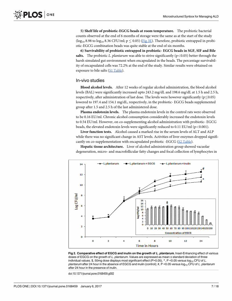
Growth of the probiotic was found to increase with increasing concentrations of EGCG upto
60mg, subsequent to which, a hormesis effect was observed. Growth enhancing effect of
EGCG for L. plantarum was significantly (p�0.05) more (10.55±0.78 to 12.8 ±0.5 log10 units)
than that induced by inulin, the most commonly used prebiotic (10.55± 0.78 to 11.4±0.38 log10
units) at corresponding concentrations (Fig 2).
Co-microencapsulation of probiotic-EGCG and its characterisation
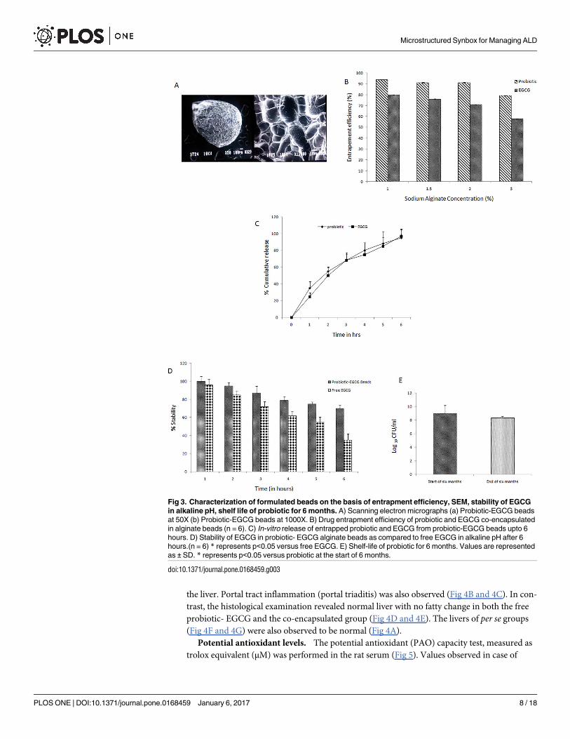
1) Size of microparticles and Scanning Electron Microscopy (SEM) images. Size of pro-
biotic-EGCG beads significantly increased to 75.4±7.8μm as compared to the beads incorpo-
rating only probiotic as described earlier [15]. SEM micrographs show smooth surface and
presence of probiotic rods in the beads (Fig 3A).
2) Percentage entrapment efficiency of combination beads. The entrapment efficiency
of both probiotic and EGCG decreased with an increase in the concentration of sodium algi-
nate. The maximum EE of 94.00±0.13% for probiotic and 80.00±0.21% for EGCG was
observed with 1% sodium alginate. Minimum entrapment was observed with the highest tried
concentration (3%) of sodium alginate (79.40±0.13% for probiotic and 57.74±0.34% for
EGCG) (Fig 3B). EE of probiotic when entrapped alone was found to be 80% [15] but upon
co-encapsulation with EGCG, it increased considerably to 94%.
3) In-vitro release studies. Significant quantities of probiotic (95%) and EGCG (97%)
were released from the probiotic-EGCG beads within 6 hours (Fig 3C).
4) Stability of EGCG at intestinal pH. EGCG entrapped in probiotic-EGCG combina-
tion was significantly more stable than free EGCG under alkaline conditions (representing pH
of the intestine) ensuring better protection (Fig 3D).
Microstructured Synbox for Managing ALD
PLOS ONE | DOI:10.1371/journal.pone.0168459 January 6, 2017 6 / 18
5) Shelf life of probiotic-EGCG beads at room temperature. The probiotic bacterial
counts observed at the end of 6 months of storage were the same as at the start of the study
(log10 8.98 to log10 8.36 CFU/ml; p� 0.05) (Fig 3E). Therefore, probiotic entrapped in probi-
otic-EGCG combination beads was quite stable at the end of six months.
6) Survivability of probiotic entrapped in probiotic- EGCG beads in SGF, SIF and Bile
salts. The probiotic L. plantarum was able to strive significantly (p<0.05) better through the
harsh simulated gut environment when encapsulated in the beads. The percentage survivabil-
ity of encapsulated cells was 72.2% at the end of the study. Similar results were obtained on
exposure to bile salts (S1 Table).
In-vivo studies
Blood alcohol levels. After 12 weeks of regular alcohol administration, the blood alcohol
levels (BAL) were significantly increased upto 243.2 mg/dL and 198.6 mg/dL at 1.5 h and 2.5 h,
respectively, after administration of last dose. The levels were however significantly (p�0.05)
lowered to 197.4 and 154.1 mg/dL, respectively, in the probiotic- EGCG beads supplemented
group after 1.5 and 2.5 h of the last administered dose.
Plasma endotoxin levels. The plasma endotoxin levels in the control rats were observed
to be 0.16 EU/ml. Chronic alcohol consumption considerably increased the endotoxin levels
to 0.54 EU/ml. However, on co-supplementing alcohol administration with probiotic- EGCG
beads, the elevated endotoxin levels were significantly reduced to 0.11 EU/ml (p<0.001).
Liver function tests. Alcohol caused a marked rise in the serum levels of ALT and ALP
while there was no significant change in AST levels. Activities of liver enzymes dropped signifi-
cantly on co-supplementation with encapsulated probiotic -EGCG (S2 Table).
Hepatic tissue architecture. Liver of alcohol administration group showed vacuolar
degeneration, micro- and macrofollicular fatty changes and focal collection of lymphocytes in
Fig 2. Comparative effect of EGCG and inulin on the growth of L. plantarum. Inset-Enhancing effect of various
doses of EGCG on the growth of L. plantarum. Values are expressed as mean ± standard deviation of three
individual values. $, 50mg dose displays most significant effect (P>0.05), *, P <0.05 versus log10 CFU of L.
plantarum after 24 hour in the absence of EGCG and inulin (control); #, P <0.05 versus log10 CFU of L. plantarum
after 24 hour in the presence of inulin.
doi:10.1371/journal.pone.0168459.g002
Microstructured Synbox for Managing ALD
PLOS ONE | DOI:10.1371/journal.pone.0168459 January 6, 2017 7 / 18
the liver. Portal tract inflammation (portal triaditis) was also observed (Fig 4B and 4C). In con-
trast, the histological examination revealed normal liver with no fatty change in both the free
probiotic- EGCG and the co-encapsulated group (Fig 4D and 4E). The livers of per se groups
(Fig 4F and 4G) were also observed to be normal (Fig 4A).
Potential antioxidant levels. The potential antioxidant (PAO) capacity test, measured as
trolox equivalent (μM) was performed in the rat serum (Fig 5). Values observed in case of
Fig 3. Characterization of formulated beads on the basis of entrapment efficiency, SEM, stability of EGCG
in alkaline pH, shelf life of probiotic for 6 months. A) Scanning electron micrographs (a) Probiotic-EGCG beads
at 50X (b) Probiotic-EGCG beads at 1000X. B) Drug entrapment efficiency of probiotic and EGCG co-encapsulated
in alginate beads (n = 6). C) In-vitro release of entrapped probiotic and EGCG from probiotic-EGCG beads upto 6
hours. D) Stability of EGCG in probiotic- EGCG alginate beads as compared to free EGCG in alkaline pH after 6
hours.(n = 6) * represents p<0.05 versus free EGCG. E) Shelf-life of probiotic for 6 months. Values are represented
as ± SD. * represents p<0.05 versus probiotic at the start of 6 months.
doi:10.1371/journal.pone.0168459.g003
Microstructured Synbox for Managing ALD
PLOS ONE | DOI:10.1371/journal.pone.0168459 January 6, 2017 8 / 18
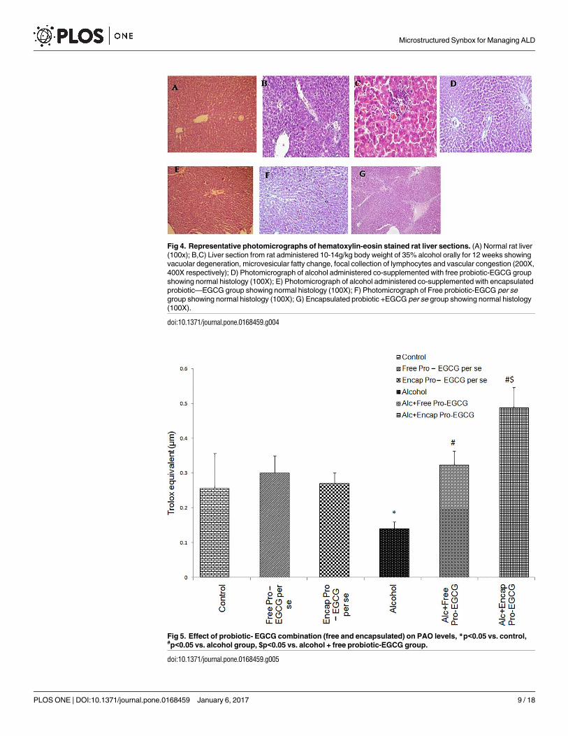
Fig 4. Representative photomicrographs of hematoxylin-eosin stained rat liver sections. (A) Normal rat liver
(100x); B,C) Liver section from rat administered 10-14g/kg body weight of 35% alcohol orally for 12 weeks showing
vacuolar degeneration, microvesicular fatty change, focal collection of lymphocytes and vascular congestion (200X,
400X respectively); D) Photomicrograph of alcohol administered co-supplemented with free probiotic-EGCG group
showing normal histology (100X); E) Photomicrograph of alcohol administered co-supplemented with encapsulated
probiotic—EGCG group showing normal histology (100X); F) Photomicrograph of Free probiotic-EGCG per se
group showing normal histology (100X); G) Encapsulated probiotic +EGCG per se group showing normal histology
(100X).
doi:10.1371/journal.pone.0168459.g004
Fig 5. Effect of probiotic- EGCG combination (free and encapsulated) on PAO levels, *p<0.05 vs. control,#p<0.05 vs. alcohol group, $p<0.05 vs. alcohol + free probiotic-EGCG group.
doi:10.1371/journal.pone.0168459.g005
Microstructured Synbox for Managing ALD
PLOS ONE | DOI:10.1371/journal.pone.0168459 January 6, 2017 9 / 18

alcoholic rats were significantly lower than control and treated rats. The entrapped probiotic-
EGCG treated group showed values even greater than the control animals (p� 0.05). Per segroups were similar to the control group. Since the PAO levels of animals treated with probi-
otic- EGCG co-encapsulated beads was significantly increased, hence, individual antioxidants
(SOD, catalase and glutathione reductase) were also studied in this group (data not shown).
Levels for each of these moeities were significantly higher than the control and the ALD group.
Assessment of intestinal permeability. The compromised intestinal permeability marks
the onset of ALD as it allows the releases of endotoxins into the serum. Increased intestinal
permeability on chronic alcohol administration was confirmed by the presence of bands of
both lactulose (disaccharide) and mannitol (monosaccharide) in serum, indicating their trans-
port across the gut wall. Treatment with entrapped probiotic-EGCG reduced the permeability
as only mannitol (monosaccharide) band was observed on the plate. The EGCG beads, on the
other hand had no effect on the intestinal permeability which was elevated by alcohol adminis-
tration (Fig 6 inset). Rf of mannitol was recorded as 0.49 whereas the Rf of lactulose was 0.61.
Molecular mechanisms of protection
Assay for NF-κB/p50, TNF-α and IL-12/40 subunit. The alcohol treated rats showed a
marked escalation of TNF-α and IL-12/p40 subunit values in comparison to those exhibited in
the control group. On co-supplementing with probiotic- EGCG beads, the enhanced (p<0.05)
levels were attenuated to normal values (Fig 7B and 7C). NF-kB/p-50 was also inactivated by
Fig 6. HPTLC Chromatogram (inset) and chromatograph showing the differential sugars i.e. lactulose and
mannitol released as a marker of intestinal permeability. Inset- A,G–lactulose standard; B,H—mannitol
standard; C- Alcohol group showing the presence of both the sugars in the serum due to increased permeability; D-
Alcohol + Encapsulated probiotic-EGCG group showing only mannitol, no disruption of gut permeability; E- Alcohol
fed; F- Alcohol + Free probiotic-EGCG group showing only mannitol, confirming intact gut permeability
doi:10.1371/journal.pone.0168459.g006
Microstructured Synbox for Managing ALD
PLOS ONE | DOI:10.1371/journal.pone.0168459 January 6, 2017 10 / 18
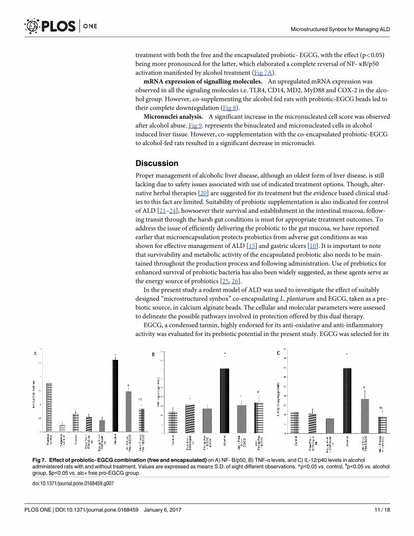
treatment with both the free and the encapsulated probiotic- EGCG, with the effect (p<0.05)
being more pronounced for the latter, which elaborated a complete reversal of NF- κB/p50
activation manifested by alcohol treatment (Fig 7A).
mRNA expression of signalling molecules. An upregulated mRNA expression was
observed in all the signaling molecules i.e. TLR4, CD14, MD2, MyD88 and COX-2 in the alco-
hol group. However, co-supplementing the alcohol fed rats with probiotic-EGCG beads led to
their complete downregulation (Fig 8).
Micronuclei analysis. A significant increase in the micronucleated cell score was observed
after alcohol abuse. Fig 9. represents the binucleated and micronucleated cells in alcohol
induced liver tissue. However, co-supplementation with the co-encapsulated probiotic-EGCG
to alcohol-fed rats resulted in a significant decrease in micronuclei.
Discussion
Proper management of alcoholic liver disease, although an oldest form of liver disease, is still
lacking due to safety issues associated with use of indicated treatment options. Though, alter-
native herbal therapies [20] are suggested for its treatment but the evidence based clinical stud-
ies to this fact are limited. Suitability of probiotic supplementation is also indicated for control
of ALD [21–24], howsoever their survival and establishment in the intestinal mucosa, follow-
ing transit through the harsh gut conditions is must for appropriate treatment outcomes. To
address the issue of efficiently delivering the probiotic to the gut mucosa, we have reported
earlier that microencapsulation protects probiotics from adverse gut conditions as was
shown for effective management of ALD [15] and gastric ulcers [10]. It is important to note
that survivability and metabolic activity of the encapsulated probiotic also needs to be main-
tained throughout the production process and following administration. Use of prebiotics for
enhanced survival of probiotic bacteria has also been widely suggested, as these agents serve as
the energy source of probiotics [25, 26].
In the present study a rodent model of ALD was used to investigate the effect of suitably
designed “microstructured synbox” co-encapsulating L. plantarum and EGCG, taken as a pre-
biotic source, in calcium alginate beads. The cellular and molecular parameters were assessed
to delineate the possible pathways involved in protection offered by this dual therapy.
EGCG, a condensed tannin, highly endorsed for its anti-oxidative and anti-inflammatory
activity was evaluated for its prebiotic potential in the present study. EGCG was selected for its
Fig 7. Effect of probiotic- EGCG combination (free and encapsulated) on A) NF- B/p50, B) TNF-α levels, and C) IL-12/p40 levels in alcohol
administered rats with and without treatment. Values are expressed as mean±S.D. of eight different observations. *p<0.05 vs. control, #p<0.05 vs. alcohol
group, $p<0.05 vs. alc+ free pro-EGCG group.
doi:10.1371/journal.pone.0168459.g007
Microstructured Synbox for Managing ALD
PLOS ONE | DOI:10.1371/journal.pone.0168459 January 6, 2017 11 / 18
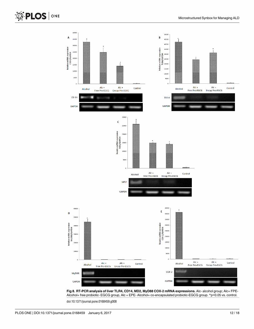
Fig 8. RT-PCR analysis of liver TLR4, CD14, MD2, MyD88 COX-2 mRNA expressions. Alc- alcohol group; Alc+ FPE-
Alcohol+ free probiotic- EGCG group, Alc + EPE- Alcohol+ co-encapsulated probiotic-EGCG group. *p<0.05 vs. control.
doi:10.1371/journal.pone.0168459.g008
Microstructured Synbox for Managing ALD
PLOS ONE | DOI:10.1371/journal.pone.0168459 January 6, 2017 12 / 18
phenolic nature and the fact that phenol decarboxylase and inducible acid phenol reductase
activities possessed by L. plantarum endow it with the capacity to metabolize phenolic acids
[8,9,27]. This will make EGCG a highly specific growth promoter, while inulin a general
polysaccharide may equally support the growth of gut associated pathogenic bacteria. EGCG
demonstrated significant and better prebiotic effect for L. plantarum, in comparison to inulin,
the well established prebiotic [28]. Howsoever, the growth promoting effect of EGCG was
observed only upto 60 mg beyond which hormesis [29] might set in; that may be due to the
accumulation of toxic metabolites which exacerbate stress related responses including cell det-
rimental pH alterations [30]. Hormesis is defined as adaptive response of cells and organisms
to intermittent stress [29].
Sodium alginate showed optimum entrapment efficiency to co-encapuslate EGCG and L.
plantarum at 1% concentration. Higher concentrations increase the viscosity of the solution
reducing its mobility during stirring and hence a reduced entrapment was observed. Presence
of EGCG conferred (i) improved probiotic survivability against harsh GIT conditions in
Fig 9. Micronuclei analysis in the hepatocytes of alcohol-fed rats. Effect of co-enapsulated Probiotic-EGCG on the extent of micronuclei
formation in hepatocytes of alcohol administered rats. Values are expressed as percentage of micronucleated cells. *p<0.001 vs. control, #p<0.01
vs. control, #$p<0.05 vs. control; Inset- Dividing cells showing binuclei (BN) and micronuclei (MN) in hepatocytes of alcohol-fed rats.
doi:10.1371/journal.pone.0168459.g009
Microstructured Synbox for Managing ALD
PLOS ONE | DOI:10.1371/journal.pone.0168459 January 6, 2017 13 / 18
simulated studies (acidic pH and presence of bile salts), and (ii) better entrapment (94%) of
probiotic in the ‘synbox’ in contrast to when probiotic was used alone (80% entrapment).
EGCG being a large molecule (140 kDa), probably plugs the alginate mesh both against the
entry of acidic, enzymatic and alkaline contents of gut and leakiness of the entrapped L. plan-tarum. Size of the beads increased significantly when both EGCG and probiotic were incorpo-
rated as compared to when these agents were entrapped individually. These results are in
concordance with an earlier study wherein size of microparticles varied with the type and con-
tent of probiotic and prebiotic used [31]. SEM images of the ‘microstructured synbox’ revealed
a smooth surface with the observance of L. plantarum on the surface.
In order to predict the performance of the ‘synbox’, the beads were tested in-vitro (under
simulated conditions) for their potential to preserve the viability of entrapped probiotic under
adverse conditions encountered in the stomach (extreme pH conditions) and intestine (micro-
aerophilic conditions, bile salts). The co-encapsulated beads showed significantly better results
(in terms of survival rate in simulated condition, in-vitro release and shelf-life) than those
exhibited by probiotic [15] and EGCG beads individually (data communicated elsewhere).
And and Kaliaspathy [32] reported similar results wherein the incorporation of Himaize starch
increased the survival rate of L. acidophilus.In the present study, the observed BAL in rats confirms the appropriate alcohol consump-
tion and its break down in the liver to generate potentially dangerous by-products that con-
tribute to alcohol induced liver damage. It may be said that the excessive alcohol consumption
led to endotoxemia (elevated plasma endotoxemia levels in alcohol administered rats) which
further attributed to the leaky gut syndrome as observed by the increased epithelial and para-
cellular transport of both lactulose (a dissacharide) and mannitol (a monosaccharide) in the
alcoholic rats. Similar clinical conditions are detected in patients with alcoholic hepatitis.
‘Microstructured synbox’ treated group gave a direct indication of restoration of tight junc-
tions as evidenced by presence of only mannitol in the in the serum of these animals.
The compromised gut permeability following high intake of alcoholic beverages also pro-
motes systemic passage of carcinogenic substances leading to decreased cell metabolism and cel-
lular immunity followed by DNA damage and cell death [33]. We presently report an increased
formation of micronuclei (MNi) (which originates from acentric chromosome fragments or
whole chromosomes that are not included in the main daughter nuclei during nuclear division)
in the hepatocytes of ALD rats taken as an indicator of genotoxic response to carcinogenic
agents [34, 35]. The MNi frequency was reduced in the ‘microstructured synbox’ treated group.
Liver inflammation and damage was indicated by the liver function tests. The elevated levels
of hepatic markers, (ALT and ALP) were restored after administration of combination beads
(supplementary data). The liver histoarchitecture was restored in the alcohol fed and combina-
tion beads treated group.
Another factor that controls the pathophysiology of ALD is generation of ROS due to the acti-
vation of kupffer cells in liver that causes oxidative stress. The PAO assay performed in the present
study indicated increased antioxidant levels (of SOD, catalase and GSH) in treatment group.
Evidence suggests that endotoxin mediated overwhelmed (redox imbalance) system leads
to activation of stress sensitive signaling pathways such as induction of NF-kB. NF-kB depen-
dent gene expression in kupffer cells contribute to alcohol induced liver injury [21, 36–39]
through inflammatory mediators including TNF-α. Significant inhibition of activation of NF-
kB by co-encapsulated beads, might have presently, suppressed the liver injury. This is consis-
tent with reports where green tea polyphenols have been shown to inhibit NF-KB activation in
ischaemia reperfusion liver injury and streptozotocin induced diabetic rats [40]. This may fur-
ther down regulate TNF-α [12], as also observed presently, coupled with the restoration of
antioxidative status.
Microstructured Synbox for Managing ALD
PLOS ONE | DOI:10.1371/journal.pone.0168459 January 6, 2017 14 / 18
Though, TNF-α is a promising target for various therapeutic diseases, yet certain side
effects are now associated with its use in immunocompromised patients. Thus newer thera-
peutic targets [41] viz. IL-12/p 40 which when downregulated, blocks IL-12, IL-23 and TNF-α,
are being explored. Animals treated presently with co-encapsulated beads brought down the
levels of not only TNF-α but also IL-12/p 40 subunit.
In continuation, diminished expression of TLR4, MD2, CD14, MyD88 and COX-2 was
recorded as compared to their strong expression in case of alcohol administered rats. This
lower gene expression of the signalling molecules after treatment with ‘microstructured
synbox’ indicated the blockade in the binding of LPS to TLR4, therefore, downregulaing the
other associated effector molecules. It is known that endotoxin binds specifically to TLR4, a
trans-membrane protein. TLR4 in association with MD2 recognizes the LPS/ CD14 com-
plex. Thus, CD14, TLR4 and MD2 are the major intrinsic components of receptor complex
which play an important role in signal transduction of LPS. The TLR4/MD2 complex fur-
ther activates a signaling pathway via the recruitment of adaptor proteins including MyD88
and cvclooxygenase-2 (COX-2), an inducible enzyme of macrophages catalysing the conver-
sion of arachidonic acid to prostaglandins which are potent inflammatory mediators caus-
ing hepatic injury. Our findings revealed that combination beads significantly inhibited the
expression of these genes and transduction of signals at the membrane level disrupting the
intracellular activation. We observed similar results in an earlier study where catechin acted
as a chain breaking inhibitor of the signalling molecules involved in LPS signalling [11].
These results are also in accordance with an earlier study where Lactobacillus amylovorushas been found to inhibit TLR4 signalling triggered by enterotoxigenic E. coli in Caco-2 cell
lines and pig explants [42]. Present study thus provides an insight on the enhanced efficacy
of developed EGCG-probiotic co-encapsulated synbox beads in suppressing an array of
molecules operative in the pathogenesis of ALD.
The above mentioned observations indicated that alcohol gavage caused significant endo-
toxaemia coupled with decreased activities of hepatic antioxidants and increased frequency of
micronuclei generation in rats. Treatment with EGCG-probiotic coencapsulated beads attenu-
ated levels of endotoxin, increased hepatic antioxidants along with decrease in the number of
micronuclei and amelioration of disruptive histoarchitecture. This may be due to the antioxi-
dative properties of EGCG and the probiotic. Flavonoids are known to localize near the mem-
brane surface, trapping directly any free radicals generated in lipid environment or in the
aqueous phase, while probiotics reduce the oxidative stress due to their ability to improve gut
barrier function by maintaining the intestinal permeability [21]. Combination beads thus
showed a multifactorial effect against increased endotoxin levels and genotoxicity, as well as
disrupted histoarchitecture and antioxidative status.
Conclusions
Our findings suggesting sequential inhibition of signal transduction without incurring prohib-
itive toxicity or loss of innate immunity, may be of importance in designing strategies like the
presently used combination of L. plantarum with EGCG in a ‘synbox’ for management of this
highly prevalent and significant clinical manifestation. Use of these agents, alone or in con-
junction with conventionally prescribed drugs may lower the dose and hence associated side-
effects of latter, besides conferring significant complementary health benefits. These results
need to be confirmed in humans in order to validate the applicability of the prepared formula-
tion. Thus, it is important to realise that full potential of such folkloric natural remedies can be
exploited only if the basic biology is combined with the modern day technology to result in
effective therapies.
Microstructured Synbox for Managing ALD
PLOS ONE | DOI:10.1371/journal.pone.0168459 January 6, 2017 15 / 18
Supporting Information
S1 Table. Log10 CFU of L. plantarum entrapped in probiotic- EGCG beads in SGF, SIF and
Bile salts.
(DOCX)
S2 Table. Effect of free probiotic and encapsulated probiotic-EGCG on hepatic markers in
the serum of control and alcohol-administered rats.
(DOCX)
S1 Text. Micronuclei analysis method.
(DOCX)
S2 Text. Measurement of intestinal permeability.
(DOCX)
Acknowledgments
The authors acknowledge Sophisticated Analytical Instrumentation Facility (SAIF)/Central
Instrumentation Laboratory, Panjab University, Chandigarh, India, for providing assistance in
scanning electron microscopic analysis of the samples.
Author Contributions
Conceptualization: PR KC IPK.
Formal analysis: PR SA UJK IPK.
Investigation: PR SA UJK KC IPK.
Methodology: SA UJK.
Resources: PR IPK KC.
Software: SA UJK.
Supervision: PR KJC IPK.
Validation: PR SA UJK KC IPK.
Visualization: PR SA UJK IPK.
References1. Park JH, Kim Y, Kim SH. Green tea extract (Camellia sinensis) fermented by Lactobacillus fermentum
attenuates alcohol-induced liver damage. Biosci Biotechnol Biochem. 2012; 23: 2294–2230.
2. Kolida S, Tuohy K, Gibson GR. Prebiotic effects of inulin and oligofructose. Bri J Nutri. 2002; 87: S193–
197.
3. Babu G, Nithyalakshmi V. Influence of prebiotic composition on probiotic survivability in calcium alginate
coated symbiotic microcapsules at thermal incubation. Agriculture J. 2011; 6: 231–236.
4. Rishi P, Mavi SK, Bharrhan S, Shukla G, Tewari R. Protective efficacy of probiotic alone or in conjunc-
tion with a prebiotic in Salmonella-induced liver damage. FEMS Microbiol Ecol. 2009; 69: 222–230. doi:
10.1111/j.1574-6941.2009.00703.x PMID: 19496820
5. Vodnar DC, Socaciu C. Green tea increases the survival yield of Bifidobacteria in simulated gastrointes-
tinal environment and during refrigerated conditions. Chem Central J. 2012; 6: 61.
6. Si W, Gonga J, Tsaoa R, Kalab M, Yang R, Yin Y. Bioassay-guided purification and identification of anti-
microbial components in Chinese green tea extract. J Chromatography. 2006; 1125: 204–210
Microstructured Synbox for Managing ALD
PLOS ONE | DOI:10.1371/journal.pone.0168459 January 6, 2017 16 / 18
7. Du GJ, Zhang Z, Wen XD, Yu C, Calway T, Yuan CS et al. Epigallocatechin Gallate (EGCG) is the most
effective cancer chemopreventive polyphenol in green tea. Nutrients. 2012; 4: 1679–1691. doi: 10.
3390/nu4111679 PMID: 23201840
8. Osawa R, Kuroiso K, Goto S, Shimizu A. Isolation of tannin-degrading Lactobacilli from humans and fer-
mented foods. Appl Environ Microbiol. 2000; 66: 3093–3097. PMID: 10877812
9. Vaquero I, Marcobal A, Muñoz R. Tannase activity by lactic acid bacteria isolated from grape must and
wine. Int J Food Microbiol. 2004; 96: 199–204. doi: 10.1016/j.ijfoodmicro.2004.04.004 PMID:
15364474
10. Singh PK, Kaur IP. Synbiotic (probiotic and ginger extract) loaded floating beads: a novel therapeutic
option in an experimental paradigm of gastric ulcer. J Pharm Pharmacol. 2012; 64: 207–217. doi: 10.
1111/j.2042-7158.2011.01397.x PMID: 22221096
11. Bharrhan S, Koul A, Chopra K, Rishi P. Catechin suppresses an array of signalling molecules and mod-
ulates alcohol-induced endotoxin mediated liver injury in a rat model. Plos One. 2011; 6: e2063.
12. Bharrhan S, Chopra K, Arora SK, Toor JS, Rishi P. Down-regulation of NF-{kappa}B signalling by poly-
phenolic compounds prevents endotoxin-induced liver injury in a rat model. Innate Immun. 2011; 18:
70–79. doi: 10.1177/1753425910393369 PMID: 21239456
13. Yuan G, Gong Z, Zhou X, Zhangq P, Sun X, Li X. Epigallocatechin-3-Gallate ameliorates alcohol-
induced liver injury in rats. Int J Mol Sci. 2006; 7: 204–219.
14. Kaviarasan S, Sundarapandiyan R, Anuradha CV. Epigallocatechin Gallate, a green tea phytochemical,
attenuates alcohol-induced hepatic protein and lipid damage. Toxicol Mechanisms Methods. 2008; 18:
645–652.
15. Arora S, Kaur IP, Chopra K, Rishi P. Efficiency of double layered microencapsulated probiotic to modu-
late pro-inflammatory molecular markers for the management of alcoholic liver disease. Mediators
Inflammat. 2014: 1–11.
16. Krasaekoopt W, Bhandari B, Deeth H. Evaluation of encapsulation techniques of probiotics for yoghurt.
Int Dairy J. 2003; 13: 3–13.
17. Krasaekoopt W, Bhandari B, Deeth H. The influence of coating materials on some properties of alginate
beads and survivability of microencapsulated probiotic bacteria. Int Dairy J. 2004; 14: 737–743.
18. Noth R, Lange-Gumfeld J, Stuber E, Kruse ML, Ellrichmann M, Hasler R, et al. Increased intestinal per-
meability and tight junction disruption by altered expression and localization of acculin in a murine graft
versus host disease model. BMC Gastroenterol. 2011; 11: 109, doi: 10.1186/1471-230X-11-109 PMID:
21977944
19. Schmid S. An evaluation of the micronuclei test using triethylenemelamine, trimethylphosphate and niri-
dazole. Mutat Res. 1975; 28: 101–106. PMID: 1095914
20. Kim MS, Ong M, Qui X. Optimal management for alcoholic liver disease: Conventional medications, nat-
ural therapy or combination? World J Gastroenterol. 2016; 22: 8–23. doi: 10.3748/wjg.v22.i1.8 PMID:
26755857
21. Forsyth CB, Farhadi A, Jakate SM, Tang Y, Shaikh M, Keshavarzian A. Lactobacillus GG treatment
ameliorates alcohol-induced intestinal oxidative stress, gut leakiness, and liver injury in a rat model of
alcoholic steatohepatitis. Alcohol. 2009; 43: 163–172. doi: 10.1016/j.alcohol.2008.12.009 PMID:
19251117
22. Chang B, Sang L, Wang Y, Tong J, Zhang D, Wang B. The protective effect of VSL#3 on intestinal per-
meability in a rat model of alcoholic intestinal injury. BMC Gastroenterol. 2013; 13: 1–8
23. Li F, Duan K, Wang C, McClain C, Feng W. Probiotics and alcoholic liver disease: Treatment and poten-
tial mechanisms. Gastroenterol Res Pract. 2016; Article ID 5491465, 11 pages.
24. Barone R, Rappa F, Macaluso F, Caruso Bavisotto C, Sangiorgi C, Di Paola G, et al. Alcoholic liver dis-
ease: A mouse model reveals protection by Lactobacillus fermentum. Clin Transl Gastroenterol. 2016;
7:e138. doi: 10.1038/ctg.2015.66 PMID: 26795070
25. Yan AW, Fouts DE, Brandl J, Starkel P, Torralba Schott E. Enteric dysbiosis associated with a mouse
model of alcoholic liver disease. Hepatology. 2011; 53:96–105. doi: 10.1002/hep.24018 PMID:
21254165
26. Li P, Burr GS, Gatlin DM, Hume ME, Patnaik S, Castille FL, et al. Dietary supplementation of short-
chain fructooligosaccharide influences gastrointestinal microbiota composition and immunity character-
istics of Pacific white shrimp, Litopenaeus vannamei, cultured in a recirculating system. J Nutr. 2007;
137:2763–2768 PMID: 18029496
27. Barthelmebs L, Divies C, Cavin JF. Molecular characterization of the phenolic acid metabolism in the
lactic acid bacteria Lactobacillus plantarum. Le Lait. 2001; 81: 161–171.
28. Kolida S, Tuohy K, Gibson GR. Prebiotic effects of inulin and oligofructose. Br J Nutr. 2002; 87: S193–
S197 doi: 10.1079/BJNBJN/2002537 PMID: 12088518
Microstructured Synbox for Managing ALD
PLOS ONE | DOI:10.1371/journal.pone.0168459 January 6, 2017 17 / 18
29. Mattson MP, Hormensis defined. Ageing Res Rev. 2008; 7:1–7. doi: 10.1016/j.arr.2007.08.007 PMID:
18162444
30. Axling U, Olsson C, Xu J, Fernandez C, Larsson S, Strom K et al. Green tea powder and Lactobacillus
plantarum affect gut microbiota, lipid metabolism and inflammation in high-fat fed C57BL/6J mice. Nutr
Metab. 2012; 9:105.
31. Chavarri M, Marañon I, Ares R, Ibañez FC, Marzo F, del Carmen Villaran M. Microencapsulation of a
probiotic and prebiotic in alginate-chitosan capsules improves survival in simulated gastro-intestinal
conditions. Int J Food Microbiol. 2010; 42:185–189.
32. And CI, Kailasapathy K. Effect of co-encapsulation of probiotics with prebiotics on increasing the viabil-
ity of encapsulated bacteria under in vitro acidic and bile salt conditions and in yogurt. J Food Sci 2005;
70: M18–M23.
33. Ramirez A, Saldanha PH. Micronucleus investigation of alcoholic patients with oral carcinomas. Genet
Mol Res. 2002; 30: 246–260.
34. Castelli E, Hrelia P, Maffei F, Fimognari C, Foschi FG, Caputo F et al. Indicators of genetic damage in
alcoholics: reversibility after alcohol abstinence. Hepatogastroenterol. 1999; 46: 1664–1668.
35. Huttner E, Gotze A, Nikolova T. Chromosomal aberrations in humans as genetic endpoints to assess
the impact of pollution. Mutat Res. 1999; 445: 251–257. PMID: 10575434
36. Lu YC, Yeh WC, Ohashi P. LPS/TLR4 signal transduction pathway. Cytokine. 2008; 42: 145–151. doi:
10.1016/j.cyto.2008.01.006 PMID: 18304834
37. Wheeler MD. Endotoxin and Kupffer cell activation in alcoholic liver disease. Alcohol Res Health. 2003;
27: 300–306. PMID: 15540801
38. Schwabe RF, Seki E, Brenner DA. Toll-like receptor signaling in the liver. Gastroenterol. 2006; 130:
1886–1900.
39. Hegazy SK, El-Bedewy M.M. Effect of probiotics on pro-inflammatory cytokines and NF-κB activation in
ulcerative colitis. World J Gastroenterol. 2010; 16: 4145–4151. doi: 10.3748/wjg.v16.i33.4145 PMID:
20806430
40. Han X, Shen T, Lou H. Dietary polyphenols and their biological significance. Int J Mol Sci 2007; 8: 950–
988.
41. Barrie AM, Scott EP. The interleukin-12 family of cytokines: Therapeutic targets for inflammatory dis-
ease mediation. Clinical Appl Immunol Rev. 2005; 5: 225–240.
42. Finamore A, Roselli M, Imbinto A, Seeboth J, Oswald IP, Mengheri E. Lactobacillus amylovorus inhibits
the TLR4 inflammatory signaling triggered by enterotoxigenic Escherichia coli via modulation of the neg-
ative regulators and involvement of TLR2 in intestinal Caco-2 cells and pig explants. Plos One. 2014; 9:
e94891. doi: 10.1371/journal.pone.0094891 PMID: 24733511
Microstructured Synbox for Managing ALD
PLOS ONE | DOI:10.1371/journal.pone.0168459 January 6, 2017 18 / 18