Embed Size (px)
DESCRIPTION
jurnal
Citation preview
© 2002 Nature Publishing Group918 | DECEMBER 2002 | VOLUME 3 www.nature.com/reviews/genetics
R E V I E W S
Candida albicans is an OPPORTUNISTIC COMMENSAL.Virtuallyall of us carry it in our gastrointestinal and genitourinarytracts and, to a lesser extent, on our skin. When immunesystems are weak (for example, as a result of cancerchemotherapy, HIV infection or in neonates) or whenthe competing flora are eliminated (for example, afterantibiotic treatment), C. albicans colonizes and invadeshost tissues. Although HIV patients frequently sufferfrom recurring oral CANDIDIASIS and sometimes die fromadvanced oesophageal colonization, infections (such asthrush and vaginitis) of mucosal tissues are usually notlife threatening. However, if the organism gains access tothe blood stream (a condition known as candidaemia),by invasion of host tissues or by contamination of in-dwelling catheters, the infection can progress to thegrowth of fungal masses in the kidney, heart or brain.
C. albicans is the fourth most common hospital-acquired infection in the United States, the treatment ofwhich is estimated to cost more than US $1 billionannually1,2. Because C. albicans and other fungalpathogens are eukaryotes and therefore share many oftheir biological processes with humans, most anti-fungal drugs cause deleterious side effects and, at thedoses used, are FUNGISTATIC rather than FUNGICIDAL. So, it isan important goal of C. albicans research to identifyappropriate targets for anti-fungal technologies.
Understanding the biology of this opportunist, in all itsmorphological and biochemical states, is necessary forthe development of therapies that will prevent or treatcandidiasis in susceptible patients.
Rapid advances in the understanding of many basicbiological processes in C. albicans have been made as aresult of their similarity to well-studied processes inSaccharomyces cerevisiae. S. cerevisiae, which divergedfrom C. albicans 140–841 million years ago3,4, is anindispensible guide for studying aspects of cell-cycleprogression, signal transduction, mating, metabolismand cell-wall biosynthesis in C. albicans. In addition,S. cerevisiae has been used for preliminary testing of hypotheses that were later addressed directly in C. albicans (for example, see REF. 5).
Despite the many processes that are conservedbetween these distant cousins, there are also significantdifferences. For example, S. cerevisiae grows exclusivelyby budding off round yeast cells or elongated pseudo-hyphal cells, whereas C. albicans is more morpho-logically diverse, forming true hyphae (BOX 1) andCHLAMYDOSPORES. This diversity is thought to aid its survival, growth and dissemination in the mamm-alian host. Such features are less readily studied using S. cerevisiae as a model and highlight the importance ofstudying C. albicans itself.
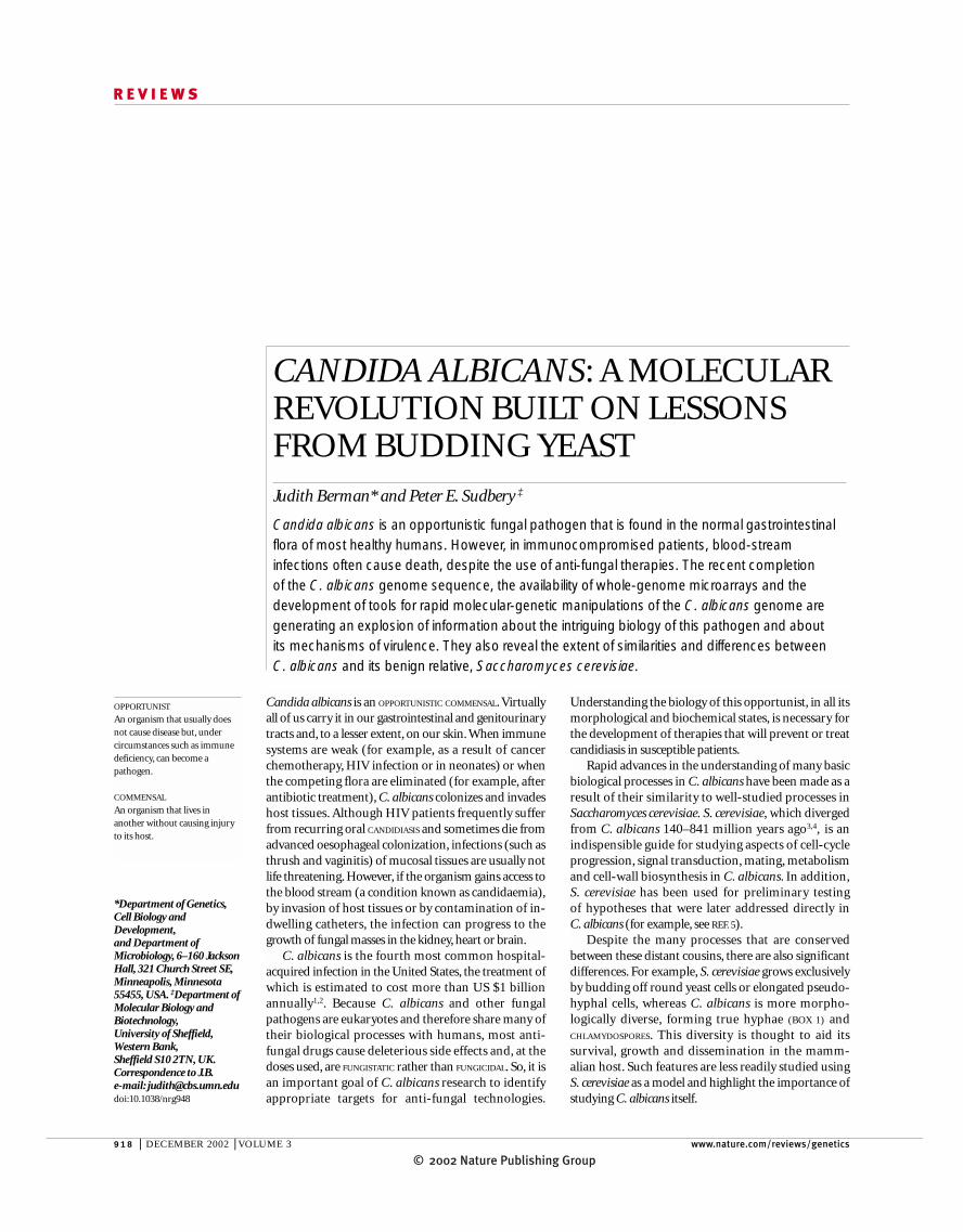
CANDIDA ALBICANS: A MOLECULARREVOLUTION BUILT ON LESSONSFROM BUDDING YEAST Judith Berman* and Peter E. Sudbery ‡
Candida albicans is an opportunistic fungal pathogen that is found in the normal gastrointestinalflora of most healthy humans. However, in immunocompromised patients, blood-streaminfections often cause death, despite the use of anti-fungal therapies. The recent completion of the C. albicans genome sequence, the availability of whole-genome microarrays and thedevelopment of tools for rapid molecular-genetic manipulations of the C. albicans genome aregenerating an explosion of information about the intriguing biology of this pathogen and about its mechanisms of virulence. They also reveal the extent of similarities and differences between C. albicans and its benign relative, Saccharomyces cerevisiae.
OPPORTUNIST
An organism that usually doesnot cause disease but, undercircumstances such as immunedeficiency, can become apathogen.
COMMENSAL
An organism that lives inanother without causing injuryto its host.
*Department of Genetics,Cell Biology andDevelopment,and Department ofMicrobiology, 6–160 JacksonHall, 321 Church Street SE,Minneapolis, Minnesota55455, USA. ‡Department ofMolecular Biology andBiotechnology,University of Sheffield,Western Bank,Sheffield S10 2TN, UK.Correspondence to J.B.e-mail: [email protected]:10.1038/nrg948

© 2002 Nature Publishing GroupNATURE REVIEWS | GENETICS VOLUME 3 | DECEMBER 2002 | 919
R E V I E W S
CANDIDIASIS
Infection with a Candida species.It often refers to the infection ofmucosal surfaces, such as themouth, vagina, skin oroesophagus.
FUNGISTATIC
The ability to inhibit the growthof fungi. Fungistatic agents cankeep an infection in check butusually do not completelyeliminate the fungus from thehost.
FUNGICIDAL
The ability to kill fungi.Fungicides have the potential toclear a fungal infection from thehost.
CHLAMYDOSPORES
Thick-walled round cells thatsometimes form at the ends ofhyphae or pseudohyphae inresponse to nutrient stress orother stresses.
SEPTIN
A protein that forms a ring-shaped scaffold-like structure atthe incipient bud site in yeastcells and pseudohyphal cells andat the incipient site of septationin true hyphae.
GERM TUBE
The elongating structure thatevaginates from a round yeastcell when it is induced to formtrue hyphae.
analysis of biological questions in C. albicans. How doesthis pathogen respond to environmental stimuli? Whatalters its morphogenesis programmes? What possiblemating interactions does it undergo? And how does itorganize and reorganize its genome while growing invitro or in mammalian host cells and tissues?
Technical challenges and solutionsGenetic manipulations of C. albicans have been fraughtwith difficulties that stem from the lack of a useful sex-ual cycle and a lack of molecular tools. Today, reverse-genetic approaches, in which genes are first identified bytheir sequence and then both genomic copies aresequentially deleted or mutated, are commonly used.Clearly, this approach requires some previous knowl-edge of the biological process of interest, emphasizingthe benefit of using a well-established model organismsuch as S. cerevisiae.
Genetic manipulations that are carried out easily inS. cerevisiae are much more laborious in C. albicansbecause of the lack of a complete sexual cycle in thispresumed obligate diploid. Both conventional andmolecular-genetic analysis have therefore proved diffi-cult. However, recent advances in molecular-genetictechniques, together with the availability of the genomesequence, have revolutionized research in this organ-ism. Moreover, data from the Candida GenomeSequencing Project6, together with sophisticatedcloning approaches7, have revealed the existence of‘mating-type-like’ loci that, when homozygous, candirect the formation of recombinants between diploidstrains7–9. So, conventional genetic techniques mightsoon be available in C. albicans.
This review provides examples of how S. cerevisiaemodels guided the early molecular studies of C. albicansbiology and how new tools are facilitating the direct
Box 1 | Differences between yeast, pseudohyphae and true hyphae
Candida albicans can exist in three forms that have distinct shapes: yeast cells (also known as blastospores), pseudohyphalcells and true hyphal cells.Yeast cells are round to ovoid in shape and separate readily from each other. Pseudohyphaeresemble elongated, ellipsoid yeast cells that remain attached to one another at the constricted septation site and usuallygrow in a branching pattern that is thought to facilitate foraging for nutrients away from the parental cell and colony. Truehyphal cells are long and highly polarized, with parallel sides and no obvious constrictions between cells. Actin is alwayslocalized at the tip of the growing hypha89. A basal SEPTIN band (green) forms transiently at the junction of the mother celland the evaginating GERM TUBE; the first true hyphal septum forms distal to the mother cell and well within the germtube66. The sub-apical cells become highly vacuolated and do not branch or bud until the ratio of cytoplasm to vacuolarmaterial increases significantly63. All three cell types have a single nucleus per cell before mitosis. Important differencesbetween yeast, pseudohyphal and true hyphal cells include the degree of polarized growth, the positioning of the septinring (green in diagram and micrographs, and black in light microscope images) and of the true septum relative to themother cell, the movement of the nucleus (blue line in diagram; stained with DAPI, blue in micrographs) relative to themother cell and the degree to which daughter cells are able to separate into individuals. GFP, green fluorescent protein.
Yeast
Pseudohyphae
True hyphae
Septin–GFP

© 2002 Nature Publishing Group920 | DECEMBER 2002 | VOLUME 3 www.nature.com/reviews/genetics
R E V I E W S
green fluorescent protein (GFP) has been codon opti-mized for expression in C. albicans24,25, and morerecently, codon-modified GFP has been altered to gen-erate the cyan (CFP) and yellow (YFP) versions of thefluorescent proteins. Codon-optimized GFP, YFP andCFP genes have also been coupled with one of two selec-table markers (URA3 or HIS1). This generated cassettesthat, when amplified by PCR, can be inserted in-frameat the carboxyl terminus of any gene of interest to tag itsproduct and visualize it in vivo26.
Use of S. cerevisiae to study C. albicans. To circumventthe difficulties of carrying out genetic studies directly in C. albicans, many C. albicans genes have been identifiedand/or analysed using S. cerevisiae as a ‘surrogate’. Forexample, many C. albicans genes were cloned by theirability to complement a mutation in S. cerevisiae. Thisapproach is not as important as it once was, becausehomologues can be identified on the basis of theirsequence similarity, as a result of the C. albicans genomesequencing project. Nonetheless, if a gene does functionin S. cerevisiae, then the effects of mutant alleles can nowbe tested in S. cerevisiae before the more laborious processof testing them in C. albicans (see, for example, REF. 27).
C. albicans genes have also been cloned on the basisof their ability to interfere with an S. cerevisiae process.Czf1, a putative transcription factor, interferes in a dom-inant manner with the cell-cycle arrest that is inducednormally in S. cerevisiae in response to matingpheromone28. The C. albicans INT1 gene encodes a pro-tein that is present at the septin rings of yeast andhyphal cells. When expressed in S. cerevisiae, INT1induces the formation of highly elongated cells thatresemble hyphal germ tubes and are much more elon-gated than S. cerevisiae pseudohyphae29. In these cells,Int1 associates with septin proteins, causing them toform abnormal spiral structures30. Int1 also affects mor-phogenesis and virulence in C. albicans31.
Some C. albicans genes were cloned on the basis oftheir ability to confer new properties to S. cerevisiae, suchas the ability to make S. cerevisiae cells adhere to humancells32–34. Of these, two encode cell-wall proteins that areimportant for adhesion in C. albicans, whereas oneaffects adhesion of S. cerevisiae cells through an indirectmechanism. Another important example was the isola-tion of Cph1, the C. albicans homologue of S. cerevisiaeSte12 — the transcription factor that is activated by themating pheromone resonse MAP kinase cascade duringmating and filamentous growth. It was isolated in ascreen for genes that, when overexpressed in S. cerevisiae,enhanced pseudohyphal formation35.
S. cerevisiae is also used as a substitute for C. albicansin studies of host interaction. In the human host,neutrophils represent the first line of defence against C. albicans, although macrophages and dendritic cellsalso have a role in the immune response to candidialinfection36. When cultured macrophages are incubatedwith C. albicans cells, the macrophages ingest the yeastcells. However, wild-type C. albicans strains proceed togrow hyphae that lyse the macrophage membranes andescape from the cells37. Before C. albicans microarrays
Laboratory studies of C. albicans use a small numberof strains that have been engineered with one or moreAUXOTROPHIC markers. Plasmids that carry autonomouslyreplicating sequences (ARSs) are available for transfor-mation at high frequency and for expressing genes in anon-chromosome-specific context10. Although it isdesirable that these plasmids remain extrachromoso-mal, even when they carry two ARSs, they integrate intothe genome primarily by homologous recombination.
Another significant challenge is posed by theunconventional C. albicans codon usage — C. albicanstranslates the CUG codon as serine, rather than the‘universal’ leucine11. For this reason, many heterolo-gous markers do not function in C. albicans unless theCUG codons are first modified. Nonetheless, many C. albicans genes are at least partially functional in S. cerevisiae, which facilitated their identification bycomplementation studies.
Transformation and mutagenesis. In the past few years,several crucial tools have greatly enhanced our ability tomanipulate C. albicans genetically (reviewed in REF. 12).Methods for transformation were modified from proto-cols for transformation of S. cerevisiae and Pichia pas-toris, which is the methanol-trophic yeast that is usedprimarily for protein production13. In 1993, a recyclableURA3 cassette was adapted for multiple sequentialtransformations of C. albicans strains that were auxo-trophic for URA3 (REF. 14; FIG. 1a). The numerous singleand double mutant strains that were generated usingthis ‘Ura-blaster’ strategy provided the first genetic evi-dence of signalling pathways that were important for C. albicans morphogenesis and virulence. More recently,it has become clear that uracil auxotrophy affects theability of C. albicans cells to adhere to human tissues. Italso affects the virulence of C. albicans in a mousemodel of systemic candidiasis15–17.
A PCR-mediated transformation system similar tothat used in S. cerevisiae18 has been developed for use inC. albicans19,20, obviating the need to clone a genebefore disrupting it (FIG. 1b). Strain BWP17, which istriple auxotrophic (ura3, his1 and arg4) has made thegeneration of double mutants simpler by allowingsequential transformation steps without the need toregenerate a single selectable marker.
Other tools, including a plasmid that can be used totest whether a gene is essential (FIG. 1c) and dominantselectable markers coupled with an excision system thatis based on the S. cerevisiae 2-µm FLP/FRT SYSTEM21, havealso been developed in the past few years (TABLE 1).Furthermore, systems that rely on in vitro transposi-tion22 into C. albicans genomic DNA to generate het-erozygous or homozygous mutant strains are beingdeveloped (D. Davis, V. Bruno, L. Loza, S. Filler and A. Mitchell, personal communication; A. Uhl and A. D. Johnson, personal communication), and antisensemRNA expression can be used to generate mutantgrowth phenotypes23. In addition, promoters that aredesigned to study gene expression and several heterolo-gous reporter genes that are designed to monitor geneexpression are now available (TABLE 1). For example,
AUXOTROPHIC
Requiring a nutritionalsupplement to grow.
PROTOTROPH
A cell that can grow in theabsence of nutritionalsupplements.
FLP/FRT SYSTEM
A recombination system that isadapted from the Saccharomycescerevisiae 2-µm plasmid. FLPencodes a site-specificrecombinase, and Frt is the FLPrecombinase target site.Expression of FLP mediatesexcision of any sequence that isflanked by Frt sites.

© 2002 Nature Publishing GroupNATURE REVIEWS | GENETICS VOLUME 3 | DECEMBER 2002 | 921
R E V I E W S
A B C
Parental allelesA
Cb
Ca
Ba
Bb
X Y Z
A B C X Y Z
A B C X Y Z
A B C
A HIS1
HIS1
ARG45′ URA3 URA3 3′
UAU1
Z
Parental alleles
First amplified deletion cassette
5′ gene-specific primer
X Y Z
A B C
A HIS1 Z
A ARG4 Z
Second amplified deletion cassette
X Y Z
A B C X Y Z
A B
Transform with Ura-blasterdisruption cassette
Heterozygousdisruption strain(Ura+)
5-FOA selection forUra recombinants
hisG URA3 hisG Y Z
A B hisG URA3 hisG Y Z
A B C X Y Z
A B hisG Y Z
A B hisG URA3 hisG Y Z
A B hisG Y Z
URA3
A B hisG URA3 hisG Y Z
A B C X Y Z
A B hisG
hisG Y Z
Transform with Ura-blasterdisruption cassette
Intrachromosomalrecombination
Step 1: transformation
Select for His+, screen forheterozygous deletion strains
A HIS1 Z
A ARG4 Z
Select for Arg+, screen forheterozygous deletion strains
Heterozygousdisruption strain(Ura+)
Homozygous disruption strain(Ura+)
Arg+ Ura–
Arg– Ura+
Arg– Ura–
GOI+/+
URA3
~70 nt of homologywith 'A'
~20 nt of homology with 5′region of marker/vector
3′ gene-specific primer
~20 nt of homology with 5′region of marker/vector
~70 nt of homologywith 'Z'
ARG4
5′ gene-specific primer
~70 nt of homologywith 'A'
~20 nt of homology with 5′region of marker/vector
3′ gene-specific primer
~20 nt of homology with 5′region of marker/vector
~70 nt of homologywith 'Z'
Step 2: gene conversion orbreak-induced replication
Arg+ Ura–
GOI+/–
Step 3: URA3 recombination
Arg+ Ura–
GOI+/–
Arg+ Ura+
GOI–/–
Figure 1 | Methods of gene disruption or deletion. A | The Ura-blaster methoduses a recyclable URA3 cassette (yellow) flanked by repeats (purple). Selection forUra+ PROTOTROPHS is used to isolate transformants that carry the Ura-blastercassette. Counterselection on 5-fluoro-orotic acid (5-FOA) identifies isolates thathave lost the URA3 sequences through recombination between the repeats. PCRand Southern blotting are carried out to verify that the desired genomic changeshave occurred. ABC and XYZ denote regions of sequence identity that are usedfor homologous integration of the disruption cassette into the genomic copy of thegene of interest. B | PCR-mediated transformation uses 5′ and 3′ primers thatinclude short regions (~20 nucleotides (nt)) of homology to the marker vector(black section of arrow) and ~70 nt of sequence homology to the sites of insertion(green section of arrow). These usually correspond to sequences that flank theentire open reading frame to be deleted. A fragment that carries the 70-nthomologous flanking sequences and the selectable marker (His1 and Arg4 here,but Ura3 can also be used) is amplified (a) and then used to transform Candidaalbicans strains. Sequential transformation with amplified fragments that carry thesame flanking sequences and two different selectable markers is required togenerate a homozygous deletion strain (b). C | A rapid method for disrupting bothalleles of a targeted gene after single transformation with the UAU1 markercassette Ura3∆3′–ARG4–Ura3∆5′ (a). PCR-mediated transformation with UAU1into the gene of interest (GOI ) is achieved by selecting for Arg+ transformants (step1) and subsequently selecting for Arg+ Ura+ recombinants (step 3), which arethought to arise after mitotic gene conversion or break-induced replication (step 2).Arg+ Ura+ recombinants are checked for the absence of the wild-type allele andthe presence of both UAU1 and the recombined URA3 derivative that arises byrecombination between the two incomplete copies of URA3 (b). Becauseconversion of the wild-type allele is often accompanied by homozygosis of distalgenes on the chromosome, the phenotype of the disrupted gene should beverified by conventional gene-disruption approaches and by complementation ofthe mutation with a wild-type copy of the gene. If the gene of interest is essential, awild-type copy cannot be lost. In this case, Arg+ Ura+ transformants are thought tooccur by duplication of the wild-type gene before step 2.

© 2002 Nature Publishing Group922 | DECEMBER 2002 | VOLUME 3 www.nature.com/reviews/genetics
R E V I E W S
C. albicans cells, in which the expression of genes thatencode the principal enzymes of the glyoxylate cycle,isocitrate lyase (ICL1) and malate synthase (MLS1), wasalso elevated during phagocytosis. Subsequent deletionof C. albicans ICL1 yielded a strain that was less virulentin the systemic mouse model of candidiasis38.
became available, S. cerevisiae microarrays were used to study the global expression profiles of S. cerevisiaecells during their phagocytosis by macrophages38.S. cerevisiae cells that were isolated from PHAGOLYSOSOMES
had elevated expression of genes that encode enzymes of the GLYOXYLATE CYCLE. This prompted the study of
PHAGOLYSOSOME
An organelle in a phagocytic cellthat is formed by fusion of aningested particle (for example,a Candida cell) with a lysosome,which has hydrolytic enzymesthat are used to digest theparticle.
GLYOXYLATE CYCLE
A metabolic pathway forconverting two acetyl CoAmolecules to a four-carbondicarboxylic acid. The cycle ispresent in bacteria, plants andfungi, but not in mammals.
Table 1 | Molecular tools that are commonly used in the study of Candida albicans
Tools Properties/comments References
Selectable markers
URA3 Selection: uridine prototrophy, counterselection 14,103on 5-FOA; Ura− cells have reduced virulence
HIS1 Selection: histidine prototrophy 20
ARG4 Selection: arginine prototrophy 20
IMH3 Wild-type allele effective only at high copy, 25,104–106resistant alleles function at single copy andhomology with endogenous copy reducestargeted integration efficiency
pUAU — cassette that carries URA3 PCR-mediated transformation for arginine 107flanked by 5′ and 3′ portions (including prototrophy, followed by selection for~500 bp of overlap) of ARG4 recombination between the URA3 fragments
(while maintaining selection for Ura+ cells), yields some isolates in which both copies of the gene of interest have been disrupted; mitotic recombination might make homozygoussequences distal to the insertion site
Promoters
ADH1 High levels of expression 108,109
ACT1 High levels of expression; stronger than ADH1 25,78,110,111
GAL1 Induced ~10–12-fold with galactose, repressed 112with glucose; 3–4-fold weaker than ACT1
PCK1 Induced on succinate or, at higher levels (up to 111,113100-fold), with casamino acids (acid digests of casein treated to eliminate or reduce vitamins);repressed with glucose
MAL2 Induced ~3–4-fold by maltose and sucrose, 114,115repressed by glucose
MET3 Repressed up to 85-fold in the presence of 116methionine and/or cysteine
Tetracycline-regulatable Escherichia coli Up to 500-fold repression; requires two components 117tetR fused to Hap4 (Saccharomyces (TetR and TetO) inserted in the genome; a lack of cerevisiae) activation domain; promoter to homology to the C. albicans genome improves the be regulated contains tetO binding site frequency with which non-homologous recombination
generates the desired integrants
Heterologous reporter genes
Kluyveromyces lactis LAC4 Does not work well as a single copy 118(β-galactosidase)
Streptococcus thermophilus lacZ Expression levels much higher than those of 119(β-galactosidase) LAC4 in K. lactis; no C. albicans homologue
Renilla reniformis luciferase Can be detected at low levels of expression; 120no C. albicans homologue; no CUG codons
Aequorea victoria GFP Codon optimized for use in C. albicans 24,25
Modified GFPs, YFPs and CYPs Codon optimized and available in cassettes for 26gene replacement or fusion protein constructionthrough PCR-mediated transformation
Flp/FRT in vivo expression system Flp recombinase driven by a test promoter is 105used to excise a marker flanked by FRT sites;the timing of marker excision reflects the timewhen the test promoter was first active
5-FOA, 5-fluoro-orotic acid; ACT1, actin 1; ADH1, alcohol dehydrogense 1; ARG4, arginine 4; CFP, cyan fluorescent protein, GAL1,galactose 1; GFP, green fluorescent protein; HIS1, histidine 1; IMH3, inosine 5′-monophosphate dehydrogenase 3; PCK1,phosphoenolpyruvate carboxykinase 1; MAL2, maltose 2; MET3, methionine 3; TetO, tetracycline operator; TetR, tetracycline repressor;URA3, uracil 3; YFP, yellow fluorescent protein.

© 2002 Nature Publishing GroupNATURE REVIEWS | GENETICS VOLUME 3 | DECEMBER 2002 | 923
R E V I E W S
conozole have an increased frequency of chromosome4 loss or chromosome 3 gain57. The mechanism bywhich these events affect fluconozole resistance is notclear. Although there are multidrug transporters onchromosomes 4 and 3, ERG11 — the gene that isimportant for ergosterol biosynthesis and that, whenmutated, confers resistance to fluconozole — is onchromosome 5. So, as for sorbose use, altered chromo-some numbers might act by regulating the genes thatare necessary for drug resistance.
Genome sequence. The C. albicans genome wassequenced by the Stanford Genome Technology Center,and a draft of the assembled sequence can be down-loaded and searched at their web site. An international,collaborative annotation group is now producing anannotated database. Information about progress of thiseffort is posted at the Candida albicans GenomeInformation web site. Partial annotation is also accessi-ble at Candida DB, the European Candida Databaseweb site.
Genome sequencing has uncovered many C. albicansORFs that have obvious S. cerevisiae homologues.Among them are many of the putative homologues ofS. cerevisiae genes that are required for sexual differenti-ation and meiosis58. C. albicans also contains manygenes that have no obvious S. cerevisiae homologues,some of which are most similar to genes from otherfungi, but others that encode novel gene products6.Because most S. cerevisiae strains do not adhere to, orinvade, human tissues, gene products that have nohomologues in S. cerevisiae are considered by some tobe good candidates for genes that are important for hostinteractions. Homologues of genes that are common toall fungi, especially those that are essential for fungalgrowth, might be good candidates for broad-spectrumanti-fungal targets. C. albicans genes that lack humanhomologues are considered especially promising in thisrespect, because they are less likely to cause the negativeside effects that are associated with most anti-fungaltherapies.
The S. cerevisiae genome is thought to have undergone a duplication ~100 million years ago59. TheC. albicans genome contains fewer sets of duplicatedgenes with related or redundant functions60, indicatingthat it might not have undergone this duplication. Forexample, the six B-cyclin genes of S. cerevisiae corre-spond to only two obvious B-cyclin homologues inC. albicans. Even if gene sequences indicate related func-tions, their roles might be different. For example, spt3mutants in S. cerevisiae are defective in filamentousgrowth, whereas spt3 mutants in C. albicans are hyperfil-amentous61. Furthermore, genes that are essential for viability in S. cerevisiae might not be essential in C. albicans, and vice versa. Accordingly, S. cerevisiae hastwo RAS homologues and together they are essential forviability, but the single obvious RAS homologue ofC. albicans is not essential62, indicating that a pathwaycontrolled by Ras/cAMP in S. cerevisiae is either notcontrolled by Ras in C. albicans or is not important forC. albicans viability. Conversely, Abp1 in S. cerevisiae, an
DNA array analysis. The availability of the C. albicansgenome sequence facilitated the development of DNAarrays for gene-expression analysis. Incyte, Inc., generatedmicroarrays of 6,600 C. albicans open reading frames(ORFs) that had been determined on the basis ofgenomic and proprietary cDNA sequences. So far, thesearrays have been used to analyse the expression patternsof cells exposed to Itraconazole, a broad-spectrum anti-fungal drug39.As discussed below (in the section entitled‘Hyphal-specific gene transcription’), groups led by AlBrown and Haoping Liu have used partial genomemicroarrays to analyse the regulatory pathways thatorchestrate gene expression during the yeast-to-hyphaltransition. Several groups are now constructing and usingwhole-genome microarrays. The first whole-genomearrays for C. albicans (6,334 ORFs) to be published camefrom Whiteway and co-workers, who used them toanalyse the evolution of resistance to anti-fungals40 andthe yeast-to-hyphal transition41. Information about theirproduction is available at the Online link to MicroArrayLab, National Research Council of Canada.
Genome organizationC. albicans has a diploid genome that is split betweeneight pairs of chromosomes that can be separated bypulse-field gel electrophoresis42. At ~16 Mb, the haploidgenome is slightly larger than that of S. cerevisiae, per-haps because of the greater number of retrotransposonfamilies6. It contains several large families of genes thatencode proteases, lipases and cell-wall proteins that arenot present in such large gene families in S. cerevisiae.The genes of both yeasts usually lack introns6. Althoughcentromere sequences have remained elusive, severalORFs that encode conserved centromere proteins are present in the genome sequence (K. Sanyal and J. Carbon, personal communication). Telomeresequences and telomerase homologues have also beenidentified43–45.
An interesting, but poorly understood, property ofC. albicans clinical isolates is their variable karyotype46,47.As has been observed in S. cerevisiae 48, the length of thechromosome that carries the ribosomal (r)DNA ishighly variable, owing to changes in the number ofrDNA repeats49,50. A set of nested repetitive sequences —multiple repeat sequences (MRSs) — seems to be themain site of the translocations that are found in clinicalisolates. Karyotype changes are caused by the expansionand contraction of repeat sequences in the MRS, as wellas by reciprocal translocation events between MRSrepeats51–53. MRSs are found in one or two copies on allchromosomes except chromosome 3 (but, see REF. 54).
Although unintended, genome rearrangementsoccur in S. cerevisiae strains that go through severalrounds of transformation55; non-disjunction appar-ently occurs with a higher frequency in C. albicans, per-haps as a mechanism to adapt to stressful conditions.For example, the loss of chromosome 5 occurs fre-quently in strains that are forced to grow on sorbose asthe sole carbon source, presumably because a repressorof sorbose use resides on chromosome 5 (REF. 56).Similarly, strains that are resistant to the anti-fungal flu-

© 2002 Nature Publishing Group924 | DECEMBER 2002 | VOLUME 3 www.nature.com/reviews/genetics
R E V I E W S
The C. albicans hyphal form is often found at sites oftissue invasion, and cells that do not readily formhyphae often have reduced virulence64. Importantly,other Candida spp. that do not readily form true hyphaeare much less frequently isolated from the human host,indicating that they are less virulent. But strains that areunable to grow in the yeast form are also less viru-lent37,61,64,67. It is generally thought that hyphal cellsexpressing cell-wall proteins that facilitate adhesion tohuman tissues are important for tissue invasion, as wellas for escape from phagocytosis mediated by neu-trophils or macrophages. By contrast, the yeast form isthought to be important for dissemination of thepathogen through the blood stream. It is likely, there-fore, that the ability to switch between the morphologi-cal forms is important for C. albicans virulence.However, proof that this is the case is still lacking, andthe issue remains controversial among the Candidaresearch community68.
C. albicans cells that have different morphologiesalso contribute to the formation of colonies with differ-ent characteristics. First, colonies with hyphal andpseudohyphal cells invade the agar substratum. Second,the presence of hyphae and pseudohyphae causescolonies to have a CRENULATED appearance, in contrast to
actin-binding protein, is not essential, but Abp1 in C.albicans seems to be required for growth23. So, knowingthe role of a gene product in S. cerevisiae is not sufficientto infer its properties in C. albicans.
MorphogenesisMorphogenesis has been a focus of research in C. albicans because virulence is associated with the abil-ity to switch between the yeast and hyphal morpholo-gies. C. albicans grows vegetatively in at least three morphogenic forms: yeast, pseudohyphae and hyphae (BOX 1). The yeast form closely resembles the buddingyeast S. cerevisiae. The pseudohyphal form consists ofchains of elongated yeast cells that retain constrictionsat the junctions between adjacent compartments,whereas hyphae are tube-like, with sides that are paral-lel along their entire length63–66. Pseudohyphae cansometimes superficially resemble hyphae; however, thetwo states are clearly different and should not be con-fused. The term filamentous is used here where it notclear whether cells are hyphal or pseudohyphal.Although mechanistic studies of pseudohyphal growthin S. cerevisiae have been informative about pseudohy-phal growth in C. albicans, S. cerevisiae models are lessrelevant to true hyphal growth.
PHENOTYPIC SWITCHING
A change in cellular or colonyproperties that seems to beheritable, but reverses at a ratethat is much higher than couldbe caused by mutation.Examples include colonyswitching and white–opaqueswitching in Candida albicans.
CRENULATED
Having an uneven ‘saw-tooth’-like edge. Crenulated colonieshave filamentous cells thatprotrude from the edges ofthem.
a b
c d
eW
W
O
O
f
g
Figure 2 | Colony morphologies of Candida albicans. A single strain can take on different colony morphologies on differentmedia or as a consequence of PHENOTYPIC SWITCHING. a | Smooth colonies grown on salt-dextrose complete (SDC) medium; b | wrinkled colonies grown on spider medium; c | fuzzy colonies grown on milk-Tween agar; and d | embedded coloniessuspended in a matrix of rich medium that contains sucrose. e | White–opaque phenotypic switching is seen here on SDC mediummaintained at 23 °C. White cells (W) of the WO-1 strain were plated at 23 °C for three days, and opaque colonies (O) and sectorsappeared in the population. f,g | Cells in wrinked, embedded and fuzzy colonies are a mixture of yeast, pseudohyphal and truehyphal cells. A population of cells derived from different portions of wrinkled colonies is shown.

© 2002 Nature Publishing GroupNATURE REVIEWS | GENETICS VOLUME 3 | DECEMBER 2002 | 925
R E V I E W S
A cph1 efg1 mutant, in which both the MAP-kinaseand the cAMP pathways are disabled, fails to form fila-ments in most in vitro conditions, and is avirulent in asystemic mouse model of candidiasis37. This observa-tion is often cited as evidence that the ability to formhyphae or pseudohyphae is an essential virulence factor.But there are two important caveats to this interpreta-tion. First, these mutations block the expression ofhyphal-specific genes (see next section for further dis-cussion), many of which are also required for virulence.Second, cph1 efg1 mutants are able to produce filamentsunder some in vivo and in vitro conditions73. This mightbe due to the action of other pathways of hyphal-growth induction, such as the Rim101 pathway, which isactivated by alkaline pH74,75 and the Czf1 pathway,which is activated by growth in a solid matrix76 (FIG. 3).
Morphogenesis is repressed by transcriptionalinhibitors such as Tup1 (REF. 77), which associates with its DNA-binding partners Nrg1 (REFS 78,79) and Rfg1(REF. 80). Apart from pH and growth in a matrix, thenature of the environmental signals to which each ofthese pathways responds is poorly understood.
Hyphal-specific gene transcription. The conditions thatinduce hyphal growth (BOX 2) also induce the expressionof hyphal-specific genes (HSGs). Identifying HSGs iscomplicated by the fact that the conditions that inducemorphogenesis will also induce cell responses that arenot necessarily connected with morphogenesis but thatare required for physiological adaptation to the newenvironment. For the most part, induction of hyphae orpseudohyphae requires a combination of two environ-mental conditions (such as high temperature and serum,or high temperature and neutral pH). So, a gene is only
the smooth appearance of yeast colonies (FIG. 2).Different colony shapes are a consequence of differentproportions of yeast and filamentous cells in regions ofthe colony. Third, feathery projections often extendfrom the periphery of colonies that contain manyhyphal cells (FIG. 2).
Signal-transduction pathways. Several environmentalfactors (BOX 2) can induce yeast cells to form hyphaeand pseudohyphae through several signal-transduc-tion pathways (FIG. 3; TABLE 2). This probably reflectsthe variety of microenvironments in which thisopportunist must survive in vivo (reviewed in REFS
64,69,70). As in S. cerevisiae, the cAMP and the matingpheromone response–MAP kinase pathways targettranscription factors that promote morphogenesis.Inactivation of the cAMP pathway (by deletingEFG1) blocks filamentation in most conditions,whereas inactivation of the MAP-kinase pathway (bydeleting CPH1) blocks filament formation only inresponse to a limited set of conditions35,71,72. So,it seems that the cAMP pathway has a more promi-nent role in C. albicans morphogenesis than in S. cerevisiae.
Cst20
Ras1? ?
Hst7
Cek1
Cph1
Nrg1
Tup1 Tup1
Cyrl, Cap1
cAMP
Matrix
Czf1
Tpk1Tpk2
Efg1
pH
Rim8Rim20
Cph2
Tec1
Rim101
HSGs
Yeast
Rpg1 Rbf1
Pseudohyphae
Hyphae
Figure 3 | Signal-transduction pathways that regulate morphogenesis. At least fourpositive (arrowheads) and two negative (bars) pathways control morphological transitions inCandida albicans. The pathways that promote the switch from yeast to pseudohyphal andhyphal growth are shown as follows: MAP-kinase pathway in pink, cAMP pathway in green,Cph2 pathway in grey, Rim101 pH response pathway in blue and Czf1 matrix pathway inorange. Pathways that inhibit this switch are the Tup1–Nrg1–Rpg1 pathway in red and the Rbf1pathway in purple. HSGs, hyphal-specific genes. See TABLE 2 for Saccharomyces cerevisiaehomologues and gene function.
Box 2 | Morphology-inducing conditions
Yeast cells• Cell density >106 cells ml−1
• Growth below 30 °C
• pH 4.0
Pseudohyphae• pH 6.0, 35 °C
• Nitrogen-limited growth on solid medium (SLAD)
Hyphae• Serum, >34 °C
• Lees medium, 37 °C
• pH 7.0, 37 °C
Other filament-inducing conditions• Spider medium
• Engulfment by macrophages
• Mouse kidneys
• Growth in agar matrix
• Iron deprivation
• Anoxia
• n-acetyl glucosamine

© 2002 Nature Publishing Group926 | DECEMBER 2002 | VOLUME 3 www.nature.com/reviews/genetics
R E V I E W S
including known HSGs and known virulence factors.Other subsets of genes are repressed by Tup1 when it isassociated with other partners, such as Mig1.Moreover, both Nrg1 and Mig1 can repress furthersubsets of genes independently of Tup1. Haoping Liuand colleagues prepared filter arrays that were printedwith 700 different C. albicans ORFs to study genes thatregulate yeast and hyphal growth by the Cph1, Cph2and Efg1 transcription factors84. The results indicatedthat several distinct signalling pathways convergentlyregulate a common set of genes that encode cell-wallproteins and proteases. In addition, both Efg1 andCph2 regulate Tec1, a transcription factor that isimportant for morphogenesis. Because many of theknown HSGs are virulence factors, some of thesenewly discovered genes might also turn out to have arole in virulence.
Both of these studies used only a subset of thegenes in the C. albicans genome. However, their suc-cess offers a realistic prospect of understanding howenvironmental cues are translated into cellularresponses, and how the necessary complex changes inthe pattern of gene expression are orchestrated. Nowthat whole-genome microarrays on glass slides arebeing used to compare the time courses of hyphalinduction under different conditions, as well as in dif-ferent mutant strains41, this understanding is certainto become more complete. For example, several genesthat are important for hyphal growth have now beenidentified in C. albicans that are unique and that donot have homologues in S. cerevisiae or other relatedfungi41. Such genes might be especially important forC. albicans pathogenicity.
Role of the cell cycle in morphogenesis. In S. cerevisiae,morphogenesis is regulated during the cell cycle by theassociation of cyclins with the Cdc28 cyclin-dependentkinase (CDK). Association of the CDK with G1 cyclins(Cln1 and Cln2) promotes polarized growth; its associa-tion with the B-cyclins promotes ISOTROPIC growth85
(reviewed in REF. 86). The C. albicans Cln1 G1 cyclin isnot required for filamentous growth, although it seemsto promote the maintenance of filamentous growth inwild-type cells87.
Many lines of evidence indicate that pseudohy-phal growth in S. cerevisiae might involve regulationof the Clb2 Cdc28 kinase, but the mechanism bywhich this comes about is still unclear. In S. cere-visiae, transcription of CLB2, the main mitotic B-cyclin, is regulated by the forkhead transcriptionfamily members Fkh1 and Fkh2. Cells that lack thesetranscription factors have reduced periodicity ofCLB2 transcription and grow constitutively aspseudohyphae. Only one homologue of Fkh1/2,Fkh2, is present in C. albicans, and its deletion resultsin a constitutive pseudohyphal phenotype underboth yeast and hyphal growth conditions. Cells thatlack Fkh2 in C. albicans have increased levels of a B-cyclin transcript, but have reduced levels of hyphalcell-wall proteins and of enzymes that dissolve theconnections between mother and daughter yeast
considered to be an HSG when it is induced duringhyphal development, but not when only one of theseconditions applies69. Many of the HSGs that have beenisolated so far encode known or putative virulence fac-tors69,81. These include genes that encode secretedaspartyl proteases (SAP4,5,6), cell-wall proteins (HWP1),adhesins (ALS3 and ALS8) and proteins that are requiredfor invasive growth (RBT1). The Hwp1 cell-wall proteinis particularly interesting because it has been shown to bethe substrate for a transglutaminase in the host epithe-lium that forms covalent bonds that anchor the C. albi-cans cell onto the surface of the epithelium82.
None of the HSGs that have been isolated so far areactually required for hyphal or pseudohyphal morpho-genesis. Rather, they are coordinately induced by the sig-nals that also induce morphogenesis. Their expression isblocked in an efg1efg1 mutant and is induced in tup1tup1, nrg1nrg1 or rfg1rfg1 mutants, indicating that theymight be among the targets of the morphogenesis sig-nalling pathways79.
A European consortium led by Alistair Brown usedfilters with 2002 C. albicans genes to examine the com-plex regulation of gene expression that is mediated byTup1 together with Nrg1 and Mig1 (REFS 79,83). Theanalysis showed that an association of Tup1 with Nrg1targets it to a specific subset of Tup1-regulated genes,
ISOTROPIC
Growth in all directions(opposite of polarized growth).
Table 2 | Components of pathways that regulate morphogenesis
C. albicans S. cerevisiae Protein function*protein homologue
Ras1 Ras2 GTPase
Cst20 Ste20 p21-activated kinase (PAK)
Hst7 Ste7 MAP kinase kinase (MEK)
Cek1 Homologue MAP kinaseuncertain
Cph1 Ste12 Transcription factor
Cyr1 Cyr1 Adenylate cyclase
Cap1 Srv2 Adenylate-cyclase-associated protein
Tpk1/Tpk2 Tpk1/Tpk2 cAMP-dependent protein kinasecatalytic subunits
Efg1 Sok2, Phd1 Helix–loop–helix transcription factorthat binds E-boxes (CANNTG)
Tec1 Tec1 TEA/ATTS DNA-binding domain familytranscription factor
Rim20 A. niger PalA Molecular function unknown
Rim8 YGL046W Molecular function unknown
Rim101 A. niger PacC Zinc-finger transcription factor activatedby proteolytic cleavage
Czf1 No known Putative zinc-finger transcription factorhomologues
Cph2 Hms1 Helix–loop–helix transcription factor
Tup1 Tup1 Transcriptional repressor
Nrg1 Nrg1 DNA-binding partner of Tup1
Rfg1 Rox1 DNA-binding partner of Tup1
Rbf1 Rbf1 Binds to Rpg box of S. cerevisiae andC. albicans telomeric repeats
A. niger, Aspergillus niger; C. albicans, Candida albicans; S. cerevisiae, Saccharomyces cerevisiae.*The function has been confirmed in at least one of the yeast species. In many cases, it is assumed,but not proven in Candida.

© 2002 Nature Publishing GroupNATURE REVIEWS | GENETICS VOLUME 3 | DECEMBER 2002 | 927
R E V I E W S
Phenotypic switchingSeveral properties of growth in C. albicans seem to beepigenetically controlled. One of them is reversiblecolony switching, which was first characterized in 1985(REFS 91,92). Yeast cells normally form smooth, whitedome-shaped colonies. However, at low frequency, C.albicans strain 3153A can spontaneously and reversiblyconvert to a variant colony shape (such as star, ring,irregular wrinkle, hat, stipple and fuzzy). A simpler,biphasic ‘white–opaque’ switching system, found in theC. albicans strain WO-1, involves a switch betweenwhite, domed colonies that contain typical yeast cellsand opaque, flat colonies that contain characteristicallyoblong cells93 (FIG. 2). White and opaque cultures havedifferent virulence properties, and several white-spe-cific and opaque-specific gene products have beenidentified93. For example, high levels of the Efg1 tran-scription factor induce and maintain the white cell-state, whereas low Efg1 levels induce and maintain theopaque cell-state94. Tup1, a transcriptional repressor,promotes the conversion from the white to the opaquephase in cells, although it is not required for the main-tenance of either phase95. It is also known that aMADS-box consensus binding site, that is most closelyrelated to the Mcm1 binding site of S. cerevisiae, is nec-essary for expression of the opaque-specific OP4 gene96.The frequency of switching between white and opaquestates is affected by histone deacetylases97,98, indicatingthat this phenotypic switch might be controlled, at leastin part, through a regulation of chromatin structure.
cells88. These observations have led to a model inwhich Fkh2 regulates the cell-cycle processes that arenecessary for the morphogenesis of true hyphal andyeast cells.
During hyphal induction in serum, germ tubesevaginate rapidly, much earlier than events such asSPINDLE POLE BODY duplication that normally signal thestart of the cell cycle. This indicates that hyphal evagi-nation can occur independently of other cell-cycleevents89 and that the initial polarized evagination of ahyphal germ tube is distinct from budding66. It alsoraises a possibility that evaginating hyphal germtubes might have features that are analogous to S. cerevisiae mating projections — they both initiatepolarized growth in non-cycling cells in response toexternal signals and both form a disorganized septinband at their base66,89.
Interestingly, MAD2 in C. albicans, which encodesa homologue of the S. cerevisiae spindle assemblyCHECKPOINT PROTEIN, is required for virulence in miceand for survival of C. albicans in the presence ofmacrophages90. However, Mad2 is not required forgrowth in liquid or solid media, which implies thatcell-cycle checkpoints, and in particular the check-point that monitors spindle assembly, are importantfor the survival of C. albicans in the host. Perhapshost-defence mechanisms damage crucial cellularcomponents, such as the mitotic spindle, which thepathogen can only repair if it triggers the Mad2checkpoints and delays cell-cycle progression90.
SPINDLE POLE BODY
The microtubule organizingcentre in fungi. In Candidaalbicans, as in Saccharomycescerevisiae, the spindle pole bodyis embedded in the nuclearmembrane, and this membraneremains intact throughout thecell cycle.
CHECKPOINT PROTEIN
A protein that is involved in oneof the pathways that monitoraspects of cellular function (suchas replication or spindleformation) that are required forproper cell-cycle progression. Ifa defect is detected, thecheckpoint pathway delays thecell cycle so that the defect canbe corrected.
ASCOMYCETE
The class of fungi in which themeitoic progeny (ascospores) arefound in sac-like structures(asci).
a
α
MTLa
a
b
PAP OBP PIK a1
MTLα PAPOBP1α 2αPIK
2N
4N
?
?a/α
White
Opaque
a
α
a/α
White
OpaqueWhite
OpaqueWhite
Figure 4 | Relationships between mating and white–opaque phenotypic switching. a | Organization of the Candidaalbicans mating-type-like (MTL) loci. The MTL loci encode the homeodomain proteins MTLa1 (a1) and MTLα2 (α2), and thetranscription regulator MTLα1 (α1). Each MTL locus also encodes a poly(A) polymerase (PAP), a phosphatidyl inositol kinase(PIK) and a protein with sequence similarity to oxysterol binding proteins (OBP)6. b | Cells that are homozygous for MTLa orMTLα (orange or purple, respectively) can switch to the opaque state because the a1/α2 transcriptional regulator inhibits theexpression of opaque-specific genes. Opaque cells mate with good efficiency to yield tetraploid cells that express both MTLaand MTLα alleles. How cells initially become homozygous at MTL and how tetraploid cells reduce their chromosome numberto 2N is not known.
© 2002 Nature Publishing Group928 | DECEMBER 2002 | VOLUME 3 www.nature.com/reviews/genetics
R E V I E W S
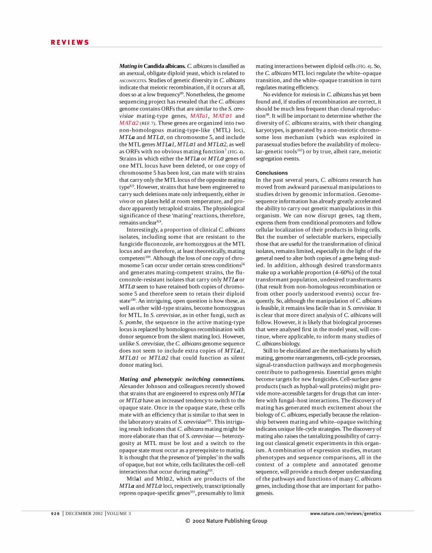
mating interactions between diploid cells (FIG. 4). So,the C. albicans MTL loci regulate the white–opaquetransition, and the white–opaque transition in turnregulates mating efficiency.
No evidence for meiosis in C. albicans has yet beenfound and, if studies of recombination are correct, itshould be much less frequent than clonal reproduc-tion99. It will be important to determine whether thediversity of C. albicans strains, with their changingkaryotypes, is generated by a non-meiotic chromo-some loss mechanism (which was exploited in parasexual studies before the availability of molecu-lar-genetic tools102) or by true, albeit rare, meioticsegregation events.
ConclusionsIn the past several years, C. albicans research hasmoved from awkward parasexual manipulations tostudies driven by genomic information. Genome-sequence information has already greatly acceleratedthe ability to carry out genetic manipulations in thisorganism. We can now disrupt genes, tag them,express them from conditional promoters and followcellular localization of their products in living cells.But the number of selectable markers, especiallythose that are useful for the transformation of clinicalisolates, remains limited, especially in the light of thegeneral need to alter both copies of a gene being stud-ied. In addition, although desired transformantsmake up a workable proportion (4–60%) of the totaltransformant population, undesired transformants(that result from non-homologous recombination orfrom other poorly understood events) occur fre-quently. So, although the manipulation of C. albicansis feasible, it remains less facile than in S. cerevisiae. Itis clear that more direct analysis of C. albicans willfollow. However, it is likely that biological processesthat were analysed first in the model yeast, will con-tinue, where applicable, to inform many studies ofC. albicans biology.
Still to be elucidated are the mechanisms by whichmating, genome rearrangements, cell-cycle processes,signal-transduction pathways and morphogenesiscontribute to pathogenesis. Essential genes mightbecome targets for new fungicides. Cell-surface geneproducts (such as hyphal-wall proteins) might pro-vide more-accessible targets for drugs that can inter-fere with fungal–host interactions. The discovery ofmating has generated much excitement about thebiology of C. albicans, especially because the relation-ship between mating and white–opaque switchingindicates unique life-cycle strategies. The discovery ofmating also raises the tantalizing possibility of carry-ing out classical genetic experiments in this organ-ism. A combination of expression studies, mutantphenotypes and sequence comparisons, all in thecontext of a complete and annotated genomesequence, will provide a much deeper understandingof the pathways and functions of many C. albicansgenes, including those that are important for patho-genesis.
Mating in Candida albicans. C. albicans is classified asan asexual, obligate diploid yeast, which is related toASCOMYCETES. Studies of genetic diversity in C. albicansindicate that meiotic recombination, if it occurs at all,does so at a low frequency99. Nonetheless, the genomesequencing project has revealed that the C. albicansgenome contains ORFs that are similar to the S. cere-visiae mating-type genes, MATa1, MATα1 andMATα2 (REF. 7). These genes are organized into twonon-homologous mating-type-like (MTL) loci,MTLa and MTLα, on chromosome 5, and includethe MTL genes MTLa1, MTLα1 and MTLα2, as wellas ORFs with no obvious mating function7 (FIG. 4).Strains in which either the MTLa or MTLα genes ofone MTL locus have been deleted, or one copy ofchromosome 5 has been lost, can mate with strainsthat carry only the MTL locus of the opposite matingtype8,9. However, strains that have been engineered tocarry such deletions mate only infrequently, either invivo or on plates held at room temperature, and pro-duce apparently tetraploid strains. The physiologicalsignificance of these ‘mating’ reactions, therefore,remains unclear8,9.
Interestingly, a proportion of clinical C. albicansisolates, including some that are resistant to thefungicide fluconozole, are homozygous at the MTLlocus and are therefore, at least theoretically, matingcompetent100. Although the loss of one copy of chro-mosome 5 can occur under certain stress conditions56
and generates mating-competent strains, the flu-conozole-resistant isolates that carry only MTLa orMTLα seem to have retained both copies of chromo-some 5 and therefore seem to retain their diploidstate100. An intriguing, open question is how these, aswell as other wild-type strains, become homozygousfor MTL. In S. cerevisiae, as in other fungi, such as S. pombe, the sequence in the active mating-typelocus is replaced by homologous recombination withdonor sequence from the silent mating loci. However,unlike S. cerevisiae, the C. albicans genome sequencedoes not seem to include extra copies of MTLa1,MTLα1 or MTLα2 that could function as silentdonor mating loci.
Mating and phenotypic switching connections.Alexander Johnson and colleagues recently showedthat strains that are engineered to express only MTLaor MTLα have an increased tendency to switch to theopaque state. Once in the opaque state, these cellsmate with an efficiency that is similar to that seen inthe laboratory strains of S. cerevisiae101. This intrigu-ing result indicates that C. albicans mating might bemore elaborate than that of S. cerevisiae — heterozy-gosity at MTL must be lost and a switch to theopaque state must occur as a prerequisite to mating.It is thought that the presence of ‘pimples’ in the wallsof opaque, but not white, cells facilitates the cell–cellinteractions that occur during mating101.
Mtla1 and Mtlα2, which are products of theMTLa and MTLα loci, respectively, transcriptionallyrepress opaque-specific genes101, presumably to limit

© 2002 Nature Publishing GroupNATURE REVIEWS | GENETICS VOLUME 3 | DECEMBER 2002 | 929
R E V I E W S
1. Beck-Sague, C. & Jarvis, W. R. Secular trends in theepidemiology of nosocomial fungal infections in the UnitedStates, 1980–1990. National Nosocomial InfectionsSurveillance System. J. Infect. Dis. 167, 1247–1251 (1993).
2. Miller, L. G., Hajjeh, R. A. & Edwards, J. E. Jr. Estimating thecost of nosocomial candidemia in the United States. Clin.Infect. Dis. 32, 1110 (2001).
3. Berbee, M. L. & Taylor, J. W. in The Mycota Vol. VIIB (edsMcLaughlin, D. J. & McLaughlin, E.) 229–246 (Springer,New York, 2000).
4. Heckman, D. et al. Molecular evidence for the earlycolonization of land by fungi and plants. Science 293,1129–1133 (2001).
5. Asleson, C. M. et al. Candida albicans INT1-inducedfilamentation in Saccharomyces cerevisiae depends onSla2p. Mol. Cell. Biol. 21, 1272–1284 (2001).
6. Scherer, S. in Candida and Candidiasis (ed. Calderone, R. A.) 259–265 (ASM Press, Washington, DC, 2002).
7. Hull, C. M. & Johnson, A. D. Identification of a mating type-like locus in the asexual pathogenic yeast Candida albicans.Science 285, 1271–1275 (1999).The Stanford University-generated genome sequencewas used to isolate regions of chromosome 5 that areheterozygous and contain genes (MTLa1, MTLα1 andMTLα2) that resemble the mating-locus genes in S. cerevisiae.
8. Hull, C. M., Raisner, R. M. & Johnson, A. D. Evidence formating of the ‘asexual’ yeast Candida albicans in amammalian host. Science 289, 307–310 (2000).
9. Magee, B. B. & Magee, P. T. Induction of mating in Candidaalbicans by construction of MTLa and MTLα strains.Science 289, 310–313 (2000).These two papers showed that C. albicans diploidcells that are homozygous for MTLa can generateapparent tetraploid recombinants when mixed withcells that are homozygous for MTLα. Reference 8showed that this reaction occurs in mice, whereasreference 9 generated in vitro recombinants at roomtemperature.
10. Pla, J., Perez-Diaz, R. M., Navarro-Garcia, F., Sanchez, M.& Nombela, C. Cloning of the Candida albicans HIS1 geneby direct complementation of a C. albicans histidineauxotroph using an improved double-ARS shuttle vector.Gene 165, 115–120 (1995).
11. Santos, M. A. & Tuite, M. F. The CUG codon is decoded invivo as serine and not leucine in Candida albicans. NucleicAcids Res. 23, 1481–1486 (1995).
12. Ernst, J. F. & Bockmühl, D. P. in Candida and Candidiasis(ed. Calderone, R. A.) 267–278 (ASM Press, Washington,DC, 2002).
13. De Backer, M. D., Magee, P. T. & Pla, J. Recentdevelopments in molecular genetics of Candida albicans.Annu. Rev. Microbiol. 54, 463–498 (2000).
14. Fonzi, W. A. & Irwin, M. Y. Isogenic strain construction andgene mapping in Candida albicans. Genetics 134, 717–728(1993).
15. Sundstrom, P., Cutler, J. E. & Staab, J. F. Reevaluation ofthe role of HWP1 in systemic candidiasis by use of Candidaalbicans strains with selectable marker URA3 targeted tothe ENO1 locus. Infect. Immun. 70, 3281–3283 (2002).
16. Lay, J. et al. Altered expression of selectable marker URA3in gene-disrupted Candida albicans strains complicatesinterpretation of virulence studies. Infect. Immun. 66,5301–5306 (1998).
17. Bain, J. M., Stubberfield, C. & Gow, N. A. Ura-status-dependent adhesion of Candida albicans mutants. FEMSMicrobiol. Lett. 204, 323–328 (2001).
18. Wach, A. PCR-synthesis of marker cassettes with longflanking homology regions for gene disruptions in S. cerevisiae. Yeast 12, 259–265 (1996).
19. Wilson, R. B., Davis, D., Enloe, B. M. & Mitchell, A. P. A recyclable Candida albicans URA3 cassette for PCRproduct-directed gene disruptions. Yeast 16, 65–70 (2000).A key methodology paper that describes an efficientPCR-based method for gene disruption that removedthe need to first clone the gene before its disruption.This system has now become a standard way togenerate homozygous gene deletions in C. albicans.
20. Wilson, R. B., Davis, D. & Mitchell, A. P. Rapid hypothesistesting with Candida albicans through gene disruption withshort homology regions. J. Bacteriol. 181, 1868–1874(1999).
21. Morschhauser, J., Michel, S. & Staib, P. Sequential gene disruption in Candida albicans by FLP-mediated site-specific recombination. Mol. Microbiol. 32, 547–556(1999).
22. Biery, M. C., Stewart, F. J., Stellwagen, A. E., Raleigh, E. A.& Craig, N. L. A simple in vitro Tn7-based transpositionsystem with low target site selectivity for genome and geneanalysis. Nucleic Acids Res. 28, 1067–1077 (2000).
23. De Backer, M. D. et al. An antisense-based functionalgenomics approach for identification of genes critical forgrowth of Candida albicans. Nature Biotechnol. 19, 235–241(2001).
24. Cormack, B. P. et al. Yeast-enhanced green fluorescentprotein (yEGFP) — a reporter of gene expression in Candidaalbicans. Microbiology 143, 303–311 (1997).
25. Morschhauser, J., Michel, S. & Hacker, J. Expression of achromosomally integrated, single-copy GFP gene in Candidaalbicans, and its use as a reporter of gene regulation. Mol.Gen. Genet. 257, 412–420 (1998).
26. Gerami-Nejad, M., Berman, J. & Gale, C. A. Cassettes forPCR-mediated construction of green, yellow and cyanfluorescent protein fusions in Candida albicans. Yeast 18,859–864 (2001).An important methodology paper that providedconvenient tools to study the localization of proteins inC. albicans cells by generating PCR-mediated fusionsto CFP, YFP and GFPs.
27. Devasahayam, G., Chaturvedi, V. & Hanes, S. D. The Ess1prolyl isomerase is required for growth and morphogeneticswitching in Candida albicans. Genetics 160, 37–48 (2002).
28. Whiteway, M., Dignard, D. & Thomas, D. Y. Dominantnegative selection of heterologous genes: isolation ofCandida albicans genes that interfere with Saccharomycescerevisiae mating factor-induced cell cycle arrest. Proc. NatlAcad. Sci. USA 89, 9410–9414 (1992).
29. Gale, C. et al. Cloning and expression of a gene encoding anintegrin-like protein in Candida albicans. Proc. Natl Acad. Sci.USA 93, 357–361 (1996).
30. Gale, C. A. et al. Candida albicans Int1p interacts with theseptin ring in yeast and hyphal cells. Mol. Biol. Cell 12,3538–3549 (2001).
31. Gale, C. et al. Linkage of adhesion, fliamentous growth, andvirulence in Candida albicans to a single gene, INT1. Science279, 1355–1358 (1998).References 30 and 31 describe the isolation andcharacterization of INT1 — a gene that is important forvirulence, adhesion and hyphal formation, under someconditions.
32. Fu, Y. et al. Expression of the Candida albicans gene ALS1 inSaccharomyces cerevisiae induces adherence to endothelialand epithelial cells. Infect. Immun. 66, 1783–1786 (1998).
33. Gaur, N. K. & Klotz, S. A. Expression, cloning, andcharacterization of a Candida albicans gene, ALA1, thatconfers adherence properties upon Saccharomycescerevisiae for extracellular matrix proteins. Infect. Immun. 65,5289–5294 (1997).
34. Fu, Y. et al. Cloning and characterization of CAD1/AAF1, agene from Candida albicans that induces adherence toendothelial cells after expression in Saccharomycescerevisiae. Infect. Immun. 66, 2078–2084 (1998).
35. Liu, H., Köhler, J. & Fink, G. R. Suppression of hyphalformation in Candida albicans by mutation of a STE12homolog. Science 266, 1723–1726 (1994).
36. Romani, L. in Candida and Candidiasis (ed. Calderone, R. A.) 223–241 (ASM Press, Washington, DC, 2002).
37. Lo, H. J. et al. Nonfilamentous C. albicans mutants areavirulent. Cell 90, 939–949 (1997).
38. Lorenz, M. C. & Fink, G. R. The glyoxylate cycle is requiredfor fungal virulence. Nature 412, 83–86 (2001).
39. De Backer, M. D. et al. Genomic profiling of the response ofCandida albicans to itraconazole treatment using a DNAmicroarray. Antimicrob. Agents Chemother. 45, 1660–1670(2001).
40. Cowen, L. E. et al. Population genomics of drug resistance inexperimental populations of Candida albicans. Proc. NatlAcad. Sci. USA 99, 9284–9289 (2002).Transcription profiling using whole-genome arrays tomonitor the changes in gene expression in fourreplicate C. albicans populations during long-termexposure to a fungicide, fluconazole.
41. Nantel, A. et al. Transcription profiling of C. albicans cellsundergoing the yeast to hyphal transition. Mol. Biol. Cell 13,3452–3465 (2002).This paper describes the first whole-genome arraystudy of the yeast-to-hyphal transition.
42. Magee, B. B. & Magee, P. T. Electrophoretic karyotypes andchromosome numbers in Candida species. J. Gen.Microbiol. 133, 425–430 (1987).
43. McEachern, M. J. & Hicks, J. B. Unusually large telomericrepeats in the yeast Candida albicans. Mol. Cell. Biol. 13,551–560 (1993).
44. Metz, A. M., Love, R. A., Strobel, G. A. & Long, D. M. Two telomerase reverse transcriptases (TERTs) expressed inCandida albicans. Biotechnol. Appl. Biochem. 34, 47–54(2001).
45. Singh, S. M., Steinberg-Neifach, O., Mian, I. S. & Lue, N. F.Analysis of telomerase in Candida albicans: a potential role intelomere end protection. Eukaryotic Cell 1 (in the press).
46. Merz, W. G., Connelly, C. & Hieter, P. Variation ofelectrophoretic karyotypes among clinical isolates ofCandida albicans. J. Clin. Microbiol. 26, 842–845 (1988).
47. Magee, P. T., Bowdin, L. & Staudinger, J. Comparison ofmolecular typing methods for Candida albicans. J. Clin.Microbiol. 30, 2674–2679 (1992).
48. Rustchenko, E. P. & Sherman, F. Physical constitution ofribosomal genes in common strains of Saccharomycescerevisiae. Yeast 10, 1157–1171 (1994).
49. Wickes, B. et al. Physical and genetic mapping of Candidaalbicans: several genes previously assigned to chromosome 1map to chromosome R, the rDNA-containing linkage group.Infect. Immun. 59, 2480–2484 (1991).
50. Rustchenko, E. P., Curran, T. M. & Sherman, F. Variations inthe number of ribosomal DNA units in morphologicalmutants and normal strains of Candida albicans and innormal strains of Saccharomyces cerevisiae. J. Bacteriol.175, 7189–7199 (1993).
51. Thrash-Bingham, C. & Gorman, J. A. DNA translocationscontribute to chromosome length polymorphisms inCandida albicans. Curr. Genet. 22, 93–100 (1992).
52. Chu, W. S., Magee, B. B. & Magee, P. T. Construction of anSfiI macrorestriction map of the Candida albicans genome.J. Bacteriol. 175, 6637–6651 (1993).
53. Chibana, H., Beckerman, J. L. & Magee, P. T. Fine-resolutionphysical mapping of genomic diversity in Candida albicans.Genome Res. 10, 1865–1877 (2000).
54. Chindamporn, A. et al. Repetitive sequences (RPSs) in thechromosomes of Candida albicans are sandwichedbetween two novel stretches, HOK and RB2, common toeach chromosome. Microbiology 144, 849–857 (1998).
55. Hughes, T. R. et al. Widespread aneuploidy revealed byDNA microarray expression profiling. Nature Genet. 25,333–337 (2000).
56. Janbon, G., Sherman, F. & Rustchenko, E. Monosomy of aspecific chromosome determines L-sorbose utilization: anovel regulatory mechanism in Candida albicans. Proc. NatlAcad. Sci. USA 95, 5150–5155 (1998).The authors discovered that forcing cells to grow onsorbose leads to a high level of chromosome 5 loss.When cells are returned to a medium that containsglucose, the remaining chromosome 5 is duplicated,showing plasticity of the C. albicans genome and thatloss of a chromosome can confer benefits undersome stress conditions.
57. Perepnikhatka, V. et al. Specific chromosome alterations in fluconazole-resistant mutants of Candida albicans. J. Bacteriol. 181, 4041–4049 (1999).
58. Tzung, K. W. et al. Genomic evidence for a complete sexualcycle in Candida albicans. Proc. Natl Acad. Sci. USA 98,3249–3253 (2001).
59. Wolfe, K. H. & Shields, D. C. Molecular evidence for anancient duplication of the entire yeast genome. Nature 387,708–713 (1997).
60. Seoighe, C. et al. Prevalence of small inversions in yeastgene order evolution. Proc. Natl Acad. Sci. USA 97,14433–14437 (2000).
61. Laprade, L., Boyartchuk, V. L., Dietrich, W. F. & Winston, F.Spt3 plays opposite roles in filamentous growth inSaccharomyces cerevisiae and Candida albicans and isrequired for C. albicans virulence. Genetics 161, 509–519(2002).
62. Feng, Q., Summers, E., Guo, B. & Fink, G. Ras signaling isrequired for serum-induced hyphal differentiation in Candidaalbicans. J. Bacteriol. 181, 6339–6346 (1999).
63. Gow, N. A. R. in Candida and Candidiasis (ed. Calderone,R. A.) 145–158 (ASM Press, Washington, DC, 2002).
64. Odds, F. C. Candida and Candidosis 2nd edn (BaillièreTindall, London, 1988).
65. Merson-Davies, L. A. & Odds, F. C. A morphology index forcharacterization of cell shape in Candida albicans. J. Gen.Microbiol. 135, 3143–3152 (1989).
66. Sudbery, P. E. The germ tubes of Candida albicans hyphaeand pseudohyphae show different patterns of septin ringlocalization. Mol. Microbiol. 41, 19–31 (2001).This paper shows that there are fundamentaldifferences in cell-cycle organization between theswitch from unbudded yeast cells to hyphae and topseudohyphae.
67. Braun, B. R., Head, W. S., Wang, M. X. & Johnson, A. D.Identification and characterization of TUP1-regulated genesin Candida albicans. Genetics 156, 31–44 (2000).
68. Gow, N., Brown, A. & Odds, F. Fungal morphogenesis andhost invasion. Curr. Opin. Microbiol. 5, 366 (2002).
69. Brown, A. J. P. in Candida and Candidiasis (ed. Calderone,R. A.) 87–93 (ASM Press, Washington, DC, 2002).
70. Liu, H. Transcriptional control of dimorphism in Candidaalbicans. Curr. Opin. Microbiol. 4, 728–735 (2001).
71. Kohler, J. R. & Fink, G. R. Candida albicans strainsheterozygous and homozygous for mutations in mitogen-

© 2002 Nature Publishing Group930 | DECEMBER 2002 | VOLUME 3 www.nature.com/reviews/genetics
R E V I E W S
activated protein kinase signaling components have defectsin hyphal development. Proc. Natl Acad. Sci. USA 93,13223–13228 (1996).
72. Leberer, E. et al. Signal transduction through homologs of theSte20p and Ste7p protein kinases can trigger hyphalformation in the pathogenic fungus Candida albicans. Proc.Natl Acad. Sci. USA 93, 13217–13222 (1996).
73. Riggle, P. J., Andrutis, K. A., Chen, X., Tzipori, S. R. &Kumamoto, C. A. Invasive lesions containing filamentousforms produced by a Candida albicans mutant that isdefective in filamentous growth in culture. Infect. Immun. 67,3649–3652 (1999).
74. Davis, D., Edwards, J. E. Jr, Mitchell, A. P. & Ibrahim, A. S.Candida albicans RIM101 pH response pathway is requiredfor host–pathogen interactions. Infect. Immun. 68,5953–5959 (2000).
75. El Barkani, A. et al. Dominant active alleles of RIM101 (PRR2)bypass the pH restriction on filamentation of Candidaalbicans. Mol. Cell. Biol. 20, 4635–4647 (2000).References 74 and 75 describe the identification of theRim101 protein as the regulator of hyphal developmentin response to alkaline pH. At alkaline pH, the carboxylterminus of Rim101 is removed by proteolyticcleavage. The truncated protein acts a dominantinducer of alkaline-specific genes and a repressor ofacid-induced genes.
76. Brown, D. H., Giusani, A. D., Chen, X. & Kumamoto, C. A.Filamentous growth of Candida albicans in response tophysical environmental cues and its regulation by the uniqueCZF1 gene. Mol. Microbiol. 34, 651–662 (1999).
77. Braun, B. R. & Johnson, A. D. Control of filament formation inCandida albicans by the transcriptional repressor TUP1.Science 277, 105–109 (1997).
78. Braun, B. R., Kadosh, D. & Johnson, A. D. NRG1, arepressor of filamentous growth in C. albicans, is down-regulated during filament induction. EMBO J. 20, 4753–4761(2001).
79. Murad, A. M. et al. NRG1 represses yeast-hyphamorphogenesis and hypha-specific gene expression inCandida albicans. EMBO J. 20, 4742–4752 (2001).
80. Kadosh, D. & Johnson, A. D. Rfg1, a protein related to theSaccharomyces cerevisiae hypoxic regulator Rox1, controlsfilamentous growth and virulence in Candida albicans. Mol.Cell. Biol. 21, 2496–2505 (2001).
81. Braun, B. R. & Johnson, A. D. TUP1, CPH1 and EFG1 makeindependent contributions to filamentation in Candidaalbicans. Genetics 155, 57–67 (2000).
82. Staab, J. F., Bradway, S. D., Fidel, P. L. & Sundstrom, P.Adhesive and mammalian transglutaminase substrateproperties of Candida albicans Hwp1. Science 283,1535–1538 (1999).
83. Murad, A. M. et al. Transcript profiling in Candida albicansreveals new cellular functions for the transcriptionalrepressors CaTup1, CaMig1 and CaNrg1. Mol. Microbiol. 42,981–993 (2001).References 79 and 83 describe transcription profilingof more than 2,000 genes in tup1, nrg1 and mig1mutants. The results broadly confirmed the model thatNrg1 and Mig1 target the repressing activity of Tup1 todifferent subsets of genes.
84. Lane, S., Birse, C., Zhou, S., Matson, R. & Liu, H. DNA arraystudies demonstrate convergent regulation of virulencefactors by Cph1, Cph2, and Efg1 in Candida albicans. J. Biol. Chem. 276, 48988–48996 (2001).Transcription profiling experiments that compared theexpression of 700 genes in single cph1, cph2 and efg1mutants showed that each gene transduced signalsfrom different environmental inputs but targeted theinduction of a common set of hyphal-specific genes.
85. Lew, D. J. & Reed, S. I. Cell cycle control of morphogenesis inbudding yeast. Curr. Opin. Genet. Dev. 5, 17–23 (1995).
86. Rua, D., Tobe, B. T. & Kron, S. J. Cell cycle control of yeastfilamentous growth. Curr. Opin. Microbiol. 4, 720–727 (2001).
87. Loeb, J. D., Sepulveda-Becerra, M., Hazan, I. & Liu, H. A G1 cyclin is necessary for maintenance of filamentousgrowth in Candida albicans. Mol. Cell. Biol. 19, 4019–4027(1999).
88. Bensen, E. S., Filler, S. G. & Berman, J. A forkheadtranscription factor is important for true hyphal as well asyeast morphogenesis in Candida albicans. Eukaryotic Cell 1,787–798 (2002).
89. Hazan, I., Sepulveda-Becerra, M. & Liu, H. Hyphalelongation is regulated independently of cell cycle in Candida albicans. Mol. Biol. Cell 13, 134–145 (2002).
90. Bai, C., Ramanan, N., Wang, Y. M. & Wang, Y. Spindleassembly checkpoint component CaMad2p isindispensable for Candida albicans survival and virulencein mice. Mol. Microbiol. 45, 31–44 (2002).
91. Slutsky, B., Buffo, J. & Soll, D. R. High-frequencyswitching of colony morphology in Candida albicans.Science 230, 666–669 (1985).
92. Pomes, R., Gil, C. & Nombela, C. Genetic analysis ofCandida albicans morphological mutants. J. Gen.Microbiol. 131, 2107–2113 (1985).
93. Soll, D. R. High-frequency switching in Candida albicans.Clin. Microbiol. Rev. 5, 183–203 (1992).
94. Sonneborn, A., Tebarth, B. & Ernst, J. F. Control ofwhite–opaque phenotypic switching in Candida albicansby the Efg1p morphogenetic regulator. Infect. Immun. 67,4655–4660 (1999).
95. Zhao, R., Lockhart, S. R., Daniels, K. & Soll, D. R. Roles ofTUP1 in switching, phase maintenance, and phase-specific gene expression in Candida albicans. EukaryoticCell 1, 353–365 (2002).
96. Lockhart, S. R., Nguyen, M., Srikantha, T. & Soll, D. R. A MADS box protein consensus binding site is necessaryand sufficient for activation of the opaque-phase-specificgene OP4 of Candida albicans. J. Bacteriol. 180,6607–6616 (1998).
97. Srikantha, T., Tsai, L., Daniels, K., Klar, A. J. S. & Soll, D.R. The histone deacetylase genes HDA1 and RPD3 playdistinct roles in regulation of high-frequency phenotypicswitching in Candida albicans. J. Bacteriol. 183,4614–4625 (2001).
98. Klar, A. J., Srikantha, T. & Soll, D. R. A histonedeacetylation inhibitor and mutant promote colony-typeswitching of the human pathogen Candida albicans.Genetics 158, 919–924 (2001).
99. Taylor, J. W., Geiser, D. M., Burt, A. & Koufopanou, V. Theevolutionary biology and population genetics underlyingfungal strain typing. Clin. Microbiol. Rev. 12, 126–146(1999).
100. Rustad, T. R., Stevens, D. A., Pfaller, M. A. & White, T. C.Homozygosity at the Candida albicans MTL locusassociated with azole resistance. Microbiology 148,1061–1072 (2002).
101. Miller, M. G. & Johnson, A. D. White–opaque switching inCandida albicans is controlled by mating-type locushomeodomain proteins and allows efficient mating. Cell110, 293–302 (2002).The authors revealed an intriguing relationshipbetween white–opaque phenotypic switching andthe ability to mate. Cells that have only one MTLlocus switch more frequently to the opaque state,and cells that are opaque mate with higherefficiency.
102. Scherer, S. & Magee, P. T. Genetics of Candida albicans.Microbiol. Rev. 54, 226–241 (1990).
103. Kirsch, D. R. & Whitney, R. R. Pathogenicity of Candidaalbicans auxotrophic mutants in experimental infections.Infect. Immun. 59, 3297–3300 (1991).
104. Kohler, G. A., White, T. C. & Agabian, N. Overexpressionof a cloned IMP dehydrogenase gene of Candida albicansconfers resistance to the specific inhibitor mycophenolicacid. J. Bacteriol. 179, 2331–2338 (1997).
105. Staib, P. et al. Host-induced, stage-specific virulencegene activation in Candida albicans during infection. Mol.Microbiol. 32, 533–546 (1999).
106. Beckerman, J., Chibana, H., Turner, J. & Magee, P. T.Single-copy IMH3 allele is sufficient to confer resistance tomycophenolic acid in Candida albicans and to mediatetransformation of clinical Candida species. Infect. Immun.69, 108–114 (2001).
107. Enloe, B., Diamond, A. & Mitchell, A. P. A single-transformation gene function test in diploid Candidaalbicans. J. Bacteriol. 182, 5730–5736 (2000).
108. Bailey, D. A., Feldmann, P. J., Bovey, M., Gow, N. A. &Brown, A. J. The Candida albicans HYR1 gene, which isactivated in response to hyphal development, belongs toa gene family encoding yeast cell wall proteins. J.Bacteriol. 178, 5353–5360 (1996).
109. Bertram, G., Swoboda, R. K., Gooday, G. W., Gow, N. A. &Brown, A. J. Structure and regulation of the Candidaalbicans ADH1 gene encoding an immunogenic alcoholdehydrogenase. Yeast 12, 115–127 (1996).
110. Delbruck, S. & Ernst, J. F. Morphogenesis-independentregulation of actin transcript levels in the pathogenic yeastCandida albicans. Mol. Microbiol. 10, 859–866 (1993).
111. Rademacher, F., Kehren, V., Stoldt, V. R. & Ernst, J. F. A Candida albicans chaperonin subunit (CaCct8p) as asuppressor of morphogenesis and Ras phenotypes in C. albicans and Saccharomyces cerevisiae. Microbiology144, 2951–2960 (1998).
112. Gorman, J. A., Chan, W. & Gorman, J. W. Repeated use ofGAL1 for gene disruption in Candida albicans. Genetics129, 19–24 (1991).
113. Leuker, C. E., Sonneborn, A., Delbruck, S. & Ernst, J. F.Sequence and promoter regulation of the PCK1 geneencoding phosphoenolpyruvate carboxykinase of thefungal pathogen Candida albicans. Gene 192, 235–240(1997).
114. Geber, A., Williamson, P. R., Rex, J. H., Sweeney, E. C. &Bennett, J. E. Cloning and characterization of a maltasegene involved in sucrose utilization. J. Bacteriol. 174,6992–6996 (1992).
115. Brown, D. H. Jr, Slobodkin, I. V. & Kumamoto, C. A. Stabletransformation and regulated expression of an induciblereporter construct in Candida albicans using restrictionenzyme-mediated integration. Mol. Gen. Genet. 251,75–80 (1996).
116. Care, R. S., Trevethick, J., Binley, K. M. & Sudbery, P. E.The MET3 promoter: a new tool for Candida albicansmolecular genetics. Mol. Microbiol. 34, 792–798 (1999).
117. Nakayama, H. et al. Tetracycline-regulatable system totightly control gene expression in the pathogenic fungusCandida albicans. Infect. Immun. 68, 6712–6719 (2000).
118. Leuker, C. E., Hahn, A. M. & Ernst, J. F. β-Galactosidase ofKluyveromyces lactis (Lac4p) as reporter of geneexpression in Candida albicans and C. tropicalis. Mol. Gen.Genet. 235, 235–241 (1992).
119. Uhl, M. A. & Johnson, A. D. Development of Streptococcusthermophilus lacZ as a reporter gene for Candida albicans.Microbiology 147, 1189–1195 (2001).
120. Srikantha, T. et al. The sea pansy Renilla reniformisluciferase serves as a sensitive bioluminescent reporter fordifferential gene expression in Candida albicans. J.Bacteriol. 178, 121–129 (1996).
AcknowledgementsWe thank the many Candida albicans researchers who discussedand provided results before publication. J.B. is supported by theNational Institutes of Health, USA, and a Burrough WellcomeScholar Award. P.E.S. is supported by the Wellcome Trust forBiomedical Research, UK.
Online links
DATABASESThe following terms in this article are linked online to:European Candida Database:http://genolist.pasteur.fr/CandidaDBAbp1 | arg4 | Cph1 | Czf1 | efg1 | his1 | HWP1 | ICL1 | INT1 | MAD2 | Mig1 | MLS1 | Nrg1 | RBT1 | Rim101 | SAP4 | SAP5 |SAP6 | spt3 | Tup1 | URA3Saccharomyces Genome Database:http://genome-www.stanford.edu/SaccharomycesCdc28 | Clb2 | Cln1 | Cln2 | Fkh1 | Fkh2 | MATa1 | MATα1 |MATα2 | Ste12 | URA3
FURTHER INFORMATIONCandida albicans Genome Information: http://genome-www.stanford.edu/fungi/CandidaCandida Genome Sequencing Project: http://sequence-www.stanford.edu/group/candida/index.htmlEuropean Candida Database:http://genolist.pasteur.fr/CandidaDBMicroArray Lab, National Research Council of Canada:http://www.bri.nrc.ca/microarraylabStanford Genome Technology Center: http://www-sequence.stanford.edu/group/candida/index.htmlAccess to this interactive links box is free online.