Embed Size (px)
Citation preview

Biochem. J. (2012) 444, 00–00 (Printed in Great Britain) doi:10.1042/BJ20112103 1
Bax and Bcl-xL exert their regulation on different sites of the ceramidechannelMeenu N. PERERA*, Shang H. LIN*, Yuri K. PETERSON†, Alicja BIELAWSKA‡, Zdzlaw M. SZULC‡, Robert BITTMAN§ andMarco COLOMBINI*1
*Department of Biology, University of Maryland, College Park, MD 20742, U.S.A., †Department of Pharmaceutical and Biomedical Sciences, Medical University of South Carolina,Charleston, SC 29403, U.S.A., ‡Department of Biochemistry and Molecular Biology, Medical University of South Carolina, Charleston, SC 29403, U.S.A., and §Department ofChemistry and Biochemistry, Queens College, City University of New York, Flushing, NY 11367, U.S.A.
The present study demonstrates the important structural featuresof ceramide required for proper regulation, binding andidentification by both pro-apoptotic and anti-apoptotic Bcl-2family proteins. The C-4 = C-5 trans-double bond has littleinfluence on the ability of Bax and Bcl-xL to identify and bindto these channels. The stereochemistry of the headgroup andaccess to the amide group of ceramide is indispensible for Baxbinding, indicating that Bax may interact with the polar portionof the ceramide channel facing the bulk phase. In contrast, Bcl-xL binding to ceramide channels is tolerant of stereochemicalchanges in the headgroup. The present study also revealed thatBcl-xL has an optimal interaction with long-chain ceramides
that are elevated early in apoptosis, whereas short-chain ceramidesare not well regulated. Inhibitors specific for the hydrophobicgroove of Bcl-xL, including 2-methoxyantimycin A3, ABT-737and ABT-263 provide insights into the region of Bcl-xL involvedin binding to ceramide channels. Molecular docking simulationsof the lowest-energy binding poses of ceramides and Bcl-xLinhibitors to Bcl-xL were consistent with the results of ourfunctional studies and propose potential binding modes.
Key words: ABT-263, ABT-737, apoptosis, Bcl-2 family protein,ceramide analogue, ceramide channel, 2-methoxyantimicin A3,mitochondrion.
INTRODUCTION
The intrinsic pathway of apoptosis, a type of programmed celldeath, is regulated at the mitochondrial level by the Bcl-2 familyof proteins. Permeabilization of the MOM (mitochondrial outermembrane) leads to the release of key IMS (intermembrane space)proteins, such as cytochrome c, which irreversibly commit thecell to the execution phase of apoptosis. The Bcl-2 proteinshave pro-survival members, such as Bcl-xL, Bcl-2 and Mcl-1,that maintain the integrity of the MOM in normal cells andpro-apoptotic members, such as Bax, Bak and Bid, that initiatethe apoptotic process and facilitate IMS protein release when thecell is damaged or an apoptogenic signal is delivered. Whetherthe anti-apoptotic or pro-apoptotic Bcl-2 proteins prevail willdetermine whether IMS proteins are released by a pore through theMOM and thus the fate of the cell. There are multiple pathwaysproposed for the release of IMS proteins from mitochondria[1–4], and even Bax itself is able to form channels [5,6].Whatever the nature of the pathway, it must be tightly regulatedto prevent unwanted cell death. One such proposed pathwayfor IMS protein release is formed by the bioactive sphingolipidceramide.
The involvement of ceramide in apoptosis is widely recognized,and intracellular ceramide levels increase in response to manydifferent apoptotic stimuli; specifically, it is the elevation ofceramide in mitochondria that is pro-apoptotic [7–10]. Elevationof ceramide in mitochondria can occur in a number of ways[11,12], including transfer of ceramides from mitochondria-associated membranes [13]. In fact, whereas Bax/Bak-knockoutcells are known to be highly resistant to apoptosis [14,15], it was
shown recently that this resistance is accompanied by a deficit inceramide generation, as Bak regulates ceramide metabolism byactivating a critical ceramide synthase to elevate mitochondrialceramide levels [16]. Moreover, whereas metabolism of ceramideto other forms, such as glucosylceramide, can render cellsless susceptible to cytotoxic agents [17–19], blocking ceramidemetabolism can sensitize multidrug-resistant cells to death [20–22]. Thus the steady-state level of ceramide is an important stepin the apoptotic programme in a variety of cell types [18,23],and cytotoxic stimuli [8,9,20] and ceramide channel formationcan be demonstrated at these physiological levels of ceramidein mammalian mitochondria and yeast mitochondria [1,24,25].Ceramide channels have also been visualized in liposomes usingtransmission electron microscopy [26]. These structures are stableand have been shown to be large enough for the release of allknown IMS proteins released from mitochondria during apoptosis[25].
The current model of the ceramide channel highlights thestructural features that are important for channel formation [27](see Figure 1 for structures of ceramide and analogues). The amidenitrogen and carbonyl group allow for the formation of ceramidecolumns. Adjacent ceramide columns are held together by theC-1 and C-3 hydroxy groups forming the lumen of the channel.The length of the hydrocarbon chains of ceramide, althoughphysiologically important [28,29], is relatively unimportant forchannel formation [30]. The sphingoid base is typically 18 carbonsin length with a C-4 = C-5 trans-double bond. The ceramidespecies in mammalian cells have fatty-acyl chains that range from12 to 24 carbons in length. It is important to note that ceramidechannels are highly stable rigid structures that are held together by
Abbreviations used: C2-Cer, N-acetyl-D-erythro-sphingosine; C16-Cer, N-palmitoyl-D-erythro-sphingosine; C18:1-Cer, N-oleoyl-D-erythro-sphingosine;C20-Cer, N-arachidoyl-D-erythro-sphingosine; C24-Cer, N-lignoceroyl-D-erythro-sphingosine; DNP, 2,4-dinitrophenol; IMS, intermembrane space; 2-MeAA3,2-methoxyantimycin A3; MOE, Molecular Operating Environment; MOM, mitochondrial outer membrane; MOMP, MOM permeabilization..
1 To whom correspondence should be addressed (email [email protected]).
c© The Authors Journal compilation c© 2012 Biochemical Society
2 M. N. Perera and others
Figure 1 Structures of ceramides and analogues used
Coded boxes highlight the key areas of the ceramide molecule that are important for ceramide channel formation (top). The codes indicate the location on the ceramide molecule where changes wereintroduced (S for stereochemistry, H for hydroxy group, T for trans-double bond, A for amide linkage, L for hydrophobic chain length) and the analogues are named throughout the paper usingthese short codes followed by a number as indicated.
a vast network of hydrogen bonds [27,31] and not simply lipidicpores of a transient non-specific nature.
It was found that ceramide channels are regulated by the Bcl-2family of proteins. The pro-survival Bcl-2 members, Bcl-xL andCed-9 (the Caenorhabditis elegans homologue of Bcl-2) wereshown to antagonize channel formation and disassemble ceramidechannels [24]. Conversely, a pro-apoptotic Bcl-2 member, Bax,can enhance ceramide channels directly [31]. The regulation ofceramide channels was demonstrated by the addition of purifiedBcl-2 family proteins to isolated mammalian mitochondria (whichhave other Bcl-2 family proteins naturally present in thosemembranes), yeast mitochondria (which do not have potentiallyinterfering Bcl-2 family proteins), and planar phospholipidbilayers (which are devoid of any proteins or specialized lipids).These results highlight the ability of Bcl-2 family proteins toidentify and regulate ceramide channels. These interactions arehighly specific and do not require other proteins or specializedlipids, demonstrating for the first time that a channel formedof lipids can be regulated by a protein. Remarkably, this regulationwas in accordance with the known physiological function of theseBcl-2 proteins.
In the present study, we set out to understand further theinteraction of ceramide channels and Bcl-2 family proteins. Weprovide insights into the specific sites on the ceramide channelwhere Bax and Bcl-xL exert their regulatory effects. Moreover,
to investigate the binding site on Bcl-xL for ceramide, we testedspecific inhibitors of Bcl-xL that bind to a known location onthe protein. Molecular docking studies were conducted withceramides and Bcl-xL inhibitors to probe for the ceramide-binding site and reveal the lowest-energy binding modes of thesemolecules to Bcl-xL. We offer mechanistic insights as to howthe specific interactions that we discovered could lead to theregulation of ceramide channels and discuss the implications ofthese results.
EXPERIMENTAL
Reagents
C2-Cer (N-acetyl-D-erythro-sphingosine), C4-Cer (N-butyryl-D-erythro-sphingosine), C8-Cer (N-octanoyl-D-erythro-sphingosine), C16-Cer (N-palmitoyl-D-erythro-sphingosine),C18:1-Cer (N-oleoyl-D-erythro-sphingosine), C20-Cer (N-arachidoyl-D-erythro-sphingosine) and C24-Cer (N-lignoceroyl-D-erythro-sphingosine) were obtained from Avanti PolarLipids. Antimycin A, DNP (2,4-dinitrophenol), and fatty-acid-depleted BSA were purchased from Sigma. The analogues ofD-erythto-C16-ceramide and the analogues T4 and L5 (Figure 1)were synthesized as described previously [32–36]. Horseheart cytochrome c was purchased from Acros, 2-MeAA3
c© The Authors Journal compilation c© 2012 Biochemical Society
Bax and Bcl-xL bind to different sites on the ceramide channel 3
(2-methoxyantimycin A3) was purchased from Enzo LifeSciences, and ABT-737 and ABT-263 were purchased fromChemie Tek.
Preparation of rat liver mitochondria
Rat liver mitochondria were isolated from male Sprague–Dawleyrats by differential centrifugation of tissue homogenates asdescribed previously [37] with modifications described in [25].BSA medium was used to wash the liver during the excision andinitial centrifugation steps and was removed by centrifugationat 8700 g (twice) in BSA-free medium. The final mitochondrialpellet was resuspended in ∼3–5 ml of ice-cold sucrose-freeisotonic FH buffer (280 mM mannitol, 0.1 mM EGTA and 2 mMHepes, pH 7.4). The mitochondrial intactness was determinedfrom the rate of cytochrome c oxidation compared with the ratemeasured after mild hypotonic shock [38] as described below.The animal use protocols were approved by the InstitutionalAnimal Care and Use Committee. The animals were killed by aprocedure consistent with the Panel on Euthanasia of the AVMA(American Veterinary Medical Association). The animal facilityused to house the animals is accredited by AAALAC (Associationfor Assessment and Accreditation of Laboratory AnimalCare).
Purification of recombinant proteins
Recombinant human Bax was purified as described previously[39] with modifications described in [31]. One change madewas to reduce the Tris concentration from 20 mM to 10 mMin both dialysis steps; this change had no effect of the activityof the protein. The protein was shell-frozen in small aliquotsin thin-walled glass tubes using ethanol/solid CO2 and storedat − 84 ◦C. A fresh monomeric Bax sample was thawed beforeeach experiment. Standardizing the concentration of Bax to5–10 μg/ml with FH buffer (10 mM Tris/HCl, pH 8.0, solutioncould also be used) before activation improved reproducibilitybetween different Bax preparations. This monomeric Bax wasactivated using 10% β-octyl glucoside to a final concentrationof 1% and incubated for 30 min on ice; this activated Baxwas used in the experiments throughout the study. Full-lengthrecombinant Bcl-xL was isolated as described previously [40]and the following changes are recommend. Only 5 ml of overnightculture was inoculated into 1 litre of LB (Luria–Bertani) mediumand this was allowed to incubate with shaking until the D600
reached 0.600. The culture was then induced with 0.01 mMIPTG (isopropyl β-D-thiogalactopyranoside) for 2 h at 37 ◦C.After the cells were harvested by centrifugation, the pellet wasresuspended in 40 ml of PBS and incubated on ice for 20 min with140 units of lysozyme and 35 μM PMSF (final concentration).The cells were then subjected to two passes through a Frenchpress and centrifuged to remove cell fragments. The lysatewas then incubated with glutathione–agarose beads for 2 h at4 ◦C and packed into a column. The beads were washed with10 column volumes of ice-cold PBS containing 35 μM PMSFand another 10 column volumes of 20 mM Tris/HCl (pH 8.0).Then, 10 ml of the same buffer supplemented with 5 units ofbiotinylated thrombin were added to the column and allowed toincubate overnight at 4 ◦C. The protein was eluted, and 80 μl ofstreptavidin beads were added to remove the remaining thrombin.After a 30 min incubation with rotation, the beads were removedby centrifugation and the sample was filter-sterilized. Glycerol(10%) was added to the protein and 100 μl aliquots were rapidlyshell-frozen in ethanol/solid CO2. The published method included
the addition of Triton X-100 to the cells along with the PMSFand lysozyme before French pressing of the cells. This greatlyincreased the yield (1.4 mg/ml with Triton X-100 comparedwith 165 μg/ml without Triton X-100). Both methods resultedin proteins with identical function in our assays.
Cytochrome c oxidation assay
The ability of cytochrome c to translocate through the MOMand become oxidized by cytochrome c oxidase (found onthe mitochondrial inner membrane) is the rate-limiting step,thus this assay is a measurement of the permeability of theMOM. Whereas measurements from mitochondrial release assaysindicate that mitochondria were permeabilized at some time point,perhaps transiently, one of the advantages of using this dynamiccytochrome c oxidation assay is that real-time permeabilityof the MOM can be measured. Thus this assay allows us togarner more information about the nature and stability of thechannels in question. This assay was performed as describedpreviously [41] with the following modifications to optimizereproducibility as mitochondria were found to be more stableat higher concentrations. In a typical experiment, unless notedotherwise in the Figure legends, mitochondria were diluted in FHbuffer at 4 ◦C to a concentration of 0.5 mg/ml, in small batches justbefore the assay. Then, 50 μl aliquots were dispersed in 650 μlof room temperature (??◦C) reaction buffer [160 mM mannitol, Q1
pH 7.25, 60 mM KCl, 5 mM DNP and 1.3 μM antimycin A(antimycin A was tested in our system with Bcl-xL and ceramideand no effect was seen at the highest concentration testedwhich was 2.7 μM)]. The final mitochondrial proteinconcentration was 36 μg/ml. The mitochondria were allowed toacclimate at room temperature for 5 min in a microfuge tube. Thenceramide or a ceramide analogue (dissolved in propan-2-ol at1 mg/ml) was delivered to the mitochondria, while the suspensionwas vortex-mixed for 30 s to achieve effective dispersal of thesphingolipid. After dispersal, the mixture was incubated for10 min at room temperature followed by addition of cytochrome c(12 μl; final concentration 27 μM) and immediate measurementof the absorbance at 550 nm for a period of 2 min. The initialrate of decline of absorbance of reduced cytochrome c was usedas a measure of the permeability of the MOM to cytochrome c;ε550 (red-ox) = 18.5 mM− 1·cm− 1. The amount of vehicle addedwas less than 4% of the sample volume, and all vehicle controlswere treated in an identical way. Rates were corrected for the rateof oxidation observed with vehicle alone and this was very closeto the untreated rate arising from a small number of damagedmitochondria. The maximal rate of cytochrome c oxidation wasassessed by subjecting the mitochondria to mild hypotonic shockas described previously [38] with the following modifications.Briefly, mitochondria were diluted in double-distilled water to1 mg/ml and incubated on ice for 10 min before adding 2×FHbuffer (FH buffer with double the amount of additives) torestore the isotonic conditions. Then, 50 μl of this 0.5 mg/mlmitochondrial stock was added to the reaction buffer and tested forcytochrome c oxidation as described below. In these experiments,the intactness of the mitochondrial preparations was greater than85%. The rate of cytochrome c oxidation was converted tothe percentage permeabilization of mitochondria by taking thisrate as a percentage of the maximal rate of oxidation obtainedby subjecting the mitochondria to mild hypotonic shock. Thepermeabilization induced by ceramide alone is given for eachexperiment in the Figure legends. It is important to note that evena relatively low percentage of permeabilization of mitochondria,as seen with our Bax experiments, is physiologically relevant
c© The Authors Journal compilation c© 2012 Biochemical Society
4 M. N. Perera and others
[1,42,43], and even low levels of cytochrome c release aresufficient to induce apoptosis in whole cells [44,45].
It should be pointed out that, unlike liposomes, mitochondriaare complex organelles whose function decays with time. Thuswe are limited in what experiments can be performed withinthe functional time window. Also, it is necessary to monitor thestate of the mitochondria. Before experiments are performed,the mitochondria are tested for the ability of C16-Cer topermeabilize the MOM and for the ability of the Bcl-2 familyproteins to alter this permeabilization. These tests are repeatedas time passes to ensure that any results obtained in theexperimental trials are meaningful. The disadvantages of usinga ‘living’ organelle are more than compensated for by performingexperiments in the natural membrane environment. Clearly,the greatest level of understanding is reached by performingexperiments at various levels of complexity, from the intact cellto the pure components in a defined system.
Testing Bcl-2 proteins on channels formed by ceramide and itsanalogues
The sensitivity of isolated mitochondria to permeabilizationby added ceramide or analogues varied from one preparationto another. Therefore experiments with analogues werealways performed in parallel with experiments with C16-Cer.Furthermore, the amounts of C16-Cer and analogue added wereadjusted (dilutions were made to keep the volume addedto the sample constant at 10 μl) so as to induce approximatelythe same extent of MOMP (MOM permeabilization). This wasdone because some analogues of ceramide may insert more orless readily into mitochondria or have a different propensity forchannel formation. By finding conditions that result in the samedegree of MOMP, we achieve the same level of channel formation.The permeabilization was measured as the rate of cytochrome coxidation. Bax, Bcl-xL, 2-MeAA3, ABT-737 and ABT-263 wereadded along with the mitochondria for 5 min of incubation at roomtemperature before sphingolipid dispersal. Stock solutions of 2-MeAA3 (1.9 mM in propan-2-ol), ABT-737 (0.7 mM in DMSO)and ABT-263 (0.7 mM in DMSO) were diluted to the appropriateconcentration with the same solvent so that the volume addedto reach the concentration indicated in the Figures remainedconstant (10, 5 and 5 μl respectively). Results are shown as apercentage change comparing the effect of adding a Bcl-2 proteinin combination with ceramide (or an analogue) compared withthe addition of ceramide (or the analogue) alone.
Molecular docking simulations of potential Bcl-xL inhibitors
Modelling, simulations and visualizations were performed usingMOE (Molecular Operating Environment) version 2011.10(Chemical Computing Group). Simulations were performed on aDell E8500 with an Intel Core 2 Duo @ 3.16 GHz using WindowsXP operating system. All other computational procedures wereperformed using a Dell XPS M1530 with an Intel Core2Duo processor T8300 @2.40 GHz with 2 GB RAM usingWindows Vista operating system. The structural file used as inputfor analysis and docking simulations was PDB code 1PQ1. Beforeanalysis and simulations, the main-chain protein was protonatedat pH 7.5 with a salt concentration of 0.2 M and the structureswere energy-minimized using the Amber99 forcefield and Bornsolvation model. Simulations focused on the entire protein surfaceand Bcl-xL was held rigid while the ligand was flexed. Initialplacement calculated 500 poses per molecule using trianglematching placement with London dG scoring, the top 250 poses
were then refined using forcefield placement and Affinity dGscoring. The resulting interaction after refinement is called Escore 2. This is a measure of the strength of the interaction, withstronger interactions yielding a more negative number. Ligandswere prepared in MOE, protonated at pH 7.5, and chiralityformalized within the ligand database. The top poses generatedfrom each ligand were then minimized in place as a systemwith Bcl-xL to allow the protein to flex as well as the ligand.System minimization also used the Amber99 forcefield and Bornsolvation model.
Statistical analysis
All results shown are means +− S.E.M. for three or four trials, andeach experiment was repeated on three separate mitochondrialpreparations. Student’s t test was performed to obtain P values andidentify a statistically significant change from the additive value(for Bax experiments) or individual C16-Cer or analogue addition(for Bcl-xL experiments). *P < 0.05, **P < 0.01, ***P < 0.001.
RESULTS AND DISCUSSION
Previous results [24,31] have indicated that the recognitionbetween the ceramide channel and Bcl-2 proteins is highlyspecific. These findings prompted the present study in whichwe used a dynamic cytochrome c accessibility assay to assessthe influence of Bax and Bcl-xL on the ability of ceramideand its analogues to permeabilize the MOM. To determine thespecific site(s) of interaction of these Bcl-2 proteins on ceramidechannels, we prepared ceramide analogues with changes indifferent functional groups thought to be important for channelformation. The analogues that retained channel-forming abilityare illustrated in Figure 1. Experiments with ceramide and ananalogue were performed in parallel. Before each experiment,the amounts of ceramide or ceramide analogue were adjusted soas to induce approximately the same extent of MOMP. In thisway, we compensated for any differences in the propensity forchannel formation and tested the Bcl-2 family proteins on thesame degree of channel presence. As a result, the ability ofthe Bcl-2 family proteins to alter the MOMP induced by ceramidecould be compared with that for the analogue.
When different doses of ceramide or ceramide analogues wereapplied to achieve a comparable degree of permeabilization,the amount of Bcl-2 family protein added was kept thesame. This is because the amount of protein added greatlyexceeded the number of channels present (for additional details,see the Supplemental Online Data at http://www.BiochemJ.org/bj/444/bj4440000add.htm). Thus the binding is not of veryhigh affinity or stoichiometry. Previous studies showed that thechannels are responsive to the effective concentration of addedprotein in a manner consistent with a dynamic equilibrium. Thusdespite adding different amounts of the sphingolipid to achievethe same degree of permeabilization, it makes sense to use thesame effective dose of Bcl-2 family protein to detect differencesin effect on channel-mediated permeability.
The results described were obtained at room temperature,whereas apoptosis, in mammalian cells, typically takes placeat 37 ◦C. Thus differences in rates of reaction, fluidity, etc.resulting for a difference in temperature could easily affect thereported values, but are unlikely to change the fundamentalconclusions about the specificity of the sites of interactionwith ceramide associated with Bax and Bcl-xL. Indeed, keyexperiments reproduced at 37 ◦C yield similar results as seen at23 ◦C (see Figures 4A and 5C, and Supplementary Figures S1A
c© The Authors Journal compilation c© 2012 Biochemical Society
Bax and Bcl-xL bind to different sites on the ceramide channel 5
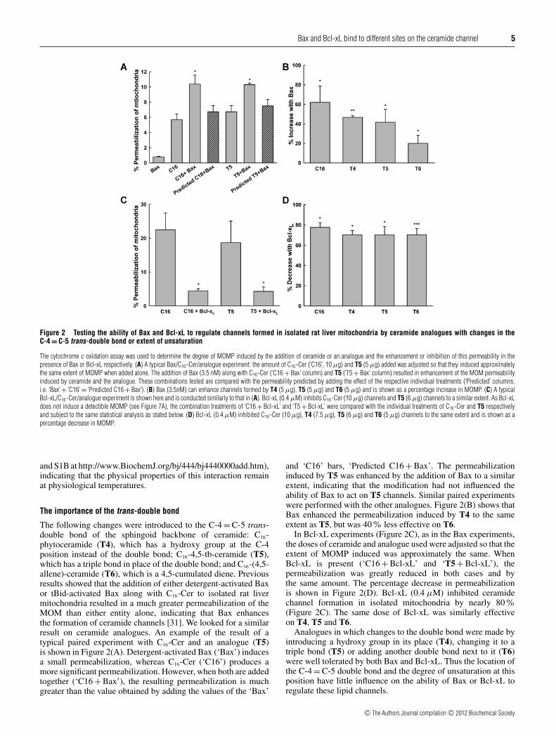
Figure 2 Testing the ability of Bax and Bcl-xL to regulate channels formed in isolated rat liver mitochondria by ceramide analogues with changes in theC-4 = C-5 trans-double bond or extent of unsaturation
The cytochrome c oxidation assay was used to determine the degree of MOMP induced by the addition of ceramide or an analogue and the enhancement or inhibition of this permeability in thepresence of Bax or Bcl-xL respectively. (A) A typical Bax/C16-Cer/analogue experiment: the amount of C16-Cer (‘C16’, 10 μg) and T5 (5 μg) added was adjusted so that they induced approximatelythe same extent of MOMP when added alone. The addition of Bax (3.5 nM) along with C16-Cer (‘C16 + Bax’ column) and T5 (‘T5 + Bax’ column) resulted in enhancement of the MOM permeabilityinduced by ceramide and the analogue. These combinations tested are compared with the permeability predicted by adding the effect of the respective individual treatments (‘Predicted’ columns,i.e. ‘Bax’ + ’C16’ = ‘Predicted C16 + Bax’). (B) Bax (3.5nM) can enhance channels formed by T4 (5 μg), T5 (5 μg) and T6 (5 μg) and is shown as a percentage increase in MOMP. (C) A typicalBcl-xL/C16-Cer/analogue experiment is shown here and is conducted similarly to that in (A). Bcl-xL (0.4 μM) inhibits C16-Cer (10 μg) channels and T5 (6 μg) channels to a similar extent. As Bcl-xLdoes not induce a detectible MOMP (see Figure 7A), the combination treatments of ‘C16 + Bcl-xL’ and ‘T5 + Bcl-xL’ were compared with the individual treatments of C16-Cer and T5 respectivelyand subject to the same statistical analysis as stated below. (D) Bcl-xL (0.4 μM) inhibited C16-Cer (10 μg), T4 (7.5 μg), T5 (6 μg) and T6 (5 μg) channels to the same extent and is shown as apercentage decrease in MOMP.
and S1B at http://www.BiochemJ.org/bj/444/bj4440000add.htm),indicating that the physical properties of this interaction remainat physiological temperatures.
The importance of the trans-double bond
The following changes were introduced to the C-4 = C-5 trans-double bond of the sphingoid backbone of ceramide: C16-phytoceramide (T4), which has a hydroxy group at the C-4position instead of the double bond; C16-4,5-tb-ceramide (T5),which has a triple bond in place of the double bond; and C16-(4,5-allene)-ceramide (T6), which is a 4,5-cumulated diene. Previousresults showed that the addition of either detergent-activated Baxor tBid-activated Bax along with C16-Cer to isolated rat livermitochondria resulted in a much greater permeabilization of theMOM than either entity alone, indicating that Bax enhancesthe formation of ceramide channels [31]. We looked for a similarresult on ceramide analogues. An example of the result of atypical paired experiment with C16-Cer and an analogue (T5)is shown in Figure 2(A). Detergent-activated Bax (‘Bax’) inducesa small permeabilization, whereas C16-Cer (‘C16’) produces amore significant permeabilization. However, when both are addedtogether (‘C16 + Bax’), the resulting permeabilization is muchgreater than the value obtained by adding the values of the ‘Bax’
and ‘C16’ bars, ‘Predicted C16 + Bax’. The permeabilizationinduced by T5 was enhanced by the addition of Bax to a similarextent, indicating that the modification had not influenced theability of Bax to act on T5 channels. Similar paired experimentswere performed with the other analogues. Figure 2(B) shows thatBax enhanced the permeabilization induced by T4 to the sameextent as T5, but was 40% less effective on T6.
In Bcl-xL experiments (Figure 2C), as in the Bax experiments,the doses of ceramide and analogue used were adjusted so that theextent of MOMP induced was approximately the same. WhenBcl-xL is present (‘C16 + Bcl-xL’ and ‘T5 + Bcl-xL’), thepermeabilization was greatly reduced in both cases and bythe same amount. The percentage decrease in permeabilizationis shown in Figure 2(D). Bcl-xL (0.4 μM) inhibited ceramidechannel formation in isolated mitochondria by nearly 80%(Figure 2C). The same dose of Bcl-xL was similarly effectiveon T4, T5 and T6.
Analogues in which changes to the double bond were made byintroducing a hydroxy group in its place (T4), changing it to atriple bond (T5) or adding another double bond next to it (T6)were well tolerated by both Bax and Bcl-xL. Thus the location ofthe C-4 = C-5 double bond and the degree of unsaturation at thisposition have little influence on the ability of Bax or Bcl-xL toregulate these lipid channels.
c© The Authors Journal compilation c© 2012 Biochemical Society
6 M. N. Perera and others
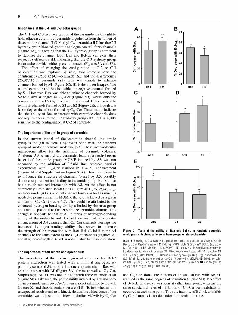
Importance of the C-1 and C-3 polar groups
The C-1 and C-3 hydroxy groups of the ceramide are thought tohold adjacent columns of ceramide together to form the lumen ofthe ceramide channel. 3-O-Methyl-C16-ceramide (H2) has the C-3hydroxy group blocked, yet this analogue can still form channels(Figure 3A), suggesting that the C-1 hydroxy group is sufficientto stabilize the channel. Both Bax and Bcl-xL can exert theirrespective effects on H2, indicating that the C-3 hydroxy groupis not a site at which either protein interacts (Figures 3A and 3B).
The effect of changing the configuration at C-2 or C-3of ceramide was explored by using two stereoisomers: theenantiomer (2R,3S,4E)-C16-ceramide (S1) and the diastereomer(2S,3S,4E)-C16-ceramide (S2). Bax was unable to enhancechannels formed by S1 (Figure 2C). S1 is the mirror image of thenatural ceramide and Bax is unable to recognize channels formedby S1. However, Bax was able to enhance channels formed byS2 to a similar degree as C16-Cer (Figure 2D), where only theorientation of the C-3 hydroxy group is altered. Bcl-xL was ableto inhibit channels formed by S1 and S2 (Figure 2E), although to alesser degree than those formed by C16-Cer. These results indicatethat the ability of Bax to interact with ceramide channels doesnot require access to the C-3 hydroxy group (H2), but is highlysensitive to the configuration at C-2 of ceramide.
The importance of the amide group of ceramide
In the current model of the ceramide channel, the amidegroup is thought to form a hydrogen bond with the carbonylgroup of another ceramide molecule [27]. These intermolecularinteractions allow for the assembly of ceramide columns.Analogue A3, N-methyl-C16-ceramide, features a methyl groupinstead of the amide group. MOMP induced by A3 was notenhanced by the addition of 3.5 nM Bax, whereas parallelexperiments with C16-Cer resulted in a 40% enhancement(Figure 4A and Supplementary Figure S1A). Thus Bax is unableto influence the structure of channels formed by A3, possiblydue to a requirement for binding to the amide group. Bcl-xL alsohas a much reduced interaction with A3, but the effect is notcompletely diminished as with Bax (Figure 4B). (2S,3R,4E)-C16-urea-ceramide (A4) is a potent channel former as half as much isneeded to permeabilize the MOM to the level achieved by a givenamount of C16-Cer (Figure 4C). This could be attributed to theenhanced hydrogen-bonding ability afforded by the urea groupand thus the potential to further stabilize ceramide columns. Thischange is opposite to that of A3 in terms of hydrogen-bondingability of the molecule and Bax addition resulted in a greaterenhancement of A4 channels than C16-Cer channels. Perhaps theincreased hydrogen-bonding ability also serves to increasethe strength of the interaction with Bax. Bcl-xL inhibits the A4channels to the same extent as the C16-Cer channels (Figures 4Cand 4D), indicating that Bcl-xL is not sensitive to the modification.
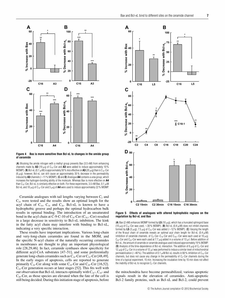
The importance of tail length and apolar bulk
The importance of the apolar region of ceramide for Bcl-2protein interaction was tested with a minimal analogue, N-palmitoylserinol (L5). It only has one aliphatic chain. Bax wasable to interact with L5 (Figure 5A) almost as well as C16-Cer.Surprisingly, Bcl-xL was not able to inhibit these channels at all(Figure 5B). Likewise, the permeability induced by a very-short-chain ceramide analogue, C2-Cer, was also not inhibited by Bcl-xL(Figure 5C and Supplementary Figure S1B). To test whether thisunexpected result was due to kinetic delays, the added doses of theceramides was adjusted to achieve a similar MOMP by C2-Cer
Figure 3 Tests of the ability of Bax and Bcl-xL to regulate ceramideanalogues with changes to polar headgroups or stereochemistry
(A and B) Blocking the C-3 hydroxy group does not reduce the channel’s sensitivity to 3.5 nMBax (5 μg of C16-Cer, 2 μg of H2, yielding ∼10 % MOMP) or 0.4 μM Bcl-xL (7.5 μg ofC16-Cer, 5 of μg H2, yielding ∼12 % MOMP). (C) Bax (2 nM) is sensitive to changes inthe stereochemistry found in analogue S1. Mitochondria were treated with 10 μg each of S1and C16-Cer (∼20 % MOMP). (D) Channels formed by analogue S2 (5 μg) interact with Bax(3.5 nM) similarly to those formed by C16-Cer (5 μg) (∼8 % MOMP). (E) Bcl-xL (0.4 μM)inhibits C16-Cer (3.5 μg) channels more strongly than those formed by S1 and S2 (10 and3.5 μg respectively, yielding ∼10 % MOMP).
and C16-Cer alone. Incubations of 15 and 30 min with Bcl-xLresulted in the same degrees of inhibition (Figure 5D). No effectof Bcl-xL on C2-Cer was seen at either time point, whereas thesame substantial level of inhibition of C16-Cer permeabilizationoccurred at each time point. Thus the inability of Bcl-xL to inhibitC2-Cer channels is not dependent on incubation time.
c© The Authors Journal compilation c© 2012 Biochemical Society
Bax and Bcl-xL bind to different sites on the ceramide channel 7
Figure 4 Bax is more sensitive than Bcl-xL to changes in the amide groupof ceramide
(A) Blocking the amide nitrogen with a methyl group prevents Bax (3.5 nM) from enhancingchannels made by A3 (10 μg of C16-Cer and A3 were added to induce approximately 10 %MOMP). (B) Bcl-xL (0.1 μM) is approximately 50 % less effective on A3 (20 μg) than is C16-Cer(6 μg); however, Bcl-xL can still cause an approximately 35 % decrease in the permeabilityinduced by A3 channels (∼11 % MOMP). (C and D) Analogue A4 contains a urea group, whichincreases the hydrogen-bonding ability of the molecule. Whereas Bax is more effective on A4than C16-Cer, Bcl-xL is similarly effective on both. For these experiments, 3.5 nM Bax, 0.1 μMBcl-xL and 10 μg of C16-Cer and 5 μg of A4 were used to induce approximately 22 % MOMP.
Ceramide analogues with tail lengths varying between C2 andC24 were tested and the results show an optimal length for theacyl chain of C16, C18 and C20. Bcl-xL is known to have ahydrophobic groove and perhaps the optimal hydrocarbon bulkresults in optimal binding. The introduction of an unsaturatedbond in the acyl chain at C-9-C-10 of C18-Cer (C18:1-Cer) resultedin a large decrease in sensitivity to Bcl-xL inhibition. The kinkin the fatty acyl chain may interfere with binding to Bcl-xL,indicating a very specific interaction.
These results have important implications. Various long-chainand very-long-chain ceramides are found in the MOM, andthe specific N-acyl chains of the naturally occurring ceramidesin membranes are thought to play an important physiologicalrole [28,29,46]. In fact, ceramide synthases show specificity fordifferent acyl-CoA substrates [47–51], and some preferentiallygenerate long-chain ceramides such as C16-Cer or C24-Cer [48,49].In the early stages of apoptosis, cells are reported to generateprimarily C16-Cer along with some C18-Cer and C20-Cer [16,52].C24-Cer generation occurs at later stages [52]. This fits well withour observation that Bcl-xL interacts optimally with C16-, C18- andC20-Cer, as those species are elevated when the fate of the cell isstill being decided. During this initiation stage of apoptosis, before
Figure 5 Effects of analogues with altered hydrophobic regions on theregulation by Bcl-xL and Bax
(A) Bax (2 nM) enhances MOMP formed by L5 (10 μg), which has a truncated sphingoid base(10 μg of C16-Cer was used, ∼30 % MOMP). (B) Bcl-xL (0.4 μM) does not inhibit channelsformed by L5 (3 μg); 7.5 μg of C16-Cer was added (∼12 % MOMP). (C) Varying the lengthof the N-acyl chain of ceramide reveals an optimal acyl chain length for Bcl-xL (0.4 μM)inhibition of ceramide channels. of C2-Cer–C18-Cer and C18:1-Cer were each used at 10 μg;C20-Cer and C24-Cer were each used at 7.7 μg added in a volume of 13 μl. Before addition ofBcl-xL, the amount of ceramide or ceramide analogue used induced approximately 19 % MOMP.(D) Analysis of the time-dependence of Bcl-xL interaction. The addition of 6 μg of C2-Cer and12 μg of C16-Cer in a volume of 12 μl was performed to induce a similar level of mitochondrialpermeabilization (∼40 %). The addition of 0.1 μM Bcl-xL results in 60 % inhibition of C16-Cerchannels, but does not cause any change in the permeability of C2-Cer channels during thetime of a typical experiment, 15 min. Increasing the incubation time by 15 min does not affectthe inability of Bcl-xL to recognize C2-Cer channels.
the mitochondria have become permeabilized, various apoptoticsignals result in the elevation of ceramides. Anti-apoptoticBcl-2 family proteins, such as Bcl-xL and Bcl-2, could prevent
c© The Authors Journal compilation c© 2012 Biochemical Society

8 M. N. Perera and others
these higher ceramide levels from forming channels resulting inMOMP. Bax activation and heterodimerization with Bcl-xL wouldrelieve this inhibition and, at the same time, any excess Bax couldfavour ceramide channel formation and/or growth. Clearly, theseinteractions result in many opportunities for controlling this keydecision-making step.
Many studies have been carried out with cell-permeant C2-and C6-ceramides to trigger apoptosis in cancer cells. It is alsoevident that many enzymes that metabolize ceramide do notrecognize C2-ceramides [53,54], thus allowing these moleculesto act over a longer time scale. Interestingly, the ability of short-and long-chain ceramides to form channels is about the same, andmixtures of long- and short-chain ceramides are actually morepotent [55]. Thus it is possible that these short-chain ceramidesmay be more effective than long-chain ceramides therapeuticallyin promoting apoptosis because Bcl-xL is unable to recognize orregulate these species, while Bax retains the ability to promotechannel formation by these ceramides.
Inhibitors provide insights into the region of Bcl-xL that binds tothe ceramide channel
Cells overexpressing pro-survival proteins Bcl-xL and Bcl-2 arehighly resistant to cell death, and many types of cancer indeeddisplay this phenotype. Three known small-molecule inhibitors ofBcl-xL and Bcl-2 are 2-MeAA3, ABT-737 and ABT-263. Theseinhibitors bind to the hydrophobic groove on Bcl-2 and Bcl-xL[24,56–58], effectively neutralizing the effects of pro-survivalprotein overexpression and sensitizing cells to death. Since Bcl-xL interacts with the ceramide channel through the hydrophobicregion, the inhibitors should interfere with the interaction ifbinding involves the hydrophobic groove.
Results from the 2-MeAA3 experiments are shown inFigure 6(A). Whereas the highest concentration of 2-MeAA3
tested (2.7 μM) had no effect on ceramide-induced MOMP,Bcl-xL (0.7 μM) inhibited the ceramide-induced MOMP byapproximately 50%. 2-MeAA3 produced a dose-dependentrecovery of ceramide-induced MOMP, indicating that 2-MeAA3
might bind to the same site on Bcl-xL as the ceramide channel.Unlike 2-MeAA3, ABT-737 displayed a strong inhibition ofceramide permeabilization at 2.5 and 5 μM, whereas lowerconcentrations had either no effect or a slightly stimulatory effecton ceramide permeabilization (Figure 6B). In the presence of0.7 μM Bcl-xL, ABT-737 produced a dose-dependent recoveryof ceramide permeabilization with 50% recovery atapproximately 1 μM and complete recovery at 2.5 μM. A stronginhibition of ceramide channels by ABT-737 is evident at 5 μMeven in the presence of Bcl-xL. ABT-263, the orally availableanalogue of ABT-737, was also able to cause a dose-dependentrecovery of ceramide permeabilization in the presence of Bcl-xL, but did not inhibit ceramide permeabilization at any of theconcentrations tested when Bcl-xL was not present (Figure 6C).Thus the inhibitory effects of Bcl-xL on ceramide channels canbe reversed by the addition of Bcl-xL-targeted inhibitors that bindto the hydrophobic groove, suggesting competition and that thehydrophobic groove is likely to be the site at which Bcl-xL bindsto the ceramide channel.
Molecular docking of ceramides and inhibitors to Bcl-xL wereconducted to determine the lowest-energy site and mode ofbinding on Bcl-xL. The region of lowest-energy binding wasfirst determined probing the whole surface of Bcl-xL and thehydrophobic groove was the best, or near best, scored region for allligands tested. The top ten poses for each of the ligands are shownin Figure 7 and the average energy of these were calculated and
Figure 6 The hydrophobic groove of Bcl-xL is involved in binding toceramide channels
Inhibitors known to bind to the hydrophobic groove of Bcl-xL were assayed for ability to preventBcl-xL binding to ceramide channels in isolated rat liver mitochondria. (A–C) Increasingconcentrations of 2-MeAA3, ABT-737 or ABT-263 were tested with C16-Cer alone (10 μg) andthe combination of C16-Cer and Bcl-xL (0.7μM).
are shown as a horizontal bar. The more negative the number, thestronger the interaction. ABT-737 and ABT-263 had the lowest-energy score, and therefore best binding, followed by C16-Cer.Especially noteworthy is the weaker average binding of C2-Cercompared with C16-Cer, consistent with the experimental results.To explore further the potential interaction between Bcl-xL andC16-Cer, the lowest-energy pose was subjected to global systemenergy minimization to allow both the protein and the ligand to
c© The Authors Journal compilation c© 2012 Biochemical Society

Bax and Bcl-xL bind to different sites on the ceramide channel 9
Figure 7 E Score 2 of ten best poses of molecular docking simulations
Docking simulations of Bcl-xL to C16-Cer (C16), C2-Cer (C2), ABT-737, ABT-263, 2-MeAA3
or palmitoylserine (P-Serine) yielded structures of various degrees of interaction energy. Thelower the E Score, the better the interaction. The average of the ten best interacting structures(poses) for each ligand is shown as a horizontal line.
Figure 8 Molecular structures of C16-Cer bound to Bcl-xL from dockingsimulations and possible interaction with a ceramide channel
(A) How Bcl-xL might bind to a ceramide molecule at the end of a ceramide column that is part
COLOUR
of a ceramide channel. One of the top poses of C16-Cer bound to Bcl-xL leaves one chain free.This was lined up with a segment of the proposed structure of a ceramide channel, showing howBcl-xL might bind to the end of one ceramide column, disrupting the column’s interaction withthe adjacent phospholipid bilayer. (B) C16-Cer nestled in the hydrophobic grove of Bcl-xL. Thisis one of the top ten poses after energy minimization.
flex. These results were considered a plausible prediction of theinteraction between ceramide and Bcl-xL (Figure 8B).
One of the top ten poses shows ceramide binding to Bcl-xLwith only one chain (Figure 8A). When this is placed on topof a segment of the model of a ceramide channel, this providesmechanistic support for a hypothetical mechanism by which Bcl-xL destabilizes a ceramide channel.
Molecular dynamics simulations [26,27] indicate that theceramide channel forms an hourglass shape in the membrane,allowing the channel to properly interface with the phospholipidbilayer and thus avoiding exposure of apolar chains tothe aqueous environment (Supplementary Figures S2 andS3 at http://www.BiochemJ.org/bj/444/bj4440000add.htm). Thisresults in a curvature in the ceramide columns forming the
channel that is positive in the direction normal to the planeof the membrane [27]. This curvature reduces the distortion ofthe phospholipids in close proximity to the ceramide channel.Thus interfacing the ceramide molecules that would tend tohave their long axis parallel with the membrane plane with thephospholipids whose long axis is normal to that plane requiresa compromise distortion of both to minimize the overall energylevel. This low-energy compromise might be interfered with bythe interaction with Bcl-xL. Bcl-xL could bind at this interface,displacing the local phospholipids and allowing the ceramidecolumn to relax by reducing the positive curvature. Evidencefrom the present study indicates that the hydrophobic groove ofBcl-xL is interacting with the hydrophobic chains of the ceramidemolecules in a channel, chains that are buried within themembrane apolar environment. This is consistent with previousresults by Siskind et al. [24], who demonstrated that a Bcl-xLdeletion mutant lacking the transmembrane domain is unable todisassemble ceramide channels, suggesting that the membrane-active form of Bcl-xL is required for ceramide channel regulation.Thus the binding of Bcl-xL to the end of one column andresulting relaxation of that column (Supplementary Figure S4at http://www.BiochemJ.org/bj/444/bj4440000add.htm) wouldresult in a mismatch with the curvature of adjacent ceramidecolumns and thus the generation of mechanical stress. Thehighly hydrogen-bonded structure of the channel would resultin the propagation of the mechanical stress to the rest ofthe channel (Supplementary Figure S5 at http://www.BiochemJ.org/bj/444/bj4440000add.htm). This high-energy structure wouldthen lead to the destabilization of the channel, shifting the dynamicequilibrium towards channel disassembly. This mechanism isconsistent with previous results [24] that indicate the formationof a 1:1 complex between Bcl-xL and the channel, requiring thatthe effects of this binding propagate through the channel structurein an allosteric manner.
As for Bax, the results indicate that the chirality of themolecule’s polar region and specifically the accessibility ofthe amide group are key for Bax binding. This indicates thatBax may also be acting at the interface of the channel and thephospholipid membrane where there is access to the carboxamidegroup. However, unlike Bcl-xL, ceramide binding might favourthe positive curvature of the ceramide columns, stabilizing thechannel at the channel–membrane interface. This stabilizationcan result in the enlargement of ceramide channels to an optimalsize (Supplementary Figure S6 at http://www.BiochemJ.org/bj/444/bj4440000add.htm). Again, these results are consistentwith previous findings [31], as the binding of Bax influencesthe ceramide channel to enlarge to a size that fits optimally withthe binding site of Bax. Previous results also indicated that mul-tiple Bax structures bind to a single ceramide channel, presumablyall acting to stabilize the ceramide channel in a particular state.
Concluding remarks
Beyond the details, it is worth noting that highly organized lipidassemblies, ceramide channels in this case, take a form dictated bythe properties of their monomeric subunits and the environment inwhich they exist. However, the forms of these superstructures canbe modified by interactions with regulating molecules, in this caseBcl-2 family proteins. Thus cells can control these superstructuresat various levels: by controlling the supply of specific monomersat the membrane or cell compartment of interest, by changing thelipid environment of the membrane, and by producing proteins (orother regulating factors) that alter the form or stability of the finalstructure. Perhaps, in thinking about lipids, one must go beyond
c© The Authors Journal compilation c© 2012 Biochemical Society

10 M. N. Perera and others
thinking about lipid phases, fluidity and phase transitions. Lipidscan form superstructures with unique properties and the ability tobe regulated much like the structures formed by proteins.
AUTHOR CONTRIBUTION
Meenu Perera designed the study, purified the proteins used, performed all mitochondrialexperiments, analysed data and wrote the paper. Shang Lin aided in repeating a portion ofmitochondrial experiments. Yuri Peterson designed and ran molecular docking simulationsand analysed output data. Alicja Bielawska, Zdzlaw Szulc and Robert Bittman providedceramide analogues. Marco Colombini provided intellectual guidance and wrote the paper.All authors contributed to editing the paper before submission.
ACKNOWLEDGEMENTS
We extend our gratitude to Richard Youle and Antonella Antignani for providing the plasmidwe used to express full-length Bax and for assistance with the purification procedure. Wealso extend our heartiest thanks to Marie Hardwick for the full-length Bcl-xL plasmid andHeather Lamb for recommending the modifications to the purification procedure. We thankRichard Trager for technical assistance with the molecular simulations. Finally, we thankVidyaramanan Ganesan for isolating rat liver mitochondria.
FUNDING
This work was supported by the National Science Foundation [grant number MCB-1023008 (to M.C.)], National Institutes of Health [grant number HL-083187 (to R.B.)],National Institutes of Health/National Cancer Institute [grant number P01 CA097132-01A1 (to A.B.)] and National Institutes of Health/National Center for Research ResourcesSouth Carolina COBRE (Centers of Biomedical Research Excellence) [grant number P20RR017677 (to Z.M.S.)].
REFERENCES
1 Siskind, L. J., Kolesnick, R. N. and Colombini, M. (2006) Ceramide forms channels inmitochondrial outer membranes at physiologically relevant concentrations.Mitochondrion 6, 118–125
2 Dejean, L. M., Martinez-Caballero, S., Guo, L., Hughes, C., Teijido, O., Ducret, T., Ichas,F., Korsmeyer, S. J., Antonsson, B., Jonas, E. A. and Kinnally, K. W. (2005) OligomericBax is a component of the putative cytochrome c release channel MAC, mitochondrialapoptosis-induced channel. Mol. Biol. Cell 16, 2424–2432
3 Chiara, F., Castellaro, D., Marin, O., Petronilli, V., Brusilow, W. S., Juhaszova, M., Sollott,S. J., Forte, M., Bernardi, P. and Rasola, A. (2008) Hexokinase II detachment frommitochondria triggers apoptosis through the permeability transition pore independent ofvoltage-dependent anion channels. PLoS ONE 3, e1852
4 Montessuit, S., Somasekharan, S. P., Terrones, O., Lucken-Ardjomande, S., Herzig, S.,Schwarzenbacher, R., Manstein, D. J., Bossy-Wetzel, E., Basanez, G., Meda, P. andMartinou, J. C. (2010) Membrane remodeling induced by the dynamin-related proteinDrp1 stimulates Bax oligomerization. Cell 142, 889–901
5 Lovell, J. F., Billen, L. P., Bindner, S., Shamas-Din, A., Fradin, C., Leber, B. and Andrews,D. W. (2008) Membrane binding by tBid initiates an ordered series of events culminatingin membrane permeabilization by Bax. Cell 135, 1074–1084
6 Terrones, O., Antonsson, B., Yamaguchi, H., Wang, H. G., Liu, J., Lee, R. M., Herrmann,A. and Basanez, G. (2004) Lipidic pore formation by the concerted action of proapoptoticBAX and tBID. J. Biol. Chem. 279, 30081–30091
7 Birbes, H., El Bawab, S., Hannun, Y. A. and Obeid, L. M. (2001) Selective hydrolysis of amitochondrial pool of sphingomyelin induces apoptosis. FASEB J. 15, 2669–2679
8 Birbes, H., Luberto, C., Hsu, Y. T., El Bawab, S., Hannun, Y. A. and Obeid, L. M. (2005) Amitochondrial pool of sphingomyelin is involved in TNFα-induced Bax translocation tomitochondria. Biochem. J. 386, 445–451
9 Dai, Q., Liu, J., Chen, J., Durrant, D., McIntyre, T. M. and Lee, R. M. (2004) Mitochondrialceramide increases in UV-irradiated HeLa cells and is mainly derived from hydrolysis ofsphingomyelin. Oncogene 23, 3650–3658
10 Matsko, C. M., Hunter, O. C., Rabinowich, H., Lotze, M. T. and Amoscato, A. A. (2001)Mitochondrial lipid alterations during Fas- and radiation-induced apoptosis. Biochem.Biophys. Res. Commun. 287, 1112–1120
11 Siskind, L. J. (2005) Mitochondrial ceramide and the induction of apoptosis. J. Bioenerg.Biomembr. 37, 143–153
12 Mullen, T. D. and Obeid, L. (2011) Ceramide and apoptosis: exploring the enigmaticconnections between sphingolipid metabolism and programmed cell death. AnticancerAgents Med. Chem., in the press
13 Stiban, J., Caputo, L. and Colombini, M. (2008) Ceramide synthesis in the endoplasmicreticulum can permeabilize mitochondria to proapoptotic proteins. J. Lipid Res. 49,625–634
14 Wei, M. C., Zong, W. X., Cheng, E. H., Lindsten, T., Panoutsakopoulou, V., Ross, A. J.,Roth, K. A., MacGregor, G. R., Thompson, C. B. and Korsmeyer, S. J. (2001) ProapoptoticBAX and BAK: a requisite gateway to mitochondrial dysfunction and death. Science 292,727–730
15 Zong, W. X., Lindsten, T., Ross, A. J., MacGregor, G. R. and Thompson, C. B. (2001)BH3-only proteins that bind pro-survival Bcl-2 family members fail to induce apoptosis inthe absence of Bax and Bak. Genes Dev. 15, 1481–1486
16 Siskind, L. J., Mullen, T. D., Romero Rosales, K., Clarke, C. J., Hernandez-Corbacho,M. J., Edinger, A. L. and Obeid, L. M. (2010) The BCL-2 protein BAK is required forlong-chain ceramide generation during apoptosis. J. Biol. Chem. 285, 11818–11826
17 Liu, Y. Y., Han, T. Y., Giuliano, A. E. and Cabot, M. C. (2001) Ceramide glycosylationpotentiates cellular multidrug resistance. FASEB J. 15, 719–730
18 Komori, H., Ichikawa, S., Hirabayashi, Y. and Ito, M. (1999) Regulation of intracellularceramide content in B16 melanoma cells: biological implications of ceramideglycosylation. J. Biol. Chem. 274, 8981–8987
19 Itoh, M., Kitano, T., Watanabe, M., Kondo, T., Yabu, T., Taguchi, Y., Iwai, K., Tashima, M.,Uchiyama, T. and Okazaki, T. (2003) Possible role of ceramide as an indicator ofchemoresistance: decrease of the ceramide content via activation of glucosylceramidesynthase and sphingomyelin synthase in chemoresistant leukemia. Clin. Cancer Res. 9,415–423
20 Lavie, Y., Cao, H., Volner, A., Lucci, A., Han, T. Y., Geffen, V., Giuliano, A. E. and Cabot,M. C. (1997) Agents that reverse multidrug resistance, tamoxifen, verapamil, andcyclosporin A, block glycosphingolipid metabolism by inhibiting ceramide glycosylationin human cancer cells. J. Biol. Chem. 272, 1682–1687
21 Liu, Y. Y., Patwardhan, G. A., Bhinge, K., Gupta, V., Gu, X. and Jazwinski, S. M. (2011)Suppression of glucosylceramide synthase restores p53-dependent apoptosis in mutantp53 cancer cells. Cancer Res. 71, 2276–2285
22 Patwardhan, G. A., Zhang, Q. J., Yin, D. M., Gupta, V., Bao, J. X., Senkal, C. E., Ogretmen,B., Cabot, M. C., Shah, G. V., Sylvester, P. W. et al. (2009) A new mixed-backboneoligonucleotide against glucosylceramide synthase sensitizes multidrug-resistant tumorsto apoptosis. PloS ONE 4, e6938
23 Chapman, J. V., Gouaze-Andersson, V., Messner, M. C., Flowers, M., Karimi, R., Kester,M., Barth, B. M., Liu, X., Liu, Y. Y., Giuliano, A. E. and Cabot, M. C. (2010) Metabolism ofshort-chain ceramide by human cancer cells: implications for therapeutic approaches.Biochem. Pharmacol. 80, 308–315
24 Siskind, L. J., Feinstein, L., Yu, T., Davis, J. S., Jones, D., Choi, J., Zuckerman, J. E., Tan,W., Hill, R. B., Hardwick, J. M. and Colombini, M. (2008) Anti-apoptotic Bcl-2 familyproteins disassemble ceramide channels. J. Biol. Chem. 283, 6622–6630
25 Siskind, L. J., Kolesnick, R. N. and Colombini, M. (2002) Ceramide channels increase thepermeability of the mitochondrial outer membrane to small proteins. J. Biol. Chem. 277,26796–26803
26 Samanta, S., Stiban, J., Maugel, T. K. and Colombini, M. (2011) Visualization of ceramidechannels by transmission electron microscopy. Biochim. Biophys. Acta 1808,1196–1201
27 Anishkin, A., Sukharev, S. and Colombini, M. (2006) Searching for the moleculararrangement of transmembrane ceramide channels. Biophys. J. 90, 2414–2426
28 Megha, Sawatzki, P., Kolter, T., Bittman, R. and London, E. (2007) Effect of ceramideN-acyl chain and polar headgroup structure on the properties of ordered lipid domains(lipid rafts). Biochim. Biophys. Acta 1768, 2205–2212
29 Pruett, S. T., Bushnev, A., Hagedorn, K., Adiga, M., Haynes, C. A., Sullards, M. C., Liotta,D. C. and Merrill, A. H. (2008) Biodiversity of sphingoid bases (“sphingosines”) andrelated amino alcohols. J. Lipid Res. 49, 1621–1639
30 Siskind, L. J., Davoody, A., Lewin, N., Marshall, S. and Colombini, M. (2003)Enlargement and contracture of C2-ceramide channels. Biophys. J. 85, 1560–1575
31 Ganesan, V., Perera, M. N., Colombini, D., Datskovskiy, D., Chadha, K. and Colombini,M. (2010) Ceramide and activated Bax act synergistically to permeabilize themitochondrial outer membrane. Apoptosis 15, 553–562
32 Usta, J., El Bawab, S., Roddy, P., Szulc, Z. M., Yusuf, Hannun, A. and Bielawska, A. (2001)Structural requirements of ceramide and sphingosine based inhibitors of mitochondrialceramidase. Biochemistry 40, 9657–9668
33 Brockman, H. L., Momsen, M. M., Brown, R. E., He, L., Chun, J., Byun, H. S. and Bittman,R. (2004) The 4,5-double bond of ceramide regulates its dipole potential, elasticproperties, and packing behavior. Biophys. J. 87, 1722–1731
34 He, L., Byun, H. S. and Bittman, R. (2000) A stereocontrolled, efficient synthetic route tobioactive sphingolipids: synthesis of phytosphingosine and phytoceramides fromunsaturated ester precursors via cyclic sulfate intermediates. J. Org. Chem. 65,7618–7626
c© The Authors Journal compilation c© 2012 Biochemical Society
Bax and Bcl-xL bind to different sites on the ceramide channel 11
35 He, L., Byun, H. S. and Bittman, R. (2000) Stereoselective preparation of ceramide and itsskeleton backbone modified analogues via cyclic thionocarbonate intermediates derivedby catalytic asymmetric dihydroxylation of α,β-unsaturated ester precursors. J. Org.Chem. 65, 7627–7633
36 Bieberich, E., Hu, B., Silva, J., MacKinnon, S., Yu, R. K., Fillmore, H., Broaddus, W. C.and Ottenbrite, R. M. (2002) Synthesis and characterization of novel ceramide analogs forinduction of apoptosis in human cancer cells. Cancer Lett. 181, 55–64
37 Parsons, D. F., Williams, G. R. and Chance, B. (1966) Characteristics of isolated andpurified preparations of the outer and inner membranes of mitochondria. Ann. N.Y. Acad.Sci. 137, 643–666
38 Douce, R., Bourguignon, J., Brouquisse, R. and Neuburger, M. (1987) Isolation of plantmitochondria: general principles and criteria of integrity. Methods Enzymol. 148,403–415
39 Suzuki, M., Youle, R. J. and Tjandra, N. (2000) Structure of Bax: coregulation of dimerformation and intracellular localization. Cell 103, 645–654
40 Basanez, G., Zhang, J., Chau, B. N., Maksaev, G. I., Frolov, V. A., Brandt, T. A., Burch, J.,Hardwick, J. M. and Zimmerberg, J. (2001) Pro-apoptotic cleavage products of Bcl-xLform cytochrome c-conducting pores in pure lipid membranes. J. Biol. Chem. 276,31083–31091
41 Wojtczak, L., Zaluska, H., Wroniszewska, A. and Wojtczak, A. B. (1972) Assay for theintactness of the outer membrane in isolated mitochondria. Acta Biochim. Pol. 19,227–234
42 Kluck, R. M., Esposti, M. D., Perkins, G., Renken, C., Kuwana, T., Bossy-Wetzel, E.,Goldberg, M., Allen, T., Barber, M. J., Green, D. R. and Newmeyer, D. D. (1999) Thepro-apoptotic proteins, Bid and Bax, cause a limited permeabilization of the mitochondrialouter membrane that is enhanced by cytosol. J. Cell Biol. 147, 809–822
43 von Ahsen, O., Renken, C., Perkins, G., Kluck, R. M., Bossy-Wetzel, E. and Newmeyer,D. D. (2000) Preservation of mitochondrial structure and function after Bid- orBax-mediated cytochrome c release. J. Cell Biol. 150, 1027–1036
44 Li, F., Srinivasan, A., Wang, Y., Armstrong, R. C., Tomaselli, K. J. and Fritz, L. C. (1997)Cell-specific induction of apoptosis by microinjection of cytochrome c: Bcl-xL hasactivity independent of cytochrome c release. J. Biol. Chem. 272, 30299–30305
45 Zhivotovsky, B., Orrenius, S., Brustugun, O. T. and Doskeland, S. O. (1998) Injectedcytochrome c induces apoptosis. Nature 391, 449–450
46 Grosch, S., Schiffmann, S. and Geisslinger, G. (2012) Chain length-specific properties ofceramides. Prog. Lipid Res. 51, 50–62
47 Menuz, V., Howell, K. S., Gentina, S., Epstein, S., Riezman, I., Fornallaz-Mulhauser, M.,Hengartner, M. O., Gomez, M., Riezman, H. and Martinou, J. C. (2009) Protection of C.elegans from anoxia by HYL-2 ceramide synthase. Science 324, 381–384
48 Venkataraman, K., Riebeling, C., Bodennec, J., Riezman, H., Allegood, J. C., Sullards,M. C., Merrill, A. H. and Futerman, A. H. (2002) Upstream of growth and differentiationfactor 1 (uog1), a mammalian homolog of the yeast longevity assurance gene 1 (LAG1),regulates N-stearoyl-sphinganine (C18-(dihydro)ceramide) synthesis in a fumonisinB1-independent manner in mammalian cells. J. Biol. Chem. 277, 35642–35649
49 Mizutani, Y., Kihara, A. and Igarashi, Y. (2005) Mammalian Lass6 and its related familymembers regulate synthesis of specific ceramides. Biochem. J. 390, 263–271
50 Laviad, E. L., Albee, L., Pankova-Kholmyansky, I., Epstein, S., Park, H., Merrill, A. H. andFuterman, A. H. (2008) Characterization of ceramide synthase 2: tissue distribution,substrate specificity, and inhibition by sphingosine 1-phosphate. J. Biol. Chem. 283,5677–5684
51 Riebeling, C., Allegood, J. C., Wang, E., Merrill, A. H. and Futerman, A. H. (2003) Twomammalian longevity assurance gene (LAG1) family members, trh1 and trh4, regulatedihydroceramide synthesis using different fatty acyl-CoA donors. J. Biol. Chem. 278,43452–43459
52 Kroesen, B. J., Jacobs, S., Pettus, B. J., Sietsma, H., Kok, J. W., Hannun, Y. A. and de Leij,L. F. (2003) BcR-induced apoptosis involves differential regulation of C16 andC24-ceramide formation and sphingolipid-dependent activation of the proteasome. J.Biol. Chem. 278, 14723–14731
53 Wijesinghe, D. S., Massiello, A., Subramanian, P., Szulc, Z., Bielawska, A. and Chalfant,C. E. (2005) Substrate specificity of human ceramide kinase. J. Lipid Res. 46,2706–2716
54 Phillips, S. C., Triola, G., Fabrias, G., Goni, F. M., DuPre, D. B. and Yappert, M. C. (2009)cis- versus trans-ceramides: effects of the double bond on conformation and H-bondinginteractions. J. Phys. Chem. B. 113, 15249–15255
55 Perera, M. N., Ganesan, V., Siskind, L. J., Szulc, Z. M., Bielawski, J., Bielawska, A.,Bittman, R. and Colombini, M. (2012) Ceramide channels: influence of molecularstructure on channel formation in membranes. Biochim. Biophys. Acta 1818,1291–1301
56 Manion, M. K., O’Neill, J. W., Giedt, C. D., Kim, K. M., Zhang, K. Y. and Hockenbery,D. M. (2004) Bcl-XL mutations suppress cellular sensitivity to antimycin A. J. Biol. Chem.279, 2159–2165
57 Tse, C., Shoemaker, A. R., Adickes, J., Anderson, M. G., Chen, J., Jin, S., Johnson, E. F.,Marsh, K. C., Mitten, M. J., Nimmer, P. et al. (2008) ABT-263: a potent and orallybioavailable Bcl-2 family inhibitor. Cancer Res. 68, 3421–3428
58 Wendt, M. D., Shen, W., Kunzer, A., McClellan, W. J., Bruncko, M., Oost, T. K., Ding, H.,Joseph, M. K., Zhang, H., Nimmer, P. M. et al. (2006) Discovery and structure-activityrelationship of antagonists of B-cell lymphoma 2 family proteins with chemopotentiationactivity in vitro and in vivo. J. Med. Chem. 49, 1165–1181
Received 29 November 2011/9 April 2012; accepted 11 April 2012Published as BJ Immediate Publication 11 April 2012, doi:10.1042/BJ20112103
c© The Authors Journal compilation c© 2012 Biochemical Society
Proof Delivery Form
FOR PUBLISHER'S USE ONLY
Article Type ____________
Pages with colour figs (main article)_______________
Supplementary online data?TypesetBIC ____No. of pages ____Not Typeset ________
MultimediaAnimation _________3D Interactive Structure _________Movie __________Other __________
CommentaryBJ20____/_______
Figures/schemes/tables to be replaced Main article figure _____________
Main article scheme _____________Main article table _____________
Supp data figure _____________Supp data scheme _____________Supp data table _____________
Revised proof to author? ________
Other comments
Journal and Article number: BIC 132
BJ reference: BJ2011/2103
Number of colour figures: Figures S2, S3, S4,S5 and S6
Number of pages (not including this page): 4
This proof contains supplementary online material.
Biochemical Journal
Please print out your proof, mark any corrections needed, and return it, together with the offprint order form, by FAXto +44 (0)20 7685 2469 as soon as possible (no later than 48 hours after receipt).
• You are responsible for correcting your proofs! Errors not found may appear in the journal.• The proof is sent to you for correction of typographical errors only, and revision of the substance of the text is not permitted.• Please answer carefully any queries from the subeditor.• A new copy of a figure must be provided if correction is required.
Notes:
1. The quality of half-tone and colour figures will be checked by the editorial office.
2. The volume number indicated on the proof is tentative only.
3. If you have any comments, however minor, on the handling of your paper, please let us know. Please quote the paper’s reference number when doing so.
4. If you have any queries, please contact the editorial office by email ([email protected]) or by telephone on 020 7685 2410 (+44 20 7685 2410 from
outside the U.K.).
Semantic Mark Up:In order to facilitate semantic mark up of your paper, please ensure that where appropriate you have included all databaseidentifiers (e.g. UniProt numbers, PDB accession numbers, GenBank accession numbers) for the following: gene sequences,sequence alignments, protein sequences, protein 3D structures etc.
Please return this form with your proof
Queries for author:
Q1: Please provide written confirmation (e-mail or fax is fine) that Dr Ganesan and Dr Samanta are happy with the content andthe context of this citation.
Typesetter’s queries to Publisher:
Non-printed material (for Publisher’s use only):
Primary: S9
Secondary: S2
Please return this form with your proof
Biochem. J. (2012) 444, 00–00 (Printed in Great Britain) doi:10.1042/BJ20112103
SUPPLEMENTARY ONLINE DATABax and Bcl-xL exert their regulation on different sites of the ceramidechannelMeenu N. PERERA*, Shang H. LIN*, Yuri K. PETERSON†, Alicja BIELAWSKA‡, Zdzlaw M. SZULC‡, Robert BITTMAN§ andMarco COLOMBINI*1
*Department of Biology, University of Maryland, College Park, MD 20742, U.S.A., †Department of Pharmaceutical and Biomedical Sciences, Medical University of South Carolina,Charleston, SC 29403, U.S.A., ‡Department of Biochemistry and Molecular Biology, Medical University of South Carolina, Charleston, SC 29403, U.S.A., and §Department ofChemistry and Biochemistry, Queens College, City University of New York, Flushing, NY 11367, U.S.A.
Comparison of the number of channels per mitochondrion and theamount of Bcl-2 family proteins present: why the effectiveconcentration of Bcl-2 family protein is important for channelregulation
The calculation of the number of channels present is based onthe fact that only a fraction of the mitochondrial populationis permeabilized as measured by adenylate kinase release. Forisolated rat liver mitochondria, there are 7.2×10⁹ mitochondriaper mg of mitochondrial protein [1]. In an experiment where160 μg of mitochondrial protein is used and 10 μg of ceramide,20% MOMP is achieved as measured by cytochrome coxidation, and only 28% of adenylate kinase is released(V. Ganesan and S. Samanta, personal communication). Thus,Q1
of the 1.2×10⁶ mitochondria, only 0.3×10⁶ mitochondriahave channels. Statistically speaking, it is unlikely that themitochondria that were permeabilized would contain multiplechannels, whereas the others contain none. Thus the number ofpermeabilized mitochondria are a good measure of the numberof ceramide channels present. However, the amounts of Bax
(3.5 nM, 1.5×10¹² molecules) and Bcl-xL (0.4 μM, 1.5×10¹⁴
molecules) are well in excess of the amount of mitochondria andchannels. Thus a dynamic equilibrium must exist between boundand free protein, and it is the effective concentration of proteinthat determines the growth or destabilization of the ceramidechannels. The amount of added protein is unrelated to the numberof channels present. The probability that a channel will have aprotein bound to it depends on the free protein concentration,either in aqueous solution or on or in the membrane.
Supplementary Figures
The supplementary Figures provide supporting information anddrawings to clarify our interpretation of the results presented.In these drawings, the membrane is shown, for simplicity, asa lipid bilayer. For the sake of clarity, it makes sense to keepthe illustrations simple. Clearly, the MOM is more complex,containing proteins and a variety of lipids that may or may notcontribute to the structure of the ceramide channel and influenceits interaction with the Bcl-2 family proteins.
1 To whom correspondence should be addressed (email [email protected]).
c© The Authors Journal compilation c© 2012 Biochemical Society
M. N. Perera and others
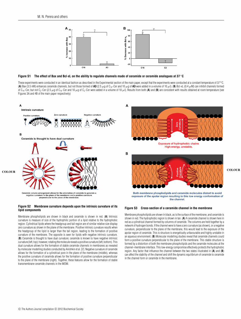
Figure S1 The effect of Bax and Bcl-xL on the ability to regulate channels made of ceramide or ceramide analogues at 37◦C
These experiments were conducted in an identical fashion as described in the Experimental section of the main paper, except that the experiments were conducted at a constant temperature of 37◦C.(A) Bax (3.5 nM) enhances ceramide channels, but not those formed of A3 (2.5 μg of C16-Cer and 10 μg of A3 were added in a volume of 10 μl). (B) Bcl-xL (0.4 μM) can inhibit channels formedof C16-Cer, but not C2-Cer (2.5 μg of C16-Cer and 10 μg of C2-Cer were added in a volume of 10 μl). Results from both (A) and (B) are consistent with results obtained at room temperature (seeFigures 3A and 4B of the main paper respectively)
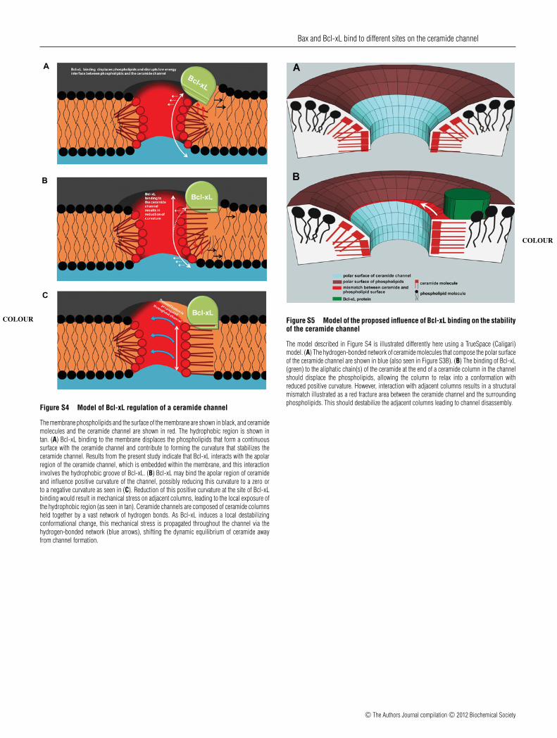
Figure S2 Membrane curvature depends upon the intrinsic curvature of itslipid components
Membrane phospholipids are shown in black and ceramide is shown in red. (A) Intrinsic
COLOUR
curvature is measure of size of the hydrophilic portion of a lipid relative to the hydrophobicregion. Cylindrical lipids where the headgroup and tail region are of similar relative size displayzero curvature as shown in the plane of the membrane. Positive intrinsic curvature results whenthe headgroup of the lipid is larger than the tail region, leading to the formation of positivecurvature of the membrane. The opposite is seen for lipids with negative intrinsic curvature.(B) Ceramide is thought to have dual curvature; ceramide is known to have negative intrinsiccurvature (left, top); however, rotating the molecule reveals a positive curvature (left, bottom). Thisdual curvature allows for the formation of stable ceramide channels in membranes as revealedby molecular modelling studies conducted by Anishkin et al. [2]. Negative curvature of ceramideallows for the formation of a cylindrical pore in the plane of the membrane (middle), whereasthe positive curvature of ceramide allows for the formation of positive curvature perpendicularto the plane of the membrane (right). Together, these features allow for the formation of stabletransmembrane ceramide channels in the MOM.
Figure S3 Cross-section of a ceramide channel in the membrane
Membrane phospholipids are shown in black, as is the surface of the membrane, and ceramide is
COLOUR
shown in red. The hydrophobic region is shown in tan. (A) A ceramide channel is shown here inred as a cylindrical channel formed by columns of ceramide. The columns are held together by anetwork of hydrogen bonds. If the channel were to have a zero curvature (as shown), or a negativecurvature, perpendicular to the plane of the membrane, this would lead to the exposure of theapolar region of ceramide. This is structure is energetically unfavourable and highly unstable inan aqueous environment. (B) Molecular modelling studies reveal that ceramide channels couldform a positive curvature perpendicular to the plane of the membrane. This stable structure isformed by a distortion of both the membrane phospholipids and the ceramide molecules at thechannel–membrane interface. This low-energy compromise effectively protects the hydrophobicregion. Any factor that influence the channel between the two states illustrated in (A) and (B)can affect the stability of the channel and shift the dynamic equilibrium of ceramide to ceramidein the channel form or ceramide in the membrane.
c© The Authors Journal compilation c© 2012 Biochemical Society
Bax and Bcl-xL bind to different sites on the ceramide channel
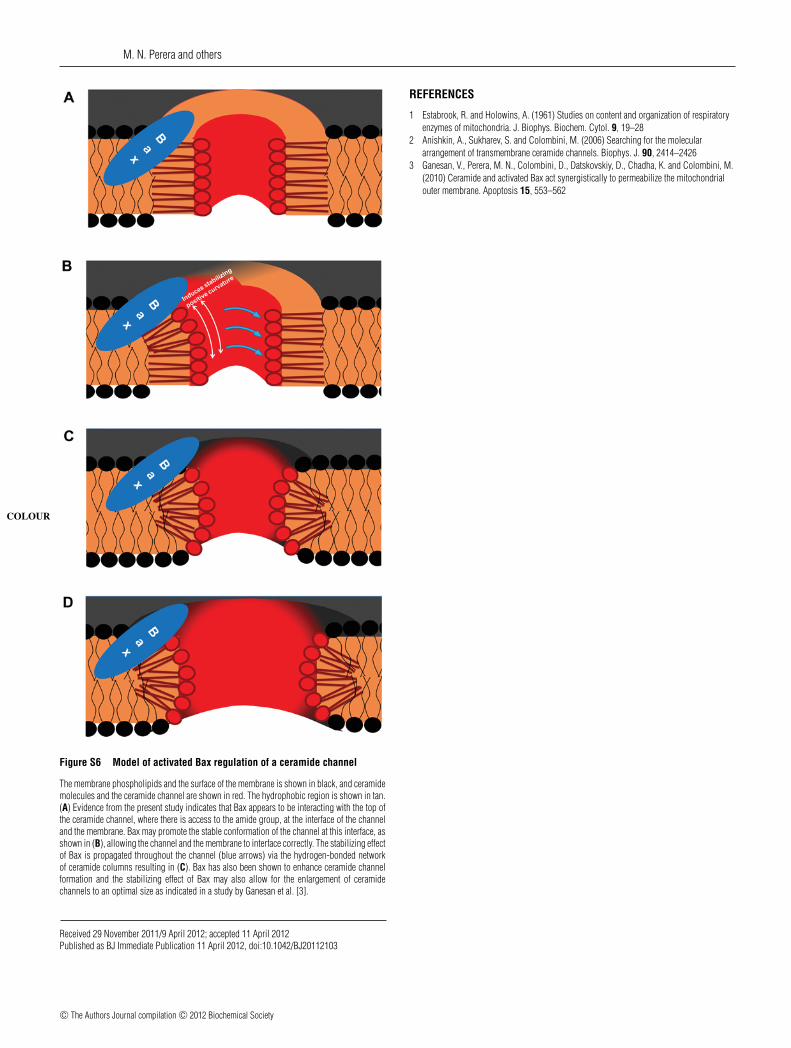
Figure S4 Model of Bcl-xL regulation of a ceramide channel
The membrane phospholipids and the surface of the membrane are shown in black, and ceramide
COLOUR
molecules and the ceramide channel are shown in red. The hydrophobic region is shown intan. (A) Bcl-xL binding to the membrane displaces the phospholipids that form a continuoussurface with the ceramide channel and contribute to forming the curvature that stabilizes theceramide channel. Results from the present study indicate that Bcl-xL interacts with the apolarregion of the ceramide channel, which is embedded within the membrane, and this interactioninvolves the hydrophobic groove of Bcl-xL. (B) Bcl-xL may bind the apolar region of ceramideand influence positive curvature of the channel, possibly reducing this curvature to a zero orto a negative curvature as seen in (C). Reduction of this positive curvature at the site of Bcl-xLbinding would result in mechanical stress on adjacent columns, leading to the local exposure ofthe hydrophobic region (as seen in tan). Ceramide channels are composed of ceramide columnsheld together by a vast network of hydrogen bonds. As Bcl-xL induces a local destabilizingconformational change, this mechanical stress is propagated throughout the channel via thehydrogen-bonded network (blue arrows), shifting the dynamic equilibrium of ceramide awayfrom channel formation.
Figure S5 Model of the proposed influence of Bcl-xL binding on the stabilityof the ceramide channel
The model described in Figure S4 is illustrated differently here using a TrueSpace (Caligari)
COLOUR
model. (A) The hydrogen-bonded network of ceramide molecules that compose the polar surfaceof the ceramide channel are shown in blue (also seen in Figure S3B). (B) The binding of Bcl-xL(green) to the aliphatic chain(s) of the ceramide at the end of a ceramide column in the channelshould displace the phospholipids, allowing the column to relax into a conformation withreduced positive curvature. However, interaction with adjacent columns results in a structuralmismatch illustrated as a red fracture area between the ceramide channel and the surroundingphospholipids. This should destabilize the adjacent columns leading to channel disassembly.
c© The Authors Journal compilation c© 2012 Biochemical Society
M. N. Perera and others
Figure S6 Model of activated Bax regulation of a ceramide channel
The membrane phospholipids and the surface of the membrane is shown in black, and ceramidemolecules and the ceramide channel are shown in red. The hydrophobic region is shown in tan.
COLOUR
(A) Evidence from the present study indicates that Bax appears to be interacting with the top ofthe ceramide channel, where there is access to the amide group, at the interface of the channeland the membrane. Bax may promote the stable conformation of the channel at this interface, asshown in (B), allowing the channel and the membrane to interface correctly. The stabilizing effectof Bax is propagated throughout the channel (blue arrows) via the hydrogen-bonded networkof ceramide columns resulting in (C). Bax has also been shown to enhance ceramide channelformation and the stabilizing effect of Bax may also allow for the enlargement of ceramidechannels to an optimal size as indicated in a study by Ganesan et al. [3].
REFERENCES
1 Estabrook, R. and Holowins, A. (1961) Studies on content and organization of respiratoryenzymes of mitochondria. J. Biophys. Biochem. Cytol. 9, 19–28
2 Anishkin, A., Sukharev, S. and Colombini, M. (2006) Searching for the moleculararrangement of transmembrane ceramide channels. Biophys. J. 90, 2414–2426
3 Ganesan, V., Perera, M. N., Colombini, D., Datskovskiy, D., Chadha, K. and Colombini, M.(2010) Ceramide and activated Bax act synergistically to permeabilize the mitochondrialouter membrane. Apoptosis 15, 553–562
Received 29 November 2011/9 April 2012; accepted 11 April 2012Published as BJ Immediate Publication 11 April 2012, doi:10.1042/BJ20112103
c© The Authors Journal compilation c© 2012 Biochemical Society


![Synthetic Bax-Anti Bcl2 combination module actuated by ......Bcl 2 levels and elevating Bax levels [14–18]. It indicates that the combination of Bax protein and anti-Bcl 2 molecule](https://img.pdfslide.us/doc/110x75/6113a58ae4fe0d22082a45c6/synthetic-bax-anti-bcl2-combination-module-actuated-by-bcl-2-levels-and.jpg)