Embed Size (px)
Citation preview


Dr Samia Esmat Dr Samia Esmat
Professor of Dermatology Professor of Dermatology
Cairo University Cairo University
Basic Immunology from the Dermatologic point of view

The Immune System• Protection from foreign
macromolecules or invading organisms (viruses, bacteria, protozoa or even larger parasites).
• Autoimmunity
immune responses against our own proteins.
• Tumor immunity.
Against our own aberrant cells.

SKIN AS AN ORGAN OF PROTECTION
• Our first line of defense against foreign organisms are barrier tissues (skin and mucous membranes).
• The skin represent a major barrier against the outside environment, being constantly exposed to microbial mechanical and physical insults.
• The skin protection is not only mechanical.

SKIN AS A PART OF THE IMMUNE SYSTEM
• It uses the immune system for protection.
• Has the capacity to generate an immune response through the SALT (skin associated lymphoid tissues).
TYPES OF THE IMMUNE REACTION
• There are two types of immune reaction to invadors.
• A rapid more primitive reaction called the innate immunity.
• A later highly specific more developed adaptive immune system.
• Both types of the immune response can be generated in the skin

Immune system• Innate immunity: Composed of heriditary components that provide an
immediate "first-line" of defense to continuously protect against pathogens.
• Adaptive (acquired) immunity: The body can develop a specific immunity Humoral or
cellular to target particular pathogens. This response takes days to develop, and so is not
effective at preventing an initial invasion, but it will normally prevent any subsequent infection, and also aids in clearing up longer-lasting infections.
DEFENCE MECHANISMS USED BY THE HOST IMMEDIATELY AFTER ENCOUNTERING A FOREIGN LIGAND

• This is the immunity one is born with.
• It is the only form of immunity in primitive organisms.
Innate immunity
Innate immunity
• The first line of defense
- It discriminates between self and non-self .
- Distinguish between pathogenic and non-pathogenic microbes.
- It plays an important role in triggering the adaptive immune response.

The major functions of innate immune system include:
• The identification and removal of foreign substances present in organs, tissues, the blood and lymph, by specialized cells.
• Recruiting immune cells to sites of infection, through the production of chemical factors, including cytokines.
• Activation of the complement cascade
• Activation of the adaptive immune system through antigen presentation.
• Immediately or within several hours after exposure to the stimulus.
• Stop the early spread of infection.• Provide the necessary stimulus for long-
lasting adaptive immunity, which is specific to the pathogen.
Innate immune responses

Mediated throughI. Cells of the innate immune system - Phagocytic cells (neutrophils, monocytes, and macrophages.
-- Cells that release inflammatory .mediators (basophils, mast cells, and eosinophils).
-Dendritic cells. - Natural killer cells (NK cells) and the gamma delta Tcells
II. cytokines, chemokines and polyreactive antibodies.
III. Anti microbial peptides.IV. Complement activation.V.Inflammation

INNATE IMMUNITY ADAPTIVE IMMUNITY
Trigger PAMP Specific antigens
Action Min-hours Days to weeks
Receptors PRR as TLR TCR,BCR
Memory No Yes
Communications Cytokines and chemokines
Effectors ComplementAntigen presentationPhagocytosis
Complement Antigen presentationAntibodiesCytotoxicity
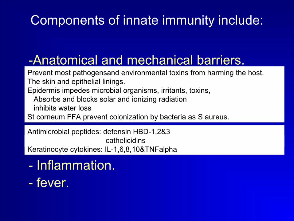
Components of innate immunity include:
-Anatomical and mechanical barriers.
-Pattern recognition receptorsPRR that recognize PAMP.
-Antigen-nonspecific defense chemicals. -The alternative complement pathways. - Phagocytosis. - Inflammation. - fever.
Prevent most pathogensand environmental toxins from harming the host.The skin and epithelial linings.Epidermis impedes microbial organisms, irritants, toxins, Absorbs and blocks solar and ionizing radiation inhibits water lossSt corneum FFA prevent colonization by bacteria as S aureus.
Activated by microbial surface without antibody formationAntimicrobial peptides: defensin HBD-1,2&3 cathelicidinsKeratinocyte cytokines: IL-1,6,8,10&TNFalpha

Anti microbial peptides
• Secreted by the human epithelia including the epidermis (Keratinocytes) to exhibit the capacity of an innate chemical defense.
• Antiliukoproteases.
• Dramcidin (SWEAT GLANDS)
• Human B defensin 1-4.
• Lysosyme, Psoriasin (Sebocytes), RNase7,LL-37/hCAP18 (urogenital tract).
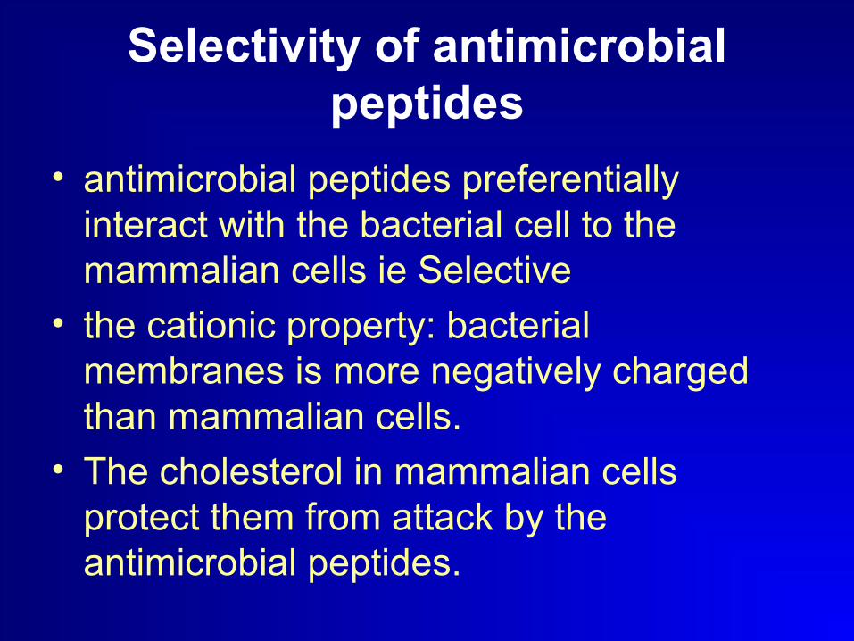
Selectivity of antimicrobial peptides
• antimicrobial peptides preferentially interact with the bacterial cell to the mammalian cells ie Selective
• the cationic property: bacterial membranes is more negatively charged than mammalian cells.
• The cholesterol in mammalian cells protect them from attack by the antimicrobial peptides.

Induction and Actions • Induced by bacterial and proinflammatory
products or cytokines as well as toll like receptor stimulation.
• Broad spectrum anti bacterial and variable anti fungal and antivrial activities.
• Disrupting membranes, interfering with metabolism, and targeting cytoplasmic components
• Attract immature dendritic cells and memory T cells via CCR6 role in adaptive immunity

IDENTIFICATION OF THE INVADOR
• On the organism:
Pathogen associated molecular patterns (PAMPS),
• On the effector cells:
Pattern recognition receptors
(PRP).
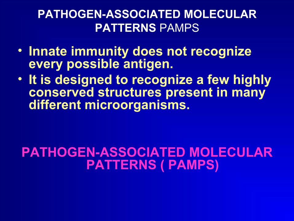
PATHOGEN-ASSOCIATED MOLECULAR PATTERNS PAMPS
• Innate immunity does not recognize every possible antigen.
• It is designed to recognize a few highly conserved structures present in many different microorganisms.
PATHOGEN-ASSOCIATED MOLECULAR PATTERNS ( PAMPS)

1. Must be shared by large groups of pathogens and thus must represent general patterns rather then specific structures.
2. Must be conserved products of microbial metabolism which are not subject to antigenic variability.
3. pathogens cannot "change" them because they are essential for the survival or pathogenicity of the microorganisms. Any attempts to change them could be lethal to the microbe or render it nonpathogenic.
4. The recognized structures must be absolutely distinct from self-antigens. The major consequence of this requirement is the ability of the innate immune system to discriminate between self and non-self.
PATHOGEN-ASSOCIATED MOLECULAR PATTERNS PAMPS (CONT)
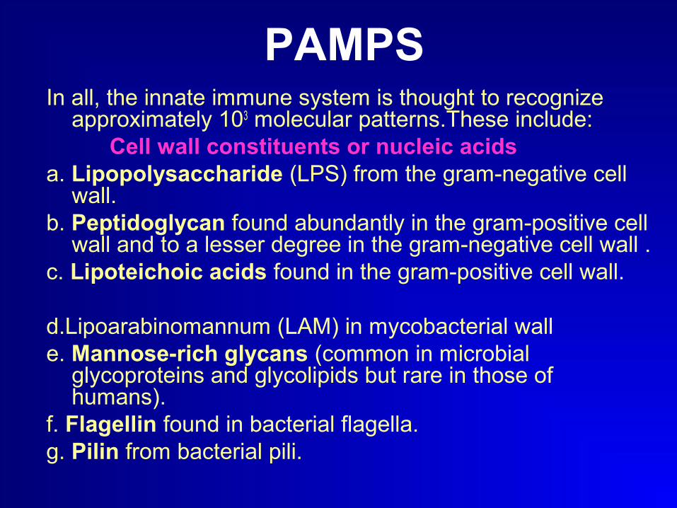
In all, the innate immune system is thought to recognize approximately 103 molecular patterns.These include:
Cell wall constituents or nucleic acidsa. Lipopolysaccharide (LPS) from the gram-negative cell
wall.b. Peptidoglycan found abundantly in the gram-positive cell
wall and to a lesser degree in the gram-negative cell wall .c. Lipoteichoic acids found in the gram-positive cell wall.
d.Lipoarabinomannum (LAM) in mycobacterial walle. Mannose-rich glycans (common in microbial
glycoproteins and glycolipids but rare in those of humans).
f. Flagellin found in bacterial flagella.g. Pilin from bacterial pili.
PAMPS
PAMPS (CONT)
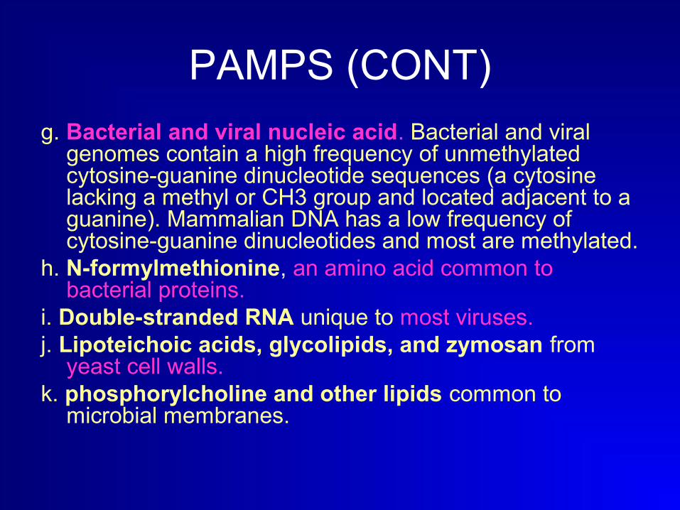
g. Bacterial and viral nucleic acid. Bacterial and viral genomes contain a high frequency of unmethylated cytosine-guanine dinucleotide sequences (a cytosine lacking a methyl or CH3 group and located adjacent to a guanine). Mammalian DNA has a low frequency of cytosine-guanine dinucleotides and most are methylated.
h. N-formylmethionine, an amino acid common to bacterial proteins.
i. Double-stranded RNA unique to most viruses. j. Lipoteichoic acids, glycolipids, and zymosan from
yeast cell walls.k. phosphorylcholine and other lipids common to
microbial membranes.
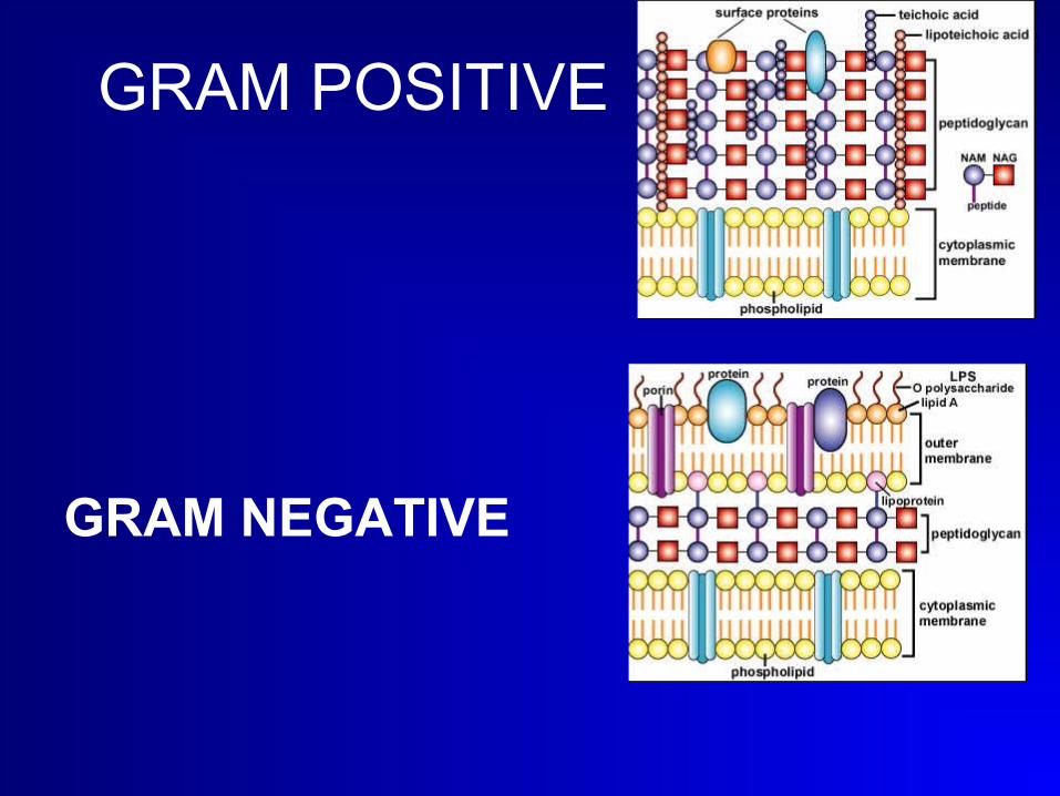
GRAM POSITIVE
GRAM NEGATIVE
Pattern-recognition Receptors(PRR)
To recognize these microbial molecules, various body defense cells have on their surface a variety of receptors called
Pattern-recognition Receptors capable of binding specifically to
PAMPS
Pattern Recognition Receptors
• The cornerstone of the innate immune system is comprised of germline-encoded receptors (not specific recognition of individual pathogens, but rather a coarse-grain encoding of the
species).
• These PRRs are activated upon recognition of “Pathogen-Associated Molecular Patterns” or PAMPs.

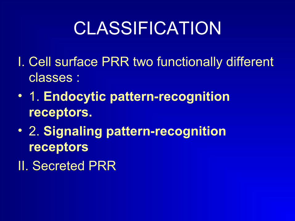
CLASSIFICATION
I. Cell surface PRR two functionally different classes :
• 1. Endocytic pattern-recognition receptors.
• 2. Signaling pattern-recognition receptors
II. Secreted PRR
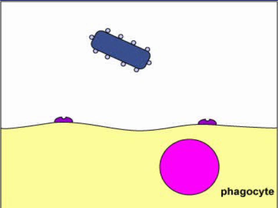
ENDOCYTIC PATTERN-RECOGNITION RECEPTORS
Found on the surface of phagocytes and promote the attachment of microorganisms to phagocytes and their subsequent engulfment and destruction.

Signaling pattern-recognition receptors
• Binding of microbial molecules to the receptor promotes the synthesis and secretion of intracellular regulatory molecules such as cytokines that is crucial to initiating innate immunity and adaptive immunity.
• Toll-like receptors and• CD14. • NOD (nucleotide-binding oligomerization
domain proteins)

Toll-like receptors
• A series of Signaling pattern-recognition receptors known as toll-like receptors (TLRs) play a major role in innate immunity and the induction of adaptive immunity.

THE TOLL
• Mamalian TLRs owe their name to a closely related receptor called Toll, first identified in Drosophila in 1988.
• TOLL: german word of fantastical or strange.
• They recognize and bind to PAMPS (conserved molecular components associated with microorganisms)

TLRs• TLRs belong to the IL-1 receptor family.
• Defined by the presence of extracellular leucine-rich repeats and an intracellular Toll/interleukin-1 receptor domain.
• Linked to a signaling pathway that involves the IL-1-receptor kinase and NF- B.
Toll like receptors
Upon binding of the extracellular ligand recognition domain to specific PAMPs, changes in the intracellular domain result in initiation of signaling events leading to
• Inflammatory responses and/or
• Release of antimicrobial agents.


Toll-Like Receptors Responding to Lipopolysaccharide (LPS)
from the Gram-Negative Cell Wall
CD14• CD14 is found on monocytes, macrophages,
and neutrophils.• promotes the ability of TLR-4 to respond to LPS
and peptidoglycan. Which causes secretion of proinflammatory cytokines such as IL-1, IL-6, IL-8, TNF-alpha.
• These cytokines then bind to cytokine receptors on target cells and initiate inflammation and activate both the complement pathways and the coagulation pathway
NOD (nucleotide-binding oligomerization domain) proteins
• NOD1 and NOD2 • allow intracellular recognition of
peptidoglycan components released during growth or phagocytosis ( muramyl dipeptide)
• Binding of the muramyl dipetides to NOD1 or NOD2 leads to the activation of genes coding for proinflammatory cytokines.
Secreted pattern recognition receptors
• Secreted pattern-recognition receptors. These bind to microbial cell walls and enable them to be recognized by the complement pathways and phagocytes.
• Eg mannan-binding lectin is synthesized by the liver and released into the bloodstream. MBL recognizes carbohydrate patterns, found on the surface of a large number of pathogenic micro-organisms, including bacteria, viruses, protozoa and fungi.

Signaling pathways - NF-kB pathway It is not known whether TLRs can productively couple to
the IL-1R signaling machinery or whether a parallel set of proteins is used. - A number of specific molecules are known to be involved.
• adaptor molecules such as the myeloid differentiation factor 88 MyD88,
• Toll/IL-1R (TIR) domain containing adaptor protein (TIRAP), and TIR domain-containing adapter inducing interferon (TRIF). and TRAFs (TNF receptor associated factors).
• Other key signaling proteins include IL-1 receptor associated kinases (IRAKs) such as IRAK1, 2, and 4, transforming growth factor kinase (TAK-1), IkB kinases (IKKs),

INNATE IMMUNITY ADAPTIVE IMMUNITY
Trigger PAMP Specific antigens
Action Min-hours Days to weeks
Receptors PRR as TLR TCR,BCR
Memory No Yes
Communications Cytokines and chemokines
Effectors ComplementAntigen presentationPhagocytosis
Complement Antigen presentationAntibodiesCytotoxicity

MEMBERS OF THE TRLs FAMILY
• The first characterized member of the mammalian family of TLRs was TLR4 which was shown to trigger the pro-inflammatory NF-kB pathway upon binding to LPS with the assistance of CD14 molecule.
• The completion of the human genome project led to the identification of numerous putative TLRs in the genome.
• These TLRs differ from each other in ligand specificities, expression patterns, and target genes they induce.
• Different combinations of TLRs appear in different cell types.
• Seem to appear in pairs.

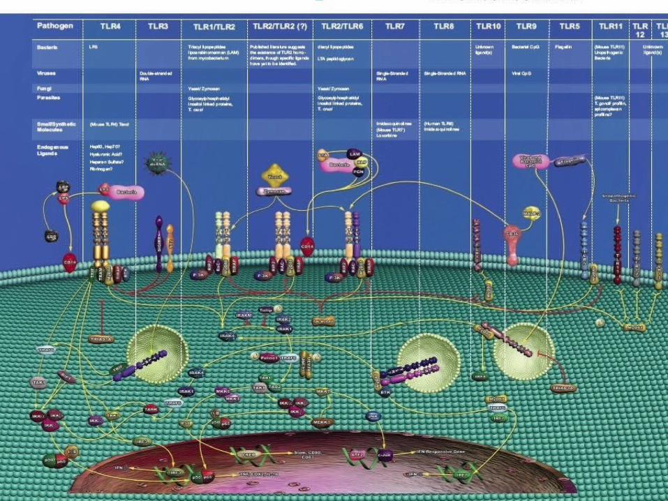
Different TLRs
• 1. TLRs found on cell surfaces: a. TLR-1/TLR-2 pairs bind uniquely bacterial lipopeptides and
glycosylphosphatidylinositol (GPI)-anchored proteins in parasites;b. TLR-2/TLR-6 pairs bind lipoteichoic acid from gram-positive cell walls and zymosan from fungi;c. TLR-2 plays a role in binding peptidoglycan fragments (glycopeptides);d. TLR-4/TLR-4 pairs bind lipopolysaccharide from gram-negative cell walls;e. TLR-5* binds bacterial flagellin;
• 2. TLRs found in the membranes of the endosomes used to degrade pathogens:
• a. TLR-3* binds double-stranded viral RNA;b. TLR-7* binds uracil-rich single-stranded viral RNA such as in HIV;c. TLR-8* binds single-stranded viral RNA;d. TLR-9* binds unmethylated cytosine-guanine dinucleotide sequences (CpG DNA) found in bacterial and viral genomes.
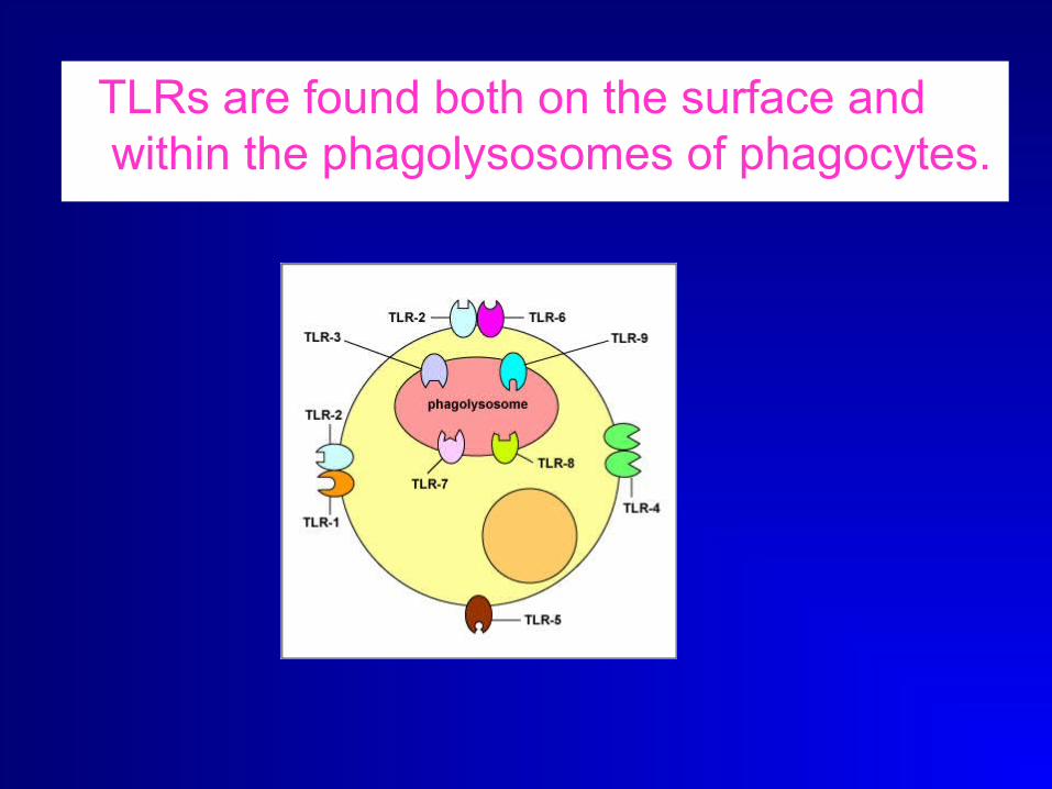
TLRs are found both on the surface and within the phagolysosomes of phagocytes.
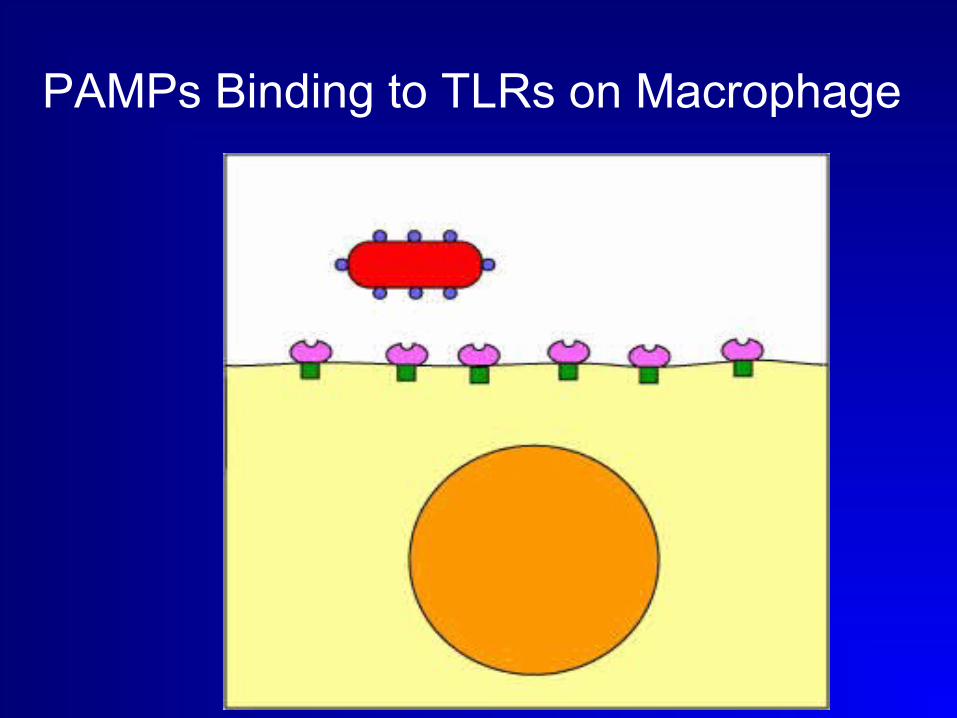
PAMPs Binding to TLRs on Macrophage

The TLRs found in the membranes of the phagosomes Recognizing Viral Double-Stranded RNA after phagocytosis

ROLE IN INNATE IMMUNITY• The binding of a microbial molecule to its TLR transmits
a signal to the cell's nucleus inducing the expression of genes coding for the synthesis of cytokines.
• Many of the TLRs, especially those that bind to bacterial and fungal cell wall components stimulate
IL -1, TNF-alpha, and IL-8. trigerring innate immune defenses such as inflammation, fever, and phagocytosis in order to provide an immediate response against the invading microorganism.
• Most of the TLRs that bind to viral components trigger the synthesis of interferons that block viral replication within infected host cells.
• Cytokines such as interleukin-6 (IL-6) that promotes B-lymphocyte activity and interleukin-12 that promotes T-lymphocyte activity are also produced.
Role in adaptive immunity
• TLRs trigger various secondary signals needed for
- humoral immunity (the production of antibodies).
-cell-mediated immunity (the production of cytotoxic T-lymphocytes and additional cytokines).
• Without innate immune responses there could be no adaptive immunity.
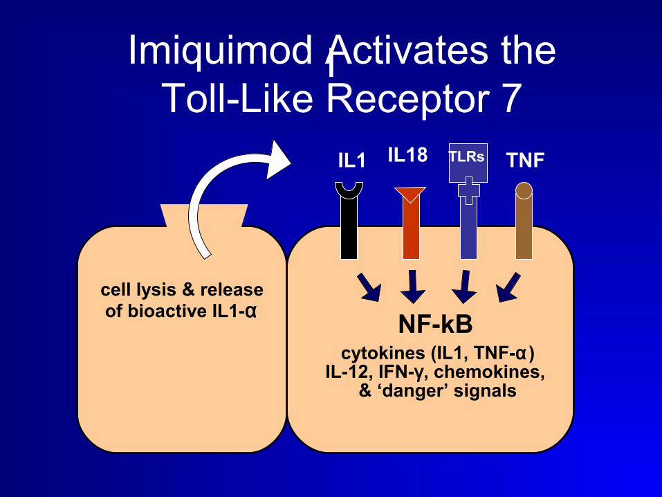
I
cell lysis & releaseof bioactive IL1-α
IL1 IL18 TLRs TNF
NF-kBcytokines (IL1, TNF-α)
IL-12, IFN-γ , chemokines, & ‘danger’ signals
Imiquimod Activates theToll-Like Receptor 7
1) These antigens activate B-lymphocytes by binding to their specific toll-like receptors rather than to B-cell receptors.
2) allow B-lymphocytes to mount an antibody without the requirement of interaction with T-lymphocytes.
3)TI antigens are PAMPS such as lipopolysaccharide (LPS) and bacterial nucleic acid.
4)Antibody molecules generated against TI antigens are often called "natural antibodies" because they are always being made against bacteria present in the body. IgM isotype and do not give rise to a memory response.
T-independent (TI) antigens and humoral immunity:
The Complement system
• a biochamical cascade of the immune system that helps, or “complements”, the ability of antibodies to clear pathogens or mark them for destruction by other cells.
• The cascade is composed of many plasma proteins, synthesized in the liver.

• Activated by three pathways:
1) Classical: antigen antibody complex.
2) Alternative pathway: polysaccharides of microbial cell walls.
3) Lectin pathway: by the binding of the microbial carbohydrates with mannose binding lectin.
• Innate immune response uses 2 and 3
• Activation of C3 and starts the cascade.
The Complement system

The Complement system• The proteins work together to:
-trigger the recruitment of inflammatory cells,
-"tag" pathogens for destruction by other cells, and enhances phagocytosis.
- Disrupt the plasma membrane of an infected cell.
• Rrid the body of neutralized antigen-antibody complexes.
• Normal cells are less sussceptible to desruction by complement .
Cytokines • Substances secreted by various cell types to
interact with other cells to produce ceratain actions.
• Interlukins (interaction between WBCs), interferons (antiviral activities)
• colony stimulating factors induce differentiation and proliferation of hematopoeitic progenitor cells.
• Chemokines,(Inflammatory and lymphoid) chemoattraction ability.
Cytokines of the innate immune system
• Mainly cytokines withi nflammatoryand antivral capacities.
- IL-6,IL-1and TNF alpha and inflammatory chemokines.
- IFN alpha and IFN beta.
Macrophages
• Expresses PPR to identify organisms.• Receptors for antibodies and complement that
enhances phagocytosis.• Destroy the organisms by toxic intracellular molecules
as:superoxide anions, hydroxyl radicals,nitrous oxide, etc.
• Antigen presenting capacity (much less than Langerhan’s cells)
• Releases G-CSF, AND GM CSF that stimulate the devision and release of neutrophils from the bone marrow.
Neutrophils• The enter the blood stream to enter the site
of infection through the complex effect of proinflammatory mediators,adhesion molecules chemoattractants and chemokines.
• Phagocytosis is enhanced by coating the organism by antibodies and complement that bind to their receptors on the neutrophils
• Phagocytose and kills the organisms by oxygen (H peroxide and hydroxyl radicals) dependant or independent mechanisms (myeloperoxidase or lysosyme).

Eosinophils • Protective agains parasites.
• Antibodies bind the parasite and then the eosinophils
• Weak phagocytic activity.
• Release toxic substances to the parasite as major basic ptn, Eosinophil cataionic protein, E peroxidase Eneurotoxin,
• Prostaglandins leukotrienes and cytokines.
• Important in allergic reactions.
Basophil and mast cells• Basophil in the blood and mast cell in the
tissues.
• Only cutaneous mast cells express receptors for the anaphylatoxin C5 a so when activated by binding antigen specific antibodies only a local reaction occur.
• Palys an important role in immediate allergic reaction, urticaria and angioedema.
• Mast cells are involved in TLR-mediated responses against Gram-negative bacteria.

Natural killer cells• Pattern recognition receptors (TLR 3,9) it
can identify and eliminate infected or malignant cells.
• Adhere and kill targets coated with IgG as they carry receptors for it (antibody dependant cellular cytotoxicity ADCC).
• Activation of killer activating receptors that recognize the abnormal cells and kills them by secreting perforin and injecting granzyme that kills the cells by inducing apoptosis.
Natural killer cells
• MHC shuts off the killer signals.
• Tumour cells and viral infected cells are vulnerable.as they sometimes mask MHC to escape cytotoxic T cells
inflammation• One of the first responses of the immune
system to infection or irritation.
• Inflammation is stimulated by chemical factors released by injured cells and cells of the innate immune syste and serves to establish a physical barrier against the spread of infection, and to promote healing of any damaged tissue following the clearance of pathogens.[3]

Inflammation
• Chemical factors produced during inflammation (histamine, bradykinin, leukotrienes, serotonin and prostaglandin, sensitize pain receptors, cause vasodilatation at the scene, and attract phagocytes, especially neutrophils.[

INNATE IMMUNITY ADAPTIVE IMMUNITY
Trigger PAMP Specific antigens
Action Min-hours Days to weeks
Receptors PRR as TLR TCR,BCR
Memory No Yes
Communications Cytokines and chemokines
Effectors ComplementAntigen presentationPhagocytosis
Complement Antigen presentationAntibodiesCytotoxicity
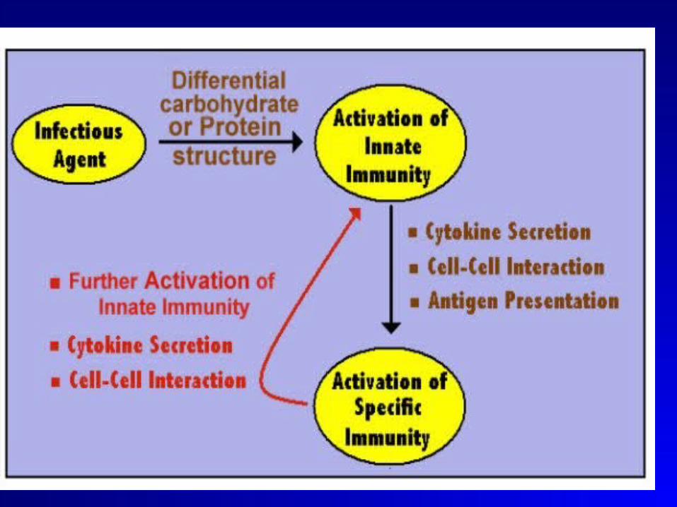
interactions between the innate and adaptive immune
• The interactions between the innate and adaptive immune systems are crucial to promote proinflammatory reactions against pathogens and to ensure maintenance of vital self-tolerance.
• TLRs are expressed on both innate and adaptive immune cells and are critically involved in this interplay.
• TLR-stimulated dendritic cells induce specific T cells to differentiate into TH-1, TH-2 or T-reg

Adaptive immunity
• Cells of the innate immune system effectively prevent free growth of bacteria within the body.
• however, many pathogens have evolved mechanisms allowing them to evade the innate immune system and generates a threshold level of antigen which triggers .
the adaptive immune system include:
Functions of the adaptive immune system
• the recognition of specific “non-self” antigens in the presence of “self”, during the process of antigen presentation.
• the generation of tailored responses to eliminate specific pathogens.
• the development of immunologic memory in which each pathogen is “remembered” by a signature antibody. These memory cells can be called upon to quickly eliminate a pathogen on subsequent infections .

Adaptive immune system
• The cells of the adaptive immune system are
• B cells and T cells are the major types of lymphocytes.
• The process starts by antigen presentation.
• Adaptive immunity relies on the capacity of immune cells to distinguish between the body's own cells and unwanted invaders
With the exception of non-nucleated cells all cells are capable of presenting antigen and of activating the adaptive response.
- Some cells are specially equipped to present antigen, and to prime naive T cells and are termed professional (APC).
- Dendritic cells and B-cells (and to a lesser extent macrophages. are equipped with special receptors that allow for enhanced activation of T cells.
Antigen presenting cells ( APC)

LNGERHAN’ S CELLS
• Dentritic Cells of the epidermis.
• Expresses CD1, S100 ptn, Vimentin, Birbeck granules associated molecule (langerin) and MHC II.
• Derived from the bone marrow from CD34 precursor cells.
• Factors behind Dcs stimulation of either Th1 or Th2 is still not determined but immature dendtritic cells induce tolerence through activation of Treg .


Antigen presentation
• Resident Langerhan’s cell engulf’s the exogenous antigen or express the edogenous one
• Starts emigration to the lymph nodes to meet the T cells .
• During this trip it develops some changes to become similar to mature Dendritic cell.
Changes of LCs during migration
- Decrease molecules involved in antigen uptake as birbeck’s granules,Fc receptors and those mediating the attachement to neighboring keratinocytes ( E-Cadherins).
- Increase expression of receptors involved in tissue homing at the lymph nodes as CD44. and surface molecules necessary for antigen presentation and Tcell priming asCD40,54,58,80,86

Antigen presentation
• APC to B cells and T cells is not the same.
• B cells can identify the whole antigen by antibodies on thier surface
• T cells only identify the antigen when processed into peptides bound to specific surface molecules on APC.

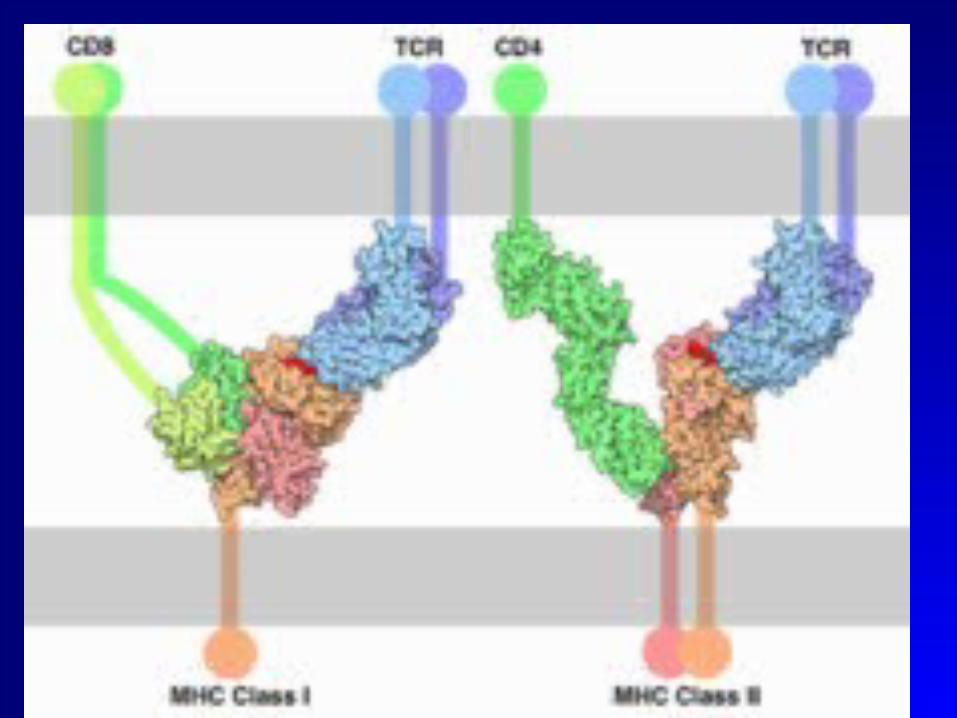
AP TO T CELLS
• Tceels identify the processed antigen bound to MHC on the surface of Dendritic cells.
• CD4 T cells identify antigens bound to MHC II while Cytotoxic CD8 T cells identify antigen T cells bound to MHC II
• Exogenous and endogenous antigen presentation.

TYPES OF AP to T cells
• Exogenous antigens are engulfed by the APC, processed and presented in association with MHC II.
• Endogenous antigens (VIRUS AND TUMOURS) are processed and presented in association with MHC I

T cells• T cells develop and mature in the thymus
after migration of the stem cells from the bone marrow.
• At the thymus only T cells that can recognize foreign and not self antigen in the MHC peptide complex get a survival signal (positive selection) and pass to the circulation and lymphnodes..
• Those who fail have affinity to self antigens receive signals for apoptosis (negative selection) thus no auto attack.
•
Types of T cells
• Immature T cells Express both CD4 and CD8 molecules.
• Later with the development of the Tcell receptor, they either express CD4 and become T helper cell that binds antigens in MHCII or express CD8 molecule and becomes T cytotoxic cell that binds antigens on MHCI.

The T CELL RECEPTOR (TCR)• It is the part responsible for recognition
of the specific antigen and the further T cell response.
• TCR are transmembrane molecules that are mainly of the α/β type while only 10% are of the γ/δ type in body and skin.
γ/δT cells
• Do not follow the classic way of antigen recognition may be able od direct antigen recognition.
• May play a role in innate immunity.
• Increase in the skin in leprosy and lieshmaniasis.
TCR
• Can recognize a huge number of antigens encoded by more than 400 genes thet are modified and rearranged to cover an endless number of antigens by recombination activation genes
• Rag-1 Rag-2 .
• when deffective ----combined immune defficiency.
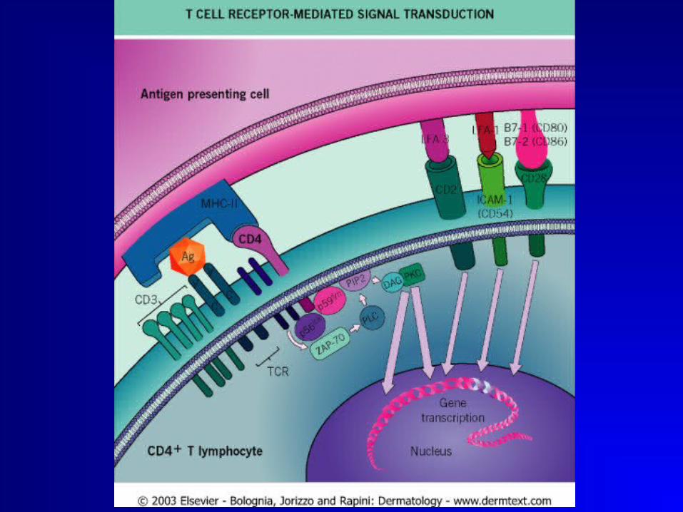
TCR SIGNALLING
• CD3 is an important part of TCR responsible for transmission of the signal to the cell that encodes for the cytokine needed to stimulate the required response for that particular antigen.

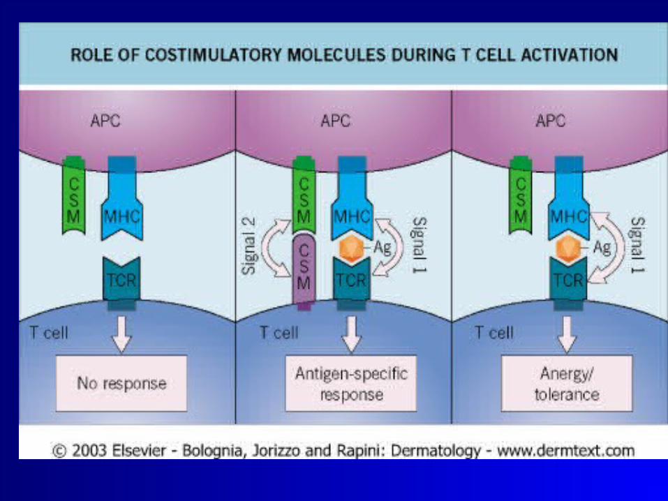
Costimulatory molecules
• B 7 FAMILY
• CYTOKINES (TNF IL-1 AND IL-6)
• CELL ADHESION MOLECULES ARE VERY IMPORTANT FOR COMPLETION OF THE T CELL RESPONSE OTHER WISE ANERGY AND FAILURE OF T CELL STIMULATION OCCURS.
CLONAL EPANSION
• After proper antigen presentation and costimulation T cell becomes activated .
• Memory T cells develop
Memory T cells
- Central memory T cells: at the lymphnode, CD45RO+CD45RA- CCR7+ Have no effector function. They stimulate dendritic cells to produce il-12 upon secondary stimulation and differentiate into ccr7- cells.
- Effector memory Tcells (TEM): CD45RO+CD45RA- CCR7- develop receptors to migrate to the inflamed tissues (e.g CLA in the skin). Have an effector function.

Skin T cells
• Majority is :
• In the dermis.
• CD4 OR CD8.
• α/β TCR.
• memory phenotype CD45RO+CD45RA-
• Skin homing receptor CLA(cutaneous lymphocyte associated antigen).
Effector T cell function
• After recognition of the antigen
• CD4: T helper cells(Th):
-activate the immune system to combat the antigen including both T and B cells.
CD8 T cytotoxic cells(Tc):
Antiviral and anti tumour responses
T helper response
• According to the type of antigen recognized by the Th cells they secrete different cytokine patterns that will further stimulate different parts of the immune system.
• T0 secretes a wide variety if cytokines which then develops into Th1 or Th2 with more restricted type of cytokine secretion.

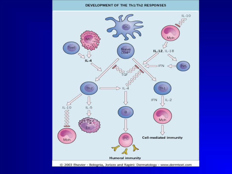
Th 1 cells
• IL-12 stimulates differnetiation of Th0 into Th1 which secretes:
• IL-2: stimulates both Th and Tc proliferation.
• IFN GAMMA: activates macrophages and NK cells and IL-12.
i.e a cell mediated inflammatory response
As in granuloma and autoimmunity.
Th2 cells
• IL-4: Stimulates Bcells to produce IgE and stimulates furhter Th 2 response and inhibits TH1 response.
• IL-5: promotes eosinophil growth.
• IL-6:
• IL-10: inhibits TH1 response.
Mediates humoral immunity.

Th1/Th2 decision
• Very important to achieve the required state if immunity .
• Depends upon the type of antigen presented and the cytokines it stimulates.
• The Dendritic cells, the toll like receptor, Dose of antigen,Genetic background of the host, The APC and its cytokines,The costimulatory molecules.
Th3 cells
• Transforming growth factor B .
• Helps IgA production.
• Supresses both Th1 and Th2 responses.
Tcytotoxic cells CD8
• Direct killing of the organism or the abnormal or infected cells.
• TC1 and TC2 in cytokine pattenrs.
• Viral and antitumour activities.
Regulatory T cells (Tregs)
• CD4 + T cells.
• Secretes large amounts of IL-10.
• Suppressor effect on both immune responses.
• Produced by immature inactive dendritic cells.
• Impotant for tolerance towards self antigens and regulating inflammation.
The B lymphocyte
• B cells function to protect the host by producing antibodies that identify and neutralize foreign objects like bacteria and viruses.
• B Cells are the major cells involved in the creation of humoral immunity.

Antibodies
• Antibodies (or immunoglobulin, Ig), are large Y-shaped proteins used by the immune system to identify and neutralize foreign objects.
• In mammals there are five types of antibody: IgA, IgD, IgE, IgG, and IgM, differing in biological properties, each has evolved to handle different kinds of antigens.
B cells
• Upon activation, B cells produce antibodies, each of which recognizes a unique antigen, and neutralize specific pathogens.[1]
• Like the T cell receptor, B cells express a unique B cell receptor (BCR), in this case, an immobilized antibody molecule. The BCR recognizes and binds to only one particular antigen.

Bcell Vs T cell ACTIVATION
• T cells recognize their cognate antigen in a processed form - as a peptide in the context of an MHC molecule,[1] while B cells recognize antigens in their native form.[
• B- cells receives additional signals from a helper T cell (predominately Th2 type)), to further differentiates into an effector cell, known as a plasma cell]
Plasma cells
• Short lived cells (2-3 days) which secrete antibodies that bind to antigens, making them easier targets for phagocytes, and trigger the complement cascade. [
• About 10% of plasma cells will survive to become long-lived antigen specific memory B cells primed to produce specific antibodies and respond quickly if the same pathogen re-infects the host; while the host experiences few, if any, symptoms.


THANK YOU