Embed Size (px)
Citation preview
Basic Bacteriology

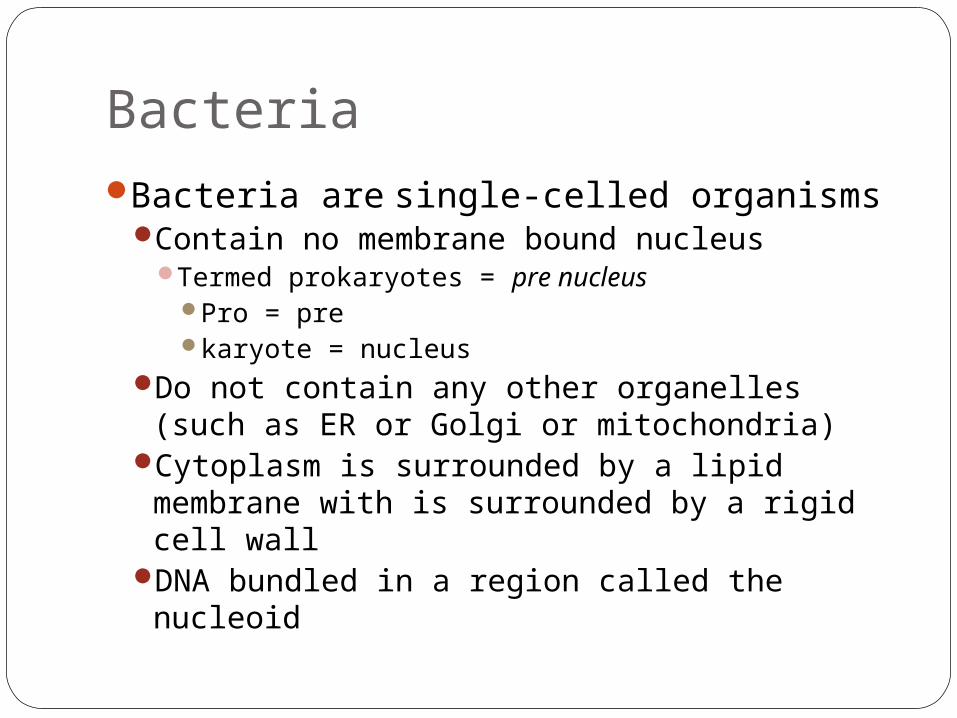
BacteriaBacteria are single-celled organisms
Contain no membrane bound nucleusTermed prokaryotes = pre nucleus
Pro = prekaryote = nucleus
Do not contain any other organelles (such as ER or Golgi or mitochondria)
Cytoplasm is surrounded by a lipid membrane with is surrounded by a rigid cell wall
DNA bundled in a region called the nucleoid
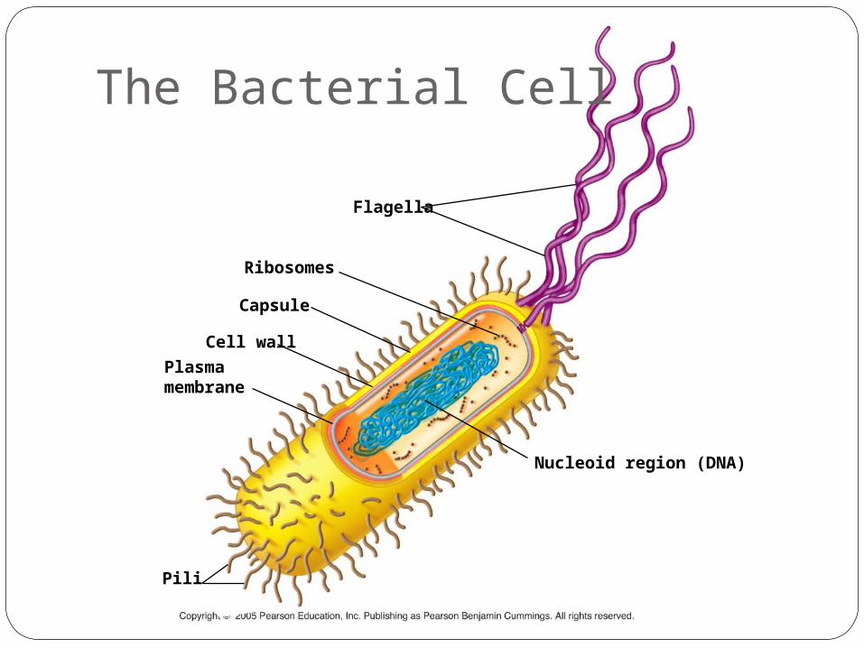
Flagella
Ribosomes
Capsule
Cell wall
Plasmamembrane
Nucleoid region (DNA)
Pili
The Bacterial Cell
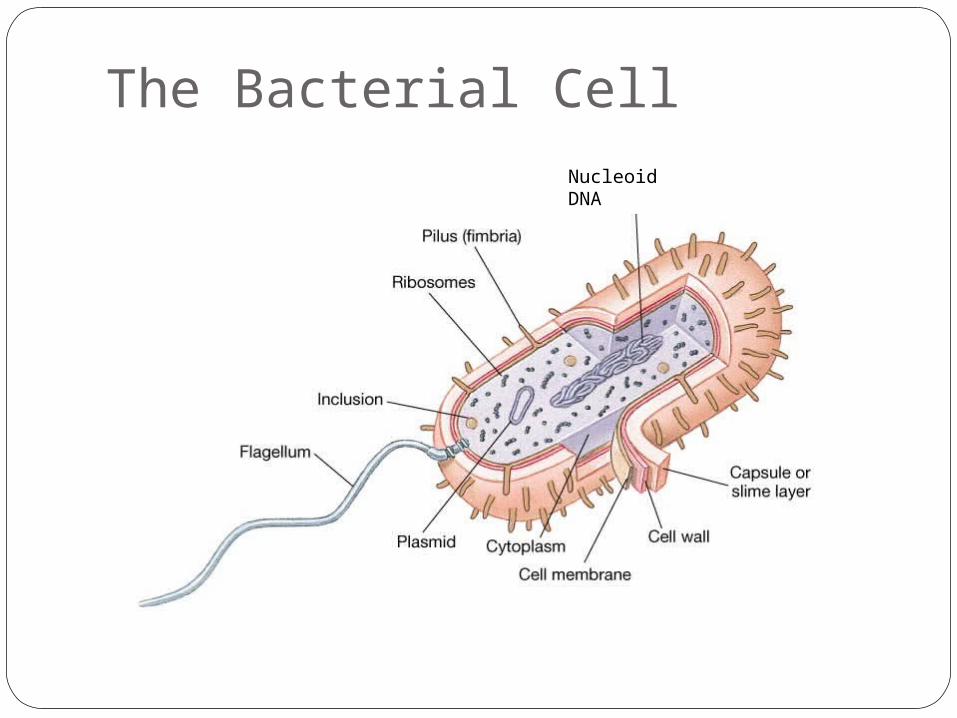
The Bacterial CellNucleoid DNA
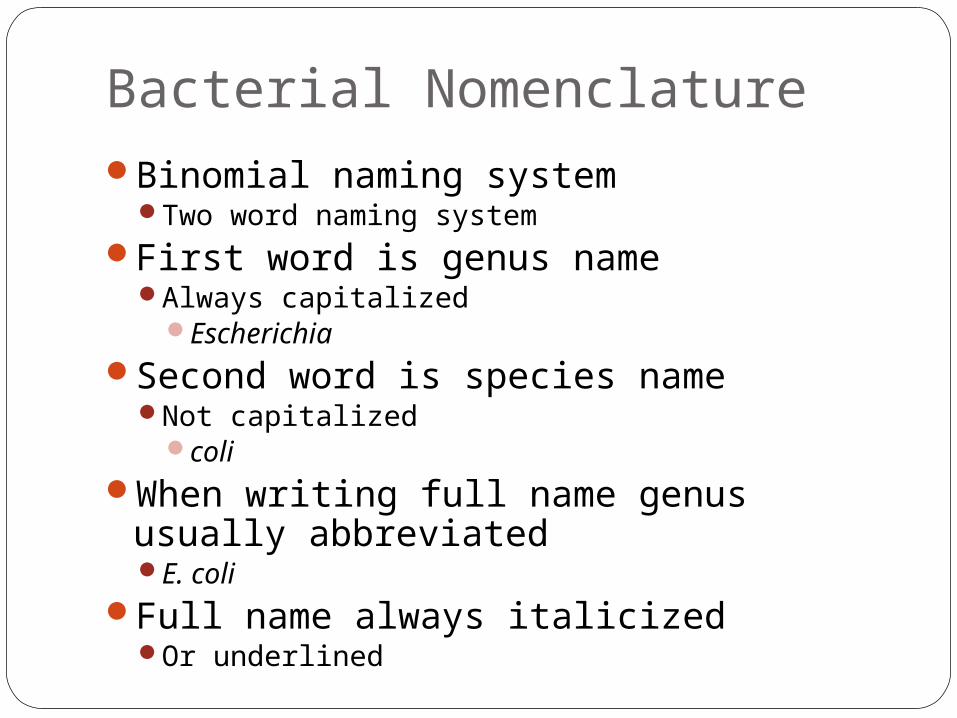
Bacterial NomenclatureBinomial naming system
Two word naming systemFirst word is genus name
Always capitalizedEscherichia
Second word is species nameNot capitalized
coliWhen writing full name genus usually
abbreviatedE. coli
Full name always italicizedOr underlined
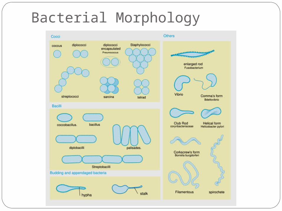
Bacterial Morphology
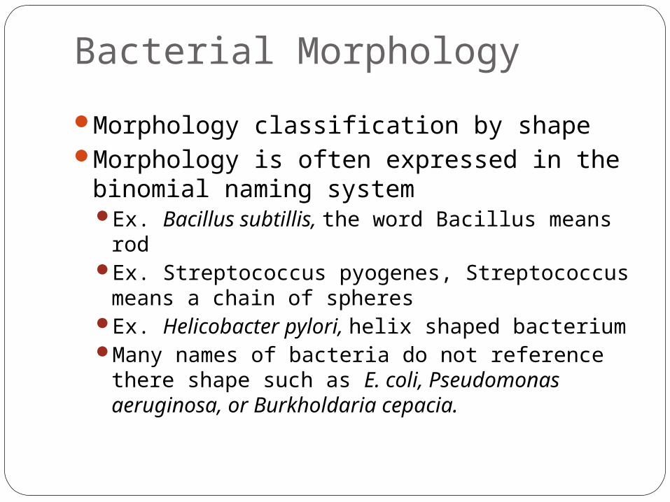
Morphology classification by shapeMorphology is often expressed in the
binomial naming systemEx. Bacillus subtillis, the word Bacillus means
rodEx. Streptococcus pyogenes, Streptococcus
means a chain of spheresEx. Helicobacter pylori, helix shaped bacteriumMany names of bacteria do not reference there
shape such as E. coli, Pseudomonas aeruginosa, or Burkholdaria cepacia.
Bacterial Morphology
Examples of Morphology
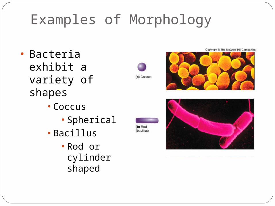
• Bacteria exhibit a variety of shapes
• Coccus• Spherical
• Bacillus• Rod or
cylinder shaped
Examples of Morphology
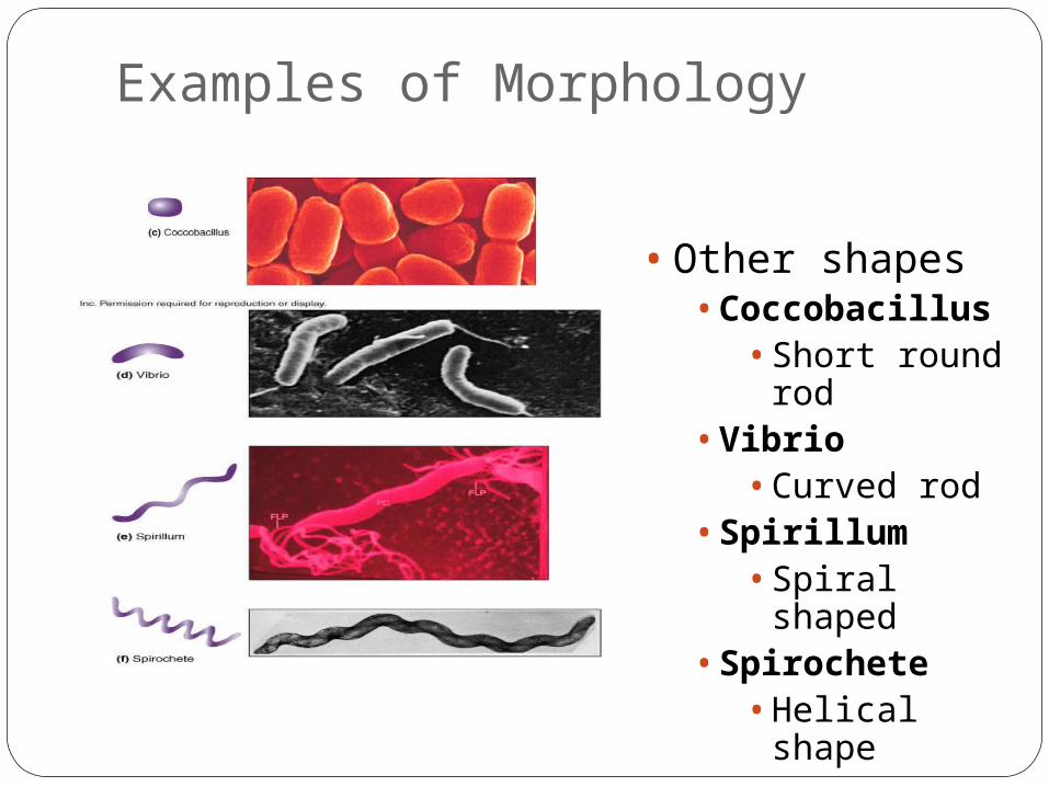
• Other shapes• Coccobacillus
• Short round rod
• Vibrio• Curved rod
• Spirillum• Spiral shaped
• Spirochete• Helical shape
Bacterial Anatomy and Structures
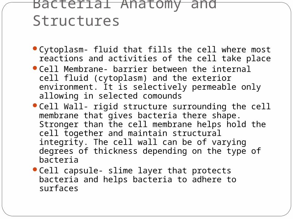
Cytoplasm- fluid that fills the cell where most reactions and activities of the cell take place
Cell Membrane- barrier between the internal cell fluid (cytoplasm) and the exterior environment. It is selectively permeable only allowing in selected comounds
Cell Wall- rigid structure surrounding the cell membrane that gives bacteria there shape. Stronger than the cell membrane helps hold the cell together and maintain structural integrity. The cell wall can be of varying degrees of thickness depending on the type of bacteria
Cell capsule- slime layer that protects bacteria and helps bacteria to adhere to surfaces
Bacterial Anatomy and Structures
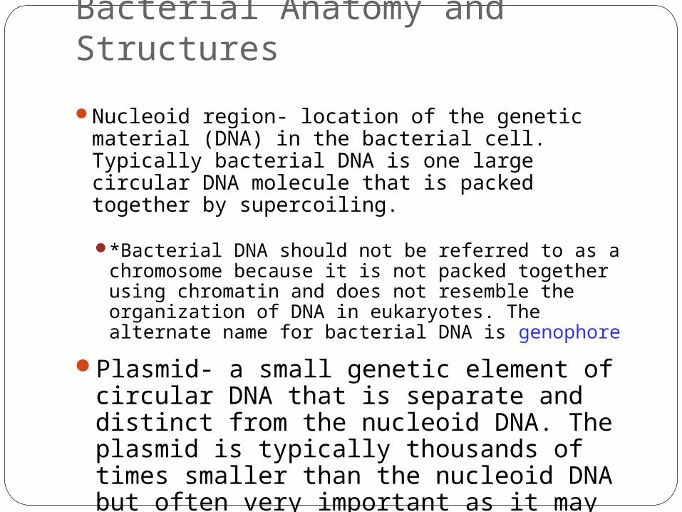
Nucleoid region- location of the genetic material (DNA) in the bacterial cell. Typically bacterial DNA is one large circular DNA molecule that is packed together by supercoiling.
*Bacterial DNA should not be referred to as a chromosome because it is not packed together using chromatin and does not resemble the organization of DNA in eukaryotes. The alternate name for bacterial DNA is genophore
Plasmid- a small genetic element of circular DNA that is separate and distinct from the nucleoid DNA. The plasmid is typically thousands of times smaller than the nucleoid DNA but often very important as it may carry genes for antibiotic drug resistance
Bacterial Anatomy and Structures
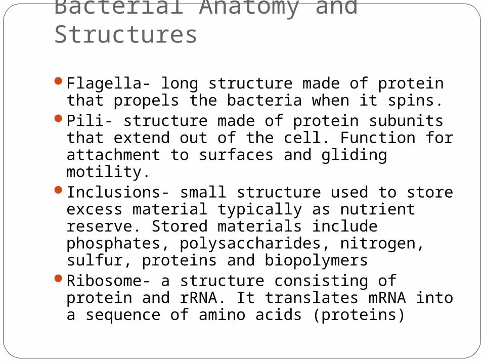
Flagella- long structure made of protein that propels the bacteria when it spins.
Pili- structure made of protein subunits that extend out of the cell. Function for attachment to surfaces and gliding motility.
Inclusions- small structure used to store excess material typically as nutrient reserve. Stored materials include phosphates, polysaccharides, nitrogen, sulfur, proteins and biopolymers
Ribosome- a structure consisting of protein and rRNA. It translates mRNA into a sequence of amino acids (proteins)
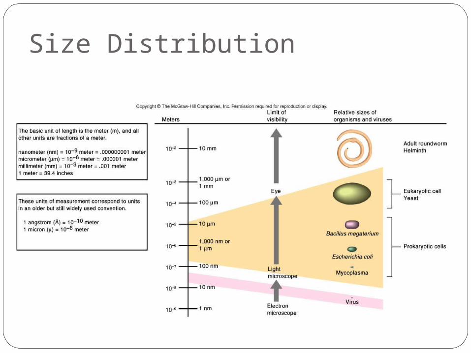
Size Distribution
Size in the Microbial World
Tremendous range in sizeSmallest virus approximately 1/1,000,000th size of
largest eukaryotic cell
Prokaryotic CellsComparing Prokaryotic and Eukaryotic
CellsProkaryote comes from the Greek words
for prenucleus.Eukaryote comes from the Greek words
for true nucleus.
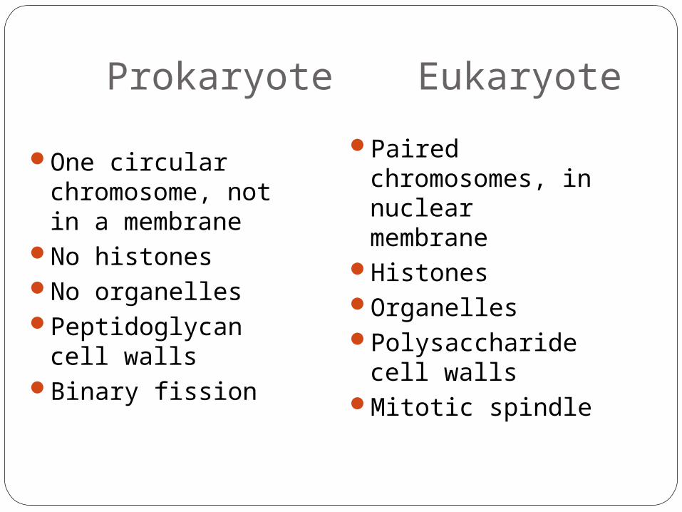
One circular chromosome, not in a membrane
No histonesNo organellesPeptidoglycan cell
wallsBinary fission
Prokaryote Eukaryote
Paired chromosomes, in nuclear membrane
HistonesOrganellesPolysaccharide
cell wallsMitotic spindle
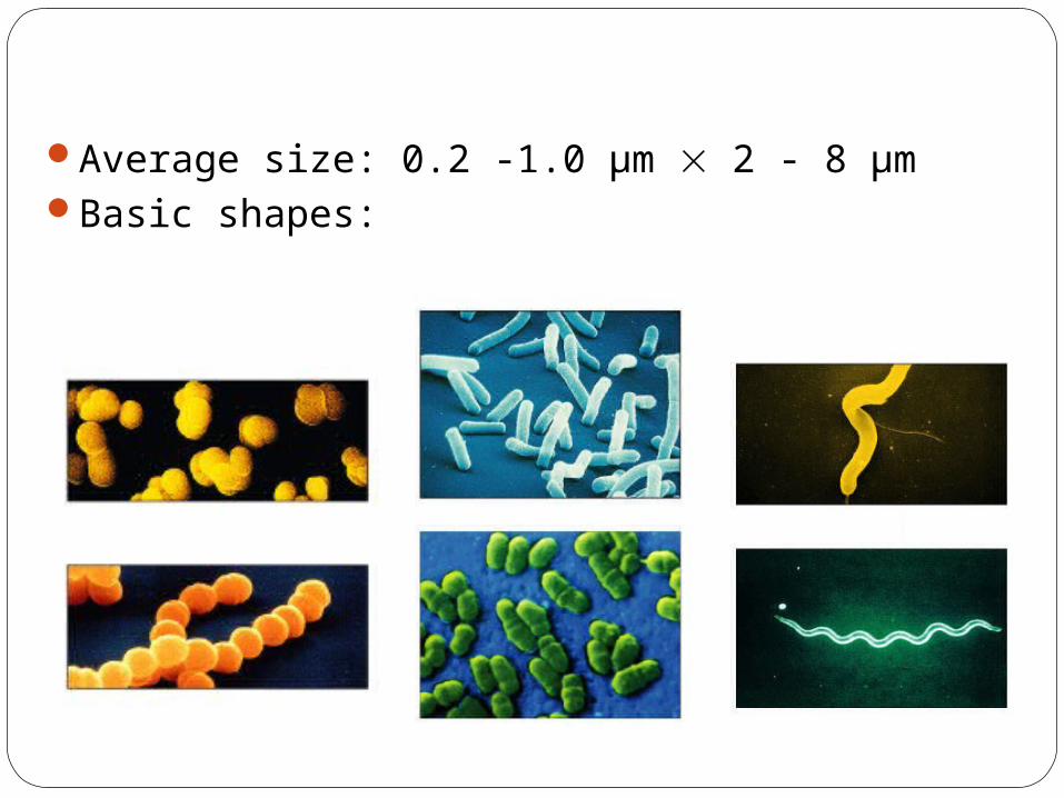
Average size: 0.2 -1.0 µm 2 - 8 µmBasic shapes:
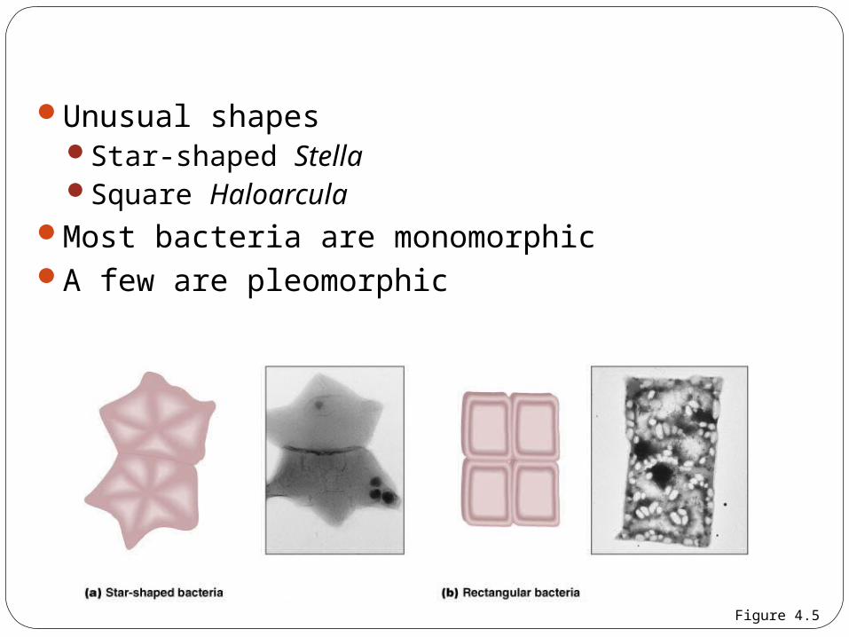
Unusual shapesStar-shaped StellaSquare Haloarcula
Most bacteria are monomorphicA few are pleomorphic
Figure 4.5
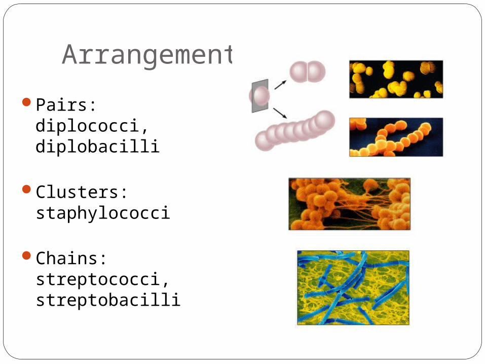
Pairs: diplococci, diplobacilli
Clusters: staphylococci
Chains: streptococci, streptobacilli
Arrangements
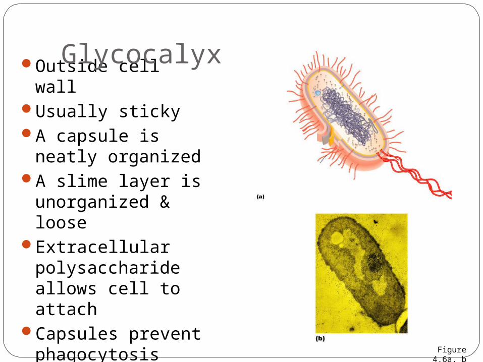
Outside cell wallUsually stickyA capsule is neatly
organizedA slime layer is
unorganized & loose
Extracellular polysaccharide allows cell to attach
Capsules prevent phagocytosis
Glycocalyx
Figure 4.6a, b
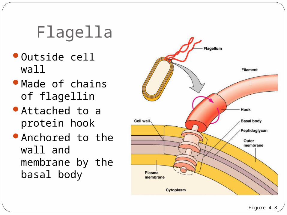
Outside cell wallMade of chains of
flagellinAttached to a
protein hookAnchored to the
wall and membrane by the basal body
Flagella
Figure 4.8
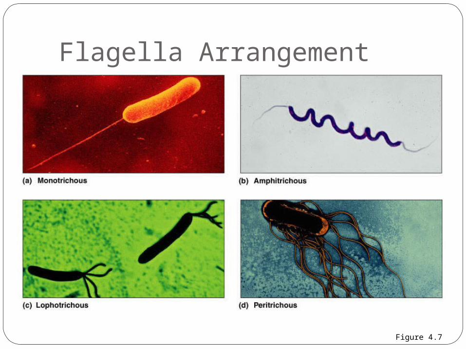
Flagella Arrangement
Figure 4.7
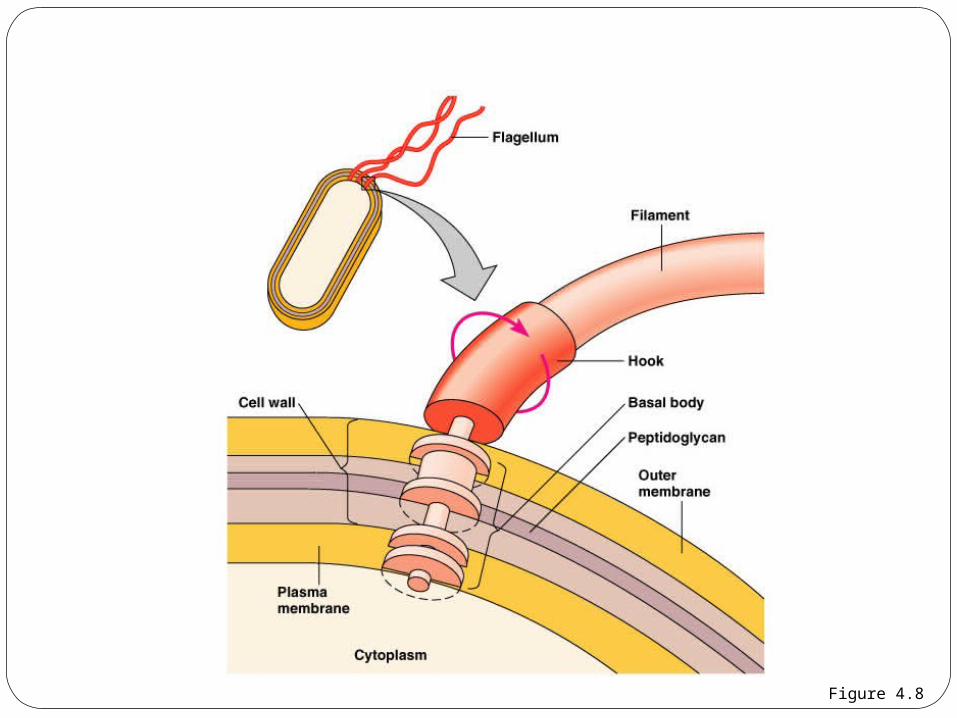
Figure 4.8
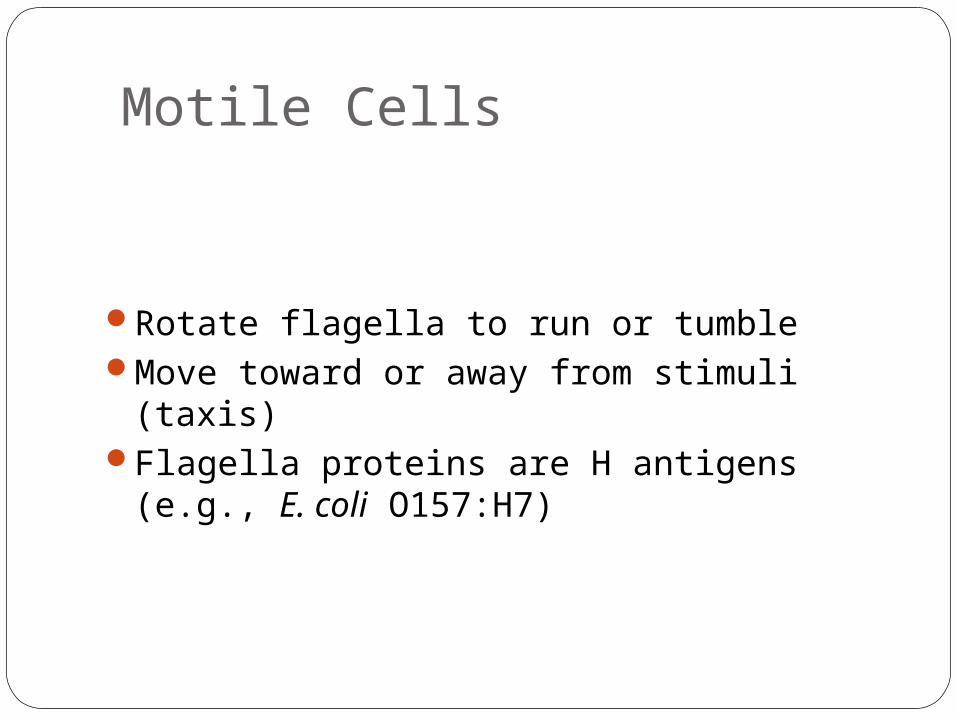
Rotate flagella to run or tumbleMove toward or away from stimuli (taxis)Flagella proteins are H antigens
(e.g., E. coli O157:H7)
Motile Cells
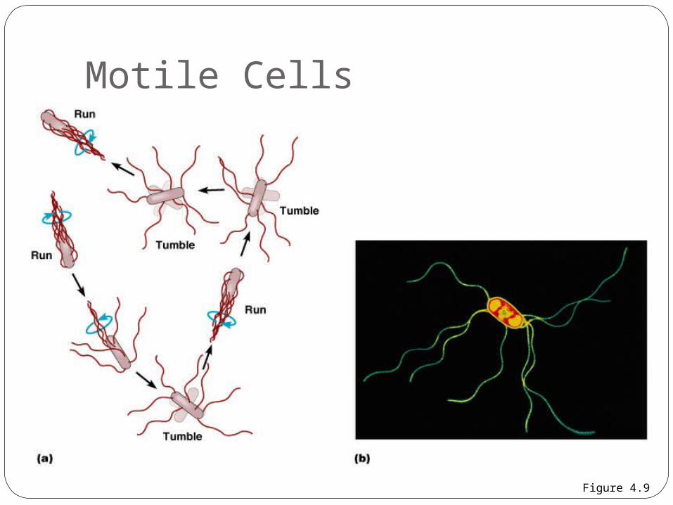
Motile Cells
Figure 4.9
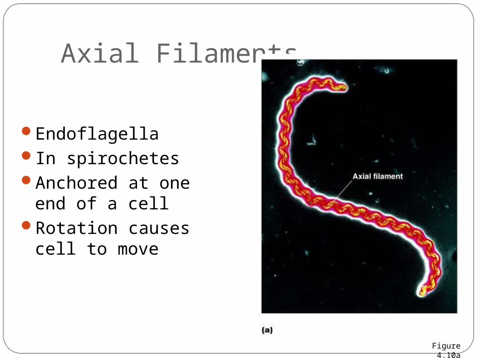
EndoflagellaIn spirochetesAnchored at one
end of a cellRotation causes
cell to move
Axial Filaments
Figure 4.10a
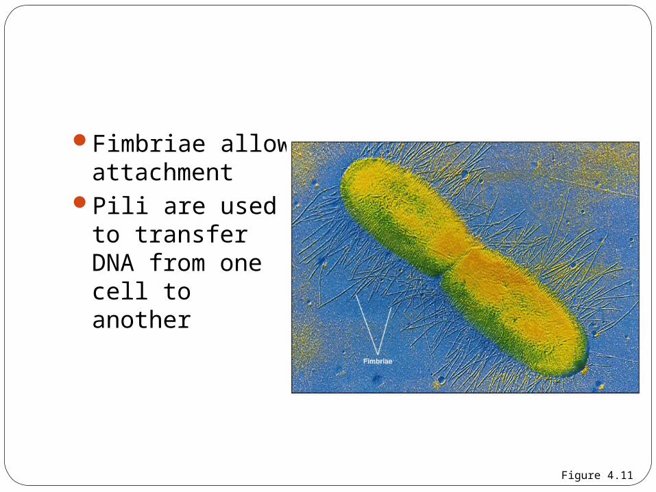
Fimbriae allow attachment
Pili are used to transfer DNA from one cell to another
Figure 4.11
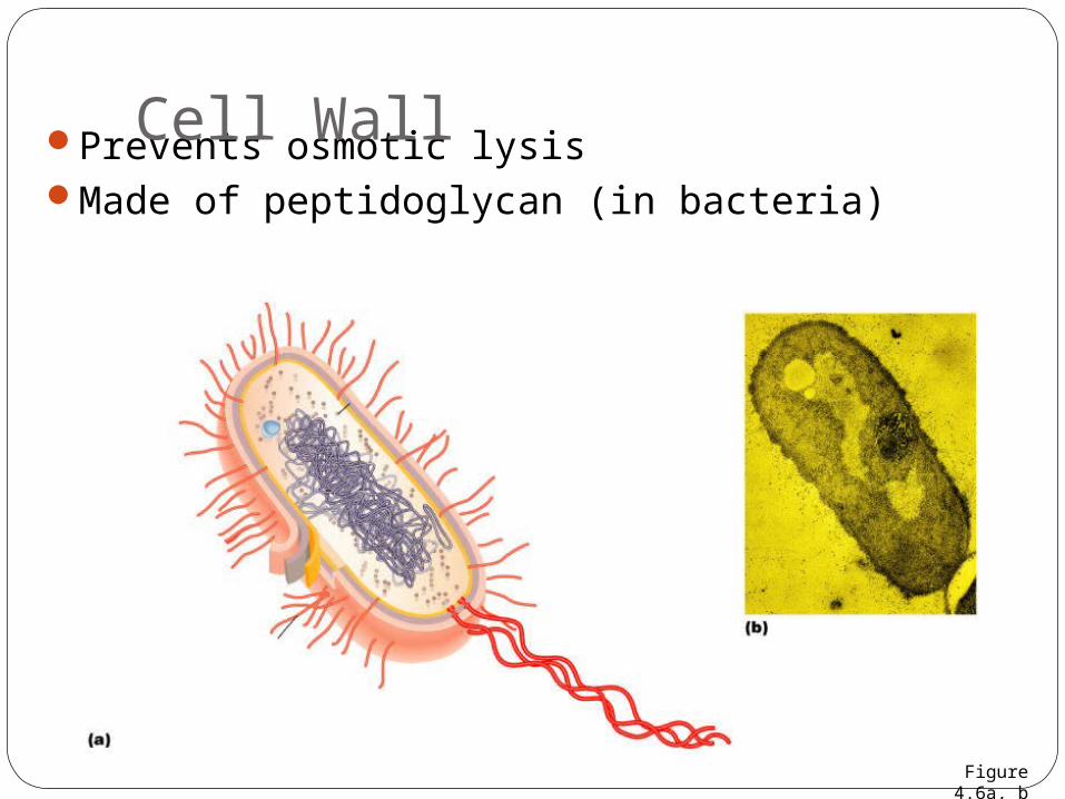
Prevents osmotic lysisMade of peptidoglycan (in bacteria)
Cell Wall
Figure 4.6a, b
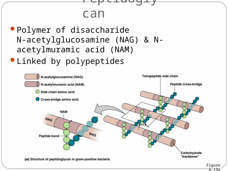
Polymer of disaccharideN-acetylglucosamine (NAG) & N-acetylmuramic acid (NAM)
Linked by polypeptides
Peptidoglycan
Figure 4.13a
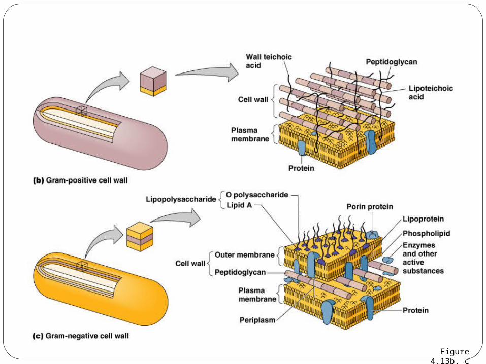
Figure 4.13b, c
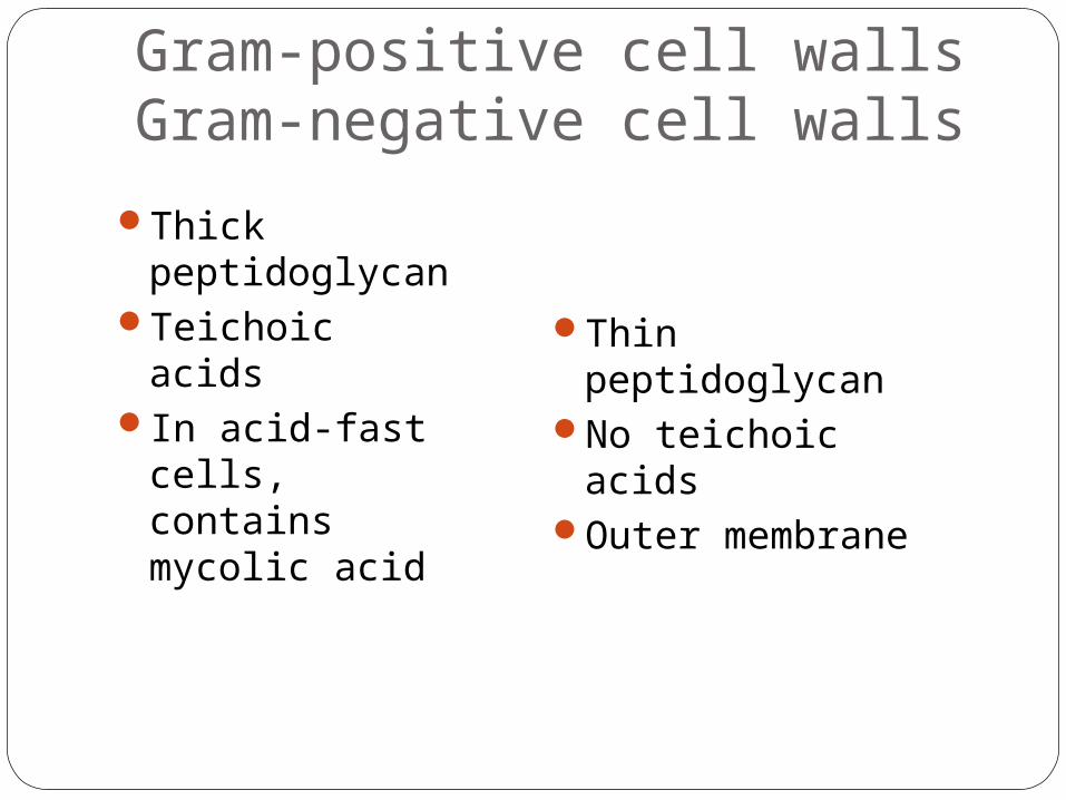
Thick peptidoglycan
Teichoic acidsIn acid-fast
cells, contains mycolic acid
Gram-positive cell wallsGram-negative cell walls
Thin peptidoglycan
No teichoic acidsOuter membrane
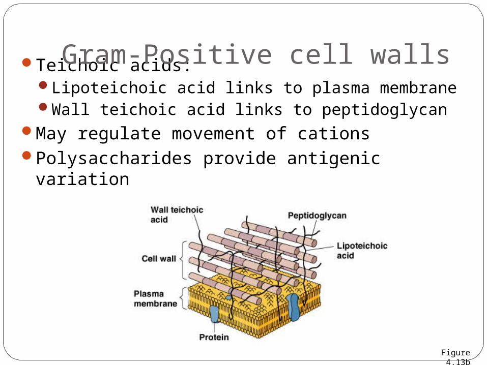
Teichoic acids:Lipoteichoic acid links to plasma membraneWall teichoic acid links to peptidoglycan
May regulate movement of cationsPolysaccharides provide antigenic variation
Gram-Positive cell walls
Figure 4.13b
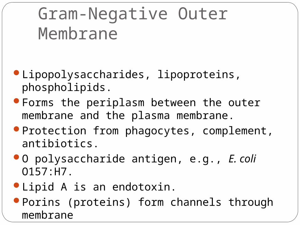
Lipopolysaccharides, lipoproteins, phospholipids.Forms the periplasm between the outer
membrane and the plasma membrane.Protection from phagocytes, complement,
antibiotics.O polysaccharide antigen, e.g., E. coli O157:H7.Lipid A is an endotoxin.Porins (proteins) form channels through
membrane
Gram-Negative Outer Membrane
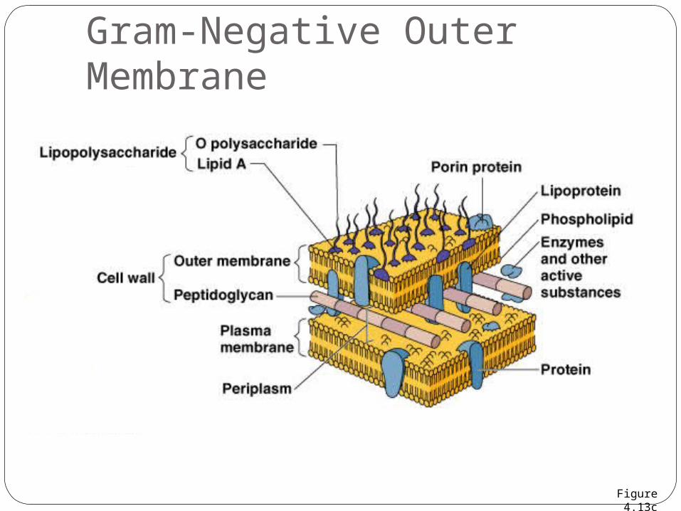
Gram-Negative Outer Membrane
Figure 4.13c

Crystal violet-iodine crystals form in cellGram-positive
Alcohol dehydrates peptidoglycanCV-I crystals do not leave
Gram-negativeAlcohol dissolves outer membrane and leaves
holes in peptidoglycanCV-I washes out
Gram Stain Mechanism
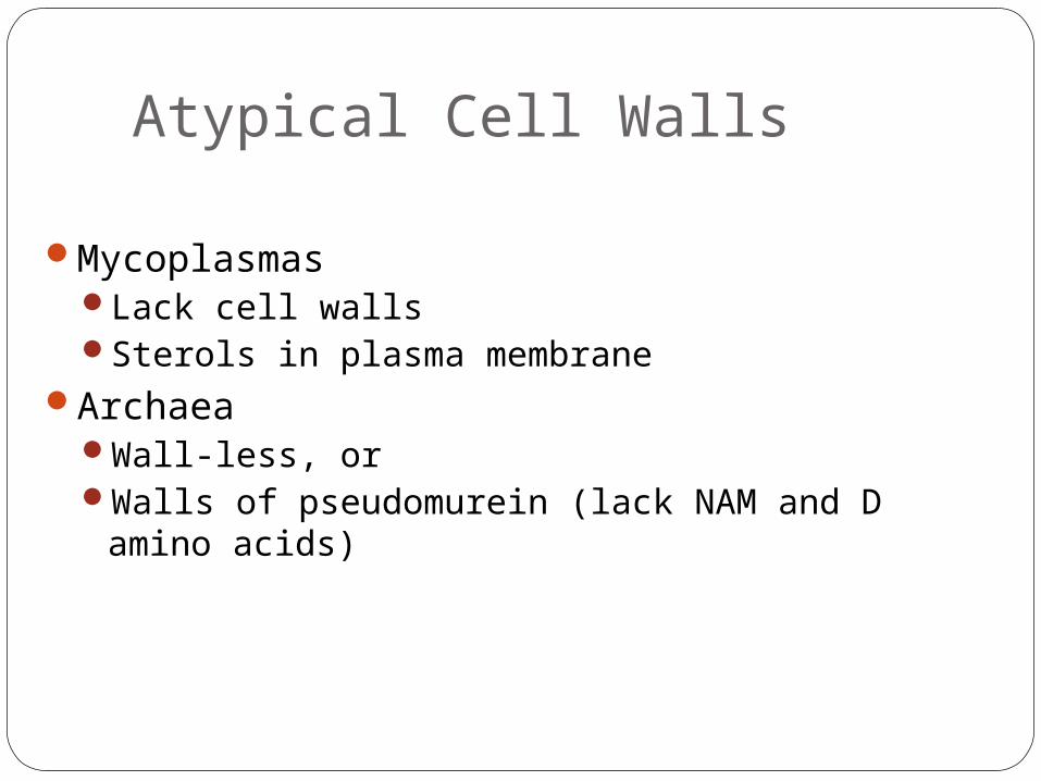
MycoplasmasLack cell wallsSterols in plasma membrane
ArchaeaWall-less, orWalls of pseudomurein (lack NAM and D amino
acids)
Atypical Cell Walls
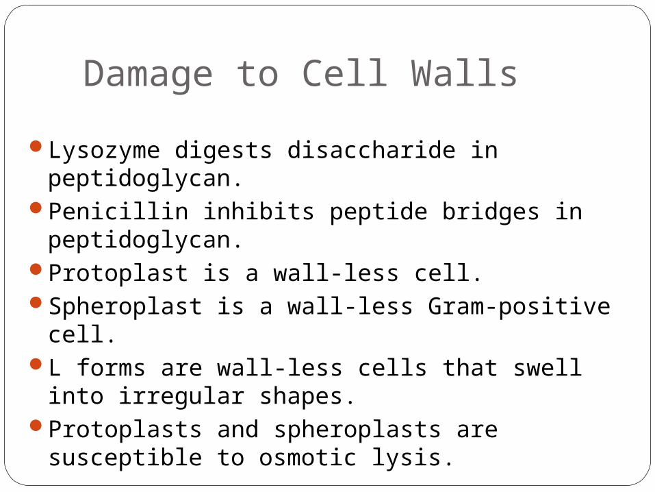
Lysozyme digests disaccharide in peptidoglycan.Penicillin inhibits peptide bridges in
peptidoglycan.Protoplast is a wall-less cell.Spheroplast is a wall-less Gram-positive cell.L forms are wall-less cells that swell into
irregular shapes.Protoplasts and spheroplasts are susceptible to
osmotic lysis.
Damage to Cell Walls
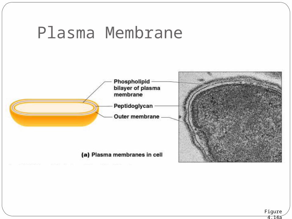
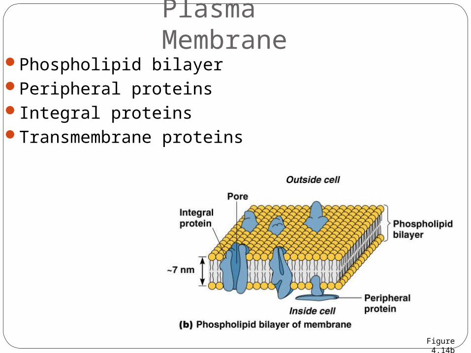
Plasma Membrane
Figure 4.14a
Plasma Membrane
Phospholipid bilayerPeripheral proteinsIntegral proteinsTransmembrane proteins
Figure 4.14b
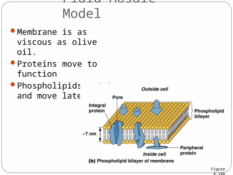
Membrane is as viscous as olive oil.
Proteins move to function
Phospholipids rotate and move laterally
Fluid Mosaic Model
Figure 4.14b