Embed Size (px)
Citation preview
Necrotizing Fasciitis Basem Attum, MD
Megan Mignemi, MD Jonathan G. Schoenecker, MD
Addison K. May, MD, FACS, FCCM William Obremskey, MD, MPH, MMHC Vanderbilt University Medical Center
Created August 2017
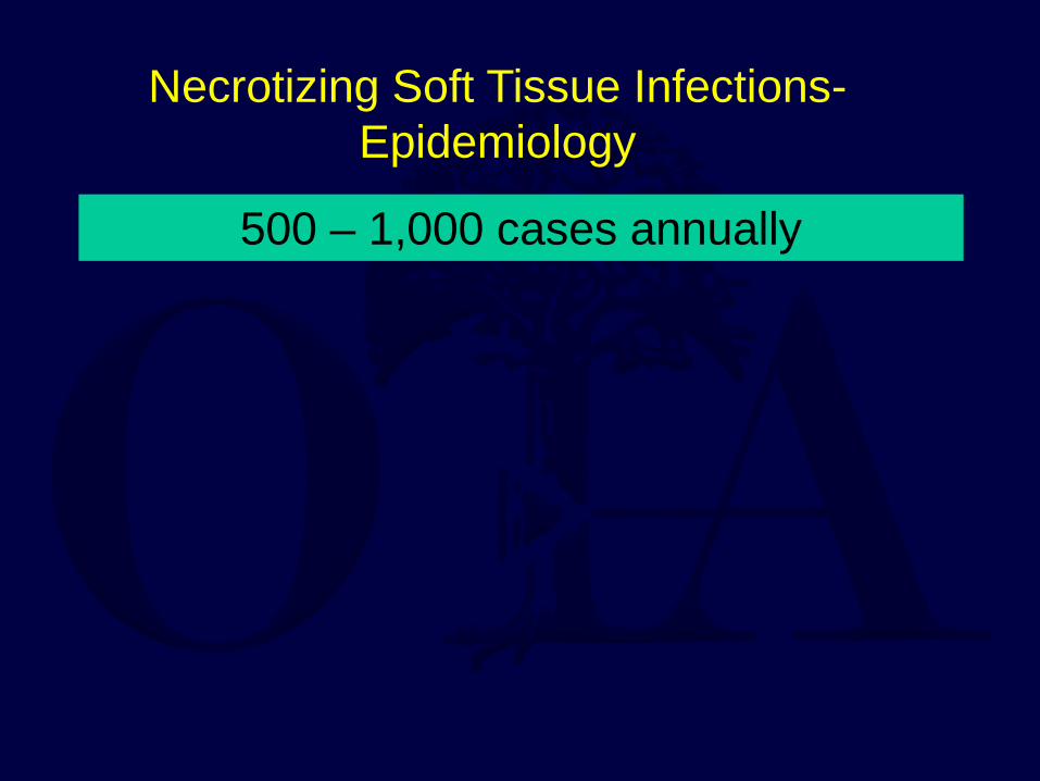
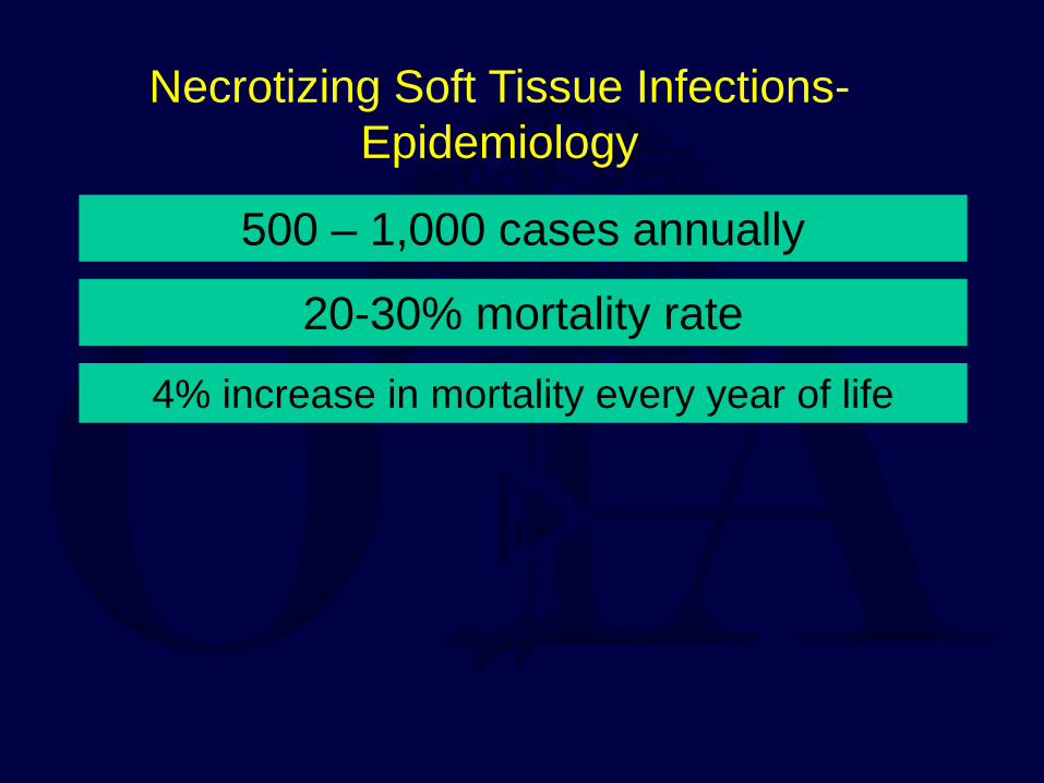
500 – 1,000 cases annually
Necrotizing Soft Tissue Infections-Epidemiology
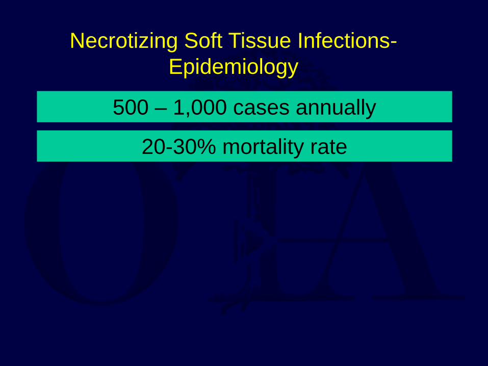
500 – 1,000 cases annually
20-30% mortality rate
Necrotizing Soft Tissue Infections-Epidemiology
500 – 1,000 cases annually
20-30% mortality rate 4% increase in mortality every year of life
Necrotizing Soft Tissue Infections-Epidemiology
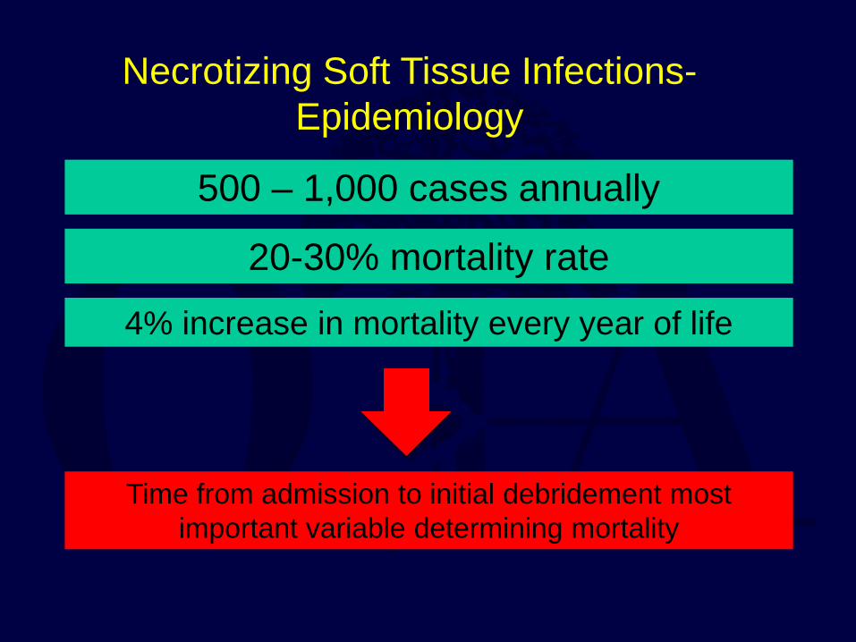
500 – 1,000 cases annually
20-30% mortality rate 4% increase in mortality every year of life
Time from admission to initial debridement most important variable determining mortality
Necrotizing Soft Tissue Infections-Epidemiology
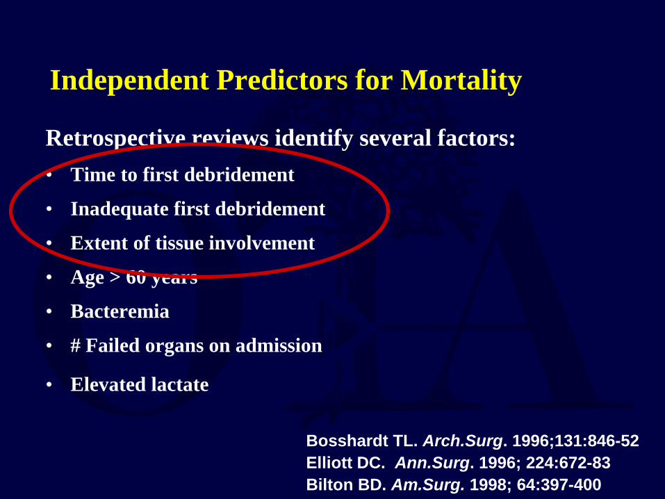
Retrospective reviews identify several factors: • Time to first debridement
• Inadequate first debridement
• Extent of tissue involvement
• Age > 60 years
• Bacteremia
• # Failed organs on admission
• Elevated lactate
Independent Predictors for Mortality
Bosshardt TL. Arch.Surg. 1996;131:846-52 Elliott DC. Ann.Surg. 1996; 224:672-83 Bilton BD. Am.Surg. 1998; 64:397-400
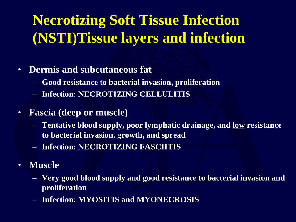
Necrotizing Soft Tissue Infection (NSTI)Tissue layers and infection
• Dermis and subcutaneous fat – Good resistance to bacterial invasion, proliferation – Infection: NECROTIZING CELLULITIS
• Fascia (deep or muscle) – Tentative blood supply, poor lymphatic drainage, and low resistance
to bacterial invasion, growth, and spread – Infection: NECROTIZING FASCIITIS
• Muscle – Very good blood supply and good resistance to bacterial invasion and
proliferation – Infection: MYOSITIS and MYONECROSIS
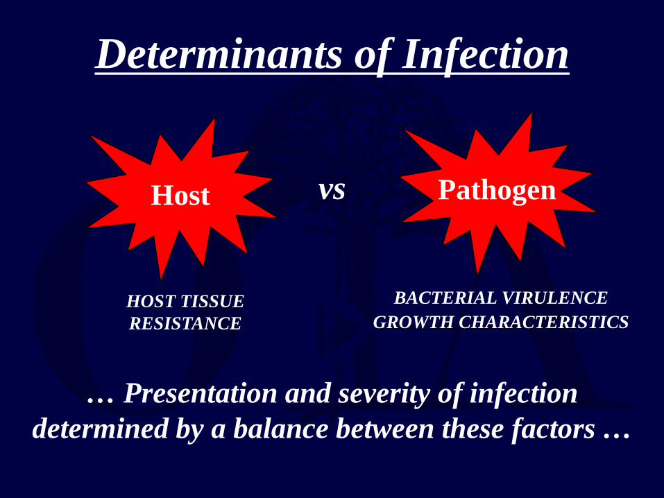
Determinants of Infection
Pathogen Host vs
HOST TISSUE RESISTANCE
BACTERIAL VIRULENCE GROWTH CHARACTERISTICS
… Presentation and severity of infection determined by a balance between these factors …
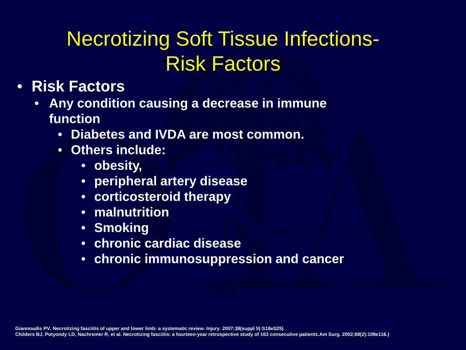
• Risk Factors • Any condition causing a decrease in immune
function • Diabetes and IVDA are most common. • Others include:
• obesity, • peripheral artery disease • corticosteroid therapy • malnutrition • Smoking • chronic cardiac disease • chronic immunosuppression and cancer
1. Giannoudis PV. Necrotizing fasciitis of upper and lower limb: a systematic review. Injury. 2007;38(suppl 5):S18eS25) Childers BJ, Potyondy LD, Nachreiner R, et al. Necrotizing fasciitis: a fourteen-year retrospective study of 163 consecutive patients.Am Surg. 2002;68(2):109e116.)
Necrotizing Soft Tissue Infections- Risk Factors
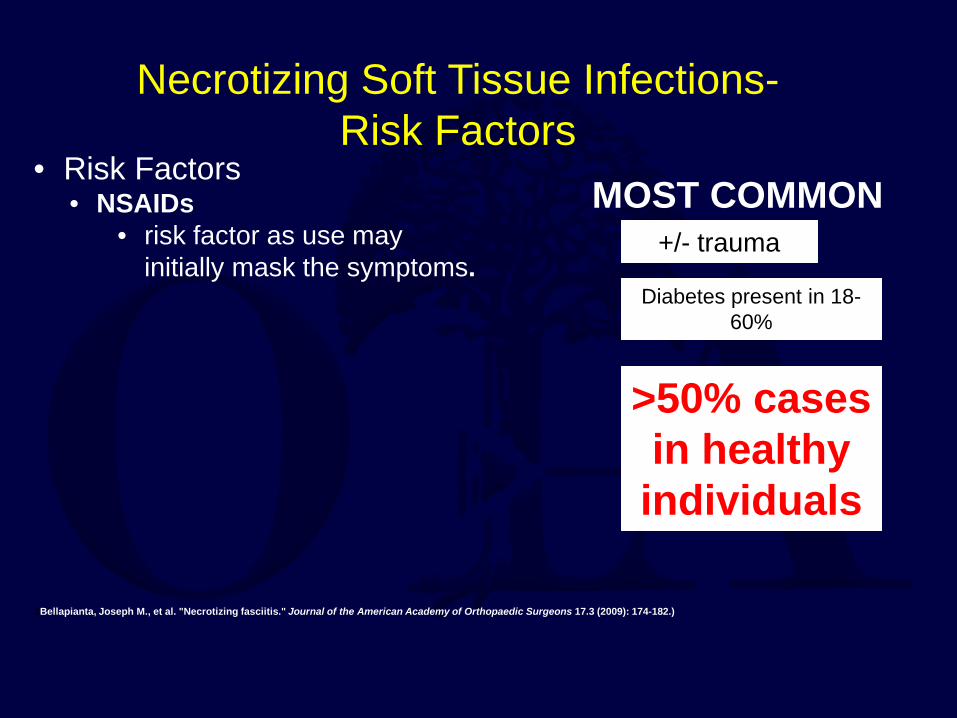
• Risk Factors • NSAIDs
• risk factor as use may initially mask the symptoms.
1Bellapianta, Joseph M., et al. "Necrotizing fasciitis." Journal of the American Academy of Orthopaedic Surgeons 17.3 (2009): 174-182.)
Diabetes present in 18-60%
>50% cases in healthy individuals
+/- trauma MOST COMMON
Necrotizing Soft Tissue Infections- Risk Factors
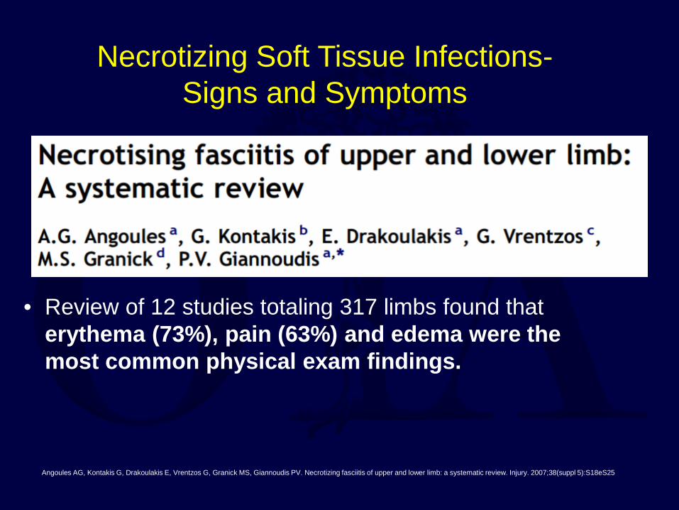
• Review of 12 studies totaling 317 limbs found that erythema (73%), pain (63%) and edema were the most common physical exam findings.
Angoules AG, Kontakis G, Drakoulakis E, Vrentzos G, Granick MS, Giannoudis PV. Necrotizing fasciitis of upper and lower limb: a systematic review. Injury. 2007;38(suppl 5):S18eS25
Necrotizing Soft Tissue Infections- Signs and Symptoms
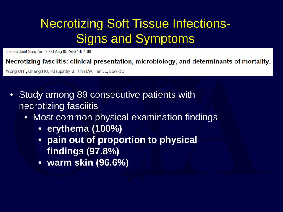
• Study among 89 consecutive patients with necrotizing fasciitis
• Most common physical examination findings • erythema (100%) • pain out of proportion to physical
findings (97.8%) • warm skin (96.6%)
Necrotizing Soft Tissue Infections- Signs and Symptoms
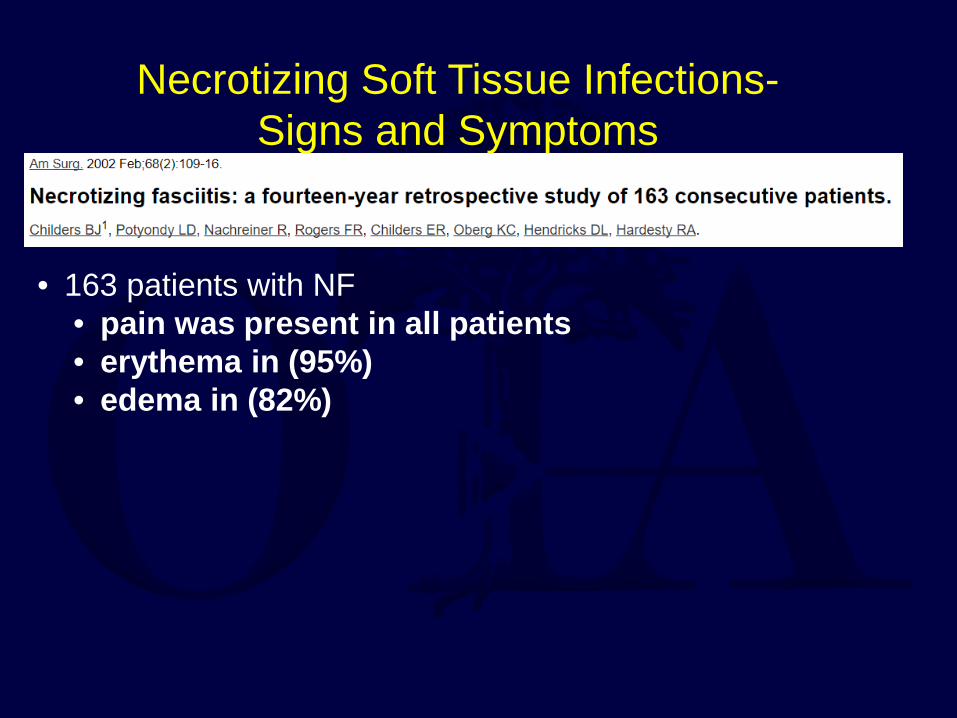
• 163 patients with NF • pain was present in all patients • erythema in (95%) • edema in (82%)
Necrotizing Soft Tissue Infections- Signs and Symptoms
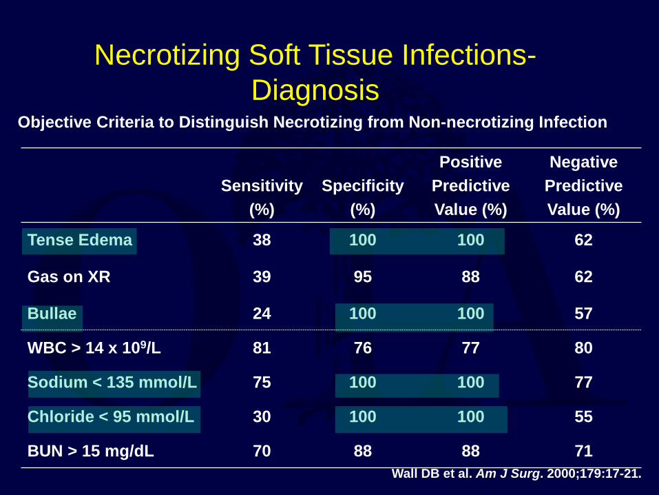
Sensitivity (%)
Specificity (%)
Positive Predictive Value (%)
Negative Predictive Value (%)
Tense Edema 38 100 100 62
Gas on XR 39 95 88 62
Bullae 24 100 100 57
WBC > 14 x 109/L 81 76 77 80
Sodium < 135 mmol/L 75 100 100 77
Chloride < 95 mmol/L 30 100 100 55
BUN > 15 mg/dL 70 88 88 71
Objective Criteria to Distinguish Necrotizing from Non-necrotizing Infection
Wall DB et al. Am J Surg. 2000;179:17-21.
Necrotizing Soft Tissue Infections- Diagnosis
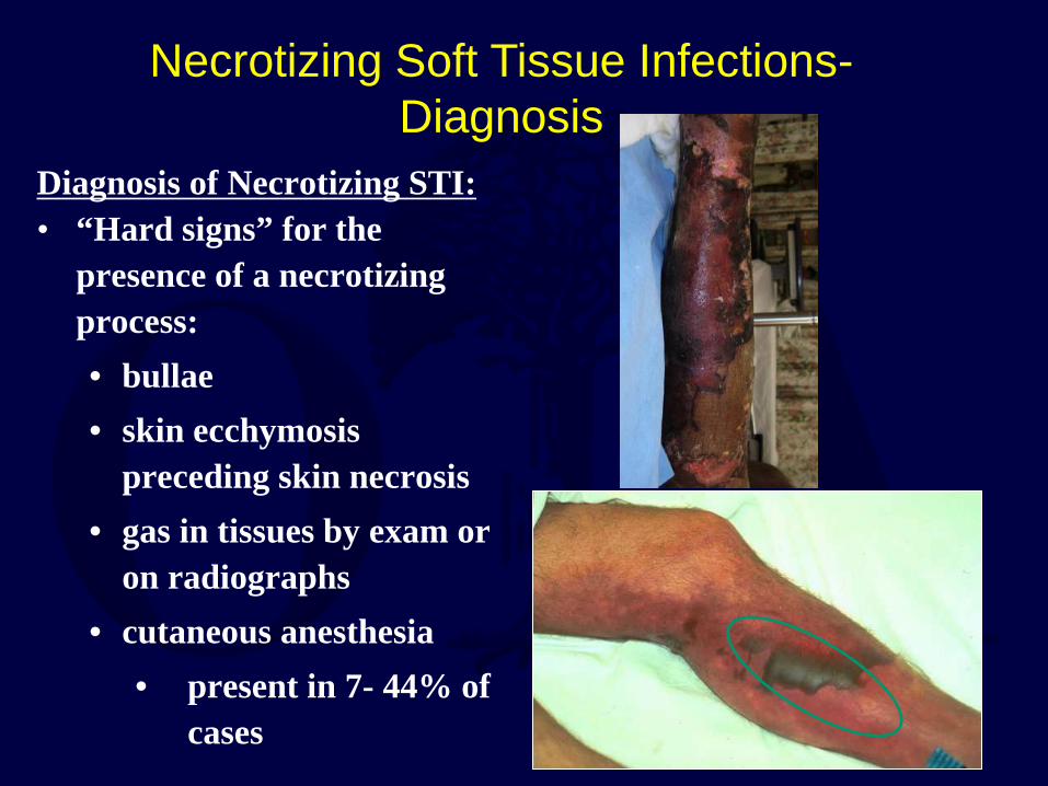
Diagnosis of Necrotizing STI: • “Hard signs” for the
presence of a necrotizing process: • bullae • skin ecchymosis
preceding skin necrosis • gas in tissues by exam or
on radiographs • cutaneous anesthesia
• present in 7- 44% of cases
Necrotizing Soft Tissue Infections- Diagnosis
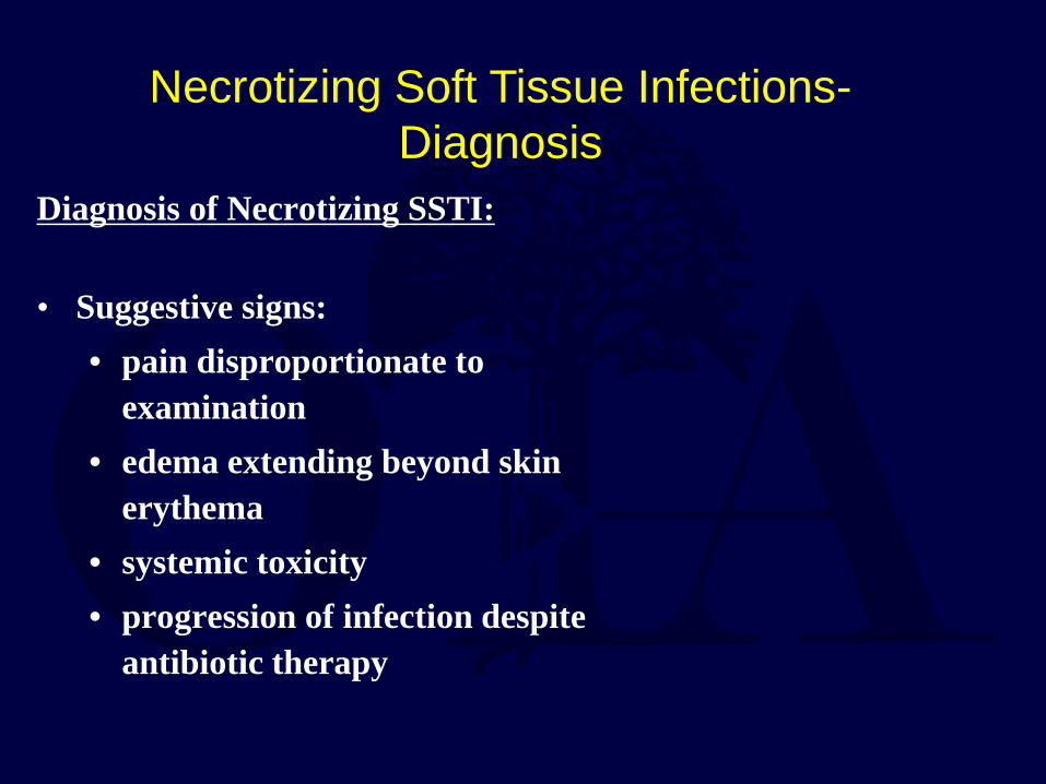
• Suggestive signs: • pain disproportionate to
examination • edema extending beyond skin
erythema • systemic toxicity • progression of infection despite
antibiotic therapy
Necrotizing Soft Tissue Infections- Diagnosis
Diagnosis of Necrotizing SSTI:
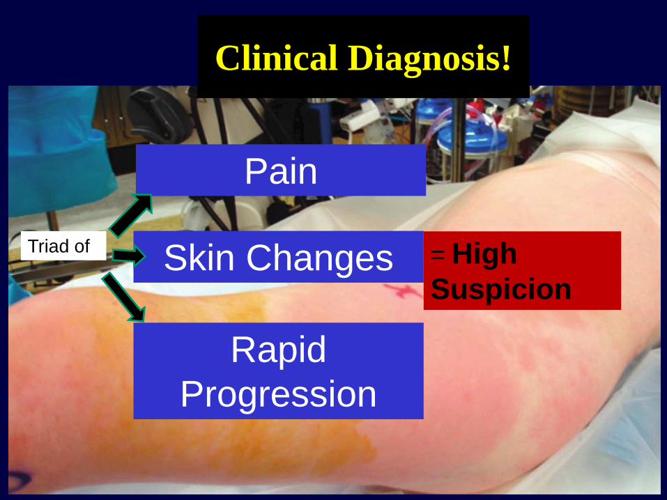
Clinical Diagnosis!
Skin Changes
Pain
Rapid Progression
Triad of = High Suspicion
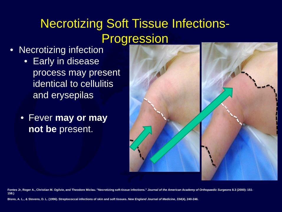
• Necrotizing infection • Early in disease
process may present identical to cellulitis and erysepilas
• Fever may or may
not be present.
Fontes Jr, Roger A., Christian M. Ogilvie, and Theodore Miclau. "Necrotizing soft-tissue infections." Journal of the American Academy of Orthopaedic Surgeons 8.3 (2000): 151-158.)
Bisno, A. L., & Stevens, D. L. (1996). Streptococcal infections of skin and soft tissues. New England Journal of Medicine, 334(4), 240-246.
Necrotizing Soft Tissue Infections- Progression
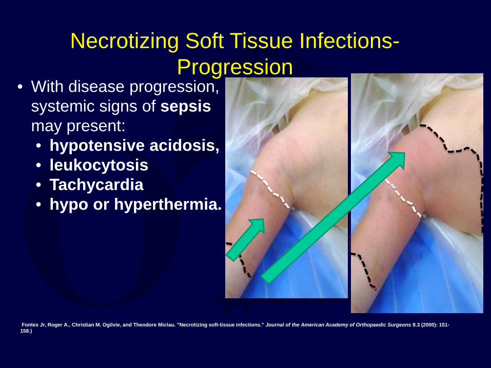
• With disease progression, systemic signs of sepsis may present: • hypotensive acidosis, • leukocytosis • Tachycardia • hypo or hyperthermia.
Fontes Jr, Roger A., Christian M. Ogilvie, and Theodore Miclau. "Necrotizing soft-tissue infections." Journal of the American Academy of Orthopaedic Surgeons 8.3 (2000): 151-158.)
Necrotizing Soft Tissue Infections- Progression
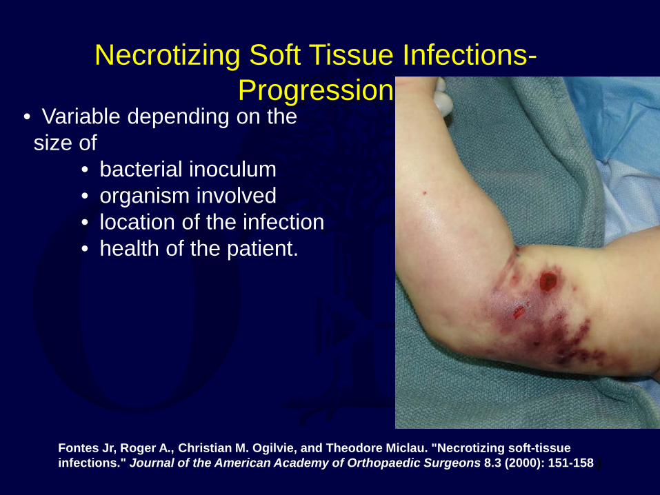
• Variable depending on the size of
• bacterial inoculum • organism involved • location of the infection • health of the patient.
Fontes Jr, Roger A., Christian M. Ogilvie, and Theodore Miclau. "Necrotizing soft-tissue infections." Journal of the American Academy of Orthopaedic Surgeons 8.3 (2000): 151-158.)
Necrotizing Soft Tissue Infections- Progression
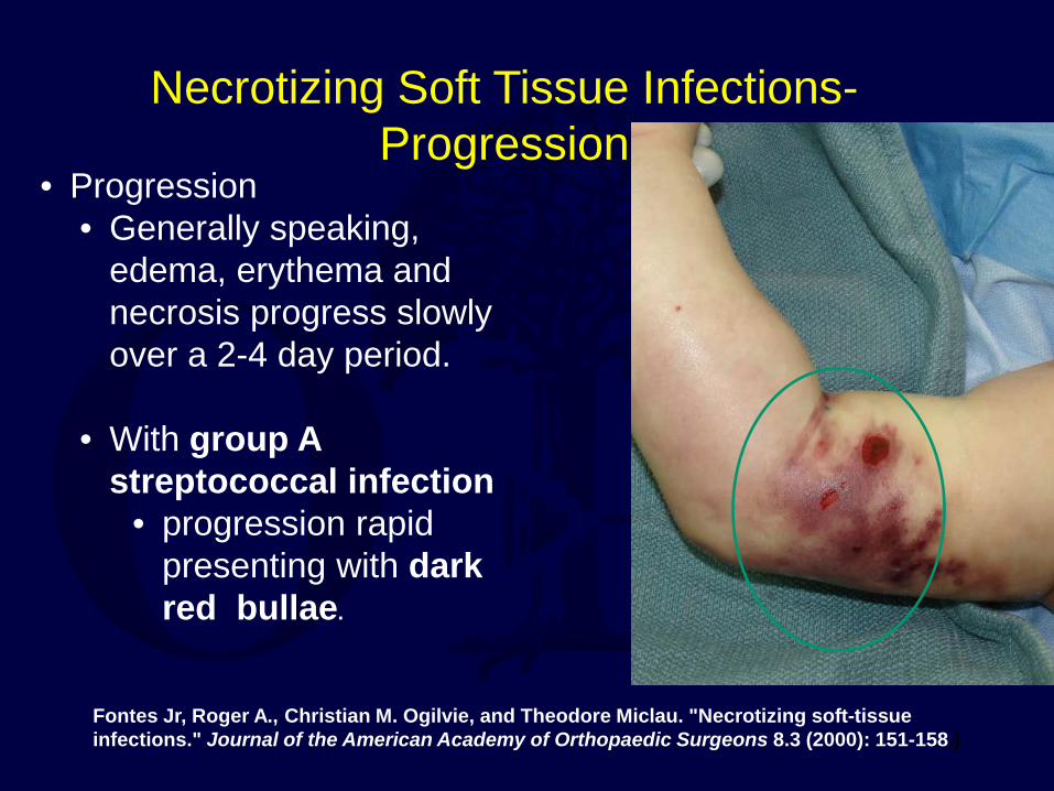
• Progression • Generally speaking,
edema, erythema and necrosis progress slowly over a 2-4 day period.
• With group A
streptococcal infection • progression rapid
presenting with dark red bullae.
Fontes Jr, Roger A., Christian M. Ogilvie, and Theodore Miclau. "Necrotizing soft-tissue infections." Journal of the American Academy of Orthopaedic Surgeons 8.3 (2000): 151-158.)
Necrotizing Soft Tissue Infections- Progression
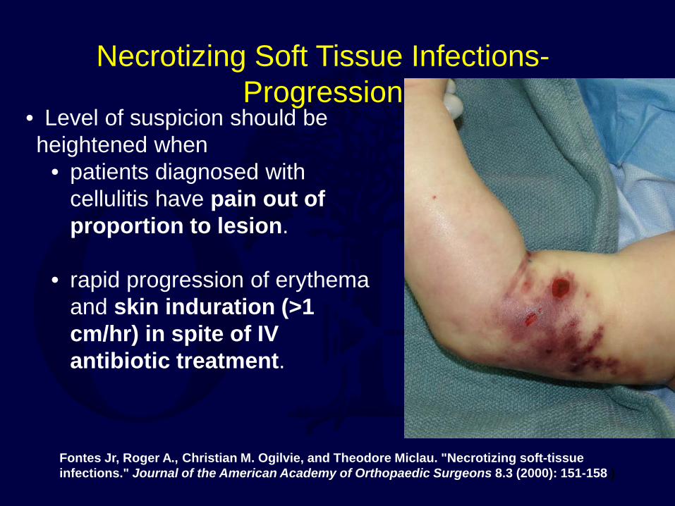
• Level of suspicion should be heightened when
• patients diagnosed with cellulitis have pain out of proportion to lesion.
• rapid progression of erythema
and skin induration (>1 cm/hr) in spite of IV antibiotic treatment.
Fontes Jr, Roger A., Christian M. Ogilvie, and Theodore Miclau. "Necrotizing soft-tissue infections." Journal of the American Academy of Orthopaedic Surgeons 8.3 (2000): 151-158.)
Necrotizing Soft Tissue Infections- Progression
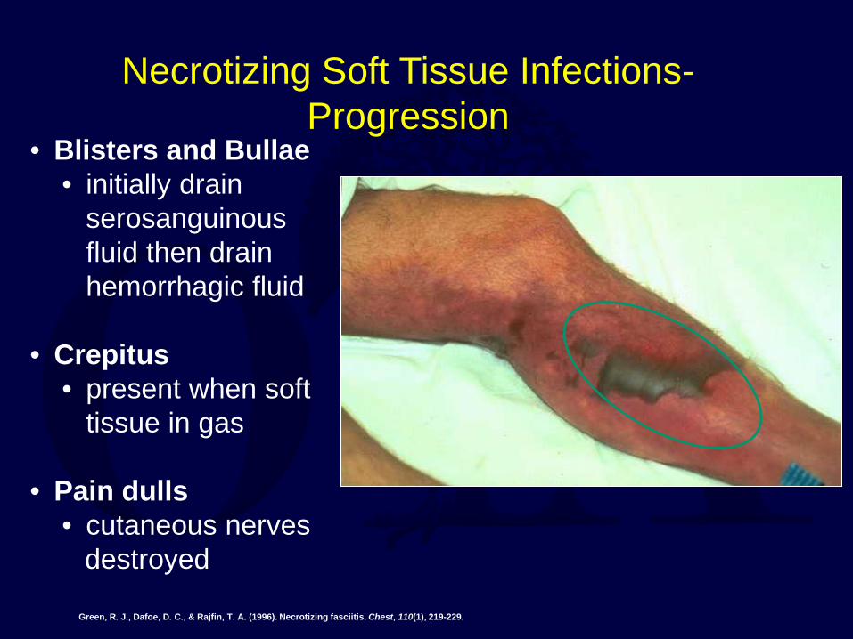
• Blisters and Bullae • initially drain
serosanguinous fluid then drain hemorrhagic fluid
• Crepitus
• present when soft tissue in gas
• Pain dulls
• cutaneous nerves destroyed
Green, R. J., Dafoe, D. C., & Rajfin, T. A. (1996). Necrotizing fasciitis. Chest, 110(1), 219-229.
Necrotizing Soft Tissue Infections- Progression
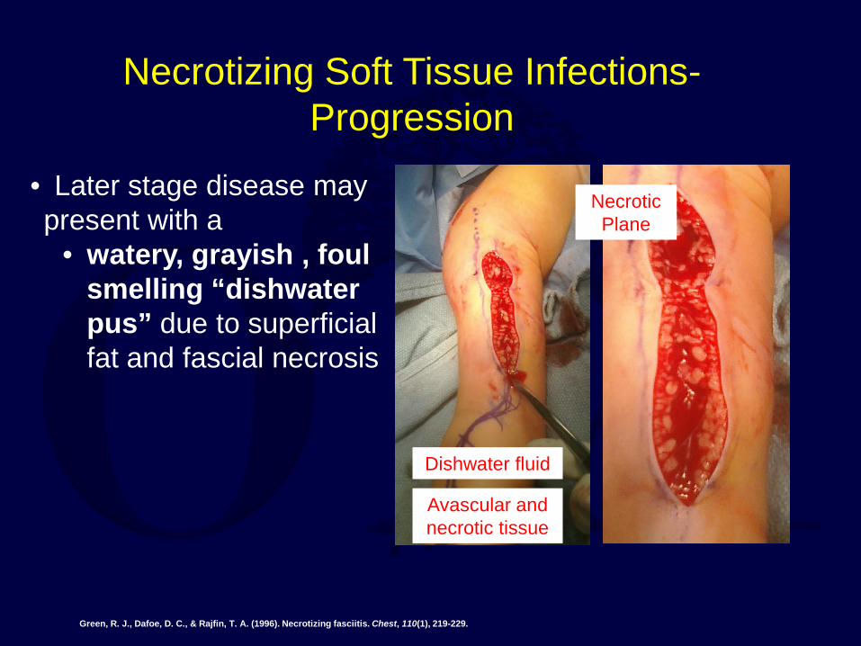
• Later stage disease may present with a
• watery, grayish , foul smelling “dishwater pus” due to superficial fat and fascial necrosis
Green, R. J., Dafoe, D. C., & Rajfin, T. A. (1996). Necrotizing fasciitis. Chest, 110(1), 219-229.
Necrotizing Soft Tissue Infections- Progression
Necrotic Plane
Dishwater fluid
Avascular and necrotic tissue
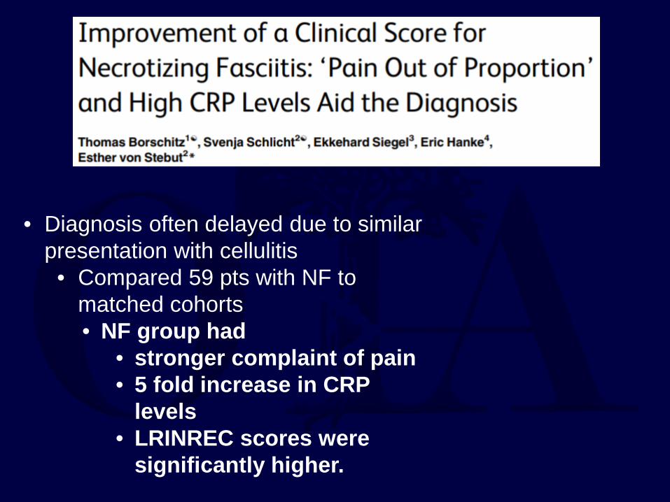
• Diagnosis often delayed due to similar presentation with cellulitis
• Compared 59 pts with NF to matched cohorts • NF group had
• stronger complaint of pain • 5 fold increase in CRP
levels • LRINREC scores were
significantly higher.
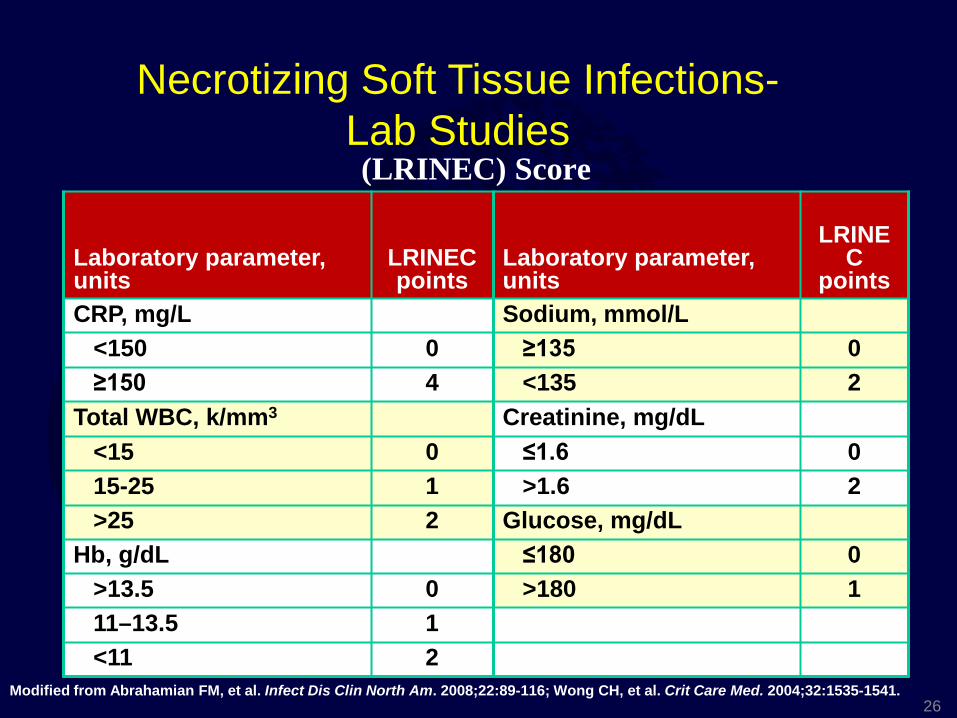
Necrotizing Soft Tissue Infections- Lab Studies
26
(LRINEC) Score
26 Modified from Abrahamian FM, et al. Infect Dis Clin North Am. 2008;22:89-116; Wong CH, et al. Crit Care Med. 2004;32:1535-1541.
Laboratory parameter, units
LRINEC points
Laboratory parameter, units
LRINEC
points CRP, mg/L Sodium, mmol/L <150 0 ≥135 0 ≥150 4 <135 2 Total WBC, k/mm3 Creatinine, mg/dL <15 0 ≤1.6 0 15-25 1 >1.6 2 >25 2 Glucose, mg/dL Hb, g/dL ≤180 0 >13.5 0 >180 1 11–13.5 1 <11 2
Necrotizing Soft Tissue Infections- Lab Studies
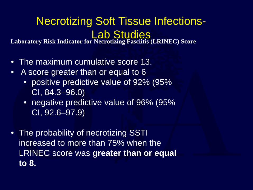
Laboratory Risk Indicator for Necrotizing Fasciitis (LRINEC) Score
• The maximum cumulative score 13. • A score greater than or equal to 6
• positive predictive value of 92% (95% CI, 84.3–96.0)
• negative predictive value of 96% (95% CI, 92.6–97.9)
• The probability of necrotizing SSTI increased to more than 75% when the LRINEC score was greater than or equal to 8.
Necrotizing Soft Tissue Infections- Lab Studies
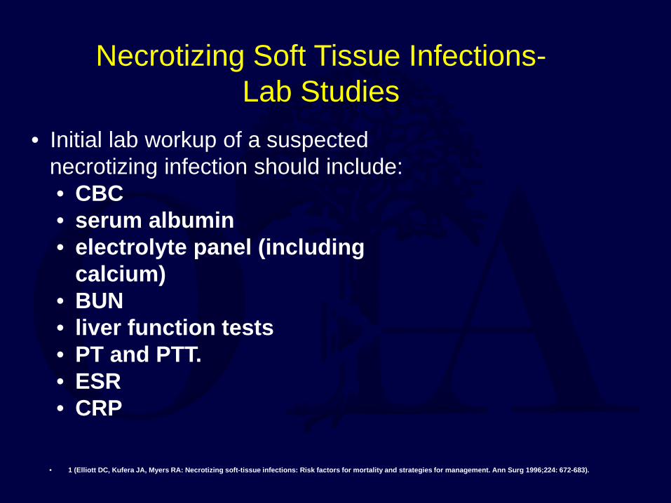
• Initial lab workup of a suspected necrotizing infection should include:
• CBC • serum albumin • electrolyte panel (including
calcium) • BUN • liver function tests • PT and PTT. • ESR • CRP
• 1 (Elliott DC, Kufera JA, Myers RA: Necrotizing soft-tissue infections: Risk factors for mortality and strategies for management. Ann Surg 1996;224: 672-683).
Necrotizing Soft Tissue Infections- Lab Studies
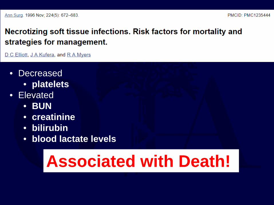
• Decreased • platelets
• Elevated • BUN • creatinine • bilirubin • blood lactate levels
Necrotizing Soft Tissue Infections- Lab Studies
Associated with Death!
Laboratory Risk Indicator for Necrotizing Fasciitis (LRINEC) Score
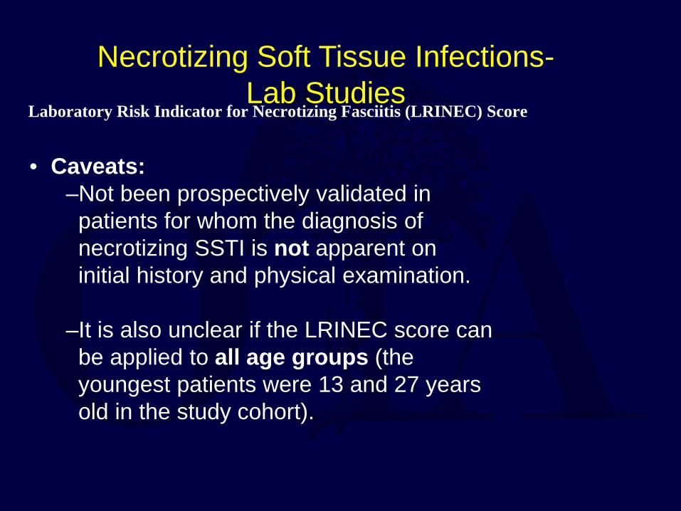
• Caveats: –Not been prospectively validated in patients for whom the diagnosis of necrotizing SSTI is not apparent on initial history and physical examination.
–It is also unclear if the LRINEC score can be applied to all age groups (the youngest patients were 13 and 27 years old in the study cohort).
Necrotizing Soft Tissue Infections- Lab Studies
31
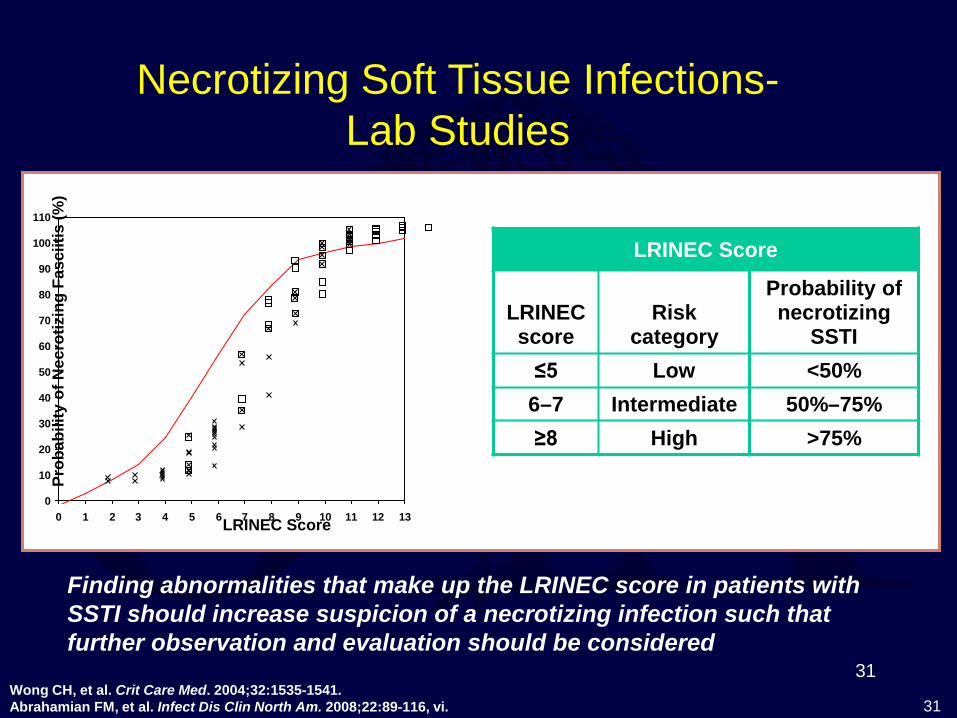
LRINEC Score: Corresponding Risk and Probability of Necrotizing SSTI
31
Finding abnormalities that make up the LRINEC score in patients with SSTI should increase suspicion of a necrotizing infection such that further observation and evaluation should be considered
Wong CH, et al. Crit Care Med. 2004;32:1535-1541. Abrahamian FM, et al. Infect Dis Clin North Am. 2008;22:89-116, vi.
LRINEC Score
LRINEC score
Risk category
Probability of necrotizing
SSTI ≤5 Low <50%
6–7 Intermediate 50%–75% ≥8 High >75%
0
10
20
30
40
50
60
70
80
90
100
110
0 1 2 3 4 5 6 7 8 9 10 11 12 13LRINEC Score
Prob
abili
ty o
f Nec
rotiz
ing
Fasc
iitis
(%)
Necrotizing Soft Tissue Infections- Lab Studies
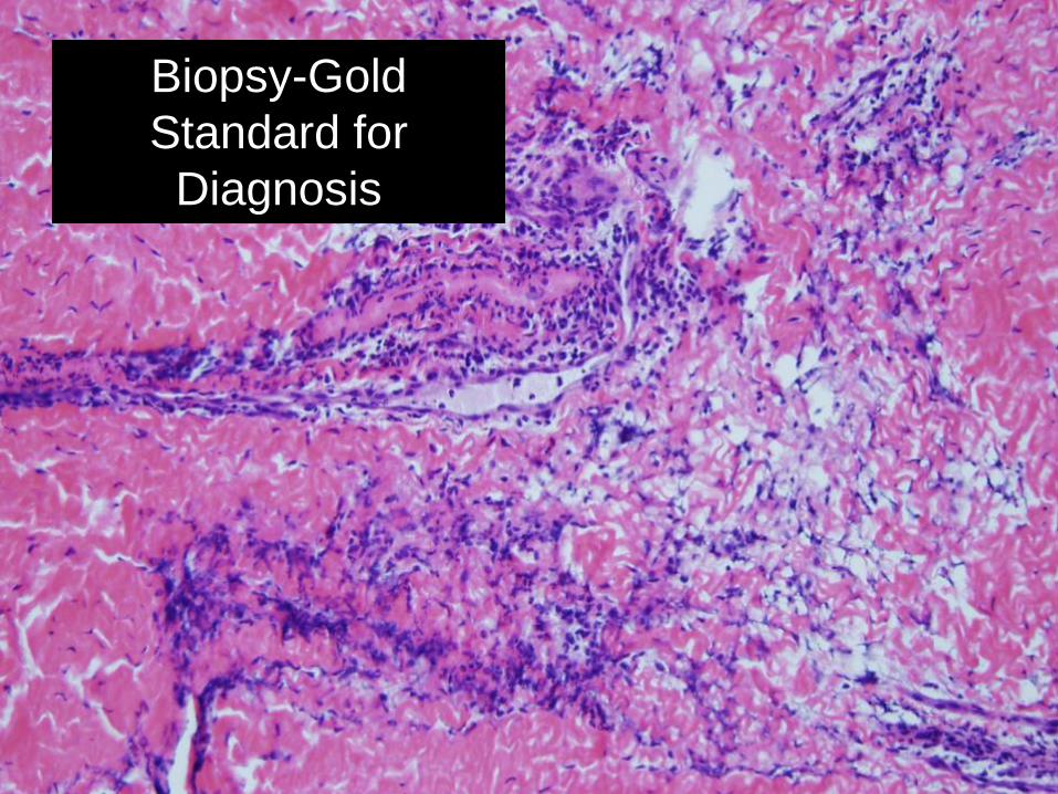
Tissue Diagnosis Biopsy-Gold Standard for Diagnosis
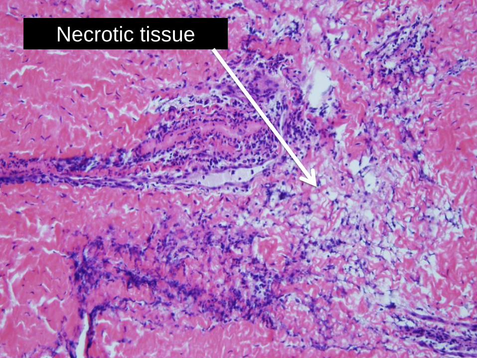
Tissue Diagnosis Necrotic tissue
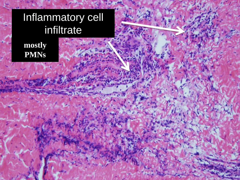
Tissue Diagnosis Inflammatory cell
infiltrate mostly PMNs
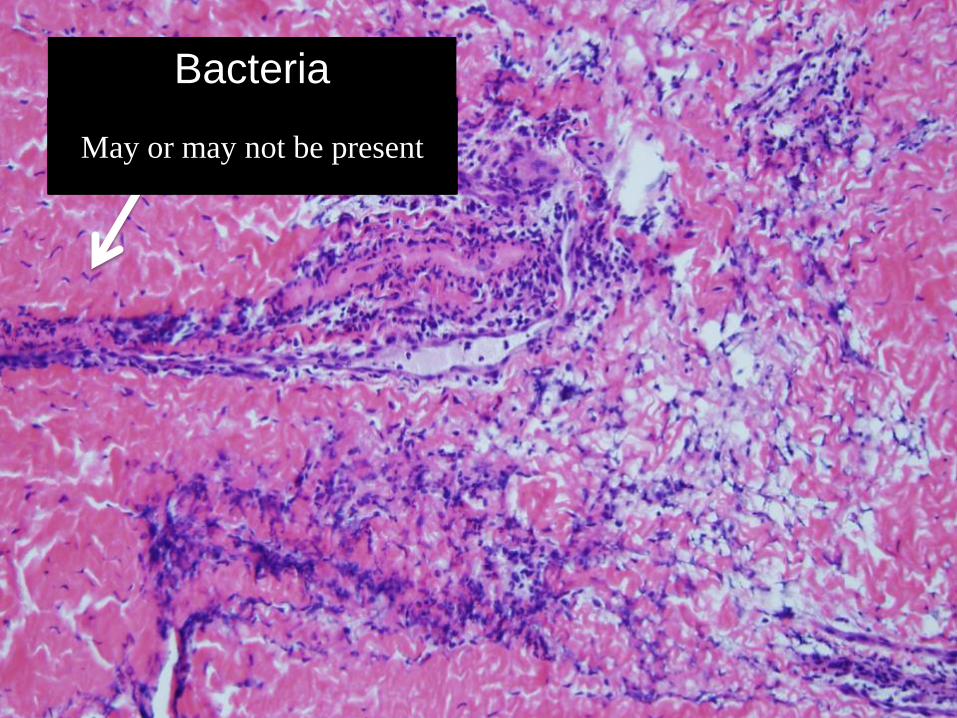
Tissue Diagnosis Bacteria
May or may not be present
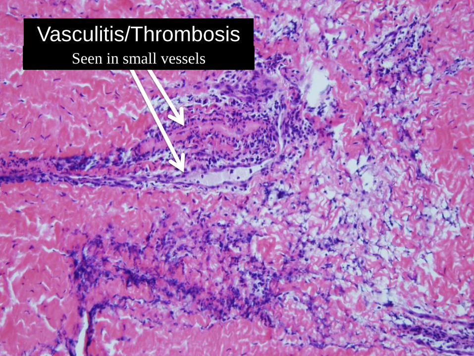
Tissue Diagnosis Vasculitis/Thrombosis
Seen in small vessels
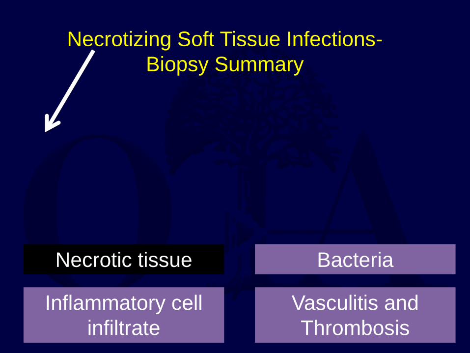
Necrotic tissue
Inflammatory cell infiltrate
Bacteria
Vasculitis and Thrombosis
Necrotizing Soft Tissue Infections- Biopsy Summary
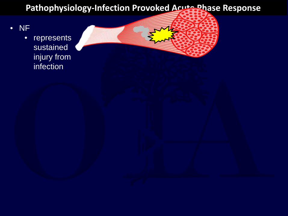
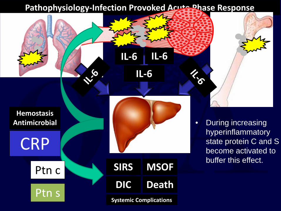
Pathophysiology-Infection Provoked Acute Phase Response
• NF • represents
sustained injury from infection
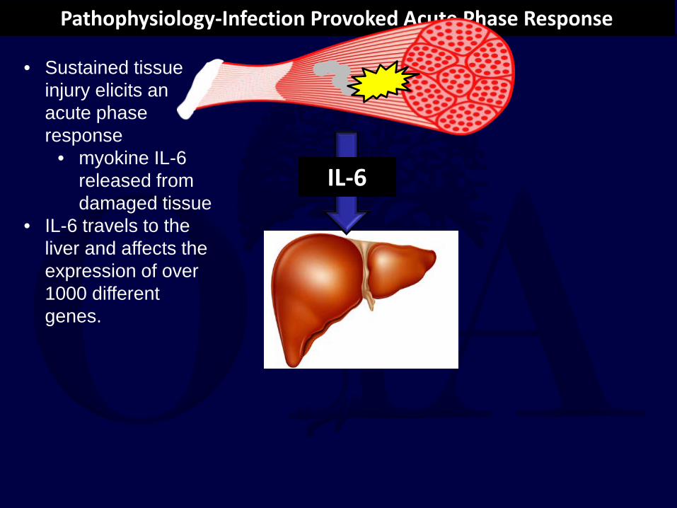
Pathophysiology-Infection Provoked Acute Phase Response
IL-6
• Sustained tissue injury elicits an acute phase response
• myokine IL-6 released from damaged tissue
• IL-6 travels to the liver and affects the expression of over 1000 different genes.
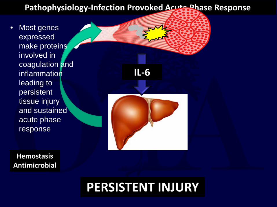
Pathophysiology-Infection Provoked Acute Phase Response
IL-6
Hemostasis Antimicrobial
PERSISTENT INJURY
• Most genes expressed make proteins involved in coagulation and inflammation leading to persistent tissue injury and sustained acute phase response
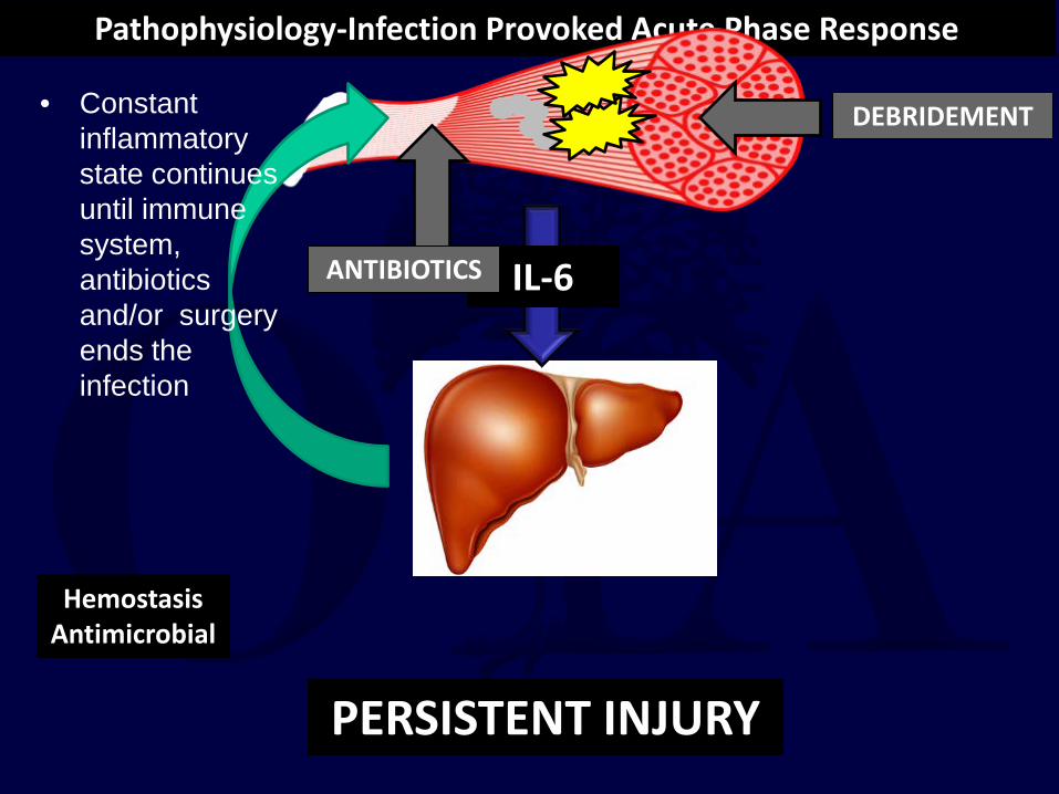
Pathophysiology-Infection Provoked Acute Phase Response
IL-6
DEBRIDEMENT
PERSISTENT INJURY
• Constant inflammatory state continues until immune system, antibiotics and/or surgery ends the infection
ANTIBIOTICS
Hemostasis Antimicrobial
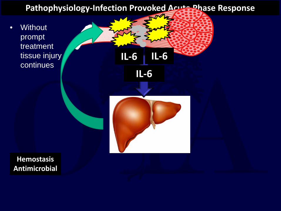
Pathophysiology-Infection Provoked Acute Phase Response
IL-6
IL-6 IL-6
• Without prompt treatment tissue injury continues
Hemostasis Antimicrobial
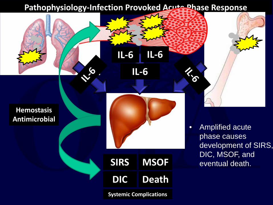
Pathophysiology-Infection Provoked Acute Phase Response
IL-6
Hemostasis Antimicrobial
IL-6 IL-6
SIRS
DIC
MSOF
Death Systemic Complications
• Amplified acute phase causes development of SIRS, DIC, MSOF, and eventual death.

Pathophysiology-Infection Provoked Acute Phase Response
IL-6
Hemostasis Antimicrobial
IL-6 IL-6
SIRS
DVT
PE
Death Systemic Complications
CRP SIRS
DIC
MSOF
Death Systemic Complications
• CRP • rapid acute phase
reactants which increases significantly in response to tissue from nec fasc
Pathophysiology-Infection Provoked Acute Phase Response
IL-6
Hemostasis Antimicrobial
IL-6 IL-6
SIRS
DVT
PE
Death Systemic Complications
CRP
Ptn s
Ptn c SIRS
DIC
MSOF
Death Systemic Complications
• During increasing hyperinflammatory state protein C and S become activated to buffer this effect.
Pathophysiology-Infection Provoked Acute Phase Response
IL-6
Hemostasis Antimicrobial
IL-6 IL-6
SIRS
DVT
PE
Death Systemic Complications
CRP
Ptn s
Ptn c SIRS
DIC
MSOF
Death Systemic Complications
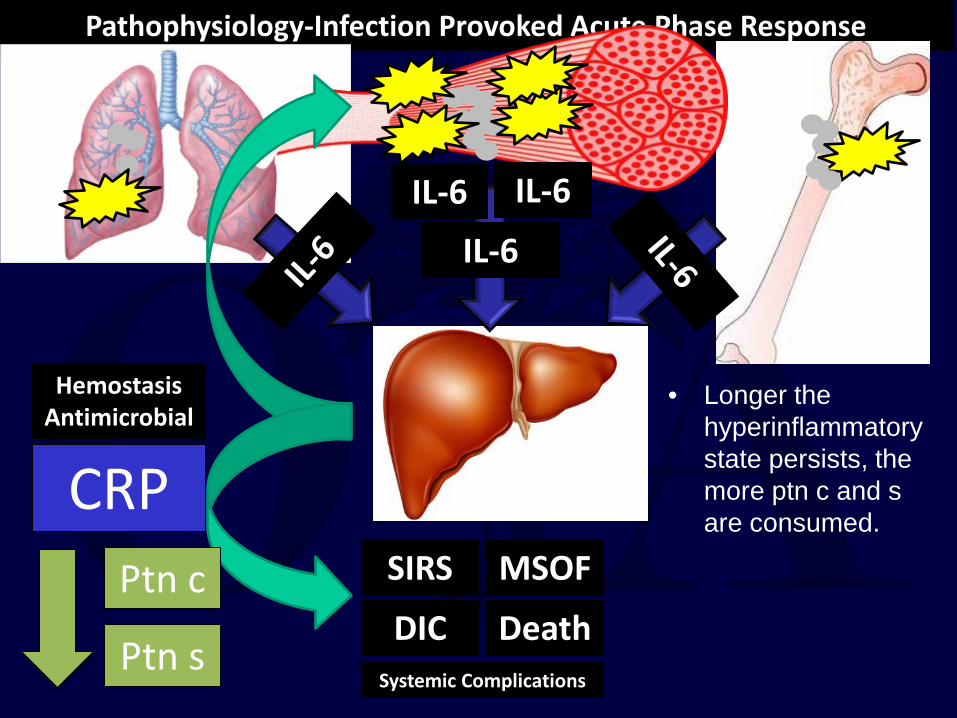
• Longer the hyperinflammatory state persists, the more ptn c and s are consumed.

Pathophysiology-Infection Provoked Acute Phase Response
IL-6
Hemostasis Antimicrobial
IL-6 IL-6
SIRS
DVT
PE
Death Systemic Complications
CRP
Ptn s
Ptn c
X SIRS
DIC
MSOF
Death Systemic Complications
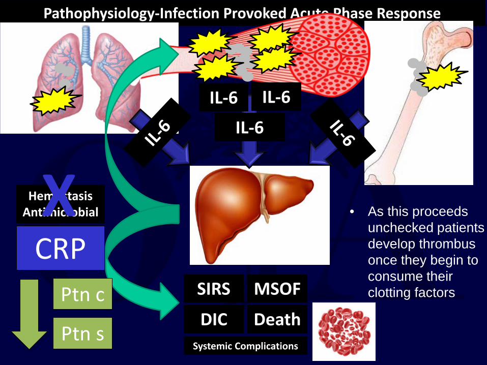
• As proteins consumed, ability to buffer coagulation decreases
Pathophysiology-Infection Provoked Acute Phase Response
IL-6
Hemostasis Antimicrobial
IL-6 IL-6
SIRS
DVT
PE
Death Systemic Complications
CRP
Ptn s
Ptn c
X SIRS
DIC
MSOF
Death Systemic Complications
• As this proceeds unchecked patients develop thrombus once they begin to consume their clotting factors
Pathophysiology-Infection Provoked Acute Phase Response
IL-6
Hemostasis Antimicrobial
IL-6 IL-6
SIRS
DVT
PE
Death
Systemic Complications
CRP
Ptn s
Ptn c SIRS
DIC
MSOF
Death
Systemic Complications
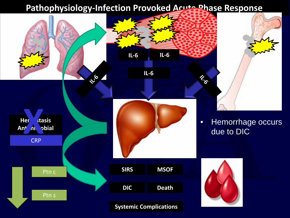
• Hemorrhage occurs due to DIC X
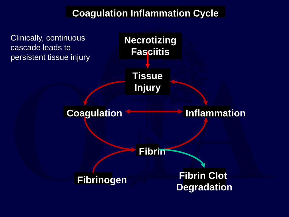
Necrotizing Fasciitis
Tissue Injury
Coagulation Inflammation
Fibrin
Fibrinogen Fibrin Clot Degradation
Coagulation Inflammation Cycle
Clinically, continuous cascade leads to persistent tissue injury
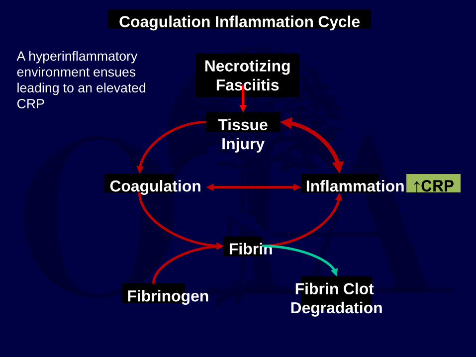
Necrotizing Fasciitis
Tissue Injury
Coagulation Inflammation
Fibrin
Fibrinogen Fibrin Clot Degradation
↑CRP
Coagulation Inflammation Cycle
A hyperinflammatory environment ensues leading to an elevated CRP
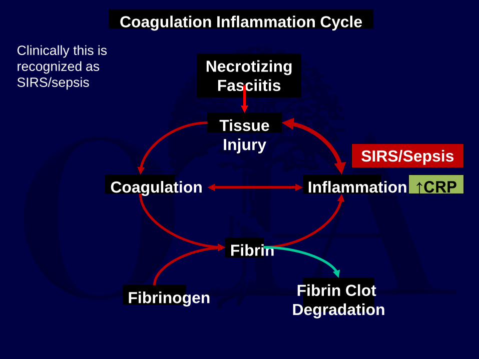
Necrotizing Fasciitis
Tissue Injury
Coagulation Inflammation
Fibrin
Fibrinogen Fibrin Clot Degradation
Coagulation Inflammation Cycle
SIRS/Sepsis
↑CRP
Clinically this is recognized as SIRS/sepsis
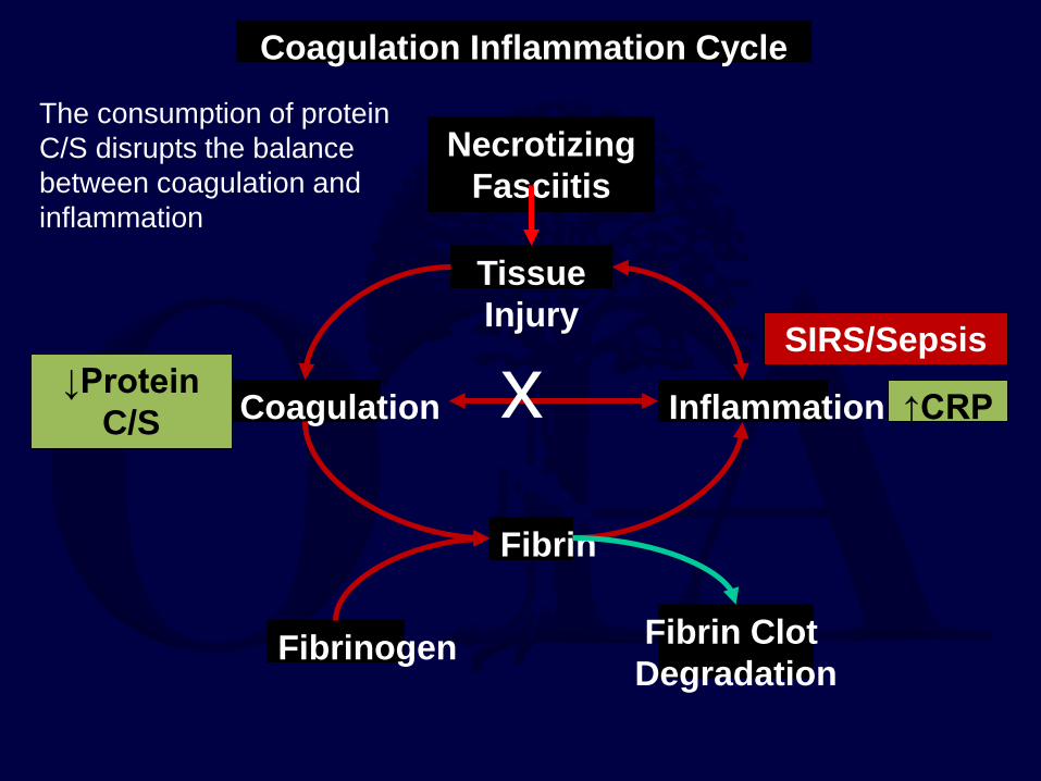
Necrotizing Fasciitis
Tissue Injury
Coagulation Inflammation
Fibrin
Fibrinogen Fibrin Clot Degradation
↓Protein C/S ↑CRP
Coagulation Inflammation Cycle
SIRS/Sepsis
x
The consumption of protein C/S disrupts the balance between coagulation and inflammation
Necrotizing Fasciitis
Tissue Injury
Coagulation Inflammation
Fibrin
Fibrinogen Fibrin Clot Degradation
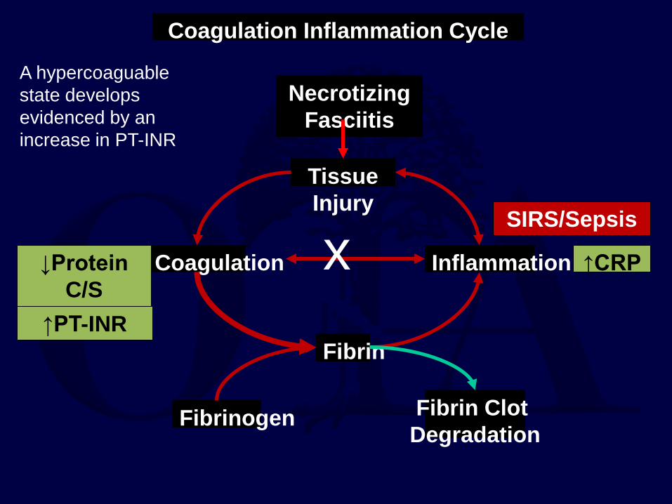
↑PT-INR
↓Protein C/S
↑CRP
Coagulation Inflammation Cycle
x
A hypercoaguable state develops evidenced by an increase in PT-INR
SIRS/Sepsis
Tissue Injury
Coagulation Inflammation
Fibrin
Fibrinogen Fibrin Clot Degradation
↑PT-INR
↓Protein C/S ↑CRP
Necrotizing Fasciitis
Coagulation Inflammation Cycle
x
As the coagulation factors are consumed, the patient becomes hypocoaguable, as evidenced by an elevated INR
SIRS/Sepsis
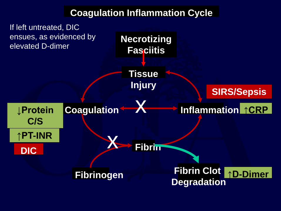
Necrotizing Fasciitis
Tissue Injury
Coagulation Inflammation
Fibrin
Fibrinogen Fibrin Clot Degradation
↑PT-INR
↓Protein C/S
↑CRP
↑D-Dimer
Coagulation Inflammation Cycle
DIC
x x
If left untreated, DIC ensues, as evidenced by elevated D-dimer
SIRS/Sepsis
Tissue Injury
Coagulation Inflammation
Fibrin
Fibrinogen Fibrin Clot Degradation
↑PT-INR
↓Protein C/S
↑CRP
↑D-Dimer
Coagulation Inflammation Cycle
DIC
x x
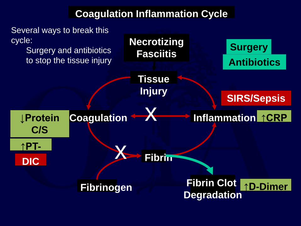
Several ways to break this cycle:
Surgery and antibiotics to stop the tissue injury
Surgery Antibiotics
Necrotizing Fasciitis
SIRS/Sepsis
Tissue Injury
Coagulation Inflammation
Fibrin
Fibrinogen Fibrin Clot Degradation
↑PT-INR
↓Protein C/S
↑CRP
↑D-Dimer
Necrotizing
Fasciitis
Coagulation Inflammation Cycle
DIC
x x
Steroids Surgery Antibiotics
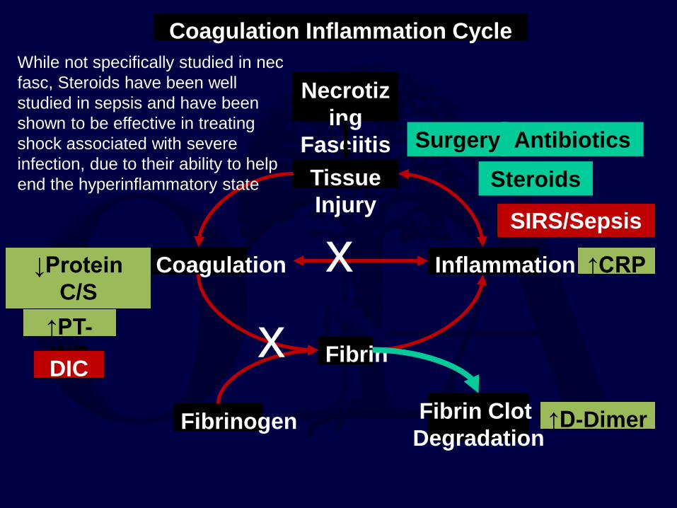
While not specifically studied in nec fasc, Steroids have been well studied in sepsis and have been shown to be effective in treating shock associated with severe infection, due to their ability to help end the hyperinflammatory state
SIRS/Sepsis
Tissue Injury
Coagulation Inflammation
Fibrin
Fibrinogen Fibrin Clot Degradation
↑PT-INR
↓Protein C/S
↑CRP
↑D-Dimer
Coagulation Inflammation Cycle
DIC
x x
Vitamin K
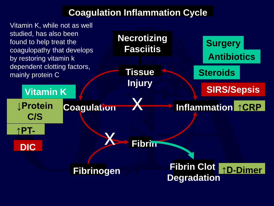
Vitamin K, while not as well studied, has also been found to help treat the coagulopathy that develops by restoring vitamin k dependent clotting factors, mainly protein C
Steroids
Surgery Antibiotics
Necrotizing Fasciitis
SIRS/Sepsis
• Radiographs not normally indicated in the work up in necrotizing fasciitis. • In clostridial
myonecrosis • gas in the muscle
bellies
Chapnick, E. K., & Abter, E. I. (1996). Necrotizing soft-tissue infections. Infectious disease clinics of North America, 10(4), 835-855.
Necrotizing Soft Tissue Infections- Imaging
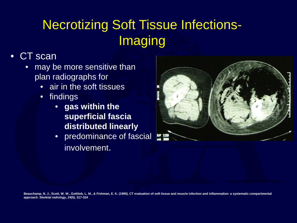
• CT scan • may be more sensitive than
plan radiographs for • air in the soft tissues • findings
• gas within the superficial fascia distributed linearly
• predominance of fascial involvement.
Beauchamp, N. J., Scott, W. W., Gottlieb, L. M., & Fishman, E. K. (1995). CT evaluation of soft tissue and muscle infection and inflammation: a systematic compartmental approach. Skeletal radiology, 24(5), 317-324
Necrotizing Soft Tissue Infections- Imaging
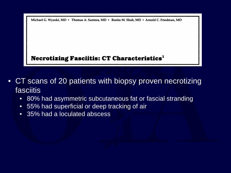
• CT scans of 20 patients with biopsy proven necrotizing fasciitis
• 80% had asymmetric subcutaneous fat or fascial stranding • 55% had superficial or deep tracking of air • 35% had a loculated abscess
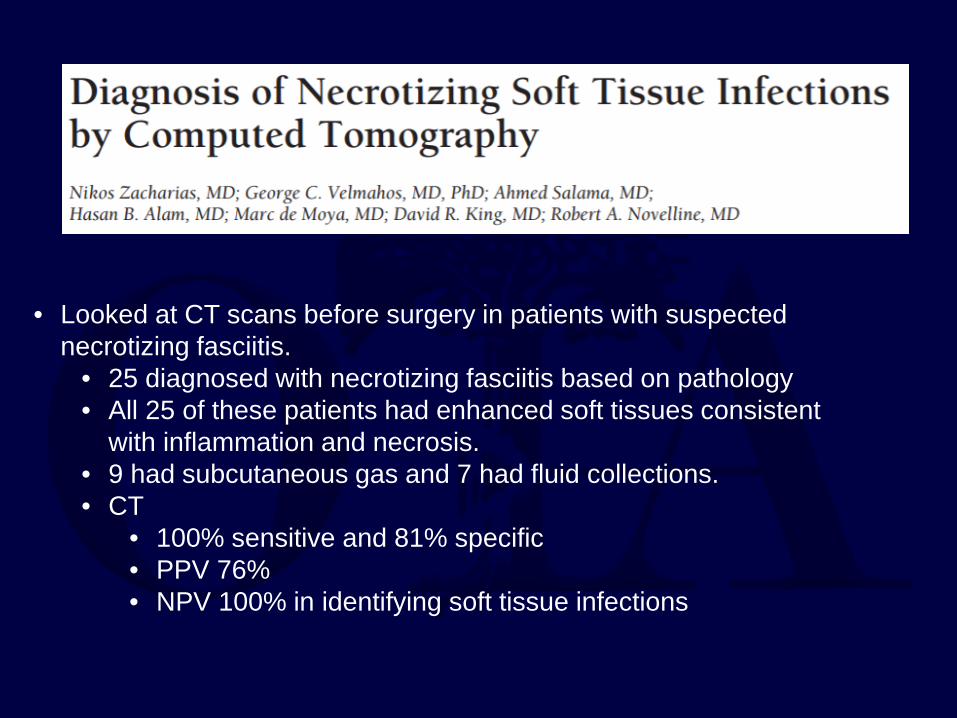
• Looked at CT scans before surgery in patients with suspected
necrotizing fasciitis. • 25 diagnosed with necrotizing fasciitis based on pathology • All 25 of these patients had enhanced soft tissues consistent
with inflammation and necrosis. • 9 had subcutaneous gas and 7 had fluid collections. • CT
• 100% sensitive and 81% specific • PPV 76% • NPV 100% in identifying soft tissue infections
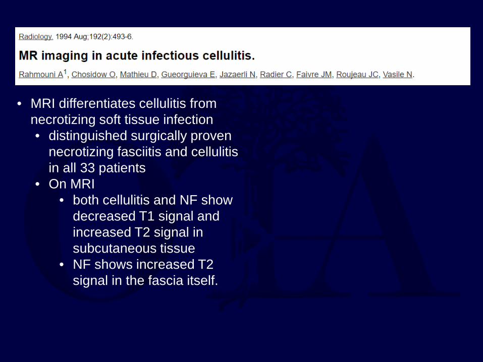
• MRI differentiates cellulitis from necrotizing soft tissue infection • distinguished surgically proven
necrotizing fasciitis and cellulitis in all 33 patients
• On MRI • both cellulitis and NF show
decreased T1 signal and increased T2 signal in subcutaneous tissue
• NF shows increased T2 signal in the fascia itself.
Necrotizing Soft Tissue Infections- Imaging
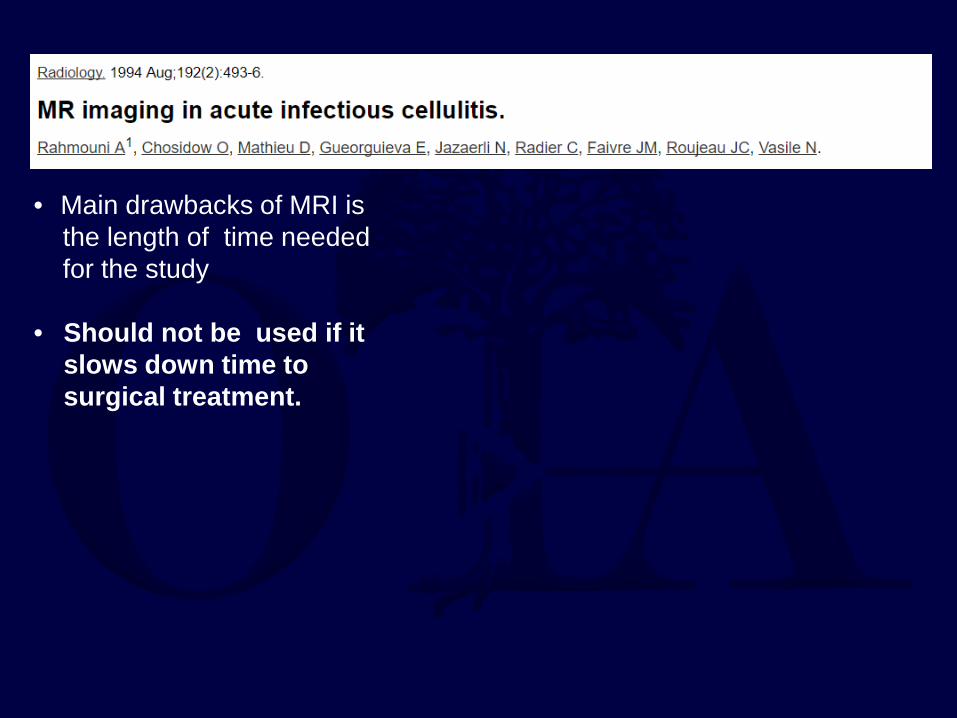
• Main drawbacks of MRI is the length of time needed for the study • Should not be used if it
slows down time to surgical treatment.
Necrotizing Soft Tissue Infections- Imaging
Streptococcal Group A strep (Strep. pyogenes)
• incubation period 1-3 days
• fulminant course due to streptolysin /
hemolysins / hyaluronidase
• “flesh-eating” bacteria – streptococcal pyrogenic exotoxin (speA, speB, speC)
Feingold DS Arch Dermatol 1996; 132:67-70
Necrotizing Soft Tissue Infections- Bacteriology
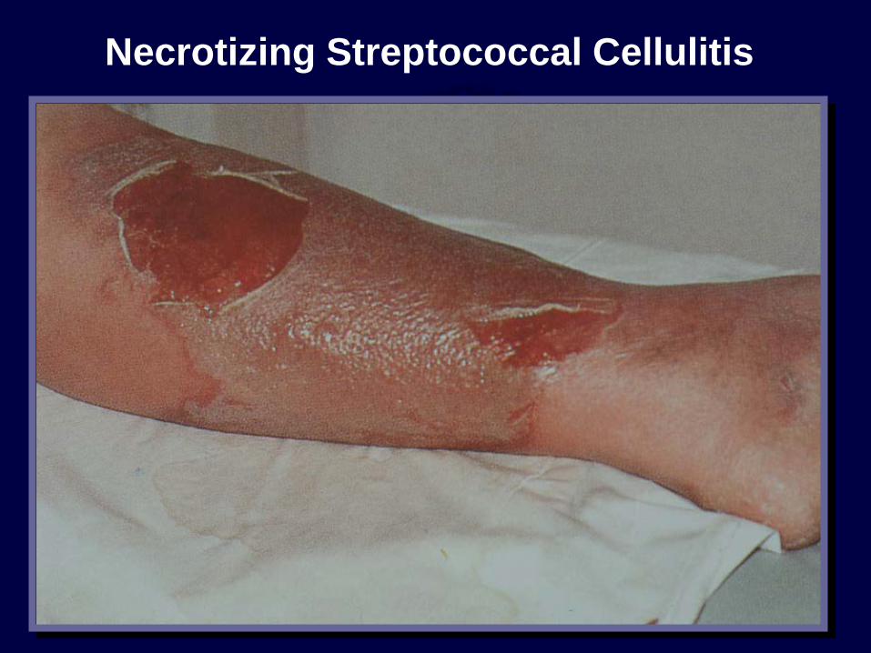
Necrotizing Streptococcal Cellulitis
Streptococcal Group B strep (Strep. Agalacitiae)
a. More common in pts with altered resistance
• diabetes, cancer, neonatal, etc
b. Short incubation period
Necrotizing Soft Tissue Infections- Bacteriology

Clostridial species a. incubation period 1-2 days
b. very fulminant course due to toxins
– C. perfringens – 20 known exotoxins
c. local gas, brownish discharge, high fever, high mortality
d. C. perfringens, C. septicum
Stevens DL Clin Inf Dis 1997; 25:S160-64
Necrotizing Soft Tissue Infections- Bacteriology
Gram negative bacillary E. coli, Kleb, Proteus, others • incubation period 7-14 days
• fever (FUO), local symptoms, sepsis
• may be mixed
Necrotizing Soft Tissue Infections- Bacteriology
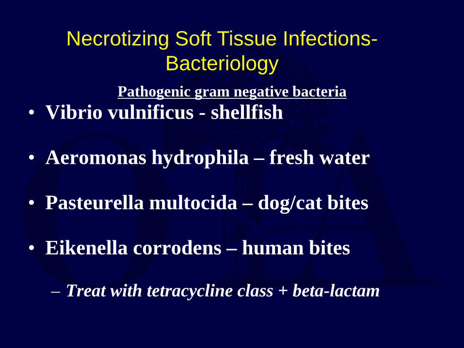
Pathogenic gram negative bacteria • Vibrio vulnificus - shellfish
• Aeromonas hydrophila – fresh water
• Pasteurella multocida – dog/cat bites
• Eikenella corrodens – human bites
– Treat with tetracycline class + beta-lactam
Necrotizing Soft Tissue Infections- Bacteriology
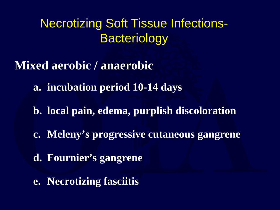
Mixed aerobic / anaerobic
a. incubation period 10-14 days
b. local pain, edema, purplish discoloration
c. Meleny’s progressive cutaneous gangrene
d. Fournier’s gangrene
e. Necrotizing fasciitis
Necrotizing Soft Tissue Infections- Bacteriology
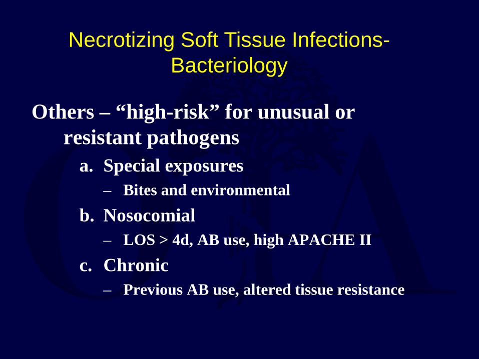
Others – “high-risk” for unusual or resistant pathogens
a. Special exposures – Bites and environmental
b. Nosocomial – LOS > 4d, AB use, high APACHE II
c. Chronic – Previous AB use, altered tissue resistance
Necrotizing Soft Tissue Infections- Bacteriology
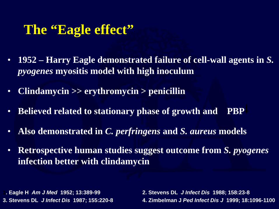
The “Eagle effect”
• 1952 – Harry Eagle demonstrated failure of cell-wall agents in S. pyogenes myositis model with high inoculum
• Clindamycin >> erythromycin > penicillin
• Believed related to stationary phase of growth and PBP
• Also demonstrated in C. perfringens and S. aureus models
• Retrospective human studies suggest outcome from S. pyogenes infection better with clindamycin
1. Eagle H Am J Med 1952; 13:389-99 2. Stevens DL J Infect Dis 1988; 158:23-8 3. Stevens DL J Infect Dis 1987; 155:220-8 4. Zimbelman J Ped Infect Dis J 1999; 18:1096-1100
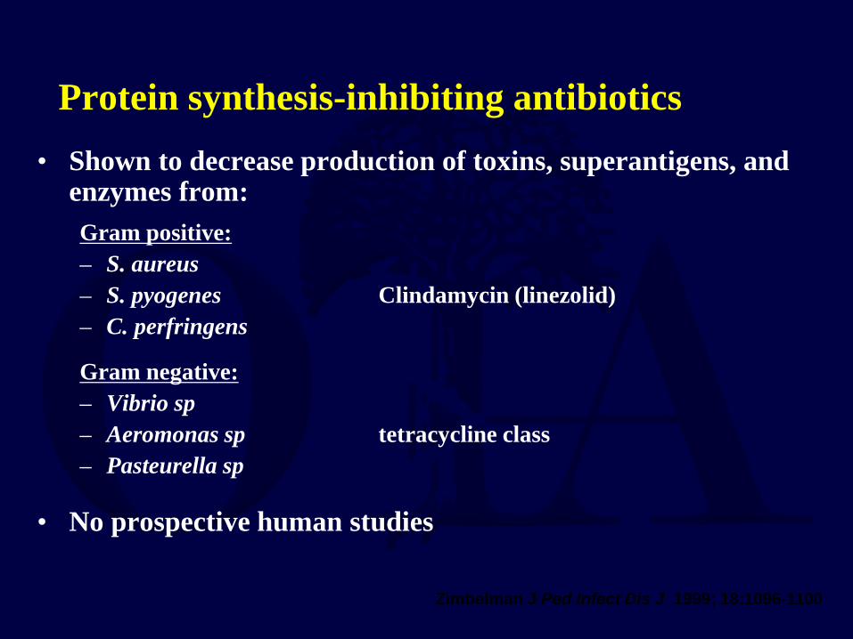
Protein synthesis-inhibiting antibiotics • Shown to decrease production of toxins, superantigens, and
enzymes from: Gram positive: – S. aureus – S. pyogenes Clindamycin (linezolid) – C. perfringens
Gram negative: – Vibrio sp – Aeromonas sp tetracycline class – Pasteurella sp
• No prospective human studies
Zimbelman J Ped Infect Dis J 1999; 18:1096-1100
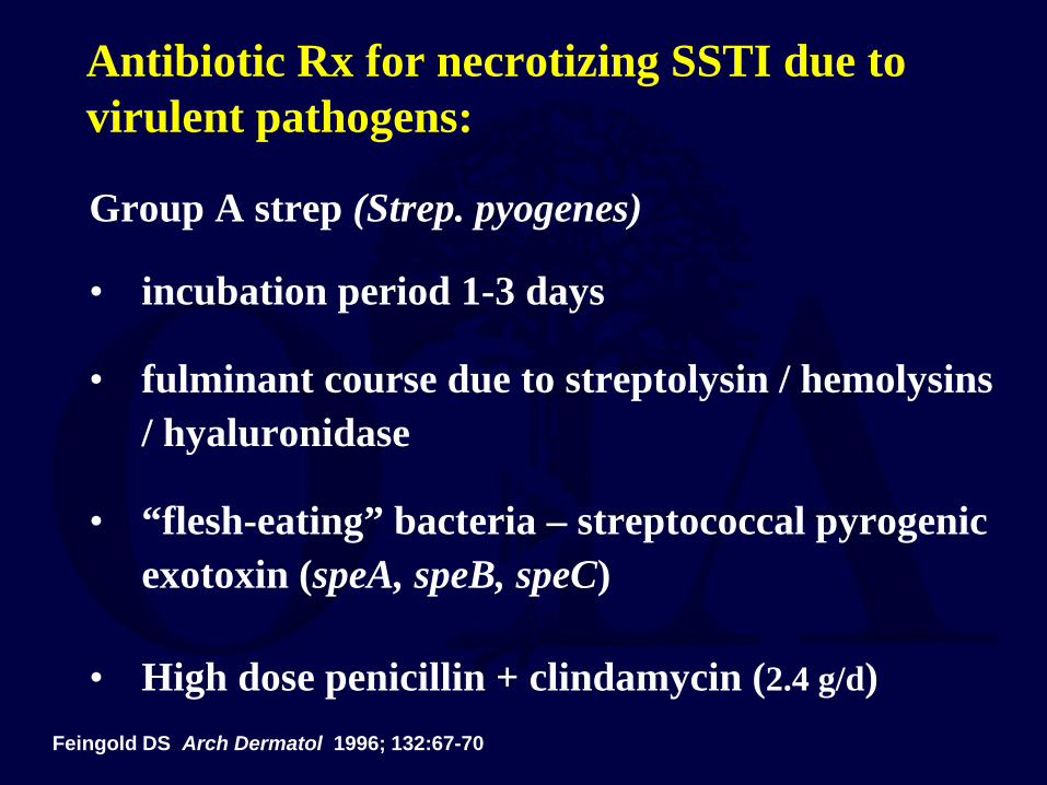
Antibiotic Rx for necrotizing SSTI due to virulent pathogens:
Group A strep (Strep. pyogenes)
• incubation period 1-3 days
• fulminant course due to streptolysin / hemolysins
/ hyaluronidase
• “flesh-eating” bacteria – streptococcal pyrogenic exotoxin (speA, speB, speC)
• High dose penicillin + clindamycin (2.4 g/d)
Feingold DS Arch Dermatol 1996; 132:67-70
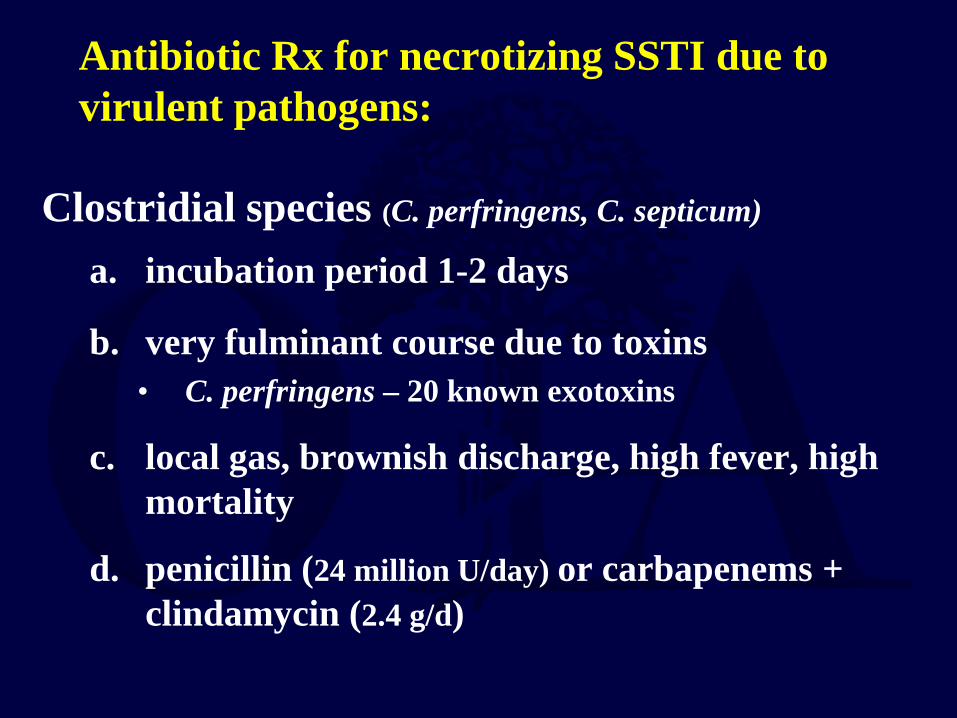
Antibiotic Rx for necrotizing SSTI due to virulent pathogens:
Clostridial species (C. perfringens, C. septicum)
a. incubation period 1-2 days b. very fulminant course due to toxins
• C. perfringens – 20 known exotoxins c. local gas, brownish discharge, high fever, high
mortality d. penicillin (24 million U/day) or carbapenems +
clindamycin (2.4 g/d)
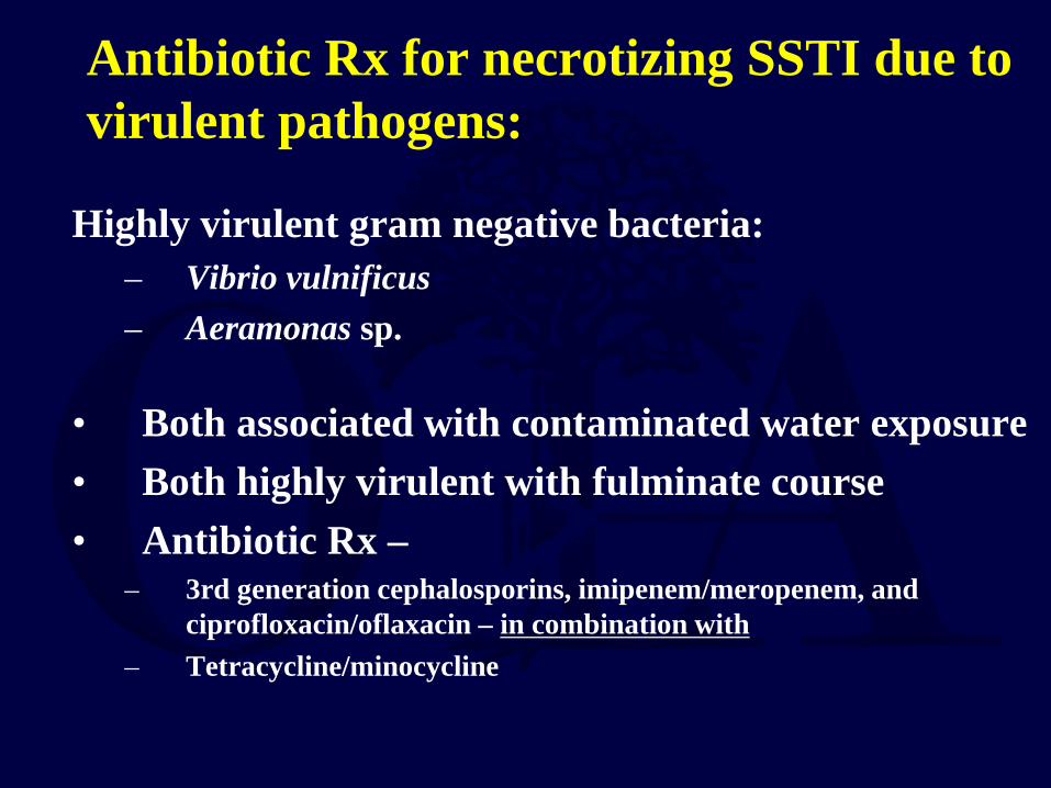
Antibiotic Rx for necrotizing SSTI due to virulent pathogens:
Highly virulent gram negative bacteria: – Vibrio vulnificus – Aeramonas sp.
• Both associated with contaminated water exposure • Both highly virulent with fulminate course • Antibiotic Rx –
– 3rd generation cephalosporins, imipenem/meropenem, and ciprofloxacin/oflaxacin – in combination with
– Tetracycline/minocycline
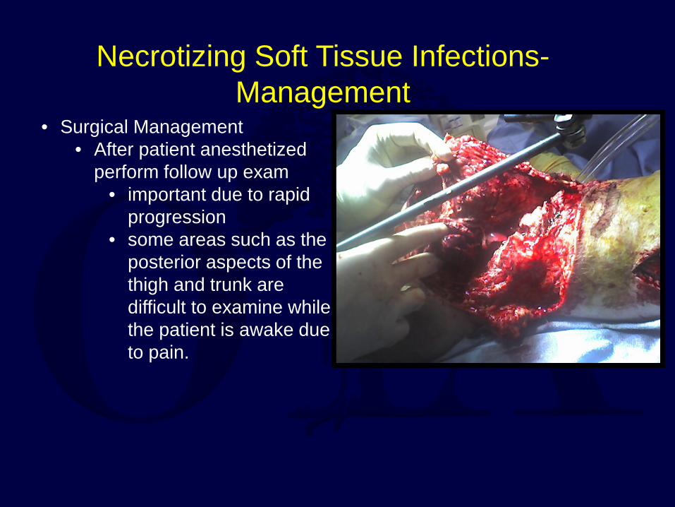
• Surgical Management • After patient anesthetized
perform follow up exam • important due to rapid
progression • some areas such as the
posterior aspects of the thigh and trunk are difficult to examine while the patient is awake due to pain.
Necrotizing Soft Tissue Infections- Management
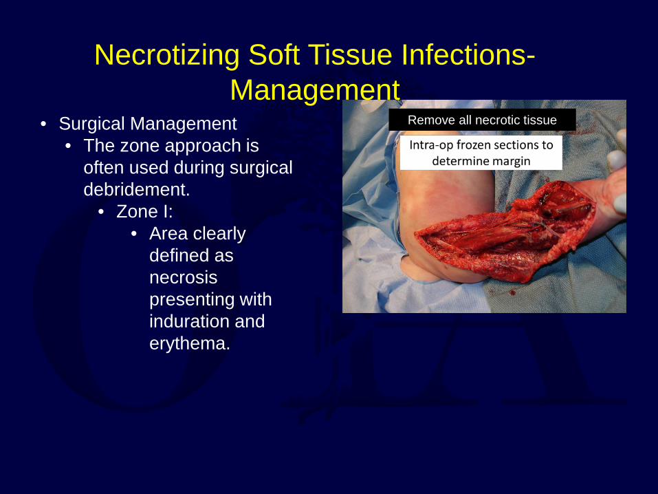
• Surgical Management • The zone approach is
often used during surgical debridement.
• Zone I: • Area clearly
defined as necrosis presenting with induration and erythema.
Remove all necrotic tissue
Necrotizing Soft Tissue Infections- Management
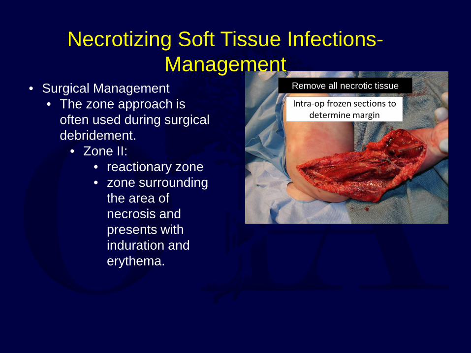
• Surgical Management • The zone approach is
often used during surgical debridement.
• Zone II: • reactionary zone • zone surrounding
the area of necrosis and presents with induration and erythema.
Remove all necrotic tissue
Necrotizing Soft Tissue Infections- Management
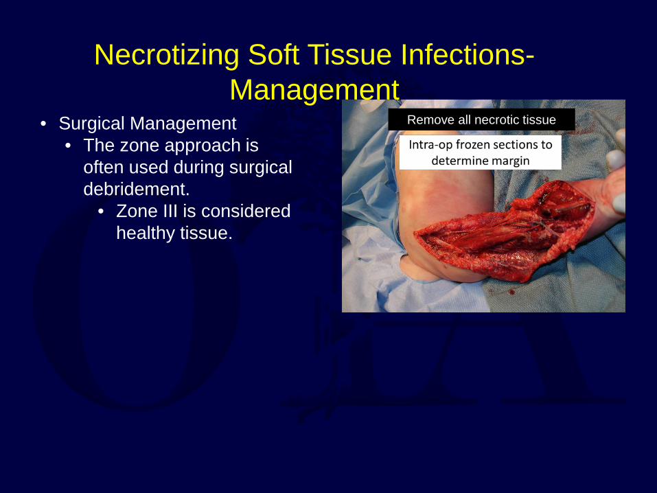
• Surgical Management • The zone approach is
often used during surgical debridement.
• Zone III is considered healthy tissue.
Remove all necrotic tissue
Necrotizing Soft Tissue Infections- Management
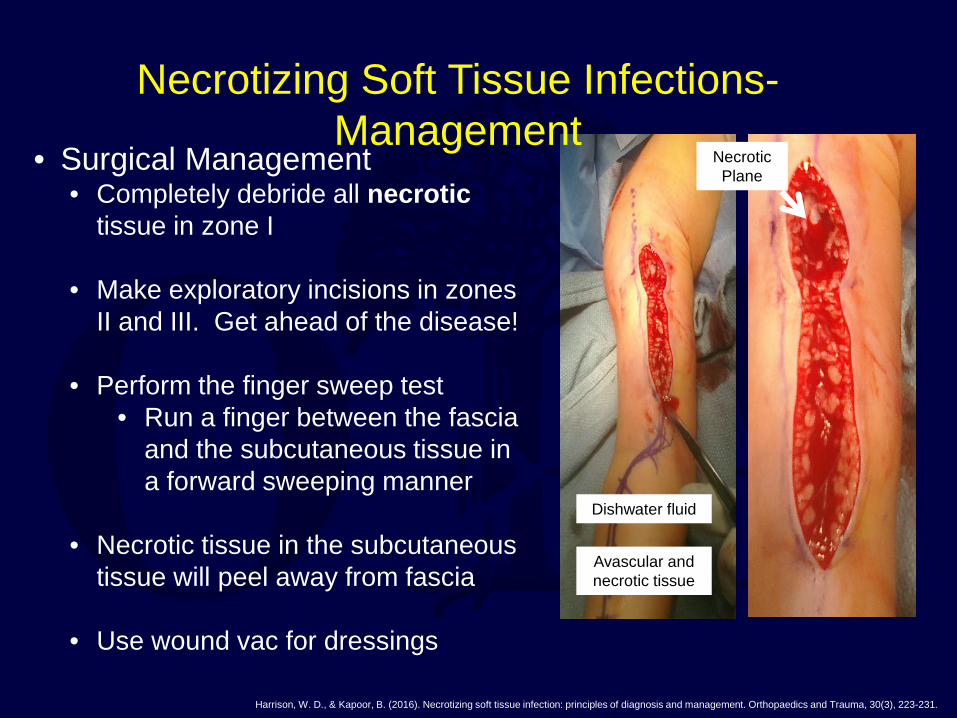
• Surgical Management • Completely debride all necrotic
tissue in zone I • Make exploratory incisions in zones
II and III. Get ahead of the disease!
• Perform the finger sweep test • Run a finger between the fascia
and the subcutaneous tissue in a forward sweeping manner
• Necrotic tissue in the subcutaneous
tissue will peel away from fascia
• Use wound vac for dressings
Harrison, W. D., & Kapoor, B. (2016). Necrotizing soft tissue infection: principles of diagnosis and management. Orthopaedics and Trauma, 30(3), 223-231.
Necrotic Plane
Dishwater fluid
Avascular and necrotic tissue
Necrotizing Soft Tissue Infections- Management
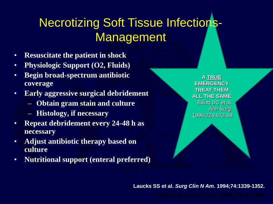
• Resuscitate the patient in shock • Physiologic Support (O2, Fluids) • Begin broad-spectrum antibiotic
coverage • Early aggressive surgical debridement
– Obtain gram stain and culture – Histology, if necessary
• Repeat debridement every 24-48 h as necessary
• Adjust antibiotic therapy based on culture
• Nutritional support (enteral preferred)
Elliott D et al. Am J Surg. 2000;179:361-366.
Laucks SS et al. Surg Clin N Am. 1994;74:1339-1352.
A TRUE EMERGENCY TREAT THEM
ALL THE SAME Elliott DC et al.
Ann Surg. 1996;224:672-68
Necrotizing Soft Tissue Infections- Management
• Surgical debridement is mainstay of therapy
• Aggressive EARLY incision or debridement of all involved tissues
• Average number of débridements 3-4 / patient
• Primary Closure of wounds once tissue improves
• STSG or flap coverage most commonly used for coverage of tissue defects
• Studies suggest that early and aggressive surgical therapy can reduce mortality to < 10%
Bosshardt TL. Arch.Surg. 1996;131:846-52 Elliott DC. Ann.Surg. 1996; 224:672-83
Gunter OL Surg. Infect. 2008; 9:443-450 Bilton BD. Am.Surg. 1998; 64:397-400
Necrotizing Soft Tissue Infections- Management
Adjuvants
• IGG • Steroids
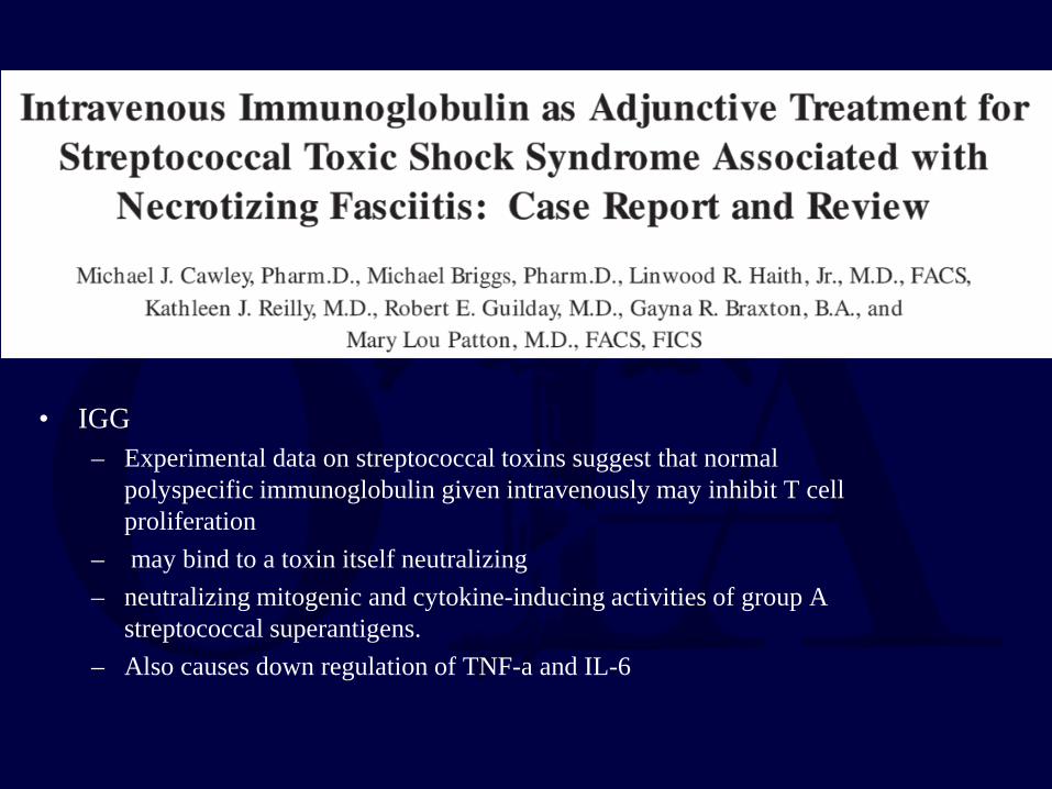
Adjuvants
• IGG – Experimental data on streptococcal toxins suggest that normal
polyspecific immunoglobulin given intravenously may inhibit T cell proliferation
– may bind to a toxin itself neutralizing – neutralizing mitogenic and cytokine-inducing activities of group A
streptococcal superantigens. – Also causes down regulation of TNF-a and IL-6
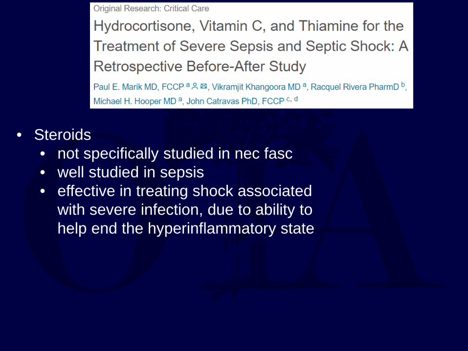
• Steroids • not specifically studied in nec fasc • well studied in sepsis • effective in treating shock associated
with severe infection, due to ability to help end the hyperinflammatory state
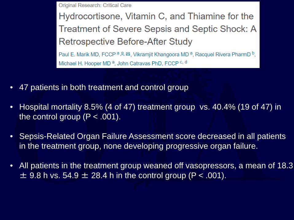
• 47 patients in both treatment and control group • Hospital mortality 8.5% (4 of 47) treatment group vs. 40.4% (19 of 47) in
the control group (P < .001). • Sepsis-Related Organ Failure Assessment score decreased in all patients
in the treatment group, none developing progressive organ failure. • All patients in the treatment group weaned off vasopressors, a mean of 18.3
± 9.8 h vs. 54.9 ± 28.4 h in the control group (P < .001).
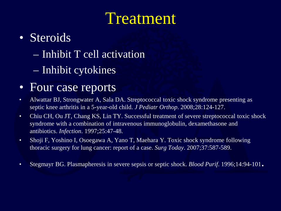
Treatment • Steroids
– Inhibit T cell activation – Inhibit cytokines
• Four case reports • Alwattar BJ, Strongwater A, Sala DA. Streptococcal toxic shock syndrome presenting as
septic knee arthritis in a 5-year-old child. J Pediatr Orthop. 2008;28:124-127. • Chiu CH, Ou JT, Chang KS, Lin TY. Successful treatment of severe streptococcal toxic shock
syndrome with a combination of intravenous immunoglobulin, dexamethasone and antibiotics. Infection. 1997;25:47-48.
• Shoji F, Yoshino I, Osoegawa A, Yano T, Maehara Y. Toxic shock syndrome following thoracic surgery for lung cancer: report of a case. Surg Today. 2007;37:587-589.
• Stegmayr BG. Plasmapheresis in severe sepsis or septic shock. Blood Purif. 1996;14:94-101.
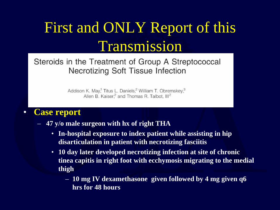
First and ONLY Report of this Transmission
• Case report – 47 y/o male surgeon with hx of right THA
• In-hospital exposure to index patient while assisting in hip disarticulation in patient with necrotizing fasciitis
• 10 day later developed necrotizing infection at site of chronic tinea capitis in right foot with ecchymosis migrating to the medial thigh
– 10 mg IV dexamethasone given followed by 4 mg given q6 hrs for 48 hours

How to Avoid Transmission?
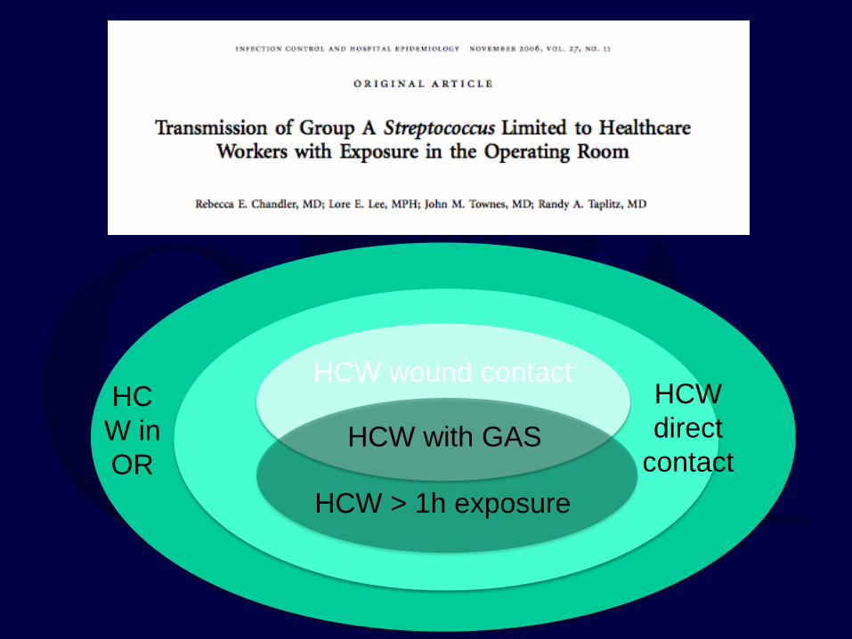
HCW in OR
HCW direct
contact HCW > 1h exposure
HCW wound contact
HCW with GAS
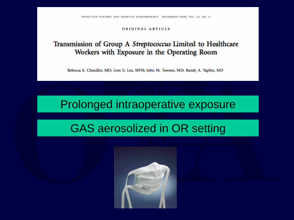
Prolonged intraoperative exposure
GAS aerosolized in OR setting

Prolonged intraoperative exposure
GAS aerosolized in OR setting
X
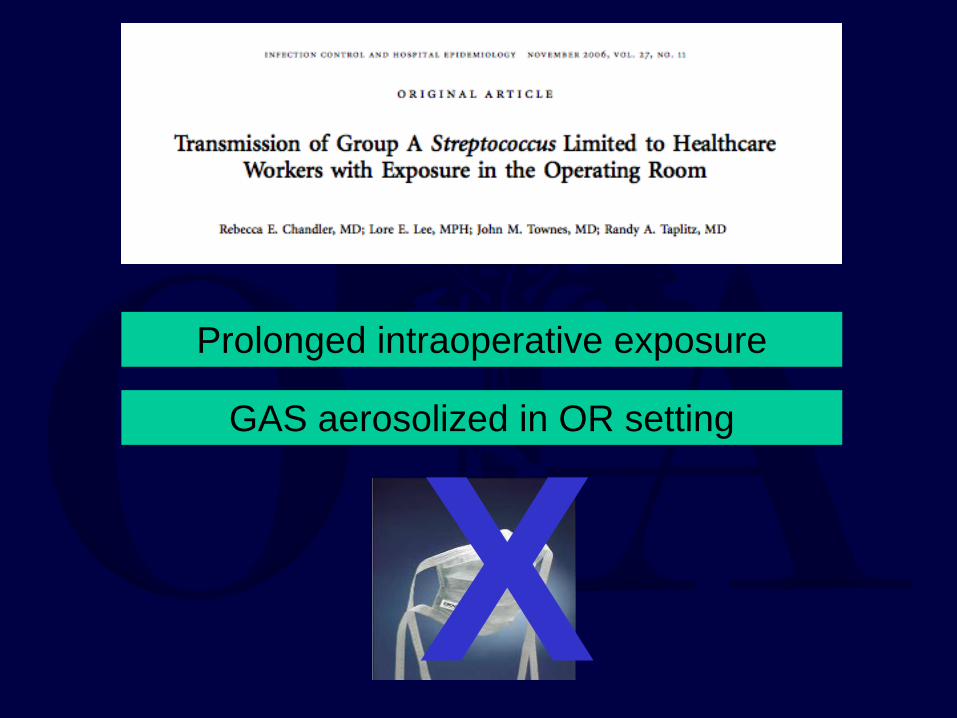
Prolonged intraoperative exposure
GAS aerosolized in OR setting
X
Index patient: 34 yo M LE nec fasc, taken to OR for debridement
Index patient: 34 yo M LE nec fasc, taken to OR for debridement
48 h postop HCW 1 (resp therapist): + GAS pharyngitis
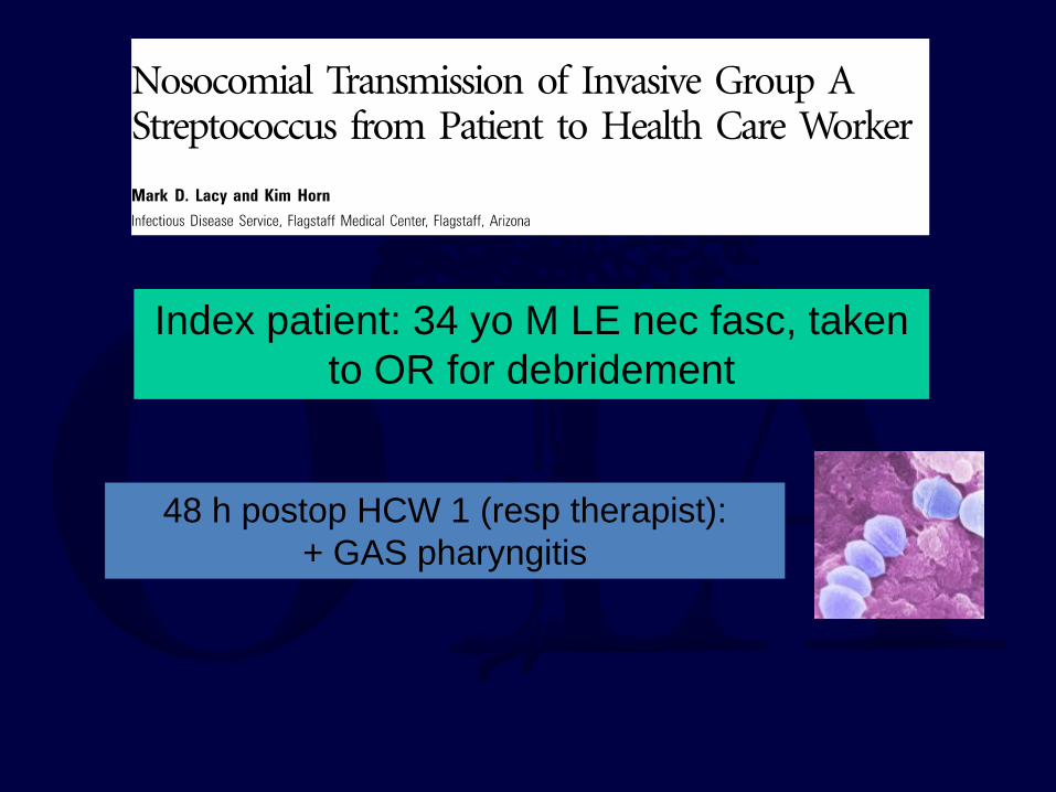
Index patient: 34 yo M LE nec fasc, taken to OR for debridement
48 h postop HCW 1 (resp therapist): + GAS pharyngitis
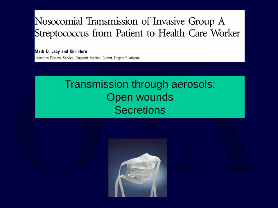
Transmission through aerosols: Open wounds
Secretions
Transmission through aerosols: Open wounds
Secretions
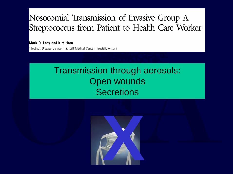
Transmission through aerosols: Open wounds
Secretions
X
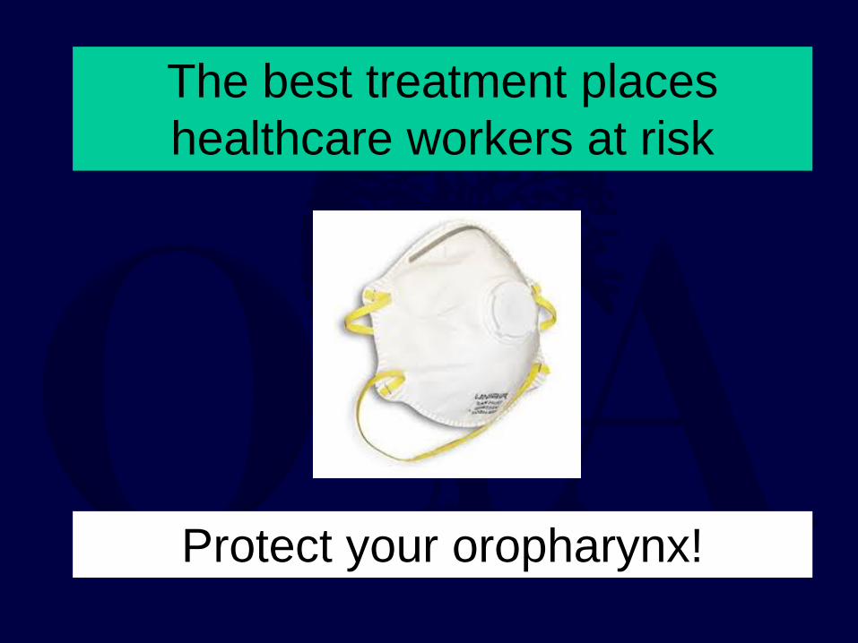
The best treatment places healthcare workers at risk
Protect your oropharynx!
• HARD signs = necrotizing infection and require OR – Bullae, cutaneous anesthesia, ecchymosis, tense edema, gas
• Early and aggressive surgical debridement improves outcome
with achievable mortality of < 10%
• Empiric surgical exploration if in doubt!
• Protect your oropharynx
• Consider Steroids or IGG in “Toxic” patient (Strep)
• Transfer patient AFTER initial debridement
Summary