Embed Size (px)
Citation preview
LEARNING AND MEMORY NEUROREPORT
0959-4965 & Lippincott Williams & Wilkins Vol 11 No 16 9 November 2000 3641
Basal ganglia involvement in memory-guided movement sequencing
V. Menon,1,3,4,CA R. T. Anagnoson,1,4 G. H. Glover2 and A. Pfefferbaum5
Departments of 1Psychiatry and Behavioral Sciences and 2Radiology, and 3Program in Neuroscience, Stanford University Schoolof Medicine, Stanford, CA 94305-5719; 4VA Palo Alto Health Care System, Palo Alto, CA 94304; 5SRI International, Menlo Park,
CA 94025, USA
CACorresponding Author
Received 12 July 2000; accepted 3 September 2000
The basal ganglia (BG) are thought to play a critical role inmotor planning and movement sequencing. While electrophy-siological and imaging studies have shown that the dorso-lateralprefrontal cortex (DLPFC) is involved in working memory(WM), the involvement of the BG in this process is not wellunderstood. We used a motor sequencing task to investigatethe differential role of BG nuclei in memory-guided movement.Signi®cant activation was observed in the DLPFC and posteriorputamen and globus pallidus (GP), with a trend in the caudateand no differences in the anterior putamen. We then inves-tigated the effect of BG out¯ow on thalamic activation usingfunctional connectivity analysis. Activation in the posterior
putamen�GP was found to be correlated with thalamicactivation only in the hemisphere contralateral to movement.These results provide the ®rst fMRI evidence that the BG maymodulate activity in the thalamus during working memory-guided movement sequencing. Our ®ndings suggest that the BGactivation may re¯ect increased motor sequencing demandsduring the memory-guided movement condition and, speci®-cally, that the posterior putamen and GP may play a role inmaintenance of representations in WM in a manner thatcontributes to planning and temporal organization of motorsequencing. NeuroReport 11:3641±3645 & 2000 LippincottWilliams & Wilkins.
Key words: Basal ganglia; Caudate; DLPFC; fMRI; Motor sequencing; Putamen; Working memory
INTRODUCTIONThe basal ganglia (BG) play an essential role in adaptivebehavior in general and movement initiation, control andsequencing in particular. Although the BG have beenprimarily thought to be involved in motor functions,researchers have argued that the BG may play a moredirect role in supporting cognitive operations [1±3]. Thisargument has been supported by a number of studieswhich have noted a pro®le of cognitive performancede®cits in patients with BG disease [4±6]. BG-relatedcognitive dysfunction might result from a number offactors, including de®cits in the allocation of attentionresources, the maintenance of representations in workingmemory, the temporal organization of behavior, planning,or the self-elaboration of internal strategies, which resem-ble dysfunctions of processes that are commonly consid-ered to be controlled by the frontal lobes [5].
In the present study we investigated the role of variousBG nuclei in supporting cognitive operations by examiningits involvement in working memory-guided movementsequencing. Electrophysiological studies in primates haveexamined the role of the BG in memory-guided movementbut there have been no such studies in humans to date.Mushiake and Strick [7] investigated the activity of globuspallidus (GP) neurons in Macaque monkeys during se-quential reaching tasks with and without memory require-
ments and reported that several GP neurons werepreferentially active during working memory-guidedmovement. Electrophysiological studies have also foundmemory-contingent neurons in the substantia nigra duringvisual and saccade responses [8,9]. However, the preciserole played by the striatum in visuo-spatial WM is still notwell understood, as some electrophysiological studies havefound delay related neuronal activity in the caudate [10,11]while others have failed to ®nd any such activity in thecaudate or anterior putamen [12]. Levy et al. [13] examinedthe role of the monkey caudate in spatial and non-spatialWM tasks using PET imaging and found that the caudatehead was active during spatial WM and the caudate tailwas active during nonspatial WM. Other electrophysiologi-cal studies have suggested that the caudate and putamennuclei of the BG primarily perform an integrative role inbinding sensory inputs to motor response [14]. Kermadiand Joseph [15] have reported that neurons in the caudateappear to play a role in the construction, but not storage ofspatial sequencing.
To date, no imaging studies have examined the differ-ential activation of BG nuclei in the context of memory-guided movements. Although, in principle, functionalmagnetic resonance imaging (fMRI) provides adequateresolution to image the basal ganglia at the spatial scale ofthe individual nuclei, activating these nuclei with fMRI has
proven to be dif®cult. In this study we investigated thedifferential role of various BG nuclei during memory-guided movement sequencing and the effect of BG out¯owon thalamic activation. We modi®ed a motor sequencingtask, which we had previously shown to reliably activatethe BG [16], to include a delay component and investigatedactivation related to WM in the DLPFC, which is com-monly activated by the executive component of WM tasks[17±19], and various nuclei of the BG. Previous studies thathave investigated BG function have mainly used purelymotor tasks which activate the motor output regions of theBG [20±22]. Our memory-guided movement sequencingtask was analogous to those used in electrophysiologicalstudies [7].
MATERIALS AND METHODSSubjects: Ten healthy right-handed subjects (six men andfour women; ages 20±35 years) participated in the studyafter the study was explained to each and written informedconsent was obtained. The human subjects committee atStanford University and the Palo Alto VA Health CareSystem approved this protocol. No subject had more than1 mm of head motion in any axis during the scan.
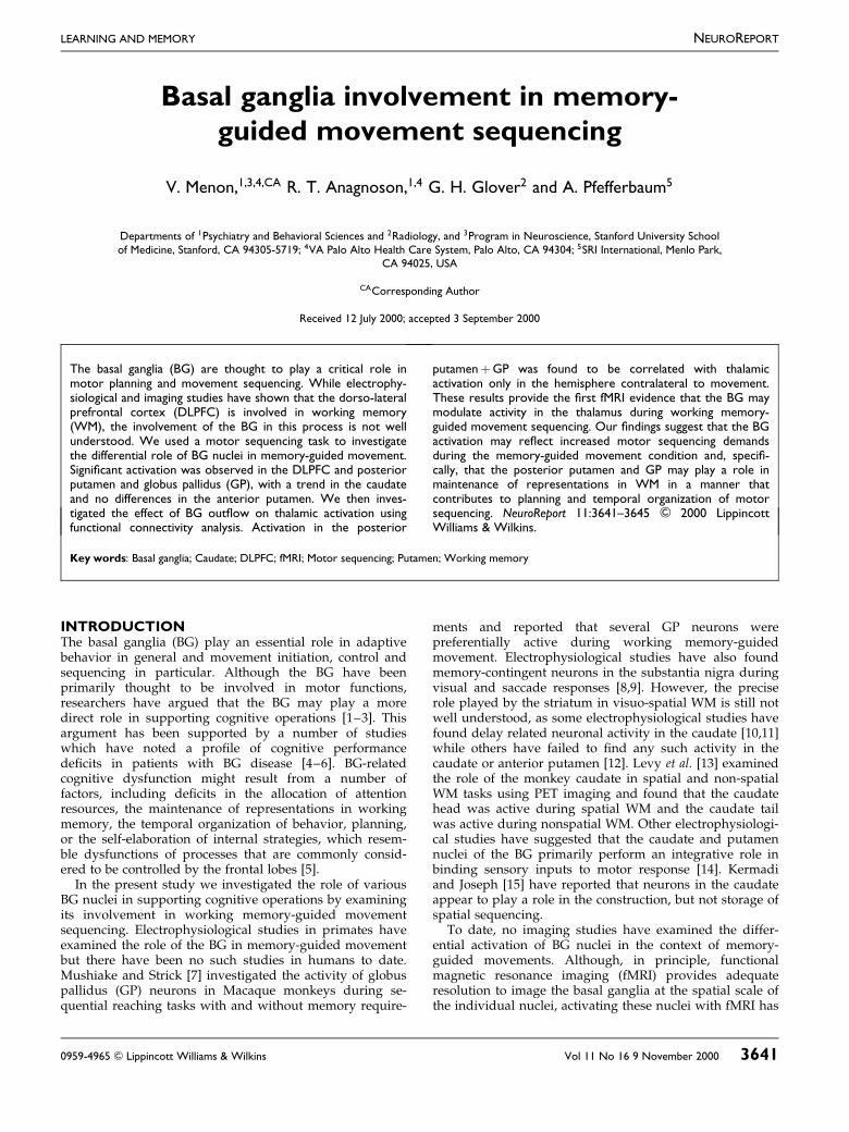
Experimental design: The tasks consisted of alternatingepochs of motor sequencing with and without WM de-mands. Trials (8 s each) were presented using a blocked-design with ®ve trials per block and six blocks of eachcondition (12 blocks total) presented in an ABAB... para-digm. Brief instructions preceded each block. Motor se-quencing involving ®nger movements do not elicit reliablefMRI activation of the BG; we therefore designed a task,involving arm movements, which reliably activated thecaudate, putamen and GP [16]. In both task conditions,subjects rested the closed ®st of their right hand on thebase of a palm-shaped keypad. Movement consisted oftouching, with thumb and fore®nger pinched together, oneof four locations 15 cm away from the base and returningto the base position. Subjects practiced the task brie¯y for2 min 40 s (four epochs) 30 min before the scan and weremonitored visually during the scan to verify consistent taskperformance. Numbers between 1 and 4 were presentedbinaurally and subjects made arm movements to corre-sponding locations on the keypad. In the memory-guided(experimental) condition, subjects heard three numbers insuccession and had to respond with the arm movementsafter a 3.5 s delay. In the control condition, numbers werepresented with an ISI of 1.5 s and subjects moved their armimmediately after each number was presented (Fig. 1). Thenumber of movements and auditory stimuli were balancedacross conditions.
Data acquisition: Images were acquired on a conven-tional 1.5 T GE (Milwaukee, WI) scanner using a quad-rature whole head coil. Twelve axial slices (6 mm thick,0 mm skip), roughly extending ÿ20 to �52 mm relative tothe anterior commissure, were imaged with a temporalresolution of 4 s at 120 time points using a T2�-weightedgradient echo spiral pulse sequence (TR� 1000 ms,TE� 40 ms, ¯ip angle� 408 and 4 interleaves) [23]. The ®eldof view was 310 mm and the effective inplane spatialresolution was 4.35 mm.
Data analysis: Images were corrected for movementusing least square minimization, normalized to stereotaxicTalairach coordinates [24], resampled every 2 mm usingsinc interpolation and smoothed with a 4 mm Gaussiankernel. Statistical analysis was performed on individualsubject data to determine which voxels showed signi®-cantly greater activation during the memory-guided, com-pared to the control condition. The general linear modeland the theory of Gaussian random ®elds as implementedin SPM96 were used to analyze the fMRI data. This methodimplements a multivariate regression analysis and correctsfor temporal and spatial autocorrelations in the fMRI data[25]. For each subject, voxel-wise t-statistics were com-puted and normalized to Z scores to provide a statisticalmeasure of activation that is independent of sample size.
ROI analysis: Regions of interest (ROIs) were constructedbased upon known neuroanatomical landmarks [26] andguided by the Talairach atlas [24] for caudate head,anterior putamen, posterior putamen and GP, and DLPFC(BA 9/46). We also examined activation in the thalamussince it is the primary target of BG output. Given thelimited spatial resolution of the fMRI scan we did notattempt to differentiate activation in the various thalamicnuclei. The anterior commissure was used to demarcateanterior and posterior putamen and GP. Separate left andright hemisphere ROIs were constructed for each region.The percentage of voxels activated above a threshold ofZ . 1.645 ( p , 0.05) within each ROI was determined foreach subject. Regional differences in activation were exam-ined using ANOVA.
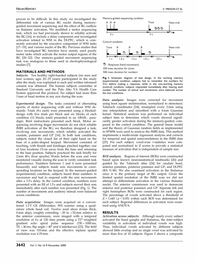
RESULTSActivation across subjects: Although nearly every subjectactivated the basal ganglia and thalamus, the inter-subjectvariability in activation of individual voxels was high.Thus, individual voxels activated by different subjectsshowed little overlap and no single voxel was activated bymore than ®ve of 10 subjects. Figure 2 shows a composite
Memory-guided sequencing condition
Control condition
Tone Next trial
- Response (hand movement)250 msec duration for tones500 msec duration for numbers
Next trial
5 s 3 s
Tone
1 s 1.5 s 1.5 s 1.5 s 2.5 s
# # #
# # #
Fig. 1. Schematic diagram of task design. In the working memory(experimental) condition, subjects had to remember the numbers for3.5 s before making a response, while in the non-working memory(control) condition, subjects responded immediately after hearing eachnumber. The number of stimuli and movements were balanced acrossthe two conditions.
NEUROREPORT V. MENON ET AL.
3642 Vol 11 No 16 9 November 2000
image of the number of voxels activated by each subjectand captures some of the variability in activation acrosssubjects. Although the precise voxel activated in eachsubject in the basal ganglia and thalamus were quitevariable, six of 10 subjects activated the caudate, six of 10activated the anterior putamen, nine of 10 activated theposterior putamen�GP and nine of 10 activated thethalamus. All 10 subjects activated the DLPFC. This map isshown for illustrative purposes only. Statistical analysis of
activation across subjects was conducted using an ANOVAon the percentage of voxels activated by each subject ineach region of interest.
Region of interest analysis: A two-way repeated-meas-ures ANOVA with factors ROI (DLPFC, caudate, anteriorputamen, posterior putamen�GP) and hemisphere (leftand right) was used to investigate the pro®le of signi®cantregional differences in activation. There was no ROI 3
Fig. 2. Composite map of activation across 10 subjects in the dorsolateral prefrontal cortex, basal ganglia, and thalamus during the memory-guidedcompared to a non-working-memory guided movement sequencing task. The number of subjects who showed signi®cant activation (Z . 1.65, p , 0.05)in each voxel was computed and displayed according to the adjoining scale. The precise voxels activated by each subject in the basal ganglia and thalamuswere quite variable. Six of 10 subjects activated the caudate, six of 10 activated the anterior putamen, nine of 10 activated the posteriorputamen� globus pallidus and nine of 10 activated the thalamus. All 10 subjects activated the dorsolateral prefrontal cortex. This map is shown forillustrative purposes only. Statistical analysis of activation across subjects was conducted using an ANOVA on the percentage of voxels activated by eachsubject in each region of interest.
BASAL GANGLIA AND MEMORY-GUIDED MOVEMENT SEQUENCING NEUROREPORT
Vol 11 No 16 9 November 2000 3643

hemisphere (F(3,27)� 0.120, p� 0.948) interaction, or maineffect of hemisphere (F(1,9)� 0.388, p� 0.549), but a signi®-cant main effect of ROI was found (F(3,27)� 3.197,p� 0.039). We compared activation in the memory-guidedand control conditions using a two-tailed t-test. Signi®cantactivation differences were found in the DLPFC(t(9)� 3.036, p� 0.014) and posterior putamen�GP(t(9)� 2.545, p� 0.031), but not the caudate (t(9)� 2.052,p� 0.070) or anterior putamen (t(9)� 1.700, p� 0.122; Fig.2). Signi®cant activation was also found in the thalamus(t(9)� 2.555, p� 0.031). Figure 3 compares activation acrosssubjects in the DLPFC, BG and thalamus.
Basal ganglia and thalamus connectivity analysis: Wethen investigated the effect of BG out¯ow on thalamicactivation separately in each hemisphere. Left thalamicactivation was signi®cantly correlated with left posteriorputamen�GP activation (Spearman R� 0.66, p� 0.04; Fig.4), but right thalamus activation was not related to rightposterior putamen�GP activation (Spearman R� 0.27,p� 0.46). Activation in the left and right thalami was alsosigni®cantly correlated (Spearman R� 0.83, p� 0.003).
DISCUSSIONThe motor sequencing task used in the present study hasbeen previously shown to activate the caudate, anteriorputamen, and the posterior putamen�GP [16]. The currentstudy compared activation between motor sequencing con-ditions with and without WM demands. Consistent withprevious imaging studies [18,19], we observed signi®cantactivation of the left and right DLPFC. In addition, signi®-cant activation was detected in the posterior putamen�GP,
with a trend toward signi®cant activation in the caudate.No signi®cant activation was detected in the anterior puta-men. There were no signi®cant hemispheric differences inactivation, suggesting that the overall activation is notentirely due to increased motor demands during the mem-ory-guided task. It should be noted that the motor compo-nents of the task were balanced across the WM and non-WM conditions. Our results provide the ®rst evidence forposterior putamen�GP involvement during working mem-ory-guided movement sequencing in humans. These resultsare in agreement with electrophysiological studies in pri-mates [7] which have indicated that GP activation is mod-ulated by memory requirements during motor sequencing.
In addition to signi®cant activation in the BG, we foundincreased activation in the thalamus. Furthermore, thalamicactivation was found to be correlated with posterior puta-
Left hemisphere
Right hemisphere
DLPFC
Cauda
te
Ant pu
tamen
Post
putam
en1
GP
Thalam
us
12
10
8
6
4
2
0
Perc
enta
ge o
f vox
els
activ
ated
Fig. 3. Mean and S.E. of activation in the dorsolateral prefrontal cortex,basal ganglia and thalamus regions of interest (ROI) during the memory-guided minus the control condition. The percentage of voxels within eachROI activated by each subject above a threshold of Z . 1.65 ( p , 0.05)was calculated used as the measure of activation.
Fig. 4. Relationship between posterior putamen� globus pallidus andthalamus activation. (a) Scatterplot of left posterior putamen� globuspallidus and left thalamus activation. (b) Scatterplot of the same activationafter rank ordering to correct for outliers and non-normal distribution,corresponding to the use of a non-parametric Spearman correlation. Theactivations were signi®cantly correlated ( p , 0.05; Spearman correlation).The percentage of voxels activated by each subject in each ROI was usedas the measure of activation.
(a)
Left posterior putamen 1 globus pallidus activation
22 0 2 4 6 8 10 12 1421
1
3
5
7
9
11
Left
tha
lam
us a
ctiv
atio
n
(b)
Left posterior putamen 1 globus pallidus ranks
0 2 4 6 8 10 120
2
4
6
8
10
12
Left
tha
lam
us r
anks
NEUROREPORT V. MENON ET AL.
3644 Vol 11 No 16 9 November 2000
men�GP activation in the left but not the right hemisphere.The increase in thalamic activation contralateral to themovement may therefore be related to increased motorcoordination demands during the memory condition. Ourresults provide the ®rst fMRI evidence that thalamic activa-tion may be related to BG out¯ow and further suggest thatthe thalamic input from the BG is increased during workingmemory-guided movement. These activations may be re-lated to the additional motor sequencing demands that arepresent during the memory-guided, but not the controlcondition. Given recent neuroanatomical evidence for inter-hemispheric thalamic connectivity [27,28] we also investi-gated the relationship between left and right thalamicactivation. A signi®cant relation between left and rightthalamic activation was found suggesting that the left hemi-sphere (contralateral to movement) basal ganglia out¯owmay play a role in the bilateral integration of motorplanning systems via inter-thalamic cross-talk.
While electrophysiological studies have speci®cally in-vestigated the role of the BG in memory operations in thecontext of movement sequencing, imaging studies in hu-mans have focused on its role in WM tasks in general.These studies have suggested that the caudate head plays arole in visuo-spatial [29,30] but not verbal working mem-ory tasks [18,29]. However, Owen et al. [31] have reportedde®cits in GP activation, but not caudate or putamen, inParkinson's patients during spatial WM. Within the contextof movement sequencing, Menon et al. [16] compared BGactivation during externally and self-paced sequences ofarm movements and reported a dissociation in anteriorand posterior putamen and GP activation. They found thatboth externally paced and self-paced movements activatedthe posterior putamen�GP but only the externally pacedmovements activated the anterior caudate and putamen.Based on these results, as well as animal [32] and imagingstudies, they suggest that the anterior caudate and puta-men may be involved in sensory to motor stimulus-response mapping and the posterior putamen and GP maybe involved in the motor response itself.
In the present study, stimulus-response associationswere completely balanced across the two conditions. Thus,statistically signi®cant differences were not found in thecaudate and anterior putamen. Elliott and Dolan [30] havenoted that caudate activation is not modulated by increasein WM delay and several electrophysiological studies havefailed to ®nd delay related activation in the caudate [12].However, signi®cant activation was detected in the poster-ior putamen�GP which together receive sensorimotorinput and send output to the sensorimotor cortex via thethalamus [1,33]. Given the lack of evidence, from bothneuroimaging [30] and electrophysiological studies [12,15],of delay speci®c modulation of activation in the BG, ourresults suggest that the observed activation may re¯ectincreased motor sequencing demands during the memory-guided movement condition. Since motor sequencing alonehas been shown to activate more extensive regions of theBG [16], the present results suggest that the posteriorputamen�GP may be speci®cally involved in the con-struction, but not the storage of movement sequences.
CONCLUSIONTaken together, our ®ndings are compatible with thehypothesis that the BG plays a role in maintenance ofrepresentations in working memory in a manner thatcontributes to planning and temporal organization ofmotor sequencing. More broadly, our ®ndings suggest thatthe BG supports operations that could contribute to plan-ning of action. Given the neuroanatomical projections be-tween the BG and DLPFC via the thalamus [34], the BGmay also exert a modulatory in¯uence on the DLPFC andcontribute to adaptive behavior. Our ®ndings also suggestthat the paradigm developed in the present study can beused to investigate cognitive and motor abnormalities indisorders such as ADHD, Parkinson's disease and schizo-phrenia, in which BG output is known to be disrupted[31,35].
REFERENCES1. Alexander GE, DeLong MR and Strick PL. Annu Rev Neurosci 9, 357±381
(1986).
2. Graybiel AM. Schizophr Bull 23, 459±469 (1997).
3. Graybiel AM. Neurobiol Learn Mem 70, 119±136 (1998).
4. Owen AM, Iddon JL, Hodges JR et al. Neuropsychologia 35, 519±532
(1997).
5. Dubois B and Pillon B. J Neurol 244, 2±8 (1997).
6. Stebbins GT, Gabrieli JD, Masciari F et al. Neuropsychologia 37, 503±510
(1999).
7. Mushiake H and Strick PL. J Neurophysiol 74, 2754±2758 (1995).
8. Hikosaka O and Wurtz RH. J Neurophysiol 49, 1285±1301 (1983).
9. Hikosaka O, Sakamoto M and Miyashita, N. Exp Brain Res 95, 457±472
(1993).
10. Hikosaka O, Sakamoto M and Usui S. J Neurophysiol 61, 780±798 (1989).
11. Hikosaka O and Sakamoto M. Brain Dev 8, 454±461 (1986).
12. Rolls ET. Rev Neurol (Paris) 150, 648±660 (1994).
13. Levy R, Friedman HR, Davachi L et al. J Neurosci 17, 3870±3882 (1997).
14. Lidsky TI, Manetto C and Schneider JS. Brain Res 356, 133±1346 (1985).
15. Kermadi I and Joseph JP. J Neurophysiol 74, 911±933 (1995).
16. Menon V, Glover GH and Pfefferbaum A. Neuroreport 9, 1567±1573
(1998).
17. Romo R, Brody CD, Hernandez A et al. Nature 399, 470±473 (1999).
18. Smith EE and Jonides J. Science 283, 1657±1661 (1999).
19. D'Esposito M, Detre JA, Alsop DC et al. Nature 378, 279±281 (1995).
20. Lehericy S, van de Moortele PF, Lobel E et al. Ann Neurol 44, 398±404
(1998).
21. Reichenbach JR, Feiwell R, Kuppusamy K et al. Magn Reson Imaging 16,
281±287 (1998).
22. Bucher SF, Seelos KC, Stehling M et al. Neurology 45, 180±182 (1995).
23. Glover GH and Lai S. Magn Reson Med 39, 361±368 (1998).
24. Talairach J and Tournoux P. Co-planar Stereotaxic Atlas of the Human
Brain. New York: Thieme; 1998.
25. Friston KJ, Ashburner J, Frith CD et al. Hum Brain Mapp 2, 165±189
(1995).
26. Duvernoy H. The Human Brain: Surface, Three-Dimensional Sectional
Anatomy. New York: Springer-Verlag; 1991.
27. Pare D and Steriade M. Neurosci Lett 154, 96±100 (1993).
28. Raos V and Bentivoglio M. J Comp Neurol 332, 145±154 (1993).
29. Postle BR and D'Esposito M. Brain Res Cogn Brain Res 8, 107±115 (1999).
30. Elliott R and Dolan RJ. J Neurosci 19, 5066±5073 (1999).
31. Owen AM, Doyon J, Dagher A et al. Brain 121, 949±965 (1998).
32. McDonald RJ and White NM. Behav Neurosci 107, 3±22 (1993).
33. Parent A and Hazrati LN. Brain Res Brain Res Rev 20, 91±127 (1995).
34. Middleton FA and Strick PL. Science 266, 458±461 (1994).
35. Marie RM, Barre L, Dupuy B et al. Neurosci Lett 260, 77±80 (1999).
Acknowledgements: This research was supported by the Department of Veterans Affairs, grants from NIH (AA05965,MH30854, MH58007), and a NARSAD Young Investigator Award to V.M.
BASAL GANGLIA AND MEMORY-GUIDED MOVEMENT SEQUENCING NEUROREPORT
Vol 11 No 16 9 November 2000 3645