Embed Size (px)
Citation preview

Ballast Water Investigation System
BallastWISE
Environmental Project no. 1972 December 2017

2 The Danish Environmental Protection Agency / Ballast Water Investigation System
Publisher: The Danish Environmental Protection Agency
Editors:
Per Over Poulsen, Unit-One
Kirsten Engell-Sørensen, Fishlab
Torben Madsen, DHI
Louise Schlüter, DHI
Edina Chua, DHI
Nick Blackburn, Bioras
Pia Hacky, Bioras
ISBN: 978-87-93614-46-8
The Danish Environmental Protection Agency publishes reports and papers about research and development projects
within the environmental sector, financed by the Agency. The contents of this publication do not necessarily represent
the official views of the Danish Environmental Protection Agency. By publishing this report, the Danish Environmental
Protection Agency expresses that the content represents an important contribution to the related discourse on Danish
environmental policy.
Sources must be acknowledged.

The Danish Environmental Protection Agency / Ballast Water Investigation System 3
Contents
1. Introduction 4
2. Development of the BallastWISE system 5
2.1 Principles of analysis 5
2.2 Choice of camera 7
2.3 Optical chambers 10
2.4 Precision optics 11
2.5 Prototype construction 15
2.6 BallastWISE software 17
2.7 Patenting of the BallastWISE technology 18
3. Conclusions 19
4. References 20
Annex 1. MEPC 71/INF.25 (2017) 21

4 The Danish Environmental Protection Agency / Ballast Water Investigation System
1. Introduction
Invasive aquatic species present a major threat to the marine ecosystems, and shipping has
been identified as a major pathway for introducing species to new environments. The problem
increased as trade and traffic volume expanded over the last few decades, and in particular
with the introduction of steel hulls, allowing vessels to use water instead of solid materials as
ballast. The effects of the introduction of new species have in many areas of the world been
devastating.
Quantitative data show the rate of bio-invasions is continuing to increase at an alarming rate.
As the volumes of seaborne trade continue overall to increase, the problem may not yet have
reached its peak. With the Ballast Water Management Convention, coming into force in Sep-
tember 2017, all ships in international traffic are required comply with the Ballast Water Man-
agement Convention by the International Maritime Organization (IMO, 2004).
According to Regulation D2 of the Ballast Water Performance Standard the maximum allowed
number of live organisms in ballastwater discharge is less than 10 viable organisms per cubic
metre greater than or equal to 50 micrometres in minimum dimension and less than 10 viable
organisms per milliliter less than 50 micrometres in minimum dimension and greater than or
equal to 10 micrometres.
The IMO Ballastwater Convention (IMO, 20014) has been ratified, and the requirements for
the discharge of ballast water will come into force in September 2017. The United States has
had requirements for the discharge of ballast water since 2013. Compliance with regulations
for ballast water discharge will be assessed by regular inspections on board ships. If the In-
spector is not satisfied with the ship's handling of ballast water, he or she can take samples of
the discharged ballast water. The problem is that there is no quick and precise measuring
methods for quantification of organisms in ballast water. Quantitative analysis of organisms in
ballastwater is necessary for the approval of new ballastwater treatment systems, and for
periodical monitoring and random checks of installations on ships. Control of organisms be-
tween 10-50 µm is a particular challenge, since it is a diverse group consisting of many differ-
ent algae (phytoplankton), flagellates, ciliates and other microzooplankton. Quantification of
organisms between 10-50 µm is performed routinely by DHI for type approval of ballast water
wastewater treatment systems. This work is today done manually, using methods that are not
part of the port authority control. There is therefore an international need for methods and
associated analytical technology that is easy to use and validated across from the convention-
al methods of quantification of living plankton.
The aim of the BallastWISE project was to develop a system for rapid and automatic evalua-
tion of the concentration of live organisms between 10 – 50 µm by detecting organisms that
are motile and/or organisms which contain chlorophyll using image- and motion analysis.
The BallastWISE method is designed for use by port State control officers to evaluate compli-
ance with the D-2 standard, or as part of BWMS to monitor efficiency during ballasting and
deballasting operations, and ship operators wishing to carry out self-control.
The BallastWISE technology has been described in an Information paper for IMO (Internation-
al Maritime Organization (IMO) (MEPC 71/INF.25).

The Danish Environmental Protection Agency / Ballast Water Investigation System 5
2. Development of the BallastWISE system
2.1 Principles of analysis BallastWISE detects organisms by their movement and/or by detecting chlorophyll. Some
organisms are heterotrophic and they can only be detected by their movement, however most
heterotrophic, planktonic organisms in the size above 10µm are actually motile. Some auto-
trophic organisms swim, while others do not. A BallastWISE analysis is divided into two stag-
es; first an analysis with white light where only moving organisms are detected and where the
background is ignored (see image below).
Figure 1. Analysis of a water sample with Alexandrium sp. cells (dinoflagellates with a size
around 20 µm). White light was used, and only moving organisms were detected while the
background noise was ignored. The blue trajectories are tracks from swimming Alexandrium
sp. cells against a complex background.
Secondly, an analysis is performed which looks at all organisms containing chlorophyll that are
detectable in the field of view, both motile and non motile. However, the background is “re-
moved” optically by way of a high pass filter, which makes the light source, and all reflected
light from it, invisible (see below).

6 The Danish Environmental Protection Agency / Ballast Water Investigation System
Figure 2. Blue high-power LED light source (430 nm) excites the Chla pigments of phytoplank-
ton cells. The excited cells produce a fluorescent signal at >500 nm and an optical highpass
filter will block virtually all other light.
Fluorescence, however, is visible because it occurs at a longer wavelength. The light source is
carefully chosen to maximize fluorescence of chlorophyll a (430nm). Light emitting diodes
(LEDs) are available that give a narrow output spectrum centered around that wavelength.
Below is a first look at the result of looking at a drop of water containing an algal culture with
this method. This confirmed that the camera is sensitive enough to see the fluorescence from
individual cells.

The Danish Environmental Protection Agency / Ballast Water Investigation System 7
Figure 3. Fluorescent signal from a drop of water containing an algal culture, excited with light
at 430 nm.
The window below shows an analysis of Alexandrium using the technique. Note the almost
completely uniform and dark background where only organisms containing chlorophyll are
visible.
Figure 4. Analysis of a water sample with containing Alexandrium cells, as described in Fig. 1.
Blue light was used, and all fluorescent organisms, containing Chla, were detected. The blue
trajectories are tracks from swimming Alexandrium sp. cells.
2.2 Choice of camera This first prototype consisted of a high quality industrial video camera (1600 x 1200 pixels,
1/1.8” CCD sensor, 4,4um pixel size) and a zoom lens, which is specially designed to give a
magnification of up to 1x. The magnification was set to 0.5x, which gives a pixel size of 8,8um.
8 The Danish Environmental Protection Agency / Ballast Water Investigation System
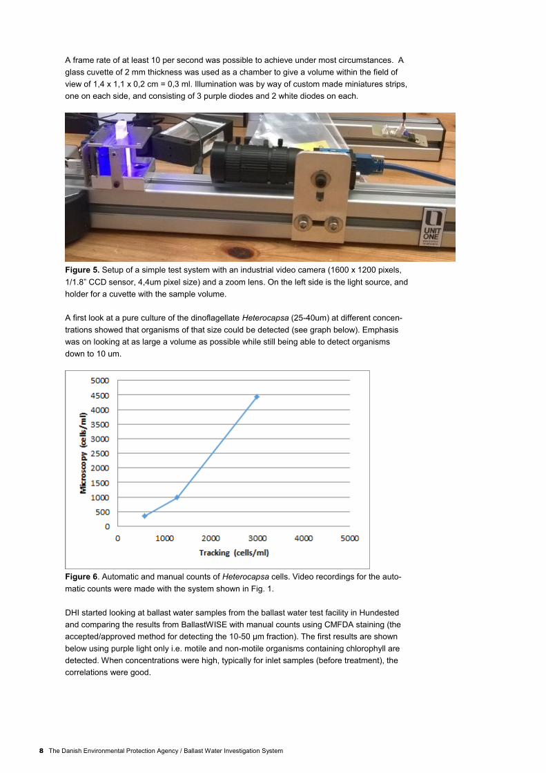
A frame rate of at least 10 per second was possible to achieve under most circumstances. A
glass cuvette of 2 mm thickness was used as a chamber to give a volume within the field of
view of 1,4 x 1,1 x 0,2 cm = 0,3 ml. Illumination was by way of custom made miniatures strips,
one on each side, and consisting of 3 purple diodes and 2 white diodes on each.
Figure 5. Setup of a simple test system with an industrial video camera (1600 x 1200 pixels,
1/1.8” CCD sensor, 4,4um pixel size) and a zoom lens. On the left side is the light source, and
holder for a cuvette with the sample volume.
A first look at a pure culture of the dinoflagellate Heterocapsa (25-40um) at different concen-
trations showed that organisms of that size could be detected (see graph below). Emphasis
was on looking at as large a volume as possible while still being able to detect organisms
down to 10 um.
Figure 6. Automatic and manual counts of Heterocapsa cells. Video recordings for the auto-
matic counts were made with the system shown in Fig. 1.
DHI started looking at ballast water samples from the ballast water test facility in Hundested
and comparing the results from BallastWISE with manual counts using CMFDA staining (the
accepted/approved method for detecting the 10-50 µm fraction). The first results are shown
below using purple light only i.e. motile and non-motile organisms containing chlorophyll are
detected. When concentrations were high, typically for inlet samples (before treatment), the
correlations were good.
The Danish Environmental Protection Agency / Ballast Water Investigation System 9
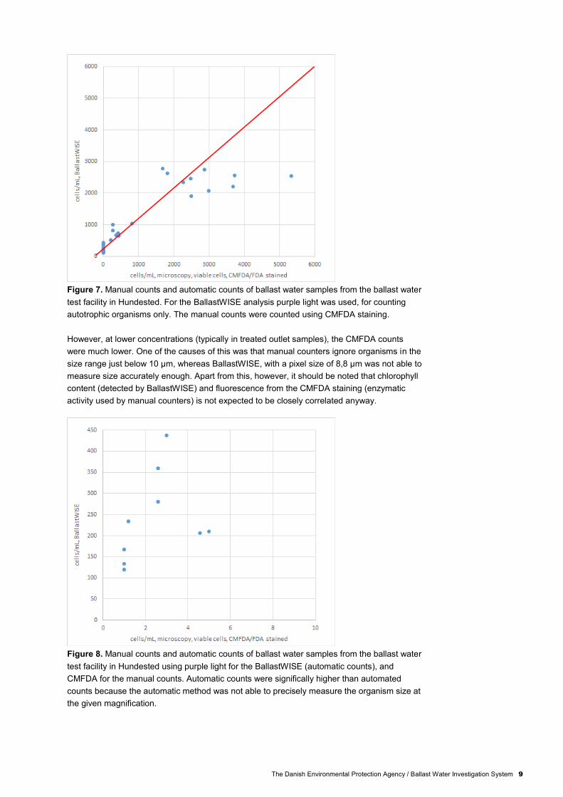
Figure 7. Manual counts and automatic counts of ballast water samples from the ballast water
test facility in Hundested. For the BallastWISE analysis purple light was used, for counting
autotrophic organisms only. The manual counts were counted using CMFDA staining.
However, at lower concentrations (typically in treated outlet samples), the CMFDA counts
were much lower. One of the causes of this was that manual counters ignore organisms in the
size range just below 10 µm, whereas BallastWISE, with a pixel size of 8,8 µm was not able to
measure size accurately enough. Apart from this, however, it should be noted that chlorophyll
content (detected by BallastWISE) and fluorescence from the CMFDA staining (enzymatic
activity used by manual counters) is not expected to be closely correlated anyway.
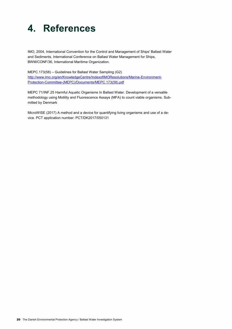
Figure 8. Manual counts and automatic counts of ballast water samples from the ballast water
test facility in Hundested using purple light for the BallastWISE (automatic counts), and
CMFDA for the manual counts. Automatic counts were significally higher than automated
counts because the automatic method was not able to precisely measure the organism size at
the given magnification.

10 The Danish Environmental Protection Agency / Ballast Water Investigation System
2.3 Optical chambers This prototype is basically the same as prototype 1, but with a higher magnification and small-
er chamber. The chamber was replaced with a 1mm thick glass capillary. Magnification was
increased to 1x giving a pixel size of 4,4um and the aperture was decreased to f/8 or f/ll to give
a larger depth of field. This decision was based on the assumption that a higher measurement
precision is required. Plotting size distributions as the one shown below (distribution of organ-
ism area in um2) of ballast water samples consistently showed something close to a continu-
ous size spectrum. Choosing the correct cutoff point in size to accurately get the size fraction
>10 um i.e. >100 um2 resembles a kind of calibration because of the lack of extreme accuracy
in size determination.
Figure 9. The organisms size distributions measured with BallastWISE prototype system. The
distribution of the cell area in µm2 of organism from ballast water samples is shown.
A set of results from DHI of ballast water samples from the test facility in Hundested with a 300
µm2 area threshold are shown below. The 300 µm2 value was chosen as a cutoff point be-
cause of a halo effect around objects which makes them look bigger than they are. There are
more detailed analysis on the size estimation problem to follow. The correlations have not
changed much in relation to the tests with the lower magnification, and in particular, the
CMFDA/FDA counts still show up much lower after treatment. It was decided to aim for yet
higher measurement precision to resolve the problem.

The Danish Environmental Protection Agency / Ballast Water Investigation System 11
Figure 10. Comparison of manual and automated analysis of ballastwater samples where the
automatic system had 1x magnification and pixel size 4.4 µm, and the manual counts were as
described above. The BallastWISE counts included cells <10 µm due to inaccurate size
measurements. Therefore the CMFDA/FDA counts still showed up much lower that the auto-
mated counts.
2.4 Precision optics In order to increase precision of cell size measurements a new prototype was based on a
Navitar 2x Magnistar bi-telecentric lens. This lens has the ability to adjust the aperture. Below
is an illustration of the importance of stopping down to (preferably) f/ll. Although cells can be
seen at larger apertures, they become diffuse halos when out of focus and this creates false
size estimations even though they can be tracked. At f/ll, this effect almost disappears, as well
as making sure that the whole depth of the chamber is in focus.
Figure 11. Left: Aperture f/4, showing the cells clearly with a bright halo around each cell,
which results in a false size estimate. On the right side the aperture was f/11, which resulted in
correct cell size estimates.
DHI analysed ballast water samples from the test facility in Hundested and close analysis of
the original videos revealed again the problem of accurately determining the size threshold on
the lower limit of 10µm (see below).

12 The Danish Environmental Protection Agency / Ballast Water Investigation System
Figure 12. Analysis of ballast water samples from Hundested, where cells close to the mini-
mum size limit of 10 µm were included in the automatic counts.
Fishlab has worked with cultures of a wide variety of organisms within the size range of 10-50
µm. Below are comparisons of sizes measured manually and with BallastWISE in white and
violet light.
Figure 13. Comparisons of sizes measured manually and with BallastWISE system using
white light.

The Danish Environmental Protection Agency / Ballast Water Investigation System 13
Figure 14. Comparisons of sizes measured manually and with BallastWISE system using
violet light.
A number of analysis were performed over time in order to estimate the variation in Ballast-
WISE estimates as well as to see what effect chamber residence time has on estimates. The
next 4 plots are of larger organisms (15 – 25 µm), and it can be seen that BallastWISE finds
them with reasonable accuracy and with relatively good consistency. The drop off with time is
usually caused by cells settling on the walls of the chamber. Only swimming organisms were
registered during these analysis.
0
200
400
600
800
1.000
1.200
0 20 40 60 80 100
Cells
(cells
/ml)
Time (sec.)
Ciliates, white light
Auto count
Manual count
Cell size: 18 µm
A)

14 The Danish Environmental Protection Agency / Ballast Water Investigation System
Figure 15. Automatic and manual counts of organisms, over a time span of 90 sec. with
measurements every 10 seconds: A) Ciliates, using white light; B) Mesodinium rubrum using
purple light; C) Teleaulax acuta using purple light; D) Teleaulax acuta using white light
The next 2 plots, show the ability of BallastWISE to reject cells that are below 10µm. A value
of 200 cells/ml corresponds to just 1 cell per video sequence.
0
2.000
4.000
6.000
8.000
10.000
12.000
0 20 40 60 80 100
Cells
/ml
Time (sec.)
Mesodinium rubrum time series, purple light
Auto count
Manual count
Cell size: M.rubrum L=25, D=20
B)
0
500
1.000
1.500
2.000
2.500
3.000
0 20 40 60 80 100
Cells
/ml
Time (sec.)
Teleaulax acuta, purple light
Auto count
Manual count
Cell size: L=15,
C)
0
500
1.000
1.500
2.000
2.500
3.000
0 20 40 60 80 100
Cells
/ml
Time (sec.)
Teleaulax acuta, white light
Auto count
Manual count
D)

The Danish Environmental Protection Agency / Ballast Water Investigation System 15
Figure 16. Automatic and manual counts of organisms smaller than 10 µm, over a time span
of 90 sec. with measurements every 10 seconds. The cells were not included in the automatic
counts due to their small size. A) Teleaulax amphioxeia (L= 8µm, D=3µm), using purple light
B) Small ciliates (L= 6µm, D=4µm), using white light
2.5 Prototype construction The final prototype that was constructed within the time frame of the project, consisted of the
same camera as was used throughout and the Navitar Magnistar 2x bi-telecentric lens set at
f/ll. The lens has no focusing option so the camera is mounted on a miniature rack and pinion
sleigh. A custom chamber fixture was machines from POM. It accepts a small interchangeable
chamber, which is constructed as a sandwich. The top of the fixture connects to the two inflow
holes in the chamber with an o-ring seal and the sample can be pumped through the system.
However, we decided to replace the small chamber with a 0.7mm flat glass capillary with sili-
con tubes attached at each end in order to avoid having to clean the channels within the fixture
top. This will be the disposable chamber solution for the time being.
0
5.000
10.000
15.000
20.000
25.000
30.000
35.000
40.000
0 20 40 60 80 100
Cells
/ml
Time (sec.)
Teleaulax amphioxeia, purple light
Auto count
Manual count
Cell size: L=8, D=3
A)
0
5.000
10.000
15.000
20.000
25.000
30.000
35.000
40.000
45.000
0 20 40 60 80 100
Cells
/ml
Time (sec.)
Small ciliates, white light
Auto count
Manual count
Cell size: L=5.6, D=4
B)

16 The Danish Environmental Protection Agency / Ballast Water Investigation System
Figure 17. The final prototype, constructed within the time frame of the project, using the
same camera as was used throughout and a Navitar Magnistar 2x bi-telecentric lens set at f/ll.
Figure 18. A custom chamber fixture machined from POM. The bottom part is holding a clear
measuring chamber, the top part is shown on the right.
A small peristaltic pump creates flow from a sample reservoir through the chamber and out to
an outflow reservoir. Custom designed electronics with an on-board microcontroller allow con-
trol of lighting and pump automatically from the main BallastWISE application. The small field
of view of only 3,5 x 2,5 mm allows for a single and more powerful focussed LED rather than
the small strip and this allows for a higher intensity of the purple LED, in particular, without
transferring heat to the optical chamber.
The number of analysis with this prototype are limited, but both DHI and Fishlab have reported
good usability. Below is a plot of ballast water discharge samples where BallastWISE and

The Danish Environmental Protection Agency / Ballast Water Investigation System 17
manual counts show similar results, however concentrations are relatively high compared to
concentrations in discharge in previous studies where viable counts were much lower.
Figure 19. Plot of ballast water discharge samples where BallastWISE and manual counts
using the using CMFDA staining show similar results.
2.6 BallastWISE software The layout of the BallasWISE user interface is shown in Fig. 20. The large area at the left is
the video display, which is live during filling, analysis, and emptying the optical chamber. This
gives good visual feedback to the operator. The operator types in a sample description in the
“Source” field, puts a sample in place, and presses the start button. The analysis will start and
the indicators on the panel show how the analysis is progressing and how much time is re-
maining. It also shows the intermediate results of the concentration of motile organisms and
the concentration of pigmented organisms after each field is processed.
Figure 20. Layout of the BallasWISE user interphase.

18 The Danish Environmental Protection Agency / Ballast Water Investigation System
2.7 Patenting of the BallastWISE technology One of the objectives of the project was to investigate whether the BallastWISE technology is
patentable, and if it is the case to apply for a patent.
A patent investigation showed that the technology is likely to be patentable. Therefore, the
project partners decided to apply for a patent based on the present project (Ballast Water
Investigation System (BallastWISE) and a previous project funded by the Danish Maritime
Fund, where a system for quantification of organisms >50 µm was developed (Kontrol af bal-
lastvand, project number 2013-064).
After the completion of the BallastWISE project, an international patent application for patent
application was filed (MicroWISE, 2017).

The Danish Environmental Protection Agency / Ballast Water Investigation System 19
3. Conclusions
The BallastWISE project succeeded in developing a system for rapid and automatic evaluation
of the concentration of live organisms between 10 – 50 µm by detecting organisms that are
motile and/or organisms which contain chlorophyll using image- and motion analysis.
Major findings during the project:
● It is possible for a modern industrial video camera to detect chlorophyll content down
to cells sizes of 10um or less using a 430nm LED for excitation and a high pass filter
of 500 nm placed in front of the camera
● The smallest moving organisms can be detected and tracked at 2x magnification
● Organisms can be fairly accurately measured by the software in order to determine
whether they fit within the 10-50um range, but due to the continuous size distribution
of organisms in typical samples, it can be difficult to give an absolutely precise esti-
mate of cell counts with the size range
● The 2x magnification gives a volume with the field of view of 0,35 x 0,25 x 0,07 = 6µl,
which means that 160 fields need to be processed in order to process 1ml of sample.
However, an ongoing statistical analysis could cut the analysis short if feasible
● The analysis process can be fully automated and will take a maximum of 30 minutes
but probably in the order of 10 minutes in practice
● The BallastWISE technology is patent pending
20 The Danish Environmental Protection Agency / Ballast Water Investigation System
4. References
IMO, 2004, International Convention for the Control and Management of Ships' Ballast Water
and Sediments, International Conference on Ballast Water Management for Ships,
BWM/CONF/36, International Maritime Organization.
MEPC.173(58) – Guidelines for Ballast Water Sampling (G2)
http://www.imo.org/en/KnowledgeCentre/IndexofIMOResolutions/Marine-Environment-
Protection-Committee-(MEPC)/Documents/MEPC.173(58).pdf
MEPC 71/INF.25 Harmful Aquatic Organisms In Ballast Water. Development of a versatile
methodology using Motility and Fluorescence Assays (MFA) to count viable organisms. Sub-
mitted by Denmark
MicroWISE (2017) A method and a device for quantifying living organisms and use of a de-
vice. PCT application number: PCT/DK2017/050131

The Danish Environmental Protection Agency / Ballast Water Investigation System 21
Annex 1. MEPC 71/INF.25 (2017)
[Tekst - Slet ikke efterfølgende linje, sektionsskifte]

22 The Danish Environmental Protection Agency / Ballast Water Investigation System

The Danish Environmental Protection Agency / Ballast Water Investigation System 23

24 The Danish Environmental Protection Agency / Ballast Water Investigation System

The Danish Environmental Protection Agency / Ballast Water Investigation System 25

The Danish Environmental
Protection Agency
Haraldsgade 53
2100 København Ø
www.mst.dk
Ballast Water Investigation System
The BallastWISE project succeeded in developing an automated system for rapid
evaluation of the concentration of live organisms between 10 – 50 µm by detecting
organisms that are motile and/or organisms which contain chlorophyll in an optical
chamber and using image- and motion analysis. It is possible for a modern industrial
video camera to detect chlorophyll content down to cells sizes of 10um or less using
a 430nm LED for excitation and a high pass filter placed in front of the camera to
remove the primary illumination. Movement of heterotrophic organisms is detected
using white light. The smallest moving organisms can be detected and tracked at 2x
magnification. Organisms can be measured by the software to determine whether
they fit within the 10-50um range, but due to the continuous size distribution of organ-
isms in typical samples, it can be difficult to give a very precise estimate of cell
counts with the size range. Several measurements need to be performed on for each
sample to give a statistically sound result. This is done by controlling flow through a
pump and can take between 10 to 30 minutes per sample. The BallastWISE techno-
logy is patent pending.