Embed Size (px)
Citation preview
RPS DEGREE COLLEGE BALANA (MAHENDERGARH)-123029
Lab Manual
Chemistry (HC 5th & HC 6th Semester)
Department of Chemistry
Experiment 1
Aim:-To prepare and analysepure sample of Potassium
Trisoxalatoferrate(III) Trihydrate, K3[Fe(C2O4)3].3H2O
APPARATUS AND CHEMICALS:
K2C2O4.H2O, funnel, FeCl3.6H2O, filter paper, K3Fe(CN)6 solution, 100-mL
beaker, H2SO4 solution, test tubes, distilled water, opaque objects
THEORY: Potassium trisoxalatoferrate(III) trihydrate, K3[Fe(C2O4)3].H2O
is a green crystalline salt, soluble in hot water but rather insoluble when
cold. It can be prepared by the reaction of K2C2O4.H2O with FeCl3.6H2O.
3K2C2O4.H2O(aq) + FeCl3.6H2O(aq) → K3Fe(C2O4)3].3H2O(aq) + 3KCl(aq)
The complex anion is photo-sensitive.This means that upon exposure to light
of anappropriate wavelength (<450 rim in this case) the Fe(C2O4)3-
3undergoes an intramolecularredox reaction in which the Fe(III) anion is
reduced to Fe(II) while one of the oxalate groupsis oxidized to CO2.
[Fe(C2O4)3]3-
−> Fe2+
+ 5/2 C2O42-
+CO2(g)
As mentioned above, light causes an internal electron-transfer reaction to
occur in [Fe(C2O4)2]3-
ion, producing CO2 and Fe2+
ions. The Fe2+
that is
produced can readily be detected by adding a solution of potassium
ferricyanide K3Fe(CN)6. A deep blue colored ferroferri cyanide complex is
formed.
Fe2+
+ Fe(CN)63-
−> Fe[Fe(CN)6] –
ferroferricyanide deep blue.
PROCEDURE: A. Preparation of K3[Fe(C2O4)3].3H2O
1. Weigh approximately 9.0 g of hydrated potassium oxalate, K2C2O4.H2O
into a 250 mL beaker.
2. Add 30 mL of distilled water and heat to dissolve (do not boil).
3. In a second small beaker dissolve 4.4 g of FeCl3.6H2O in a minimum
amount of cold water (10-15 mL). Add the FeCl3.6H2O solution to the warm
oxalate solution and stir with a glass rod. Allow the product to crystallize
(away from strong sunlight) by cooling the solution in an ice-water mixture.
4. Collect the crystalline product by filtration. The product is
K3[Fe(C2O4)3].3H2O. B.
Blueprinting
1. Wet a piece of filter paper with [Fe(C2O4)2] 3-
solution.
2. Leave it to dry. (Meanwhile you can follow part C)
3. Place small opaque objects (coins, keys, etc.) on the paper.
4. Irradiate for few minutes using a light source (If not available you may
use bright sunlight)
5. Dip the paper into potassium ferricyanide solution (CAUTION potassium
ferricyanide is poisonous. Avoid contact with your skin. If it happens
immediately wash your skin with plenty of water.)
6. Remove the developed blueprint and dip in a beaker of distilled water to
wash off excess ferricyanide solution. Explain your observations.
C. Photochemical Reaction of [Fe(C2O4)2]3"
1. Dissolve 0.7 g of your complex in 100 mL of distilled water in a flask.
Add 3 mL of 2 M H2SO4 and swirl the mixture. To each three labeled test
tubes add 10 mL of this solution.
2. Keep one tube away from the light source as the control and irradiate the
remaining two tubes with the light source for 1 and 5 minutes respectively.
3. To all three tubes add 1 mL of 0. 1 M potassium ferricyanide solution
K3Fe(CN)6.
4. Record and explain your observations.

Experiment:2
Aim:-To verify Beer-Lambert law for KMnO4 and determine the
concentration of the given KMnO4 solution.
Chemical Required:- solid KMn04.
Apparatus Required:- Spectrophotometer or Elico colorimeter,
measuring flasks (100ml and 1000ml), weight box, fractional
weights, graph papers.
OBJECTIVE:-
In it we used Beer Lamberts law, this law was dependent on
absorbance phenomena.For it number of standard solutions of
different concentrations are prepared. Their absorbance is
determined. Then a plot of A vs c is drawn. It is a straight line
passing through the origin. This proves the validity of Beer-
Lambert law. Then the absorbance of the unknown solution is
determined under the same experimental conditions. The
concentration corresponding to this absorbance is read from the
calibration graph.
PROCEDURE:-
(i) Prepare a stock solution of 10-3M KMn04 by dissolving
0.0316g solid KMn04 in one liter distilled water.
.
(ii) Took four 100ml flat-bottomed measuring flasks and name
them as A, B, C and D respectively. (iii)Now pipette out 20,
40, 60 and 80ml of stock solution of KMn04 into flask A, B, C
and D respectively. Make the solution up to the given mark in
Conical flask by dilution with distilled water in every 100ml
flask.
20ml stock solution+ 40ml stock sol.+ 60ml
stock sol+ 80ml stock sol+
80ml distilled 60ml water 40ml
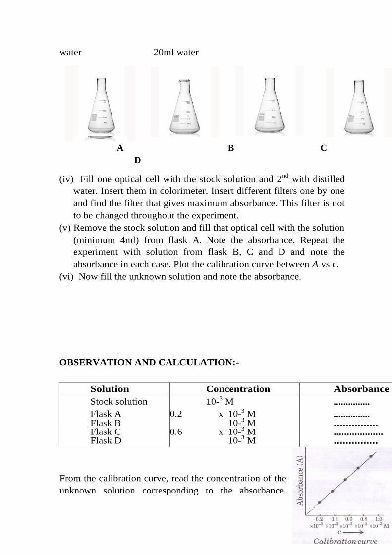
water 20ml water
A B C
D
(iv) Fill one optical cell with the stock solution and 2nd
with distilled
water. Insert them in colorimeter. Insert different filters one by one
and find the filter that gives maximum absorbance. This filter is not
to be changed throughout the experiment.
(v) Remove the stock solution and fill that optical cell with the solution
(minimum 4ml) from flask A. Note the absorbance. Repeat the
experiment with solution from flask B, C and D and note the
absorbance in each case. Plot the calibration curve between A vs c.
(vi) Now fill the unknown solution and note the absorbance.
OBSERVATION AND CALCULATION:-
From the calibration curve, read the concentration of the
unknown solution corresponding to the absorbance.
Solution Concentration Absorbance
Stock solution 10-3 M ...............
Flask A 0.2 x 10⁻³ M ...............
Flask B
0.4 x
10-3 M ...............
Flask C 0.6 x 10⁻³ M ...................
Flask D
0.8 x
10-3 M ...............
Further a straight line verified the Beer-Lambert's law.
RESULT:
The concentration of given KMnO4 solution is…………………
.
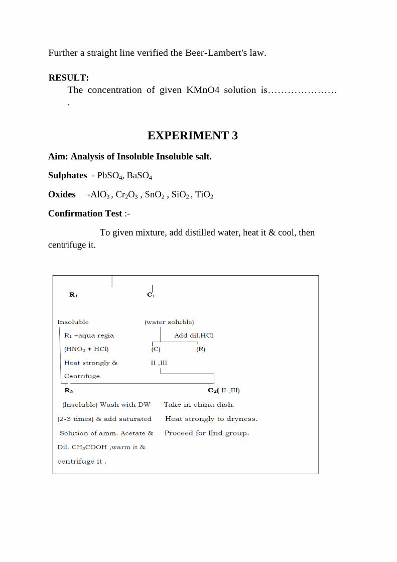
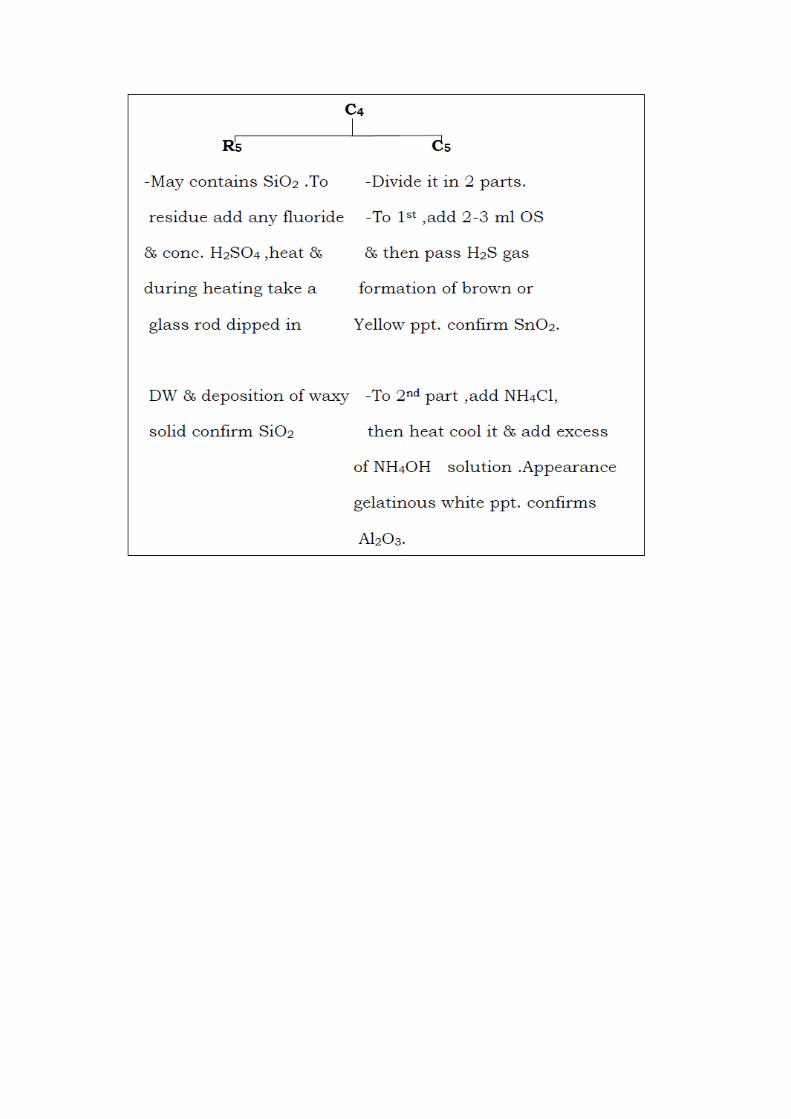
EXPERIMENT 3
Aim: Analysis of Insoluble Insoluble salt.
Sulphates - PbSO4, BaSO4
Oxides -AlO3 , Cr2O3 , SnO2 , SiO2 , TiO2
Confirmation Test :-
To given mixture, add distilled water, heat it & cool, then
centrifuge it.
-If all test for halides are –ve , then proceed for another salts .
-then , do fusion with residue R3 :- [Done in Ni crucible ]
Two Methods :-
1 At the bottom of Ni crucible make a layer of NaOH pallets + few
crystal of KNO3+ add residue part.
2 Ni crucible + (K2CO3 + Na2CO3) + KNO3& then add residue ,then
strongly heat the Ni crucible upto red hot ,cool it at room temp. & add
saturated solution of Na2CO3 & then centrifuge it .

Semester 6th
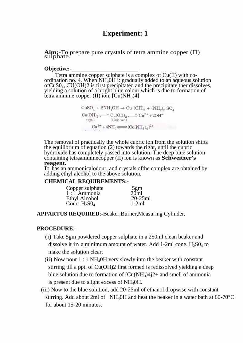
Experiment: 1
Aim:-To prepare pure crystals of tetra ammine copper (II) sulphate. Objective:-____________________
Tetra ammine copper sulphate is a complex of Cu(II) with co-ordination no. 4. When NH40H i: gradually added to an aqueous solution ofCuS04, CU(OH)2 is first precipitated and the precipitate ther dissolves, yielding a solution of a bright blue colour which is due to formation of tetra ammine copper (II) ion, [Cu(NH3)4]
The removal of practically the whole cupric ion from the solution shifts the equilibrium of equation (2) towards the right, until the cupric hydroxide has completely passed into solution. The deep blue solution containing tetraamminecopper (II) ion is known as Schweitzer's reagent. It has an ammonicalodour, and crystals ofthe complex are obtained by adding ethyl alcohol to the above solution. CHEMICAL REQUIREMENTS:-
Copper sulphate 5gm 1 : 1 Ammonia 20ml Ethyl Alcohol 20-25ml Conc. H2S04 1-2ml
APPARTUS REQUIRED:-Beaker,Burner,Measuring Cylinder.
PROCEDURE:-
(i) Take 5gm powdered copper sulphate in a 250ml clean beaker and
dissolve it in a minimum amount of water. Add 1-2ml cone. H2S04 to
make the solution clear.
(ii) Now pour 1 : 1 NH40H very slowly into the beaker with constant
stirring till a ppt. of Cu(OH)2 first formed is redissolved yielding a deep
blue solution due to formation of [Cu(NH3)4j2+ and smell of ammonia
is present due to slight excess of NH40H.
(iii) Now to the blue solution, add 20-25ml of ethanol dropwise with constant
stirring. Add about 2ml of NH40H and heat the beaker in a water bath at 60-70°C
for about 15-20 minutes.
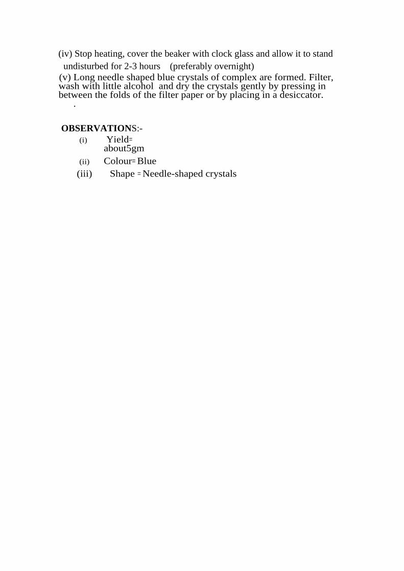
(iv) Stop heating, cover the beaker with clock glass and allow it to stand
undisturbed for 2-3 hours (preferably overnight)
(v) Long needle shaped blue crystals of complex are formed. Filter, wash with little alcohol and dry the crystals gently by pressing in between the folds of the filter paper or by placing in a desiccator. .
OBSERVATIONS:-
(i) Yield= about5gm
(ii) Colour= Blue
(iii) Shape = Needle-shaped crystals
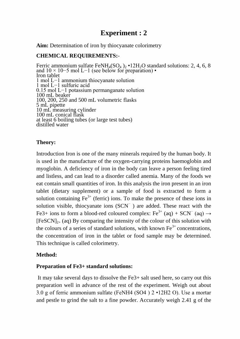
Experiment : 2
Aim: Determination of iron by thiocyanate colorimetry
CHEMICAL REQUIREMENTS:- Ferric ammonium sulfate FeNH4(SO4 )2 •12H2O standard solutions: 2, 4, 6, 8 and 10 × 10−5 mol L−1 (see below for preparation) • Iron tablet 1 mol L−1 ammonium thiocyanate solution 1 mol L−1 sulfuric acid 0.15 mol L−1 potassium permanganate solution 100 mL beaker 100, 200, 250 and 500 mL volumetric flasks 5 mL pipette 10 mL measuring cylinder 100 mL conical flask at least 6 boiling tubes (or large test tubes) distilled water
Theory:
Introduction Iron is one of the many minerals required by the human body. It
is used in the manufacture of the oxygen-carrying proteins haemoglobin and
myoglobin. A deficiency of iron in the body can leave a person feeling tired
and listless, and can lead to a disorder called anemia. Many of the foods we
eat contain small quantities of iron. In this analysis the iron present in an iron
tablet (dietary supplement) or a sample of food is extracted to form a
solution containing Fe3+
(ferric) ions. To make the presence of these ions in
solution visible, thiocyanate ions (SCN− ) are added. These react with the
Fe3+ ions to form a blood-red coloured complex: Fe3+
(aq) + SCN− (aq) →
[FeSCN]2+ (aq) By comparing the intensity of the colour of this solution with
the colours of a series of standard solutions, with known Fe3+
concentrations,
the concentration of iron in the tablet or food sample may be determined.
This technique is called colorimetry.
Method:
Preparation of Fe3+ standard solutions:
It may take several days to dissolve the Fe3+ salt used here, so carry out this
preparation well in advance of the rest of the experiment. Weigh out about
3.0 g of ferric ammonium sulfate (FeNH4 (SO4 ) 2 •12H2 O). Use a mortar
and pestle to grind the salt to a fine powder. Accurately weigh 2.41 g of the
powder into a 100 mL beaker and add 20 mL of concentrated sulfuric acid
(see safety notes). Leave powder to soak in acid overnight. The next day,
carefully pour the acid/ powder slurry into a 500 mL volumetric flask,
rinsing the beaker into the flask a few times with water, then make up to the
mark with distilled water. Let this solution stand for several days until the
ferric ammonium sulfate powder has fully dissolved. If possible, insert a
magnetic stirrer bar and stir the solution to speed up this dissolving process.
Use a pipette to transfer 20 mL of ferric ion solution to a 200 mL volumetric
flask and make up to the mark with distilled water. This gives a solution with
[Fe3+] = 0.001 mol L−1. To prepare a 2 × 10⁻⁵ mol L−1 standard solution
pipette 10 mL of the 0.001 mol L−1 solution into a 500 mL volumetric flask,
add 10 mL of 1 mol L−1 sulphuric acid, and then make up to the mark with
distilled water. Repeat this procedure inseparate 500 mL volumetric flasks,
pipetting in 20, 30, 40 and 50 mL of 0.001 mol L−1 Fe3+ solution in turn, to
obtain 4, 6, 8 and 10 × 10−5 mol L−1 solutions respectively.
Preparation of 1 mol L−1 ammonium thiocyanate solution:
Weigh 38 g of solid ammonium thiocyanate into a 500 mL volumetric flask
and make up to the mark with distilled water. 3. Preparation of 0.15 mol L−1
potassium permanganate solution (only required for analysis of iron tablet):
Weigh 2.4 g of solid potassium permanganate into a 100 mL volumetric
flask and make up to the mark with distilled water.
Preparation of iron tablet for analysis:
1. Place iron tablet in a 100 mL beaker and use a measuring cylinder to add
20 mL of 1 mol L−1 sulfuric acid. Allow the tablet’s coating to break down
and its contents to dissolve. You may help this process by using a stirring
rod to carefully crush the tablet and stir the solution. (NB: iron tablets
sometimes contain filler materials that may not fully dissolve in acid)
2. Once the iron tablet is dissolved, add 0.15 mol L−1 potassium
permanganate solution dropwise, swirling the beaker after each addition.
Iron tablets usually contain ferrous sulfate, with iron present as Fe2+ ions.
Since Fe2+ does not form a coloured complex with thiocyanate,
permanganate ions are added to oxidise all the Fe2+ to form Fe3+ ions. For
the first few drops of permanganate, the purple colour will disappear
immediately upon addition to the iron solution; however, as further drops are
added the colour will begin to linger for a little longer. Stop adding
potassium permanganate drops when the purple colour persists for several
seconds after addition − usually no more than about 2 mL of 0.15 mol L−1
permanganate solution will be required.
3. Transfer the iron solution to a 250 mL volumetric flask, rinsing the beaker
with distilled water a few times and transferring the washings to the
volumetric flask. Make up to the mark with distilled water, stopper the flask
and mix well.
4. Use a pipette to transfer 5 mL of iron solution to a 100mL volumetric
flask and make up to the markwith distilled water. This diluted solution will
be used for colorimetric analysis.
Preparation of food sample for analysis:
1. Accurately weigh a few grams (typically 2 − 5 g is required, depending on
iron content of sample) of your food sample into a crucible.
2. Heat the crucible over a bunsen burner (see Figure 1) until the sample is
reduced completely to ash, or (preferably) combust the sample directly in the
bunsen flame (as shown in Figure 2), reducing it to ash. NB: be very careful
with the bunsen flame while heating/combusting your sample. Also beware
that the crucible will become very hot during this process, so handle it only
with crucible tongs − or preferably not at all − until it has cooled.
3. When the sample and crucible have cooled, use a stirring rod to crush the
ash to a fine powder (see Figure 3). Use a measuring cylinder to add 10 mL
of 1 mol L−1 hydrochloric acid and stir for 5 minutes, making sure that all
the ash is soaked.
4. Add 5 mL of distilled water and filter the solution into a 100 mL conical
flask to remove the ash. This filtered solution will be used for colorimetric
analysis.
Colorimetric analysis: this analysis method applies to samples prepared
using either of the two methods above (iron tablets or food samples).
1. Accurately measure 10 mL of your sample solution into a clean, dry
boiling tube/test tube. NB: this is most accurately done using a 10 mL
pipette; however, it is possible to do this accurately enough (and with less
hassle) using a clean 10 mL measuring cylinder if you measure carefully.
2. Next, measure 10 mL of each Fe3+ standard solution into separate boiling
tubes (one standard per tube) in order of increasing concentration, beginning
with the 2 × 10⁻⁵ mol L−1 standard. It is a good idea to first rinse your
pipette or measuring cylinder with a few mL of the 2 × 10⁻⁵ mol L−1
standard). NB: Make sure you label each boiling tube appropriately with the
name of the sample or standard it contain. A test tube rack is very useful for
holding and transporting your tubes (see Figure 4). Alternatively you can use
a large beaker to hold them.
3. Using a 10 mL measuring cylinder, measure 10 mL of 1 mol L−1
ammonium thiocyanate solution into each of six small clean vessels − six
boiling tubes is ideal. You should now have one measured portion of
thiocyanate solution for each of your iron solutions.
4. As quickly as possible, pour 10 mL of thiocyanate solution (the portions
measured out above) into each of your iron solutions.
5. Mix the solutions by swirling. A stable red colour will appear over the
next few minutes.
6. Allow the red colour to develop for 15 minutes. Then estimate the
concentration of Fe3+ ions in your iron sample by identifying which of your
Fe3+ standards matches its colourmost closely. Figure 4 illustrates the range
of colour intensities that you can expect from your set of Fe3+ standards.
Tip: If you are using boiling/test tubes all of identical sizes, the best way to
compare colours is by looking at your solutions from above − looking down
into the tubes (see Figure 5).
7. If the colour of your unknown iron solution is stronger than the colour of
your highest concentration Fe3+ standard you will need to modify the above
procedure. In the case of an iron tablet, you should repeat the analysis with a
more dilute solution of the dissolved iron tablet. In the case of a food sample,
you should repeat the analysis using a smaller mass of your food.
Calculations

1. Assume that the concentration of Fe3+ in your unknown iron solution is
approximately equal to that of the Fe3+ standard whose colour was the
closest match.
2. Use this concentration to calculate the mass of iron (in mg) in your
original tablet or food sample


![OF The SAMBO Federation of India [SFI] Constitution of the SFI 2014.pdf · THE SAMBO FEDERATION OF INDIA 11 Hatta Bazar, Mohindergarh , Haryana 123029, INDIA CHAPTER A : MEMORANDUM](https://img.pdfslide.us/doc/110x75/5ac262fb7f8b9a433f8e01ab/of-the-sambo-federation-of-india-sfi-constitution-of-the-sfi-2014pdfthe-sambo.jpg)
![CeC),17 ank GOVERNMENT OF HARYANA •gftzrr rr MOH ROM ... · The Deputation period of [085550] Vikram Singh CV PTI from GMS Saidpur (Mahendergarh) [4446] to GSSS Chamarian (Rohtak)](https://img.pdfslide.us/doc/110x75/5f682a6c876bf358de45261b/cec17-ank-government-of-haryana-agftzrr-rr-moh-rom-the-deputation-period.jpg)