Embed Size (px)
Citation preview

BACTERIOLOGY OF ABSCESSES WITH SPECIAL REFERENCE TO
PHENOTYPING AND GENOTYPING OF METHICILLIN RESISTANT
STAPHYLOCOCCUS AUREUS IN GOVT. STANLEY MEDICAL COLLEGE &
HOSPITAL, CHENNAI.
Dissertation submitted in THE TAMILNADU DR.M.G.R.MEDICAL UNIVERSITY
in partial fulfilment of the regulationsfor the award of the degree of
M.D. (MICROBIOLOGY) BRANCH - IV
GOVERNMENT STANLEY MEDICAL COLLEGE
& HOSPITAL
THE TAMILNADU DR.M.G.R.MEDICAL UNIVERSITY
CHENNAI
MARCH 2010
brought to you by COREView metadata, citation and similar papers at core.ac.uk
provided by ePrints@TNMGRM (Tamil Nadu Dr. M.G.R. Medical University)
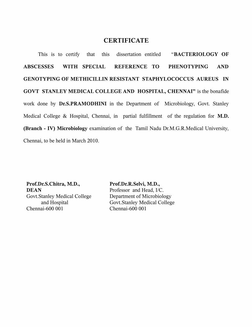
CERTIFICATE
This is to certify that this dissertation entitled “BACTERIOLOGY OF
ABSCESSES WITH SPECIAL REFERENCE TO PHENOTYPING AND
GENOTYPING OF METHICILLIN RESISTANT STAPHYLOCOCCUS AUREUS IN
GOVT STANLEY MEDICAL COLLEGE AND HOSPITAL, CHENNAI” is the bonafide
work done by Dr.S.PRAMODHINI in the Department of Microbiology, Govt. Stanley
Medical College & Hospital, Chennai, in partial fulfillment of the regulation for M.D.
(Branch - IV) Microbiology examination of the Tamil Nadu Dr.M.G.R.Medical University,
Chennai, to be held in March 2010.
Prof.Dr.S.Chitra, M.D.,DEANGovt.Stanley Medical College
and HospitalChennai-600 001
Prof.Dr.R.Selvi, M.D., Professor and Head, I/C.Department of MicrobiologyGovt.Stanley Medical College Chennai-600 001
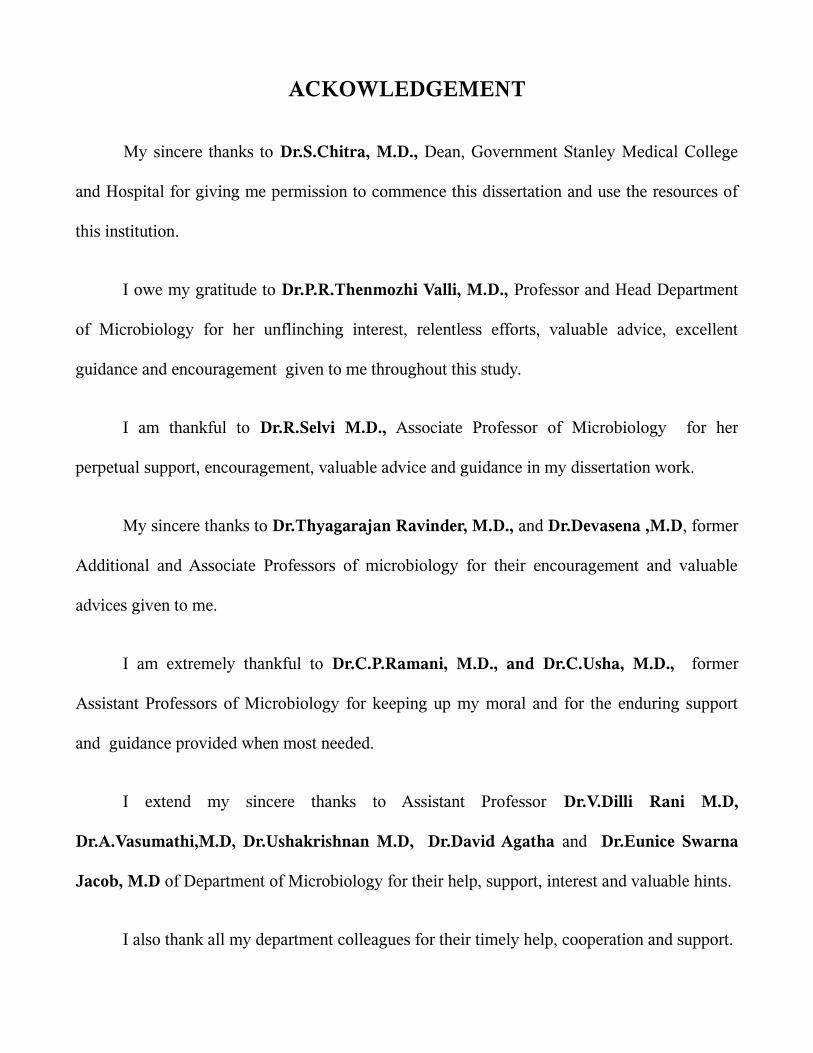
ACKOWLEDGEMENT
My sincere thanks to Dr.S.Chitra, M.D., Dean, Government Stanley Medical College
and Hospital for giving me permission to commence this dissertation and use the resources of
this institution.
I owe my gratitude to Dr.P.R.Thenmozhi Valli, M.D., Professor and Head Department
of Microbiology for her unflinching interest, relentless efforts, valuable advice, excellent
guidance and encouragement given to me throughout this study.
I am thankful to Dr.R.Selvi M.D., Associate Professor of Microbiology for her
perpetual support, encouragement, valuable advice and guidance in my dissertation work.
My sincere thanks to Dr.Thyagarajan Ravinder, M.D., and Dr.Devasena ,M.D, former
Additional and Associate Professors of microbiology for their encouragement and valuable
advices given to me.
I am extremely thankful to Dr.C.P.Ramani, M.D., and Dr.C.Usha, M.D., former
Assistant Professors of Microbiology for keeping up my moral and for the enduring support
and guidance provided when most needed.
I extend my sincere thanks to Assistant Professor Dr.V.Dilli Rani M.D,
Dr.A.Vasumathi,M.D, Dr.Ushakrishnan M.D, Dr.David Agatha and Dr.Eunice Swarna
Jacob, M.D of Department of Microbiology for their help, support, interest and valuable hints.
I also thank all my department colleagues for their timely help, cooperation and support.

I express many thanks to all the technical staff and other staff members of the
Department of Microbiology & Immunology for their kind cooperation to carry out this work
successfully.
I also extend my thanks to all the patients who participated in my study.

DECLARATION
I, Dr.S.PRAMODHINI, solemnly declare that this dissertation “BACTERIOLOGY
OF ABSCESSES WITH SPECIAL REFERENCE TO PHENOTYPING AND
GENOTYPING OF METHICILLIN RESISTANT STAPHYLOCOCCUS AUREUS
IN GOVT STANLEY MEDICAL COLLEGE AND HOSPITAL, CHENNAI” is the
bonafide work done by me at the Department of Microbiology, Government Stanley Medical
College and Hospital, Chennai, under the guidance and supervision of
Prof.Dr.P.R.THENMOZHI VALLI, M.D., Professor of Microbiology, Government Stanley
Medical College, Chennai-600 001.
This dissertation is submitted to The Tamil Nadu Dr. M. G. R. Medical University,
Chennai in partial fulfillment of the University regulations for the award of degree of M.D.
Branch IV Microbiology examinations to be held in March 2010.
Place: Chennai.Date: Dr. S.PRAMODHINI
CONTENTS
SL.NO. TITLE PAGE NO.
1. INTRODUCTION 1
2. AIMS AND OBJECTIVES 4
3. REVIEW OF LITERATURE 5
4. MATERIALS AND METHODS 31
5. RESULTS 45
6. DISCUSSION 59
7. SUMMARY 67
8. CONCLUSION 70
9. ANNEXURES
APPENDIX
BIBLIOGRAPHY
MASTER CHART
KEY TO MASTER CHART
INTRODUCTION
An abscess is a localized collection of purulent inflammatory tissue caused by
suppuration deep within a tissue, an organ or a confined space. It is produced by deep seeding
of pyogenic bacteria into a tissue. It may involve skin, dermis, fasciae, muscles, and even
bones.58 They occur in many parts of the body as superficial infections or as deep-seated
infections associated with any internal organ. Any organism isolated from them may be of
significance.
Abscesses that develop as a result of introduction of the normal flora into a normally
sterile body site are often polymicrobial in nature 25 Flora can gain access to the sterile site by
direct extension or secondary to laceration or perforation. Because of the uniqueness of the
normal flora at various body sites, the microbiology of such abscesses is generally predictable
by their location.
Organisms may enter the tissue by direct implantation (eg, penetrating trauma with a
contaminated object); spread from an established, contiguous infection; dissemination via
lymphatic or hematogenous routes from a distant site; or migration from a location where there
are resident flora into an adjacent, normally sterile area because of disruption of natural barriers

Staphylococcus aureus and Group A ß-haemolytic streptococci are the most prevalent
aerobes in skin and soft tissue abscesses and are isolated at all body sites7.In contrast, organisms
that colonize the mucous membranes predominated in infections adjacent to these membrane6 ,17
In this fashion, organisms of the gastrointestinal and cervical flora (enteric Gram-negative
bacilli and Bacteroides fragilis group) were found most often in intra-abdominal and buttock
and leg lesions.9 Group A ß-haemolytic streptococci, pigmented Prevotella and Porphyromonas
spp., and Fusobacterium spp. were most commonly found in lesions of the mouth, head, neck
and fingers8 .
Appropriate management of mixed aerobic and anaerobic infections requires the
administration of antimicrobials that are effective against both aerobic and anaerobic
components of infection in addition to surgical correction and drainage of pus. A number of
factors should be considered when choosing appropriate antimicrobial agents. They should be
effective against all target organisms, induce little or no resistance, achieve sufficient level in
infected site, have minimal toxicity and have maximal stability and longevity.
Staphylococcus aureus has been reported as a major cause of community and hospital
acquired infections.48 The organism has a differential ability to spread and cause outbreaks in
hospitals.62 Infections causes by S.aureus used to respond to beta lactam and related group of
antibiotics. However, due to development of methicillin resistance amongst S.aureus isolates
(MRSA); treatment of these infections has become problematic. Indiscriminate use of multiple
antibiotics, prolonged hospital stay, intravenous drug abuse, carriage of MRSA in nose are few
important risk factors for MRSA acquisition31 . Burns and Orthopaedics are two such high risk
units where patients are on multiple antibiotics and have a long stay in hospital59. Currently, the
treatment options for MRSA infections are limited to very few and expensive drugs like
Teicoplanin and Vancomycin. Thus, control of MRSA is essential to curtail the introduction and
spread of infection.
This study was aimed to determine the bacteriological profile,the antibiotic
susceptibility patterns of organisms isolated and the incidence of methicillin resistant
staphylococcus aureus in abscesses.
AIMS AND OBJECTIVES
• To study the prevalence of aerobes and anaerobes in abscess
• To characterize the aerobes and anaerobes
• Phenotypic and Genotypic characterization of antibiotic susceptibility of isolated
organism
• To study the incidence of methicillin resistant Stapylococcus aureus among the isolate
• To monitor antibiotic sensitivity pattern of MRSA and to formulate definite antibiotic
policy to reduce the incidence of MRSA infection.
REVIEW OF LITERATURE
An abscess is a collection of pus that has accumulated in a cavity formed by the
tissue on the basis of an infectious process or other foreign materials . It is a defensive reaction
of the tissue to prevent the spread of infectious materials to other parts of the body.The
organisms or foreign materials destroys the adjacent cells, resulting in the release of cytokines.
The cytokines trigger an inflammatory response, which draws large numbers of leucocytes to
the area and increases the regional blood flow.
Abscess wall or capsule is formed by the adjacent healthy cells in an attempt to keep the
pus from infecting neighboring structures. The final structure of the abscess tends to prevent
immune cells from attacking bacteria in the pus, or from reaching the causative organism or
foreign object.
Abscesses can develop in many parts of the body, but they usually involve the skin
surface. Common sites affected in superficial abscess include the armpits, groin, rectal area
(perirectal abscess), the external vaginal area (Bartholin abscess), and along the tailbone
(pilonidal abscess). Deep seated abscesses affect the brain, kidneys, liver (hepatic abscess),
lungs, teeth (dental abscess), and tonsils (peritonsillar abscess)24
Immunopathogenesis of abscess formation
Bacterial Pathogens Induce Abscess Formation by CD4+ T-Cell Activation via the CD28-
B7-2 Costimulatory Pathway .The mechanism by which T cells mediate abscess formation
requires the role of T-cell activation and the contribution of antigen-presenting cells via
CD28-B7 costimulation. Blockade of T-cell activation via the CD28-B7 pathway in
animals with CTLA4Ig prevented abscess formation following challenge with different
bacterial pathogens. Abscess formation in vivo and T-cell activation in vitro required
costimulation by B7-2 but not B7-1. These results demonstrate that abscess formation by
pathogenic bacteria is under the control of a common effector mechanism that requires T-
cell activation via the CD28-B7-2 pathway2
Abscess Causes
Abscesses are typically caused by either an inflammatory reaction to an infectious
process or less commonly, to a foreign substance within the body, which triggers the body's
immune system to form a cavity or capsule to contain the infection and prevent it from
spreading to other parts of the body. The interior of the abscess liquefies, and pus develops
(which contains dead cells, bacteria, and other debris). This area then begins to expand,
creating increasing tension and inflammation of the overlying skin.
Individuals with any of the following conditions are at higher risk for developing
abscesses :
Chronic steroid therapy, Chemotherapy, Diabetes, Cancer,Dialysis for kidney failure,
AIDS, Sickle cell disease, Leukemia, Peripheral vascular disease, Crohn's disease, Ulcerative
colitis, Severe burns, Severe trauma
Abscess Symptoms
The symptoms of an abscess vary depending on the location of the abscess, but in general,
individuals will experience the following:
• -Most often, an abscess becomes a painful, compressible mass that is red, warm to the
touch, and tender.
• -As an abscess progresses, it may "point" and come to a head. Pustular drainage and
spontaneous rupture may occur.
• -Most abscesses will continue to get worse without care and proper incision and
drainage. The infection can potentially spread to deeper tissues, and even into the
bloodstream.
• -If the infection spreads, fever, nausea, vomiting, increasing pain, and increasing skin
redness may develop.
Manifestations of abscess:
The cardinal symptoms and signs of any kind of inflammatory process are redness
(rubor), heat (calor), swelling (tumor), pain (dolor) and loss of function 27. Major complications
are spreading of the abscess material to adjacent or remote tissues and extensive regional tissue
death (gangrene). Abscesses in most parts of the body rarely heal themselves, so prompt
medical attention is indicated at the first suspicion of an abscess. An abscess could potentially
be fatal (although this is rare) if it compresses vital structures such as the trachea in the context
of a deep neck abscess
Abscesses that develop as a result of introduction of the normal flora into a normally
sterile body site are often polymicrobial in nature25. Most deep seated abscess and necrotizing
lesions involving anaerobes are polymicrobial and may include obligate anaerobes, facultative
anaerobes or microaerophiles as concomitant microorganisms .These microorganisms acting in
concert with trauma, vascular stasis or tissue necrosis, lower oxygen tension and oxidation
reduction potential in tissues and provide favourable conditions for obligate anaerobe to
multiply 32.
Mechanisms of Microbial Synergy:
Microbial synergy may increase the net pathogenic effect and hence the severity of infection
in several ways:
(i) oxygen consumption by aerobic bacteria induces tissue hypoxia and a lowering of the
redox potential, which favors the growth of anaerobic bacteria;
(ii) specific nutrients produced by one bacterium may encourage the growth of fastidious
and potentially pathogenic cohabiting microorganisms; and
(iii) some anaerobes are able to impair host immune cell function and thus provide a
competitive advantage for themselves as well as for other, cohabiting,
microorganisms 46
Mixed anaerobic infections
Anaerobiasis was discovered in 1861 by Louis Pasteur who introduced the terms
aerobes and anaerobes to designate, respectively, microorganism that live in the presence and
absence of oxygen within the next 30 years,many diverse type of anaerobic bacteria were
isolated and described . the association of non sporing anaerobes with infections in humans
was recognized as early as 18971.
"Anaerobic bacteria are organisms that require reduced oxygen tension for growth,
failing to grow on the surface of solid media in 10% CO2 in air. (In contrast, microaerophilic
bacteria can grow in an atmosphere of 10% CO2 in air or under anaerobic or aerobic conditions,
although they grow best in the presence of only a small amount of atmospheric oxygen, and
facultative bacteria can grow in the presence or absence of air.
Anaerobes can infect normal hosts and those with compromised resistance or damaged
tissues. Symptoms depend on site of infection. Anaerobes are often accompanied by aerobic
organisms23. Hundreds of species of nonsporulating anaerobes are part of the normal flora of
the skin, mouth, GI tract, and vagina. If this commensal relationship is disrupted (eg, by
surgery or other trauma, poor blood supply, tissue necrosis), a few of these species can cause
infections with high morbidity and mortality. After becoming established in a primary site,
organisms can spread hematogenously to distant sites. Because aerobic and anaerobic bacteria
frequently are found in the same infected site, appropriate procedures for isolation and culture
are necessary to keep from overlooking the anaerobes. Anaerobes can be the major cause of
infection in the pleural spaces and lungs; in intra-abdominal, gynecologic, CNS, upper
respiratory tract, and cutaneous diseases; and in bacteremia.
The principal anaerobic gram-positive cocci that produce disease are the peptococci and
the peptostreptococci, which are part of the normal flora of the mouth, upper respiratory tract,
and large intestine. The principal anaerobic gram-negative bacilli include Bacteroides fragilis ,
Prevotella melaninogenica, and Fusobacterium sp. The B. fragilis group is part of the normal
bowel flora and includes the anaerobic pathogens most frequently isolated from intra-
abdominal infections. Organisms in the Prevotella group and Fusobacterium sp are part of the
indigenous oral flora 23,25.
Anaerobic infections can usually be characterized by the following features:
• They tend to occur as localized collections of pus or abscesses
• The reduced O2 tension and low oxidation-reduction potential that prevail in avascular
and necrotic tissues are critical for their survival
• When bacteremia occurs, it usually does not lead to disseminated intravascular
coagulation (DIC) and purpura.
Some anaerobic bacteria possess distinct virulence factors. Those of B. fragilis probably
account for its frequent isolation from clinical specimens despite its relative rarity in normal
flora. This organism has a polysaccharide capsule that apparently stimulates abscess formation.
An experimental model of intra-abdominal sepsis has shown that B. fragilis alone can cause
abscesses, whereas other Bacteroides sp require the synergistic effect of another organism25.
Another virulence factor, a potent endotoxin, is implicated in septic shock associated with
severe Fusobacterium pharyngitis.
Because of their fastidiousness, they are difficult to isolate and are often overlooked.
Their isolation requires appropriate methods of collection, transportation, and cultivation of
specimens. Treatment is complicated by factors which include slow growth, increasing
resistance to antimicrobial agents, and the polymicrobial synergistic nature of the infection.
Morbidity and mortality are as great from anaerobic and mixed bacterial sepsis as from sepsis
caused by a single aerobic organism. Anaerobic infections are often complicated by deep-
seated tissue necrosis.
Bacteria causing cutaneous abscesses are typically indigenous to the skin of the involved
area. For abscesses on the trunk, extremities, axillae, or head and neck, the most common
organisms are Staphylococcus aureus and Streptococci .
Staphylococcus aureus is a medically important bacterial pathogen that is a common
cause of superficial and deep-seated abscesses in humans. Most S. aureus isolates produce
either a serotype 5 or 8 capsular polysaccharide (CP) that has been shown to enhance bacterial
virulence 2.
In an elegant series of clinical observations and laboratory studies published in 1880 and
1882, Ogston described staphylococcal disease and its role in sepsis and abscess formation.
More than 100 years later, Staphylococcus aureus remains a versatile and dangerous pathogen
in humans. The frequencies of both community-acquired and hospital-acquired staphylococcal
infections have increased steadily, with little change in overall mortality 18. Treatment of these
infections has become more difficult because of the emergence of multidrug-resistant strains.
Staphylococcal Components and Products
S. aureus is a member of the Micrococcaceae family . The genus Staphylococcus has at
least 35 species.On microscopical examination, the organisms appear as gram-positive cocci in
clusters. S. aureus is distinguished from other staphylococcal species on the basis of the gold
pigmentation of colonies and positive results of coagulase, mannitol-fermentation, and
deoxyribonuclease tests. Some are members of the normal flora of the skin and mucous
membranes of humans; others cause suppuration, abscess formation, a variety of pyogenic
infections, and even fatal septicemia. The pathogenic staphylococci often hemolyze blood,
coagulate plasma, and produce a variety of extracellular enzymes and toxins 28.
Genome
The staphylococcal genome consists of a circular chromosome (of approximately 2800
bp, with prophages, plasmids, and transposons. Genes governing virulence and resistance to
antibiotics are found on the chromosome, as well as the extrachromosomal elements 38 .These
genes are transferred between staphylococcal strains, species, or other gram-positive bacterial
species through the extrachromosomal elements 47 .
Antigenic Structure
Staphylococci contain antigenic polysaccharides and proteins as well as other substances
important in cell wall structure. Peptidoglycan, a polysaccharide polymer containing linked
subunits, provides the rigid exoskeleton of the cell wall. Peptidoglycan is destroyed by strong
acid or exposure to lysozyme. It is important in the pathogenesis of infection: It elicits
production of interleukin-1 (endogenous pyrogen) and opsonic antibodies by monocytes, and it
can be a chemoattractant for polymorphonuclear leukocytes, have endotoxin-like activity, and
activate complement.
Teichoic acids, which are polymers of glycerol or ribitol phosphate, are linked to the
peptidoglycan and can be antigenic. Antiteichoic acid antibodies detectable by gel diffusion
may be found in patients with active endocarditis due to S aureus 28 .
Protein A is a cell wall component of many S aureus strains that binds to the Fc portion
of IgG molecules except IgG3. The Fab portion of IgG bound to protein A is free to combine
with a specific antigen. Protein A has become an important reagent in immunology and
diagnostic laboratory technology; for example, protein A with attached IgG molecules directed
against a specific bacterial antigen will agglutinate bacteria that have that antigen
("coagglutination").
Some S aureus strains have capsules, which inhibit phagocytosis by polymorphonuclear
leukocytes unless specific antibodies are present. Most strains of S aureus have coagulase, or
clumping factor, on the cell wall surface; coagulase binds nonenzymatically to fibrinogen,
yielding aggregation of the bacteria.
Various Enzymes and Toxins Produced by Staphylococci 40
• β--lactamase - Breaks down penicillin
• Catalase - Converts hydrogen peroxide into water and oxygen and reduces killing by
phagocytosis.
• Coagulase - Reacts with prothrombin to form a complex that can cleave fibrinogen and
cause the formation of a fibrin clot; fibrin may also be deposited on the surface of
staphylococci, which may protect them from destruction by phagocytic cells;
coagulase production is synonymous with invasive pathogenic potential.
• DNase - Destroys DNA.
• Enterotoxins - Are divided into heat-stable toxins of six known types (A, B, C1, C2, D,
E); responsible for the gastrointestinal upset typical of food poisoning.
• Exfoliative toxins (A and B ) -Causes loss of the surface layers of the skin in scalded-
skin syndrome.
• Hemolysins- Alpha hemolysin destroys erythrocytes and causes skin destruction , Beta
hemolysin destroys erythrocytes and sphingomyelin around nerves, Gamma hemolysin
and Delta hemolysin destroys erythrocytes.
• Hyaluronidase -Also known as spreading factor; breaks down hyaluronic acid located
between cells, allowing for penetration and spread of bacteria.
• Panton Valentine leukocidin - Inhibits phagocytosis by granulocytes and destroy these
cells by forming pores in the phagosomal membranes.
• Lipases - Break down lipids.
• Nuclease- Breaks down nucleic acids.
• Protein A - Is antiphagocytic by competing with neutrophils for the Fc portion of

specific opsonins.
• Proteases - Break down proteins.
• Toxic shock syndrome toxin-1- Is associated with the fever, shock, and
multisystem syndrome involvement of toxic shock syndrome.
Genetic Regulation of Virulence-Determinant Expression
The expression of staphylococcal virulence determinants is regulated by several systems
that are sensitive to environmental signals. These systems consist of two proteins (two
component systems), a sensor kinase, and a response regulator 28 .There are several well-
described two-component regulatory systems in S aureus. These include agr, the best described,
sae RS, srrAB, arlSR, and lytRS.
The accessory gene regulator (agr) is essential in quorum-sensing control of gene
expression. It controls the preferential expression of surface adhesins (protein A, coagulase, and
fibronectin binding protein) and production of exoproteins (toxins such as TSST-1) depending
upon the growth phase (and hence bacterial density).
The most extensively studied gene, agr, induces the expression of exoprotein
(extracellular protein) while suppressing the expression of surface protein through a bacterial-
density–sensing octapeptide. Surface proteins are predominantly synthesized during the
exponential growth phase, and the secreted proteins are synthesized during the stationary
phase . This sequential expression of genes may have clinical importance.
Different stages of staphylococcal infection appear to require different panels of
virulence determinants. During the initial stages of infection, the expression of surface proteins
that bind extracellular-matrix molecules favors successful colonization of host tissues, whereas
the synthesis of exoproteins favors the spread to adjacent tissues.
Epidemiology of Staphylococcal Disease
Humans are a natural reservoir of S. aureus. Thirty to 50 percent of healthy adults are
colonized, with 10 to 20 percent persistently colonized10. Persons colonized with S. aureus are
at increased risk for subsequent infections45. Rates of staphylococcal colonization are high
among patients with type 1 diabetes, intravenous drug users, patients undergoing hemodialysis,
surgical patients, and patients with the acquired immunodeficiency syndrome.
Transmission
Persons colonized with S. aureus strains are at increased risk of becoming infected with
these strains. Most cases of nosocomial infection are acquired through exposure to the hands of
health care workers after they have been transiently colonized with staphylococci from their
own reservoir or from contact with an infected patient.
Temporal Trends in S. aureus Disease
Staphylococcus aureus has been reported as a major cause of community and hospital
acquired infections. The organism has a differential ability to spread and cause outbreaks in
hospitals. Infections causes by S.aureus used to respond to beta lactam and related group of
antibiotics. However, due to development of methicillin resistance amongst S.aureus isolates
(MRSA); treatment of these infections has become problematic. Indiscriminate use of multiple

antibiotics, prolonged hospital stay, intravenous drug abuse, carriage of MRSA in nose are few
important risk factors for MRSA acquisition 31.
The epidemiology of MRSA has continued to evolve since its first appearance more than
three decades ago. Initially, there were sporadic reports of methicillin resistance amongst
nosocomial S. aureus isolates but later MRSA became a well established hospital acquired
pathogen with few reports of community acquired isolates. Recent studies report an increased
prevalence of community acquired MRSA with different risk factors compared to the earlier
investigations from Detroit which first reported community acquired MRSA59.
Pathogenesis of Staphylococcal Disease
The prototype of a staphylococcal lesion is the furuncle or other localized abscess28.
Groups of S aureus established in a hair follicle lead to tissue necrosis (dermonecrotic factor).
Coagulase is produced and coagulates fibrin around the lesion and within the lymphatics,
resulting in formation of a wall that limits the process and is reinforced by the accumulation of
inflammatory cells and, later, fibrous tissue. Within the center of the lesion, liquefaction of the
necrotic tissue occurs (enhanced by delayed hypersensitivity), and the abscess "points" in the
direction of least resistance. Drainage of the liquefied center necrotic tissue is followed by slow
filling of the cavity with granulation tissue and eventual healing.
Focal suppuration (abscess) is typical of staphylococcal infection. From any one focus,
organisms may spread via the lymphatics and bloodstream to other parts of the body.
Suppuration within veins, associated with thrombosis, is a common feature of such
dissemination. In osteomyelitis, the primary focus of S aureus growth is typically in a terminal
blood vessel of the metaphysis of a long bone, leading to necrosis of bone and chronic
suppuration. S aureus may cause pneumonia, meningitis, empyema, endocarditis, or sepsis with
suppuration in any organ. Staphylococci of low invasiveness are involved in many skin
infections (eg, acne, pyoderma, or impetigo). Anaerobic cocci (peptostreptococcus) participate
in mixed anaerobic infections.
Staphylococci also cause disease through the elaboration of toxins, without apparent
invasive infection. Bullous exfoliation, the scalded skin syndrome, is caused by the production
of exfoliative toxins. Toxic shock syndrome is associated with TSST-1 28.
The virulence of S. aureus infection is remarkable, given that the organism is a
commensal that colonizes the nares, axillae, vagina, pharynx, or damaged skin surfaces.
Infections are initiated when a breach of the skin or mucosal barrier allows staphylococci access
to adjoining tissues or the bloodstream. Whether an infection is contained or spreads depends
on a complex interplay between S. aureus virulence determinants and host defense mechanisms.
.
Host Response to Infection
The typical pathological finding of staphylococcal disease is abscess formation.
Leukocytes are the primary host defense against S. aureus infection. The migration of
leukocytes to the site of infection results from the orchestrated expression of adhesion
molecules on endothelial cells. This cytokine-mediated process is triggered by bacteria and
tissue-based macrophages. After infection, cytokines are first demonstrable within vessels,
extending into tissues as inflammatory cells migrate to the sites of infection.S. aureus–infected
endothelial cells also express intercellular adhesion molecule 1 (CD54), vascular-cell adhesion
molecule 1 (CD106), and MHC class I molecules and probably contribute to this process
Mechanisms of Resistance to Antimicrobial Agents
Staphylococci are variably sensitive to many antimicrobial drugs. Resistance falls into
several classes: 28
1. β--lactamase production is common, is under plasmid control, and makes the
organisms resistant to many penicillins (penicillin G, ampicillin, ticarcillin,
piperacillin, and similar drugs). The plasmids are transmitted by transduction and
perhaps also by conjugation.
2. Resistance to nafcillin (and to methicillin and oxacillin) is independent of beta
-lactamase production. The mecA gene for nafcillin resistance resides on the
chromosome, and the gene encodes a low-affinity penicillin binding protein
(PBP2 or PBP2a).
3. In the United States, S aureus and S lugdunensis are considered to be susceptible
to vancomycin if the minimum inhibitory concentration (MIC) is 2 µg/mL; of
intermediate susceptibility if the MIC is 4–8 µg/mL; and resistant if the MIC is
16 µg/mL.
The mechanism of vancomycin resistance is associated with increased cell wall
synthesis and alterations in the cell wall and is not due to the van genes found in enterococci. S
aureus strains of intermediate susceptibility to vancomycin "VISA." usually are nafcillin-
resistant but generally are susceptible to oxazolidinones and to quinupristin/dalfopristin.
4. Since 2002, several isolates of vancomycin-resistant S aureus (VRSA) strains
were isolated from patients in the United States. The isolates contained the
vancomycin resistance gene vanA from enterococci and the nafcillin resistance
gene mecA Both of the initial VRSA strains were susceptible to other antibiotics.
Vancomycin resistance in S aureus is of major concern worldwide.
5. Plasmid-mediated resistance to tetracyclines, erythromycins, aminoglycosides,
and other drugs is frequent in staphylococci.
6. "Tolerance" implies that staphylococci are inhibited by a drug but not killed by it
—ie, there is great difference between minimal inhibitory and minimal lethal
concentrations of an antimicrobial drug. Patients with endocarditis caused by a
tolerant S aureus may have a prolonged clinical course compared with patients
who have endocarditis caused by a fully susceptible S aureus. Tolerance can at
times be attributed to lack of activation of autolytic enzymes in the cell wall.
SPECIMEN SAMPLING METHODS:
SPECIMEN:
In clinical microbiology a clinical specimen represents a portion or quantity of human
material that is tested, examined, or studied to determine the presence or absence of particular
microorganisms.40
Important concerns regarding specimens need emphasis:
1. The specimen selected should adequately represent the diseased area and also may
include additional sites (e.g.,liver and blood specimens) in order to isolate and identify
potential agents of the particular disease process.
2. A quantity of specimen adequate in amount to allow a variety of diagnostic testing
should be obtained.

3. Attention must be given to specimen collection in order to avoid contamination from the
many varieties of microorganisms indigenous to the skin and mucous membranes .
4. The specimen should be forwarded promptly to the clinical laboratory.
5. If possible, the specimen should be obtained before antimicrobial agents have been
administered to the patient.
SPECIMEN TRANSPORT:
Skin or mucus membranes were decontaminated using alcohol or povidone iodine. The
specimens were purulent exudate aspirated from abscesses Aspirates of purulent fluid and
tissue samples are considered to be preferable to swabs because they will maintain the
conditions required to sustain microbial viability. Prompt delivery of the specimen to the
laboratory is considered to be of utmost importance , particularly if anaerobic bacteria are
being investigated. The material is aspirated with a needle and syringe. Most of the time it is
practical to remove the needle, cap the syringe with its original seal, and bring the specimen
directly to the clinical laboratory. For specimens that cannot be transferred to the laboratory
within 1-2 hours, storage at room temperature is considered to be appropriate for the
maintainance of aerobic and anaerobic microorganisms 32 .
SPECIMEN PROCESSING:
Gram Stain
Despite being used for over a century Gram’s stain is still the most important stain in
microbiology and is widely used as a rapid technique for guiding antibiotic therapy. Meislin et
al .36 reported that the Gram stain reliably indicates sterile and mixed abscesses, as well as those
containing pure Staphylococcus aureus. Similarly, this procedure may also facilitate
identification of the etiological agent where there is a higher probability of one microorganism
being involved
CULTURE OF SPECIMEN & ANTIBIOGRAM:
Routine analysis of pus specimens normally involves the use of selective and non
selective agar media to culture aerobic and anaerobic bacteria For anaerobic culture, the
specimens were collected in cooked meat broth (CMB, Hi-Media) and incubated at 37ºC for 48
hours. The media used for aerobic incubation were 5% sheep blood agar, MacConkey agar, 7%
salt agar and chocolate agar. Chocolate agar was incubated aerobically with 5% to 10% CO2.
The media used for anaerobic incubation were Blood agar, Neomycin Blood agar, Bacteroides
Bile esculin agar. Anaerobic incubation was done with P. aeruginosa as a biological indicator
and alkaline methylene blue glucose as a chemical indicator. 4
Following incubation under aerobic or anaerobic conditions for 24 to 48hours,
qualitative and semiquantitative assessments of the cultures are normally made. Aerobes were
identified using standard microbiological methods and anaerobes were processed upto
identification level .
Antibiograms are most frequently performed for the aerobic pathogens, particularly if
they are cultured in abundance and with minimal cohabiting microflora. If aerobes are absent,
anaerobes should be suspected and investigated more thoroughly.
METHICILLIN RESISTANT STAPHYLOCOCCUS AUREUS
Methicillin-resistant Staphylococcus aureus (MRSA) were first reported in 1961 in UK
and have since become a major nosocomial pathogen worldwide. The reservoir of MRSA is
infected and colonized patients, and the major mode of transmission from patient to patient is
on the contaminated hands of healthcare workers. It is axiomatic that the sooner an MRSA
infection is diagnosed, and the susceptibility to antimicrobial agents established, the sooner
appropriate therapy and control measures can be initiated. Laboratory diagnosis and
susceptibility testing are crucial steps in treating, controlling and preventing MRSA
infections.15.
The organism is often sub-categorized as Community-Associated MRSA (CA-MRSA)
or Hospital-Associated MRSA (HA-MRSA) depending upon the circumstances of acquiring
disease About 75 percent of CA-MRSA infections are localized to skin and soft tissue and
usually can be treated effectively; however CA-MRSA strains display enhanced virulence,
spreading more rapidly and causing illness much more severe than traditional HA-MRSA
infections, which can affect vital organs and lead to widespread infection (sepsis), toxic shock
syndrome and necrotizing pneumonia
Methicillin resistance in staphylococci is due to the acquisition of the mecA gene, which
encodes the low-affinity penicillin-binding protein( 2a or 2’). Presence of the mecA gene
defines the Staphylococcus as methicilin resistance( MR), while absence of the gene from a
staphylococcal strain indicates methicillin susceptibility (MS). Methicillin resistance can be
difficult to detect because mecA-positive strains can differ in their level of expression of
resistance. Resistance is typically heterogeneous with only a few cells, one in 10 4 or 10 6 ,
expressing the phenotype. 11
In vitro testing condition can be modified to enhance the expression of oxacillin

resistance,they are as follows,13
a) preparation of inocula using the direct inoculum suspension procedure.
b) incubation of test at temperature no greater than 35 degree Celsius
c) obtaining final test reading after a full 24 hours of incubation .
d) supplementation of Muller Hinton Agar with 2% NaCl
The extended incubation allows the slower growing resistant subpopulation sufficient
time to grow to detectable number. In addition zone of inhibition must be examined using
transmitted light and growth is considered significant.
In the routine laboratories, phenotypical methods are preferred in detecting methicilin
resistance, but it is time consuming and there are some difficulties in detecting all of the
resistant isolates. Many factors including inoculum size, incubation time, incubation
temperature, beta lactam antibiotic being tested, pH of the culture medium and NaCl
concentration have a major effect on the expression and therefore the detection of resistance.
Hence, the tests based on detection of genotype are more accurate than phenotypic methods.
More recently, PCR-based methods have been used routinely by reference laboratories
as their standard method for detecting the mecA gene15 Occasional susceptible strains carrying
a non-functional or non-expressed mecA, will also be detected, but the presence of mecA is
generally considered to indicate a potential for resistance and is used as a marker to identify
MRSA.
DETECTION OF METHICILLIN RESISTANT STAPHYLOCOCCUS AUREUS:
Disk diffusion methods:
The disk agar diffusion was performed by following the recommendations of the
Clinical Laboratory Standards 13. 1 μg oxacillin disks were applied onto each plate. After
incubation at 35 °C for 24 hours, the zone diameters were documented in millimeters.
Cefoxitin is a potent inducer of the mecA regulatory system and an accurate surrogate
marker for the detection of MRSA in routine susceptibility testing for disk diffusion 52.Testing a
surrogate marker of resistance may provide a more accurate indication of oxacillin resistance
than testing oxacillin itself. This is because cefoxitin serves to induce greater expression of
PBP2a in mecA containing strains of staphylococci and also function as a test reagent to detect
resistance. It is suggested that no special medium or incubation temperature is required with
cefoxitin .
Cefoxitin disk diffusion testing is now recommended by CLSI as a preferred method of
detection of oxacillin resistance in staphylococci; however, it is important to report the findings
from cefoxitin disc diffusion test as indicative of either oxacillin susceptibility or resistance;
cefoxitin report should not be reported.
Oxacillin agar screening methods
Mueller-Hinton agar (MHA) plates containing 4% NaCl and 6 µg/ml of oxacillin were
prepared. Plates were spot inoculated with 0.5 Mc Farland suspension of the isolate and
incubated at 35 0 C for 24 h. Plates were observed carefully in transmitted light for any growth.
Any growth after 24 h was considered oxacillin resistant 52

Quality control strains - methicillin sensitive S. aureus (MSSA) ATCC 25923 and
methicillin resistant S. aureus (MRSA) ATCC 43300 - were used as negative and positive
controls, respectively.
Molecular methods :
The fact that high-level resistance to penicillinase-resistant penicillins is generally
related to the presence of the mecA gene means that a genotypic method for the detection of
mecA allows rapid and unambiguous characterization of this resistance mechanism. The earliest
molecular methods for the detection of mecA relied on either radiolabelled or digoxigenin
(DIG)-labelled DNA probes.3 The non-radioactive DIG-labelled probe performed as well as the
radioactive label, enabling the safer utilization of the test system in a diagnostic laboratory.
More recently, PCR-based methods have been used routinely by reference laboratories
as their standard method for detecting the mecA gene.18 Occasional susceptible strains carrying
a non-functional or non-expressed mecA, will also be detected, but the presence of mecA is
generally considered to indicate a potential for resistance and is used as a marker to identify
MRSA.
Generally speaking, MRSA PCR assays that use a single amplification step are both
robust and simple to perform. However, simple assays of this type are vulnerable to the
presence of inhibitors, which will lead to a false-negative result, and the addition of a second set
of primers to amplify a gene which is always present within staphylococci has been a very
common control method. Primers directed against the nuc, coa and gyrA genes have been used
for this purpose..15
Treatment of S. aureus Infection
Abscesses and other closed suppurating lesions are treated by drainage, which is
essential, and antimicrobial therapy.28 Acute hematogenous osteomyelitis responds well to
antimicrobial drugs. In chronic and recurrent osteomyelitis, surgical drainage and removal of
dead bone is accompanied by long-term administration of appropriate drugs, but eradication of
the infecting staphylococci is difficult. Hyperbaric oxygen and the application of vascularized
myocutaneous flaps have aided healing in chronic osteomyelitis.
Bacteremia, endocarditis, pneumonia, and other severe infections due to S aureus
require prolonged intravenous therapy with a - -lactamase-resistant penicillin. Vancomycin is
often reserved for use with nafcillin-resistant staphylococci. If the infection is found to be due
to non- --lactamase-producing S aureus, penicillin G is the drug of choice, but only a small
percentage of S aureus strains are susceptible to penicillin G.28
Patients unable to tolerate vancomycin have been treated with fluoroquinolones,
trimethoprim–sulfamethoxazole, clindamycin, or minocycline. Quinolones with enhanced
antistaphylococcal activity have recently become available, but their use may also be limited by
the development of resistance during therapy.
Newer antimicrobial agents such as linezolide, daptomycin, and
quinupristin/dalfopristin 28 are generally reserved for patients with serious staphylococcal or
enterococcal infections that are resistant to the more traditional agents, who are failing
clinically or who are highly allergic.
Prevention of Staphylococcal Disease
The use of topical agents to eliminate staphylococcal colonization in high-risk groups,
such as patients undergoing hemodialysis or surgery, has been shown to reduce the incidence of
subsequent infections. Mupirocin, a topical antistaphylococcal agent that inhibits RNA and
protein synthesis, eliminates nasal colonization in carriers and can reduce the incidence of
wound infections.41 In healthcare environments, MRSA can survive on surfaces and fabrics,
including privacy curtains or garments worn by care providers. Complete surface sanitation is
necessary to eliminate MRSA in areas where patients are recovering from invasive procedures.
Testing patients for MRSA upon admission, isolating MRSA-positive patients, decolonization
of MRSA-positive patients, and terminal cleaning of patients' rooms and all other clinical areas
they occupy is the current best practice protocol for nosocomial MRSA
At present, prevention of the spread of infection relies on the application of appropriate
principles of infection control. These approaches have been effective in reducing the
nosocomial spread of staphylococcal infection.
.
MATERIALS AND METHODS
STUDY DESIGN: Prospective Cohort study
The present study was carried out in department of microbiology in Govt. Stanley
Medical College and Hospital ,Chennai.
STUDY PERIOD:
Over a period of one year from May 2008 to June 2009.
MATERIALS:
Pus sample taken from a case of total 120 patients with abscess, superficial and deep
attending surgical and allied surgical departments as outpatients and inpatients in Stanley
Medical College and Hospital, Chennai were included in the study.
METHODOLOGY:
SPECIMEN COLLECTION & TRANSPORT:
The specimen were collected by aspiration in sterile syringe and needle, tip of the needle
sealed with sterile rubber cork, transported to the laboratory and processed immediately within
1-2 hours of collection..
SPECIMEN PROCESSING:
Once the specimen reached the laboratory, smears were prepared by smearing the
purulent material on clean glass slide. Gram staining of the smear done ,examined under
microscope and findings were recorded..
For aerobic culture specimens were inoculated onto Blood agar, Mac conkey agar plate
and incubated aerobically at 37 degree Celsius for 18 to 24 hours.6
For anaerobic culture specimen were inoculated onto freshly prepared Blood agar,
Neomycin Blood agar and Bacteroides Bile Esculin agar plate. Inoculated plates were kept in
anaerobic jar with media facing upwards. Commercially available gas pack was cut open in the
corner and placed inside the jar and the lid of the jar was closed immediately and kept for
incubation for 48 hours at 37ºC. A plate inoculated with pseudomonas was put in the jar which
served as a control to check the maintanence of anaerobiasis.6
Each specimen was also inoculated into Robertson cooked meat media and incubated at
37 ºC for 48 hours. 67
On the second day, the aerobically incubated plates were examined for any growth. If
growth was seen organism were identified by gram staining, motility, colony morphology and
biochemical reactions.
GPC were identified on the following media as mentioned in appendix (6).
Differentiating characters of gram negative bacilli were identified based on biochemical
reaction as mentioned in appendix (7)
On the third day anaerobic jar was opened Blood agar, Neomycin Blood agar and
Bacteroides Bile Esculin agar plates were examined for any growth. All different types of
colony were marked.
From each colony the following were done:
i) Gram stain and the findings were recorded.
ii) Inoculated onto Blood agar incubated aerobically at 37 ºC to check for
aerotolerance.
iii) Inoculated onto Blood agar and incubated anaerobically at 37 ºC for 48 hours.
From RCM which was incubated for 48 hours the following were done
i. Gram stain and the findings were recorded.
ii. Blood agar, Mac conkey agar plate were inoculated and incubated aerobically at 37 ºC
for 18 to 24 hours.
iii. Blood agar, Neomycin Blood agar and Bacteroides Bile Esculin agar plate were
inoculated and incubated anaerobically in jar at 37 ºC s for 48hrs.
On fourth day, the Blood agar plate put up to check aerotolerance was examined for any
growth and findings recorded. Any colony which showed aerotolerant negative was considered
as obligate anaerobe. From RCM subcultured aerobic plates were examined for any growth.
Growth was identified with the help of biochemical reactions.
On fifth day anaerobic jar was opened and the blood agar plates where individual
colonies were subcultured was examined for growth. If the corresponding blood agar plate kept
for aerotolerance did not show any growth, then the bacteria was an anaerobe and appropriate
test was put up for identification.
Level I Identification
Gram stain and colonial morphology of a pure isolate provides presumptive
identification of anaerobic organisms 6
OrganismsCell
shapeGramreaction
Aerotolerance
Distinguishing characteristics
Bacteroides fragilis B - -
Growth on BBE with colony size >1mm in diameter.
Pigmented Bacteroides species
CBB
- -
Dark pigmenting or brickredfluorescing colony.
B. ureolyticus like group.
.B - -Pitting colonies
F. nucleatum (presumptive) B - -
Slender bacillus with pointed ends; bread crumbs speckling colony.
Anaerobic gram negative bacillus.
BCB
- -
Anaerobic gram negative coccus
C - -
Anaerobic gram positive coccus
C + -
C.perfringens (presumptive) B + -
Double zone of beta hemolysis; box car shaped cells.
Other clostridium species
B + ± Spores seen on Grams stain
Anaerobic gram positive bacillus
B + ±No boxcar shaped cells;no spores
B - BacillusCB - Coccobacillus
± - Most strains negative ,some weakly positive.
Antibiotic identification disc:
Many anaerobes have characteristic susceptibility pattern to colistin (10µg),
vancomycin(5 µg) and kanamycin (1 mg) disk as mentioned in appendix (11). The pattern
generated will usually confirm a dubious gram stain reaction and aid in subdividing the
anaerobic gram negative bacilli into groups.6
STAPHYLOCOCCUS AUREUS:
Staphylococcus aureus was identified by its gram stain ,colony morphology , catalase
test and coagulase test 37
Gram stain:
On gram staining, Gram positive cocci in clusters was seen.
Colony morphology:
Nutrient agar: Showed 1 to 3mm diameter, circular, smooth, low convex, glistening,
densely opaque colonies with golden yellow pigmentation.
Blood agar: Showed colonies surrounded by narrow zone of beta hemolysis
Mac conkey agar: Showed small size pink coloured colonies .
Further confirmation was done by slide and tube coagulase test , growth on
mannitol salt agar and DNase test by standard microbiological technique as recommended by
CLSI guidelines.
Slide coagulase test:
A staphylococcal colony was emulsified in a drop of saline to form a smooth milky
suspension. Similar suspension was made with a positive and negative control strains. To the
suspension, a loopful of plasma was added. Coarse clumping of cocci visible to naked eye
within 10 seconds was considered positive and absence of clumping as negative. 37
Tube coagulase test:
Staphylococcus to be tested was grown in brain heart infusion broth overnight at 37ºC .
To 0.1ml of this culture , 0.5 ml of undiluted plasma was added and incubated at 37ºC in a
water bath for upto 4 hours. Tubes were examined at 1,2,and 4 hrs for clot formation by tilting
the tube at 90º. Any degree of clot formation was considered positive. If the plasma remained
wholly liquid it was considered negative 37
Deoxyribonuclease test:
DNase was detected by heavily spot inoculating several colonies of the organism on
DNase test medium. After 24 hours of incubation at 37 ºC, the plate is flooded with 3.6%
hydrochloric acid. After few minutes, the medium under and around the inoculum became clear
indicating hydrolysis of DNA37

Mannitol salt agar:
The organism was inoculated in mannitol salt agar and incubated at 37 ºC for 24 hours.
S.aureus produced yellow coloured colonies. A positive control was put using S.aureus ATCC
25923.
Biochemical reactions
Indole Negative
MR Positive
VP Positive
Mannitol Fermented
Urease Positive
DETECTION OF METHICILLIN RESISTANT STAPHYLOCOCCUS AUREUS
Disk diffusion test:
Reagents for the Disk Diffusion Test
Müeller-Hinton Agar Medium containing 2% NaCl
Turbidity standard for inoculum preparation
To standardize the inoculum density for a susceptibility test, a BaSO4 turbidity standard,
equivalent to a 0.5 McFarland standard or its optical equivalent (e.g., latex particle suspension),
was used. A BaSO4 0.5 McFarland standard was prepared as follows:

A 0.5-ml aliquot 1.175% w/v BaCl2 is added to 99.5 ml of 1% H2SO4 with constant
stirring to maintain a suspension 17
Inoculum Preparation
At least three to five well-isolated colonies of the same morphological type were
selected from an agar plate culture. The top of each colony was touched with a loop,
and the growth was transferred into a tube containing 4 to 5 ml of a suitable broth
medium. The broth culture was incubated at 35°C until it achieved the turbidity of the 0.5
McFarland standard (usually 2 to 6 hours) .The turbidity of the actively growing broth
culture was adjusted with sterile McFarland standard. This results in a suspension
containing approximately 1 x 10⁸CFU/ml
Inoculation of Test Plates
The dried surface of a Müeller-Hinton agar plate was inoculated by streaking the
swab over the entire sterile agar surface. This procedure was repeated by streaking two
more times, rotating the plate approximately 60° each time to ensure an even distribution
of inoculum. As a final step, the rim of the agar was swabbed. The lid was left ajar for 3
to 5 minutes, but no more than 15 minutes, to allow for any excess surface moisture to
be absorbed before applying the drug impregnated disks. 17
Application of Discs to Inoculated Agar Plates
The predetermined battery of antimicrobial discs which included Penicillin(10u),
Ampicillin (10µg), Cefotaxime(30µg), Amikacin(30 µg), Erythromycin (15 µg),
Ciprofloxacin(5µg), Oxacillin (1µg) was dispensed onto the surface of the inoculated agar plate.
Each disc must be pressed down to ensure complete contact with the agar surface.
Reading Plates and Interpreting Results
The diameters of the zones of complete inhibition (as judged by the unaided eye) were
measured, including the diameter of the disc. Zones were measured to the nearest whole
millimeter, using sliding calipers or a ruler, which was held on the back of the inverted petri plate.
Transmitted light (plate held up to light) was used to examine the for light growth of methicillin-
resistant colonies, within apparent zones of inhibition17. Any discernable growth within zone of
inhibition was indicative of methicillin resistance.
The sizes of the zones of inhibition were interpreted by referring to the NCCLS Table-2
Volume 20; No 1:2000 (Zone Diameter Interpretive Standards)and reported as susceptible,
intermediate or resistant to the agents that have been tested as mentioned in appendix( 14)
Oxacillin disk diffusion method
Oxacillin disk (1 µg) diffusion method was carried out on Mueller-Hinton agar
supplemented with 2% NaCl to detect MRSA according to the CLSI guidelines. The plates
were incubated at 35°C and results were recorded after 24 hrs of incubation. Isolates were
considered resistant when the diameter of inhibition was ≤10 mm, intermediate resistance when
the diameter was 11-12 mm and sensitive when the diameter was ≥13 mm. .16
Cefoxitin disc diffusion test
Cefoxitin is a potent inducer of the mecA regulatory system and an accurate surrogate
marker for the detection of MRSA in routine susceptibility testing for disk diffusion The
Clinical and Laboratory Standards Institute (CLSI) guidelines (2006) has recommended
cefoxitin disc diffusion method for the detection of MRSA. This is performed by using a 30 µg
cefoxitin disc and an inhibition zone diameter of ≤21 mm is reported as methicillin resistant
and ≥22 mm is considered as methicillin sensitive. 16
Quality control strains - methicillin sensitive S. aureus (MSSA) ATCC 25923 and
methicillin resistant S. aureus (MRSA) ATCC 43300 - were used as negative and positive
controls, respectively.
Oxacillin screen agar:
Muller-Hinton agar (MHA) plates containing 4% NaCl and 6 µg/ml of oxacillin were
prepared. To perform oxacillin screen test, a swab dipped in 0.5 Mc Farland suspension of the
isolate was deposited as a spot on agar surface and incubated at 35 0 C for 24 h. Plates were
observed carefully in transmitted light for any growth. Any growth after 24 h was considered
oxacillin resistant.59
MOLECULAR METHODS:
PCR assay for detection of mecA gene:
Extraction of DNA:
1.5 ml of overnight bacterial culture was taken & centrifuged at 12000 rpm at 4°C for
5 min.
To the pellet 330µl of TE buffer and 4 µl of Lysozyme was added.
About 16.7 µl of SDS was added to the sample and incubated for 10 min at 50 °C
After incubation 17 µl of 0.5 M EDTA was added and incubated for 10 min at 50°C
Then 10 µl of proteinase K was added to the sample and incubated for 3 hours at room
temperature .
Equal volume of phenol-chloroform was added to the sample and mixed well
The sample was centrifuged for 10,000 rpm for 10 minutes
The supernatant was transferred to new tube and 0.1 volume of sodium acetate and two
volume of 95% ethanol was added
Tube was incubated at -20 °C overnight
The sample was centrifuged for 10,000 rpm for 10 minutes
The pellet was washed with 70% ethanol
The pellet was allowed to dry and resuspended in 20 µl of TE buffer
MASTER MIX
dNTP mix 20nM
Taq buffer
Taq polymerase enzyme
Forward primer (5’CAT TTT GAG TTC TGC ACT ACC 3’)
Reverse primer (5’GCA ATA CAA TCG CAC ATA CAT TAA TAG 3’)
An optimal negative control was employed using 1 µl molecular grade water
PCR AMPLIFICATION
The following were the conditions adopted for PCR amplification

Initial denaturation at 94° C for 5 min
Cycle denaturation at 94°C for 30 secs
Primer annealing at 60 °C for 1min 20 secs
Extension 72 °C for 1min 20 secs
The PCR was carried out for35 cycles
A final extension at 72°C for 7 min
ANALYSIS BY GEL ELECTROPHORESIS 48
Preparation of agarose gel
To prepare 1% agarose gel 0.5gms agarose powder was mixed with 50ml of electrophoresis
buffer, then heated in a microwave oven, mixed well Until the agarose was uniformly dissolved.
After cooling to about 60oC ethidium bromide* was added to the gel(final concentration 0.5
ug/ml) to facilitate visualization of DNA after electrophoresis. After cooling the solution, it
is poured into a casting tray containing a sample comb and allowed to solidify at room
temperature
After the gel hardens enough, the gel was mounted in electrophoresis tank.
Electrophoresis buffer is poured into the electrophoresis tank so that the gel was completely
immersed
the comb carefully removed
Ethidium Bromide is mutagenic and should be handled with extreme caution. Dispose of
the contaminated tip into a dedicated ethidium bromide waste container.
Gel electrophoresis
Electrical leads connected.As the DNA amplified by PCR was charged negative, it
migrates from cathode to anode.
2.0 μ l of 6 x loading buffer was addedto each tube containing the PCR reactant and
mixed.
The mixture was slowly loaded into the slots of the submerged gel using a Micropipette.
Marker DNAs of known size was loaded into slots
Constant voltage of 50-150 V was applied to allow the gel run until the Bromophenol
Blue have migrated ¾ the length of the gel.
All PCR products were analysed in 1.5% agarose gel, stained with ethidium bromide and
observed under UV transilluminator. Amplicons of 310 bp58 were consistent with mecA gene
amplification

RESULTS
The study was performed during May 2008 to June 2009 at Department of Microbiology
& Surgery, which included 120 cases of superficial and deep abscesses Govt Stanley Medical
College and Hospital This study was done to find the bacteriological profile, antibiotic
susceptibility pattern and the incidence of MRSA in the abscesses. The results were analysed
as followed
TABLE – 1
AGE AND SEX DISTRIBUTION OF CASES (n = 120 CASES)
Age in years Male Female Total
1- 20 17 8 25(20.83%)
21 - 40 25 22 47(39.17%)
41 - 60 31 11 42(35%)
61 – 80 3 3 6(5%)
Total 76(63.33%) 44(36.67%) 120
Out of 120 cases taken for study, 25 (20.83%) were between 1-20 years, 47 (39.17%)
were between 21-40 years, 42 (35%) were between 41-60 years and 6 (5%) were between 61 –
80 years. Maximum cases were recorded between 21-40 years followed by 41-60 years age
group. The study group included 76 (63.33%) males and 44 (36.67%) females. In all age
group the sex distribution was predominantly male.
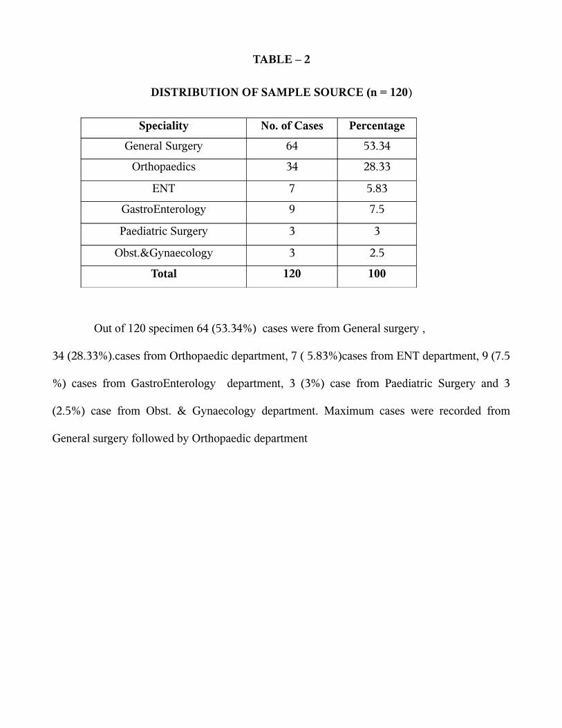
TABLE – 2
DISTRIBUTION OF SAMPLE SOURCE (n = 120)
Speciality No. of Cases Percentage
General Surgery 64 53.34
Orthopaedics 34 28.33
ENT 7 5.83
GastroEnterology 9 7.5
Paediatric Surgery 3 3
Obst.&Gynaecology 3 2.5
Total 120 100
Out of 120 specimen 64 (53.34%) cases were from General surgery ,
34 (28.33%).cases from Orthopaedic department, 7 ( 5.83%)cases from ENT department, 9 (7.5
%) cases from GastroEnterology department, 3 (3%) case from Paediatric Surgery and 3
(2.5%) case from Obst. & Gynaecology department. Maximum cases were recorded from
General surgery followed by Orthopaedic department

TABLE - 3DISTRIBUTION OF ABSCESS AS PER SITE (n=120)
S.NOLocation of
AbscessNo of cases
Type of AbscessSuperficial
AbscessDeep Abscess
1 Neck 5 4 1
2 Breast 13 13 -
3 Axilla 7 7 -
4 Arm 5 2 3
5 Hand 11 7 4
6 Inguinal 2 2 -
7 Gluteal 11 1 10
8 Perianal 10 10 -
9 Thigh 2 - 2
10 Leg 4 4 -
11 Feet 7 6 1
12 Chestwall 3 3 -
13 Bone & Joint 5 - 5
14 Liver 7 - 7
15 Gall Bladder 2 - 2
16 Abd. Wall 7 - 7
17 Appendix 3 - 3
18 Ear 7 3 4
19 Lung 4 - 4
20 Genital 5 2 3
Total (%) 120(100%) 64(53.33%) 56(46.67%)
Out of 120 pus samples taken, 64 (53.33%) were from superficial abscess and 56
(46.67%) were from deep abscess.

TABLE - 4
MICROBIOLOGIC CHARACTERISTICS OF ABSCESSES BY LOCATION (n = 120)
S.No. Abscess Location No. of Cases Aerobic Anaerobic Both
1 Neck 5 4 1 0
2 Breast 13 7 6 0
3 Axilla 7 5 2 0
4 Arm 5 4 1 0
5 Hand 11 8 2 1
6 Inguinal 2 1 0 1
7 Gluteal 11 9 0 2
8 Perianal 10 7 2 1
9 Thigh 2 2 0 0
9 Leg 4 4 0 0
10 Foot 7 6 1 0
11 Chestwall 3 3 0 0
12 Bone & Joint 5 5 0 0
13 Liver 7 4 3 0
14 Gall Bladder 2 2 0 0
15 Abd. Wall 7 3 4 0
16 Appendix 3 2 0 1
17 Ear 7 4 3 0
18 Lung 4 4 0 0
19 Genital 5 5 0 0
Total(%) 120(100%) 89(74.17%) 25(20.83%) 6(5%)
Out of 120 pus samples, samples with aerobic isolates only were 89 (74.17%), samples
with anaerobic isolates only were 25 (20.83%) and samples with both aerobic and anaerobic
isolates were 6 (5%).
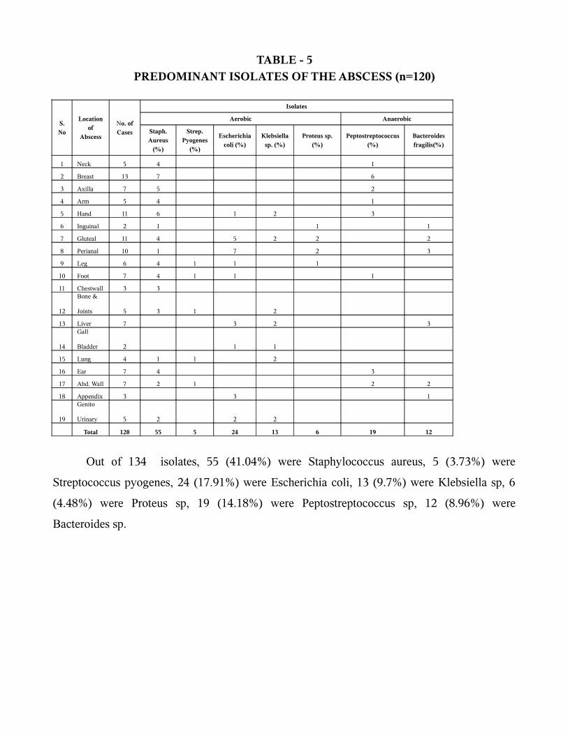
TABLE - 5PREDOMINANT ISOLATES OF THE ABSCESS (n=120)
S.
No
Location of
Abscess
No. of
Cases
Isolates
Aerobic Anaerobic
Staph. Aureus
(%)
Strep. Pyogenes
(%)
Escherichia
coli (%)
Klebsiella
sp. (%)
Proteus sp.
(%)
Peptostreptococcus
(%)
Bacteroides
fragilis(%)
1 Neck 5 4 1
2 Breast 13 7 6
3 Axilla 7 5 2
4 Arm 5 4 1
5 Hand 11 6 1 2 3
6 Inguinal 2 1 1 1
7 Gluteal 11 4 5 2 2 2
8 Perianal 10 1 7 2 3
9 Leg 6 4 1 1 1
10 Foot 7 4 1 1 1
11 Chestwall 3 3
12
Bone &
Joints 5 3 1 2
13 Liver 7 3 2 3
14
Gall
Bladder 2 1 1
15 Lung 4 1 1 2
16 Ear 7 4 3
17 Abd. Wall 7 2 1 2 2
18 Appendix 3 3 1
19
Genito
Urinary 5 2 2 2
Total 120 55 5 24 13 6 19 12
Out of 134 isolates, 55 (41.04%) were Staphylococcus aureus, 5 (3.73%) were
Streptococcus pyogenes, 24 (17.91%) were Escherichia coli, 13 (9.7%) were Klebsiella sp, 6
(4.48%) were Proteus sp, 19 (14.18%) were Peptostreptococcus sp, 12 (8.96%) were
Bacteroides sp.
TABLE - 6
PREDOMINANT ISOLATES OF SUPERFICIAL ABSCESS (n=71)S
.No.
Loc
atio
n o
f Ab
sces
s
No.
of
Cas
es
IsolatesAerobic Anaerobic
Sta
ph
. au
reu
s
Str
ep. p
yoge
nes
Esc
her
ich
ia c
oli
Kle
bsi
ella
sp
.
Pro
teu
s sp
.
Pep
tost
rep
toco
ccu
s
Bac
tero
ides
fra
gili
s
1 Neck 4 42 Breast 13 7 63 Axilla 7 5 24 Arm 2 25 Hand 7 3 1 2 26 Inguinal 2 1 1 17 Gluteal 1 1 18 Perianal 10 1 7 2 39 Leg 4 3 110 Feet 6 3 1 1 111 Chestwall 3 312 Ear 3 2 113 Genital 2 2 1
Total 64 36 2 10 3 4 12 4
Out of 64 superficial abscess, 5 were aerobic isolates and 16 were anaerobic isolates. Of
55 aerobic isolates, 36 were Staphylococcus aureus,2 were Streptococcus pyogenes,10 were
Escherichia coli, 3 were Klebsiella spp, 4 were Proteus spp. Of 16 anaerobic isolates, 12 were
Peptostreptococcus spp, 4 were Bacterodes fragilis group.
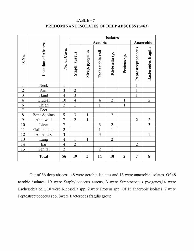
TABLE - 7PREDOMINANT ISOLATES OF DEEP ABSCESS (n=63)
S.N
o.
Loc
atio
n o
f Abs
cess
No.
of
Cas
es
IsolatesAerobic Anaerobic
Sta
ph
. au
reu
s
Str
ep. p
yoge
nes
Esc
her
ich
ia c
oli
Kle
bsi
ella
sp.
Pro
teus
sp.
Pep
tost
rep
toco
ccu
s
Bac
tero
ides
fra
gili
s
1 Neck 1 12 Arm 3 2 13 Hand 4 3 14 Gluteal 10 4 4 2 1 26 Thigh 2 1 1 17 Feet 1 18 Bone &joints 5 3 1 29 Abd. wall 7 2 1 2 210 Liver 7 3 2 311 Gall bladder 2 1 112 Appendix 3 3 113 Lung 4 1 1 214 Ear 4 2 215 Genital 2 2 1
Total 56 19 3 14 10 2 7 8
Out of 56 deep abscess, 48 were aerobic isolates and 15 were anaerobic isolates. Of 48
aerobic isolates, 19 were Staphylococcus aureus, 3 were Streptococcus pyogenes,14 were
Escherichia coli, 10 were Klebsiella spp, 2 were Proteus spp. Of 15 anaerobic isolates, 7 were
Peptostreptococcus spp, 8were Bacterodes fragilis group

TABLE – 8
TOTAL NUMBER OF AEROBES AND ANAEROBES ISOLATED FROM THE ABSCESSES (n=134)
Organism Total (%)Aerobic 103(76.87%)
Gram positive cocci 60(44.77%)
Staphylococcus aureus 55(41.04%)
Streptococcus pyogenes 5(3.73%)
Gram negative bacilli 43(32.09%)
Escherichia coli 24(17.91%)
Klebsiella spp 13(9.7%)
Proteus spp 6(4.48%)
Anaerobic 31(23.14%)
Gram positive cocci 19(14.18%)
Peptostreptococcus spp 19(14.18%)
Gram negative bacilli 12(8.96%)
Bacteroides fragilis 12(8.96%)
Out of 134 isolates, 103 (76.87%) were aerobic isolates , which included 60 (44.77%)
gram positive cocci and 43 (32.09%)gram negative bacilli .Total anaerobic isolates were
31(23.14%),which included 19 (14.18%) gram positive cocci and 12 (8.96%) gram negative
bacilli
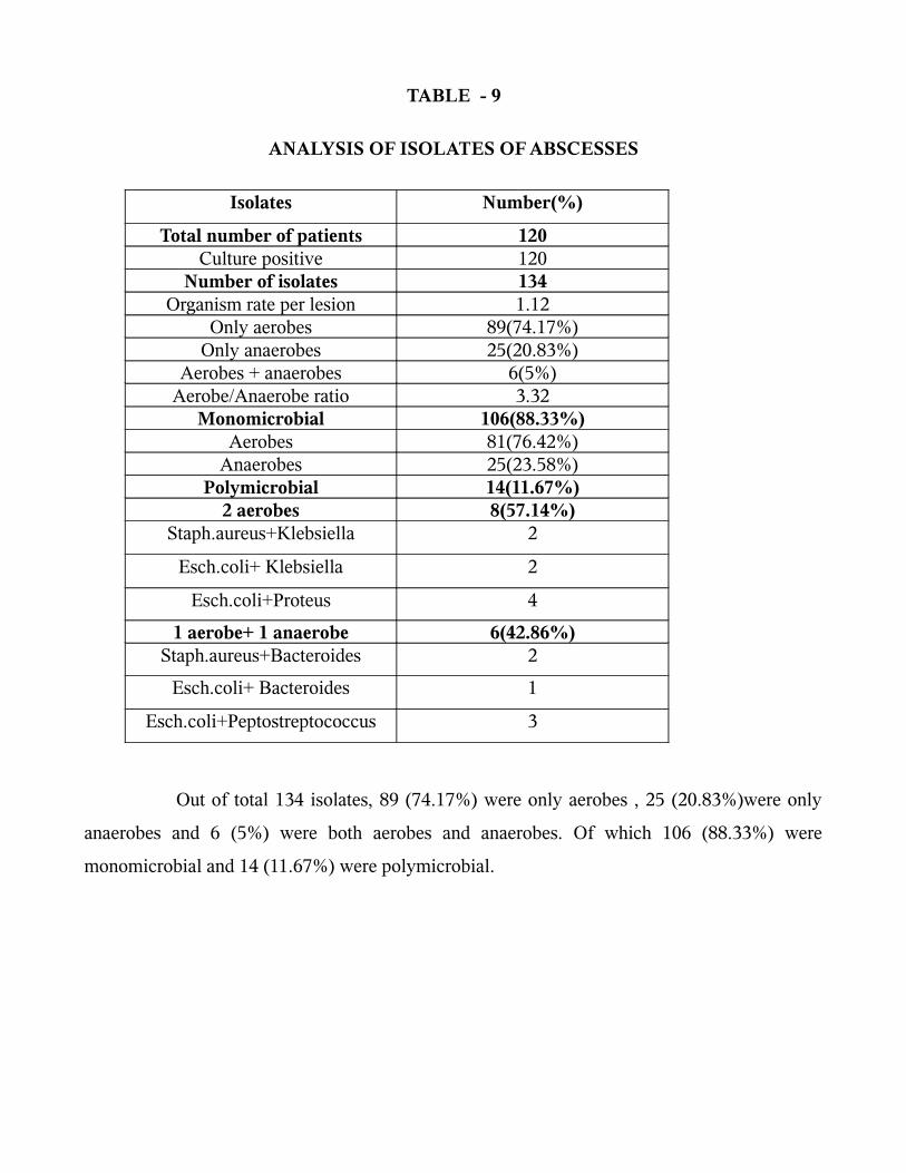
TABLE - 9
ANALYSIS OF ISOLATES OF ABSCESSES
Isolates Number(%)
Total number of patients 120Culture positive 120
Number of isolates 134Organism rate per lesion 1.12
Only aerobes 89(74.17%)Only anaerobes 25(20.83%)
Aerobes + anaerobes 6(5%)Aerobe/Anaerobe ratio 3.32
Monomicrobial 106(88.33%)Aerobes 81(76.42%)
Anaerobes 25(23.58%)Polymicrobial 14(11.67%)
2 aerobes 8(57.14%)Staph.aureus+Klebsiella 2
Esch.coli+ Klebsiella 2
Esch.coli+Proteus 4
1 aerobe+ 1 anaerobe 6(42.86%)Staph.aureus+Bacteroides 2
Esch.coli+ Bacteroides 1
Esch.coli+Peptostreptococcus 3
Out of total 134 isolates, 89 (74.17%) were only aerobes , 25 (20.83%)were only
anaerobes and 6 (5%) were both aerobes and anaerobes. Of which 106 (88.33%) were
monomicrobial and 14 (11.67%) were polymicrobial.
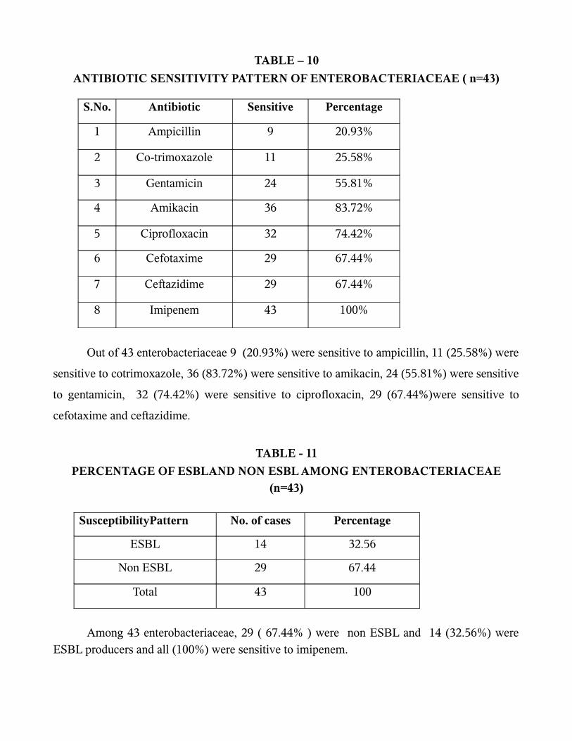
TABLE – 10
ANTIBIOTIC SENSITIVITY PATTERN OF ENTEROBACTERIACEAE ( n=43)
S.No. Antibiotic Sensitive Percentage
1 Ampicillin 9 20.93%
2 Co-trimoxazole 11 25.58%
3 Gentamicin 24 55.81%
4 Amikacin 36 83.72%
5 Ciprofloxacin 32 74.42%
6 Cefotaxime 29 67.44%
7 Ceftazidime 29 67.44%
8 Imipenem 43 100%
Out of 43 enterobacteriaceae 9 (20.93%) were sensitive to ampicillin, 11 (25.58%) were
sensitive to cotrimoxazole, 36 (83.72%) were sensitive to amikacin, 24 (55.81%) were sensitive
to gentamicin, 32 (74.42%) were sensitive to ciprofloxacin, 29 (67.44%)were sensitive to
cefotaxime and ceftazidime.
TABLE - 11
PERCENTAGE OF ESBLAND NON ESBL AMONG ENTEROBACTERIACEAE (n=43)
SusceptibilityPattern No. of cases Percentage
ESBL 14 32.56
Non ESBL 29 67.44
Total 43 100
Among 43 enterobacteriaceae, 29 ( 67.44% ) were non ESBL and 14 (32.56%) were ESBL producers and all (100%) were sensitive to imipenem.

TABLE - 12ANTIBIOTIC SENSITIVITY PATTERN OF STREPTOCOCCI (n=5)
S.No. Antibiotic Sensitive Percentage%
1 Penicillin 5 100%
2 Ampicillin 4 80%
3 Co-trimoxazole 3 60%
4 Erythromycin 5 100%
5 Ciprofloxacin 5 100%
Out of 5 Beta hemolytic streptococci 3 (60%) were sensitive to cotrimoxazole, 4 (80%)
were sensitive to ampicillin and all (100%) were sensitive to penicillin, erythromycin and
ciprofloxacin
TABLE - 13ANTIBIOTIC SUSCEPTIBILITY PATTERN OF STAPH. AUREUS (n=55)
S.No. Antibiotic Sensitive (%) Intermediate (%) Resistance (%)
1 Penicillin 2(3.63%) - 53(96.37%)
2 Ampicillin 7(12.72%) - 48(87.28%)
3 Cefotaxime 29(52.73%) 4(7.27%) 22(40%)
4 Amikacin 40(72.73%) 7(12.73%) 8(14.54%)
5 Erythromycin 25(45.46%) 6(10.9%) 24(43.64%)
6 Ofloxacin 43(78.19%) - 12(21.81%)
7 Oxacillin 37(67.27%) 18(32.73%)
Out of 55 strains, 2 (3.63%) were sensitive to penicillin, 7 (12.72%) were sensitive to
ampicillin, 25 (45.46%)were sensitive to erythromycin, 29 (52.73%) were sensitive to
cefotaxime, 37 (67.27%) were sensitive to oxacillin,40 (72.73%) were sensitive to amikacin
and 43 (78.19%)were sensitive to ofloxacin
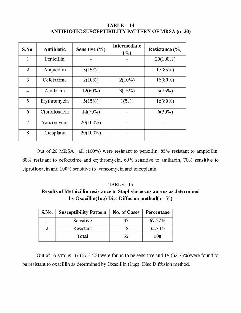
TABLE - 14ANTIBIOTIC SUSCEPTIBILITY PATTERN OF MRSA (n=20)
S.No. Antibiotic Sensitive (%)Intermediate
(%)Resistance (%)
1 Penicillin - - 20(100%)
2 Ampicillin 3(15%) - 17(85%)
3 Cefotaxime 2(10%) 2(10%) 16(80%)
4 Amikacin 12(60%) 3(15%) 5(25%)
5 Erythromycin 3(15%) 1(5%) 16(80%)
6 Ciprofloxacin 14(70%) - 6(30%)
7 Vancomycin 20(100%) - -
8 Teicoplanin 20(100%) - -
Out of 20 MRSA , all (100%) were resistant to pencillin, 85% resistant to ampicillin,
80% resistant to cefotaxime and erythromycin, 60% sensitive to amikacin, 70% sensitive to
ciprofloxacin and 100% sensitive to vancomycin and teicoplanin.
TABLE - 15
Results of Methicillin resistance to Staphylococcus aureus as determinedby Oxacillin(1µg) Disc Diffusion method( n=55)
S.No. Susceptibility Pattern No. of Cases Percentage
1 Sensitive 37 67.27%
2 Resistant 18 32.73%
Total 55 100
Out of 55 strains 37 (67.27%) were found to be sensitive and 18 (32.73%)were found to
be resistant to oxacillin as determined by Oxacillin (1µg) Disc Diffusion method.
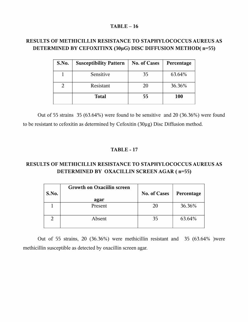
TABLE – 16
RESULTS OF METHICILLIN RESISTANCE TO STAPHYLOCOCCUS AUREUS AS DETERMINED BY CEFOXITINX (30µG) DISC DIFFUSION METHOD( n=55)
S.No. Susceptibility Pattern No. of Cases Percentage
1 Sensitive 35 63.64%
2 Resistant 20 36.36%
Total 55 100
Out of 55 strains 35 (63.64%) were found to be sensitive and 20 (36.36%) were found
to be resistant to cefoxitin as determined by Cefoxitin (30µg) Disc Diffusion method.
TABLE - 17
RESULTS OF METHICILLIN RESISTANCE TO STAPHYLOCOCCUS AUREUS AS DETERMINED BY OXACILLIN SCREEN AGAR ( n=55)
S.No.Growth on Oxaciilin screen
agarNo. of Cases Percentage
1 Present 20 36.36%
2 Absent 35 63.64%
Out of 55 strains, 20 (36.36%) were methicillin resistant and 35 (63.64% )were
methicillin susceptible as detected by oxacillin screen agar.
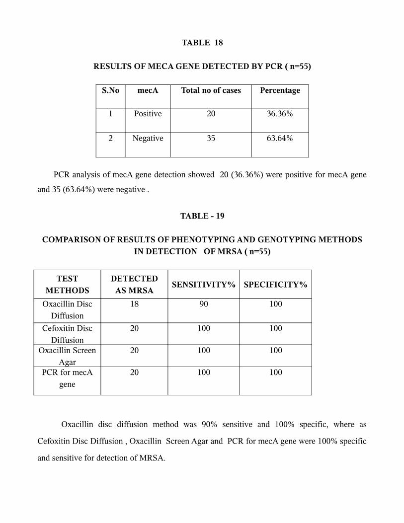
TABLE 18
RESULTS OF MECA GENE DETECTED BY PCR ( n=55)
S.No mecA Total no of cases Percentage
1 Positive 20 36.36%
2 Negative 35 63.64%
PCR analysis of mecA gene detection showed 20 (36.36%) were positive for mecA gene
and 35 (63.64%) were negative .
TABLE - 19
COMPARISON OF RESULTS OF PHENOTYPING AND GENOTYPING METHODS IN DETECTION OF MRSA ( n=55)
TEST METHODS
DETECTED AS MRSA
SENSITIVITY% SPECIFICITY%
Oxacillin DiscDiffusion
18 90 100
Cefoxitin DiscDiffusion
20 100 100
Oxacillin ScreenAgar
20 100 100
PCR for mecAgene
20 100 100
Oxacillin disc diffusion method was 90% sensitive and 100% specific, where as
Cefoxitin Disc Diffusion , Oxacillin Screen Agar and PCR for mecA gene were 100% specific
and sensitive for detection of MRSA.

DISCUSSION
Abscesses, cellulitis and necrotizing bacterial infections are seen more commonly in
developing countries because of the higher incidence of malnutrition with its resultant
immunosupression..Abscesses are accumulation of pus in tissue and any organism isolated
from them may be of significance.
The type and location of abscess will determine the specific management of abscesses.
The management will therefore differ considerably, but there are overriding principles of
management for all abscess and these will include: (1)drainage of pus (2) removal of necrotic
tissue and foreign material(3)correction of predisposing cause(4) amtimicrobial cover. The
choice of antimicrobial will initially be empirical,with the view of the known pyogenic
bacteria of most type of abscesses and the result of culture and sensitivity will indicate the
exact type required.43
In the present study pus samples from 120 cases of abscesses were taken between age
group 1 to 80 years. Maximum cases were in age group 21 to 40 years.(39.17%)( Table 1).Out
of 120 cases 76(63.33%) cases were male and 44(36.67%)cases were female. In all age group
the sex distribution was predominantly male. In study conducted by Christian et al,60%
isolates were male and 40% were female. 15
Maximum cases of abscesses were recorded from General Surgery department (53.34%)
followed by Orthopaedics department(28.33%) (Table 2) High case turn over in surgery
department occurs due to following factors like severe trauma, burns, diabetes, cancer,
chemotherapy, Aids, peripheral vascular disease etc.
In this study distribution of 120 samples as per site showed 53.33% were from
superficial abscess and 46.67% were from deep abscess (Table 3). Similar study done by
Itzhak Brook et al28. showed 676 (52.16%) cases were from superficial abscesses and 620
( 47.84%) cases were from deep abscesses.
Microbiological characteristics of abscesses showed in this study showed out of 120
samples,74.17% had aerobic isolates only ,20.83% had anaerobic isolates only and 5% had
mixed infections.(Table 4) Santosh Saini et al50 in his study observed 87.5%, had only aerobic
bacteria isolates, 5% had anaerobes only and mixed aerobic and anaerobic bacteria were
present in 7.5%
Study on isolates of 64 superficial abscesses showed , predominant aerobes were: S
aureus (36 isolates), and predominant anaerobes were anaerobic Gram-positive cocci (12
isolates) (Table 5& 6) Study by Itzhak Brook and Sydney M. Finegold et al20 on specimens
from 209 cutaneous abscesses cultured for aerobic and anaerobic microorganisms showed
predominant aerobes recovered were: S aureus (89 isolates), and predominant anaerobes
recovered were anaerobic Gram-positive cocci (79 isolates ) similar to this study.
Another study by Meislin et al41 and Ghoneim et al23 in concluded that Staphylococcus
aureus was present in half or more of axillary abscesses, finger and toe paronycia ,breast
abscesses. ,abscesses of the trunk, extremities, hand, buttocks and inguinal regions.
Whitehead et al65 in his study concluded perineal abscesses involving the vulvovaginal,
inguinal, scrotal, perianal, and buttock regions, by contrast, typically yield cultures containing
fecal flora, such as Enterobacteriaceae, Bacteroides spp, anaerobic gram positive cocci and
alpha hemolytic or nonhemolytic streptococci.22
Study on 56 isolates of deep abscess in this study showed predominant aerobic isolates
were Enterobacteriaceae and anaerobic isolates were Bacteroides spp (Table 5&7) Similar
study by Itzhak Brook et al 28 ,observed that in deep abscesses ,predominant aerobic isolates
were Enterobacteriaceae and group D streptococci and predominant anaerobic isolates were
Bacteroides fragilis group and Peptostreptococcus spp
Bacteroides species are significant clinical pathogens and are found in most anaerobic
infections, with an associated mortality of more than 19%. The bacteria maintain a complex and
generally beneficial relationship with the host when retained in the gut, but when they escape
this environment they can cause significant pathology, including bacteremia and abscess
formation in multiple body sites.
Intra-abdominal sepsis is the most common infection caused by Bacteroides. After
disruption of the intestinal wall, rupture of the diverticula, or other perforations due to a
surgical wound, malignancies, or appendicitis, members of the normal flora infiltrate the
normally sterile peritoneal cavity, and the resultant infections reflect the gut flora composition.
During the early, acute stage of infection (approximately 20 h), the aerobes, such as E. coli, are
the most active members of infection, establishing preliminary tissue destruction and reducing
the oxidation-reduction potential of the oxygenated tissue. Once sufficient oxygen has been
removed to allow the anaerobic Bacteroides species to replicate, these bacteria begin to
predominate during the second, chronic stage of infection25
In this study 44.77% isolates were gram positive and 32.09% were gram negative
bacilli. (Table 8). S Mohanty et al 53 in his study observed 45.96 % were gram positive cocci
and 54.04% were gram negative bacilli.
Our study showed 23.14% anaerobes from pyogenic infection .In study conducted by
Reena Set et al 47 on prevalence of anaerobic bacteria in pyogenic infections showed 18.7%
anaerobes from pyogenic infection .
Overall the predominant isolates of both superficial and deep abscesses in this study
group was Staphylococcus aureus around 41.04% in both superficial and deep abscesses.
Santosh Saini et al 50 in his study on abscesses showed 40% isolates of Staphylococcus aureus.
In another study by R.M.Banda et al 46 showed that Staphylococcus aureus was the
predominant isolates 51.8% from surgical bacterial infection, of which 37.55% were from
abscess .
In this study ,out of 120 culture positive cases, 106 (88.33%) cases were monomicrobial
and 14(11.67%) cases were polymicrobial, in which 8 (57.14%) cases had two aerobes. (Table
9) Study by Santosh Saini et al50 showed out of 40 culture positive cases, 26 (65%)were
monomicrobial and 14(35%) were polmicrobial of which 8 (57%) cases had two aerobes
similar to this study
Surgical infections, such as abscesses, secondary peritonitis, necrotising fascitis and
wounds with devitalized tissues are largely polymicrobial, and the role of both aerobic and
anaerobic bacteria in the pathogenesis of these infections is well recogniszed. Microbial
synergy may increase the net pathogenic effect and hence the severity of infection in several
ways7.
In the present study antibiotic susceptibility pattern of enterobactericeae showed out of
43 isolates, 29 (67.44%) were sensitive to third generation cephalosporins, cefotaxime and
ceftazidine and 14(32.56%) were resistant to it. All isolates were sensitive to imipenem.(Table
10)
In the present study the occurence of ESBL producing enterobacteriaceae in abscesses
was 32.56%. (Table 11). Shukla et al 54reported prevalence of ESBL in enterobactericeae was

found to be 36.1% similar to our study. All the ESBL's in the study were sensitive to imepenem
(100%) which correlated with the study by Baby Padmini S.5
ESBL producing organisms pose a major problem for clinical therapeutics. The
incidence of ESBL producing strains among clinical isolates has been steadily increasing over
the past few years resulting in limitation of therapeutic options.2 Initially restricted to hospital
acquired infections, they have also been isolated from infections in outpatients. Major
outbreaks involving ESBL strains have been reported from all over the world, thus making
them emerging pathogens
Sensitivity pattern of 5 isolates of Streptococcus pyogenes showed 100% sensitivity to
penicillin, erythromycin, ciprofloxacin. (Table12).S pyogenes is the main human pathogen
associated with local or systemic invasion and poststreptococcal immunologic disorders.
Although humans can be asymptomatic nasopharyngeal or perineal carriers of S pyogenes, the
organism should be considered abnormal if it is detected by culture. The portal of entry
determines the principal clinical picture. In each case, however, there is a diffuse and rapidly
spreading infection that involves the tissues and extends along lymphatic pathways with only
minimal local suppuration.32 Almost all Streptococcus pyogenes are susceptible to penicillin G,
and most are susceptible to erythromycin which correlates with our study.
Antibiotic susceptibility pattern of 55 isolates of Staphylococcus aureus showed , 37
(67.27%) isolates were sensitive and 18 (32.73%) were resistant to oxacillin(Table 13)
In our study resistance pattern of MRSA isolates were as follows, penicillin 100%,
ampicillin 85%, cephotaxime & erythromycin 80%,ciprofloxacin 30%,amikacin 25% and all
strains were 100% sensitive to vancomycin.(Table 14)
More than 80% of MRSA were found to be resistant to majority of antibiotics tested like
cephalexin, ciprofloxacin, penicillin, co-trimoxazole, gentamicin, erythromycin and
tetracycline and 60.5% to amikacin, no strains resistant to vancomycinin study conducted by S
Anupurba et al 55.
Study conducted by Vidhani et al 63 showed similar pattern of resistance of MRSA to
beta lactams like penicillin and amoxycillin was 100% while 87.4% isolates were resistant to
augmentin. Cefotaxime resistance was seen in 78.5% of isolates.
In this study, oxacillin disc diffusion method detected 18(32.73%) cases of MRSA from
55 isolates with sensitivity of 90% and specificity of 100%.(Table15&19). KB Anand et al 34 in
his study showed 87.5% specificity and 100%sensitivity by oxacillin disc diffusion method.
Cefoxitin disc diffusion method detected 20 (36.36%)cases out of 55 isolates as MRSA
which accounts for 100% sensitivity and specificity(Table16&19)) Cefoxitin is a potent inducer
of the mecA regulatory system and an accurate surrogate marker for the detection of MRSA in
routine susceptibility testing for disk diffusion 18. It is suggested that no special medium or
incubation temperature is required with cefoxitin as for oxacillin.
Cefoxitin disk diffusion testing is now recommended by CLSI as a preferred method of
detection of oxacillin resistance in staphylococci; however, it is important to report the findings
from cefoxitin disc diffusion test as indicative of either oxacillin susceptibility or resistance;
cefoxitin report should not be reported
MRSA detection by oxacillin screen agar method isolated 20(36.36%) cases with 100%
sensitivity and 100% specificity.(Table 17&19)) Chambers et al reported that the sensitivity of
this method approaches 100% for the detection of methicillin resistant S. aureus and 95% for
the coagulase-negative strains14

Detection of mecA gene by PCR was positive for 20 (36.36%) cases with 100%
sensitivity and specificity.(Table 18&19) mecA encodes PBP 2a (also termed PBP 29), an
inducible 76-kDa PBP that determines methicillin resistance. The heterogeneous nature of
methicillin resistance is an inherent limitation to the accuracy of susceptibility testing mecA
detection tests based on PCR or DNA hybridization will correctly identify even the most
heterogeneous of strains and should be considered the gold standard 14for methicillin
resistance.
Isolation rate of MRSA in this study was 36.36% .K Rajaduraipandi et al 33 in his study
showed that 33.6% of strains were obtained from pus among clinical isolates. Similar
observation was made by Mehta40, who in his study on control of MRSA in a tertiary care
center, had reported an isolation rate of 33% from pus and wound swabs.
Methicillin-resistant strains of staphylococci were identified immediately upon the
introduction of methicillin into clinical practice Resistance was termed “intrinsic” because it
was not due to destruction of the antibiotic by beta lactamase. Methicillin resistant
Staphylococcus aureus (MRSA) strains were initially described in 1961 and emerged in the last
decade as one of the most important nosocomial pathogens38. Infected and colonized patients
provide the primary reservoir and transmission is mainly through hospital staff 39. The risk
factors which contribute to MRSA are excessive antibiotic usage, prolonged hospitalization,
intravascular catheterization and hospitalisation in intensive care unit.16 .With the increased
incidence of MRSA, the effectiveness of penicillin and cephalosporins is questioned. In fact
many stains of MRSA exhibit resistance to both beta -lactams and aminoglycosides59. Hence, a
knowledge of prevalence of MRSA and their antimicrobial profile becomes necessary in the
selection of appropriate empirical treatment of these infections
SUMMARY
1. Among 120 cases taken for study, maximum number of cases were in age group 21 –
40 years (39.17%)
2. Sex distribution showed male preponderance (71.3%) among the 120 isolates.
3. Majority of samples were from general surgery (53.34%) followed by Orthopedics
department (28.33%)
4. Among 120 cases, 64(53.33%) cases were taken from superficial abscess and
56(46.67%) cases were taken deep abscess.
5. Out of 120 pus samples, 89 (74.17%) samples had only aerobic isolates 25 (20.83%)
samples had only anaerobic isolates and 6 (5%)samples had both aerobes and
anaerobes..Of which 106 (88.33%) were monomicrobial and 14 (11.67%) were
polymicrobial.
6. Of total 134 isolates, 44.77% and 32.09% were gram positive and gram negative
aerobic isolates respectively, 14.18% and 8.96% were gram positive and gram negative
anaerobic isolates respectively.
7. Study on isolates of 64 superficial abscesses showed , predominant aerobes were: S
aureus (36 isolates), and predominant anaerobes were anaerobic Gram- positive cocci.
Study on 56 isolates of deep abscess in this study showed predominant aerobic isolates

were Enterobacteriaceae and anaerobic isolates were Bacteroides spp
8. Out 134 isolates, 103(76.87%) were aerobic isolates, of which 60(44.77%) were gram
positive cocci and 43(32.09%) were gram negative bacilli and 31(23.14%) were
anaerobic isolates, of which 19(14.18%) were gram positive cocci and 12(8.96%)were
gram negative bacilli.
9. Of 43 enterobacteriaceae ,14(32.56%) were ESBL and 29(67.44%) were non ESBL
producer.All isolates were 100% sensitive to imipenem.
10. Isolation of Streptococcus pyogenes 5(3.73%) from 60(44.77%) aerobic gram positive
cocci showed 100% sensitivity to penicillin, erythromycin and ciprofloxacin.
11. Staphylococcus aureus were predominant isolates of abscesses constituting to about
55(41.04%) ot total 134 isolates.
12. Antibiotic sensitivity result of Staphylococcus aureus by disc diffusion method with
oxacillin showed ,out of 55 strains ,37(67.28%) were detected as methicillin sensitive
and 18(32.72%) were detected as methicillin resistant.
13. Out of 55 isolates of Staphylococcus aureus ,35(63.64%) isolates were detected
methicillin sensitive and 20(36.36%)were detected as methicillin resistant by Cefoxitin
disc diffusion and Oxacillin Screen Agar method.
14. PCR analysis of mecA gene detection showed 20(36.36%) were mecA gene positive

and 35(63.64%) were mecA gene negative.
15. All isolates of Staphylococcus aureus were 100% sensitive to vancomycin and
teicoplanin.

CONCLUSION
Among 120 cases of superficial and deep abscesses studied for bacteriological profile
showed 134 isolates, of which 88.33% were monomicrobial and 11.67% were polymicrobial.
Of 76.87% aerobic isolates, 44.77% were gram positive cocci and 32.09% were gram negative
bacilli. Of 23.14% anaerobic isolates , 14.18% were gram positive cocci and 8.96%were gram
negative bacilli.
Of 134 isolates, Staphylococcus aureus was predominant isolate (41.04%) from both
superficial and deep abscess, of which 36.36% were detected as Methicillin resistant .MRSA
emerge to be significant problematic pathogen in the surgical settting with vancomycin
probably the only reliable choice of this infection. The conventional phenotypic methods
,Cefoxitin disc diffusion method and Oxacillin screen agar had high degree of sensitivity and
specificity, in concordance with ‘gold standard’ mecA gene detection by polymerase chain
reaction.
Essential infection control practices should include hand washing by hospital
personnel, basic cleaning of all surface levels (hand touch sites) , increased barrier precautions
and isolation of patients colonized or infected with MRSA.. A multidisciplinary approach,
coordinated participation of microbiologists, clinicians, nursing personnel, hospital infection
control team is necessary for management of MRSA producing infection. Continuous
monitoring of antimicrobial susceptibility pattern in individual settings together with judicious
use is emphasized to minimize emergence of drug resistant bacteria.
BIBILOGRAPHY
1 Alex. C.. Sonnerwith, Chapter 82, Gradwohl’s Clinical laboratory methods and
diagnostic, eighth edition, volume 2, Anaerobic bacteria pg 1876-1885.
2. Ananthkrishnan AN, Kanungo R, Kumar A, Badrinath S, Detection of ESBL producers
among surgical wound infections and burns patients in JIPMER, Indian J Med Microbiol
2000;18(4):160-165.
3. Arthur O. Tzianabos, Anil Chandraker, Wiltrud Kalka-Moll, Francesca
Stingele, Victor M. Dong, Robert W. Finberg,Robert Peach, and Mohamed H. Sayegh
Bacterial Pathogens Induce Abscess Formation by CD4+ T-Cell Activation via the
CD28-B7-2 Costimulatory Pathway American society of Microbiology, Infection and
Immunity, December 2000, p. 6650-6655, Vol. 68, No. 12
4. Archer GL, Pennell E. Detection of methicillin resistance in staphylococci by using a
DNA probe. Antimicrob Agents Chemother 1990; 34: 1720–4.
5. Baby Padimini S, Appala Raju B,Mani KR.,Detection of Enterobacteriaceae producing
CTXM ESBL from a Tertiary care hospital in South India.,Indian journal of medical
microbiology.2008;26(2):163-166.
6. Bailey & Scott’s, Diagnostic Microbiology,Anaerobic Bacteriology, Chapter 44 , page
463-476
7. Bowler P.G., Duerden B.I., Armstrong D.G.. Wound microbiology and associated
approaches to wound management. Clin Microbiol Rev 2001;244-69
8. Brook I.. Bacteroides infections in children. J Med Microbiol 1995;43:92-8.
9. Brook, I. 2002. Pediatric Anaerobic Infection: Diagnosis and Management. Marcel
Dekker, New York, N.Y
10. Brook I., Frazier E. H. Aerobic and anaerobic bacteriology of wounds and cutaneous
abscesses. Arch. Surg. 1990; 125: 1445–1551.
11. Brook I. Microbiology of abscesses of the head and neck in children. Ann. Otol. Rhinol.
Laryngol. 1987; 96: 429–433. 12. Brook I., Frazier E. H. Aerobic and anaerobic
microbiology in intra-abdominal infections associated with diverticulitis. J. Med.
Microbiol. 2000; 49: 827–830.
12. Brook I, Frazier E H. Clinical features and Aerobic and anaerobic microbiological
characteristics of cellulitis. Arch Surg. 1995, 130(7); 786 – 92.
13. Casewell MW, Hill RLR. The carrier state: methicillin-resistant Staphylococcus aureus.
J Antimicrob Chemother 1986;18:Suppl A:1-12.
14. Chambers HF. Methicillin resistance in Staphylococci:Molecular and biochemical basis
and clinical implications. Clin Microbiol Rev 1997; 10 : 781-91.
15. Christian H Jacobus, Christopher J Lindsell , Sabrina D Leach , Gregory J Fermann,
Amy Beth Kressel and Laura E Rue ,Prevalence and demographics of methicillin
resistant Staphylococcus aureus in culturable skin and soft tissue infections in an urban
emergency department,BMC Emergency Medicine 2007
16. Clinical and Laboratory Standards Institute / NCCLS Performance standards for
Antimicrobial disc diffusion tests; Approved standards. 9th ed. CLSI Document M2-
M9. Wayne Pa: Clinical and Laboratory Standards Institute; 2006.
17. Connie R.Mahon, George Manuselis, Textbook of Diagnostic Microbiology, Second
edition, Chapter 3, Page 53-91

18. Derek F. J. Brown1, David I. Edwards2, Peter M. Hawkey3, Journal of Antimicrobial
Chemotherapy 2005 56(6):1000-1018; Guidelines for the laboratory diagnosis and
susceptibility testing of methicillin-resistant Staphylococcus aureus (MRSA)
19. Doebbeling BN. The epidemiology of methicillin resistant Staphylococcus aureus
colonization and infection. J Chemotherapeutics 1995; 7 (99 -103)
20. Finegold, S. M. 1977. Anaerobic Bacteria in Human Disease. Academic
Press, Inc., New York, N.Y.
21. Franklin D. Lowy, M.D N Engl J Med 1998; 339(27):2025. Staphylococcus aureus
Infections
22. G Garcea, T Lloyd, M Jacobs, A Cope, A Swann, D Berry, Role of microbiological
investigations in the management of non-perineal cutaneous abscesses, Postgraduate
Medical Journal 2003;79:519-521)
23. Ghoneim, ATM, McGolrick J, Blick et al. Aerobic and Anaerobic bacteriology of
subcutaneous abscess. Br J Surg 1981;68:498
24. Gorbach, Barlett, Blacklow, Infectious Disease, third edition, Cutaneous Abscesses
and Ulcers, Chapter 1-34, page 1159
25. Hannah M. Wexler ,Bacteroides ; Clinical Microbiology Reviews, October 2007, p.
593-621, Vol. 20, No. 4
26. Harrison's Principles of Internal Medicine, 17e Chapter 157. Infections Due to Mixed
Anaerobic Organisms
27. Infection and Immunity, December 2000, p. 6650-6655, Vol. 68, No. 12
Copyright © 2000, American Society for Microbiology

28. Itzhak Brook, The British Society for Antimicrobial Chemotherapy Journal of
Antimicrobial Chemotherapy (2002) 50, 805-810 2002
29. Itzhak Brook MD1 and Sydney M. Finegold , Aerobic and Anaerobic Bacteriology of
Cutaneous Abscesses in Children, Journal of American Academy of Paediatrics, Vol.
67 No. 6 June 1981, pp. 891-895
30. Itzhak Brook MD1 and Sydney M. Finegold MD1 Aerobic and Anaerobic Bacteriology
of Cutaneous Abscesses in Children PEDIATRICS Vol. 67 No. 6 June 1981, pp. 891-
895
31. Jan.V.Hirschmann,Harrison’s Principle of Internal Medicine, Eleventh Edition,Chapter
87,Localised Infection & Abscesses,page 478-485
32. Jawetz, Melnick & Adelberg’s, Medical Microbiology, Twenty fourth edition, Chapter
14,Page224-230
33. K Rajaduraipandi1, KR Mani2, K Panneerselvam1, M Mani3, M Bhaskar4, P
Manikandan5 , Prevalence and antimicrobial susceptibility pattern of methicillin
resistant Staphylococcus aureus: A multicentre study. ,Year : 2006 | Volume : 24 |
Issue : 1 | Page : 34-38
34. KB Anand, P Agrawal, S Kumar, K Kapila ,Comparison of cefoxitin isc diffusion test,
oxacillin screen agar, and PCR for mecA gene for detection of MRSA Year : 2009 |
Volume : 27 | Issue : 1 | Page : 27-29 )
35. Kluytmans J, Belkum AV, Verbrugh H. Nasal Carriage of Staphylococcus aureus;
Epidemiology, underlying mechanisms and associated risk. Clinical Microbiology Rev
1997; Volume 505-520

36. Koneman’s Color Atlas & Textboook of Diagnostic Microbiology, Sixth
edition, Chapter 16,The Anaerobic Bacteria,page 870-940
37. Mackie & McCartney, Practical Medical Microbiology, Fourteenth edition, Chapter
11, page 245-261
38. Maple PAC, Hamilton- Miller JMT, Brumfitt W. World wide antibiotic resistance in
methicillin resistant Staphylococcus aureus. Lancet 1989;1:537-540.
39. McDonald M. The epidemiology of methicillin resistant Staphylococcus aureus:
Surgical relevance 20 years on. Aust N Z J Surg 1997;67:682-685.).
40. Mehta AP, Rodrigues C, Sheth K, Jani S, Hakimiyan A, Fazalbhoy N. Control of
methicillin resistant Staphylococcus aureus in a tertiary care Centre-A five-year study. J
Med Microbiol 1998;16:31-4.
41. Meislin H W, Lerner S A, Graves M H, McGehee M D, Kocka F E, Morello J A, Rosen
P. Cutaneous abscesses-Anaerobic and aerobic bacteriology and outpatient management.
Ann Intern Med. 1977;87:145–149
42. Novick RP. The staphylococcus as a molecular genetic system. In: Novick RP, ed.
Molecular biology of the staphylococci. New York: VCH, 1990:1-37.
43. Oxford Textbook Of Surgery,second edition,volume 3,edited by Sir Peter J Morris,
William C Wood, Chapter 47.1, Surgery of bacterial disease Pg 3219- 3222)
44. Prescott,Harley,Klein,Microbiology,Chapter 36 ,Clinical Microbiology,
page 827-844
45. Reagan DR, Doebbeling BN, Pfaller MA, et al. Elimination of coincident
Staphylococcus aureus nasal and hand carriage with intranasal application of mupirocin
calcium ointment. Ann Intern Med 1991;114:101-106

46. RM Banda, AS Muula ,Surgical bacterial infections and antimicrobial susceptibility
patterns at Lilonge Central Hospital, Malawi Medical Journal, 2000)
47. R Set, L Sequeira, SA Angadi, GV Koppikar, Set Reena, Sequeira Loraine , Prevalence
of anaerobic bacteria in pyogenic infections; Year : 1997, Volume : 15 , Issue : 3 Page
: 121-122
48. Sambrook and Russel Molecular Cloning,A Laboratory Manual, ,Third Edition,Volume
2 ;pages 8.20 -8.22
49. Sanford MD, Widmer AF, Bale MJ, Jones RN, Wenzel RP. Efficient detection and long-
term persistence of the carriage of methicillin-resistant Staphylococcus aureus. Clin
Infect Dis 1994;19:1123-1128
50. Santosh Saini; Naveen Gupta; Aparna; Lokveer; M.S. Griwan Surgical
infections: a microbiological study .Brazilian Journal of Infectious Diseases vol.8
no.2 Salvador Apr. 2004
51. Schaberg DR, Zervos MJ. Intergeneric and interspecies gene exchange in gram-positive
cocci. Antimicrob Agents Chemother 1986;30:817-822.
52. Sheagren JN. Staphylococcus aureus. The persistent pathogen. New Engl J Med 1984;
310: 1368-73, 1437
53. S Mohanty, Arti Kapil, B Dhawan, BK Das ,Bacteriological and antimicrobial
susceptibility profile of soft tissue infections from Northern India ,Year : 2004
Volume : 58 Issue : 1 Page : 10-15 )
54. Shukla, R Tiwari, M Agrawal Prevalence of extended spectrum -lactamase producing
klebsiella pneumoniae in a tertiary care hospital Indian journal of Medical
Microbiology Year : 2004 Volume : 22 Issue : 2 Page : 87-91
55. S Anupurba, MR Sen, G Nath, BM Sharma, AK Gulati, TM Mohapatra Prevalence of
methicillin resistant staphylococcus aureus in a tertiary referral hospital in eastern Uttar
Pradesh, 2003 | Volume : 21 | Issue : 1 | Page : 49-51 )
56. Stevens D.L. Infections of the skin, muscle and soft tissues. In: Fauci AS, Braun WE,
Isselbacher KJ, Wilson JD, Martin JB, Kasper DL et al editors. Harrison's Principles of
Internal Medicine. 14th ed. New York: McGraw Hill;
57. Structural rationale for the modulation of abscess formation by Staphylococcus aureus
capsular polysaccharides Arthur O. Tzianabos, Julia Y. Wang, and JeaCLee
58. Sutter V.L., Citron D.M., Edelstein M.A.C., Finegold S.M. Processing of clinical
specimens and isolation and identification procedures. In: Wadsworth Anaerobic
Bacteriology Manual. 4th ed. Belmont: Star Publishing; 1985:23-70.
59. Swenson JM, Patel JB, Jorgensen JK. Special phenotypic methods for detecting
antibacterial resistance, Chapter 74. In: Murray PR, Baron ES, Jorgensen JH, Landry
ML Pfaller MA, editors. Manual of Clinical Microbiology, 9th ed. Washington DC:
ASM Press; 2007. p. 1175-76.
60. Swenson JM, Lonsway D, McAllister S, Thompson A, Jevitt L, Patel JB. Detection of
mecA-mediated Resistance Using Cefoxitin Disk Diffusion (DD) in a Collection of
Staphylococcus aureus Expressing Borderline oxacillin MICs. Diagn Microbiol Infect
Dis 2007;58:33-9.
61. Thornsberry C. The development of antimicrobial resistance in staphylococci. J
Antimicrob Chemotherap 1998;21 (Suppl.C):9-16. 1998:827-30
62. Tucker C. Acute and chronic inflammation. In: Cotran, Kumar, Collins, Robbins's
Pathologic Basis of Disease. 6th ed. Philadelphia: WB Saunders Co.; 1999:50-89.
63. Vidhani S, Mehndiratta PL, Mathur MD ; Study of methicillin resistant S. aureus

(MRSA) isolates from high risk patients ; Indian Journal of Medical Microbiology Year
: 2001 | Volume : 19 | Issue : 2 | Page : 13
64. VK Beena, GR Kumari, PG Shivananda ,Prevalence of Bacteroides fragilis group in
human infections ,Year : 1995 | Volume : 13 | Issue : 1 | Page : 18-21
65. Whitehead SM, Leach RD, Eykyn SJ, et al. The Aetiology of perirectal sepsis BrJ Surg
1982;69;166
66. Williams DN, Peterson PK. Endocarditis caused by coagulase negative
staphylococci. Infection 1979;7:5-9
67. Wren M.W.D. The culture of clinical specimen for anaerobic bacteria: a comparison of
three regimen. J Med Microbiol 1977;10:195-201.

APPENDIX
1. Peptone Water
Peptone 10 gSodium chloride 5gDistilled water 1 litre
Dissolve the ingredients in warm water, adjust the pH to 7.4-7.5 and filter. Distribute as required and autoclave at 121ºC for 15 minutes.
2. Nutrient Agar
Ingredients Grams/litre
Peptic digest of animal tissue 5Sodium Chloride 5Beef extract 1.5Yeast extract 1.5Agar 15.00
Final pH(at 25º C) 7.4 ±0.2.
Suspend 28 gms in 1000 ml of distilled water. Heat to boiling to dissolve the medium
completely. Sterilize by autoclaving at 15 lbs pressure (121ºC) for 15 minutes and pour into
sterile petridishes.
3. Blood agar
Sterile sheep blood 5 mlNutrient agar 100ml
Autoclave the nutrient agar base at 121°C for 15 minutes. Cool to 45-50 ºC and add
blood with sterile precautions and distribute in petri dishes.
4. Mac Conkey Agar
Ingredients Grams/litrePeptic digest of animal tissue 17Proteose peptone 3Lactose 10Bile salts 1.5Sodium Chloride 5
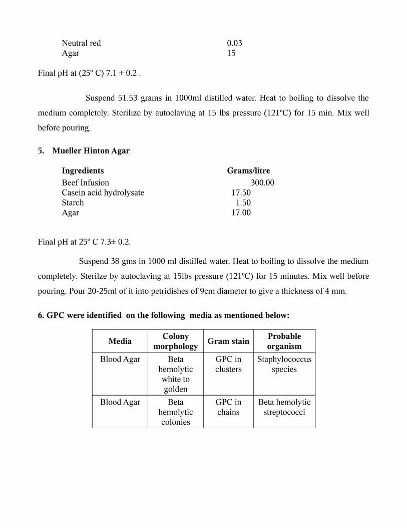
Neutral red 0.03Agar 15
Final pH at (25º C) 7.1 ± 0.2 .
Suspend 51.53 grams in 1000ml distilled water. Heat to boiling to dissolve the
medium completely. Sterilize by autoclaving at 15 lbs pressure (121ºC) for 15 min. Mix well
before pouring.
5. Mueller Hinton Agar
Ingredients Grams/litre
Beef Infusion 300.00Casein acid hydrolysate 17.50Starch 1.50Agar 17.00
Final pH at 25º C 7.3± 0.2.
Suspend 38 gms in 1000 ml distilled water. Heat to boiling to dissolve the medium
completely. Sterilze by autoclaving at 15lbs pressure (121ºC) for 15 minutes. Mix well before
pouring. Pour 20-25ml of it into petridishes of 9cm diameter to give a thickness of 4 mm.
6. GPC were identified on the following media as mentioned below:
MediaColony
morphologyGram stain
Probable organism
Blood Agar Beta hemolytic white to golden
GPC in clusters
Staphylococcus species
Blood Agar Beta hemolytic colonies
GPC in chains
Beta hemolytic streptococci

7. Differentiating characters of gram negative bacilli for their identification were as follows
Org
anis
ms
Mot
ility
Cat
alas
e
Oxi
dase
Indo
le
Met
hyl R
ed
Vog
es P
rosk
auer
Cit
rate
TS
I
Glu
cose
Lac
tose
Sucr
ose
Mal
tose
Man
nito
l
Eshenchia coli
+ + ─ + + ─ ─
A/A withgas
+ + ─ + +Klebsiella Pneumoniae
─ + ─ ─ ─ + +
A/Awith gas
+ + + + +Klebsiella oxytoca
─ + ─ + ─ + +
A/A
+ + + + +Citrobacter freundii
+ + ─ ─ + ─ +
K/Awith gas & H2S
+ ± + + +Citrobacter koseri
+ + ─ ─ + ─ +
K/A with gas
+ ± ± + +Proteus vulgaris
+ + ─ + + ─ V
K/Awith gas & H2S
+ ─ ─ + ─Proteus mirabilis
+ + ─ ─ + ─ V
K/Awith gas & H2S
+ ─ ─ ─ ─
8. Robertson Cooked meat mediumIngredients Grams/litreBrain heart solids 98Proteose peptone 20
Dextrose 2Sodium Chloride 5
Final pH at 25 ºC 7.2 ±0.2.
To each test tube add 1 part of the ingredients and 3 parts of distilled water, mix
thoroughly and allow to stand for 15 minutes until all particles are thoroughly wetted. Sterilize
by autoclaving at 15 lbs pressure (121º C) for 15 minutes.
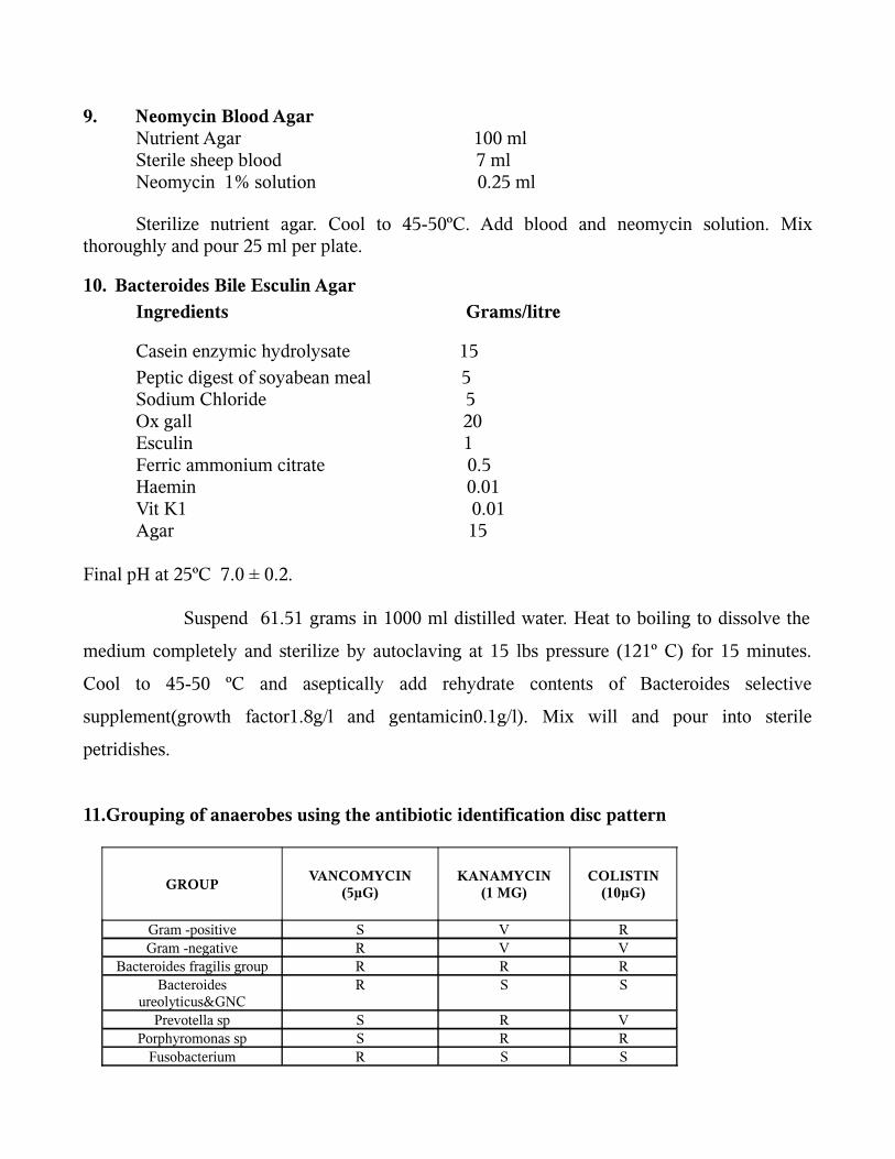
9. Neomycin Blood AgarNutrient Agar 100 mlSterile sheep blood 7 mlNeomycin 1% solution 0.25 ml
Sterilize nutrient agar. Cool to 45-50ºC. Add blood and neomycin solution. Mix thoroughly and pour 25 ml per plate.
10. Bacteroides Bile Esculin Agar
Ingredients Grams/litre
Casein enzymic hydrolysate 15
Peptic digest of soyabean meal 5Sodium Chloride 5Ox gall 20Esculin 1Ferric ammonium citrate 0.5Haemin 0.01Vit K1 0.01Agar 15
Final pH at 25ºC 7.0 ± 0.2.
Suspend 61.51 grams in 1000 ml distilled water. Heat to boiling to dissolve the
medium completely and sterilize by autoclaving at 15 lbs pressure (121º C) for 15 minutes.
Cool to 45-50 ºC and aseptically add rehydrate contents of Bacteroides selective
supplement(growth factor1.8g/l and gentamicin0.1g/l). Mix will and pour into sterile
petridishes.
11.Grouping of anaerobes using the antibiotic identification disc pattern
GROUPVANCOMYCIN
(5µG)KANAMYCIN
(1 MG)COLISTIN
(10µG)
Gram -positive S V RGram -negative R V V
Bacteroides fragilis group R R RBacteroides
ureolyticus&GNCR S S
Prevotella sp S R VPorphyromonas sp S R R
Fusobacterium R S S

12. Mannitol salt agar
Ingredients Grams/litre
Beef extract 0.1
Peptone 1
NaCl 7.5
Mannitol 1
Agar 1.5
Phenol red 0.0025
Distilled water 1000
The above ingredients were dissolved in 1000ml of dissolved in distilled water, mixed
thoroughly. Heated with frequent agitation and pH adjusted to 7.6. Sterilised by autoclaving
and poured in plates.
13. Oxacillin screen agar
Ingredients
Mueller Hinton Agar 100gms
NaCl 4%
Distilled water 1000ml
Oxacillin 6µg/ml
Mueller Hinton Agar was dissolved in 1 litre of distilled water and NaCl -4gms/100ml
was added to it. Mixed thoroughly.Sterilised byautoclaving. Cooled to about 45-50º C.
Oxacillin solution 6µg/ml was added to it and poured in plates.

14. NCCLS Table-2 Volume 20; No 1: 2000 (Zone Diameter Interpretive Standards).
S.No DrugDisk
content. µg
Resistant mm or
less
Intermediate mm
Sensitive mm or more.
1. GNB Ampicillin 10 13 14-16 17Cotrimoxazole 25 10 11-15 16
Ciprofloxacin 5 15 16-20 21
Cefotaxime 30 14 15-22 23
Ceftazidime 30 14 15-17 18
Gentamicin 10 12 13-14 15
Amikacin 30 14 15-16 17
Imipenem 10 13 14-15 16
2. Staphylococci Erythromycin 15 13 14-22 23Penicillin 10 U 28 - 29
Oxacillin 1 10 11-12 13
(CONS) 17 - 18
Cotrimoxazole 25 10 11-15 16
Cefotaxime 30 14 15-22 23
Ciprofloxacin 5 15 16-20 21
Gentamicin 10 12 13-14 15
Amikacin 30 14 15-16 17
Vancomycin 30 - - 15
3. Streptococci Erythromycin 15 15 16-20 21Penicillin 10 U 19 20-27 28
Cephalexin 30 14 15-17 18
Cotrimoxazole 25 10 11-15 16
Controls S.aureus ATCC 25923, E. coli ATCC 25922 and P. aeruginosa ATCC 27853 was put
up with each batch
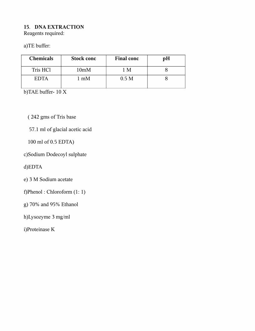
15. DNA EXTRACTION Reagents required:
a)TE buffer:
Chemicals Stock conc Final conc pH
Tris HCl 10mM 1 M 8
EDTA 1 mM 0.5 M 8
b)TAE buffer- 10 X
( 242 gms of Tris base
57.1 ml of glacial acetic acid
100 ml of 0.5 EDTA)
c)Sodium Dodecoyl sulphate
d)EDTA
e) 3 M Sodium acetate
f)Phenol : Chloroform (1: 1)
g) 70% and 95% Ethanol
h)Lysozyme 3 mg/ml
i)Proteinase K

PROFORMA
1.Patient’s name: 2. Age/Sex:
3.Ip/Op no: 4.Occupation:
5.Address: 6.Ward:
7.Patient’s consent:
8.Chief complaints:
Swelling – Onset , Duration, Progression
Associated symptoms: Pain/Discharge/Ulcer
9.Past history:
10.Personal history:
11.General Examination:
Nutrition,Anaemia,Lymphadenopathy,Edema
12.Local Examination:
Swelling- size,shape,margin,consistency,base
Discharge-colour,consistency,odour,granules,gas/crepitus
13.Diagnosis:
14.Investigations:
15.Treatment & Management:
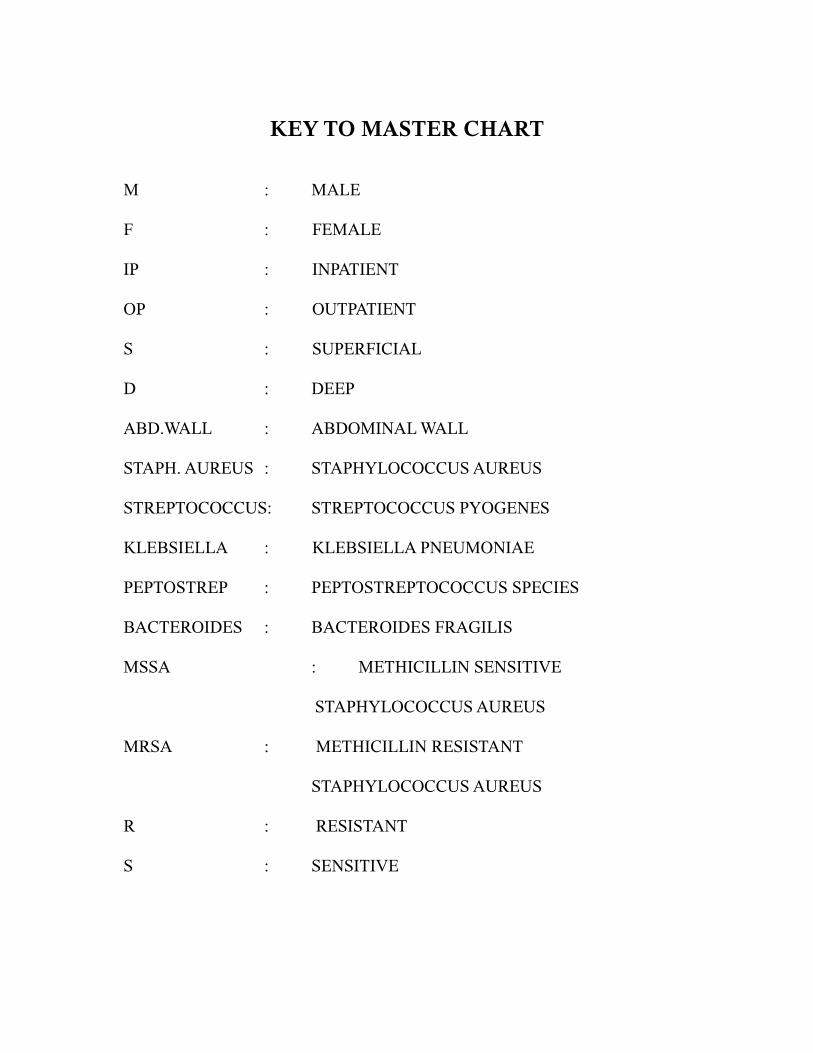
KEY TO MASTER CHART
M : MALE
F : FEMALE
IP : INPATIENT
OP : OUTPATIENT
S : SUPERFICIAL
D : DEEP
ABD.WALL : ABDOMINAL WALL
STAPH. AUREUS : STAPHYLOCOCCUS AUREUS
STREPTOCOCCUS: STREPTOCOCCUS PYOGENES
KLEBSIELLA : KLEBSIELLA PNEUMONIAE
PEPTOSTREP : PEPTOSTREPTOCOCCUS SPECIES
BACTEROIDES : BACTEROIDES FRAGILIS
MSSA : METHICILLIN SENSITIVE
STAPHYLOCOCCUS AUREUS
MRSA : METHICILLIN RESISTANT
STAPHYLOCOCCUS AUREUS
R : RESISTANT
S : SENSITIVE