Embed Size (px)
Citation preview
Biochimica et Biophysica Acta 1800 (2010) 134–143
Contents lists available at ScienceDirect
Biochimica et Biophysica Acta
j ourna l homepage: www.e lsev ie r.com/ locate /bbagen
Review
Bacterial toxin and effector glycosyltransferases
Yury Belyi a, Klaus Aktories b,⁎a Gamaleya Research Institute, Moscow 123098, Russiab Institut für Experimentelle und Klinische Pharmakologie und Toxikologie, Albert-Ludwigs-Universität Freiburg, Albertstr. 25, D-79104 Freiburg, Germany
⁎ Corresponding author. Tel.: +49 761 2035301; fax:E-mail address: [email protected]
0304-4165/$ – see front matter © 2009 Elsevier B.V. Adoi:10.1016/j.bbagen.2009.07.022
a b s t r a c t
a r t i c l e i n f oArticle history:Received 4 May 2009Received in revised form 14 July 2009Accepted 18 July 2009Available online 30 July 2009
Keywords:GlycosyltransferaseRho proteinBacterial protein toxinGlucosylationClostridial glucosylating toxinClostridium difficile toxinLegionella pneumophilaElongation factor 1A
Clostridial glucosylating cytotoxins, including Clostridium difficile toxins A and B, Clostridium novyi α-toxin,and Clostridium sordellii lethal toxin, are major virulence factors and causative agents of human diseases.These toxins mono-O-glucosylate (or mono-O-GlcNAcylate) a specific threonine residue of Rho/Ras-proteins, which is essential for the function of the molecular switches. Recently, a related group ofglucosyltransferases from Legionella pneumophila has been identified. These Legionella glucosyltransferasesmodify the large GTPase elongation factor eEF1A at a serine residue by mono-O-glucosylation, therebyinhibiting protein synthesis of target cells. Recent results on structures, functions and biological roles of bothgroups of bacterial toxin glucosyltransferases will be discussed.
© 2009 Elsevier B.V. All rights reserved.
1. Introduction
Cytosolic mono-O-glucosylation is an important molecular mech-anism by which various bacterial protein toxins and effectors targeteukaryotic cells. So far, twomajor groups of bacterial toxins have beendescribed that possess glucosyltransferase activity. One group iscomprised of the clostridial glucosylating cytotoxins, which areproduced by Clostridium difficile, Clostridium novyi, and Clostridiumsordellii [18,41,58,60,62,97,113]. These exotoxins are released bythe clostridia into the environment and are able to enter eukaryotictarget cells by an inherent cellular uptake mechanism. Once insidethe target cell, the toxins modify Rho- and Ras-subfamily proteins byO-glucosylation at specific threonine residues. In addition, onememberof this toxin family, Clostridium novyi α-toxin, modifies proteins byattachment of N-acetyl-glucosamine (GlcNAc). The second group ofbacterial glucosyltransferases is produced by Legionella pneumophila,and these enzymes modify eukaryotic elongation factor EF1A bymono-O-glucosylation at a specific serine residue. Instead of being termedtoxins, these bacterial glucosyltransferases are often called effectors,because translocation of the bacterial enzymes into the cytosol ofeukaryotic target cells depends on direct contact of the pathogen withhost cells. Thepresent reviewcoversboth groupsof glucosyltransferasesand their eukaryotic targets, their structure, function, and intracellularconsequences of their actions, as well as the medical impact and role inhost–pathogen interaction during infection.
+49 761 2035311.urg.de (K. Aktories).
ll rights reserved.
2. The family of clostridial glucosylating toxins
Members of the family of clostridial glucosylating toxins areClostridium difficile toxins A and B (TcdA, TcdB), C. sordellii lethal toxin(TcsL) and hemorrhagic toxin, and the α-toxin from C. novyi thatcatalyzes a GlcNAcylation. In addition to the C. difficile prototype toxinsA and B, several isoforms have been described for C. difficile toxins[92,93]. All these toxins are 50 to 90% identical in their amino acidsequences. They are large proteins of 250 to 308 kDa. Hence, they arealso called large clostridial cytotoxins.
These toxins are groups in the carbohydrate-active enzyme (CAZY)family GT44. This family has now more than 30 members, includingputative glycosyltransferases from Clostridium perfringens, Escherichiacoli, Citrobacter rodentium, Photobacterium profundum, Pseudomonasfluorescens and various species of Chlamydia and Chlamidophila (http://www.cazy.org/fam/GT44.html).
2.1. Glucosylating toxins are important virulence factors
From a medical point of view the C. difficile toxins A and B are themost important in terms of prevalence and pathogenicity. In the late1970s, it was recognized that C. difficile toxins A and B are the causativeagents of antibiotic-associated diarrhea and pseudomembranous colitisas a consequence of treatment with antibiotics, which destroy thenormalmicroflora of the gut and allow colonization and proliferation ofC. difficile bacteria [7,8,67]. Although the precise pathogenetic mechan-isms of induction of diarrhea and colitis are not known, it is generallyaccepted that the toxin-induced glucosylation of Rho GTPases is central
135Y. Belyi, K. Aktories / Biochimica et Biophysica Acta 1800 (2010) 134–143
to the action of theC. difficile toxins [67]. Elderly patients are particularlyat risk for severe sequelae, prolonged stay in hospitals and even lethaloutcomes of C. difficile infections. C. difficile-induced diseases receivedadditional attention recently, when hypervirulent strains of C. difficilewere isolated, that produced more than 10 times higher concentrationof toxins than previous isolates [79].
The larger amount of toxin production is apparently due to changesin the pathogenicity locus of the toxins. At least 5 tcdA–E genes areresponsible for toxin production: tcdA and tcdB are the structural genesof both toxins, tcdC encodes a negative regulator of toxin expression,tcdD appears to be responsible for activation of toxin gene expressionand tcdE encodes a holin-like protein likely involved in toxin release. Inthe hypervirulent strain the negative regulator is deleted, which resultsin higher toxin production.
Historically, toxinAwasdesignated as anenterotoxin,whereas toxinB was termed a cytotoxin. This distinction was based on findings inanimal models showing that C. difficile toxin A but not toxin B causeddisease after intragastral application [75]. In contrast, toxin B was ingeneral 100 to 1000-fold more potent in inducing cytotoxic effects incell culture than toxin A, most likely due to differential receptor binding[113]. Later however, it was shown that toxin B is also able to potentlyinduce destruction ofmucosal gut tissue in humans [91]. Recently it wasreported in anexcellent studyusing genetic approaches that toxin B, butnot toxin A, is crucial for virulence of C. difficile in diverse animalmodels[76].
The lethal toxin produced by C. sordellii and the α-toxin fromC. novyi, appear to be involved in gas gangrene syndrome [13,95,105].The samemay be true for the less characterized C. sordellii hemorrhagictoxin. Lethal toxin may also play a role in toxic shock syndromeassociatedwith abortion and gynecological infections [25] and deaths ofdrug users [68].
2.2. Multidomain structure of the toxins
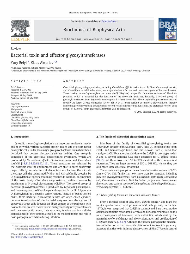
All members of the glycosyltransferase toxin family share 26 to 76%sequence identity and are structurally and functionally organized in asimilar module-like manner [18]. At least four domains “ABCD” canbe distinguished in toxins A and B of C. difficile [58], which are putativelyinvolved in biological activity (A-domain), receptor binding (B-domain),auto-proteolytic cleavage during toxin-processing (C-domain), anddelivery of the A-domain into the cytosol (D-domain) (Fig. 1).
The A-domain, which harbors the glucosyltransferase activity, islocated at the N-terminus of the toxins. This domain is most wellcharacterized and will be described in more detail below. Thereceptor-binding domain (B), which consists of polypeptide repeats,is located at the C-terminus [111,112]. Because this B-domain exhibits
Fig. 1. ABCD-model of clostridial glucosylating toxins. The clostridial glucosylating toxins are(note, C. novyi α-toxin possesses GlcNAcylation activity). The B-domain consisting of polypecatalytic cleavage of the toxins (arrow: cleavage site) and is a cysteine-protease with the catais necessary for activation of the cysteine protease. The D-domain is likely involved in the(indicated) suggested to be important for insertion of the toxin into endosome membranes
sequence similarity to the carbohydrate binding region of theglucosyltransferase from Streptococcus mutans, it was suggested earlyon that this part of the toxin is involved in binding to a carbohydrate-containing receptor [111]. Accordingly, antibodies generated againstthis part of the molecule blocked cytotoxic activity [38], and toxinfragments containing this domain are able to compete with theholotoxin and block the toxin effects [39]. A part of the polypeptiderepeats of the C-terminus of C. difficile toxin A has been crystallizedrecently [44,50], showing a solenoid-like structure with 32 repeatsconsisting of 15–21 amino acid residues and seven repeats consisting of30 residues. The repeats form β-hairpins, arranged in pairs with eachadjacent pair of hairpins rotated by 120° to the next pair, resulting in ascrew-like structure of a left-handed β-solenoid helix [44]. Co-crystallization with a derivate of the trisaccharide α-Gal(1,3)β-Gal(1,4)βGlcNAc confirmed the carbohydrate binding capacity of thedomain [44].
As mentioned above only the A-domain, harboring the glucosyl-transferase activity, is translocated into the cytosol [84,94]. Therefore,cleavage of the toxin is required. Recently, itwas shown that cleavage ofthe toxin occurs auto-catalytically by a cysteine protease activity, whichis harbored in the C-domain, covering residues 544–955, directly down-stream of the glucosyltransferase domain [33]. Cys-698 and His-653have been shown to be part of the catalytic dyad, which in addition toAsp-587 might participate in the auto-cleavage reaction [33]. Thecysteine protease activity is enhanced by inositol hexakisphosphate(InsP6) [89]. InsP6 binds to the C-Domain, causing a conformationalchange that activates the auto-catalytic activity [32].
Structure and function of the D-domain, which is located betweenresidues 955 and 1852, are least understood. A small region in theprimary sequence between residues 965 and 1128 is characterized byhydrophobic amino acids and is suggested to participate in formation oftransmembrane structure during pore formation and translocation ofthe toxin into the cytosol [113]. Pore formation induced by the toxin hasbeen shown in artificial lipid membranes, as well as by the release ofradioactive rubidium ions (86Rb) from preloaded cells under low pHconditions, whichmimic the pH of endosomes [6,43]. However, so far itis not clear how pore formation relates to the delivery of the toxin intothe cytosol.
2.3. Toxin up-take
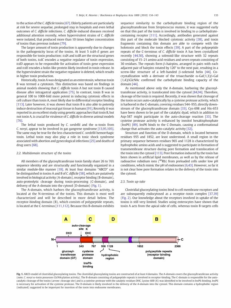
Clostridial glucosylating toxins bind to cell membrane receptors andare subsequently endocytosed as a receptor–toxin complex [37,39](Fig. 2). Our knowledge about the receptors involved in uptake of thetoxins is still very limited. Studies using enterocytes have shown thattoxin A acts from the apical side of cells, whereas toxin B targets cells
constructed of at least 4 domains. The A-domain covers the glucosyltransferase activityptide repeats is involved in receptor-binding. The C-domain is responsible for the auto-lytic residues DHC. Lysine-600 (K) was identified to be involved in InsP6-binding. InsP6delivery of the A-domain into the cytosol. This domain contains a hydrophobic region.
Fig. 2. Up-take of clostridial glucosylating toxins. The clostridial glucosylating toxins bindwith their C-terminal B-domain to the membrane receptor of target cells. After endocytosis, thetoxin inserts into the endosomemembranemost likely involving the hydrophobic part of the D-domain. Cellular InsP6 activates the cysteine protease C-domain. This results in cleavage ofthe toxin and release of the glucosyltransferase A-domain into the cytosol. In the cytosol Rho GTPases are glucosylated and thereby inactivated. Insert: Rho proteins are GTP bindingproteins, which are active in the GTP-bound state. The nucleotide exchange is facilitated by guanine nucleotide exchange factors (GEFs). In the GTP-bound form the Rho GTPases interactwith a large variety of effectors and induce multiple signaling events. The active state of Rho proteins is terminated by GTP hydrolysis stimulated by GTPase-activating proteins (GAPs).
136 Y. Belyi, K. Aktories / Biochimica et Biophysica Acta 1800 (2010) 134–143
from the basolateral side [102]. It has been suggested that toxin A bindsto carbohydrates [23,71,104,106], including α-Gal(1,3)β-Gal(1,4)βGlcNAc on rabbit erythrocytes and hamster brush bordermembranes. Proteins have also been discussed as receptors for toxinA, including sucrase-isomaltase in rabbit gut [86] and glycoprotein 96(gp96) in human colonocytes [81]. However, it is not clearwhether theyare relevant as receptors of toxin A in humans. The receptor of toxin B iscompletely unknown and the same is true for the other clostridialglucosylating toxins.
After receptor-binding, the toxins are taken up by endocytosisthrough pathways that are still not clear and end up in acidicendosomes, from where they translocate into the cytosol [6,87]. Thetranslocation mechanism is still largely enigmatic, but appears todepend on the low pH of endosomes, because bafilomycin, a specificinhibitor of the vacuolar H+-ATPase, blocks toxin up-take. Most likelythe low pH of endosomes induces conformational changes, that favorinsertion of the toxin into the membrane [6,87]. Interestingly, a pH-pulse (pHb5.5) at the cell membrane allows membrane insertion andpore formation by toxins without prior endocytosis [6,43]. Poreformation and toxin up-take appear to be cholesterol-dependent [43].It is still not clear, where or at what point in the uptake process theabove-mentioned auto-catalytic processing of the toxins occurs (e.g.,in the vesicles or in the cytosol), which finally results in release of theglucosyltransferase domain. Inositol-hexakisphosphate, which isrequired for auto-catalysis is present at relatively high concentrations(~100 µM) in the cytosol, but the concentration in endosomes isunknown.
2.4. Glucosylation of Rho GTPases by clostridial toxins
Once in the cytosol, the clostridial toxins glucosylate small GTPasesof the Rho and Ras superfamily [64,65,85,100]. The structure, functionand roles of these substrates in cellular processes have beendescribed in
detail in several excellent recent reviews and will be mentioned onlybriefly here [17,35,48,57,114]. The ~20 different Rho GTPases aremolecular switches involved in several cellular signaling pathways. TheGTPases are active in theGTP-bound formand inactivewithGDP bound.Hydrolysis of GTP is caused by inherent GTPase activity which can befacilitated by numerous GTPase-activating proteins (GAPs). Activationof the GTPases occurs after nucleotide exchange induced by guaninenucleotide exchange factors (GEFs), which release GDP and allowrebinding of GTP. Thebest studied small GTPases of this family are RhoA,Rac and Cdc42 isoforms. They control organization of the cytoskeletonand regulate cellular motility, and participate in the regulation oftranscription, cell cycle progression, apoptosis, transformation and theactivity of numerous other cellular enzymes. Modification of low-molecularGTPases by the toxins occurs at a Thr35/37, dependingon theRho GTPase isoforms [64]. Important differences in substrate specificityhave been detected among the various clostridial glucosyltransferases.WhereasC. difficile toxins A and B andC. novyiα-toxinmodifymost Rho,Rac and Cdc42 isoforms C. sordellii lethal toxin glucosylates Rac but notRhoA or Cdc42 in intact cells [63,85]. However, C. sordellii lethal toxinalso glucosylates Ras GTPases, including Rap, Ral und Ras isoforms. Allclostridial glucosylating toxins with the exception of C. novyi α-toxinuse UDP-glucose as a cosubstrate, whereas the α-toxin uses UDP-GlcNAc as a sugar donor [99].
Glucosylation of Rho or Ras GTPases inhibits activation of theGTPases by GEFs and blocks interaction with their effectors [98,109] aswell as the cycling of Rho GTPases between the membrane localizationand cytosolic localization [40] (Fig. 3). Glucosylated Rho proteins arelocated at the membrane. Most importantly, the toxin-inducedglucosylation inhibits the active conformation of Rho/Ras GTPases[42,109]. This fact also explains why the stimulation of the GTPaseactivity byGAPs is also blocked after glucosylation. Althoughnot studiedin detail, it is thought that attachment of N-acetylglucosamine causesthe same functional consequences.

Fig. 3. Functional consequences of the glucosylation of Rho GTPases. Glucosylation of Rho proteins by clostridial glucosylating toxins has several consequences for Rho GTPases. Theglucosylation inhibits the active conformation of Rho proteins. This causes: (1) blockade of Rho activation by GEFs; (2) inhibition of the interaction of Rho proteins with effectors;(3) inhibition of the stimulation of the GTP hydrolyzing activity of Rho proteins by GAPs; and (4) blockade of the cycling of Rho proteins between membranes and the cytosol (in thecytosol Rho proteins are bound to guanine nucleotide dissociation inhibitors (GDIs)). Ras proteins are modified by some clostridial glucosylating toxins (e.g., C. sordellii lethal toxin)with the same consequences. Modified from [60].
137Y. Belyi, K. Aktories / Biochimica et Biophysica Acta 1800 (2010) 134–143
Because Rho GTPases have major effects on the cytoskeleton, effortshave been made to differentiate toxin effects on cell morphology fromthose on cell survival. Results from these studies suggested that themorphological changes induced by toxin B are largely dependent on Racprotein because cells expressing non-glucosylatable Rac1-Q61L wereprotected against toxin B, whereas this was not the case when RhoA-Q63L was expressed [47]. On the other hand, cell death by toxin Binduced apoptosis appears to dependonRhoA, because a specific toxinBisoform (from C. difficile 1470 serotype F), which does not accept RhoAas a substrate but instead only modifies Rac, does not cause apoptosis[54]. Similarly, lethal toxin from C. sordellii, which modifies Rac but notRhoA, is able to induce apoptosis but in this case toxin-inducedapoptosis appears to depend on glucosylation of Ras and modulation ofphosphatidylinositol-3-kinase [31].
2.5. Structure of the catalytic A-domain of clostridial glucosyltransferases
The crystal structures of the catalytic A-domains of toxin B, lethaltoxin and α-toxin have been solved [90,116]. These studies show thatall these toxins belong to the GT-A family of glycosyltransferases[15,73,107], which is characterized by a common catalytic core (243amino acids in length for toxin B) with a mixedα/β-fold and a central6-stranded β sheet (Fig. 4). As is typical for the GT-A family, all but oneβ strand are parallel; strand 5 is antiparallel. The catalytic core of theclostridial toxins is surrounded by helical structures (309 residues inlength for toxin B). The four N terminal helices appear to form asubdomain, which might be involved in membrane binding. Indeed, ithas been suggested that this part of lethal toxin binds to phospha-tidylserine at membranes [80].
Another typical feature of theGT-A-family is the so-calledDXDmotif[19,73,115], which is involved in coordination of manganese (Mn2+).The motif, which includes Asp286 and Asp288 in toxin B, also interactswith the pyrophosphate of the sugar donor UDP-glucose [90] (Fig. 4).Asp288 directly binds toMn2+,whereas Asp286 coordinatesMn2+via awatermolecule. Asp286 togetherwith Arg273 and Asp270 forms a tightnetwork of hydrogen bonds to the hydroxyl groups (positions 3″, 4″ and6″) of theglucosemolecule. The ribosemoiety ofUDP-glucose isfixedbyTyr284, Ser269 and also by Asp286. Among other residues, Trp102 isimportant for positioning of the uracil ring of UDP by aromatic stacking[90]. All these residues have been shown to be essential for enzymeactivity [60]. Exchange of these residues with alanine caused a severalhundred fold drops in glucosyltransferase and glucohydrolase activities.
Comparison of the structures of the catalytic domains of differentclostridial glucosyltransferases with or without UDP-glucose boundsuggests that Trp520 is of major importance for the glucosylationreaction (Fig. 5). In the absence of Mn2+ and without UDP-glucose,Trp520 and the mobile chain segment of residues 519–525 exhibit alarge shift of ~15 Å fromanopen conformation to a closedUDP-glucose-binding conformation [116], allowing bonding of the glycoside oxygenof UDP-glucose with Trp520-Nε1. Accordingly, substitution of Trp520with alanine or even phenylalanine inhibits glucosyltransferase activity.
The glucosylation of Rho/Ras GTPases by clostridial glucosyltrans-ferases causes retention of the α-anomeric configuration of UDP-glucose [42,109]. Whereas the molecular mechanism of invertingglucosylation reactions (e.g., conversion of the α-anomeric configura-tion of UDP-glucose to the β-anomeric configuration) is well-under-stood, e.g., by a direct SN2-like displacement reaction [73], the retainingreaction is less well-understood. As deduced from comparison of thecrystal structures obtained from toxin A, lethal toxin and α-toxin, amodel for the glucosylation reaction has been proposed, which largelydepends on Trp520 and suggests a circular type of reaction [116].
2.6. Substrate specificity
The crystal structures alsoprovide anexplanation for the cosubstratespecificity of the clostridial toxins, e.g., it is now very well-understoodwhy α-toxin from C. novyi uses UDP-GlcNAc but not UDP-glucose as asugar donor [99]. It appears that two amino acids in the vicinity of thecatalytic cleft are responsible for the cosubstrate specificity. Toxins A, Band lethal toxin, which all use UDP-glucose, have isoleucine andglutamine in equivalent positions (Ile-383 and Glu-385 in toxin B),whereas C. novyi α-toxin has serine and alanine residues in therespective positions [61,116]. Substitution of Ile-383 with serine orGlu385 with alanine favored the acceptance of UDP-GlcNAc forglycosylation by toxin B, but change of both residues to that of α-toxin completely converted the donor specificity from UDP-glucose toUDP-GlcNAc. Apparently, the bulkier side chains of Ile383 and Glu385limit the space of the catalytic cleft for binding of UDP-GlcNAc and theexchange of these side chains with smaller groups causes a dramaticdrop in the Km-value for UDP-GlcNAc from ~900 µM to ~25 µM.
Although somespecific regionsand residuesof theRhoGTPaseshavebeen identified to play a role in substrate–enzyme interaction, theprotein substrate specificity is less clear. Arg455, Asp461, Lys463 andGlu472 aswell as helixα17 are involved in protein substrate recognition

Fig. 4. A. Structure of the glucosyltransferase A-domain of toxin B. The catalytic core of the glucosyltransferase A-domain is given in blue. Additional amino acids and regions of the A-domain, likely not directly involved in catalysis are given in red. B. Amino acid involved in the interaction with the sugar donor UDP-glucose. Data are from [90,116].
138 Y. Belyi, K. Aktories / Biochimica et Biophysica Acta 1800 (2010) 134–143
by toxin B. Of particular interest is the finding that introduction ofhelixα17 from toxin B into C. sordellii lethal toxin inhibits modificationof Ras but allows glucosylation of RhoA [59]. Of course, co-crystallizationof the catalytic domain of the toxin with the substrate GTPase isnecessary for a complete understanding of the molecular basis for theenzyme–substrate specificities.
Fig. 5. Conformational changes occurring after binding of UDP-glucose and Mn2+ to the thundergoes a major conformational change after binding of UDP-glucose andMn2+ to the apo
3. The family of L. pneumophila glucosyltransferases
3.1. Intracellular biology of L. pneumophila
Legionella is a fastidious gram-negative bacterium, causing severeoften fatal pneumonia in humans, known as Legionnaires' disease. This
e apo-enzyme. Deduced from a series of crystal structures, it is suggested that Trp520-enzyme resulting in an open or closed conformation of the glucosyltransferase domain.

139Y. Belyi, K. Aktories / Biochimica et Biophysica Acta 1800 (2010) 134–143
infection ranks among themost common causes of severepneumonia inthe community setting, and its infectious agent is isolated in up to 40%ofthe cases of hospital-acquired pneumonia. Among the more than 50known species of Legionella, the most important human pathogen is L.pneumophila, strains ofwhichaccount for up to 90%ofmorbidity recordsdue to legionellosis [30].
L. pneumophila is able to multiply in phagocytes — either in free-living unicellular eukaryotes (amoebae and ciliated protozoa) or inmammalian cells (macrophages and monocytes). This property is adirect prerequisite for survival of this bacterium in natural aquaticsystems and for proliferation in lung tissues of infectedmacroorganisms[36].
In terms of its subcellular compartmentalization, L. pneumophila is avacuolar pathogen. After penetration into eukaryotic cells legionellaereside andmultiplywithin the phagosome. This is in obvious contrast tosome other intracellular microorganisms, such as Listeria or Shigella,which lyse the membrane of a phagosome and multiply freely incytoplasm of a target cell [46]. Subsequent to uptake, the legionellae-containing vacuole (LCV) is subjected to specialized biogenesis steps,leading to transformation of this organelle into a “cozy” niche thatsupportsmultiplicationof thebacteria andhence, this LCV is also termed“replicative phagosome” [24,56].
Prominent characteristics of LCV development include the delay inmaturation of a phagosomalmembrane, the avoidance of the degradinglysosomal pathway, the malfunction in proton pump activity and,subsequently, impaired acidification of the vacuole, the attraction ofmitochondria and components of rough endoplasmic reticulum, and theinterception of early secretory vesicles, containing host cell membra-nousmaterial and nutrients [28,51,52,66]. Concurrently, global changesin target cellmetabolismbecomeevident, including inefficient oxidativeburst generation, shift in apoptotic–antiapoptotic equilibrium, drop ingeneral protein synthesis, modulation of ubiquitination processes andNF-κB activity [5,72,74,78,96,103].
Infection of phagocytes by L. pneumophila is accompanied bynumerous alterations in cellular processes, leading to transformationof normally hostile intracellular environment into friendly nichesuitable for bacterial proliferation. A specialized dot/icm type 4 secre-tion system (T4SS) and an array of its substrates/effectors have beenshown to participate in adaptation of a target cell to inhabitation byL. pneumophila [83]. The estimated number of such substrates/effectorsreaches 30 [83], 85 [56], or almost 130 [49], but the general consensus isthat even these figures are an underestimation. In most instances themolecular mechanism of the activities of the effectors and theirsubstrates is not known. But in certain cases detailed information isavailable. Recent data demonstrate that several effector proteins targetthe small GTPasesArf1 andRab1 [55,77,82]. TheseGTP-bindingproteinsplay critical roles in tethering ER-derived vesicles and thereforemanipulation of their activities by Legionella effectors apparently allowsthe bacteria to redirect early secretory traffic.
3.2. Identification of glucosyltransferases in L. pneumophila
Recent studies have shown that among the various bacterialeffectors, which are translocated from Legionella into the host cytosolare bacterial glucosyltransferases. Initially it was observed that in thepresence of L. pneumophila cellular extract and UDP-[14C]glucoselabeling of a ~50 kDa cytoplasmic component of eukaryotic cells was
Fig. 6. Partial alignment of amino acid sequences of Lgt1/2/3 with that of toxin A from C. diffiyellow, whereas residues, which are identical in all 4 sequences are marked by red. The consA gene are lpg1368, lpg2862, lpg1488 and M30307, respectively.
detectable [9]. Further purification of the extract of L. pneumophilaPhiladelphia-1 resulted in isolation of a ~60 kDa glucosyltransferase,which was subsequently named “Lgt1”. The enzyme was present in alltested members of L. pneumophila, but was absent in strains of certainnon-pneumophila species, such as L. longbeachae, L. gormanii and L.steigerwaltii. Whether all pneumophila species or only a subset of thesebacteria contain glucosylation activity is not known. The enzymaticactivity was sugar-specific, i.e. only UDP-glucose, but not glucose, UDP-galactose, UDP-N-acetyl-galactosamine, UDP-N-acetylglucosamine,UDP-glucuronic acid or GDP-mannose served as co-substrates in thereaction [10].
Primary amino acid sequence of the glucosylating proteins sharedlittle homology with known proteins in the NCBI database. The mostnotable hit was similarity between central region of Lgt1 and the DXD-containing domains of clostridial glucosylating toxins (Fig. 6). In thissequence region several stretches of identical amino acid residues couldbe identified, including two residues found to be critically important forcatalysis and apparently representingpart of aDXDmotif (D246 andD248
in Lgt1) [10].Subsequent database searches in the sequenced genomes of four
L. pneumophila strains (Philadelphia-1, Corby, Lens and Paris) disclosedaltogether nine open reading frames with significant sequencehomology (Fig. 7). Based upon the level of identity, these gene productshave been grouped into three families: Lgt1 through Lgt3 (typerepresentatives of Philadelphia-1 strain had gene IDs lpg1368, lpg2862and lpg1488 for Lpg1, Lpg2 and Lpg3, respectively). Only one copy ofeach gene family could be found in the corresponding genome.Philadelphia-1 contained the full set of genes (i.e. lgt1, lgt2 and lgt3),whereas the other three strains included only lgt1 and lgt3. Representa-tives within each family demonstrated more than 85% amino acididentity, whereas differences between the groups were in the range of15–25%. Interestingly,whereas proteins from Lgt1, Lgt2 and Lgt3 groupsdemonstrated interfamily homology with the majority of identicalamino acid residues grouped around the DXD motifs, two additionaltranslated sequences (lpg1491 in Philadelphia-1 and lpp1447 in Parisstrains) displayed considerable similarity only to the C-terminal regionof the Lgt3 members and did not contain the canonical DXDmotif. Dueto these data they were not included into this scheme.
In order to establish the enzymatic activity of these putativeglucosyltransferases, the coding sequences from the Philadelphia-1strain of L. pneumophila were expressed in E. coli as recombinantproteins and tested in vitro in the UDP-[14C]glucose assay. Additionally,Lgt2 and Lgt3 proteins from several other L. pneumophila strainsbelonging to different serogroups were purified in recombinant formand tested in glucosylation assays. Representatives of Lgt1, Lgt2 and Lgt3did possess glucosylation activity andmodified a ~50 kDa component inmammalian cell extracts.
In a recent study another putative glycosyltransferase produced byL. pneumophila has been identified [49]. The protein caused delay invacuolar trafficking and was termed therefore SetA (subversion ofeukaryotic vesicle trafficking A). Closer inspection of the amino acidsequences disclosed moderate homology to that of clostridialglycosylating toxins, as well as to Legionella glucosyltransferases. Inparticular, SetA possessed a DSD motif resembling the DXD motif ofthe active domain of other bacterial glycosylating enzymes[19,73,115]. Although the SetA DXD motif contains polar amino acidresidue serine in the middle of the triplet in contrast to typical non-
cile. Amino acid residues, which are identical in 2 or 3 sequences are marked by green orerved DXDmotif is indicated by asterisks. Identification codes of lgt1, lgt2, lgt3 and toxin
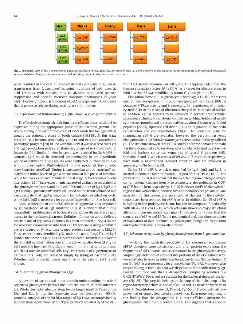
Fig. 7. Schematic view of the L. pneumophila glucosyltransferase family. Identification code of each Lgt gene is shown as deposited in the corresponding L. pneumophila sequencedgenome database. Empty rectangles indicate lack of Lgt2 group in Corby, Paris and Lens strains.
140 Y. Belyi, K. Aktories / Biochimica et Biophysica Acta 1800 (2010) 134–143
polar residues in the case of large clostridial cytotoxins or glucosyl-transferases from L. pneumophila, point mutations of both asparticacid residues with substitutions to alanine attenuated growthsuppression and specific vacuolar transport phenotypes in yeast[49]. However, molecular functions of SetA or experimental evidencethat it possesses glycosylating activity are still missing.
3.3. Expression and translocation of L. pneumophila glucosyltransferases
To effectively accomplish their functions, effector proteins should beexpressed during the appropriate phase of the bacterial growth. Thetypical timing interval for production of T4SS substrates by Legionella isusually the stationary phase of broth culture [16,118]. At this stagebacterial cells become remarkably virulent and execute transmissionphenotypeprogram[20]. In linewith this view, itwasobserved that Lgt1and Lgt2 production peaked at stationary phase of in vitro-growth ofLegionella [12]. Similar in vitro behavior was reported for SetA [49]. Incontrast, Lgt3 could be detected predominately in pre-logarithmicperiod of cultivation. These results were confirmed in infection studieswith L. pneumophila Philadelphia-1 in the model of the amoebaAcanthamoeba castellanii. During L. pneumophila and A. castellanii co-cultivation mRNA levels of lgt1were maximal at late phase of infection,while lgt3 was expressed mainly at initial stage of bacterium–amoebainteraction [12]. These experiments suggested distinctive regulation ofthe glucosyltransferases and implied differential roles of Lgt1/Lgt2 andLgt3 during L. pneumophila infection. Based on the results obtained onecan speculate that Lgt3 is important for initiation of infection cycle,while Lgt1/Lgt2 is necessary for egress of Legionella from the host cell.
Because infection of epithelial cells with Legionella is accompaniedby glucosylation of an Lgt substrate [10], it is evident that duringintracellular proliferation of bacterial cells glucosyltransferases gainaccess to their eukaryotic targets. Definite information about deliverymechanisms of Legionella enzymes has been obtained during a searchfor molecules transported into host cell by Legionella using adenylatecyclase-tagged or β-lactamase-tagged protein constructions [26,27].These experiments identified Lgt2 (under the name “LegC8”) and Lgt3(under the name “LegC5”) as T4SS-translocated substrates. However,there is still no information concerning active translocation (if any) ofLgt1 into the host cell. One should keep in mind that some proteins,which act outside microbial cells (e.g., enterotoxin of C. perfringens orLT toxin of E. coli) are released simply by lysing of bacteria [101].Whether such a mechanism is operative in the case of Lgt1 is notknown.
3.4. Substrates of glucosyltransferases Lgt
A question of exceptional importance for understanding the role ofLegionella glucosyltransferases includes the nature of their substrate(s). While clostridial glucosylating toxins target small GTPases of theRho and Ras family, the Legionella enzymes glucosylate ~50 kDaproteins. Analysis of the 50-kDa target of Lgt1 was accomplished bytandem mass-spectrometry of tryptic products isolated by SDS-PAGE
from Lgt1-treatedmammalian cell lysate. This approach identified thehuman elongation factor 1A (eEF1A) as a target for glucosylation, inwhich serine-53 was modified by mono-O-glucosylation [10].
Elongation factor eEF1A (prokaryotic homolog is EF-Tu) representsone of the key-players in ribosome-dependent synthesis [88]. Itpossesses GTPase activity and is necessary for recruitment of aminoa-cylated tRNA to the A-site of ribosomes charged with translated mRNA.In addition, eEF1A appears to be involved in several other cellularprocesses, including translational control, assembling/folding of newlysynthesizedproteins andproteosomal degradationof incorrectly-foldedpeptides [22,53], lipotoxic cell death [14] and regulation of the actincytoskeleton and cell morphology [34,45]. No structural data formammalian eEF1A are available, however the very similar yeastelongation factor 1A from Saccharomyces cerevisiaehas been crystallized[3]. The structure showed that eEF1A consists of three domains: domain1 is the G domain of ~240 residues, which is characterized by a Ras-likefold and harbors consensus sequences of typical G proteins [69].Domains 2 and 3, which consist of 89 and 107 residues respectively,have both a six-stranded β-barrel structure and are involved inaminoacyl-tRNA binding [3].
Serine-53 of eEF1A, which is modified by Lgt1, Lgt2 and Lgt3, islocated in domain I near the switch-1 region of the GTPase [10,12]. Forprokaryotic EF-Tu, it is known that the switch-1 region undergoesmajorconformational changes from β- to α-structure, depending on its GDP-orGTP-bound formrespectively [1,110]. However in eEF1A the switch-1region is notwell defined, because two additional helices (A* andA′) areinserted into this region, and no structural changes in the switch-1region have been reported for eEF1A so far. In addition, Ser-53 of eEF1Ais lacking in the prokaryotic factor, but can be compared functionallywith Ala-42 in E. coli EF-Tu, which yet again shows no major structuralalteration upon nucleotide exchange [1]. However, it is clear that thestructures of eEF1A and EF-Tu are not identical and, therefore, completeextrapolation of data obtained on prokaryotic elongation factor ontoeukaryotic molecule is obviously difficult.
3.5. Substrate recognition by glucosyltransferases from L. pneumophila
To clarify the substrate specificity of Lgt enzymes, recombinanteEF1A deletions were constructed and after protein expression, thefragments of eEF1A were used as substrates in the glucosylation assay.Surprisingly, deletions of considerable portions of the elongation factorwere still able to serve as substrates for glucosylation. Neither domain 2nor 3 of eEF1Awas necessary for glucosylation (Fig. 8A). Moreover, alsoproper folding of the G-domainwas dispensable formodification by Lgt.Finally, it turned out that a decapeptide comprising residues 50-GKGSFKYAWV-59 served as substrate for the bacterial glucosyltransfer-ase (Fig. 8B). This peptide belongs to the loop of the helix–loop–helixregion formedbyhelices A* andA′of eEF1Aand is part of thefirst turnofhelix A′. Substitution of Ser-53, Phe-54, Tyr-56 or Trp-58 with alanineprevented or largely decreased glucosylation. Even more surprising isthe finding that the decapeptide is a more efficient substrate forglucosylation than the full length eEF1A. This suggests that a specific
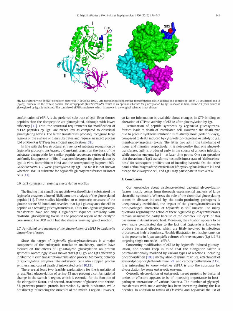
Fig. 8. Structural view of yeast elongation factor eEF1A (PDB ID: 1F60). Left, ribbon plot; right, surface representation. eEF1A consists of 3 domains (I (green), II (magenta) and III(cyan)). Domain I is the GTPase domain. The decapeptide (GKGSFKYAWV), which is an optimal substrate for glucosylation by Lgt, is shown in blue. Serine-53 (red), which isglucosylated by Lgts, is indicated. The complexed eEF1Bα molecule, which is present in the original scheme, is not shown.
141Y. Belyi, K. Aktories / Biochimica et Biophysica Acta 1800 (2010) 134–143
conformation of eEF1A is the preferred substrate of Lgt1. Even shorterpeptides than the decapeptide are glucosylated, although with lowerefficiency [11]. Thus, the structural requirements for modification ofeEF1A peptides by Lgt1 are rather low as compared to clostridialglucosylating toxins. The latter transferases probably recognize largeregions of the surface of their substrates and require an intact proteinfold of Rho/Ras GTPases for efficient modification [58].
In linewith the low structural stringency of substrate recognition byLegionella glucosyltransferases, a GeneBank search on the basis of thesubstrate decapeptide for similar peptide sequences retrieved Hsp70subfamily B suppressor 1 (Hbs1) as a possible target for glucosylationbyLgt1 in vitro. Recombinant Hbs1 and the corresponding fragment 303-GKASFAYAWV-312 were glucosylated by Lgt1. So far it is not knownwhether Hbs1 is substrate for Legionella glucosyltransferases in intactcells [11].
3.6. Lgt1 catalyzes a retaining glucosylation reaction
Thefinding that a small decapeptidewas theefficient substrate of theLegionella enzymes allowed NMR structure analysis of the glucosylatedpeptide [11]. These studies identified an α-anomeric structure of theglucose-serine-53 bond and revealed that Lgt1 glucosylates the eEF1Apeptide as a retaining glucosyltransferase. Thus, the Legionella glucosyl-transferases have not only a significant sequence similarity withclostridial glucosylating toxins in the proposed region of the catalyticcore around the DXD motif but also share a retaining type of reaction.
3.7. Functional consequences of the glucosylation of eEF1A by Legionellaglucosyltransferases
Since the target of Legionella glucosyltransferases is a majorcomponent of the eukaryotic translation machinery, studies havefocused on the effects of Lgt-catalyzed glucosylation on proteinsynthesis. Accordingly, it was shown that Lgt1, Lgt2 and Lgt3 effectivelyinhibit the in vitro transcription/translation process. Moreover, deliveryof glucosylating enzymes into eukaryotic cells also stopped proteinsynthesis and caused death of intoxicated cells [10,12].
There are at least two feasible explanations for the translationalarrest. First, glucosylation of serine-53 may prevent a conformationalchange in the switch-1 region, which is essential for the function ofthe elongation factor, and second, attachment of glucose onto serine-53, prevents protein–protein interaction by steric hindrance, whilenot directly influencing the structure of the switch-1 region. However,
so far no information is available about changes in GTP-binding oralteration of GTPase activity of eEF1A after glucosylation by Lgt.
Termination of peptide synthesis by Legionella glucosyltrans-ferases leads to death of intoxicated cell. However, the death ratedue to protein synthesis inhibition is relatively slow (order of days),compared to death induced by cytoskeleton-targeting or cytolytic (i.e.membrane-targeting) toxins. The latter two act in the timeframe ofhours and minutes, respectively. It is noteworthy that one glucosyl-transferase, Lgt3, is produced early in the course of amoeba infection,while another enzyme, Lgt1 — at later time points. One can speculatethat the action of Lgt3 transforms host cells into a state of “defenseless-ness” for subsequent proliferation of invading bacteria. On the otherhand, atfinal stages of the intracellular life cycle Legionellahas to kill andescape the eukaryotic cell, and Lgt1 may participate in such a task.
4. Conclusion
Our knowledge about virulence-related bacterial glycosyltrans-ferases mostly comes from thorough experimental analysis of largeclostridial cytotoxins. Whereas the role of the clostridial glucosylatingtoxins in disease induced by the toxin-producing pathogens isunequivocally established, the impact of the glucosyltransferases inhost–pathogen interaction of Legionella is still unclear. The manyquestions regarding the action of these Legionella glucosyltransferasesremain unanswered partly because of the complex life cycle of thisbacterium in its eukaryotic host. Moreover, the situation appears to beeven more complicated due to the fact that Legionella is known toproduce bacterial effectors, which are likely involved in infectiousprocesses, at high redundancy. Notable illustration to this phenomenonis the presence in L. pneumophila cultures of three enzymes (Lgt1/2/3)targeting single molecule — eEF1A.
Concerning modification of eEF1A by Legionella-induced glucosy-lation, one should keep in mind that the elongation factor isposttranslationally modified by various types of reactions, includingphosphorylation [108], methylation of lysine residues, attachment ofglycerylphosphorylethanolamine [29] and carboxymethylation [117].It is interesting to know whether eEF1A is also the substrate forglycosylation by some eukaryotic enzyme.
Cytosolic glycosylation of eukaryotic target proteins by bacterialtoxins or effectors appears to be of increasing importance in host–pathogen interactions of various bacteria. The number of glycosyl-transferases with toxic activity has been increasing during the lastdecades. In addition to toxins of Clostridia and Legionella the list of
142 Y. Belyi, K. Aktories / Biochimica et Biophysica Acta 1800 (2010) 134–143
putative glycosylating enzymes includes, for example, virulencefactors from C. perfringens, E. coli, C. rodentium and Chlamydia(lymphostatin LifA and related proteins) [2,4,21,70]. It is likely thatthorough study of various infectious agents aiming at identification ofeukaryotic proteins-targeting glycosyltransferases will disclose novelenzymes and hence new mechanisms used by invading bacteria totyrannize eukaryotic host.
Acknowledgements
We thank Dr. Brenda Wilson for critical reading of the manuscriptand Dr. Thomas Jank for designing Fig. 5.
References
[1] K. Abel, M.D. Yoder, R. Hilgenfeld, F. Jurnak, An alpha to beta conformationalswitch in EF-Tu, Structure 4 (1996) 1153–1159.
[2] K. Amimoto, T. Noro, E. Oishi, M. Shimizu, A novel toxin homologous to largeclostridial cytotoxins found in culture supernatant of Clostridium perfringens typeC, Microbiology 153 (2007) 1198–1206.
[3] G.R. Andersen, L. Pedersen, L. Valente, I. Chatterjee, T.G. Kinzy, M. Kjeldgaard, J.Nyborg, Structural basis for nucleotide exchange and competition with tRNA in theyeast elongation factor complex eEF1A:eEF1Balpha, Mol. Cell 6 (2000) 1261–1266.
[4] B.A. Babbin, M. Sasaki, K.W. Gerner-Schmidt, A. Nusrat, J.M. Klapproth, Thebacterial virulence factor lymphostatin compromises intestinal epithelial barrierfunction by modulating rho GTPases, Am. J. Pathol. 174 (2009) 1347–1357.
[5] S. Banga, P. Gao, X. Shen, V. Fiscus, W.X. Zong, L. Chen, Z.Q. Luo, Legionellapneumophila inhibits macrophage apoptosis by targeting pro-death members ofthe Bcl2 protein family, Proc. Natl. Acad. Sci. U. S. A. 104 (2007) 5121–5126.
[6] H. Barth, G. Pfeifer, F. Hofmann, E. Maier, R. Benz, K. Aktories, Low pH-inducedformation of ion channels by Clostridium difficile toxin B in target cells, J. Biol. Chem.276 (2001) 10670–10676.
[7] J.G. Bartlett, Historical perspectives on studies of Clostridium difficile and C.difficile infection, Clin. Infect. Dis. 46 (Suppl 1) (2008) S4–S11.
[8] J.G. Bartlett, A.B. Onderdonk, R.L. Cisneros, D.L. Kasper, Clindamycin-associatedcolitis due to a toxin-producing species of Clostridium in hamsters, J. Infect. Dis. 136(1977) 701–705.
[9] I. Belyi, M.R. Popoff, N.P. Cianciotto, Purification and characterization of a UDP-glucosyltransferase produced by Legionella pneumophila, Infect. Immun. 71 (2003)181–186.
[10] Y. Belyi, R. Niggeweg, B. Opitz, M. Vogelsgesang, S. Hippenstiel, M. Wilm, K.Aktories, Legionella pneumophila glucosyltransferase inhibits host elongationfactor 1A, Proc. Natl. Acad. Sci. U. S. A. 103 (2006) 16953–16958.
[11] Y. Belyi, M. Stahl, I. Sovkova, P. Kaden, B. Luy, K. Aktories, Region of elongationfactor 1A1 involved in substrate recognition by Legionella pneumophilaglucosyltransferase LGT1-identification of LGT1 as a retaining glucosyltransfer-ase, J. Biol. Chem. 284 (2009) 20167–20174.
[12] Y. Belyi, I. Tabakova, M. Stahl, K. Aktories, Lgt: a family of cytotoxic glucosyl-transferases produced by Legionella pneumophila, J. Bacteriol. 190 (2008)3026–3035.
[13] S.P. Boriello, K. Aktories, Clostridium perfringens, Clostridium difficile and otherClostridium species, in: S.P. Boriello, P.R. Murray, G. Funke (Eds.), Topley andWilson's Microbiology and Microbial Infections, vol. Bacteriology, EdwardArnold, 2005, pp. 1089–1136.
[14] N.M. Borradaile, K.K. Buhman, L.L. Listenberger, C.J. Magee, E.T. Morimoto, D.S.Ory, J.E. Schaffer, A critical role for eukaryotic elongation factor 1A-1 in lipotoxiccell death, Mol. Biol. Cell (2005).
[15] C. Breton, L. Snajdrova, C. Jeanneau, J. Koca, A. Imberty, Structures andmechanisms of glycosyltransferases, Glycobiology 16 (2006) 29R–37R.
[16] H. Bruggemann, A. Hagman, M. Jules, O. Sismeiro, M.A. Dillies, C. Gouyette, F. Kunst,M. Steinert, K. Heuner, J.Y. Coppee, C. Buchrieser, Virulence strategies for infectingphagocytes deduced from the in vivo transcriptional program of Legionellapneumophila, Cell. Microbiol. 8 (2006) 1228–1240.
[17] K. Burridge, K.Wennerberg, RhoandRac take center stage,Cell 116 (2004)167–179.[18] C. Busch, K. Aktories, Microbial toxins and the glucosylation of Rho family
GTPases, Curr. Opin. Struct. Biol. 10 (2000) 528–535.[19] C. Busch, F. Hofmann, J. Selzer, J. Munro, D. Jeckel, K. Aktories, A commonmotif of
eukaryotic glycosyltransferases is essential for the enzyme activity of largeclostridial cytotoxins, J. Biol. Chem. 273 (1998) 19566–19572.
[20] B. Byrne, M.S. Swanson, Expression of Legionella pneumophila virulence traits inresponse to growth conditions, Infect. Immun. 66 (1998) 3029–3034.
[21] J.H. Carlson, S. Hughes, D. Hogan, G. Cieplak, D.E. Sturdevant, G. McClarty, H.D.Caldwell, R.J. Belland, Polymorphisms in the Chlamydia trachomatis cytotoxin locusassociated with ocular and genital isolates, Infect. Immun. 72 (2004) 7063–7072.
[22] S.M. Chuang, L. Chen, D. Lambertson, M. Anand, T.G. Kinzy, K. Madura,Proteasome-mediated degradation of cotranslationally damaged proteinsinvolves translation elongation factor 1A, Mol. Cell Biol. 25 (2005) 403–413.
[23] G.F. Clark, H.C. Krivan, T.D. Wilkins, D.F. Smith, Toxin A from Clostridium difficilebinds to rabbit erythrocyte glycolipids with terminal Gal alpha 1–3Gal beta 1–4GlcNAc sequences, Arch. Biochem. Biophys. 257 (1987) 217–229.
[24] J. Coers, C. Monahan, C.R. Roy, Modulation of phagosome biogenesis by Legionellapneumophila creates an organelle permissive for intracellular growth, Nat. Cell Biol.1 (1999) 451–453.
[25] A.L. Cohen, J. Bhatnagar, S. Reagan, S.B. Zane,M.A.D'Angeli,M. Fischer,G.Killgore, T.S.Kwan-Gett, D.B. Blossom, W.J. Shieh, J. Guarner, J. Jernigan, J.S. Duchin, S.R. Zaki, L.C.McDonald, Toxic shock associated with Clostridium sordellii and Clostridiumperfringens after medical and spontaneous abortion, Obstet. Gynecol. 110 (2007)1027–1033.
[26] K.S. de Felipe, R.T. Glover, X. Charpentier,O.R. Anderson,M.Reyes, C.D. Pericone,H.A.Shuman, Legionella eukaryotic-like type IV substrates interfere with organelletrafficking, PLoS Pathog. 4 (2008) e1000117.
[27] K.S. de Felipe, S. Pampou, O.S. Jovanovic, C.D. Pericone, S.F. Ye, S. Kalachikov, H.A.Shuman, Evidence for acquisition of Legionella type IV secretion substrates viainterdomain horizontal gene transfer, J. Bacteriol. 187 (2005) 7716–7726.
[28] I. Derre, R.R. Isberg, Legionella pneumophila replication vacuole formation involvesrapid recruitment of proteins of the early secretory system, Infect. Immun. 72(2004) 3048–3053.
[29] T.E. Dever, C.E. Costello, C.L. Owens, T.L. Rosenberry, W.C. Merrick, Location ofseven post-translational modifications in rabbit elongation factor 1 alphaincluding dimethyllysine, trimethyllysine, and glycerylphosphorylethanolamine,J. Biol. Chem. 264 (1989) 20518–20525.
[30] B.M. Diederen, Legionella spp. and Legionnaires' disease, J. Infect. 56 (2008) 1–12.[31] S.C. Dreger, F. Schulz, J. Huelsenbeck, R. Gerhard, F. Hofmann, I. Just, H. Genth,
Killing of rat basophilic leukemia cells by lethal toxin from Clostridium sordellii:critical role of phosphatidylinositide 3′-OH kinase/Akt signaling, Biochemistry(Febraury 6 2009) Electronic publication ahead of print.
[32] M. Egerer, T. Giesemann, C. Herrmann, K. Aktories, Autocatalytic processing ofClostridium difficile toxin B. Binding of inositol hexakisphosphate, J. Biol. Chem. 284(2009) 3389–3395.
[33] M. Egerer, T. Giesemann, T. Jank, K.J. Satchell, K. Aktories, Auto-catalytic cleavage ofClostridium difficile toxins A and B depends on a cysteine protease activity, J. Biol.Chem. 282 (2007) 25314–25321.
[34] S. Ejiri, Moonlighting functions of polypeptide elongation factor 1: from actinbundling to zincfingerproteinR1-associatednuclear localization,Biosci. Biotechnol.Biochem. 66 (2002) 1–21.
[35] S. Etienne-Manneville, A. Hall, Rho GTPases in cell biology, Nature 420 (2002)629–635.
[36] B.S. Fields, R.F. Benson, R.E. Besser, Legionella and Legionnaires' disease: 25 yearsof investigation, Clin. Microbiol. Rev. 15 (2002) 506–526.
[37] I. Florin, M. Thelestam, Internalization of Clostridium difficile cytotoxin intocultured human lung fibroblasts, Biochim. Biophys. Acta 763 (1983) 383–392.
[38] S.M. Frey, T.D. Wilkins, Localization of two epitopes recognized by monoclonalantibodyPCG-4onClostridiumdifficile toxinA, Infect. Immun.60 (1992) 2488–2492.
[39] C. Frisch, R. Gerhard, K. Aktories, F. Hofmann, I. Just, The complete receptor-binding domain of Clostridium difficile toxin A is required for endocytosis,Biochem. Biophys. Res. Commun. 300 (2003) 706–711.
[40] H. Genth, K. Aktories, I. Just, Monoglucosylation of RhoA at threonine-37 blockscytosol-membrane cycling, J. Biol. Chem. 274 (1999) 29050–29056.
[41] H. Genth, S.C. Dreger, J. Huelsenbeck, I. Just, Clostridium difficile toxins: more thanmere inhibitors of Rho proteins, Int. J. Biochem. Cell Biol. 40 (2008) 592–597.
[42] M. Geyer, C. Wilde, J. Selzer, K. Aktories, H.R. Kalbitzer, Glucosylation of Ras byClostridium sordellii lethal toxin: consequences for the effector loop conforma-tions observed by NMR spectroscopy, Biochemistry 42 (2003) 11951–11959.
[43] T. Giesemann, T. Jank, R. Gerhard, E. Maier, I. Just, R. Benz, K. Aktories,Cholesterol-dependent pore formation of Clostridium difficile toxin A, J. Biol.Chem. 281 (2006) 10808–10815.
[44] A. Greco, J.G. Ho, S.J. Lin, M.M. Palcic, M. Rupnik, K.K. Ng, Carbohydrate recognitionby Clostridium difficile toxin A, Nat. Struct. Mol. Biol. 13 (2006) 460–461.
[45] S.R.Gross, T.G. Kinzy, Translationelongation factor 1A is essential for regulationof theactin cytoskeleton and cell morphology, Nat. Struct. Mol. Biol. 12 (2005) 772–778.
[46] S. Gruenheid, B.B. Finlay, Microbial pathogenesis and cytoskeletal function, Nature422 (2003) 775–781.
[47] I. Halabi-Cabezon, J. Huelsenbeck, M. May, M. Ladwein, K. Rottner, I. Just, H.Genth, Prevention of the cytopathic effect induced by Clostridium difficile toxin Bby active Rac1, FEBS Lett. 582 (2008) 3751–3756.
[48] S.J. Heasman, A.J. Ridley, Mammalian Rho GTPases: new insights into theirfunctions from in vivo studies, Nat. Rev. Mol. Cell Biol. 9 (2008) 690–701.
[49] M. Heidtman, E.J. Chen,M.Y.Moy, R.R. Isberg, Large-scale identification of Legionellapneumophila Dot/Icm substrates that modulate host cell vesicle trafficking path-ways, Cell. Microbiol. 11 (2009) 230–248.
[50] J.G. Ho, A. Greco, M. Rupnik, K.K. Ng, Crystal structure of receptor-binding C-terminal repeats from Clostridium difficile toxin A, Proc. Natl. Acad. Sci. U. S. A. 102(2005) 18373–18378.
[51] M.A.Horwitz, The Legionnaires' disease bacterium(Legionella pneumophila) inhibitsphagosome–lysosome fusion in human monocytes, J. Exp. Med. 158 (1983)2108–2126.
[52] M.A. Horwitz, F.R. Maxfield, Legionella pneumophila inhibits acidification of itsphagosome in human monocytes, J. Cell Biol. 99 (1984) 1936–1943.
[53] Y. Hotokezaka, U. Tobben, H. Hotokezaka, L.K. Van, B. Beatrix, D.H. Smith, T.Nakamura, M.Wiedmann, Interaction of the eukaryotic elongation factor 1Awithnewly synthesized polypeptides, J. Biol. Chem. 277 (2002) 18545–18551.
[54] J. Huelsenbeck, S. Dreger, R. Gerhard, H. Barth, I. Just, H. Genth, Difference in thecytotoxic effects of toxin B from Clostridium difficile strain VPI 10463 and toxin Bfrom variant Clostridium difficile strain 1470, Infect. Immun. 75 (2007) 801–809.
[55] A. Ingmundson, A. Delprato, D.G. Lambright, C.R. Roy, Legionella pneumophilaproteins that regulate Rab1 membrane cycling, Nature 450 (2007) 365–369.

143Y. Belyi, K. Aktories / Biochimica et Biophysica Acta 1800 (2010) 134–143
[56] R.R. Isberg, T.J. O'Connor, M. Heidtman, The Legionella pneumophila replicationvacuole: making a cosy niche inside host cells, Nat. Rev. Microbiol. 7 (2009) 13–24.
[57] A.B. Jaffe, A. Hall, Rho GTPases: biochemistry and biology, Annu. Rev. Cell Dev.Biol. 21 (2005) 247–269.
[58] T. Jank, K. Aktories, Structure and mode of action of clostridial glucosylatingtoxins: the ABCD model, Trends Microbiol. 16 (2008) 222–229.
[59] T. Jank, T. Giesemann, K. Aktories, Clostridium difficile glucosyltransferase toxin B—
essential amino acids for substrate-binding, J. Biol. Chem. 282 (2007) 35222–35231.[60] T. Jank, T. Giesemann, K. Aktories, Rho-glucosylating Clostridium difficile toxins A
and B: new insights into structure and function, Glycobiology 17 (2007) 15R–22R.[61] T. Jank, D.J. Reinert, T. Giesemann, G.E. Schulz, K. Aktories, Change of the donor
substrate specificity of Clostridium difficile toxin B by site-directed mutagenesis,J. Biol. Chem. 280 (2005) 37833–37838.
[62] I. Just, R. Gerhard, Large clostridial cytotoxins, Rev. Physiol. Biochem. Pharmacol.152 (2004) 23–47.
[63] I. Just, J. Selzer, F. Hofmann, G.A. Green, K. Aktories, Inactivation of Ras by Clos-tridium sordellii lethal toxin-catalyzed glucosylation, J. Biol. Chem. 271 (1996)10149–10153.
[64] I. Just, J. Selzer, M.Wilm, C. Von Eichel-Streiber, M.Mann, K. Aktories, Glucosylationof Rho proteins by Clostridium difficile toxin B, Nature 375 (1995) 500–503.
[65] I. Just, M. Wilm, J. Selzer, G. Rex, C. Von Eichel-Streiber, M. Mann, K. Aktories, Theenterotoxin from Clostridium difficile (ToxA) monoglucosylates the Rho proteins,J. Biol. Chem. 270 (1995) 13932–13936.
[66] J.C. Kagan, C.R. Roy, Legionella phagosomes intercept vesicular traffic fromendoplasmic reticulum exit sites, Nat. Cell Biol. 4 (2002) 945–954.
[67] C.P. Kelly, J.T. LaMont, Clostridium difficile — more difficult than ever, N. Engl. J.Med. 359 (2008) 1932–1940.
[68] A.C. Kimura, J.I. Higa, R.M. Levin, G. Simpson, Y. Vargas, D.J. Vugia, Outbreak ofnecrotizing fasciitis due to Clostridium sordellii among black-tar heroin users,Clin. Infect. Dis. 38 (2004) e87–e91.
[69] M. Kjeldgaard, J. Nyborg, B.F.C. Clark, The GTP binding motif: variations on atheme, FASEB J. 10 (1996) 1347–1368.
[70] J.M. Klapproth, M.S. Donnenberg, J.M. Abraham, H.L. Mobley, S.P. James, Productsof enteropathogenic Escherichia coli inhibit lymphocyte activation and lympho-kine production, Infect. Immun. 63 (1995) 2248–2254.
[71] H.C. Krivan, G.F. Clark, D.F. Smith, T.D. Wilkins, Cell surface binding site forClostridium difficile enterotoxin: evidence for a glycoconjugate containing thesequence Galα1–3Galβ1–4GlcNAc, Infect. Immun. 53 (1986) 573–581.
[72] T. Kubori, A. Hyakutake, H. Nagai, Legionella translocates an E3 ubiquitin ligasethat has multiple U-boxes with distinct functions, Mol. Microbiol. 67 (2008)1307–1319.
[73] L.L. Lairson, B. Henrissat, G.J. Davies, S.G. Withers, Glycosyltransferases:structures, functions, and mechanisms, Annu. Rev. Biochem. 77 (2008) 521–555.
[74] V.P. Losick, R.R. Isberg, NF-kappaB translocation prevents host cell death after low-dose challenge by Legionella pneumophila, J. Exp. Med. 203 (2006) 2177–2189.
[75] D.M. Lyerly, K.E. Saum, D.K. MacDonald, T.D. Wilkins, Effects of Clostridium difficiletoxins given intragastrically to animals, Infect. Immun. 47 (1985) 349–352.
[76] D. Lyras, J.R. O'Connor, P.M. Howarth, S.P. Sambol, G.P. Carter, T. Phumoonna, R.Poon, V. Adams, G. Vedantam, S. Johnson, D.N. Gerding, J.I. Rood, Toxin B isessential for virulence of Clostridium difficile, Nature 458 (2009) 1176–1179.
[77] M.P. Machner, R.R. Isberg, A bifunctional bacterial protein links GDI displacementto Rab1 activation, Science 318 (2007) 974–977.
[78] K.T. McCusker, B.A. Braaten, M.W. Cho, D.A. Low, Legionella pneumophila inhibitsprotein synthesis in Chinese hamster ovary cells, Infect. Immun. 59 (1991)240–246.
[79] L.C. McDonald, G.E. Killgore, A. Thompson, R.C. Owens Jr., S.V. Kazakova, S.P.Sambol, S. Johnson, D.N. Gerding, An epidemic, toxin gene-variant strain ofClostridium difficile, N. Engl. J. Med. 353 (2005) 2433–2441.
[80] B. Mesmin, K. Robbe, B. Geny, F. Luton, G. Brandolin, M.R. Popoff, B. Antonny, Aphosphatidylserine-binding site in the cytosolic fragment of Clostridium sordelliilethal toxin facilitates glucosylation of membrane-bound Rac and is required forcytotoxicity, J. Biol. Chem. 279 (2004) 49876–49882.
[81] X. Na, H. Kim, M.P. Moyer, C. Pothoulakis, J.T. LaMont, gp96 is a human colonocyteplasma membrane binding protein for Clostridium difficile toxin A, Infect. Immun.76 (2008) 2862–2871.
[82] H. Nagai, J.C. Kagan, X. Zhu, R.A. Kahn, C.R. Roy, A bacterial guanine nucleotideexchange factor activates ARF on Legionella phagosomes, Science 295 (2002)679–682.
[83] S. Ninio, C.R. Roy, Effector proteins translocated by Legionella pneumophila:strength in numbers, Trends Microbiol. 15 (2007) 372–380.
[84] G. Pfeifer, J. Schirmer, J. Leemhuis, C. Busch, D.K. Meyer, K. Aktories, H. Barth,Cellular uptake of Clostridium difficile toxin B: translocation of the N-terminalcatalytic domain into the cytosol of eukaryotic cells, J. Biol. Chem. 278 (2003)44535–44541.
[85] M.R. Popoff, O.E. Chaves, E. Lemichez, C. Von Eichel-Streiber, M. Thelestam, P.Chardin, D. Cussac, P. Chavrier, G. Flatau, M. Giry, J. Gunzburg, P. Boquet, Ras, Rap,and Rac small GTP-binding proteins are targets for Clostridium sordellii lethaltoxin glucosylation, J. Biol. Chem. 271 (1996) 10217–10224.
[86] C. Pothoulakis, R.J. Gilbert, C. Cladaras, I. Castagliuolo, G. Semenza, Y. Hitti, J.S.Montcrief, J. Linevsky, C.P. Kelly, S. Nikulasson, H.P. Desai, T.D. Wilkins, J.T.LaMont, Rabbit sucrase-isomaltase contains a functional intestinal receptor forClostridium difficile toxin A, J. Clin. Invest. 98 (1996) 641–649.
[87] M. Qa'Dan, L.M. Spyres, J.D. Ballard, pH-induced conformational changes inClostridium difficile toxin B, Infect. Immun. 68 (2000) 2470–2474.
[88] V. Ramakrishnan, Ribosome structure and the mechanism of translation, Cell 108(2002) 557–572.
[89] J. Reineke, S. Tenzer, M. Rupnik, A. Koschinski, O. Hasselmayer, A. Schrattenholz,H. Schild, C. Von Eichel-Streiber, Autocatalytic cleavage of Clostridium difficiletoxin B, Nature 446 (2007) 415–419.
[90] D.J. Reinert, T. Jank, K. Aktories, G.E. Schulz, Structural basis for the function ofClostridium difficile toxin B, J. Mol. Biol. 351 (2005) 973–981.
[91] M. Riegler, R. Sedivy, C. Pothoulakis, G. Hamilton, J. Zacheri, G. Bischof, E.Cosentini, W. Feil, R. Schiessel, J.T. LaMont, E. Wenzl, Clostridium difficile toxin B ismore potent than toxin A in damaging human colonic epithelium in vitro, J. Clin.Invest. 95 (1995) 2004–2011.
[92] M. Rupnik, V. Avesani, M. Janc, C. Von Eichel-Streiber, M. Delmée, A noveltoxinotyping scheme and correlation of toxinotypes with serogroups of Clostri-dium difficile isolates, J. Clin. Microbiol. 36 (1998) 2240–2247.
[93] M. Rupnik, V. Braun, F. Soehn, M. Janc, M. Hofstetter, R. Laufenberg-Feldmann, C.Von Eichel-Streiber, Characterization of polymorphisms in the toxin A and Bgenes of Clostridium difficile, FEMS Microbiol. Lett. 148 (1997) 197–202.
[94] M. Rupnik, S. Pabst, M. Rupnik, C. Von Eichel-Streiber, H. Urlaub, H.D. Soling,Characterization of the cleavage site and function of resulting cleavage fragmentsafter limited proteolysis of Clostridium difficile toxin B (TcdB) by host cells,Microbiology 151 (2005) 199–208.
[95] C.P. Samlaska, K.L. Maggio, Subcutaneous emphysema, Adv. Dermatol. 11 (1996)117–151.
[96] M. Santic, R. Asare, M. Doric, K.Y. Abu, Host-dependent trigger of caspases andapoptosis by Legionella pneumophila, Infect. Immun. 75 (2007) 2903–2913.
[97] J. Schirmer, K. Aktories, Large clostridial cytotoxins: cellular biology of Rho/Ras-glucosylating toxins, Biochim. Biophys. Acta 1673 (2004) 66–74.
[98] P. Sehr, G. Joseph, H. Genth, I. Just, E. Pick, K. Aktories, Glucosylation and ADP-ribosylation of Rho proteins— Effects on nucleotide binding, GTPase activity, andeffector-coupling, Biochemistry 37 (1998) 5296–5304.
[99] J. Selzer, F. Hofmann, G. Rex, M. Wilm, M. Mann, I. Just, K. Aktories, Clostridiumnovyi α-toxin-catalyzed incorporation of GlcNAc into Rho subfamily proteins,J. Biol. Chem. 271 (1996) 25173–25177.
[100] J. Selzer, F. Hofmann, G. Rex, M. Wilm, M. Mann, I. Just, K. Aktories, Clostridiumnovyi α-toxin-catalyzed incorporation of GlcNAc into Rho subfamily proteins,J. Biol. Chem. 271 (1996) 25173–25177.
[101] J.G. Smedley III, D.J. Fisher, S. Sayeed,G. Chakrabarti, B.A.McClane, The enteric toxinsof Clostridium perfringens, Rev. Physiol. Biochem. Pharmacol. 152 (2004) 183–204.
[102] H. Stubbe, J. Berdoz, J.-P. Kraehenbuhl, B. Corthésy, Polymeric IgA is superior tomonomeric IgA and IgG carrying the samevariabledomain inpreventingClostridiumdifficile toxin A damaging of T84 monolayers, J. Immunol. 164 (2000) 1952–1960.
[103] J.T. Summersgill, M.J. Raff, R.D. Miller, Interactions of virulent and avirulent Le-gionella pneumophila with human polymorphonuclear leukocytes, Microb.Pathog. 5 (1988) 41–47.
[104] S. Teneberg, I. Lönnroth, J.F.T. López, U. Galili, M.Ö. Halvarsson, J. Ångström, K.A.Karlsson, Molecular mimicry in the recognition of glycosphingolipids by Galα3-Galβ4GlcNAcβ-bindingClostridiumdifficile toxinA, humannatural antiα-galactosylIgG and the monoclonal antibody Gal-13: characterization of a binding-activehuman glycosphingolipid, non-identical with the animal receptor, Glycobiology 6(1996) 599–609.
[105] M. Tsokos, S. Schalinski, F. Paulsen, J.P. Sperhake, K. Puschel, I. Sobottka, Pathologyoffatal traumatic and nontraumatic clostridial gas gangrene: a histopathological,immunohistochemical, and ultrastructural study of six autopsy cases, Int. J. LegalMed. 122 (2008) 35–41.
[106] K.D. Tucker, T.D. Wilkins, Toxin A of Clostridium difficile binds to the humancarbohydrate antigens I, X, and Y, Infect. Immun. 59 (1991) 73–78.
[107] U.M. Unligil, J.M. Rini, Glycosyltransferase structure and mechanism, Curr. Opin.Struct. Biol. 10 (2000) 510–517.
[108] R.C. Venema, H.I. Peters, J.A. Traugh, Phosphorylation of elongation factor 1 (EF-1)and valyl-tRNA synthetase by protein kinase C and stimulation of EF-1 activity,J. Biol. Chem. 266 (1991) 12574–12580.
[109] I.R. Vetter, F. Hofmann, S. Wohlgemuth, C. Herrmann, I. Just, Structuralconsequences of mono-glucosylation of Ha-Ras by Clostridium sordellii lethaltoxin, J. Mol. Biol. 301 (2000) 1091–1095.
[110] I.R. Vetter, A. Wittinghofer, The guanine nucleotide-binding switch in threedimensions, Science 294 (2001) 1299–1304.
[111] C. Von Eichel-Streiber, M. Sauerborn, Clostridium difficile toxin A carries a C-terminal repetitive structure homologous to the carbohydrate binding region ofstreptococcal glycosyltransferases, Gene 96 (1990) 107–113.
[112] C. Von Eichel-Streiber, M. Sauerborn, H.K. Kuramitsu, Evidence for a modularstructure of the homologous repetitive C-terminal carbohydrate-binding sites ofClostridium difficile toxins and Streptcoccus mutans glucosyltransferases,J. Bacteriol. 174 (1992) 6707–6710.
[113] D.E. Voth, J.D. Ballard, Clostridium difficile toxins: mechanism of action and role indisease, Clin. Microbiol. Rev. 18 (2005) 247–263.
[114] K.Wennerberg, C.J. Der, Rho-familyGTPases: it's not onlyRac and Rho (and I like it),J. Cell Sci. 117 (2004) 1301–1312.
[115] C.A.R. Wiggins, S. Munro, Activity of the yeast MNN1α-1, 3-mannosyltransferaserequires a motif conserved in many other families of glycosyltransferases, Proc.Natl. Acad. Sci. U. S. A. 95 (1998) 7945–7950.
[116] M.O. Ziegler, T. Jank, K. Aktories, G.E. Schulz, Conformational changes andreaction of clostridial glycosylating toxins, J. Mol. Biol. 377 (2008) 1346–1356.
[117] P. Zobel-Thropp, M.C. Yang, L. Machado, S. Clarke, A novel post-translationalmodification of yeast elongation factor 1A. Methylesterification at the Cterminus, J. Biol. Chem. 275 (2000) 37150–37158.
[118] T. Zusman, G. Aloni, E. Halperin, H. Kotzer, E. Degtyar, M. Feldman, G. Segal, Theresponse regulator PmrA is amajor regulator of the icm/dot type IV secretion systemin Legionella pneumophila andCoxiella burnetii, Mol.Microbiol. 63 (2007) 1508–1523.


![Putative Glycosyltransferases and Other Plant Golgi ... · Putative Glycosyltransferases and Other Plant Golgi Apparatus Proteins Are Revealed by LOPIT Proteomics1[W] Nino Nikolovski,](https://img.pdfslide.us/doc/110x75/5beabde209d3f2ff498bfa69/putative-glycosyltransferases-and-other-plant-golgi-putative-glycosyltransferases.jpg)