Embed Size (px)
Citation preview
Skin and soft tissue infections (SSTIs) are clinical entities ofvariable presentation, etiology and severity that involve
microbial invasion of the layers of the skin and underlying softtissues. SSTIs range from mild infections, such as pyoderma, toserious life-threatening infections, such as necrotizing fasciitis.The minimum diagnostic criteria are erythema, edema,warmth, and pain or tenderness. The affected area may alsobecome dysfunctional (eg, hands and legs) depending on theseverity of the infection. A patient’s comorbidities (eg, dia-betes mellitus and AIDS) can easily transform a normally mildinfection into a rapidly advancing threat to life (1). SSTIspresent clinically diverse challenges requiring managementstrategies that efficiently and effectively identify those casesrequiring immediate attention and intervention, whethermedical or surgical, from those less severe cases.
The difficulty stems from the paucity of robust research tosupport any particular approach (2,3). Current guidelines forstratifying SSTI patients to specific treatments are basedprimarily on retrospective data and clinical experience. Eronet al (2) have presented a preliminary algorithm for managingSSTIs based on a crude numerical scale. The goal of thealgorithm is to evaluate patients expeditiously and refer themto a specific site of care treatment. Although this algorithmprovides an approach to patient stratification, it is overly sim-plified and takes into account very few patient characteristicsin the different classifications. Another schema designed fordermatologists by Elston (4) makes no attempt to differentiatecomplicated from uncomplicated SSTIs.
The primary purpose of the present paper is to reviewcurrent practice and then formulate a more comprehensive
Can J Infect Dis Med Microbiol Vol 19 No 2 March/April 2008 173
1Department of Medicine, McMaster University, Hamilton; 2Toronto General Hospital, University Health Network, Toronto, OntarioCorrespondence: Dr Vincent Ki, Internal Medicine Residency Training Programme, Health Sciences Centre, 3W10B 1200 Main Street West,
Hamilton, Ontario L8N3Z5. E-mail [email protected] and accepted for publication November 12, 2007
©2008 Pulsus Group Inc. All rights reserved
THE 2007 CJIDMM TRAINEE REVIEW ARTICLE AWARD
Bacterial skin and soft tissue infections in adults: A review of their epidemiology, pathogenesis,
diagnosis, treatment and site of care
Vincent Ki MD1, Coleman Rotstein MD FRCPC2
V Ki, C Rotstein. Bacterial skin and soft tissue infections in
adults: A review of their epidemiology, pathogenesis, diagnosis,
treatment and site of care. Can J Infect Dis Med Microbiol
2008;19(2):173-184.
Skin and soft tissue infections (SSTIs) involve microbial invasion of
the skin and underlying soft tissues. They have variable presentations,
etiologies and severities. The challenge of SSTIs is to efficiently
differentiate those cases that require immediate attention and
intervention, whether medical or surgical, from those that are less
severe. Approximately 7% to 10% of hospitalized patients are affected
by SSTIs, and they are very common in the emergency care setting. The
skin has an extremely diverse ecology of organisms that may produce
infection. The clinical manifestations of SSTIs are the culmination of
a two-step process involving invasion and the interaction of bacteria
with host defences. The cardinal signs of SSTIs involve the features of
inflammatory response, with other manifestations such as fever, rapid
progression of lesions and bullae. The diagnosis of SSTIs is difficult
because they may commonly masquerade as other clinical syndromes.
To improve the management of SSTIs, the development of a severity
stratification approach to determine site of care and appropriate
empirical treatment is advantageous. The selection of antimicrobial
therapy is predicated on knowledge of the potential pathogens, the
instrument of entry, disease severity and clinical complications. For
uncomplicated mild to moderate infections, the oral route suffices,
whereas for complicated severe infections, intravenous administration
of antibiotics is warranted. Recognition of the potential for resistant
pathogens causing SSTIs can assist in guiding appropriate selection of
antibiotic therapy.
Key Words: Bacterial, Infections, Management, Skin
Les infections bactériennes de la peau et destissus mous chez les adultes : Une analyse deleur épidémiologie, de leur pathogenèse, de leurdiagnostic, de leur traitement et du foyerd’intervention
Les infections de la peau et des tissus mous (IPTM) sont causées par une
invasion microbienne de la peau et des tissus mous sous-jacents. Leur
présentation, leur étiologie et leur gravité sont variables. Le défi des IPTM
consiste à distinguer avec efficacité les cas qui doivent être soignés
immédiatement et justifient des interventions médicales ou chirurgicales,
de ceux qui sont moins graves. De 7 % à 10 % des patients hospitalisés
souffrent d’une IPTM, et on les voit très souvent en soins d’urgence. La
peau est dotée d’une écologie très diversifiée d’organismes qui peuvent
provoquer une infection. Les manifestations cliniques des IPTM sont la
culmination d’un processus en deux étapes mettant en cause l’invasion et
l’interaction des bactéries avec les atteintes de l’hôte. Les signes cardinaux
d’IPTM sont les caractéristiques de la réponse inflammatoire
accompagnées d’autres manifestations comme la fièvre, la rapide
progression des lésions et les cloques. Il est difficile de poser un diagnostic
d’IPTM parce que ces infections imitent souvent d’autres syndromes
cliniques. Pour améliorer la prise en charge des IPTM, il est avantageux de
mettre au point une démarche de stratification de la gravité pour
déterminer le foyer des soins et le traitement empirique pertinent. La
sélection d’une thérapie antimicrobienne est préconisée dès que l’on
connaît les pathogènes potentiels, le mode d’entrée, la gravité de la
maladie et les complications cliniques. Dans le cas des complications
légères à modérées sans complications, la voie orale suffit, tandis que dans
le cas des infections graves accompagnées de complications,
l’administration des antibiotiques par voie intraveineuse s’impose. Le fait
d’admettre le risque que des pathogènes résistants soient responsables des
IPTM peut orienter le choix pertinent de la thérapie antibiotique.
10733_ki.qxd 28/03/2008 11:40 AM Page 173

clinical approach to managing patients with SSTIs. Given thehigher prevalence of bacterial infections, the present reviewdoes not include a discussion of viral, fungal or parasitic SSTIs.This approach involves an assessment of patient characteristicsin assigning infection severity through an algorithm that paral-lels the community-acquired pneumonia algorithm of severityproposed by Fine et al (5), and Halm and Teirstein (6).
EPIDEMIOLOGYGiven the variable presentation of SSTIs, an assessment oftheir incidence and prevalence has been difficult. Theestimated incidence rate of SSTIs is 24.6 per 1000 person-years(7). Because a majority of SSTIs tend to resolve within sevento 10 days, an estimate of prevalence is highly variable. Amonghospitalized patients, the estimated prevalence of SSTIs is 7%to 10% (8,9). Among all hospitalized patients with infectionsonly, SSTIs take on a more prominent role. In the emergencycare setting, SSTIs represent the third most common diagnosisafter chest pain and asthma (2). There is an increased preva-lence among men (60% to 70% of all cases) and patientsbetween 45 and 64 years of age. Approximately 70% to 75% ofall cases are managed in the outpatient setting (2,7), with manycases of SSTIs involving the lower leg region (7,9-11). Overall,the rate of complicated cellulitis is low (erysipelas 0.09 per1000 person-years; lymphadenitis 0.16% of all cellulitis cases;lymphangitis 0.16 per 1000 person-years and necrotizing fasciitis0.04 per 1000 person-years) (7).
RISK FACTORSThe presence of specific risk factors may potentiate SSTIs, andmay dictate their etiology, the course of disease and theresponse to specific treatments. The presence of risk factors fordeveloping an SSTI has not been shown to correlate with
disease severity (9). Thus, the use of risk factors for diagnosticpurposes requires further investigation.
Risk factors may be organized into two categories. First,there are patient-related factors, which may predispose to dis-ease or have prognostic implications. Risk factors in this cate-gory include critical illness, elderly age, immunocompromisedstate, liver and kidney disease, and vascular (especially lym-phatic or venous) insufficiency (1-3,9,12). Because the lowerleg has been shown to be the most frequent location for SSTIs,studies have described specific patient-related risk factors forsuch infections. A recent study by Björnsdóttir et al (11) wasable to quantify the likelihood of an SSTI of the lower limbsbased on the presence of Staphylococcus aureus and/or beta-hemolytic streptococcus in toe webs, leg erosions or ulcers,and/or prior saphenectomy. These factors independently corre-lated with the development of SSTI of the lower leg. In thesame population, if toe web bacteria were absent, the presenceof tinea pedis had moderate predictive power for an SSTI.Moreover, multiple patient-related risk factors may correlate toa poorer prognosis, more rapid progression of disease, slowerhealing and, also, more resistant pathogens. Certain riskfactors (chronic renal or liver failure, asplenia, immunocom-promised state, vascular insufficiency or neuropathy) should beconsidered in the determination of disease severity.
The second category is etiological risk factors. The mecha-nism of injury (trauma or others) or specific exposures increasesthe likelihood of SSTIs caused by specific microbes. There isoverlap between risk factors in this grouping and those listed inthe above group. A comprehensive list of these etiological riskfactors and their associated bacterial causes are presented byEron et al (2) in Table 1.
MICROBIOLOGYThe principal barrier against microbial invasion is the skin. Itconstantly interacts with the external environment and iscolonized with a diverse population of microbes. The vastmajority of colonizing flora consists of bacteria. To helporganize the distribution of flora, one can divide the body intotwo halves at the waistline. The typical organisms that colonizethe skin above the waist are usually Gram-positive species suchas Staphylococcus epidermidis, Corynebacterium species, S aureusand Streptococcus pyogenes (13). The latter two species are par-ticularly significant because they contribute to a majority ofSSTIs.
On the other hand, the typical organisms that colonize theskin below the waist are both Gram-positive and Gram-negative species. It is speculated that this difference is second-ary to the proximity to the anorectal region. Enteric species,such as Enterobacteriaceae and Enterococcus species, gravitate toand colonize this area of the skin, the so-called ‘fecal veneer’.
The usual pattern of distribution consists of larger popula-tions in the axilla, groin and intertriginous areas, where there isa higher moisture level. The microflora tend to occupy the stra-tum corneum and the upper parts of the hair follicles. Specificmicrobes tend to colonize specific anatomical structures depend-ing on tropic stimuli, site-specific biochemical interactions andtissue-specific biofilm formation. The composition of the floracan vary drastically depending on climate, genetics, age, sex,stress, hygiene, nutrition and hospitalization (13).
The exact mechanisms of interaction between the normalmicroflora and the human skin are not well understood. Amutual relationship exists between the flora and the human
Ki and Rotstein
Can J Infect Dis Med Microbiol Vol 19 No 2 March/April 2008174
TABLE 1List of etiological risk factors for skin and soft tissueinfections and their associated bacterial causes
Risk factor Associated etiological pathogen
Diabetes mellitus Staphylococcus aureus, group B streptococci,
anaerobes, Gram-negative bacilli
Cirrhosis Campylobacter fetus, Klebsiella pneumoniae,
Escherichia coli, Capnocytophaga canimorsus,
other Gram-negative bacilli, Vibrio vulnificus
Neutropenia Pseudomonas aeruginosa
Human bite wounds Oral flora (Eikenella corrodens)
Cat bite wounds Pasteurella multocida
Dog bite wounds C canimorsus, P multocida
Rat bite wounds Streptobacillus moniliformis
Animal contact Campylobacter species
Reptile contact Salmonella species
Hot tub exposure/ P aeruginosa
loofah sponge
Freshwater exposure Aeromonas hydrophila
Seawater (fish tank) V vulnificus, Mycobacterium marinum
exposure
IV drug abuse MRSA, P aeruginosa
Subcutaneous Anaerobes, especially E corrodens
drug abuse
IV Intravenous; MRSA Methicillin-resistant S aureus. Adapted from reference 2
10733_ki.qxd 28/03/2008 11:40 AM Page 174
host. In humans, the complex interactions with skin florapromote protection against colonization by other pathogenicspecies through site competition and production of antimicro-bial substances (13). The latter process produces cross-reactiveantibodies, which are active against other invasive microbes.
The microbiology of SSTIs may also vary with the means ofentry (Table 1) (2). Thus, the etiology of SSTIs may be normalhost flora transferred from the instrument of entry or transferredfrom the environment. In addition, etiologies differ betweencommunity-acquired and hospital-acquired SSTIs. Hospital-acquired SSTIs in North America showed an increase in moreresistant organisms (14). Specifically, S aureus (45.9%) (approxi-mately 40% of all cases were methicillin resistant), Pseudomonasaeruginosa (10.8%) and Enterococcus species (8.2%) ranked sig-nificantly higher than beta-hemolytic streptococci (2.3%),which constitute the majority of community-acquired SSTIs.New evidence suggest an increase in methicillin-resistantS aureus (MRSA) in community-acquired SSTIs (15-17). Thisisolate is characterized by the insertion of the staphylococcalchromosomal cassette mecA type IV and is associated with thePanton-Valentine leukocidin virulence factor (Table 2) (18).
PATHOGENESISHuman skin serves as the first line of defence against microbialinfection as a physical barrier; by secreting low pH, sebaceousfluid and fatty acids to inhibit growth of pathogens; and bypossessing its own normal flora, thus deterring colonization byother pathogenic organisms (19). Unfortunately, having pene-trated the integumentary barrier, infecting organisms may causetissue damage and may incite an inflammatory response.
Bacteria, initially in low numbers, colonize different layersof the skin architecture (ie, epidermis, dermis, subcutaneousand adipose tissues, and muscle fascia). As bacteria increase innumber where the integumentary barrier is disrupted, invasionby these colonizing bacteria ensues and an SSTI develops.Involvement of pores in the epidermis may lead to folliculitis,furuncles or carbuncles. Infection of the superficial layers ofskin is labelled erysipelas, whereas deeper involvement of thedermis and/or subcutaneous tissues is labelled cellulitis. Finally,involvement of yet deeper skin structures may lead to fasciitisand even myositis. For individuals with thick adipose tissues
(eg, overweight or obese individuals), involvement of fat tissuecauses panniculitis (20).
The clinical presentation of most SSTIs is the culminationof a two-step process. First, invasion occurs, and then a processfollows that culminates in clinical effects resulting from theinteraction of the bacteria and the host defences.
There are several means by which bacteria penetrate theskin barrier. The most common route is through a break in thebarrier. Lacerations, bite wounds, scratches, instrumentation(eg, needles), pre-existing skin conditions, wounds (eg,chicken pox or ulcer), burns and surgery are the commonmechanisms of compromising the skin barrier. These mecha-nisms permit the entry of normal skin flora and indigenousflora from the instrument of penetration. Other routes ofpenetration include contiguous spread from an adjacent infec-tion (eg, osteomyelitis), entry of water into skin pores (eg, hot-tub folliculitis) and, rarely, hematogenous seeding (ie, emboli)(1-3).
Bacterial infectionThe development of an SSTI depends on three steps – bacter-ial adherence to host cells, invasion of tissue with evasion ofhost defences and elaboration of toxins (19). Virulence genes,in most pathogenic bacteria, encode special proteins that con-fer these properties. Specific examples of the following viru-lence factors are found in Table 2.
Among the bacterial arsenal of virulence proteins, thetoxins are most potent and responsible for clinical disease (19).There are two main classes of toxins – endotoxins andexotoxins. Endotoxins are lipopolysaccharide chains foundabundantly in Gram-negative bacterial cell walls. In modestquantities, lipopolysaccharides may be beneficial by activatingthe immune system. They cause the release of chemoattrac-tants and enhance T lymphocyte activation by inducing theexpression of costimulatory molecules. Massive elaboration ofliposaccharides may, however, lead to detrimental overstimula-tion of host immune and inflammatory systems. For example,the potent endotoxin expressed by Vibrio vulnificus usuallycauses rapidly progressive SSTIs, leading to necrotizing fasciitis(21), and culminating in septic shock, disseminated intravas-cular coagulation and adult respiratory distress syndrome.
Skin and soft tissue infections review
Can J Infect Dis Med Microbiol Vol 19 No 2 March/April 2008 175
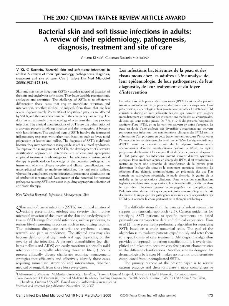
TABLE 2Examples of bacteria-specific virulence factors
Classification Bacteria Virulence factor Details
Adherence S pyogenes Fimbrillae Allow adherence to host epithelial cells
factors M protein Prevent phagocytosis
Protein F Allow access into epithelial cells to avoid detection
S aureus Clumping factor Allow adherence to host epithelial cell
Protein A Prevent antibody opsonization and phagocytosis
Exotoxins S aureus Serine protease Digest desmosome proteins and cause bullous disease (44)
Lipases Digest skin fatty acids to invade through skin barrier
Panton-Valentine Membrane pore formation, especially in neutrophils and skin tissues leading to cell lysis (45);
leukocidin predilection for mitochondria may lead to elaboration of oxidative species leading to skin necrosis
Clostridium Collagenases Connective tissue digestion, which can cause rapidly progressive disease
species Hyaluronidases Matrix protein digestion, which can cause rapidly progressive disease
Alpha-toxin Cell membrane and nerve sheath degradation; induce metabolic dysfunction through prostaglandin
elaboration
E coli Nonspecific exotoxin Intracellular signalling disruption leading to cell death
E coli Escherichia coli; S aureus Staphylococcus aureus; S pyogenes Streptococcus pyogenes. Adapted from reference 19
10733_ki.qxd 28/03/2008 11:40 AM Page 175
Exotoxins, on the other hand, are actively secreted proteinsthat cause tissue damage or dysfunction through variousmechanisms (19). They may cause tissue damage throughenzymatic reactions, cellular dysregulation or pore formation,with subsequent cell lysis. A special group of exotoxins is thesuperantigens. These are most notably produced by virulentS aureus and S pyogenes strains (19). These antigens bindconserved portions of T cell receptors and are, therefore, ableto activate a large number of T lymphocytes. The massiverelease of cytokines causes a grossly exaggerated inflammatoryresponse. SSTIs caused by these strains develop rapidly and areassociated with severe tissue necrosis. This phenomenonprecipitates toxic shock syndrome.
InflammationThe other portion of the infection process involves the hostresponse to tissue invasion and damage. As a protectiveresponse, the goals of inflammation are to rid the body of theinciting organisms and begin tissue repair. Microbial invasionor tissue damage in skin or soft tissues induces changes invascular tone to increase blood flow to the injured site.Additional changes in microvasculature promote and assist theextravasation of plasma proteins and leukocytes. These cellsand proteins migrate, accumulate and are activated at the siteof injury. With activation, cells phagocytize, and destroy for-eign matter, dead tissue or microbes. Certain pyrogeniccytokines or exotoxins cause the febrile response. The orches-tration of cells and cytokines is highly sophisticated andbeyond the scope of the present review. Ultimately, the site ofinjury is quarantined, cleared and repaired gradually (19).
Unfortunately, there may be circumstances when this processcontinues unfettered. With diabetic foot ulcer infections,S aureus infections with Panton-Valentine leukocidin produc-tion and toxic shock syndrome, the persistence of tissue damageor pathogens may perpetuate the inflammatory response. As aresult, inflammation may be the source of ongoing tissue damage(19). The tissue eventually becomes devitalized and hypoxiaensues, which predisposes to anaerobic infections, such as withClostridium species. Urgent medical attention, including surgicaldebridement of necrotic tissues and aggressive antibiotic therapy,is essential to arrest inflammation and promote healing.
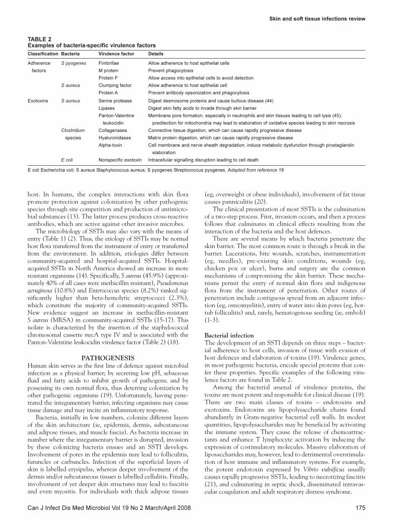
Clinical manifestations of inflammationThe cardinal manifestations of inflammation are warmth,erythema, edema, pain and dysfunction (22). Prolonged inflam-mation can lead to chronic edema, especially in the lowerextremities, and can result in a postcellulitic syndrome.Ancillary systemic signs, such as fever, hypotension and tachy-cardia, result from cytokine-induced changes in thermoregula-tion and vascular resistance. The release of cytokines may bemediated by the normal immune cell function or by bacterialtoxin stimulation. Out-of-proportion pain results from severedamage of the deep layers of skin produced by bacterial toxins,while bullous lesions are produced by toxin-mediated epidermalcleavage. Skin anesthesia, which may be present during thecourse of necrotizing fasciitis, occurs secondary to toxin-mediated nerve tissue damage. Also, violaceous lesions resultfrom toxin-mediated lysis of erythrocytes and hemorrhage (3,8).
Clinical presentationSSTIs produce a diversity of clinical manifestations. Typicalpresenting features, as mentioned above, are nonspecific, and
include erythema, edema, pain and warmth. In contrast, moresevere infections may present with more systemic signs andsymptoms, including temperature higher than 40°C or lowerthan 35°C, hypotension, heart rate faster than 100 beats/min,altered mental status, with a rapidly progressive course andextreme pain (necrotizing fasciitis and myonecrosis) (3,8). Onexamination of severe infections, one may be able to palpatecrepitus and fluctuance secondary to gas or fluid collections.With subsequent necrosis of the dermis, bullae form, which areinitially filled with clear fluid and then with hemorrhagic,violaceous fluid (3,8). As mentioned above, skin anesthesiamay be a late finding in severe skin SSTIs. Finally, ulcersdevelop in areas of high mechanical pressure (23), progressingto ischemia and necrosis (24,25).
DIFFERENTIAL DIAGNOSISBecause of its delicate and intricate anatomy and physiology,the skin is very prone to irritation, abrasions or trauma, as wellas the development of lesions generated from within its ownstructures (eg, folliculitis). Erythematous skin lesions do notalways represent infections. A broad range of differentialdiagnoses exist, which may present similar to impetigo,erysipelas or even cellulitis. Falagas and Vergidis (26) havediscussed various common and rare diseases that may mimicSSTIs (Table 3).
The general lack of data has precluded the development ofa standardized approach to categorizing the different skindiseases. For this reason, an approach to the differential diag-nosis of SSTIs may be based on the specific anatomical siteaffected. First, for skin lesions that affect the upper extremities,venous thrombophlebitis, contact dermatitis, envenomations,Sweet’s syndrome, gouty arthritis, pseudogout, erythromelalgiaand familial Hibernian fever should be considered. Second, forlesions that affect the head, acne, drug reactions, relapsingpolychondritis, herpes zoster and psoriasis should be consid-ered. Third, for chest and abdominal skin lesions, drug reac-tions, foreign body reactions, the familial fever syndromes,eosinophilic cellulitis, herpes zoster infection and carcinomaerysipeloides should be considered. Finally, for lower extremityskin lesions, deep venous thrombosis, gouty arthritis, pseudo-gout, relapsing polychondritis and erythromelalgia should beconsidered.
DIAGNOSISThe diagnosis of most SSTIs is based on clinical impression.Laboratory investigations help to confirm the diagnosis andelucidate characteristics of specific etiologies. A diagnosticapproach to a suspected SSTI is provided in Figure 1.
The first step is clinical suspicion of an SSTI. The minimumcriterion is a skin lesion with the typical inflammatory tetrad –tenderness, erythema, edema and warmth. Depending on theextent and location of infection, dysfunction of the affected area(eg, hand or foot) may also be present. The symptom that highlyincreases the suspicion of an SSTI is fever. Other signs and symp-toms, including crepitus, bullae, anesthesia and hemorrhage, aug-ment the suspicion and confirm the diagnosis.
Investigations may include blood cultures, tissue swab withculture, needle aspiration, x-ray, ultrasound and computedtomography (CT) scan or magnetic resonance imaging (MRI)screen, depending on the clinical manifestations. In thepresence of systemic symptoms, such as fever and hypotension,blood cultures help to assess for bacteremia. Blood cultures
Ki and Rotstein
Can J Infect Dis Med Microbiol Vol 19 No 2 March/April 2008176
10733_ki.qxd 28/03/2008 11:40 AM Page 176
produce a low yield, with less than 5% of cases being positive(27).
Swabs of tissue with culture, such as blood cultures, are alsolow-yield tests (27). Before swabbing, an ulcerated woundought to be debrided and cleansed with normal saline irriga-tion. The difficulty with this test is determining which posi-tive swab cultures represent pathogenic agents and whichrepresent merely skin colonization. In wounds with skin
breakdown characterized by the cardinal manifestations ofSSTIs, tissue swabs are most useful, given the high pretestprobability of infection. In addition, positive swabs of superfi-cial ulcers – without penetration to the bone – in diabeticpatients are also useful in determining the microbiologicaletiology of the underlying infection (28). However, suchswabs may not be indicative of the etiology of underlyingosteomyelitis (29).
Skin and soft tissue infections review
Can J Infect Dis Med Microbiol Vol 19 No 2 March/April 2008 177
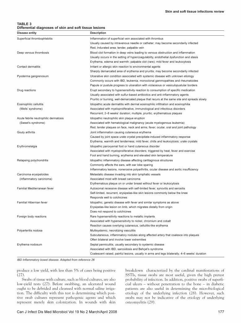
TABLE 3Differential diagnoses of skin and soft tissue lesions
Disease entity Description
Superficial thrombophlebitis Inflammation of superficial vein associated with thrombus
Usually caused by intravenous needle or catheter; may become secondarily infected
Red, indurated area; tender, palpable vein
Deep venous thrombosis Blood clot formation in deep veins leading to venous obstruction and inflammation
Usually occurs in the setting of hypercoagulability, endothelial dysfunction and stasis
Erythema, edema and warmth; palpable clot (rare); mild fever and leukocytosis
Contact dermatitis Irritant or allergic skin reaction to environmental agents
Sharply demarcated area of erythema and pruritis; may become secondarily infected
Pyoderma gangrenosum Ulcerative skin condition associated with systemic disease with unknown etiology
Commonly occurs with IBD, leukemia, monoclonal gammopathies and rheumatoides
Papule or pustule progress to ulceration with violaceous or vesiculopustular borders
Drug reactions Erupt secondary to hypersensitivity reaction to consumption of specific medication
Usually associated with sulfur-based antibiotics and anti-inflammatory agents
Pruritic or burning, well-demarcated plaque that recurs at the same site and spreads slowly
Eosinophilic cellulitis Idiopathic acute dermatitis with dermal eosinophilic infiltration and eosinophilia
(Wells’ syndrome) Associated with myeloproliferative, immunological and infectious disorders
Recurrent; 2–8 weeks’ duration; multiple, pruritic, erythematous plaques
Acute febrile neutrophilic dermatosis Idiopathic neutrophilic skin plaque eruption
(Sweet’s syndrome) Associated with hematological malignancy (acute myelogenous leukemia)
Red, tender plaques on face, neck and arms; fever; ocular, oral and joint pathology
Gouty arthritis Joint inflammation causing cutaneous erythema
Caused by joint space urate crystal precipitate-induced inflammatory response
Erythema, warmth and tenderness; mild fever, chills and leukocytosis; urate crystals
Erythromelalgia Idiopathic paroxysmal foot or hand cutaneous disorder
Associated with myeloproliferative disorders; triggered by heat, fever and exercise
Foot and hand burning, erythema and elevated skin temperature
Relapsing polychondritis Idiopathic inflammatory disease affecting cartilaginous structures
Commonly affects the ears, with ear lobe sparing
Inflammatory lesions, nonerosive polyarthritis, ocular disease and aortic insufficiency
Carcinoma erysipeloides Metastatic disease invading into skin lymphatic vessels
(inflammatory carcinoma) Associated most with breast carcinoma
Erythematous plaque on or under breast without fever or leukocytosis
Familial Mediterranean fever Autosomal recessive disease with self-limited fever, synovitis and serositis
Self-limited, recurrent, erysipelas-like skin lesions commonly below the knee
Responds well to colchicines
Familial Hibernian fever Idiopathic, genetic disease with fever and similar symptoms as above
Erysipelas-like lesion on limb, which migrates distally from origin
Does not respond to colchicines
Foreign body reactions Rare hypersensitivity reactions to metallic implants
Associated with hypersensitivity to nickel, chromium and cobalt
Reaction causes overlying cutaneous, cellulitis-like erythema
Polyarteritis nodosa Multisystemic, necrotizing vasculitis
Subcutaneous, inflammatory nodules along affected artery that coalesce into plaques
Often bilateral and involve lower extremities
Erythema nodosum Septal panniculitis, usually secondary to systemic disease
Associated with IBD, sarcoidosis and Behçet’s syndrome
Coalescent raised, painful lesions, usually in arms and legs bilaterally; 4–6 weeks’ duration
IBD Inflammatory bowel disease. Adapted from reference 26
10733_ki.qxd 28/03/2008 11:40 AM Page 177
Needle aspiration is a controversial investigation, and dif-ferent approaches exist. Some studies advise a leading edgeaspirate, while others attempt a central aspirate. The evidence,however, demonstrates no added benefit to either method. Inone study, positive cultures were attained in approximately10% of patients, regardless of method (30). Furthermore, it hasalso been demonstrated that patients with underlying diseasesor fever are more likely to have positive needle aspiratecultures (31). Needle aspirations may be most useful inpatients presenting with skin infections associated with fluid-filled vesicles.
An x-ray or ultrasound may be used to explore subdermalinvolvement. The x-ray may reveal bony involvement such aswith osteomyelitis, although its sensitivity and specificity islimited (32). In addition, x-rays may reveal air in the tissues orair fluid levels, which are indicative of gas-producing organ-isms such as Clostridium species. Ultrasound, on the otherhand, may be used to investigate fluctuance and crepitus. Thismodality is useful for detecting abscess formation or fascialinflammation (9,33). For more detailed exploration of deepersoft-tissues, a CT scan or an MRI screen may be useful. Theselatter two modalities are most helpful in diagnosing patientswith rapidly progressive skin infections, because these lesionsdo not present superficially until later in their course (3,8,9).For these rapidly progressive lesions, such as necrotizing fasci-itis, early surgical exploration may be prudent, because usualdiagnostic testing may prove equivocal (2,3). In patients withcranial lesions suspected of being SSTIs, head CT scans and/orMRI screens are indicated in patients with the following find-ings: neurological deficits, nonassessable vision, proptosis,deteriorating visual acuity, bilateral ocular edema or ophthal-moplegia, head lesions with no improvement after 24 h orswinging pyrexia not resolving within 36 h (34). In patients
suspected of central nervous system involvement, a lumbarpuncture may be necessary after the exclusion of increasedintracranial pressure.
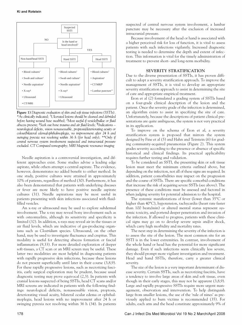
Because involvement of the head or hand is associated witha higher perceived risk for loss of function, it is vital to assesspatients with such infections vigilantly. Increased diagnostictesting is needed to determine the depth and extent of infec-tion. This information is vital for the timely administration oftreatment to prevent short- and long-term morbidity.
SEVERITY STRATIFICATIONDue to the diverse presentation of SSTIs, it has proven diffi-cult to adopt a severity stratification approach. To improve themanagement of SSTIs, it is vital to develop an appropriateseverity stratification approach to assist in determining the siteof care and appropriate empirical treatment.
Eron et al (2) formulated a grading system of SSTIs basedon a four-grade clinical description of the lesion and thepatient. Once the severity grade of the infection is determined,an algorithm exists to assist in specifying the site of care.Unfortunately, because the descriptions of patient clinical pre-sentations are quite ambiguous, the system is not very practicalin its application.
To improve on the schema of Eron et al, a severitystratification system is proposed that mirrors the systemdesigned by Fine et al (5) and Hahn and Teirstein (6) for grad-ing community-acquired pneumonias (Figure 2). This systemgrades severity according to the presence or absence of specifichistorical and clinical findings. Its practical applicabilityrequires further testing and validation.
To be considered an SSTI, the presenting skin or soft tissuelesion must meet the minimum criteria outlined above, butdepending on the infection, not all of these signs are required. Inaddition, patient comorbidities may impact on the progressionand the course of SSTIs. There are specific comorbid conditionsthat increase the risk of acquiring severe SSTIs (see above). Thepresence of these conditions must be assessed and factored inwhen judging severity for potential admission to the hospital.
The systemic manifestations of fever (lower than 35°C orhigher than 40°C), hypotension, tachycardia (heart rate fasterthan 100 beats/min) or altered mental status represent sys-temic toxicity, and portend deeper penetration and invasion ofthe infection. If allowed to progress, patients with these clini-cal signs may go on to develop severe sepsis and/or shock,which carry high morbidity and mortality rates.
The next step in determining the severity of the infection isto assess the site of the lesion. The most common site for anSSTI is in the lower extremities. In contrast, involvement ofthe whole hand or head has the potential for more significantdamage. Even if such infections seem clinically less severe,they should prompt more vigilant investigation and treatment.Head and hand SSTIs, therefore, carry a greater clinicalseverity.
The size of the lesion is a very important determinant of dis-ease severity. Certain SSTIs, such as necrotizing fasciitis, havea tendency to involve large areas of skin and soft tissue, eventhough in their early stages, this may not be apparent (3,8,9).Large and rapidly progressive SSTIs require more urgent man-agement, observation and intervention. To help distinguishlarge from smaller lesions, the use of the ‘rule of nines’ as pre-viously applied to burn victims is recommended (35). Foradults, each arm and the head constitute approximately 9% of
Ki and Rotstein
Can J Infect Dis Med Microbiol Vol 19 No 2 March/April 2008178
SuspectedSSTI
Is the hand or head involved?
NO
YES
Non-hand/head SSTI Hand SSTI Head SSTI
• Blood cultures*
• Swab and culture†
• Needle aspiration‡
• X-ray§
• Ultrasound
• Blood cultures*
• Swab and culture†
• Needle aspiration‡
• X-ray§
• Ultrasound
• CT/MRI
• Blood cultures*
• Aspiration‡
• CT/MRI¶
• Lumbar puncture**
Figure 1) Diagnostic evaluation of skin and soft tissue infections (SSTIs).*As clinically indicated; †Ulcerated lesions should be cleaned and debridedbefore having wound base swabbed; ‡Most useful if vesicle/bullae or fluidabscess present; §Seek out bone trauma and air fluid levels; ¶Indications –neurological deficits, vision nonassessable, proptosis/deteriorating acuity orcolour/bilateral edema/ophthalmoplegia, no improvement after 24 h andswinging pyrexia not resolving within 36 h (for head only); **Only ifcentral nervous system involvement suspected and intracranial pressureexcluded. CT Computed tomography; MRI Magnetic resonance imaging
10733_ki.qxd 28/03/2008 11:40 AM Page 178
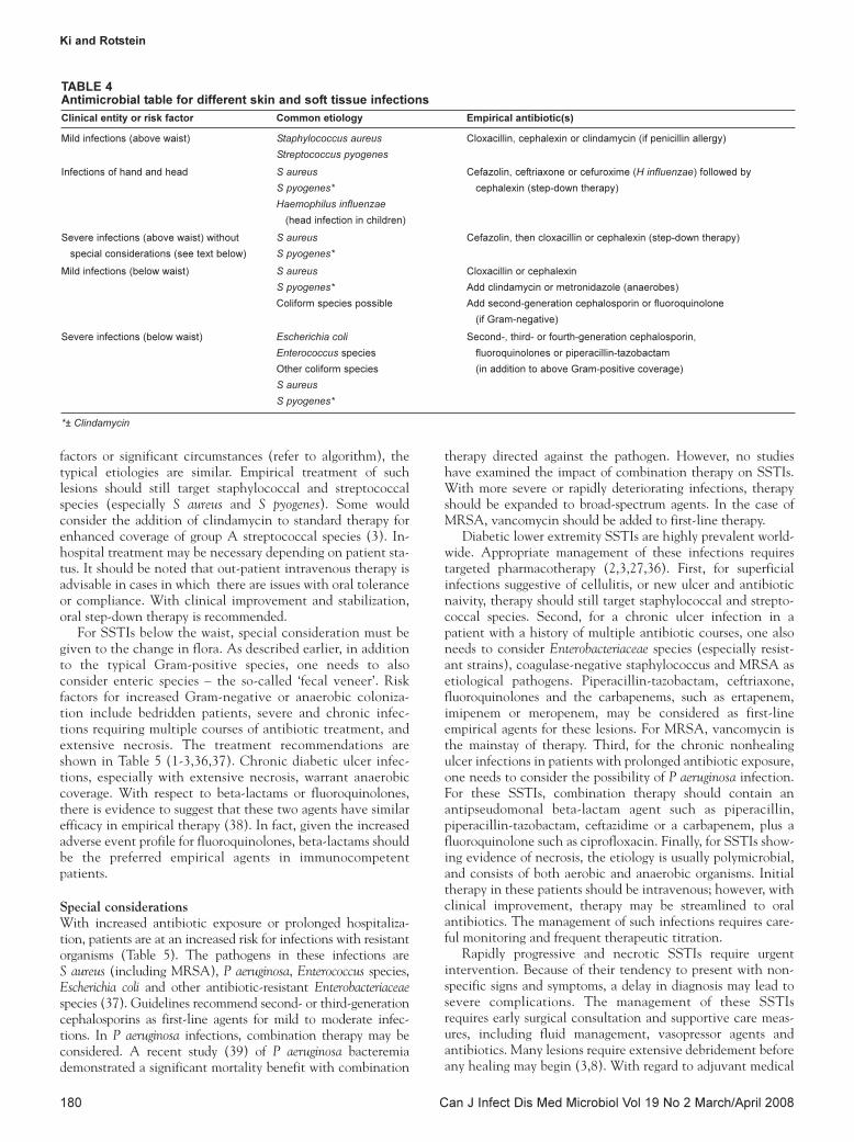
body surface area, whereas each leg, the upper torso and theabdomen (when including both anterior and posterior aspects)each constitute approximately 18% of body surface area,respectively. Any SSTI that involves more than 9% of bodysurface area should be viewed as severe (Figure 3). The excep-tion to this rule are head and hand infections. The whole headand the whole hand constitute approximately 9% and 2% ofbody surface area, respectively. The potential morbidity ofthese infections, as previously mentioned, requires a lowerthreshold for increasing severity. Therefore, for these regions,lesions covering the whole hand or one-half of the head shouldbe considered to be as more severe infections.
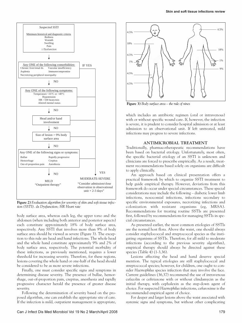
Finally, one must consider specific signs and symptoms indetermining disease severity. The presence of bullae, hemor-rhage, out-of-proportion pain, crepitus, anesthesia and rapidlyprogressive character herald the presence of greater diseaseseverity.
Following the determination of severity based on the pro-posed algorithm, one can establish the appropriate site of care.If the infection is mild, outpatient management is appropriate,
which includes an antibiotic regimen (oral or intravenous)with or without specific wound care. If, however, the infectionis severe, it is prudent to consider hospital admission or at leastadmission to an observational unit. If left untreated, mildinfections may progress to severe infections.
ANTIMICROBIAL TREATMENTTraditionally, pharmacotherapeutic recommendations havebeen based on bacterial etiology. Unfortunately, most often,the specific bacterial etiology of an SSTI is unknown andclinicians are forced to prescribe empirically. As a result, treat-ment recommendations based solely on organisms are difficultto apply clinically.
An approach based on clinical presentation offers apractical framework by which to organize SSTI treatment tohelp guide empirical therapy. However, deviations from thisframework do occur under special circumstances. These specialconsiderations may include the following – diabetic lower limbinfections, nosocomial infections, infections secondary tospecific environmental exposures, necrotizing infections andcolonization with resistant organisms (eg, MRSA).Recommendations for treating routine SSTIs are presentedfirst, followed by recommendations for managing SSTIs in spe-cial circumstances.
As presented earlier, the most common etiologies of SSTIsare the normal host flora. Above the waist, one should alwaysconsider staphylococcal and streptococcal species as the insti-gating organisms of SSTIs. Therefore, for all mild to moderateinfections (according to the previous severity algorithm),empirical therapy should always be directed against thesespecies (Table 4) (1-3,36).
Lesions affecting the head and hand deserve specialmention. The typical etiologies are still staphylococcal andstreptococcal species; however, for children, one ought to con-sider Haemophilus species infection that may involve the face.Current guidelines (36,37) recommend the use of intravenouscefazolin or ceftriaxone with or without clindamycin as theinitial therapy, with cephalexin as the step-down agent ofchoice. For suspected Haemophilus infections, cefuroxime is therecommended empirical agent of choice.
For deeper and larger lesions above the waist associated withsystemic signs and symptoms, but without other complicating
Skin and soft tissue infections review
Can J Infect Dis Med Microbiol Vol 19 No 2 March/April 2008 179
Suspected SSTI
Minimum historical and diagnostic criteriaRedness Warmth Swelling
Pain± Dysfunction
IF YES
NO
NO
NO
NO
NO
Head and/or hand involvement
Any ONE of the following symptoms:Temperature <35°C or >40°C
Hypotension HR >100 beats/min Altered mental status
Size of lesion > 9% body surface area
Any ONE of the following comorbidities:Chronic liver/renal dx Vascular insufficiency Asplenia Immunocompromise Necrotizing peripheral neuropathy
Any ONE of the following signs or symptoms: Bullae Rapidly progressive Hemorrhage Crepitus Out-of-proportion pain Anesthesia
MILD “Outpatient therapy” “Consider admission/close
observation in observationalunit × 2-3 days”
YES
MODERATE-SEVERE
Figure 2) Evaluation algorithm for severity of skin and soft tissue infec-tion (SSTI). dx Dysfunction; HR Heart rate
18%
1%
9%
1%
4.5%
4.5%
4.5%
18%
4.5%4.5%
Figure 3) Body surface area – the rule of nines
10733_ki.qxd 28/03/2008 11:40 AM Page 179
factors or significant circumstances (refer to algorithm), thetypical etiologies are similar. Empirical treatment of suchlesions should still target staphylococcal and streptococcalspecies (especially S aureus and S pyogenes). Some wouldconsider the addition of clindamycin to standard therapy forenhanced coverage of group A streptococcal species (3). In-hospital treatment may be necessary depending on patient sta-tus. It should be noted that out-patient intravenous therapy isadvisable in cases in which there are issues with oral toleranceor compliance. With clinical improvement and stabilization,oral step-down therapy is recommended.
For SSTIs below the waist, special consideration must begiven to the change in flora. As described earlier, in additionto the typical Gram-positive species, one needs to alsoconsider enteric species – the so-called ‘fecal veneer’. Riskfactors for increased Gram-negative or anaerobic coloniza-tion include bedridden patients, severe and chronic infec-tions requiring multiple courses of antibiotic treatment, andextensive necrosis. The treatment recommendations areshown in Table 5 (1-3,36,37). Chronic diabetic ulcer infec-tions, especially with extensive necrosis, warrant anaerobiccoverage. With respect to beta-lactams or fluoroquinolones,there is evidence to suggest that these two agents have similarefficacy in empirical therapy (38). In fact, given the increasedadverse event profile for fluoroquinolones, beta-lactams shouldbe the preferred empirical agents in immunocompetentpatients.
Special considerationsWith increased antibiotic exposure or prolonged hospitaliza-tion, patients are at an increased risk for infections with resistantorganisms (Table 5). The pathogens in these infections areS aureus (including MRSA), P aeruginosa, Enterococcus species,Escherichia coli and other antibiotic-resistant Enterobacteriaceaespecies (37). Guidelines recommend second- or third-generationcephalosporins as first-line agents for mild to moderate infec-tions. In P aeruginosa infections, combination therapy may beconsidered. A recent study (39) of P aeruginosa bacteremiademonstrated a significant mortality benefit with combination
therapy directed against the pathogen. However, no studieshave examined the impact of combination therapy on SSTIs.With more severe or rapidly deteriorating infections, therapyshould be expanded to broad-spectrum agents. In the case ofMRSA, vancomycin should be added to first-line therapy.
Diabetic lower extremity SSTIs are highly prevalent world-wide. Appropriate management of these infections requirestargeted pharmacotherapy (2,3,27,36). First, for superficialinfections suggestive of cellulitis, or new ulcer and antibioticnaivity, therapy should still target staphylococcal and strepto-coccal species. Second, for a chronic ulcer infection in apatient with a history of multiple antibiotic courses, one alsoneeds to consider Enterobacteriaceae species (especially resist-ant strains), coagulase-negative staphylococcus and MRSA asetiological pathogens. Piperacillin-tazobactam, ceftriaxone,fluoroquinolones and the carbapenems, such as ertapenem,imipenem or meropenem, may be considered as first-lineempirical agents for these lesions. For MRSA, vancomycin isthe mainstay of therapy. Third, for the chronic nonhealingulcer infections in patients with prolonged antibiotic exposure,one needs to consider the possibility of P aeruginosa infection.For these SSTIs, combination therapy should contain anantipseudomonal beta-lactam agent such as piperacillin,piperacillin-tazobactam, ceftazidime or a carbapenem, plus afluoroquinolone such as ciprofloxacin. Finally, for SSTIs show-ing evidence of necrosis, the etiology is usually polymicrobial,and consists of both aerobic and anaerobic organisms. Initialtherapy in these patients should be intravenous; however, withclinical improvement, therapy may be streamlined to oralantibiotics. The management of such infections requires care-ful monitoring and frequent therapeutic titration.
Rapidly progressive and necrotic SSTIs require urgentintervention. Because of their tendency to present with non-specific signs and symptoms, a delay in diagnosis may lead tosevere complications. The management of these SSTIsrequires early surgical consultation and supportive care meas-ures, including fluid management, vasopressor agents andantibiotics. Many lesions require extensive debridement beforeany healing may begin (3,8). With regard to adjuvant medical
Ki and Rotstein
Can J Infect Dis Med Microbiol Vol 19 No 2 March/April 2008180
TABLE 4Antimicrobial table for different skin and soft tissue infections
Clinical entity or risk factor Common etiology Empirical antibiotic(s)
Mild infections (above waist) Staphylococcus aureus Cloxacillin, cephalexin or clindamycin (if penicillin allergy)
Streptococcus pyogenes
Infections of hand and head S aureus Cefazolin, ceftriaxone or cefuroxime (H influenzae) followed by
S pyogenes* cephalexin (step-down therapy)
Haemophilus influenzae
(head infection in children)
Severe infections (above waist) without S aureus Cefazolin, then cloxacillin or cephalexin (step-down therapy)
special considerations (see text below) S pyogenes*
Mild infections (below waist) S aureus Cloxacillin or cephalexin
S pyogenes* Add clindamycin or metronidazole (anaerobes)
Coliform species possible Add second-generation cephalosporin or fluoroquinolone
(if Gram-negative)
Severe infections (below waist) Escherichia coli Second-, third- or fourth-generation cephalosporin,
Enterococcus species fluoroquinolones or piperacillin-tazobactam
Other coliform species (in addition to above Gram-positive coverage)
S aureus
S pyogenes*
*± Clindamycin
10733_ki.qxd 28/03/2008 11:40 AM Page 180
Skin and soft tissue infections review
Can J Infect Dis Med Microbiol Vol 19 No 2 March/April 2008 181
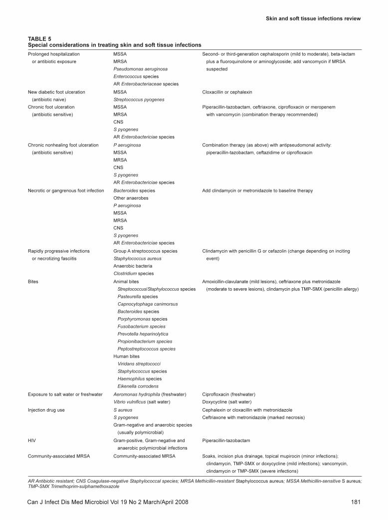
TABLE 5Special considerations in treating skin and soft tissue infections
Prolonged hospitalization MSSA Second- or third-generation cephalosporin (mild to moderate), beta-lactam
or antibiotic exposure MRSA plus a fluoroquinolone or aminoglycoside; add vancomycin if MRSA
Pseudomonas aeruginosa suspected
Enterococcus species
AR Enterobacteriaceae species
New diabetic foot ulceration MSSA Cloxacillin or cephalexin
(antibiotic naive) Streptococcus pyogenes
Chronic foot ulceration MSSA Piperacillin-tazobactam, ceftriaxone, ciprofloxacin or meropenem
(antibiotic sensitive) MRSA with vancomycin (combination therapy recommended)
CNS
S pyogenes
AR Enterobactericiae species
Chronic nonhealing foot ulceration P aeruginosa Combination therapy (as above) with antipseudomonal activity:
(antibiotic sensitive) MSSA piperacillin-tazobactam, ceftazidime or ciprofloxacin
MRSA
CNS
S pyogenes
AR Enterobactericiae species
Necrotic or gangrenous foot infection Bacteroides species Add clindamycin or metronidazole to baseline therapy
Other anaerobes
P aeruginosa
MSSA
MRSA
CNS
S pyogenes
AR Enterobactericiae species
Rapidly progressive infections Group A streptococcus species Clindamycin with penicillin G or cefazolin (change depending on inciting
or necrotizing fasciitis Staphylococcus aureus event)
Anaerobic bacteria
Clostridium species
Bites Animal bites Amoxicillin-clavulanate (mild lesions), ceftriaxone plus metronidazole
Streptococcus/Staphylococcus species (moderate to severe lesions), clindamycin plus TMP-SMX (penicillin allergy)
Pasteurella species
Capnocytophaga canimorsus
Bacteroides species
Porphyromonas species
Fusobacterium species
Prevotella heparinolytica
Propionibacterium species
Peptostreptococcus species
Human bites
Viridans streptococci
Staphylococcus species
Haemophilus species
Eikenella corrodens
Exposure to salt water or freshwater Aeromonas hydrophila (freshwater) Ciprofloxacin (freshwater)
Vibrio vulnificus (salt water) Doxycycline (salt water)
Injection drug use S aureus Cephalexin or cloxacillin with metronidazole
S pyogenes Ceftriaxone with metronidazole (marked necrosis)
Gram-negative and anaerobic species
(usually polymicrobial)
HIV Gram-positive, Gram-negative and Piperacillin-tazobactam
anaerobic polymicrobial infections
Community-associated MRSA Community-associated MRSA Soaks, incision plus drainage, topical mupirocin (minor infections);
clindamycin, TMP-SMX or doxycycline (mild infections); vancomycin,
clindamycin or TMP-SMX (severe infections)
AR Antibiotic resistant; CNS Coagulase-negative Staphylococcal species; MRSA Methicillin-resistant Staphylococcus aureus; MSSA Methicillin-sensitive S aureus;TMP-SMX Trimethoprim-sulphamethoxazole
10733_ki.qxd 28/03/2008 11:40 AM Page 181
therapy, antibiotic agents should target Gram-positive organ-isms: group A streptococcus, S aureus, group B streptococcusand Clostridium species. Current guidelines recommend intra-venous clindamycin in combination with either penicillin Gor cefazolin. Depending on the inciting event (eg, bite or envi-ronmental exposures), empirical therapy may need to bealtered to cover for specific bacterial etiology (3). Ancillaryintravenous immunoglubulin may also prove to be useful inseverely septic patients (40).
Bite wounds are at risk for developing SSTIs. In thesecases, it is important to determine the cause of the bitewound. Organisms involved in these SSTIs depend on thesource agent as mentioned above. For human bites, because ofthe resistance pattern of Eikenella species, intravenouscefoxitin or a beta-lactam/beta-lactamase inhibitor are recom-mended for first-line empirical therapy (3). For mild lesionssecondary to animal bites, treatment with oral amoxicillin-clavulanate suffices. For moderate to severe lesions, intra-venous ceftriaxone in combination with metronidazole isrecommended. For patients who have a penicillin allergy, acombination of trimethoprim-sulfamethoxazole and clin-damycin is recommended (2,3,36).
Specific types of SSTIs may result secondary to exposure tosalt water or freshwater. Current guidelines (2,3) recommenddoxycycline or ciprofloxacin for salt water- and freshwater-exposed SSTIs, respectively.
Injection drug users may develop SSTIs at injection sites.Although staphylococcal species predominate in these infec-tions, patients may present with polymicrobial infections. Theapproach to empirical therapy, therefore, must also considerGram-negative and oral anaerobic organisms. Despite the pos-sibility of Gram-negative infections, current guidelines recom-mend the combination of cephalexin or cloxacillin withmetronidazole for empirical treatment. MRSA may also beisolated in these patients due to previous multiple antibioticexposures (41). For such infections, agents directed againstMRSA are necessary (vancomycin, trimethoprim-sulfamethoxazole with rifampin, tetracycline with rifampin,fusidic acid with rifampin, linezolid, daptomycin and tigecy-cline). In cases with necrosis in which Gram-negative speciesare suspected, a third-generation cephalosporin, such as ceftri-axone, may be substituted in the above regimen (2,3,30,37).
With increasing worldwide prevalence, patients with HIVmay develop severe polymicrobial SSTIs. Although Gram-positive organisms predominantly cause the lesions in thesepatients, being immunocompromised increases HIV patients’susceptibility to infections with Gram-negative organisms andanaerobic organisms. Current guidelines recommend the use ofintravenous therapy with piperacillin-tazobactam (3).
Recently, there has been an acute rise in the incidence ofinfections involving community-associated MRSA (15,17,41).Currently recognized risk factors include the following: youngage, contact sport athlete, injection drug use, military personnel,correctional facility inmate, MRSA carrier, recurrent or recentantibiotic use, and overcrowding (41). SSTIs with this organismmay range from mild to severe. An expert committee hasdeveloped guidelines for the management of community-associated MRSA infections (41). Treatment guidelines werebased on severity and organism susceptibility testing. For minorskin infections caused by community-associated MRSA,nonantibiotic treatment consisting of soaks, incision anddrainage, and possibly topical mupirocin, suffice. Once MRSA is
confirmed in mild, non-life-threatening SSTIs, clindamycin,trimethoprim-sulfamethoxazole or doxycycline may be used asfirst-line agents. For MRSA-positive life-threatening lesions,therapy with intravenous vancomycin, clindamycin ortrimethoprim-sulfamethoxazole is recommended.
Duration of therapyThere are no guidelines to dictate specific durations of therapy.Clinicians should be guided by clinical response to drugtherapy. Follow-up is of the utmost importance. On average,treatment for most lesions requires 10 to 14 days of antibiotictherapy. It should be noted that following initiation of antibi-otic treatment, if there this no response in five days, thisshould prompt a change in the antibiotic regimen or otherinvestigations to verify the diagnosis (41).
Intravenous versus oral therapy, step-down options andadditional careThe decision to use intravenous or oral agents has been anongoing controversy. At present, there is no evidence support-ing any difference in efficacy between the two routes oftherapy. Current consensus among clinicians, however,recommends oral therapy for mild infections and intravenoustherapy for severe infections. The latter allows the achieve-ment of high drug levels with rapid delivery. Intravenoustherapy is also indicated in patients who experience oralintolerance. Moderate infections may be treated via the oralroute, or perhaps with one to two intravenous doses and thengraduating to oral therapy.
For patients with severe infections who are able to tolerateoral therapy and in whom clinical improvement has beendocumented, the goal should be to streamline therapy to theoral route as soon as possible. There is evidence to suggestthat this approach positively impacts length of stay as well(42).
In choosing an appropriate step-down oral antibiotic,there are three options (42). First, one may choose the sameactive antibiotic in oral form with high bioavailability.Antibiotics in this group include amoxicillin, cephalexin,clindamycin, trimethoprim-sulfamethoxazole, metronidazole,doxycycline and linezolid. Second, one may choose the sameactive antibiotic in oral form with moderate bioavailability.Antibiotics in this group include penicillin V, ampicillin,cloxacillin, ciprofloxacin and tetracycline. Finally, one maychoose an oral antibiotic from the same or a different classwith a similar spectrum of activity and good bioavailability(43).
It is noteworthy to include basic wound care techniques aspart of the management of SSTIs. Wound dressings that rangefrom debriding agents to growth-enhancing measures, such asvacuum-assisted closure and occasionally compression, are partof the overall management of SSTIs. These practices mayenhance the healing process.
SUMMARYSSTIs are a highly prevalent but complex and diverse group ofinfections. As a result of the diversity of their presentation,clinical management is challenging. Furthermore, their man-agement is complicated by the paucity of evidence from well-documented studies, and decisions regarding site of care andappropriate antimicrobial therapy may be inconsistent andinefficient.
Ki and Rotstein
Can J Infect Dis Med Microbiol Vol 19 No 2 March/April 2008182
10733_ki.qxd 28/03/2008 11:40 AM Page 182
Skin and soft tissue infections review
Can J Infect Dis Med Microbiol Vol 19 No 2 March/April 2008 183
One method of addressing site of care decisions is todetermine disease severity based on the combination of severalclinical findings. Disease severity should consider location,size, systemic symptoms, comorbidities and significant charac-teristics of the infection. Based on these criteria, SSTIs may beclassified as either mild or moderate to severe. Following thisstratification, one can determine the site of care: mild lesionscan be treated in the outpatient setting with oral therapy,whereas moderate to severe lesions may require hospitalizationor outpatient intravenous therapy.
Appropriate antibiotic therapy is the key to infection treat-ment. Empirical therapy should depend on several factors: poten-tial pathogens, disease severity, clinical complications and theinstrument of entry (eg, animal bite). For all uncomplicated
lesions, empirical therapy should target the typical Gram-positiveskin flora, such as S pyogenes and S aureus. For lesions below thewaist, therapy should also be directed against enteric species.Characteristics that complicate SSTIs include prolonged hospi-talization and antibiotic therapy, diabetes, rapidly progressive andnecrotic lesions, bite wounds, exposure to salt water or freshwa-ter, injection drug use, HIV and risk factors for community-associated MRSA. Empirical therapy for SSTIs in the abovesettings must include coverage of the commonly encounteredpathogens. Finally, the duration of therapy and use of oraltherapy are best determined by careful follow-up and astuteclinical judgement. It is also unknown whether current therapyguidelines for outpatient and hospitalized patient care are opti-mal with respect to treatment efficacy and health care costs.
REFERENCES1. Swartz MN. Clinical practice. Cellulitis. N Engl J Med
2004;350:904-12.2. Eron LJ, Lipsky BA, Low DE, Nathwani D, Tice AD, Volturo GA;
Expert panel on managing skin and soft tissue infections. Managingskin and soft tissue infections: Expert panel recommendations onkey decision points. J Antimicrob Chemother 2003;52:i3-17.
3. Stevens DL, Bisno AL, Chambers HF, et al; Infectious DiseasesSociety of America. Practice guidelines for the diagnosis andmanagement of skin and soft-tissue infections. Clin Infect Dis2005;41:1373-406. (Errata in 2005;41:1830, 2006;42:1219).
4. Elston DM. Optimal antibacterial treatment of uncomplicated skinand skin structure infections: Applying a novel treatmentalgorithm. J Drugs Dermatol 2005;4(6 Suppl):s15-9.
5. Fine MJ, Auble TE, Yealy DM, et al. A prediction rule to identifylow-risk patients with community-acquired pneumonia. N Engl J Med 1997;336:243-50.
6. Halm EA, Teirstein AS. Clinical practice. Management ofcommunity-acquired pneumonia. N Engl J Med 2002;347:2039-45.
7. Ellis Simonsen SM, van Orman ER, Hatch BE, et al. Cellulitisincidence in a defined population. Epidemiol Infect 2006;134:293-9.
8. Vinh DC, Embil JM. Rapidly progressive soft tissue infections.Lancet Infect Dis 2005;5:501-13.
9. Baddour LM. Epidemiology, clinical features, and diagnosis ofcellulitis. <http://www.utdol.com/utd/content/topic.do?topicKey=skin.inf/11185&selectedTitle=1150&source=searc_result2008> (Version current at February 12, 2008).
10. Cox NH. Management of lower leg cellulitis. Clin Med 2002;2:23-7.
11. Björnsdóttir S, Gottfredsson M, Thórisdóttir AS, et al. Risk factorsfor acute cellulitis of the lower limb: A prospective case-controlstudy. Clin Infect Dis 2005;41:1416-22.
12. Horowitz Y, Sperber AD, Almog Y. Gram-negative cellulitiscomplicating cirrhosis. Mayo Clin Proc 2004;79:247-50.
13. Todar K. The bacterial flora of humans.<http://textbookofbacteriology.net/normalflora.html> (Version current at February 12, 2008).
14. Rennie RP, Jones RN, Mutnick AH; SENTRY Program StudyGroup (North America). Occurrence and antimicrobialsusceptibility patterns of pathogens isolated from skin and softtissue infections: Report from the SENTRY AntimicrobialSurveillance Program (United States and Canada, 2000). Diagn Microbiol Infect Dis 2003;45:287-93.
15. King MD, Humphrey BJ, Wang YF, Kourbatova EV, Ray SM,Blumberg HM. Emergence of community-acquired methicillin-resistant Staphylococcus aureus USA 300 clone as the predominantcause of skin and soft-tissue infections. Ann Intern Med2006;144:309-17.
16. Frazee BW, Lynn J, Charlebois ED, Lambert L, Lowery D, Perdreau-Remington F. High prevalence of methicillin-resistantStaphylococcus aureus in emergency department skin and soft tissueinfections. Ann Emerg Med 2005;45:311-20.
17. Eady EA, Cove JH. Staphylococcal resistance revisited:Community-acquired methicillin resistant Staphylococcus aureus –an emerging problem for the management of skin and soft tissueinfections. Curr Opin Infect Dis 2003;16:103-24.
18. Moroney SM, Heller LC, Arbuckle J, Talavera M, Widen RH.Staphylococcal cassette chromosome mec and Panton-Valentineleukocidin characterization of methicillin-resistant Staphylococcusaureus clones. J Clin Microbiol 2007;45:1019-21.
19. McAdam AJ, Sharpe AH. Infectious diseases – bacterial infections.In: Kumar V, Abbas AK, Fausto N, eds. Robbins & CotranPathologic Basis of Disease. Philadelphia: Elsevier Inc, 2005:371-96.
20. Nauta RJ. A radical approach to bacterial panniculitis of theabdominal wall in the morbidly obese. Surgery 1990;107:134-9.
21. Oliver JD. Wound infections caused by Vibrio vulnificus and othermarine bacteria. Epidemiol Infect 2005;133:383-91.
22. Acute and chronic inflammation. In: Kumar V, Abbas AK, Fausto N, eds. Robbins & Cotran Pathologic Basis of Disease.Philadelphia: Elesvier Inc, 2005:47-86.
23. Cavanagh PR, Lipsky BA, Bradbury AW, Botek G. Treatment fordiabetic foot ulcers. Lancet 2005;366:1725-35.
24. Apelqvist J, Larsson J. What is the most effective way to reduceincidence of amputation in the diabetic foot? Diabetes Metab Res Rev2000;16:S75-83.
25. Lipsky BA, Berendt AR, Deery HG, et al; Infectious DiseasesSociety of America. Diagnosis and treatment of diabetic footinfections. Clin Infect Dis 2004;39:885-910.
26. Falagas ME, Vergidis PI. Narrative review: Diseases that masqueradeas infectious cellulitis. Ann Intern Med 2005;142:47-55.
27. Hook EW III, Hooton TM, Horton CA, Coyle MB, Ramsey PG,Turck M. Microbiologic evaluation of cutaneous cellulitis in adults.Arch Intern Med 1986;146:295-7.
28. Slater RA, Lazarovitch T, Boldur I, et al. Swab cultures accuratelyidentify bacterial pathogens in diabetic foot wounds not involvingbone. Diabet Med 2004;21:705-9.
29. Senneville E, Melliez H, Bertrand E, et al. Culture of percutaneousbone biopsy specimens for diagnosis of diabetic foot osteomyelitis:Concordance with ulcer swab cultures. Clin Infect Dis 2005;42:47-62.
30. Newell PM, Norden CW. Value of needle aspiration inbacteriologic diagnosis of cellulitis in adults. J Clin Microbiol1988;26:401-4.
31. Sachs MK. The optimum use of needle aspiration in thebacteriologic diagnosis of cellulitis in adults. Arch Intern Med1990;150:1907-12. (Erratum in 1991;151:244).
32. Lipsky BA. Osteomyelitis of the foot in diabetic patients. Clin Infect Dis 1997;25:1318-26.
33. Tsai CC, Lai CS, Yu ML, Chou CK, Lin SD. Early diagnosis ofnecrotizing fasciitis by utilization of ultrasonography. Kaohsiung J Med Sci 1996;12:235-40.
34. Howe L, Jones NS. Guidelines for the management of periorbitalcellulitis/abscess. Clin Otolaryngol Allied Sci 2004;29:725-8.
35. Berkow SG. A method of estimating the extensiveness of lesions,burns and scalds, based on surface area proportions. Arch SurgLond 1924;8:138-48.
36. Fung HB, Chang JY, Kuczynski S. A practical guide to thetreatment of complicated skin and soft tissue infections. Drugs 2003;63:1459-80.
37. Rosser WW, Pennie RA, Pila NJ; The Anti-Infective ReviewPanel. Anti-Infective Guidelines for Community-AcquiredInfections. Toronto: MUMS Guideline Clearinghouse, 2005.
10733_ki.qxd 28/03/2008 11:40 AM Page 183
Ki and Rotstein
Can J Infect Dis Med Microbiol Vol 19 No 2 March/April 2008184
38. Falagas ME, Matthaiou DK, Vardakas KZ. Fluoroquinolones vs beta-lactams for empirical treatment of immunocompetent patientswith skin and soft tissue infections: A meta-analysis of randomizedcontrolled trials. Mayo Clin Proc 2006:81:1553-66.
39. Safdar N, Handelsman J, Maki DG. Does combinationantimicrobial therapy reduce mortality in Gram-negativebacteremia? A meta-analysis. Lancet Infect Dis 2004;4:519-27.
40. Alejandria MM, Lansang MA, Dans LF, Mantaring JB. Intravenousimmunoglobulin for treating sepsis and septic shock. Cochrane Database Syst Rev 2002;(1):CD001090.
41. Barton M, Hawkes M, Moore D, et al. Guidelines for theprevention and management of community-associated methicillin-resistant Staphylococcus aureus: A perspective for Canadian healthcare practitioners. Can J Infect Dis Med Microbiol 2006;17(Suppl):1B-24B.
42. Jewesson PJ. Pharmaceutical, pharmacokinetic and otherconsiderations for intravenous to oral stepdown therapy. Can J Infect Dis 1995;6(Suppl):11A-6A.
43. Desai M, Franklin BD, Holmes AH, et al. A new approach totreatment of resistant gram-positive infections: Potential impact oftargeted IV to oral switch on length of stay. BMC Infect Dis 2006;6:94.
44. Hanakawa Y, Schecter NM, Lin C, et al. Molecular mechanisms ofblister formation in bullous impetigo and staphylococcal scaldedskin syndrome. J Clin Invest 2002;110:53-60.
45. Kaneko J, Kamio Y. Bacterial two-component and hetero-heptameric pore-forming cytolytic toxins: Structures, pore-forming mechanism, and organization of the genes. Biosci Biotechnol Biochem 2004;68:981-1003. (Erratum in2004;68:A4).
10733_ki.qxd 28/03/2008 11:40 AM Page 184

Submit your manuscripts athttp://www.hindawi.com
Stem CellsInternational
Hindawi Publishing Corporationhttp://www.hindawi.com Volume 2014
Hindawi Publishing Corporationhttp://www.hindawi.com Volume 2014
MEDIATORSINFLAMMATION
of
Hindawi Publishing Corporationhttp://www.hindawi.com Volume 2014
Behavioural Neurology
EndocrinologyInternational Journal of
Hindawi Publishing Corporationhttp://www.hindawi.com Volume 2014
Hindawi Publishing Corporationhttp://www.hindawi.com Volume 2014
Disease Markers
Hindawi Publishing Corporationhttp://www.hindawi.com Volume 2014
BioMed Research International
OncologyJournal of
Hindawi Publishing Corporationhttp://www.hindawi.com Volume 2014
Hindawi Publishing Corporationhttp://www.hindawi.com Volume 2014
Oxidative Medicine and Cellular Longevity
Hindawi Publishing Corporationhttp://www.hindawi.com Volume 2014
PPAR Research
The Scientific World JournalHindawi Publishing Corporation http://www.hindawi.com Volume 2014
Immunology ResearchHindawi Publishing Corporationhttp://www.hindawi.com Volume 2014
Journal of
ObesityJournal of
Hindawi Publishing Corporationhttp://www.hindawi.com Volume 2014
Hindawi Publishing Corporationhttp://www.hindawi.com Volume 2014
Computational and Mathematical Methods in Medicine
OphthalmologyJournal of
Hindawi Publishing Corporationhttp://www.hindawi.com Volume 2014
Diabetes ResearchJournal of
Hindawi Publishing Corporationhttp://www.hindawi.com Volume 2014
Hindawi Publishing Corporationhttp://www.hindawi.com Volume 2014
Research and TreatmentAIDS
Hindawi Publishing Corporationhttp://www.hindawi.com Volume 2014
Gastroenterology Research and Practice
Hindawi Publishing Corporationhttp://www.hindawi.com Volume 2014
Parkinson’s Disease
Evidence-Based Complementary and Alternative Medicine
Volume 2014Hindawi Publishing Corporationhttp://www.hindawi.com


![[Micro] pathogenesis](https://img.pdfslide.us/doc/110x75/55a726df1a28ab7e5e8b45a7/micro-pathogenesis.jpg)