Embed Size (px)
Citation preview
1
Blocked Autophagy Using Lysosomotropic Agents Sensitizes Resistant Prostate Tumor
Cells to the Novel Akt Inhibitor, AZD5363
Francois Lamoureux1*, Christian Thomas1*, Claire Crafter2, Masafumi Kumano1, Fan Zhang1,
Barry R Davies2, Martin E Gleave1 and Amina Zoubeidi1
1The Vancouver Prostate Centre, University of British Columbia, Vancouver, BC, Canada
2AstraZeneca, Alderley Park, Macclesfield, UK
* equal contributors
Key words: Akt inhibitor, AZD5363, autophagy, prostate cancer, chloroquine
Corresponding author:
Amina Zoubeidi, Ph.D. The Vancouver Prostate Centre 2660 Oak Street Vancouver, BC V6H 3Z6, Canada [email protected]
Research. on January 11, 2019. © 2012 American Association for Cancerclincancerres.aacrjournals.org Downloaded from
Author manuscripts have been peer reviewed and accepted for publication but have not yet been edited. Author Manuscript Published OnlineFirst on December 20, 2012; DOI: 10.1158/1078-0432.CCR-12-3114
2
Statement of translational relevance
Targeting Akt using a novel inhibitor AZD5363 activates autophagy in resistant prostate cell
lines and cooperated with Chloroquine (CHQ) to promote cell death in vitro and in vivo in
prostate tumor models. We specifically report AZD5363 monotherapy induced G2 growth arrest
and autophagy, but failed to induce significant apoptosis in PC-3 and DU145 prostate cancer
cell lines. Blocking autophagy using pharmacological inhibitors (3-methyladenine, CHQ and
bafilomycin A) or genetic inhibitors (siRNA targeting Atg3 and Atg7) enhanced cell death
induced by Akt inhibitor AZD5363 in these tumor prostate cell lines. Importantly, the
combination of AZD5363 with CHQ significantly reduced tumor volume compared with the
control group and compared with either drug alone in PC3 xenografts.
Research. on January 11, 2019. © 2012 American Association for Cancerclincancerres.aacrjournals.org Downloaded from
Author manuscripts have been peer reviewed and accepted for publication but have not yet been edited. Author Manuscript Published OnlineFirst on December 20, 2012; DOI: 10.1158/1078-0432.CCR-12-3114
3
Abstract
Introduction and objective: Prostate cancer development is often associated with deletion or silencing of tumor suppressor phosphatase and tensin homologue (PTEN), a negative regulator of the PI3K-Akt pathway, leading to resistance to various therapies in both preclinical and clinical setting. Therefore, the PI3K/Akt pathway plays a central role in various cellular processes promoting survival signaling that can contribute to the malignant phenotype, and, consequently is an attractive pharmacological target. However, as single agents, the efficacy of AKT inhibitors may be limited by resistance mechanisms that result in minimal cell death in tumor cells.
Methods: We investigated the effects of the Akt inhibitor AZD5363 on cell proliferation, cell cycle, apoptosis and Akt downstream pathway proteins. Survival mechanisms induced by AZD5363 were investigated. We then examined the impacts of inhibition of autophagy in combination with AZD5363 on cell proliferation and apoptosis. Furthermore, the anti-cancer activity of combination treatment of the lysosomotropic inhibitor of autophagy (chloroquine) with Akt inhibitor AZD5363 was evaluated in PC-3 prostate cancer xenografts.
Results: Here, we show that Akt inhibitor AZD5363 affected the Akt downstream pathway by reducing p-mTOR, p-P70S6K and p-S6K. While AZD5363 monotherapy induced G2 growth arrest and autophagy, it failed to induce significant apoptosis in PC-3 and DU145 prostate cancer cell lines. Blocking autophagy using pharmacological inhibitors (3-methyladenine, chloroquine and bafilomycin A) or genetic inhibitors (siRNA targeting Atg3 and Atg7) enhanced cell death induced by Akt inhibitor AZD5363 in these tumor prostate cell lines. Importantly, the combination of AZD5363 with chloroquine significantly reduced tumor volume by 84.9% compared with the control group and by 77.5% compared with either drug alone in PC3 xenografts.
Conclusion: Taken together, these data demonstrate that Akt inhibitor AZD5363, synergizes with the lysosomotropic inhibitor of autophagy, chloroquine, to induce apoptosis and delay tumor progression in prostate cancer models that are resistant to monotherapy AZD5363, providing a new therapeutic approach potentially translatable to patients.
Research. on January 11, 2019. © 2012 American Association for Cancerclincancerres.aacrjournals.org Downloaded from
Author manuscripts have been peer reviewed and accepted for publication but have not yet been edited. Author Manuscript Published OnlineFirst on December 20, 2012; DOI: 10.1158/1078-0432.CCR-12-3114
4
Introduction
Prostate cancer (PCa) is the most common cancer and the third most common cause of cancer
related mortality in men in the United States (1). Androgen ablation remains the standard
effective therapy for patients with advanced PCa, inhibiting proliferation and inducing apoptosis
in tumor cells (2). Unfortunately, after short-term remissions, surviving tumor cells recur with
castrate resistant prostate cancer (CRPC) and death usually within 3 years in most men (3). To
significantly improve survival in men with PCa, new therapeutic strategies to inhibit the
appearance of this phenotype must be developed.
Cancer development is often associated with deletion or silencing of tumor suppressor
phosphatase and tensin homologue (PTEN), a negative regulator of the PI3K-Akt pathway (4),
leading to resistance to various therapies in both preclinical and clinical trials (5, 6). Therefore,
PI3K/Akt/mTOR pathway plays a central role in various cellular processes including protein
synthesis, cell cycle, cell survival, cell growth, motility and angiogenesis (7) that can contribute
to the malignant phenotype. Many small molecule inhibitors targeting PI3K, Akt and/or mTOR
are currently at various stages of clinical development. Despite an impressive efficacy in certain
preclinical tumor models (7), clinical responses to small molecule inhibitors of this pathway
have been limited, because PI3K, Akt or mTOR inhibition typically promotes growth arrest
rather than cell death in solid tumors (8). In order to enhance cancer cell killing mediated by Akt
inhibitors, the pro-survival pathways activated in response to the drugs were investigated. Since
mTOR is reported to be a key node of macroautophagy (hereafter called autophagy) that
provides energy and nutrients to cancer cells during stress by a cellular self-digestion process
(9), autophagy seems to be a good candidate as an activated survival pathway.
Autophagy is a conserved process by which cells recycle macromolecules and organelles into
specialized double-membrane vesicles known as autophagosomes to maintain homeostasis
Research. on January 11, 2019. © 2012 American Association for Cancerclincancerres.aacrjournals.org Downloaded from
Author manuscripts have been peer reviewed and accepted for publication but have not yet been edited. Author Manuscript Published OnlineFirst on December 20, 2012; DOI: 10.1158/1078-0432.CCR-12-3114
5
(10, 11). The formation of autophagosomes is initiated by class III phophoinositide 3-kinase and
Beclin-1. Then, autophagosomes fuse with the lysosome, forming autophagolysosomes
promoting the intracellular degradation. The abundant cytoplasmic microtubule-associated
protein chain 3 protein (LC3-I) recruited during the formation of autophagosomes, is cleaved
and lipidated generating LC3-II form (12). The hallmark of autophagic activation is thus the
formation of cellular autophagosome punctae containing LC3-II.
So far, the role of autophagy in cancer is controversial, as the factors that determine whether
autophagy induces tumor cell death or protects tumor cells from unfavorable conditions have
not been clearly elucidated, Autophagy is reported to play an anti-tumoral role by reducing the
chromosomic instability, thus avoiding DNA damage (13), inhibiting cell proliferation (14, 15),
and protecting cells from necrosis and associated inflammation, that could promote
angiogenesis and tumor progression (13, 16). On the other hand, several studies demonstrated
that autophagy is part of tumor progression during metabolic stresses (17). Therefore,
autophagy acts as an anti-apoptotic mechanism for tumor cells under stress, maintaining
metabolism and energy needed for survival (13). Moreover, inhibition of autophagy promotes
cancer cell death (18, 19) and potentiates various anticancer therapies (20-24).
Whilst some prostate cancer cell lines such as LNCaP and PC346-Flu1 are very sensitive to
monotherapy with the AKT inhibitor AZD5363, where the compound induces significant
apoptosis at concentrations achievable in preclinical models (25), other prostate cancer cell
lines, such as DU-145 and PC3, are relatively resistant. Here we show that the novel Akt
inhibitor AZD5363 activated autophagy in resistant prostate cell lines and cooperated with
Chloroquine (CHQ) to promote cell death in vitro and in vivo in prostate tumor models. This
study strongly demonstrates the attractive prospect of blocking autophagy processes combined
with targeted therapy as a promising therapeutic approach for prostate cancer.
Research. on January 11, 2019. © 2012 American Association for Cancerclincancerres.aacrjournals.org Downloaded from
Author manuscripts have been peer reviewed and accepted for publication but have not yet been edited. Author Manuscript Published OnlineFirst on December 20, 2012; DOI: 10.1158/1078-0432.CCR-12-3114
6
Materials and Methods:
Tumor cell lines and reagents:
The human PCa cell lines PC-3 and DU-145 were purchased from the American Type Culture
Collection (2008 and 1989, respectively, ATCC-authentication by isoenzymes analysis) and
maintained in DMEM (Invitrogen-Life Technologies, Inc.) supplemented with 5% fetal bovine
serum and 2mmol/L L-glutamine. All cell lines were cultured in a humidified 5% CO2/air
atmosphere at 37oC. All cell lines were passaged for less than 3 months after resurrection.
Therapeutic agents:
Akt inhibitor, AZD5363, was kindly provided from AstraZeneca and used for in vitro and in vivo
studies. AZD5363 compound is novel synthetic Akt inhibitor that is orally bioavailable. For the in
vitro studies, AZD5363 was dissolved in dimethyl sulfoxide (DMSO) at 10mM stock solutions
and stored at -20oC. For the in vivo studies, AZD5363 was dissolved in PBS 1%
carboxymethylcellulose (CMC) and 0.5% Tween 80 (Invitrogen-Life Technologies, Inc.) at
15mg/ml and stored at 4oC. Chloroquine (CHQ) (Sigma-Aldrich, Inc) was dissolved in water for
both in vitro and in vivo experiments at the desired concentrations and stored at 4oC.
Cell proliferation and apoptosis assays:
Prostate cancer cell lines were plated in appropriate media (DMEM) with 5% FBS and treated
with AZD5363 at indicated concentration and time and cell growth was measured using the
crystal violet assay as described previously (26). Detection and quantitation of apoptotic cells
was done by flow-cytometry (described below) and western blotting analysis. Each assay was
repeated in triplicate.
Caspase-3 activity was assessed 3 days after treatment using the kit CaspACE Assay System,
Fluorometric (Promega, Madison, WI, USA). Fifty µg of total cell lysate were incubated with
Research. on January 11, 2019. © 2012 American Association for Cancerclincancerres.aacrjournals.org Downloaded from
Author manuscripts have been peer reviewed and accepted for publication but have not yet been edited. Author Manuscript Published OnlineFirst on December 20, 2012; DOI: 10.1158/1078-0432.CCR-12-3114
7
caspase-3 substrate AC-DEVD-AMC at room temperature for 4h and caspase-3 activity was
quantified in a fluorometer with excitation at 360nm and emission 460nm.
Cell cycle analysis
Prostate cancer cell lines were incubated in the absence or the presence of 10, 50 and 100μM
AZD5363 for 48h, trypsinized, washed twice and incubated in PBS containing 0.12% Triton X-
100, 0.12mM EDTA and 100μg/ml ribonuclease A; 50μg/ml propidium iodide was then added to
each sample for 20min at 4°C. Cell cycle distribution was analyzed by flow cytometry (Beckman
Coulter Epics Elite, Beckman, Inc., Miamai, FL), based on 2N and 4N DNA content. Each assay
was done in triplicate.
Western blotting analysis:
Samples containing equal amounts of protein (depending on the antibody, 5-50µg) from lysates
of cultured tumor prostate cell lines underwent electrophoresis on SDS-polyacrylamide gel and
were transferred to nitrocellulose filters. The filters were blocked in Odyssey Blocking Buffer (LI-
COR Biosciences) at room temperature for 1h and blots were probed overnight at 4°C with
1:1,000 primary antibodies to detect proteins of interests. After incubation, the filters were
washed 3 times with washing buffer (PBS containing 0.1% Tween) for 5min. Filters were then
incubated for 1h with 1:5,000 diluted Alexa Fluor secondary antibodies (Invitrogen) at room
temperature. Specific proteins were detected using ODYSSEY IR imaging system (LI-COR
Biosciences) after washing. Antibodies anti–phospho-Akt (Ser473), anti-Akt, anti-mTOR, anti-
phospho-mTOR, anti-GSK-3B, anti-phospho-GSK-3B, anti-phospho-S6, anti-caspase-3, anti-
cleaved PARP, anti-Atg7, anti-Atg3 and anti-LC3, anti-phospho-CDC2, anti-phospho-CDC25C,
anti-phospho-WEE1 from Cell Signaling Technology; and anti-vinculin and anti–β-actin from
Sigma-Aldrich.
siRNA transfection
Research. on January 11, 2019. © 2012 American Association for Cancerclincancerres.aacrjournals.org Downloaded from
Author manuscripts have been peer reviewed and accepted for publication but have not yet been edited. Author Manuscript Published OnlineFirst on December 20, 2012; DOI: 10.1158/1078-0432.CCR-12-3114
8
Cells were transfected with antisense or siRNA as described previously (27, 28). The sequence
of Atg3 siRNA corresponds to the human Atg3 site (5′-GGAAUCAAAGUUUAAGGAAACAGGU-
3′; Invitrogen Life Technologies). A scrambled siRNA (5′-CAGCGCUGACAACAGUUUCAU-3′;
Dharmacon) was used as a control for RNA interference experiments.
Immunofluorescence
Tumor cells were grown on coverslips and treated with AZD5363 at indicated concentrations
and indicated time. After treatment, cells were fixed in ice-cold methanol completed with 3%
acetone for 10min at -20oC. Cells were the washed three times with PBS and incubated with
0.2% Triton/PBS for 10min, followed by washing and 30min blocking in 3% nonfat milk before
the addition of antibody overnight to detect LC-3 (1:250). Antigens were visualized using anti-
mouse antibody coupled with FITC (1:500; 30 min). Photomicrographs were taken at 20X
magnification using Zeiss Axioplan II fluorescence microscope, followed by analysis with
imaging software (Northern Eclipse, Empix Imaging, Inc.). Puncta from 100 to 150 cells were
counted from 3 independent experiments for quantitative analysis as described previously (29).
Cells displaying >15 brightly fluorescent LC3 puncta were counted as positive. Photo-
micrographs were taken at 40x magnification using Zeiss Axioplan II fluorescence microscope.
Animal Treatment
To establish PC-3 or DU145 tumors, 2 x 10⁶ cells were inoculated s.c. in the flank region of 6-8
week-old male athymic mice (Harlan Sprague-Dawley, Inc.). When tumors reached 100mm3,
usually 3-4 weeks after injection, mice were randomly assigned to vehicle, AZD5363 alone,
CHQ alone or AZD5363 + CHQ. AZD5363 (150mg/kg; formulation in 0.5% CMC + 0.5% Tween-
80) was administered orally twice daily and CHQ (15mg/kg) was injected intra-peritoneally once
daily. Each experimental group consisted of 10 mice. Tumor volume was measured twice
Research. on January 11, 2019. © 2012 American Association for Cancerclincancerres.aacrjournals.org Downloaded from
Author manuscripts have been peer reviewed and accepted for publication but have not yet been edited. Author Manuscript Published OnlineFirst on December 20, 2012; DOI: 10.1158/1078-0432.CCR-12-3114
9
weekly (length x width x depth x 0.5432). Data points were expressed as average tumor volume
± SEM.
When tumor volume reached ≥10% of body weight, mice were sacrificed and tumors harvested
for evaluation of protein expression by western blotting analyses and immunohistochemistry. All
animal procedures were performed according to the guidelines of the Canadian Council on
Animal Care and appropriate institutional certification.
Immunohistochemistry
Immunohistochemical stains were performed on formalin-fixed and paraffin-embedded 4µm
sections of tumor samples using adequate primary antibody (supplementary materials), and the
Ventana autostainer Discover XT (Ventana Medical System) with enzyme labeled biotin
streptavidin system and solvent resistant 3,3’-diaminobenyidine Map kit. All comparisons of
staining intensities were made at 200x magnifications.
Statistical analysis
All in vitro data were assessed using the Student t test and Mann-Whitney test. Tumor volumes
of mice were compared using Kruskal-Wallis test. Overall survival was analyzed using Kaplan-
Meier curves and statistical significance between the groups was assessed with the log-rank
test (Graphpad Prism). Levels of statistical significance were set at P<0.05.
Research. on January 11, 2019. © 2012 American Association for Cancerclincancerres.aacrjournals.org Downloaded from
Author manuscripts have been peer reviewed and accepted for publication but have not yet been edited. Author Manuscript Published OnlineFirst on December 20, 2012; DOI: 10.1158/1078-0432.CCR-12-3114
10
Results
A novel Akt inhibitor AZD5363 down-regulates the Akt downstream pathway.
AZD5363, an orally bioavailable, small molecule, inhibitor of Akt (Fig 1A), potently inhibits all
three isoforms of Akt (Akt 1, 2, and 3) (25). Although these three isoforms are encoded by
separate genes, they share a common NH2-terminal pleckstrin homology (PH) domain, a
catalytic domain in the middle, and a COOH terminus (30, 31). The identity of the overall amino
acid sequence of the three isoforms is very high (∼80%); however, the COOH terminus and the
PH-linker region are more diverse (31). AZD5363 down-regulates the phosphorylation of
downstream pathway proteins in a dose-dependent manner (Fig. 1B and C), as evaluated by
western-blot in PC-3 and DU145 prostate cell lines. Indeed, AZD5363 reduced phospho-mTOR,
phospho-S6 and phospho-GSK-3β proteins, clearly demonstrating that the compound is
inhibiting AKT substrates and pathway across the dose range. However, consistent with data in
other cell lines, phosphorylation of Akt was increased in a dose-dependent manner in both PC-3
and DU145 cells (25).
Biological effects of AZD5363 on PC-3 and DU145 prostate cell lines
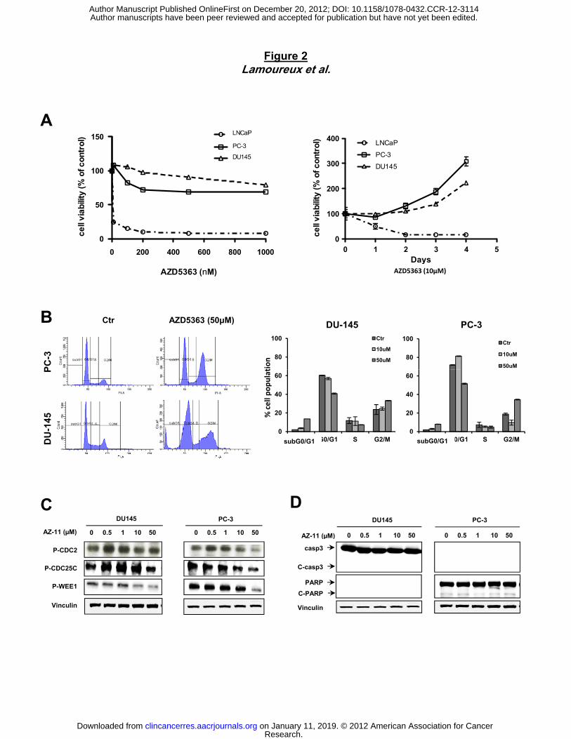
To determine the biological activity of AZD5363 on cell proliferation, we compared PC-3 and
DU145 cell lines with LNCaP cells, which are known to be very sensitive to AZD5363 (25).
AZD5363 reduced LNCaP cell viability in a dose- and time-dependent manner (≥90% inhibition
after after 3 days at 200nM and >90% reduction in cell proliferation after 2 days at 10µM). In
contrast, PC-3 and DU145 cell viability was only slightly affected in dose-dependent manner
after 24h, and indeed cell number increased after 1 day, indicating insensitivity and innate
resistance of these cancer cells to AZD5363 (Fig. 2A). We next examined the effect of AZD5363
inhibitor on cell cycle repartition in PC-3 and DU145 cancer cell lines compared to sensitive
LNCaP cells. While low dose of AZD5363 enhances subG0/G1 phase (Fig. S1A) and increases
Research. on January 11, 2019. © 2012 American Association for Cancerclincancerres.aacrjournals.org Downloaded from
Author manuscripts have been peer reviewed and accepted for publication but have not yet been edited. Author Manuscript Published OnlineFirst on December 20, 2012; DOI: 10.1158/1078-0432.CCR-12-3114
11
cleaved-PARP in the sensitive LNCaP cells (Fig. S1B), PC-3 and DU145 cells show a slight
G2/M arrest (increasing by 1.5-2 fold the cell population after 50µM AZD5363 relative to control)
after AZD5363 treatment compared to untreated cells (Fig. 2B). These results were associated
with decreased phospho-CDC2, phospho-CDC25C and phospho-WEE1 expression in PC-3 and
DU145 cell lines (Fig. 2C). No significant change was observed in subG0/G1 phase after
treatment with 10µM, even with high concentration 50µM of AZD5363 (approximately 5-fold
higher than the concentration achieved at a maximum dose tolerated in nude mice) compared to
control (Fig. 2B). These results suggest that AZD5363 fails to induce apoptosis in PC-3 and
DU145 prostate cell lines at a concentration that reflects maximal tolerated doses in preclinical
mouse models (Fig. 2B). This result was confirmed by immunoblot analysis showing that
AZD5363 did not induce cleaved caspase-3 or cleaved PARP in PC-3 and DU145 cells (Fig.
2D), while other drugs (staurosporine or MG132) induce apoptosis in these cell lines (Fig. S2A).
AZD5363 induces autophagy by inhibiting Akt/mTOR/p70S6K signaling pathway in
prostate cells.
Having demonstrated down-regulation of Akt downstream pathway, including inhibition of mTOR
pathway (Fig. 1A) and a growth arrest without apoptosis induction (Fig. 2), we focused on
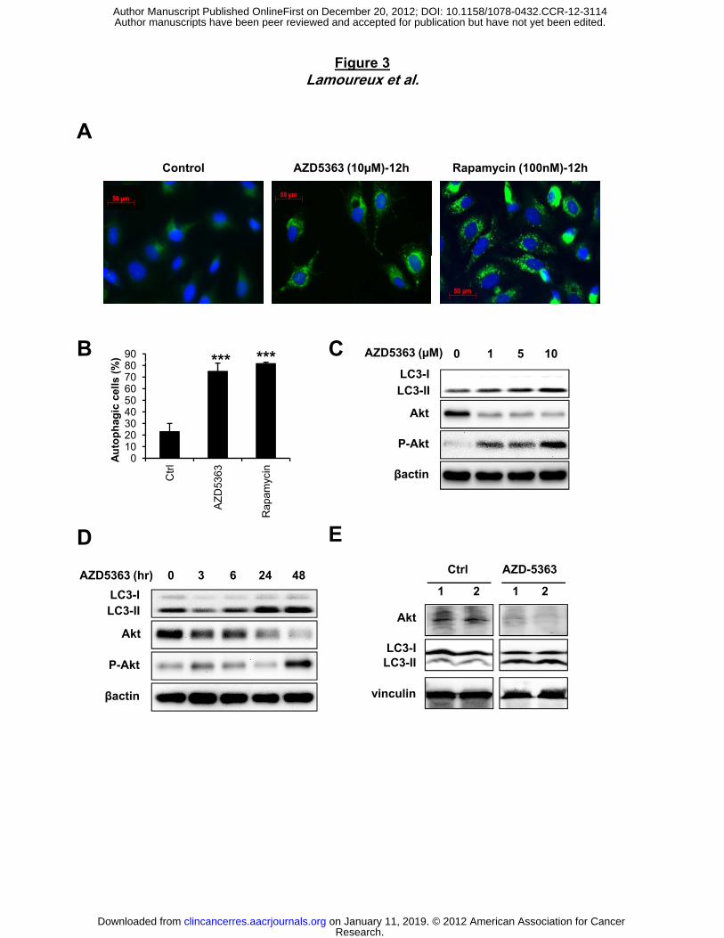
autophagy as a mechanism inducing cell survival rather than apoptosis (20). We investigated
autophagy induction in tumor prostate cells after AZD5363 treatment and found that AZD5363
induces autophagosome formation, as measured by puncta of LC3 fusion protein in PC3
prostate cancer cells compared to untreated cells (75% of positive cells after AZD5363
treatment vs 23% of positive cells in Ctrl condition; Fig. 3A and B). The level of autophagy
induction was comparable to that induced by rapamycin, an mTOR inhibitor known to induce
autophagy (32). Like with rapamycin treatment (Fig. S2B), immunoblotting revealed that
AZD5363 induced the conversion of LC3-I to LC3-II in a dose and time-dependent manner (Fig.
3C and D). We next examined if AZD5363 induces autophagy in vivo. Mice treated with
Research. on January 11, 2019. © 2012 American Association for Cancerclincancerres.aacrjournals.org Downloaded from
Author manuscripts have been peer reviewed and accepted for publication but have not yet been edited. Author Manuscript Published OnlineFirst on December 20, 2012; DOI: 10.1158/1078-0432.CCR-12-3114
12
AZD5363 show an induction of autophagy as indicated by conversion of LC3-I to LC3-II in heart
tissue relative to untreated mice (Fig. 3E).
Blocking autophagy enhances induction of apoptosis by AZD5363 in vitro.
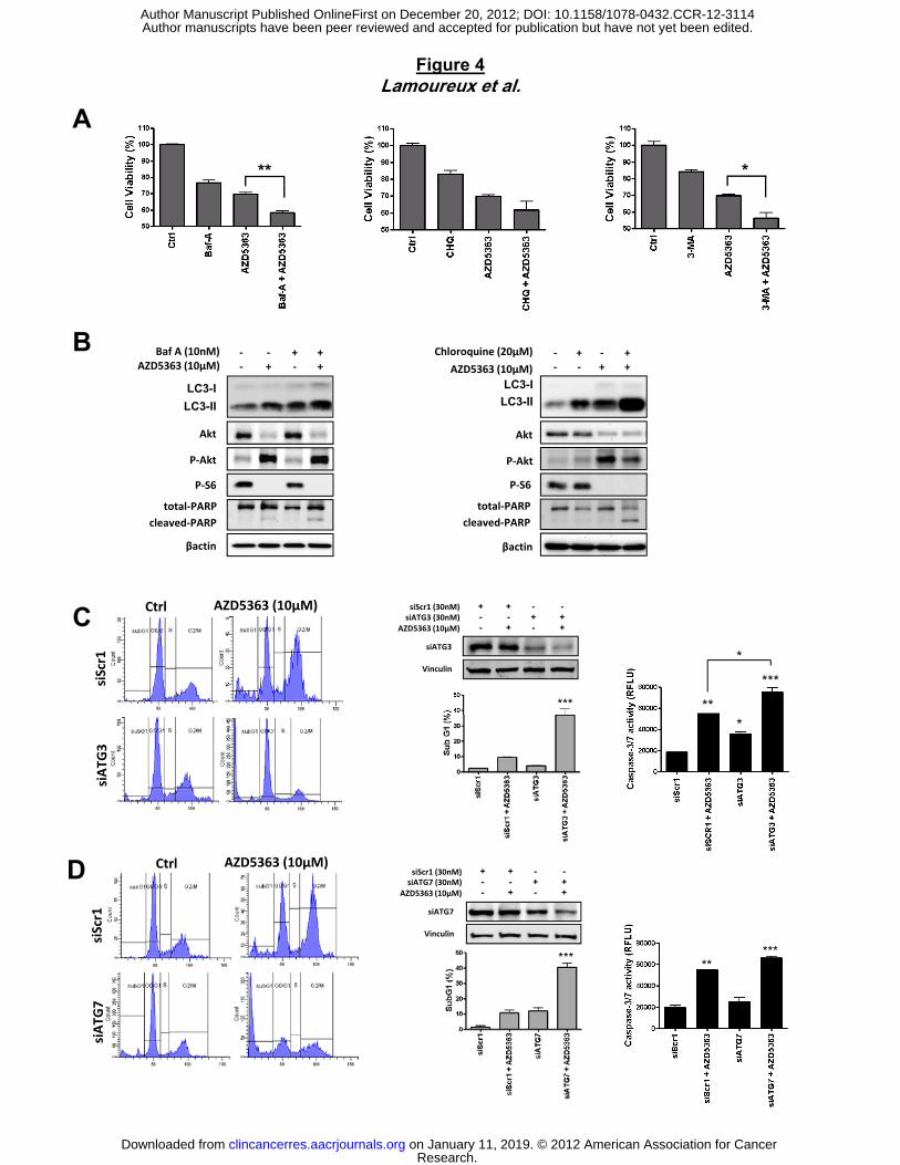
Considering that autophagy may function as a stress-activated pro-survival mechanism in
prostate cancer cells (20, 23), we hypothesized that inhibiting autophagy pathway could
promote cell death when combined with AZD5363. First, PC-3 cells were treated with AZD5363,
followed by brief exposure to pharmacological inhibitors of autophagy, bafilomycin A (Baf A),
which inhibits vacuolar-type H+-ATPase, thus blocking autophagosome maturation (33),
chloroquine (CHQ) which disrupts the function of lysosomes, and 3-methyladenine (3-MA)
which inhibits early stages of autophagosome formation (34). As shown in Figure 4A, cell
viability is significantly reduced when AZD5363 is combined with BafA, CHQ or 3-MA compared
to either drug alone (Fig. 4A). This result is accompanied by increased apoptosis as shown by
enhancing cleaved PARP level with combination treatment compared with monotherapy
AZD5363, BafA or CHQ (Fig. 4B). Furthermore, BafA or CHQ-treated cells showed increased
conversion of LC3-I to LC3-II (Fig. 4B), likely due to autophagosome accumulation (35). This
increased conversion of LC3-I to LC3-II was higher when BafA or CHQ was combined with
AZD5363. These results were confirmed using another PI3K inhibitor, PI-103, further showing
that targeting Akt induced a decrease of cell proliferation (Fig. S3A), enhanced cleaved PARP
with combination treatment compared with monotherapy PI-103, or CHQ, and increased
conversion of LC3-I to LC3-II (Fig. S3B).
To exclude off-target pharmacological effects of drugs inhibiting autophagy, we treated cells
with small interfering RNAs (siRNAs) directed against ATG3 and ATG7, which are required for
autophagosome formation (36). The knock-down of the expression of endogenous ATG-3 or
ATG-7 induced cell death by increasing significantly the sub-G1 fraction when combined with
Research. on January 11, 2019. © 2012 American Association for Cancerclincancerres.aacrjournals.org Downloaded from
Author manuscripts have been peer reviewed and accepted for publication but have not yet been edited. Author Manuscript Published OnlineFirst on December 20, 2012; DOI: 10.1158/1078-0432.CCR-12-3114
13
AZD5363, compared to control siRNA-treated cells (fig. 4C and D). This cell death is due to
enhanced apoptosis, as evidenced by a pronounced increase in casapase-3/7 activity after
combined treatment AZD5363 with siATG-3 or siATG-7, relative to control siRNA (Fig. 4C and
D). Taken together, these data strongly indicate that autophagy protects tumor cells from cell
death induced by AZ5363 and, then, blocking autophagy contributes to apoptosis when
combined with AZD5363.
Autophagy inhibition potentiates in vivo activity of clinical inhibitor AZD5363 in prostate
xenograft model.
To further investigate the therapeutic benefit of inhibiting autophagy in combination with
AZD5363, we used CHQ, which has been used in clinic for more than 50 years for treatment of
malaria and autoimmune disease. Using PC-3 and DU145 cell lines, we demonstrated that
AZD5363 and CHQ could cooperate to induce apoptosis in vitro as shown by PARP cleavage
and Caspase3 activity compared with either agent alone. To translate these results in vivo, we
used prostate xenograft models from PC-3 and DU145. When CHQ was combined with
AZD5363, the PC3 xenograft growth was reduced by 80% compared to untreated mice or those
treated with AZD5363 or CHQ monotherapy (Fig. 5A). The tumor doubling time was 2-fold
longer with combination therapy (±0.8 month) compared to either drug alone (±0.4 month) (Fig.
5B). Moreover, all animals treated with combination of AZD5363 and CHQ survived after 64
days, while 80% of untreated mice and 30-40% of mice treated with monotherapy AZD5363 or
CHQ acquired tumor burden that required euthanasia (Fig. 5C). No significant changes in
overall body weight or behavior were observed in all mice. Similar results were observed in the
DU145 xenograft model (Fig. 5D, E and F).
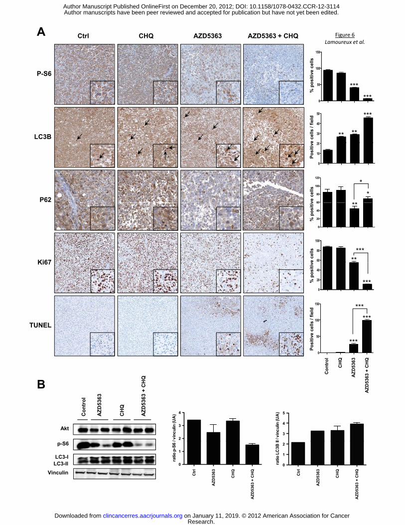
Analyses of treated tumors confirmed that the combination treatment of AZD5363 and CHQ
significantly inhibited the Akt pathway and induced marked increases in apoptosis (Fig. 6A and
Research. on January 11, 2019. © 2012 American Association for Cancerclincancerres.aacrjournals.org Downloaded from
Author manuscripts have been peer reviewed and accepted for publication but have not yet been edited. Author Manuscript Published OnlineFirst on December 20, 2012; DOI: 10.1158/1078-0432.CCR-12-3114
14
B). Indeed, p-S6 staining decreased by 40% in AZD5363 monotherapy group, and by 90%
respectively, after combination treatment. In addition, TUNEL staining (indicating degree of cell
death) showed a large increase after combination treatment, whilst Ki67 expression decreased
by ~90%, indicating reduction in cell proliferation. To detect LC3 and subsequently distinguish
between the diffuse localization of LC3-I and the punctea of LC3-II in tumor tissue, only the dark
punctea were counted as positive cells of LC3-II. Either drug alone increased autophagy by 2-
fold relative to untreated mice, whereas the combination of AZD5363 and CHQ increased LC3-II
by 3.5-fold, corroborating with the in vitro data. In Parallel to this LC3-II increase, a drastic
degradation of autophagy substrate p62 in tumor tissue was observed after AZD5363 treatment,
while CHQ slightly increased p62 expression compared to untreated group (Fig. 6A), implying
that the accumulation of LC3-II is associated with autophagosome degradation. CHQ combined
with AZD5363 partially blocked the p62 degradation induced by AZD5363 (Fig. 6A). Moreover
our data showed that Ki-67 decreases with combination therapy whereas apoptosis increases.
These data showed clearly that the combination therapy overwhelmed the cells and shift
autophagy from survival to apoptosis. These data confirmed our in vitro results and support that
inhibition of autophagy sensitizes the anti-tumor effect of AZD5363 on PCa cells.
Research. on January 11, 2019. © 2012 American Association for Cancerclincancerres.aacrjournals.org Downloaded from
Author manuscripts have been peer reviewed and accepted for publication but have not yet been edited. Author Manuscript Published OnlineFirst on December 20, 2012; DOI: 10.1158/1078-0432.CCR-12-3114
15
Discussion
Signaling through the PI3K/Akt pathway controls proliferation and apoptosis of cancer cells (37,
38) by modulating cell cycle and apoptosis regulatory proteins (39). Genetic inactivation of
PTEN through either gene deletion or mutation is common in metastatic prostate cancer leading
to Akt/PKB activation (40). Indeed, expression of phosphorylated Akt (Ser473) is correlated with
advanced human prostate cancer (41) and associated with poor clinical outcome (42),
highlighting the therapeutic interest to target Akt/PKB in prostate cancer.
AZD5363, a potent pan-Akt kinase inhibitor, inhibits the growth of a range of human tumor
xenografts as a monotherapy (25), including prostate cancer. On the basis of these data,
AZD5363 is currently being investigated in phase I clinical trials. However, we found a
difference of therapeutic response of AZD5363 to induce apoptosis in different prostate cancer
cell lines. Indeed, our data indicate that, in contrast to LNCaP and PC346C-Flu1 cells where a
concentration of 1μM AZD5363 is sufficient to induce apoptosis, AZD5363 can cause cell cycle
delay without promoting significant apoptosis in PC-3 and DU-145 prostate tumor cells.
Inhibition of Akt signaling using AZD5363 induces autophagy in prostate cells through
downregulation of mTOR pathway. Autophagy may promote cell survival or apoptosis in
different cellular contexts. Indeed, autophagy may act as a protective mechanism in tumor cells
against treatment with cytotoxic agents (16, 20, 43); understanding the consequences of
autophagy is necessary in order to rationalize the potential of combination therapies to block
this process and to sensitize the tumor cells to targeted therapies such as Akt inhibitors. In our
study, inhibiting autophagy by using inhibitors (BafA, 3-MA, or CHQ) acting at different stage of
the autolysosome formation enhances AZD5363 activity to induce apoptosis in PC-3 and DU-
145 prostate cancer cell lines. All these data suggest that autophagy plays a protective role in
the survival and growth of these two prostate cancer cell lines. Recently, Bray et al. reported
Research. on January 11, 2019. © 2012 American Association for Cancerclincancerres.aacrjournals.org Downloaded from
Author manuscripts have been peer reviewed and accepted for publication but have not yet been edited. Author Manuscript Published OnlineFirst on December 20, 2012; DOI: 10.1158/1078-0432.CCR-12-3114
16
that inhibition of mTOR pathway stimulates autophagy and eliminates RIP kinases (RIPKs) (44).
Moreover, combined mTOR and autophagy inhibition leads to an increase of ROS production,
causing necroptosis (44). Our data show clearly that neither AZD5363 (Akt inhibitor) nor PI-103
(PI3K inhibitor) has an effect on RIPKs while rapamycin (mTOR inhibitor) induces a decrease of
RIPKs expression (Fig. S4A). In addition, we show that both rapamycin and PI-103 induce ROS
production while AZD5363 failed to induce it (Fig. S4B). Our data strongly suggest that
AZD5363 induces autophagy independent of RIPKs or ROS production.
Recently, several studies have reported that CHQ has other biological effects in addition to
blocking autophagy, including inhibition of cell proliferation, and induction of apoptosis (45-47).
It follows that the mechanism of sensitization of CHQ to different drugs is currently controversial.
Indeed, Maycotte et al. reported that CHQ sensitizes breast cancer cells to cisplatin
independently of autophagy inhibition or more precisely independently of Atg12 or beclin 1 (48).
Previous reports have shown that PI3K pathway inhibitors combined with CHQ or other
lysosomotropic agents increased cell death in breast (49) or glioma (23) cell lines. In our study,
we have shown that three different inhibitors (BafA, 3-MA, or CHQ) blocking autophagy at
different stages of this process sensitize prostate tumor cells to AZD5363, leading to cell death.
This supports the interpretation that inhibition of autophagy is likely to be a mechanism that
contributes to the therapeutic benefit of giving CHQ in combination with AZD5363. Moreover,
the specific knockdown of ATG3 or ATG7 induced apoptosis when combined with AZD5363 in
prostate cancer cells as lysosomotropic agents, confirming that the inhibition of autophagy
sensitizes tumor cells to apoptosis induced by this Akt inhibitor.
CHQ has been used for decades for the treatment of malaria, rheumatoid arthritis, and has
been shown to achieve some level of anti-HIV activity (50). CHQ is also known to be an inhibitor
of autophagy by blocking acidification of the lysosome, preventing fusion with autophagosome
(51), and thus represents a clinical opportunity. Currently, around 20 clinical trials are ongoing
Research. on January 11, 2019. © 2012 American Association for Cancerclincancerres.aacrjournals.org Downloaded from
Author manuscripts have been peer reviewed and accepted for publication but have not yet been edited. Author Manuscript Published OnlineFirst on December 20, 2012; DOI: 10.1158/1078-0432.CCR-12-3114
17
involving CHQ as autophagy inhibitor to potentiate the effects of targeted therapies (such as
bortezomib, temsirolimus, or gemcitabine) in various cancers (52). Many of them have evidence
of preliminary antitumor activity. In our study, we demonstrated that blockade of autophagy at
the level of lysosomal trafficking using CHQ led to enhanced tumor cell death in response to
AZD5363 and prolonged the overall survival in two prostate tumor xenograft models, as shown
by increasing TUNEL expression and by decreasing Ki67 expression in tumor tissues. Our data
highlight the importance of autophagy as a survival signal in response to targeting the PI3K/Akt
axis in prostate cancer.
Although we do not exclude the possibility that effects of CHQ are limited to inhibition of
autophagy, we propose that CHQ-mediated inhibition of autophagy has the potential to enhance
the efficacy of AZD5363-mediated inhibition of Akt/PKB signaling and should be considered as
a combination therapy in prostate cancer.
Research. on January 11, 2019. © 2012 American Association for Cancerclincancerres.aacrjournals.org Downloaded from
Author manuscripts have been peer reviewed and accepted for publication but have not yet been edited. Author Manuscript Published OnlineFirst on December 20, 2012; DOI: 10.1158/1078-0432.CCR-12-3114
18
Acknowledgements
AZD5363 was discovered by AstraZeneca subsequent to collaboration with Astex Therapeutics
(and its collaboration with the Institute of Cancer Research and Cancer Research Technology
Limited).
Research. on January 11, 2019. © 2012 American Association for Cancerclincancerres.aacrjournals.org Downloaded from
Author manuscripts have been peer reviewed and accepted for publication but have not yet been edited. Author Manuscript Published OnlineFirst on December 20, 2012; DOI: 10.1158/1078-0432.CCR-12-3114
19
References
1. Jemal A, Siegel R, Ward E, Murray T, Xu J, Smigal C, et al. Cancer statistics, 2006. CA Cancer J Clin. 2006;56(2):106-30. Epub 2006/03/04. 2. Kyprianou N, English HF, Isaacs JT. Programmed cell death during regression of PC-82 human prostate cancer following androgen ablation. Cancer Res. 1990;50(12):3748-53. Epub 1990/06/15. 3. Gleave ME, Bruchovsky N, Moore MJ, Venner P. Prostate cancer: 9. Treatment of advanced disease. CMAJ : Canadian Medical Association journal = journal de l'Association medicale canadienne. 1999;160(2):225-32. Epub 1999/02/10. 4. Kok K, Geering B, Vanhaesebroeck B. Regulation of phosphoinositide 3-kinase expression in health and disease. Trends in biochemical sciences. 2009;34(3):115-27. Epub 2009/03/21. 5. Chalhoub N, Baker SJ. PTEN and the PI3-kinase pathway in cancer. Annual review of pathology. 2009;4:127-50. Epub 2008/09/05. 6. Comprehensive genomic characterization defines human glioblastoma genes and core pathways. Nature. 2008;455(7216):1061-8. Epub 2008/09/06. 7. Markman B, Dienstmann R, Tabernero J. Targeting the PI3K/Akt/mTOR pathway--beyond rapalogs. Oncotarget. 2010;1(7):530-43. Epub 2011/02/15. 8. Cheng CK, Fan QW, Weiss WA. PI3K signaling in glioma--animal models and therapeutic challenges. Brain Pathol. 2009;19(1):112-20. Epub 2008/12/17. 9. Levine B, Kroemer G. Autophagy in the pathogenesis of disease. Cell. 2008;132(1):27-42. Epub 2008/01/15. 10. Mizushima N, Levine B, Cuervo AM, Klionsky DJ. Autophagy fights disease through cellular self-digestion. Nature. 2008;451(7182):1069-75. Epub 2008/02/29. 11. Meijer AJ, Codogno P. Autophagy: regulation and role in disease. Critical reviews in clinical laboratory sciences. 2009;46(4):210-40. Epub 2009/06/26. 12. Tanida I, Ueno T, Kominami E. LC3 conjugation system in mammalian autophagy. The international journal of biochemistry & cell biology. 2004;36(12):2503-18. Epub 2004/08/25. 13. Mathew R, Kongara S, Beaudoin B, Karp CM, Bray K, Degenhardt K, et al. Autophagy suppresses tumor progression by limiting chromosomal instability. Genes & development. 2007;21(11):1367-81. Epub 2007/05/19. 14. Levine B, Klionsky DJ. Development by self-digestion: molecular mechanisms and biological functions of autophagy. Developmental cell. 2004;6(4):463-77. Epub 2004/04/08. 15. Pua HH, Dzhagalov I, Chuck M, Mizushima N, He YW. A critical role for the autophagy gene Atg5 in T cell survival and proliferation. The Journal of experimental medicine. 2007;204(1):25-31. Epub 2006/12/28. 16. Degenhardt K, Mathew R, Beaudoin B, Bray K, Anderson D, Chen G, et al. Autophagy promotes tumor cell survival and restricts necrosis, inflammation, and tumorigenesis. Cancer cell. 2006;10(1):51-64. Epub 2006/07/18. 17. Ogier-Denis E, Codogno P. Autophagy: a barrier or an adaptive response to cancer. Biochimica et biophysica acta. 2003;1603(2):113-28. Epub 2003/03/06. 18. Rubinsztein DC, Gestwicki JE, Murphy LO, Klionsky DJ. Potential therapeutic applications of autophagy. Nature reviews Drug discovery. 2007;6(4):304-12. Epub 2007/03/31. 19. Lum JJ, Bauer DE, Kong M, Harris MH, Li C, Lindsten T, et al. Growth factor regulation of autophagy and cell survival in the absence of apoptosis. Cell. 2005;120(2):237-48. Epub 2005/02/01. 20. Wu Z, Chang PC, Yang JC, Chu CY, Wang LY, Chen NT, et al. Autophagy Blockade Sensitizes Prostate Cancer Cells towards Src Family Kinase Inhibitors. Genes & cancer. 2010;1(1):40-9. Epub 2010/09/03.
Research. on January 11, 2019. © 2012 American Association for Cancerclincancerres.aacrjournals.org Downloaded from
Author manuscripts have been peer reviewed and accepted for publication but have not yet been edited. Author Manuscript Published OnlineFirst on December 20, 2012; DOI: 10.1158/1078-0432.CCR-12-3114
20
21. Bursch W, Ellinger A, Kienzl H, Torok L, Pandey S, Sikorska M, et al. Active cell death induced by the anti-estrogens tamoxifen and ICI 164 384 in human mammary carcinoma cells (MCF-7) in culture: the role of autophagy. Carcinogenesis. 1996;17(8):1595-607. Epub 1996/08/01. 22. Kanzawa T, Germano IM, Komata T, Ito H, Kondo Y, Kondo S. Role of autophagy in temozolomide-induced cytotoxicity for malignant glioma cells. Cell death and differentiation. 2004;11(4):448-57. Epub 2004/01/10. 23. Fan QW, Cheng C, Hackett C, Feldman M, Houseman BT, Nicolaides T, et al. Akt and autophagy cooperate to promote survival of drug-resistant glioma. Science signaling. 2010;3(147):ra81. Epub 2010/11/11. 24. Apel A, Herr I, Schwarz H, Rodemann HP, Mayer A. Blocked autophagy sensitizes resistant carcinoma cells to radiation therapy. Cancer research. 2008;68(5):1485-94. Epub 2008/03/05. 25. Davies BR, Greenwood H, Dudley P, Crafter C, Yu DH, Zhang J, et al. Preclinical Pharmacology of AZD5363, an Inhibitor of AKT: Pharmacodynamics, Antitumor Activity, and Correlation of Monotherapy Activity with Genetic Background. Molecular cancer therapeutics. 2012;11(4):873-87. Epub 2012/02/02. 26. Leung SY, Jackson J, Miyake H, Burt H, Gleave ME. Polymeric micellar paclitaxel phosphorylates Bcl-2 and induces apoptotic regression of androgen-independent LNCaP prostate tumors. The Prostate. 2000;44(2):156-63. Epub 2000/07/06. 27. Zoubeidi A, Zardan A, Wiedmann RM, Locke J, Beraldi E, Fazli L, et al. Hsp27 promotes insulin-like growth factor-I survival signaling in prostate cancer via p90Rsk-dependent phosphorylation and inactivation of BAD. Cancer research. 2010;70(6):2307-17. Epub 2010/03/04. 28. Zoubeidi A, Zardan A, Beraldi E, Fazli L, Sowery R, Rennie P, et al. Cooperative interactions between androgen receptor (AR) and heat-shock protein 27 facilitate AR transcriptional activity. Cancer Res. 2007;67(21):10455-65. 29. Jiang H, Martin V, Gomez-Manzano C, Johnson DG, Alonso M, White E, et al. The RB-E2F1 pathway regulates autophagy. Cancer Res. 2010;70(20):7882-93. Epub 2010/09/03. 30. Kumar CC, Madison V. AKT crystal structure and AKT-specific inhibitors. Oncogene. 2005;24(50):7493-501. Epub 2005/11/17. 31. Nicholson KM, Anderson NG. The protein kinase B/Akt signalling pathway in human malignancy. Cell Signal. 2002;14(5):381-95. Epub 2002/03/08. 32. Cao C, Subhawong T, Albert JM, Kim KW, Geng L, Sekhar KR, et al. Inhibition of mammalian target of rapamycin or apoptotic pathway induces autophagy and radiosensitizes PTEN null prostate cancer cells. Cancer research. 2006;66(20):10040-7. Epub 2006/10/19. 33. Yamamoto A, Tagawa Y, Yoshimori T, Moriyama Y, Masaki R, Tashiro Y. Bafilomycin A1 prevents maturation of autophagic vacuoles by inhibiting fusion between autophagosomes and lysosomes in rat hepatoma cell line, H-4-II-E cells. Cell structure and function. 1998;23(1):33-42. Epub 1998/06/25. 34. Seglen PO, Gordon PB. 3-Methyladenine: specific inhibitor of autophagic/lysosomal protein degradation in isolated rat hepatocytes. Proceedings of the National Academy of Sciences of the United States of America. 1982;79(6):1889-92. Epub 1982/03/01. 35. Rubinsztein DC, Cuervo AM, Ravikumar B, Sarkar S, Korolchuk V, Kaushik S, et al. In search of an "autophagomometer". Autophagy. 2009;5(5):585-9. Epub 2009/05/05. 36. Radoshevich L, Murrow L, Chen N, Fernandez E, Roy S, Fung C, et al. ATG12 conjugation to ATG3 regulates mitochondrial homeostasis and cell death. Cell. 2010;142(4):590-600. Epub 2010/08/21. 37. Vivanco I, Sawyers CL. The phosphatidylinositol 3-Kinase AKT pathway in human cancer. Nature reviews Cancer. 2002;2(7):489-501. Epub 2002/07/03. 38. Paez J, Sellers WR. PI3K/PTEN/AKT pathway. A critical mediator of oncogenic signaling. Cancer treatment and research. 2003;115:145-67. Epub 2003/03/05.
Research. on January 11, 2019. © 2012 American Association for Cancerclincancerres.aacrjournals.org Downloaded from
Author manuscripts have been peer reviewed and accepted for publication but have not yet been edited. Author Manuscript Published OnlineFirst on December 20, 2012; DOI: 10.1158/1078-0432.CCR-12-3114
21
39. Cully M, Shiu J, Piekorz RP, Muller WJ, Done SJ, Mak TW. Transforming acidic coiled coil 1 promotes transformation and mammary tumorigenesis. Cancer research. 2005;65(22):10363-70. Epub 2005/11/17. 40. Majumder PK, Sellers WR. Akt-regulated pathways in prostate cancer. Oncogene. 2005;24(50):7465-74. Epub 2005/11/17. 41. Malik SN, Brattain M, Ghosh PM, Troyer DA, Prihoda T, Bedolla R, et al. Immunohistochemical demonstration of phospho-Akt in high Gleason grade prostate cancer. Clinical cancer research : an official journal of the American Association for Cancer Research. 2002;8(4):1168-71. Epub 2002/04/12. 42. Kreisberg JI, Malik SN, Prihoda TJ, Bedolla RG, Troyer DA, Kreisberg S, et al. Phosphorylation of Akt (Ser473) is an excellent predictor of poor clinical outcome in prostate cancer. Cancer research. 2004;64(15):5232-6. Epub 2004/08/04. 43. Degtyarev M, De Maziere A, Orr C, Lin J, Lee BB, Tien JY, et al. Akt inhibition promotes autophagy and sensitizes PTEN-null tumors to lysosomotropic agents. The Journal of cell biology. 2008;183(1):101-16. Epub 2008/10/08. 44. Bray K, Mathew R, Lau A, Kamphorst JJ, Fan J, Chen J, et al. Autophagy suppresses RIP kinase-dependent necrosis enabling survival to mTOR inhibition. PloS one. 2012;7(7):e41831. Epub 2012/08/01. 45. Jiang PD, Zhao YL, Shi W, Deng XQ, Xie G, Mao YQ, et al. Cell growth inhibition, G2/M cell cycle arrest, and apoptosis induced by chloroquine in human breast cancer cell line Bcap-37. Cellular physiology and biochemistry : international journal of experimental cellular physiology, biochemistry, and pharmacology. 2008;22(5-6):431-40. Epub 2008/12/18. 46. Geng Y, Kohli L, Klocke BJ, Roth KA. Chloroquine-induced autophagic vacuole accumulation and cell death in glioma cells is p53 independent. Neuro-oncology. 2010;12(5):473-81. Epub 2010/04/22. 47. Fan C, Wang W, Zhao B, Zhang S, Miao J. Chloroquine inhibits cell growth and induces cell death in A549 lung cancer cells. Bioorganic & medicinal chemistry. 2006;14(9):3218-22. Epub 2006/01/18. 48. Maycotte P, Aryal S, Cummings CT, Thorburn J, Morgan MJ, Thorburn A. Chloroquine sensitizes breast cancer cells to chemotherapy independent of autophagy. Autophagy. 2012;8(2):200-12. Epub 2012/01/19. 49. Hu C, Solomon VR, Ulibarri G, Lee H. The efficacy and selectivity of tumor cell killing by Akt inhibitors are substantially increased by chloroquine. Bioorganic & medicinal chemistry. 2008;16(17):7888-93. Epub 2008/08/12. 50. Romanelli F, Smith KM, Hoven AD. Chloroquine and hydroxychloroquine as inhibitors of human immunodeficiency virus (HIV-1) activity. Current pharmaceutical design. 2004;10(21):2643-8. Epub 2004/08/24. 51. Maclean KH, Dorsey FC, Cleveland JL, Kastan MB. Targeting lysosomal degradation induces p53-dependent cell death and prevents cancer in mouse models of lymphomagenesis. The Journal of clinical investigation. 2008;118(1):79-88. Epub 2007/12/22. 52. Amaravadi RK, Lippincott-Schwartz J, Yin XM, Weiss WA, Takebe N, Timmer W, et al. Principles and current strategies for targeting autophagy for cancer treatment. Clinical cancer research : an official journal of the American Association for Cancer Research. 2011;17(4):654-66. Epub 2011/02/18.
Research. on January 11, 2019. © 2012 American Association for Cancerclincancerres.aacrjournals.org Downloaded from
Author manuscripts have been peer reviewed and accepted for publication but have not yet been edited. Author Manuscript Published OnlineFirst on December 20, 2012; DOI: 10.1158/1078-0432.CCR-12-3114
22
Legends
Figure 1. AZD5363 inhibits PI3K/Akt/mTOR pathway. A, chemical structure of AZD5363. B,
PC-3 and DU145 cells were treated with AZD5363 at indicated doses for 48 hours. Cell lysates
were analyzed by by immunoblotting with antibodies as indicated. C, a model depicting Akt-
mediated signal pathway, also involved in the suppression of autophagy via mTOR activation.
Figure 2. AZD5363 causes G2 growth arrest, but fails to induce apoptosis in PC-3 and
DU145 prostate tumor cell lines.
A, PC-3, DU145 and LNCaP cells were treated with with AZD5363 at indicated doses for 48
hours or with 10μM AZD5363 at indicated time points. And then, cell growth was determined by
crystal violet. B, C and D, PC-3 and DU145 cells were treated for 48h with AZD5363 at
indicated doses. The proportion of cells in subG0/G1, G0-G1, S, or G2-M was determined by
propidium iodide staining (B). Protein extracts were analyzed for interest proteins involved in cell
cycle (C) or in apoptosis pathway (D). ***, p<0.001.
Figure 3. AZD5363 induces autophagy in vitro and in vivo. A, PC-3 cells were treated with
10µM AZD5363 or with 100nM rapamycin for 12 hours as positive control of autophagy
induction. LC3 detected by immunostaining was anaylysed and compared with control. Puncta
represent autophagosome formation. B, quantification of puncta from (A) representing
proportion of autophagic cells. PC-3 cells were treated with AZD5363 at indicated doses for 48
hours (C) or with 10µM for indicated time points (D). Protein extracts were analysed by western
blotting for LC3-I, LC3-II, Akt and p-Akt. β-actin was used as the loading control. E, Male C57-
BL-6J black mice were treated with vehicle control or AZD5363 (100mg/kg) for 6 hours. The Akt,
LC3-I and LC3-II levels were examined in the whole protein lysates prepared from heart tissues.
Vinculin was used as the loading control.
Research. on January 11, 2019. © 2012 American Association for Cancerclincancerres.aacrjournals.org Downloaded from
Author manuscripts have been peer reviewed and accepted for publication but have not yet been edited. Author Manuscript Published OnlineFirst on December 20, 2012; DOI: 10.1158/1078-0432.CCR-12-3114
23
Figure 4. Targeting autophagy enhances AZD5363 activity to induce apoptosis in PC-3
tumor prostate cells. PC3 cells were treated with 10µM AZD5363 alone or in combination with
10nM Bafilomycin A (Baf A), or 1mM 3-MA, or 20µM chloroquine (CHQ) for 48h. Cell
proliferation was determined by crystal violet (A). Protein extracts were analyzed by western
blotting for LC3, P62, Akt, p-Akt, p-S6 and PARP. (B). C and D, PC3 cells were transiently
transfected with negative control siRNA (siScr1), siAtg3 or siATG7. PC-3 cell death was
assessed by propidium iodide staining and flow cytometry analysis after 48 hours. Western
blotting analysis or caspase-3/7 activity was determined on the cell lysates and the results are
expressed in arbitrary units and corrected for protein content. ***, p<0.001; **, p<0.01; *, p<0.05.
Figure 5. Combination treatment of AZD5363 and CHQ synergistically inhibits tumor
growth in PC-3 and DU145 prostate cancer xenograft models. Mice were treated with
150mg/kg AZD5363 twice a day by oral gavage and 25mg/kg CHQ once a day by IP injection,
starting when the tumor is palpable. The mean tumor volume (A and D) and the tumor doubling
time (B and E) were compared between the 4 groups ± SEM (n=10). Tumor doubling time was
defined as time for the first tumor volume doubling. C and F, in Kaplan-Meier curve, cancer-
specific survival was compared between the 4 groups over a 64-d (PC-3) model or 54-d (DU145
model) periods. ***, p<0.001.
Figure 6. AZD5363 and CHQ cause apoptosis in xenograft model. A, tumors were collected
after sacrifice and p-S6, Ki67, LC3 and TUNEL were evaluated by immunohistochemical
analysis (original magnification: x200). Specimens were scored and estimated in positive cells
per field or in % of positive cells. B, total proteins were extracted from the xenograft tumors and
Akt, p-S6, LC3-I and LC3-II were analyzed by western blotting. The relative levels were
Research. on January 11, 2019. © 2012 American Association for Cancerclincancerres.aacrjournals.org Downloaded from
Author manuscripts have been peer reviewed and accepted for publication but have not yet been edited. Author Manuscript Published OnlineFirst on December 20, 2012; DOI: 10.1158/1078-0432.CCR-12-3114
24
normalized with vinculin and estimated in densitometric units ±SEM; ***, p<0.001; **, p<0.01; *,
p<0.05.
Research. on January 11, 2019. © 2012 American Association for Cancerclincancerres.aacrjournals.org Downloaded from
Author manuscripts have been peer reviewed and accepted for publication but have not yet been edited. Author Manuscript Published OnlineFirst on December 20, 2012; DOI: 10.1158/1078-0432.CCR-12-3114
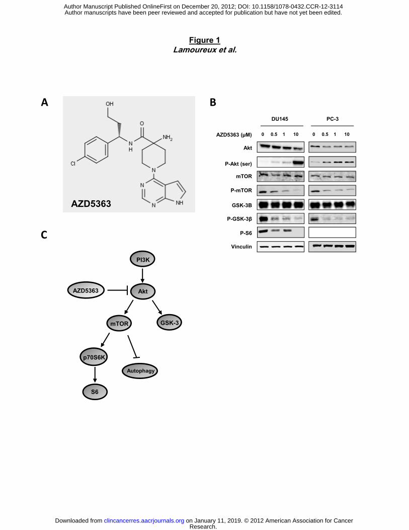
Figure 1Lamoureux et al.
B
0 0.5 1 10 0 0.5 1 10
DU145 PC-3
AZD5363 (µM)
A
P-Akt (ser)
Akt
GSK 3B
P-mTOR
mTOR
AZD5363
C
PI3K
P-S6
Vinculin
GSK-3B
P-GSK-3β
AZD5363
Akt
mTOR GSK-3
AZD5363
mTOR
p70S6K
Autophagy
S6
Research. on January 11, 2019. © 2012 American Association for Cancerclincancerres.aacrjournals.org Downloaded from
Author manuscripts have been peer reviewed and accepted for publication but have not yet been edited. Author Manuscript Published OnlineFirst on December 20, 2012; DOI: 10.1158/1078-0432.CCR-12-3114
A
Figure 2Lamoureux et al.
A
200
300
400 LNCaP
PC-3
DU145
ity (%
of c
ontr
ol)
0 1 2 3 4 50
100
Days
cell
viab
il
AZD5363 (10μM)ti
on
80
100 Ctr
10uM
50uM 80
100 Ctr
10uM
50uM
DU-145 PC-3Ctr AZD5363 (50μM)
PC-3
B
% c
ell p
opul
at
0
20
40
60
sub G1 G0/G1 S G2/M0
20
40
60
sub G1 G0/G1 S G2/MDU
-145
P
subG0/G1 subG0/G1
0 0.5 1 10 50AZ-11 (µM) 0 0.5 1 10 50
DU145 PC-3
0 0.5 1 10 500 0.5 1 10 50
DU145 PC-3
AZ-11 (µM)
C D
0 0.5 1 10 50
casp3
PARP
Vinculin
AZ 11 (µM)
C-casp3
C-PARP
0 0.5 1 10 50
P-CDC2
P-WEE1
P-CDC25C
Vinculin
Research. on January 11, 2019. © 2012 American Association for Cancerclincancerres.aacrjournals.org Downloaded from
Author manuscripts have been peer reviewed and accepted for publication but have not yet been edited. Author Manuscript Published OnlineFirst on December 20, 2012; DOI: 10.1158/1078-0432.CCR-12-3114
A
Figure 3Lamoureux et al.
Control AZD5363 (10µM)-12h Rapamycin (100nM)-12h
8090
%)B C*** *** 1 50 10AZD5363 (µM)
01020304050607080
rl 3 n
Aut
opha
gic
cells
(%
Akt
P-Akt
LC3-IILC3-I
Ctr
AZD5363
Rapamycin
D E
3 60 24 48AZD5363 (hr) Ctrl AZD-5363
βactin
3 60 24 48
P-Akt
LC3-I
Akt
AZD5363 (hr)
LC3-II Akt
1 2 1 2
LC3-ILC3-II
βactin vinculin
Research. on January 11, 2019. © 2012 American Association for Cancerclincancerres.aacrjournals.org Downloaded from
Author manuscripts have been peer reviewed and accepted for publication but have not yet been edited. Author Manuscript Published OnlineFirst on December 20, 2012; DOI: 10.1158/1078-0432.CCR-12-3114
A
** *
Figure 4Lamoureux et al.
B --
+-
-+
++
Chloroquine (20µM)
AZD5363 (10µM)
Akt
LC3-IILC3-I
--
-+
+-
++
Baf A (10nM)AZD5363 (10µM)
Akt
LC3-IILC3-I
βactin
P-S6
P-Akt
cleaved-PARP total-PARP
cleaved-PARP
P-S6
βactin
P-Akt
total-PARP
C
siSc
r1
AZD5363 (10µM)Ctrl
****
*****
+--
+-+
-+-
-++
siATG3 (30nM)AZD5363 (10µM)
siScr1 (30nM)
siATG3
Vinculin
siA
TG3
*
siSc
r1
AZD5363 (10µM)CtrlD
********
+--
+-+
-+-
-++
siATG7 (30nM)AZD5363 (10µM)
siScr1 (30nM)
siATG7
Vinculin
siA
TG7
Research. on January 11, 2019. © 2012 American Association for Cancerclincancerres.aacrjournals.org Downloaded from
Author manuscripts have been peer reviewed and accepted for publication but have not yet been edited. Author Manuscript Published OnlineFirst on December 20, 2012; DOI: 10.1158/1078-0432.CCR-12-3114
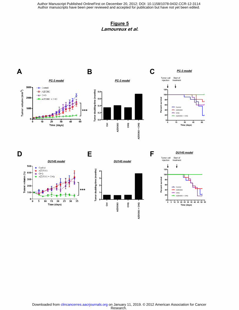
Figure 5Lamoureux et al.
A B CPC-3 model
PC-3 model
PC-3 modelTumor cellinjection
Start oftreatment
***
0 0
0.2
0.4
0.6
0.8
um
or
do
ub
ling
-tim
e (m
on
ths
)
0
20
40
60
80
100
120
Control
AZD5363
CHQ
AZD5363 + CHQ
Per
cen
t su
rviv
al
Ctr
l
AZ
D5
363
CH
Q
AZ
D5
363
+ C
HQ
0.0Tu
D E F DU145 model
0 15 30 45 600
Time (days)
2
3
4
ub
ling
-tim
e (m
on
ths
)
D E FDU145 modelDU145 model
*** 40
60
80
100
120
Control
AZD5363
Tumor cellinjection
Start oftreatment
erce
nt
surv
ival
Ctr
l
AZ
D5
363
CH
Q
AZ
D5
363
+ C
HQ
0
1
Tum
or
do
u***
0 5 10 15 20 25 30 35 40 45 50 550
20
40 AZD5363
CHQ
AZD5363 + CHQ
Time (days)
Pe
Research. on January 11, 2019. © 2012 American Association for Cancerclincancerres.aacrjournals.org Downloaded from
Author manuscripts have been peer reviewed and accepted for publication but have not yet been edited. Author Manuscript Published OnlineFirst on December 20, 2012; DOI: 10.1158/1078-0432.CCR-12-3114
A Ctrl CHQ AZD5363 AZD5363 + CHQ
P-S6
50
100
150
os
itiv
e c
ell
s
***
Figure 6Lamoureux et al.
LC3B
0
50
20
30
40
50
e c
ell
s /
fie
ld%
po
***
****
******
0
10
20
Po
siti
ve
P62 60
80
100
120
ve c
ells
**
**
Ki67 60
80
100
e c
ell
s
0
20
40
60
% p
osi
tiv **
*****Ki67
100
150
0
20
40
% p
osi
tive
s /
fie
ld
******
***
B
TUNEL
0
50
Co
ntr
ol
AZ
D53
63
CH
Q
5363
+ C
HQ
HQ
Po
siti
ve c
ells
***
B
AZ
D5
2
3
4
/ vin
cu
lin (U
A)
3
4
5
I / v
inc
ulin
(UA
)
Akt
Co
ntr
ol
AZ
D5
36
3
CH
Q
AZ
D5
36
3 +
CH
Ctr
l
AZ
D53
63
CH
Q
AZ
D53
63
+ C
HQ
0
1
ratio
p-S
6 /
Ctr
l
AZ
D5
363
CH
Q
AZ
D5
363
+ C
HQ
0
1
2
ratio
LC
3B
Ip-S6
Vinculin
LC3-IILC3-I
Research. on January 11, 2019. © 2012 American Association for Cancerclincancerres.aacrjournals.org Downloaded from
Author manuscripts have been peer reviewed and accepted for publication but have not yet been edited. Author Manuscript Published OnlineFirst on December 20, 2012; DOI: 10.1158/1078-0432.CCR-12-3114
Published OnlineFirst December 20, 2012.Clin Cancer Res Francois Lamoureux, Christian Thomas, Claire Crafter, et al. AZD5363Resistant Prostate Tumor Cells to the Novel Akt Inhibitor, Blocked Autophagy Using Lysosomotropic Agents Sensitizes
Updated version
10.1158/1078-0432.CCR-12-3114doi:
Access the most recent version of this article at:
Material
Supplementary
http://clincancerres.aacrjournals.org/content/suppl/2012/12/20/1078-0432.CCR-12-3114.DC1
Access the most recent supplemental material at:
Manuscript
Authoredited. Author manuscripts have been peer reviewed and accepted for publication but have not yet been
E-mail alerts related to this article or journal.Sign up to receive free email-alerts
Subscriptions
Reprints and
To order reprints of this article or to subscribe to the journal, contact the AACR Publications
Permissions
Rightslink site. Click on "Request Permissions" which will take you to the Copyright Clearance Center's (CCC)
.http://clincancerres.aacrjournals.org/content/early/2012/12/20/1078-0432.CCR-12-3114To request permission to re-use all or part of this article, use this link
Research. on January 11, 2019. © 2012 American Association for Cancerclincancerres.aacrjournals.org Downloaded from
Author manuscripts have been peer reviewed and accepted for publication but have not yet been edited. Author Manuscript Published OnlineFirst on December 20, 2012; DOI: 10.1158/1078-0432.CCR-12-3114