Embed Size (px)
Citation preview
Axial patterning in the developing vertebrate inner ear
TANYA T. WHITFIELD* and KATHERINE L. HAMMOND
Centre for Developmental and Biomedical Genetics, Department of Biomedical Science, University of Sheffield, UK
ABSTRACT Axial patterning in the vertebrate inner ear has been studied for over eighty years,
and recent work has made great progress towards an understanding of the molecular mecha-
nisms responsible for establishing asymmetries about the otic axes. Tissues extrinsic to the ear
provide sources of signalling molecules that are active early in development, at or before otic
placode stages, while intrinsic factors interpret these signals to establish and maintain axial
pattern. Key features of dorsoventral otic patterning in amniote embryos involve Wnt and Fgf
signalling from the hindbrain and Hh signalling from midline tissues (notochord and floorplate).
Mutual antagonism between these pathways and their downstream targets within the otic
epithelium help to refine and maintain dorsoventral axial patterning in the ear. In the zebrafish ear,
the same tissues and signals are implicated, but appear to play a role in anteroposterior, rather
than dorsoventral, otic patterning. Despite this paradox, conservation of mechanisms may be
higher than is at first apparent.
KEY WORDS: axis, inner ear, otic vesicle, otocyst, axial patterning
Introduction: the axes of the inner ear
The vertebrate inner ear mediates the sense of hearing, andit contributes—together with other sensory and motor systems—to an organism’s postural control or balance. To accomplish thelatter, the inner ear must be responsive to movement in threedimensions: hence, in most vertebrates, it displays obvious asym-metries about all three body axes—anteroposterior (AP), dors-oventral (DV) and mediolateral (ML) (Fig. 1A, B). In jawed verte-brates, the inner ear comprises three orthogonally arrangedsemicircular canals (anterior, lateral and posterior), connecting toa medially located crus commune. A series of chambers contain-ing sensory maculae are located ventral to the semicircular canalsystem, while the endolymphatic duct and sac extend from thedorsomedial part of the ear. In amniotes, a specialised hearingorgan, the cochlea, extends ventrally from posterior regions of theear.
The inner ear arises from a thickening of head ectoderm, theotic placode, which invaginates or cavitates to give rise to anepithelial vesicular structure, the otic vesicle or otocyst. Morpho-logical asymmetries first become evident at these early stages(Fig. 1C, D). In the zebrafish, cavitation begins at the anterior poleof the vesicle before the posterior, and soon afterwards, cellmovements result in a thinning of the dorsal otic epithelium and athickening of ventral epithelium (Haddon, 1997; Riley et al.,1997). In the chick, the DV axis is morphologically obvious at oticcup stages, as the developing endolymphatic duct forms an
Int. J. Dev. Biol. 51: 507-520 (2007)doi: 10.1387/ijdb.072380tw
*Address correspondence to: Dr. Tanya T. Whitfield. Centre for Developmental and Biomedical Genetics, Department of Biomedical Science, University ofSheffield, SHEFFIELD S10 2TN, UK. Fax: +44-114-276-5413. e-mail: [email protected]
0214-6282/2007/$30.00© UBC PressPrinted in Spainwww.intjdevbiol.com
Abbreviations used in this paper: AP, anteroposterior; A, anterior; BMP, bonemorphogenetic protein; D, dorsal; DV, dorsoventral; E, embryonic day(mouse stages); FGF, fibroblast growth factor; HH, Hamburger-Hamilton(chick stages); Hh, hedgehog; hpf, hours post fertilization (zebrafish stages);M, medial; ML, mediolateral; L , lateral; P, posterior; r, rhombomere; S,somite (zebrafish stages); V, ventral; Wnt, wingless/int.
outpocketing from the dorsal pole of the invaginating otic cup(Brigande et al., 2000a). Molecular studies, however, reveal thatasymmetric patterns of gene expression about all three axesappear even earlier, prior to any obvious morphological manifes-tation of asymmetry. Gene expression patterns in the otic vesicledo not always correlate precisely with the body axes, which arethus somewhat arbitrary points of reference, but as we shall see,the sources of signalling molecules that pattern the ear are oftenaligned with the body axes. For the purposes of description,therefore, the body axes are useful, and we will use these here.
Although there is a correlation between axial patterning of theotic vesicle and the final structure of the mature labyrinth, it shouldbe remembered that morphogenetic movements, cell migrationand differential rates of cell division and cell death distort therelationship between the two. Perhaps the most obvious exampleis the migration of neuroblasts that give rise to the VIIIth ganglion.In both zebrafish and amniotes, these cells are specified inpredominantly anterior regions of the otic placode and vesicle, butgive rise to neurons that occupy a predominantly ventral positionin the final pattern. Fate mapping and gene expression studies
508 T.T. Whitfield and K.L. Hammond
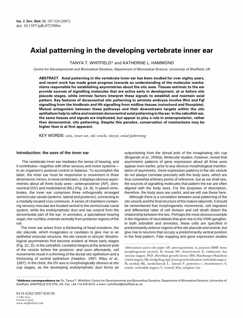
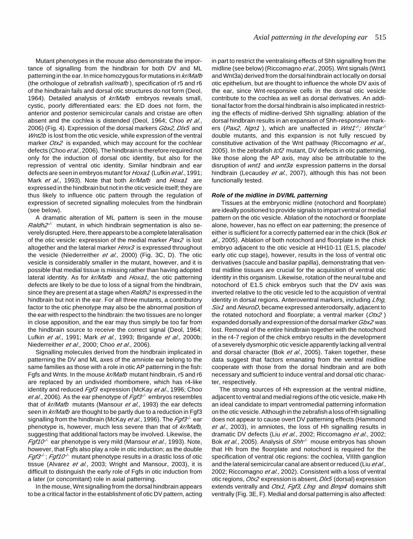
Fig. 1. Axes of the otic vesicle and adult inner ear in zebrafish and
amniotes. (A,B) Diagram of a lateral view of the inner ear of an adultzebrafish (A) and E16 mouse (B), showing asymmetries about all threebody axes. The positions of the maculae (blue) and cristae (red) areindicated. Scale bars, 500 µm. (C,D) Schematic diagram of a lateral viewof the otic vesicle of a zebrafish at 24 hpf (C) and the chick at HH24 (E4)(D). Scale bars, 100 µm. There are already obvious morphologicalasymmetries at these stages: in zebrafish, the dorsal epithelium isthinner than the ventral epithelium; in amniotes, the endolymphatic ductprotrudes from the dorsal side. In zebrafish, hair cells (blue) are differen-tiating in the presumptive utricular and saccular maculae at the anteriorand posterior poles, respectively, of the otic vesicle. In the chick, theexpression of Bmp4 marks the relative positions of all presumptivesensory areas (the mediolateral dimension is not shown) (adapted fromdata in Oh et al., 1996). Although the shape of the otic vesicle is distortedduring morphogenesis, the relative positions of the sensory patchesabout the body axes in the mature ear match those at otic vesicle stages.Abbreviations: bp, basilar papilla; ed, endolymphatic duct; sm, saccularmacula; um, utricular macula.
have been used to trace other cell and tissue movements thatshape the developing ear. In the chick, for example, fate mappingat otic cup stages has shown that there is considerable distortionof different regions of the cup as it invaginates to form a vesicle(Brigande et al., 2000a; Abelló et al., 2007). In the mouse, therelationship between cell position in the otic cup and the finalpattern is also complex: Wnt-responsive cells in the dorsomedialregion of the otic cup contribute to both dorsal and ventral oticderivatives (Riccomagno et al., 2005). In the zebrafish, thereappears to be little change in the relative positions of cells in theotic vesicle between 17 and 48 hours post fertilisation (hpf)(Haddon, 1997), but in the amphibian ear, fate mapping studiesindicate that cell mixing is still prevalent at otic vesicle stages, andso the relative positions of cells in the otic vesicle and structuresof the mature ear may be harder to interpret (Kil and Collazo,2001).
Patterns of gene expression have also been used to trace theorigin of particular structures in the ear, although this technique is
not as reliable as fate mapping, since changes in expressiondomains may reflect dynamic changes in gene transcription inaddition to cell movements. These data should therefore betreated with caution, but they are sometimes useful in indicatingthe relative positions of the anlagen of different structures in theear. For example, in the chick, the fate-mapped proneural regionof the otic cup corresponds well to the anterior domain of Fgf10expression (Abelló et al., 2007), while Bmp expression domainsat otic vesicle stages mark the relative positions of all the futuresensory patches in the ear (Oh et al., 1996; Wu and Oh, 1996)(Fig. 1D). In the mouse, a dorsomedial domain of Wnt2b expres-sion marks the outpocketing of the vesicle that will give rise to thedorsomedial endolymphatic duct (Ozaki et al., 2004). In thezebrafish, the developing utricular and saccular maculae aremarked from early stages in the otic vesicle by the expression ofvarious markers, and the relative positions of these maculaeabout the AP axis are retained in the adult ear (Fig. 1C).
Thus, asymmetries in the final adult structure of the inner earare—to a greater or lesser extent, depending on the species—reflected in early molecular and morphological asymmetries aboutthe axes of the otic vesicle. The purpose of this review is toexamine what is known about the mechanisms that establish,interpret and maintain this axial asymmetry in the developing ear.
Equipotentiality about the AP axis of the otic placodeand vesicle
One question concerning the development of any asymmetricorgan is whether there is initially a symmetric stage on whichpattern is imposed by a later symmetry-breaking event. Althoughit is not certain whether the otic vesicle is ever truly symmetricalat early stages, the A and P poles of the presumptive oticectoderm were recognised to be equipotential as early as the1930s. Rotation of presumptive ear ectoderm in amphibian (sala-mander) embryos at preplacodal stages (around the time of APaxis fixation) yielded mirror image twinned ears with either doubleA or double P character (Harrison, 1936; Hall, 1939; Harrison,1945; Yntema, 1955) (Fig. 2A-C). Similarly, manipulation of theHedgehog (Hh) signalling pathway in the fish and amphibianembryo can also result in double anterior or double posterior ears(Hammond et al., 2003; Waldman et al., 2007) (Fig. 2D-I; seefurther discussion in the section on AP patterning below). Theseenantiomorphic phenotypes demonstrate that the A and P polesof the ear initially have the potential to form either A or Pstructures; they are reminiscent of other classical developmentalaxial duplications, such as those of the chick limb followingtransplantation of the zone of polarising activity, or Drosophilasegment polarity phenotypes. Double anterior or posterior earscan also result during regeneration of otic tissue after partialablation of the placode in Xenopus : these phenotypes arestrikingly similar to those generated by the salamander rotationexperiments or by manipulations of Hh signalling in zebrafish(Waldman et al., 2007) (Fig. 2J-O).
The existence of twinned ear phenotypes also suggests thatthe poles of the otic placode may be specified independently of amechanism to assign A or P identity to either. In the zebrafish, theA and P poles of the otic placode appear to arise by the divisionof a single symmetrical prosensory domain into two. The proneuralgene atoh1b is initially expressed in a single domain (the sensory
Utricularmacula
Semicircularcanals
Lagenarmacula
Saccularmacula
Zebrafish adult ear Mouse ear, E16
D
A
V
P
M
L
D
A
V
P
M
L
um smbp
Zebrafish otic vesicle, 24hpf Chick otocyst, HH24
D
A
V
P
D
A
V
Pum
ed
Semicircularcanals
Utricularmacula
Cochlea
Saccularmacula
Endolymphaticsac
sm
B
C D
A
Axial patterning in the developing ear 509
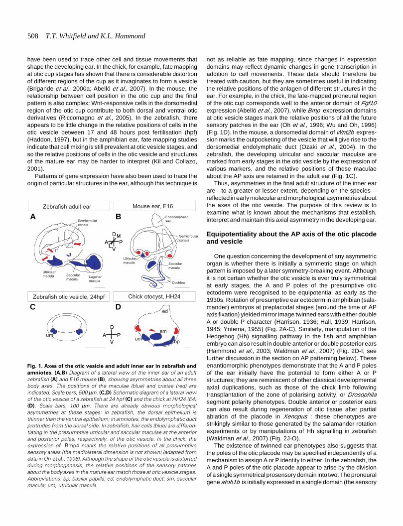
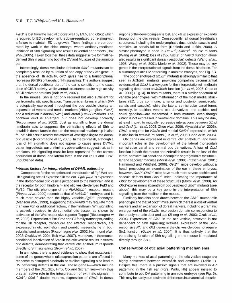
Fig. 2. Examples of axial duplications in the ears of amphibian and zebrafish embryos. (A-C)
Diagrams of ear phenotypes obtained by rotations of the otic rudiment about the AP axis in thesalamander (reproduced, with permission, from Harrison, 1945). (A) Wild-type ear pattern; (B)
double anterior ear; (C) double posterior ear. The utricular and saccular maculae (blue) and cristae(red) are highlighted for clarity. (D-I) Confocal images of ear phenotypes following manipulation ofHh signalling in the zebrafish embryo. Ears are stained with FITC-phalloidin to mark the hair bundlesof sensory hair cells in the maculae (D-F, I) or cristae (G, H). (D,G) Wild-type pattern (86 hpf); (E,H)
double anterior pattern (86 hpf) obtained by incubation of the embryo from the 10 somite stage to22 hpf in 50 µM cyclopamine to inhibit Hh signalling; (F,I) two examples of a double posteriorpattern (mirror-image saccular macula) obtained by injection of shha mRNA into the embryo at the1-cell stage (72 hpf) (reproduced with permission of the Company of Biologists from Hammond etal., 2003). (J-O) Ear phenotypes at tadpole stage (stage 48) obtained by partial otic placode or oticvesicle ablations in the Xenopus embryo at stages 24-27 (reprinted with permission of Wiley-Liss,Inc., a subsidiary of John Wiley & Sons, Inc., from Waldman et al., 2007). (J,M) Wild-type pattern;(K,N) double anterior ear obtained by ablation of the posterior half of the otic placode; (L,O) doubleposterior ear obtained by ablation of the anterior half of the otic placode. In all double anterior ears,the utricular macula is duplicated, and four cristae are present. In the double posterior ears, theutricular macula is missing, and the number of cristae is reduced. In all panels, the anterior of theembryo is to the left; (A-I) are lateral views, (J-O) are dorsal views. Abbreviations: sm, saccularmacula; so, saccular otolith; um, utricular macula; uo, utricular otolith. Asterisks indicate theposition of cristae. Scale bars: (D-I) 25 µm; (J-O) 100 µm.
G
O
B C
D E F
H I
J K L
M N
A
equivalence group) across the entireAP axis of the otic placode, but this islater segregated into two domains, oneat the anterior and one at the posteriorof the placode, which prefigure theappearance of hair cell foci (Millimakiet al., 2007). Refinement of the ex-pression domain is dependent onNotch signalling (Millimaki et al., 2007)and may also require an inhibitory sig-nal from rhombomere 5 (r5), prevent-ing hair cell differentiation in the middleof the placode (Lecaudey et al., 2007).The two foci of hair cells that arise atthe A and P poles of the ear (theprecursors of the utricular and saccu-lar maculae, respectively) initially ap-pear relatively symmetric, and theycomprise the same differentiated celltypes (hair cells and supporting cells),but the shapes, positions, sizes andpolarity patterns of the two maculaewill be very different in the mature earas they develop according to their A orP position.
Timing of axis formation in thedeveloping ear
Asymmetric patterns of gene ex-pression arise early in the otic devel-opmental programme, during or evenbefore placode stages. In zebrafish,although otic asymmetry is not mor-phologically obvious until vesiclestages, asymmetric gene expressionis apparent in the otic placode, con-comitant with or just after the resolu-tion of the prosensory domain into twoseparate domains at the 14 hpf (10somite) stage. For example, at 14 hpf,deltaA, B and D— although expressedsymmetrically about the AP axis—areexpressed on the medial, but not thelateral, side of the placode at the A andP poles (Haddon et al., 1998). By the16 hpf (14 somite) stage, hmx3 isexpressed strongly at the anterior endof the placode (Adamska et al., 2000).Similarly, in mice, Hmx3 is expressedin the anterior part of the placode, andWnt6 is detected dorsally, at E8.5(Hadrys et al., 1998; Lilleväli et al.,2006). For further examples, see Table1. These data suggest that mecha-nisms for the specification of axialasymmetry in the ear must be active ator before placode stages.
510 T.T. Whitfield and K.L. Hammond
Determination or fixation of the otic axes, however, seems tooccur some time after signs of otic asymmetry are first apparent.Evidence for this comes from extirpation and grafting experimentsperformed in the chick (Wu et al., 1998; Bok et al., 2005). In thisspecies, signs of otic AP asymmetry are visible at E1.5 (Hh10,placode stage), when, for example, Hmx3 is expressed at theanterior end of the placode. Rotation experiments have indicatedthat the otic AP axis does not become fixed, however, until aroundE2.0 (Hh12, otic cup stage). Similarly, the DV axis does not becomefixed until after E3.5, although several genes (Gbx2, Otx2 ) areexpressed asymmetrically about the DV axis from as early as E2.5(Hh14) (see Table 1). The ML axis is fixed for some characters butnot others at E2.5.
Sequential fixation of the otic AP axis followed by the DV axisappears to be a general phenomenon, although the exact timingdiffers for different species. In the salamander, for example, the
AP axis becomes fixed much earlier than in the chick, just as theneural folds are closing and before appearance of the otic pla-code; the DV axis becomes fixed somewhat later, when the oticvesicle is about to close (Harrison, 1936; Hall, 1939; Harrison,1945; Yntema, 1955).
Factors required for axis formation: extrinsic and in-trinsic signalling
The rotation experiments described above suggest that factorsboth extrinsic and intrinsic to the ear are required for specificationand fixation (determination) of the otic axes. At early stages,positioning with respect to surrounding tissues can influence axisformation, while after fixation, rotating the ear cannot overridepatterning, suggesting that factors intrinsic to the ear act tomaintain axial patterning information, and that the ear is either no
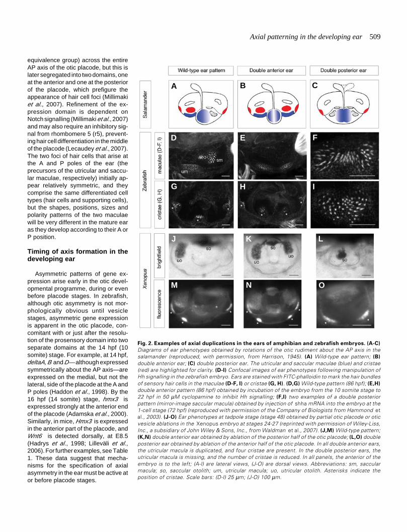
Gene Organism Expression pattern within the ear Timing of expression in the ear References
Anteroposterior
Fgf3 Mouse AV E10 (Alvarez et al., 2003; Raft et al., 2004)fgf3 Zebrafish AV 22hpf (Millimaki et al., 2007)fgf8 Zebrafish AV (strong), PV (weak) / AV only 18hpf / 24hpf (Léger and Brand, 2002)Fgf10 Chick A, later AV HH10-19 (Alsina et al., 2004)fst Zebrafish P 24hpf+ (Mowbray et al., 2001)Lfng Chick AV HH14 (Bok et al., 2005) and references withinLfng Mouse A E9.5+ (Raft et al., 2004)mfng Zebrafish AV 20S (Qiu et al., 2004)Otx1 Mouse PV and L / VL E10.25 / E11.5 (Morsli et al., 1999)otx1 Zebrafish VL 24hpf (Kwak et al., 2002)pax5 Zebrafish A (strong), P (very weak) 17hpf + (Kwak et al., 2006; Pfeffer et al., 1998)zp23 Zebrafish PM 24hpf (Kwak et al., 2002; Kwak et al., 2006)
Anteroposterior and Mediolateral
Hmx3 Chick AM initially / later L HH10+ / HH14+ (Herbrand et al., 1998)Hmx3 Mouse AM initially then becomes L E8.5 (Hadrys et al., 1998)hmx3 Zebrafish AM initially / later L 16hpf(14S+) / late vesicle stages (Adamska et al., 2000)Hmx2 Mouse Same as Hmx3 but later onset E13.5+ (Hadrys et al., 1998)GH6 Chick PL / broad L domain HH9+ / HH16 (Kiernan et al., 1997)SoHo-1 Chick As for GH6 As for GH6 (Kiernan et al., 1997)
Mediolateral
Pax2 Chick All otic epithelium - stronger M HH14 (Hidalgo-Sanchez et al., 2000)pax2a Zebrafish Initially throughout / later M 3S / 24hpf (Pfeffer et al., 1998; Riley et al., 1999)pax2b Zebrafish Initially throughout; later M ~3hr later than pax2a (Pfeffer et al., 1998)Tbx1 Mouse P / PL / ADL and PVM Placode+ / E9.5 / E10+ (Raft et al., 2004)tbx1 Zebrafish VL 24hpf (Piotrowski et al., 2003)
Dorsoventral
Dlx3 Chick Initially entire placode / later DM HH10 / HH12+ (Pera and Kessel, 1999)dlx3b Zebrafish D 24hpf (Ellies et al., 1997)Dlx5 Chick DM Similar to Dlx3 once otic pit forms (Pera and Kessel, 1999)Dlx5 Mouse Initially entire placode / restricts to D E8.0-8.5 / E10.5+ (Merlo et al., 2002) and refs withinDrapC1 Mouse Initially entire placode then DM otic cup E9.0+ (Lilleväli et al., 2006)FgfR2(III)b Mouse D E9 (Pirvola et al., 2000)Gbx2 Chick Initially entire placode / Restricts to DM HH10 / HH14 (Lin et al., 2005)Gbx2 Mouse Initially entire placode / Restricts to DM E8.5 / E9.5 (Hidalgo-Sanchez et al., 2000;
Miyazaki et al., 2006;Sánchez-Calderón et al., 2002)
gbx2 Zebrafish DM 24hpf + (Su and Meng, 2002)Otx2 Chick VM otic vesicle HH14+ (Hidalgo-Sanchez et al., 2000;
Miyazaki et al., 2006;Sánchez-Calderón et al., 2002)
Otx2 Mouse V tip of otocyst E10.25 (Morsli et al., 1999)Wnt2b Mouse D rim / pole of otocyst E9.5+ (Lin et al., 2005) and references withinWnt6 Mouse D E8.5+ (Lilleväli et al., 2006)
GENES EXPRESSED ASYMMETRICALLY ABOUT THE AXES OF THE OTIC PLACODE AND OTIC VESICLEIN ZEBRAFISH, CHICK AND MOUSE
Where complete expression data are not available, we have indicated the time at which there is obvious asymmetric expression in the ear. Abbreviations: A, anterior; M, medial; L, lateral; P, posterior;E, embryonic day (mouse); HH, Hamburger-Hamilton stage (chick); hpf, hours post fertilisation (zebrafish); S, somite stage (zebrafish).
TABLE 1
Axial patterning in the developing ear 511
longer responsive to external cues, or that these have ceased.Tissues in the vicinity of the otic ectoderm that may signal to itinclude the neural tube, notochord, overlying ectoderm, pharyn-geal endoderm, migratory neural crest streams and other perioticmesenchyme. Of these, the dorsal neural tube and midlinetissues (notochord and floorplate) have been the most exten-sively studied; the experiments described in the next sectionsillustrate their importance as sources of extrinsic patterning infor-mation. In turn, extrinsic signals are interpreted, maintained andpropagated by intrinsic factors expressed within the otic epithe-lium itself.
Candidates for extrinsic signalling factors from the dorsalneural tube, floorplate and notochord include those of the BMP,Fgf, Hh and Wnt families; note that these are, with the possibleexception of Hh for some species, also expressed within the oticepithelium, where they may act as intrinsic factors to maintain andrefine axial patterning. Many factors, both extrinsic and intrinsic tothe ear, are involved in specification of more than one axis, andeach factor is unlikely to act independently; several studiesindicate that there is interaction and cross-talk between differentsignalling pathways to pattern the ear. In the sections below, wediscuss the evidence for the role of these signalling pathways inpatterning each of the otic axes. We cover the AP axis separately,but treat the DV and ML axes together, since these are rarelyaffected independently.
Anteroposterior patterning
Role of the hindbrain in otic AP patterning The otic placode develops closely juxtaposed to the hindbrain
rhombomeres (r), each of which expresses a unique combinationof genes at the time of otic axial specification; it is thus anattractive idea that rhombomeres confer AP identity on adjacentotic tissue. In zebrafish, for example, the otic placode initiallyarises adjacent to r4, but later becomes positioned adjacent to r5,with its anterior and posterior poles (and sites of initial hair cellformation) opposite r4 and r6, respectively (Kimmel et al., 1995;Riley et al., 1997; Ernest et al., 2000). In the chick, the otic vesiclelies adjacent to r5 and r6; here, the r5/6 boundary aligns with anotic AP compartment boundary, suggesting that r5- and r6-derived factors may confer A and P identity, respectively, on theotic vesicle (Brigande et al., 2000b) (see Fig. 5).
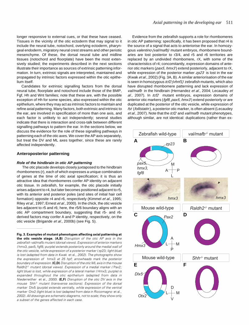
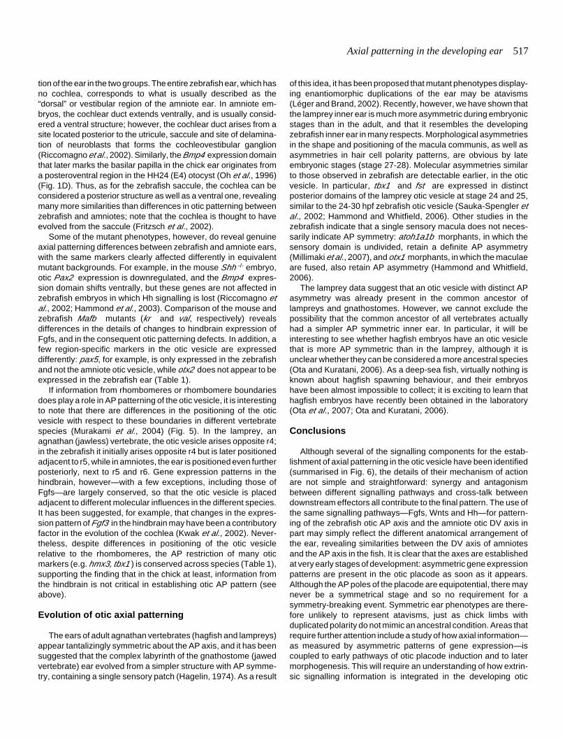
Fig. 3. Examples of mutant phenotypes affecting axial patterning at
the otic vesicle stage. (A,B) Disruption of the otic AP axis in thezebrafish val/mafb mutant (dorsal views). Expression of anterior markers( hmx3, pax5, fgf8; purple) extends posteriorly around the medial wall ofthe otic vesicle, while expression of a posterior marker ( zp23; light blue)is lost (adapted from data in Kwak et al., 2002). The photographs showthe expression of hmx3 at 25 hpf; arrowheads mark the posteriorboundary of expression. (C,D) Disruption of the otic ML axis in the mouseRaldh2-/- mutant (dorsal views). Expression of a medial marker ( Pax2;light blue) is lost, while expression of a lateral marker ( Hmx3; purple) isexpanded throughout the otic epithelium (adapted from data inNiederreither et al., 2000). (E,F) Disruption of the otic DV axis in themouse Shh-/- mutant (transverse sections). Expression of the dorsalmarker Dlx5 (purple) extends ventrally, while expression of the ventralmarker Otx2 (light blue) is lost (adapted from data in Riccomagno et al.,2002). All drawings are schematic diagrams, not to scale; they show onlya subset of the genes affected in each case.
Mouse wild-type Raldh2-/- mutant
Hmx3
Pax2
A
L M
P
val/mafb-/- mutantZebrafish wild-type
M
A P
L
zp23
pax5,
hmx3,
fgf8
hmx3 hmx3
Shh-/- mutantMouse wild-type
Dlx5
Otx2
D
L M
V
B
C D
E F
A
Evidence from the zebrafish supports a role for rhombomeresin otic AP patterning: specifically, it has been proposed that r4 isthe source of a signal that acts to anteriorise the ear. In homozy-gous valentino (val/mafb) mutant embryos, rhombomere bound-aries are lost posterior to r3/4, and r5 and r6 territories arereplaced by an undivided rhombomere, rX, with some of thecharacteristics of r4; concomitantly, expression domains of ante-rior otic markers (pax5, hmx3 ) extend posteriorly, adjacent to rX,while expression of the posterior marker zp23 is lost in the ear(Kwak et al., 2002) (Fig. 3A, B). A similar anteriorisation of the earis seen in homozygous tcf2 (vhnf1) zebrafish mutants, which alsohave disrupted rhombomere patterning and lack expression ofval/mafb in the hindbrain (Hernandez et al., 2004; Lecaudey etal., 2007). In tcf2 mutant embryos, expression domains ofanterior otic markers (fgf8, pax5, hmx3 ) extend posteriorly or areduplicated at the posterior of the otic vesicle, while expression offst (follistatin ), a posterior otic marker, is often absent (Lecaudeyet al., 2007). Note that the tcf2 and val/mafb mutant phenotypes,although similar, are not identical: duplications (rather than ex-
512 T.T. Whitfield and K.L. Hammond
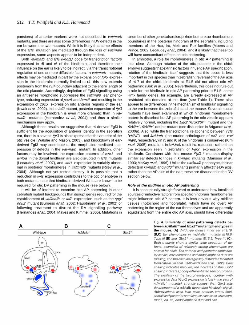
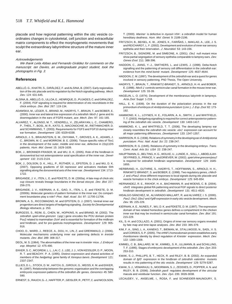
Fig. 4. Similarity of axial patterning defects be-
tween kr/Mafb-/- and Gbx2-/- mutant phenotypes in
the mouse. (A) Wild-type mouse inner ear at E16.(B,C) Ear phenotypes in kr/Mafb-/- mutants (E15.5,Type II) (B) and Gbx2-/- mutants (E15.5, Type III) (C).Both mutants show a similar wide spectrum of de-fects; examples of relatively strong phenotypes areshown for each. The anterior and posterior semicircu-lar canals, crus commune and endolymphatic duct aremissing, and the cochlea is grossly distended (adaptedfrom data in Lin et al., 2005 and Choo et al., 2006). Blueshading indicates maculae; red indicates cristae. Lightshading indicates poorly differentiated sensory organs.The similarity of the two phenotypes, together withexpression data ( Gbx2 expression is lost in the ears ofkr/Mafb-/- mutants), strongly suggest that Gbx2 actsdownstream of a kr/Mafb-dependent hindbrain signal.Abbreviations: ascc, lscc, pscc, anterior, lateral (hori-zontal) and posterior semicircular canals; cc, crus com-mune; ed, es, endolymphatic duct and sac.
pansions) of anterior markers were not described in val/mafbmutants, and there are also some differences in DV defects in theear between the two mutants. While it is likely that some effectsof the tcf2 mutation are mediated through the loss of val/mafbexpression, some aspects appear to be independent.
Both val/mafb and tcf2 (vhnf1) code for transcription factorsexpressed in r5 and r6 of the hindbrain, and therefore theirinfluence on the ear is likely to be indirect, via the transcriptionalregulation of one or more diffusible factors. In val/mafb mutants,effects may be mediated in part by the expansion of fgf3 expres-sion in the hindbrain: normally limited to r4, this now extendsposteriorly from the r3/4 boundary adjacent to the entire length ofthe otic placode. Accordingly, depletion of Fgf3 signalling usingan antisense morpholino suppresses the val/mafb ear pheno-type, reducing expression of pax5 and hmx3 and resulting in theexpansion of zp23 expression into anterior regions of the ear(Kwak et al., 2002). In the tcf2 mutant, posterior expansion of fgf3expression in the hindbrain is even more dramatic than in val/mafb mutants (Hernandez et al., 2004) and thus a similarmechanism may apply.
Although these results are suggestive that r4-derived Fgf3 issufficient for the acquisition of anterior identity in the zebrafishear, there is a caveat: fgf3 is also expressed at the anterior of theotic vesicle (Walshe and Mason, 2003), and knockdown of ear-derived Fgf3 may contribute to the morpholino-mediated sup-pression of defects in the val/mafb mutant. In addition, otherfactors may be involved: the expression patterns of wnt1 andwnt3a in the dorsal hindbrain are also disrupted in tcf2 mutants(Lecaudey et al., 2007), and wnt1 expression is variably abnor-mal in posterior rhombomeres in val/mafb mutants (Riley et al.,2004). Although not yet tested directly, it is possible that areduction in wnt expression contributes to the otic phenotype inboth mutants; note that hindbrain-derived Wnts are known to berequired for otic DV patterning in the mouse (see below).
It will be of interest to examine otic AP patterning in otherzebrafish mutant backgrounds that disrupt genes required for theestablishment of val/mafb or tcf2 expression, such as the spg/pou2 mutant (Burgess et al., 2002; Hauptmann et al., 2002) orfollowing treatment to disrupt the RA signalling pathway(Hernandez et al., 2004; Maves and Kimmel, 2005). Mutations in
a number of other genes also disrupt rhombomeres or rhombomereboundaries in the posterior hindbrain of the zebrafish, includingmembers of the Hox, Irx, Meis and Pbx families (Moens andPrince, 2002; Lecaudey et al., 2004), and it is likely that these toowill have concomitant effects on otic patterning.
In amniotes, a role for rhombomeres in otic AP patterning isless clear. Although rotation of the otic placode in the chickembryo suggests that extrinsic factors influence AP specification,rotation of the hindbrain itself suggests that this tissue is lessimportant in this species than in zebrafish: reversal of the AP axisof r4-7 of the chick hindbrain at E1.5 did not affect otic APpatterning (Bok et al., 2005). Nevertheless, this does not rule outa role for the hindbrain in otic AP patterning prior to E1.5; someHmx family genes, for example, are already expressed in APrestricted otic domains at this time (see Table 1). There alsoappear to be differences in the mechanism of hindbrain signallingto the ear between the zebrafish and the mouse. Several mousemutants have been examined in which hindbrain rhombomerepattern is disturbed but AP patterning in the otic vesicle appearsrelatively normal, including the Egr2 (Krox20)-/- mutant and theRARa-/-; RARb-/- double mutant (see discussion in Brigande et al.,2000a). Also, while the transcriptional relationship between Tcf2/vHNF1 and kr/Mafb (the murine orthologues of tcf2 and val/mafb, respectively) in r5 and r6 of the hindbrain is conserved (Kimet al., 2005), mutations in kr/Mafb result in a reduction, rather thanthe expansion seen in zebrafish, of Fgf3 expression in thehindbrain. Consistent with this, mouse Fgf3 -/- mutants displaysimilar ear defects to those in kr/Mafb mutants (Mansour et al.,1993; McKay et al., 1996). Unlike the val/mafb phenotype, the eardefects in kr/Mafb and Fgf3 -/- mutants primarily affect the DV axis,rather than the AP axis of the ear; these are discussed in the DVsection below.
Role of the midline in otic AP patterning It is conceptually straightforward to understand how localised
sources of inductive factors from specific hindbrain rhombomeresmight influence otic AP pattern. It is less obvious why midlinetissues (notochord and floorplate), which have no overt APpatterning in the region of the ear themselves and are apparentlyequidistant from the entire otic AP axis, should have differential
B CA
Axial patterning in the developing ear 513
effects on the A and P poles of the otic vesicle.Nevertheless, data from our lab suggest thatHedgehog (Hh) signalling from the notochord andfloorplate plays a role in the development of pos-terior otic structures in the zebrafish (Hammond etal., 2003). Hh genes are strongly expressed in boththe floorplate and notochord of the zebrafish em-bryo, while genes whose products are required fortransduction of the Hh signal (ptc, smo and gli ), areexpressed in the otic epithelium. Moreover, ptc1—a target of Hh signalling—is expressed in a Hh-dependent manner in the ear, suggesting that theeffect of Hh on the ear is direct. When Hh signallingis absent or severely reduced, posterior otic struc-tures fail to form and the ear displays a mirrorimage duplication of anterior regions, similar to theenantiomorphic ears observed by Harrison (Fig.2). A recent study has reported a very similarfinding in Xenopus : here, injection of mRNAcoding for the Hh inhibitor Hip results in mirrorimage duplications of anterior inner ear structures(Waldman et al., 2007). When the Hh pathway isactivated ectopically in the zebrafish, by injectionof shh or dominant negative PKA RNA, we see thereverse phenotype: ears lose anterior otic struc-tures and show a mirror image duplication ofposterior regions (Hammond et al., 2003) (Fig. 2).
posterior otic regions (Hammond et al., 2003). It is not clear,however, whether this corresponds to the time at which Hh isrequired for otic patterning. Alternative explanations (yet to betested) include the possibility that cells specified by Hh signallingmigrate to posterior regions of the otic vesicle as the ear develops.It is also possible that Hh antagonises or synergises with othermore localised AP determination factors, such as Fgf3 from r4, tomaintain otic AP pattern. For a summary of otic AP patterning inthe zebrafish, see Fig. 6A.
Intrinsic factors for interpretation of AP patterning informa-tion
As described in the previous sections, changes in extrinsicsignalling disrupt patterning in the otic vesicle, as measured bychanges both to morphological pattern and to gene expressionpatterns within the otic epithelium. If these latter genes are notmerely markers of position in the otic vesicle, but play an activerole in interpreting extrinsic signals, we would expect their loss tohave similar effects on AP patterning in the ear. Where mutationor knockdown of an intrinsic otic gene results in a phenocopy ofall or a subset of the defects seen for disruption of an extrinsicfactor, we can conclude that this gene is likely to be involved in thereception or interpretation of that signal.
At present, there are rather few examples where the loss offunction of genes intrinsic to otic epithelium yield AP patterningdefects, and none are known that precisely phenocopy thedefects seen in the zebrafish hindbrain or midline mutants. In thezebrafish, for example, a reduction in the expression or functionof pax5 (an anterior marker, expanded in the anteriorised ears ofval/mafb and tcf2 mutants) results in the loss of some anterior(utricular) hair cells, but no disruption of overall AP patterning(Kwak et al., 2006). This suggests that Pax5 plays a role in
r3
r4
r5
r6
r7
Lamprey AmnioteZebrafish
Krox20 Egr2
(Krox20)
egr2b
(krox20)
tcf2
(vhnf1)
Tcf2
(vHNF1)
val
(mafb)
kr
(Mafb)
fgf3
wnt1 Wnt1
Wnt3a
wnt3a
Fgf3
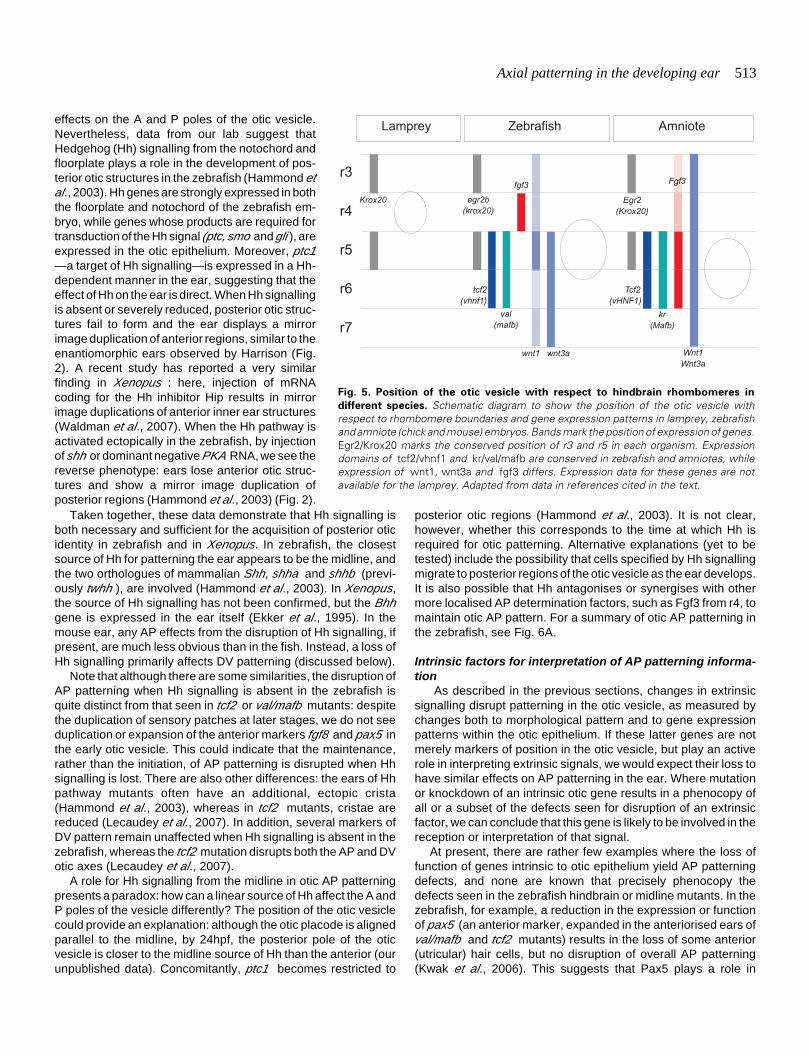
Fig. 5. Position of the otic vesicle with respect to hindbrain rhombomeres in
different species. Schematic diagram to show the position of the otic vesicle withrespect to rhombomere boundaries and gene expression patterns in lamprey, zebrafishand amniote (chick and mouse) embryos. Bands mark the position of expression of genes.Egr2/Krox20 marks the conserved position of r3 and r5 in each organism. Expressiondomains of tcf2/vhnf1 and kr/val/mafb are conserved in zebrafish and amniotes, whileexpression of wnt1, wnt3a and fgf3 differs. Expression data for these genes are notavailable for the lamprey. Adapted from data in references cited in the text.
Taken together, these data demonstrate that Hh signalling isboth necessary and sufficient for the acquisition of posterior oticidentity in zebrafish and in Xenopus. In zebrafish, the closestsource of Hh for patterning the ear appears to be the midline, andthe two orthologues of mammalian Shh, shha and shhb (previ-ously twhh ), are involved (Hammond et al., 2003). In Xenopus,the source of Hh signalling has not been confirmed, but the Bhhgene is expressed in the ear itself (Ekker et al., 1995). In themouse ear, any AP effects from the disruption of Hh signalling, ifpresent, are much less obvious than in the fish. Instead, a loss ofHh signalling primarily affects DV patterning (discussed below).
Note that although there are some similarities, the disruption ofAP patterning when Hh signalling is absent in the zebrafish isquite distinct from that seen in tcf2 or val/mafb mutants: despitethe duplication of sensory patches at later stages, we do not seeduplication or expansion of the anterior markers fgf8 and pax5 inthe early otic vesicle. This could indicate that the maintenance,rather than the initiation, of AP patterning is disrupted when Hhsignalling is lost. There are also other differences: the ears of Hhpathway mutants often have an additional, ectopic crista(Hammond et al., 2003), whereas in tcf2 mutants, cristae arereduced (Lecaudey et al., 2007). In addition, several markers ofDV pattern remain unaffected when Hh signalling is absent in thezebrafish, whereas the tcf2 mutation disrupts both the AP and DVotic axes (Lecaudey et al., 2007).
A role for Hh signalling from the midline in otic AP patterningpresents a paradox: how can a linear source of Hh affect the A andP poles of the vesicle differently? The position of the otic vesiclecould provide an explanation: although the otic placode is alignedparallel to the midline, by 24hpf, the posterior pole of the oticvesicle is closer to the midline source of Hh than the anterior (ourunpublished data). Concomitantly, ptc1 becomes restricted to
514 T.T. Whitfield and K.L. Hammond
anterior development in the ear, and specifically in the productionor survival of hair cells in the anterior macula.
In the mouse, Tbx1 has been proposed as a determinant for APpatterning within the otic vesicle (Raft et al., 2004), but it is notclear which extrinsic signals regulate its expression in the oticepithelium; expression here is not dependent on Hh signalling, forexample (Riccomagno et al., 2002). In Tbx1 -/- mouse embryos,expression of some anterior otic markers (Ngn1, NeuroD, Lfng,Fgf3 ) extends posteriorly, expression of Otx1 (aposteroventrolateral marker) is lost, and the rudiment of the VIIIthganglion, which normally occupies an anteromedial position be-neath the otic vesicle, is duplicated at the posterior (Raft et al.,2004; Arnold et al., 2006). An alternative interpretation, however,is provided by Xu et al. (2007), who claim that axial patterningdefects are due to a loss of tissue rather than a true anteriorisationof the otocyst. Although it is expressed in both the otic epitheliumand surrounding tissues, Tbx1 certainly qualifies as an intrinsicfactor for otic patterning: the requirement for Tbx1 function in theotic epithelium has been elegantly demonstrated using tissue-specific conditional knockout approaches (Arnold et al., 2006; Xuet al., 2007).
An AP organiser for the otic vesicle? The existence of phenotypes involving duplication of the otic
AP axis and the presence of localised sources of diffusiblesignalling molecules—both in the vicinity of the ear and within theotic epithelium—is reminiscent of other organ systems patternedby an organiser, and it has been proposed that an organiser mayalso exist to pattern the AP axis of the ear (Léger and Brand,2002). Although an organiser for the otic AP axis has not beendefinitively identified, this is a credible idea. Classically, anorganiser is defined operationally; it is often the source of amorphogen that establishes pattern in a concentration-depen-
dent manner. For the ear, this source of patterning informationmay either be outside the otic region (such as r4, or rhombomereboundaries) or may be within the otic epithelium itself, such as theexpression domains of fgf3 and fgf8 at the anterior, or fst at theposterior, of the zebrafish otic vesicle. Hh clearly acts as amorphogen in other systems in which there are mirror imagephenotypes (see discussion in Hammond et al., 2003), but for theear, since there is no obvious localised source of Hh or clearlylocalised response to its activity, it is not clear whether Hh is actingin an organising capacity, or is perhaps required for the establish-ment or maintenance of such an organiser. Further experimentsinvolving targeted cell ablations, or the conditional misexpressionor knockdown of genes in specific areas of otic epithelium, will berequired to identify any tissue in the ear that may have organisingproperties and to determine which of the signals are instructiveand which permissive for the establishment of otic AP patterning.
Dorsoventral and mediolateral patterning
Role of the hindbrain in DV/ML patterning Rhombomeres in the hindbrain display distinct differences in
gene expression along the DV axis and so, in addition to their rolein otic AP patterning, may also provide cues to direct DV patternin the adjacent otic placode or vesicle. Harrison provided earlyevidence that the hindbrain is required for the acquisition of dorsalotic identity. Specifically, grafting a second hindbrain ventral tothe otic placode in amphibian embryos induced the formation ofa second endolymphatic duct (ED)—normally a dorsomedialstructure—from the ventral part of the ear (Harrison, 1945). In thechick, ablation or DV rotation of r4-r7 of the neural tube indicatesthat signals from the dorsal neural tube are both necessary andsufficient for the acquisition of dorsal otic fates in this species (Boket al., 2005).
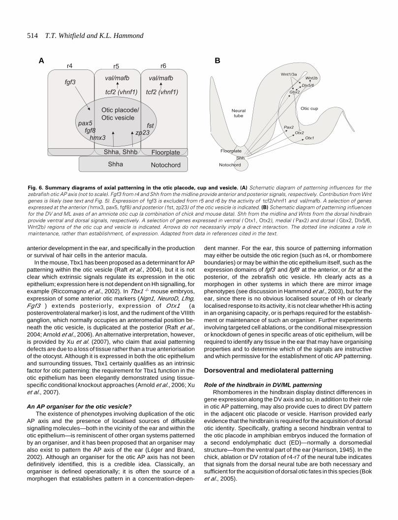
Fig. 6. Summary diagrams of axial patterning in the otic placode, cup and vesicle. (A) Schematic diagram of patterning influences for thezebrafish otic AP axis (not to scale). Fgf3 from r4 and Shh from the midline provide anterior and posterior signals, respectively. Contribution from Wntgenes is likely (see text and Fig. 5). Expression of fgf3 is excluded from r5 and r6 by the activity of tcf2/vhnf1 and val/mafb. A selection of genesexpressed at the anterior ( hmx3, pax5, fgf8) and posterior ( fst, zp23) of the otic vesicle is indicated. (B) Schematic diagram of patterning influencesfor the DV and ML axes of an amniote otic cup (a combination of chick and mouse data). Shh from the midline and Wnts from the dorsal hindbrainprovide ventral and dorsal signals, respectively. A selection of genes expressed in ventral ( Otx1, Otx2), medial ( Pax2) and dorsal ( Gbx2, Dlx5/6,Wnt2b) regions of the otic cup and vesicle is indicated. Arrows do not necessarily imply a direct interaction. The dotted line indicates a role inmaintenance, rather than establishment, of expression. Adapted from data in references cited in the text.
fgf3
r4 r6r5
Shha, Shhb
Shha
val/mafb
tcf2 (vhnf1)
Otic placode/
Otic vesicle
Notochord
Floorplate
hmx3
pax5fgf8
tcf2 (vhnf1)
val/mafb
fstzp23
Wnt1/3a
Shh
Otx2
Dlx5/6
Neural
tube
Otic cup
Notochord
Wnt2b
Gbx2
Pax2
Otx1
Floorplate
BA
Axial patterning in the developing ear 515
Mutant phenotypes in the mouse also demonstrate the impor-tance of signalling from the hindbrain for both DV and MLpatterning in the ear. In mice homozygous for mutations in kr/Mafb(the orthologue of zebrafish val/mafb ), specification of r5 and r6of the hindbrain fails and dorsal otic structures do not form (Deol,1964). Detailed analysis of kr/Mafb embryos reveals small,cystic, poorly differentiated ears: the ED does not form, theanterior and posterior semicircular canals and cristae are oftenabsent and the cochlea is distended (Deol, 1964; Choo et al.,2006) (Fig. 4). Expression of the dorsal markers Gbx2, Dlx5 andWnt2b is lost from the otic vesicle, while expression of the ventralmarker Otx2 is expanded, which may account for the cochleardefects (Choo et al., 2006). The hindbrain is therefore required notonly for the induction of dorsal otic identity, but also for therepression of ventral otic identity. Similar hindbrain and eardefects are seen in embryos mutant for Hoxa1 (Lufkin et al., 1991;Mark et al., 1993). Note that both kr/Mafb and Hoxa1 areexpressed in the hindbrain but not in the otic vesicle itself; they arethus likely to influence otic pattern through the regulation ofexpression of secreted signalling molecules from the hindbrain(see below).
A dramatic alteration of ML pattern is seen in the mouseRaldh2-/- mutant, in which hindbrain segmentation is also se-verely disrupted. Here, there appears to be a complete lateralisationof the otic vesicle: expression of the medial marker Pax2 is lostaltogether and the lateral marker Hmx3 is expressed throughoutthe vesicle (Niederreither et al., 2000) (Fig. 3C, D). The oticvesicle is considerably smaller in the mutant, however, and it ispossible that medial tissue is missing rather than having adoptedlateral identity. As for kr/Mafb and Hoxa1, the otic patterningdefects are likely to be due to loss of a signal from the hindbrain,since they are present at a stage when Raldh2 is expressed in thehindbrain but not in the ear. For all three mutants, a contributoryfactor to the otic phenotype may also be the abnormal position ofthe ear with respect to the hindbrain: the two tissues are no longerin close apposition, and the ear may thus simply be too far fromthe hindbrain source to receive the correct signal (Deol, 1964;Lufkin et al., 1991; Mark et al., 1993; Brigande et al., 2000b;Niederreither et al., 2000; Choo et al., 2006).
Signalling molecules derived from the hindbrain implicated inpatterning the DV and ML axes of the amniote ear belong to thesame families as those with a role in otic AP patterning in the fish:Fgfs and Wnts. In the mouse kr/Mafb mutant hindbrain, r5 and r6are replaced by an undivided rhombomere, which has r4-likeidentity and reduced Fgf3 expression (McKay et al., 1996; Chooet al., 2006). As the ear phenotype of Fgf3-/- embryos resemblesthat of kr/Mafb mutants (Mansour et al., 1993) the ear defectsseen in kr/Mafb are thought to be partly due to a reduction in Fgf3signalling from the hindbrain (McKay et al., 1996). The Fgf3-/- earphenotype is, however, much less severe than that of kr/Mafb,suggesting that additional factors may be involved. Likewise, theFgf10-/- ear phenotype is very mild (Mansour et al., 1993). Note,however, that Fgfs also play a role in otic induction; as the doubleFgf3-/- ; Fgf10-/- mutant phenotype results in a drastic loss of otictissue (Alvarez et al., 2003; Wright and Mansour, 2003), it isdifficult to distinguish the early role of Fgfs in otic induction froma later (or concomitant) role in axial patterning.
In the mouse, Wnt signalling from the dorsal hindbrain appearsto be a critical factor in the establishment of otic DV pattern, acting
in part to restrict the ventralising effects of Shh signalling from themidline (see below) (Riccomagno et al., 2005). Wnt signals (Wnt1and Wnt3a) derived from the dorsal hindbrain act locally on dorsalotic epithelium, but are thought to influence the whole DV axis ofthe ear, since Wnt-responsive cells in the dorsal otic vesiclecontribute to the cochlea as well as dorsal derivatives. An addi-tional factor from the dorsal hindbrain is also implicated in restrict-ing the effects of midline-derived Shh signalling: ablation of thedorsal hindbrain results in an expansion of Shh-responsive mark-ers (Pax2, Ngn1 ), which are unaffected in Wnt1-/-; Wnt3a-/-
double mutants, and this expansion is not fully rescued byconstitutive activation of the Wnt pathway (Riccomagno et al.,2005). In the zebrafish tcf2 mutant, DV defects in otic patterning,like those along the AP axis, may also be attributable to thedisruption of wnt1 and wnt3a expression patterns in the dorsalhindbrain (Lecaudey et al., 2007), although this has not beenfunctionally tested.
Role of the midline in DV/ML patterning Tissues at the embryonic midline (notochord and floorplate)
are ideally positioned to provide signals to impart ventral or medialpattern on the otic vesicle. Ablation of the notochord or floorplatealone, however, has no effect on ear patterning; the presence ofeither is sufficient for a correctly patterned ear in the chick (Bok etal., 2005). Ablation of both notochord and floorplate in the chickembryo adjacent to the otic vesicle at HH10-11 (E1.5, placode/early otic cup stage), however, results in the loss of ventral oticderivatives (saccule and basilar papilla), demonstrating that ven-tral midline tissues are crucial for the acquisition of ventral oticidentity in this organism. Likewise, rotation of the neural tube andnotochord of E1.5 chick embryos such that the DV axis wasinverted relative to the otic vesicle led to the acquistion of ventralidentity in dorsal regions. Anteroventral markers, including Lfng,Six1 and NeuroD, became expressed anterodorsally, adjacent tothe rotated notochord and floorplate; a ventral marker (Otx2 )expanded dorsally and expression of the dorsal marker Gbx2 waslost. Removal of the entire hindbrain together with the notochordin the r4-7 region of the chick embryo results in the developmentof a severely dysmorphic otic vesicle apparently lacking all ventraland dorsal character (Bok et al., 2005). Taken together, thesedata suggest that factors emanating from the ventral midlinecooperate with those from the dorsal hindbrain and are bothnecessary and sufficient to induce ventral and dorsal otic charac-ter, respectively.
The strong sources of Hh expression at the ventral midline,adjacent to ventral and medial regions of the otic vesicle, make Hhan ideal candidate to impart ventromedial patterning informationon the otic vesicle. Although in the zebrafish a loss of Hh signallingdoes not appear to cause overt DV patterning effects (Hammondet al., 2003), in amniotes, the loss of Hh signalling results indramatic DV defects (Liu et al., 2002; Riccomagno et al., 2002;Bok et al., 2005). Analysis of Shh-/- mouse embryos has shownthat Hh from the floorplate and notochord is required for thespecification of ventral otic regions: the cochlea, VIIIth ganglionand the lateral semicircular canal are absent or reduced (Liu et al.,2002; Riccomagno et al., 2002). Consistent with a loss of ventralotic regions, Otx2 expression is absent, Dlx5 (dorsal) expressionextends ventrally and Otx1, Fgf3, Lfng and Bmp4 domains shiftventrally (Fig. 3E, F). Medial and dorsal patterning is also affected:
516 T.T. Whitfield and K.L. Hammond
Pax2 is lost from the medial otocyst wall by E9.5, and Gbx2, whichis required for ED development, is down-regulated, correlating witha failure to maintain ED outgrowth. These findings are corrobo-rated by work in the chick embryo, where antibody-mediatedinhibition of Shh signalling also results in ventral ear defects (Boket al., 2005). Taken together, these data suggest a role for midline-derived Shh in patterning both the DV and ML axes of the amnioteear.
Interestingly, dorsal vestibular defects in Shh-/- mutants can becompletely rescued by mutation of one copy of the Gli3 gene. Inthe absence of Hh activity, Gli3 gives rise to a transcriptionalrepressor (Gli3R) of targets of Hh signalling. The authors suggestthat the dorsal vestibular part of the ear is sensitive to the exactdose of Gli3R activity, while ventral structures require high activityof Gli activator proteins (Bok et al., 2007).
In the mouse, Shh is not only required but also sufficient forventromedial otic specification. Transgenic embryos in which Shhis ectopically expressed throughout the otic vesicle display anexpansion of ventral and medial markers (VIIIth ganglion, Pax2 )and a reduction in dorsal (Dlx5 ) and lateral (Hmx3 ) markers. Thecochlear duct is enlarged, but does not develop correctly(Riccomagno et al., 2002). Just as signalling from the dorsalhindbrain acts to suppress the ventralising effects of Shh toestablish dorsal fates in the ear, the reciprocal relationship is alsofound: Shh acts to restrict the effects of Wnt signalling to the dorsalotic vesicle (Riccomagno et al., 2005). In the zebrafish, although aloss of Hh signalling does not appear to cause gross DV/MLpatterning defects, our preliminary observations suggest that, as inamniotes, Hh signalling must be kept repressed for the correctacquisition of dorsal and lateral fates in the ear (KLH and TTW,unpublished data).
Intrinsic factors for interpretation of DV/ML patterning Components for the reception and transduction of Fgf, Wnt and
Hh signalling are all expressed in the ear. Fgfr2(III)b is expressedin the dorsomedial otic vesicle juxtaposed to the hindbrain and isthe receptor for both hindbrain- and otic vesicle-derived Fgf3 andFgf10. The otic phenotype of the Fgfr2(III)b-/- receptor mutant(Pirvola et al., 2000) resembles that of kr/Mafb-/- embryos and ismuch more severe than the highly variable Fgf3-/- phenotype(Mansour et al., 1993), suggesting that kr/Mafb may regulate morethan one Fgf, or additional factors, in the hindbrain. Wnt signallingis actively received in dorsomedial otic tissue, as shown byactivation of the Wnt-responsive reporter Topgal (Riccomagno etal., 2005). Expression of Ptc, Smo and Gli family transcripts, codingfor the Hh receptor, transducer and effector, respectively, areexpressed in otic epithelium and periotic mesenchyme in bothzebrafish and amniotes (Riccomagno et al., 2002; Hammond et al.,2003; Ozaki et al., 2004; Bok et al., 2007). Moreover, in the mouse,conditional inactivation of Smo in the otic vesicle results in ventralotic defects, demonstrating that ventral otic epithelium respondsdirectly to Shh signalling (Brown et al., 2007).
In amniotes, there is good evidence to show that mutations insome of the genes whose otic expression patterns are affected inresponse to disrupted hindbrain or midline signalling also lead toDV patterning defects in the ear. These genes—which includemembers of the Dlx, Gbx, Hmx, Otx and Six families—may thusplay an active role in the interpretation of extrinsic signals. InDlx5-/-; Dlx6 -/- double mutants, expression of Gbx2 in dorsal
regions of the developing ear is lost, and Pax2 expression expandsthroughout the otic vesicle. Consequently, all dorsal (vestibular)structures, including the endolymphatic duct, utricle, saccule andsemicircular canals fail to form (Robledo and Lufkin, 2006). Asimilar phenotype is seen in Hmx2-/-; Hmx3-/- double mutants(Wang et al., 2004); loss of Dlx5, Hmx2 or Hmx3 function alonealso results in significant dorsal (vestibular) defects (Wang et al.,1998; Wang et al., 2001; Merlo et al., 2002). These may be keygenes in the interpretation of signals from the dorsal hindbrain. Fora summary of otic DV patterning in amniote embryos, see Fig. 6B.
The otic phenotype of Gbx2-/- mutants is strikingly similar to thatseen in kr/Mafb mutants, providing compelling circumstantialevidence that Gbx2 is a key gene for the interpretation of hindbrainsignalling dependent on kr/Mafb function (Lin et al., 2005; Choo etal., 2006) (Fig. 4). In both mutants, there is a similar spectrum ofvariable phenotypes, with malformation of the most medial struc-tures (ED, crus commune, anterior and posterior semicircularcanals and saccule), while the lateral semicircular canal formsnormally. In addition, ventral otic derivatives—the cochlea andspiral ganglion—are malformed in both mutants, even thoughGbx2 is not expressed in ventral otic domains. This may be due,at least in part, to mutually repressive interactions between Gbx2and Otx2 (Lin et al., 2005; Choo et al., 2006; Miyazaki et al., 2006).Gbx2 is required for Wnt2b and medial Dlx5/6 expression, whichis also lost in kr/Mafb mutants (Lin et al., 2005; Choo et al., 2006).
Otx genes are expressed in ventral otic epithelium, and haveimportant roles in the development of the lateral (horizontal)semicircular canal and ventral otic derivatives. A loss of Otx1function in both the mouse and zebrafish results in the loss of thelateral semicircular canal and incomplete segregation of the utricu-lar and saccular maculae (Morsli et al., 1999; Fritzsch et al., 2001;Hammond and Whitfield, 2006). Otx2-/- mice develop without ahead, precluding an examination of the ear in these embryos;however, Otx1-/-;Otx2+/- mice have much more severe cochlea andsaccule defects than Otx1-/- mice, indicating the importance ofOtx2 for development of these structures (Morsli et al., 1999). AsOtx2 expression is absent from otic vesicles of Shh-/- mutants (seeabove), this may be a key gene in the interpretation of Shhsignalling from the midline in the mouse.
Similarity has also been drawn between the Shh-/- mutant oticphenotype and that of Six1-/- mice, in which there is a loss of ventralmarkers and an expansion of dorsal markers, including a dramaticenlargement of the Wnt2b expression domain corresponding tothe endolymphatic duct and sac (Zheng et al., 2003; Ozaki et al.,2004). Expression of Six1 in the otic vesicle, however, is notdependent on Shh signalling; likewise, expression of the Shh-responsive Ptc and Gli1 genes in the otic vesicle does not requireSix1 function (Ozaki et al., 2004). It is thus unlikely that theventralising function of Shh signalling in the mouse is mediateddirectly through Six1.
Conservation of otic axial patterning mechanisms
Many markers of axial patterning at the otic vesicle stage arehighly conserved between zebrafish and amniotes (Table 1).Despite this, there is a puzzle: signals that are involved in APpatterning in the fish ear (Fgfs, Wnts, Hh) appear instead tocontribute to otic DV patterning in amniote embryos (see Fig. 6).This may be partly due to simple differences in anatomical descrip-
Axial patterning in the developing ear 517
tion of the ear in the two groups. The entire zebrafish ear, which hasno cochlea, corresponds to what is usually described as the“dorsal” or vestibular region of the amniote ear. In amniote em-bryos, the cochlear duct extends ventrally, and is usually consid-ered a ventral structure; however, the cochlear duct arises from asite located posterior to the utricule, saccule and site of delamina-tion of neuroblasts that forms the cochleovestibular ganglion(Riccomagno et al., 2002). Similarly, the Bmp4 expression domainthat later marks the basilar papilla in the chick ear originates froma posteroventral region in the HH24 (E4) otocyst (Oh et al., 1996)(Fig. 1D). Thus, as for the zebrafish saccule, the cochlea can beconsidered a posterior structure as well as a ventral one, revealingmany more similarities than differences in otic patterning betweenzebrafish and amniotes; note that the cochlea is thought to haveevolved from the saccule (Fritzsch et al., 2002).
Some of the mutant phenotypes, however, do reveal genuineaxial patterning differences between zebrafish and amniote ears,with the same markers clearly affected differently in equivalentmutant backgrounds. For example, in the mouse Shh -/- embryo,otic Pax2 expression is downregulated, and the Bmp4 expres-sion domain shifts ventrally, but these genes are not affected inzebrafish embryos in which Hh signalling is lost (Riccomagno etal., 2002; Hammond et al., 2003). Comparison of the mouse andzebrafish Mafb mutants (kr and val, respectively) revealsdifferences in the details of changes to hindbrain expression ofFgfs, and in the consequent otic patterning defects. In addition, afew region-specific markers in the otic vesicle are expresseddifferently: pax5, for example, is only expressed in the zebrafishand not the amniote otic vesicle, while otx2 does not appear to beexpressed in the zebrafish ear (Table 1).
If information from rhombomeres or rhombomere boundariesdoes play a role in AP patterning of the otic vesicle, it is interestingto note that there are differences in the positioning of the oticvesicle with respect to these boundaries in different vertebratespecies (Murakami et al., 2004) (Fig. 5). In the lamprey, anagnathan (jawless) vertebrate, the otic vesicle arises opposite r4;in the zebrafish it initially arises opposite r4 but is later positionedadjacent to r5, while in amniotes, the ear is positioned even furtherposteriorly, next to r5 and r6. Gene expression patterns in thehindbrain, however—with a few exceptions, including those ofFgfs—are largely conserved, so that the otic vesicle is placedadjacent to different molecular influences in the different species.It has been suggested, for example, that changes in the expres-sion pattern of Fgf3 in the hindbrain may have been a contributoryfactor in the evolution of the cochlea (Kwak et al., 2002). Never-theless, despite differences in positioning of the otic vesiclerelative to the rhombomeres, the AP restriction of many oticmarkers (e.g. hmx3, tbx1 ) is conserved across species (Table 1),supporting the finding that in the chick at least, information fromthe hindbrain is not critical in establishing otic AP pattern (seeabove).
Evolution of otic axial patterning
The ears of adult agnathan vertebrates (hagfish and lampreys)appear tantalizingly symmetric about the AP axis, and it has beensuggested that the complex labyrinth of the gnathostome (jawedvertebrate) ear evolved from a simpler structure with AP symme-try, containing a single sensory patch (Hagelin, 1974). As a result
of this idea, it has been proposed that mutant phenotypes display-ing enantiomorphic duplications of the ear may be atavisms(Léger and Brand, 2002). Recently, however, we have shown thatthe lamprey inner ear is much more asymmetric during embryonicstages than in the adult, and that it resembles the developingzebrafish inner ear in many respects. Morphological asymmetriesin the shape and positioning of the macula communis, as well asasymmetries in hair cell polarity patterns, are obvious by lateembryonic stages (stage 27-28). Molecular asymmetries similarto those observed in zebrafish are detectable earlier, in the oticvesicle. In particular, tbx1 and fst are expressed in distinctposterior domains of the lamprey otic vesicle at stage 24 and 25,similar to the 24-30 hpf zebrafish otic vesicle (Sauka-Spengler etal., 2002; Hammond and Whitfield, 2006). Other studies in thezebrafish indicate that a single sensory macula does not neces-sarily indicate AP symmetry: atoh1a1b morphants, in which thesensory domain is undivided, retain a definite AP asymmetry(Millimaki et al., 2007), and otx1 morphants, in which the maculaeare fused, also retain AP asymmetry (Hammond and Whitfield,2006).
The lamprey data suggest that an otic vesicle with distinct APasymmetry was already present in the common ancestor oflampreys and gnathostomes. However, we cannot exclude thepossibility that the common ancestor of all vertebrates actuallyhad a simpler AP symmetric inner ear. In particular, it will beinteresting to see whether hagfish embryos have an otic vesiclethat is more AP symmetric than in the lamprey, although it isunclear whether they can be considered a more ancestral species(Ota and Kuratani, 2006). As a deep-sea fish, virtually nothing isknown about hagfish spawning behaviour, and their embryoshave been almost impossible to collect; it is exciting to learn thathagfish embryos have recently been obtained in the laboratory(Ota et al., 2007; Ota and Kuratani, 2006).
Conclusions
Although several of the signalling components for the estab-lishment of axial patterning in the otic vesicle have been identified(summarised in Fig. 6), the details of their mechanism of actionare not simple and straightforward: synergy and antagonismbetween different signalling pathways and cross-talk betweendownstream effectors all contribute to the final pattern. The use ofthe same signalling pathways—Fgfs, Wnts and Hh—for pattern-ing of the zebrafish otic AP axis and the amniote otic DV axis inpart may simply reflect the different anatomical arrangement ofthe ear, revealing similarities between the DV axis of amniotesand the AP axis in the fish. It is clear that the axes are establishedat very early stages of development: asymmetric gene expressionpatterns are present in the otic placode as soon as it appears.Although the AP poles of the placode are equipotential, there maynever be a symmetrical stage and so no requirement for asymmetry-breaking event. Symmetric ear phenotypes are there-fore unlikely to represent atavisms, just as chick limbs withduplicated polarity do not mimic an ancestral condition. Areas thatrequire further attention include a study of how axial information—as measured by asymmetric patterns of gene expression—iscoupled to early pathways of otic placode induction and to latermorphogenesis. This will require an understanding of how extrin-sic signalling information is integrated in the developing otic
518 T.T. Whitfield and K.L. Hammond
placode and how regional patterning within the otic vesicle co-ordinates changes in cytoskeletal, cell junction and extracellularmatrix components to effect the morphogenetic movements thatsculpt the extraordinary labyrinthine structure of the mature innerear.
AcknowledgementsWe thank Leila Abbas and Fernando Giráldez for comments on the
manuscript. Ian Davies, an undergraduate project student, took thephotographs in Fig. 3.
References
ABELLÓ, G., KHATRI, S., GIRÁLDEZ, F. and ALSINA, B. (2007). Early regionaliza-tion of the otic placode and its regulation by the Notch signaling pathway. Mech.Dev. 124: 631-645.
ALSINA, B., ABELLÓ, G., ULLOA, E., HENRIQUE, D., PUJADES, C. and GIRALDEZ,F. (2004). FGF signaling is required for determination of otic neuroblasts in thechick embryo. Dev. Biol. 267: 119-134.
ADAMSKA, M., LÉGER, S., BRAND, M., HADRYS, T., BRAUN, T. and BOBER, E.(2000). Inner ear and lateral line expression of a zebrafish Nkx5-1 gene and itsdownregulation in the ears of FGF8 mutant, ace. Mech. Dev. 97: 161-165.
ALVAREZ, Y., ALONSO, M. T., VENDRELL, V., ZELARAYAN, L. C., CHAMERO,P., THEIL, T., BOSL, M. R., KATO, S., MACONOCHIE, M., RIETHMACHER, D.and SCHIMMANG, T. (2003). Requirements for FGF3 and FGF10 during innerear formation. Development 130: 6329-6338.
ARNOLD, J. S., BRAUNSTEIN, E. M., OHYAMA, T., GROVES, A. K., ADAMS, J.C., BROWN, M. C. and MORROW, B. E. (2006). Tissue-specific roles of Tbx1in the development of the outer, middle and inner ear, defective in 22q11DSpatients. Hum. Mol. Genet. 15: 1629-1639.
BOK, J., BRONNER-FRASER, M. and WU, D. K. (2005). Role of the hindbrain indorsoventral but not anteroposterior axial specification of the inner ear. Devel-opment 132: 2115-2124.
BOK, J., DOLSON, D. K., HILL, P., RÜTHER, U., EPSTEIN, D. J. and WU, D. K.(2007). Opposing gradients of Gli repressor and activators mediate Shhsignalling along the dorsoventral axis of the inner ear. Development 134: 1713-1722.
BRIGANDE, J. V., ITEN, L. E. and FEKETE, D. M. (2000a). A fate map of chick oticcup closure reveals lineage boundaries in the dorsal otocyst. Dev. Biol. 227:256-270.
BRIGANDE, J. V., KIERNAN, A. E., GAO, X., ITEN, L. E. and FEKETE, D. M.(2000b). Molecular genetics of pattern formation in the inner ear: Do compart-ment boundaries play a role? Proc. Natl. Acad. Sci. 97: 11700-11706.
BROWN, A. S., RICCOMAGNO, M. and EPSTEIN, D. J. (2007). Ventral inner earprogenitors are direct targets of hedgehog signaling. Society for DevelopmentalBiology Abstracts, p. 253.
BURGESS, S., REIM, G., CHEN, W., HOPKINS, N. and BRAND, M. (2002). Thezebrafish spiel-ohne-grenzen (spg ) gene encodes the POU domain proteinPou2 related to mammalian Oct4 and is essential for formation of the midbrainand hindbrain, and for pre-gastrula morphogenesis. Development 129: 905-916.
CHOO, D., WARD, J., REECE, A., DOU, H., LIN, Z. and GREINWALD, J. (2006).Molecular mechanisms underlying inner ear patterning defects in kreislermutants. Dev. Biol. 289: 308-317.
DEOL, M. S. (1964). The abnormalities of the inner ear in kreisler mice. J. Embryol.exp. Morphol. 12: 475-490.
EKKER, S. C., MCGREW, L. L., LAI, C. J., LEE, J. J., VON KESSLER, D. P., MOON,R. T. and BEACHY, P. A. (1995). Distinct expression and shared activities ofmembers of the hedgehog gene family of Xenopus laevis. Development 121:2337-2347.
ELLIES, D. L., STOCK, D. W., HATCH, G., GIROUX, G., WEISS, K. M. and EKKER,M. (1997). Relationship between the genomic organisation and the overlappingembryonic expression patterns of the zebrafish dlx genes. Genomics 45: 580-590.
ERNEST, S., RAUCH, G.-J., HAFFTER, P., GEISLER, R., PETIT, C. and NICOLSON,
T. (2000). Mariner is defective in myosin VIIA : a zebrafish model for humanhereditary deafness. Hum. Mol. Genet. 9: 2189-2196.
FRITZSCH, B., BEISEL, K. W., JONES, K., FARIÑAS, I., MAKLAD, A., LEE, J. S.and REICHARDT, L. F. (2002). Development and evolution of inner ear sensoryepithelia and their innervation. J. Neurobiol. 53: 143-156.
FRITZSCH, B., SIGNORE, M. and SIMEONE, A. (2001). Otx1 null mutant miceshow partial segregation of sensory epithelia comparable to lamprey ears. Dev.Genes Evol. 211: 388-396.
HADDON, C., JIANG, Y.-J., SMITHERS, L. and LEWIS, J. (1998). Delta-Notchsignalling and the patterning of sensory cell differentiation in the zebrafish ear:evidence from the mind bomb mutant. Development 125: 4637-4644.
HADDON, C. M. (1997). The development of the zebrafish ear and a quest for genesinvolved in sensory patterning. PhD Thesis, The Open University.
HADRYS, T., BRAUN, T., RINKWITZ-BRANDT, S., ARNOLD, H.-H. and BOBER,E. (1998). Nkx5-1 controls semicircular canal formation in the mouse inner ear.Development 125: 33-39.
HAGELIN, L. O. (1974). Development of the membranous labyrinth in lampreys.Acta Zool. Suppl: 1-218.
HALL, E. K. (1939). On the duration of the polarization process in the earprimordium of embryos of Amblystoma punctatum (Linn.). J. Exp. Zool. 82: 173-192.
HAMMOND, K. L., LOYNES, H. E., FOLARIN, A. A., SMITH, J. and WHITFIELD,T. T. (2003). Hedgehog signalling is required for correct anteroposterior pattern-ing of the zebrafish otic vesicle. Development 130: 1403-1417.
HAMMOND, K. L. and WHITFIELD, T. T. (2006). The developing lamprey earclosely resembles the zebrafish otic vesicle: otx1 expression can account forall major patterning differences. Development 133: 1347-1357.
HARRISON, R. G. (1936). Relations of symmetry in the developing ear of Amblystomapunctatum. Proc. Natl. Acad. Sci. 22: 238-247.
HARRISON, R. G. (1945). Relations of symmetry in the developing embryo. Trans.Conn. Acad. Arts Sci. USA 22: 238-247.
HAUPTMANN, G., BELTING, H. G., WOLKE, U., LUNDE, K., SOLL, I., ABDELILAH-SEYFRIED, S., PRINCE, V. and DRIEVER, W. (2002). spiel ohne grenzen/pou2is required for zebrafish hindbrain segmentation. Development 129: 1645-1655.
HERBRAND, H., GUTHRIE, S., HADRYS, T., HOFFMANN, S., ARNOLD, H. H.,RINKWITZ-BRANDT, S. and BOBER, E. (1998). Two regulatory genes, cNkx5-1 and cPax2, show different responses to local signals during otic placode andvesicle formation in the chick embryo. Development 125: 645-654.
HERNANDEZ, R. E., RIKHOF, H. A., BACHMANN, R. and MOENS, C. B. (2004).vhnf1 integrates global RA patterning and local FGF signals to direct posteriorhindbrain development in zebrafish. Development 131: 4511-4520.
HIDALGO-SÁNCHEZ, M., ALVARADO-MALLART, R. and ALVAREZ, I. S. (2000).Pax2, Otx2, Gbx2 and Fgf8 expression in early otic vesicle development. Mech.Dev. 95: 225-229.
KIERNAN, A. E., NUNES, F., WU, D. K. and FEKETE, D. M. (1997). The expressiondomain of two related homeobox genes defines a compartment in the chickeninner ear that may be involved in semicircular canal formation. Dev. Biol. 191:215-229.
KIL, S.-H. and COLLAZO, A. (2001). Origins of inner ear sensory organs revealedby fate map and time-lapse analyses. Dev. Biol. 233: 365-379.
KIM, F. A., SING, L. A., KANEKO, T., BIEMAN, M., STALLWOOD, N., SADL, V. S.and CORDES, S. P. (2005). The vHNF1 homeodomain protein establishes earlyrhombomere identity by direct regulation of Kreisler expression. Mech. Dev.122: 1300-1309.
KIMMEL, C. B., BALLARD, W. W., KIMMEL, S. R., ULLMANN, B. and SCHILLING,T. F. (1995). Stages of embryonic development of the zebrafish. Dev. Dyn. 203:253-310.
KWAK, S.-J., PHILLIPS, B. T., HECK, R. and RILEY, B. B. (2002). An expandeddomain of fgf3 expression in the hindbrain of zebrafish valentino mutantsresults in mis-patterning of the otic vesicle. Development 129: 5279-5287.
KWAK, S. J., VEMARAJU, S., MOORMAN, S. J., ZEDDIES, D., POPPER, A. N. andRILEY, B. B. (2006). Zebrafish pax5 regulates development of the utricularmacula and vestibular function. Dev. Dyn. 235: 3026-3038.
LECAUDEY, V., ANSELME, I., ROSA, F. and SCHNEIDER-MAUNOURY, S.
Axial patterning in the developing ear 519
(2004). The zebrafish Iroquois gene iro7 positions the r4/r5 boundary andcontrols neurogenesis in the rostral hindbrain. Development 131: 3121-3131.
LECAUDEY, V., ULLOA, E., ANSELME, I., STEDMAN, A., SCHNEIDER-MAUNOURY, S. and PUJADES, C. (2007). Role of the hindbrain in patterningthe otic vesicle: A study of the zebrafish vhnf1 mutant. Dev. Biol. 303: 134-143.
LÉGER, S. and BRAND, M. (2002). Fgf8 and Fgf3 are required for zebrafish earplacode induction, maintenance and inner ear patterning. Mech. Dev. 119: 91-108.
LILLEVÄLI, K., HAUGAS, M., MATILAINEN, T., PUSSINEN, C., KARIS, A. andSALMINEN, M. (2006). Gata3 is required for early morphogenesis and Fgf10expression during otic development. Mech. Dev. 123: 415-429.
LIN, Z., CANTOS, R., PATENTE, M. and WU, D. K. (2005). Gbx2 is required for themorphogenesis of the mouse inner ear: a downstream candidate of hindbrainsignaling. Development 132: 2309-2318.
LIU, W., LI, G., CHIEN, J. S., RAFT, S., ZHANG, H., CHIANG, C. and FRENZ, D.A. (2002). Sonic hedgehog regulates otic capsule chondrogenesis and inner eardevelopment in the mouse embryo. Dev. Biol. 248: 240-250.
LUFKIN, T., DIERICH, A., LEMEUR, M., MARK, M. and CHAMBON, P. (1991).Disruption of the Hox-1.6 homeobox gene results in defects in a regioncorresponding to its rostral domain of expression. Cell 66: 1105-1119.
MANSOUR, S. L., GODDARD, J. M. and CAPECCHI, M. R. (1993). Mice homozy-gous for a targeted disruption of the proto-oncogene int-2 have developmentaldefects in the tail and inner ear. Development 117: 13-28.
MARK, M., LUFKIN, T., VONESCH, J.-L., RUBERTE, E., OLIVO, J.-C., DOLLÉ, P.,GORRY, P., LUMSDEN, A. and CHAMBON, P. (1993). Two rhombomeres arealtered in Hoxa-1 mutant mice. Development 119: 319-338.
MAVES, L. and KIMMEL, C. B. (2005). Dynamic and sequential patterning of thezebrafish posterior hindbrain by retinoic acid. Dev. Biol. 285: 593-605.
MCKAY, I. J., LEWIS, J. and LUMSDEN, A. (1996). The role of FGF-3 in early innerear development: An analysis in normal and kreisler mutant mice. Dev. Biol.174: 370-378.
MERLO, G. R., PALEARI, L., MANTERO, S., ZEREGA, B., ADAMSKA, M.,RINKWITZ, S., BOBER, E. and LEVI, G. (2002). The Dlx5 homeobox gene isessential for vestibular morphogenesis in the mouse embryo through a BMP4-mediated pathway. Dev. Biol. 248: 157-169.
MILLIMAKI, B. B., SWEET, E. M., DHASON, M. S. and RILEY, B. B. (2007).Zebrafish atoh1 genes: classic proneural activity in the inner ear and regulationby Fgf and Notch. Development 134: 295-305.
MIYAZAKI, H., KOBAYASHI, T., NAKAMURA, H. and FUNAHASHI, J. (2006). Roleof Gbx2 and Otx2 in the formation of cochlear ganglion and endolymphatic duct.Dev. Growth Diff. 48: 429-438.
MOENS, C. B. and PRINCE, V. E. (2002). Constructing the hindbrain: insights fromthe zebrafish. Dev. Dyn. 224: 1-17.
MORSLI, H., TUORTO, F., CHOO, D., POSTIGLIONE, M. P., SIMEONE, A. andWU, D. K. (1999). Otx1 and Otx2 activities are required for the normaldevelopment of the mouse inner ear. Development 126: 2335-2343.
MOWBRAY, C., HAMMERSCHMIDT, M. and WHITFIELD, T. T. (2001). Expressionof BMP signalling pathway members in the developing zebrafish inner ear andlateral line. Mech. Dev. 108: 179-184.
MURAKAMI, Y., PASQUALETTI, M., TAKIO, Y., HIRANO, S., RIJLI, F. M. andKURATANI, S. (2004). Segmental development of reticulospinal andbranchiomotor neurons in lamprey: insights into the evolution of the vertebratehindbrain. Development 131: 983-995.
NIEDERREITHER, K., VERMOT, J., SCHUHBAUR, B., CHAMBON, P. and DOLLÉ,P. (2000). Retinoic acid synthesis and hindbrain patterning in the mouseembryo. Development 127: 75-85.
OH, S.-H., JOHNSON, R. and WU, D. K. (1996). Differential expression of bonemorphogenetic proteins in the developing vestibular and auditory sensoryorgans. J. Neurosci. 16: 6463-6475.
OTA, K. G., KURAKU, S. and KURATANI, S. (2007). Hagfish embryology withreference to the evolution of the neural crest. Nature 446: 672-675.
OTA, K. G. and KURATANI, S. (2006). The history of scientific endeavors towardsunderstanding hagfish embryology. Zool. Science 23: 403-418.
OZAKI, H., NAKAMURA, K., FUNAHASHI, J., IKEDA, K., YAMADA, G., TOKANO,H., OKAMURA, H., KITAMURA, K., MUTO, S., KOTAKI, H., SUDO, K., HORAI,
R., IWAKURA, Y. and KAWAKAMI, K. (2004). Six1 controls patterning of themouse otic vesicle. Development 131: 551-562.
PERA, E. and KESSEL, M. (1999). Expression of DLX3 in chick embryos. Mech.Dev. 89: 189-193.
PFEFFER, P. L., GERSTER, T., LUN, K., BRAND, M. and BUSSLINGER, M.(1998). Characterization of three novel members of the zebrafish Pax2/5/8family: dependency of Pax5 and Pax8 expression on the Pax2.1 (noi ) function.Development 125: 3063-3074.
PIOTROWSKI, T., AHN, D.-G., SCHILLING, T. F., NAIR, S., RUVINSKY, I.,GEISLER, R., RAUCH, G.-J., HAFFTER, P., ZON, L. I., ZHOU, Y., FOOTT, H.,DAWID, I. B. and HO, R. K. (2003). The zebrafish van gogh mutation disruptstbx1, which is involved in the DiGeorge deletion syndrome in humans. Develop-ment 130: 5043-5052.
PIRVOLA, U., SPENCER-DENE, B., XING-QUN, L., KETTUNEN, P., THESLEFF,I., FRITZSCH, B., DICKSON, C. and YLIKOSKI, J. (2000). FGF/FGFR-2(IIIb)signaling is essential for inner ear morphogenesis. J. Neurosci. 20: 6125-6134.
QIU, X., XU, H., HADDON, C., LEWIS, J. and JIANG, Y. J. (2004). Sequence andembryonic expression of three zebrafish fringe genes: lunatic fringe, radicalfringe, and manic fringe. Dev. Dyn. 231: 621-630.
RAFT, S., NOWOTSCHIN, S., LIAO, J. and MORROW, B. E. (2004). Suppressionof neural fate and control of inner ear morphogenesis by Tbx1. Development131: 1801-1812.
RICCOMAGNO, M. M., MARTINU, L., MULHEISEN, M., WU, D. K. and EPSTEIN,D. J. (2002). Specification of the mammalian cochlea is dependent on Sonichedgehog. Genes Dev. 16: 2365-2378.
RICCOMAGNO, M. M., TAKADA, S. and EPSTEIN, D. J. (2005). Wnt-dependentregulation of inner ear morphogenesis is balanced by the opposing andsupporting roles of Shh. Genes Dev. 19: 1612-1623.
RILEY, B. B., CHIANG, M., FARMER, L. and HECK, R. (1999). The deltaA geneof zebrafish mediates lateral inhibition of hair cells in the inner ear and isregulated by pax2.1. Development 126: 5669-5678.
RILEY, B. B., CHIANG, M. Y., STORCH, E. M., HECK, R., BUCKLES, G. R. andLEKVEN, A. C. (2004). Rhombomere boundaries are Wnt signaling centers thatregulate metameric patterning in the zebrafish hindbrain. Dev. Dyn. 231: 278-291.
RILEY, B. B., ZHU, C., JANETOPOULOS, C. and AUFDERHEIDE, K. J. (1997). Acritical period of ear development controlled by distinct populations of ciliatedcells in the zebrafish. Dev. Biol. 191: 191-201.
ROBLEDO, R. F. and LUFKIN, T. (2006). Dlx5 and Dlx6 homeobox genes arerequired for specification of the mammalian vestibular apparatus. Genesis 44:425-437.
SÁNCHEZ-CALDERÓN, H., MARTÍN-PARTIDO, G. and HIDALGO-SÁNCHEZ, M.(2002). Differential expression of Otx2, Gbx2, Pax2, and Fgf8 in the developingvestibular and auditory sensory organs. Brain Res. Bull. 57: 321-323.
SAUKA-SPENGLER, T., LE MENTEC, C., LEPAGE, M. and MAZAN, S. (2002).Embryonic expression of Tbx1, a DiGeorge syndrome candidate gene, in thelamprey Lampetra fluviatilis. Gene Exp. Patt. 2: 99-103.
SU, Y. and MENG, A. (2002). The expression of gbx-2 during zebrafish embryo-genesis. Mech. Dev. 113: 107-110.
WALDMAN, E. H., CASTILLO, A. and COLLAZO, A. (2007). Ablation studies on thedeveloping inner ear reveal a propensity for mirror duplications. Dev. Dyn. 236:1237-1248.
WALSHE, J. and MASON, I. (2003). Fgf signalling is required for formation ofcartilage in the head. Dev. Biol. 264: 522-536.
WANG, W., CHAN, E. K., BARON, S., VAN DE WATER, T. and LUFKIN, T. (2001).Hmx2 homeobox gene control of murine vestibular morphogenesis. Develop-ment 128: 5017-5029.
WANG, W., GRIMMER, J. F., VAN DE WATER, T. R. and LUFKIN, T. (2004). Hmx2and Hmx3 homeobox genes direct development of the murine inner ear andhypothalamus and can be functionally replaced by Drosophila Hmx. Dev. Cell7: 439-453.
WANG, W., VAN DE WATER, T. and LUFKIN, T. (1998). Inner ear and maternalreproductive defects in mice lacking the Hmx3 homeobox gene. Development125: 621-634.
WRIGHT, T. J. and MANSOUR, S. L. (2003). Fgf3 and Fgf10 are required for mouse
520 T.T. Whitfield and K.L. Hammond
otic placode induction. Development 130: 3379-3390.
WU, D. K., NUNES, F. D. and CHOO, D. (1998). Axial specificiation for sensoryorgans versus non-sensory structures of the chicken inner ear. Development125: 11-20.
WU, D. K. and OH, S.-H. (1996). Sensory organ generation in the chick inner ear.J. Neurosci. 16: 6454-6462.
XU, H., VIOLA, A., ZHANG, Z., GERKEN, C. P., LINDSAY-ILLINGWORTH, E. A.and BALDINI, A. (2007). Tbx1 regulates population, proliferation and cell fatedetermination of otic epithelial cells. Dev. Biol. 302: 670-682.
YNTEMA, C. L. (1955). Ear and Nose. In Analysis of Development, (ed. B. H. WillierP. A. Weiss and V. Hamburger), pp. 415-428. Philadelphia and London: W. B.Saunders.
ZHENG, W., HUANG, L., WEI, Z.-B., SILVIUS, D., TANG, B. and XU, P.-X. (2003).The role of Six1 in mammalian auditory system development. Development130: 3989-4000.
Published Online: 14th September 2007
Related, previously published Int. J. Dev. Biol. articles
See our Special Issue Ear Development edited by Fernando Giraldez and Bernd Fritzsch at:http://www.ijdb.ehu.es/web/contents.php?vol=51&issue=6-7
Analysis of Netrin 1 receptors during inner ear developmentTanja Matilainen, Maarja Haugas, Jordan A. Kreidberg and Marjo SalminenInt. J. Dev. Biol. (2007) 51: 409-414
Cell proliferation during the early compartmentalization of the Xenopus laevis inner earQuincy A. Quick and Elba E. SerranoInt. J. Dev. Biol. (2007) 51: 201-210
The origins of axial patterning in the metazoa: how old is bilateral symmetry?John R FinnertyInt. J. Dev. Biol. (2003) 47: 523-529
Evidence for non-axial A/P patterning in the nonneural ectoderm of Xenopus and zebrafish pregastrulaembryos.E M Read, A R Rodaway, B Neave, N Brandon, N Holder, R K Patient and M E WalmsleyInt. J. Dev. Biol. (1998) 42: 763-774
A conceptual framework for analyzing axial patterning in regenerating urodele limbs.D L StocumInt. J. Dev. Biol. (1996) 40: 773-783
Alterations in gene expression during mesoderm formation and axial patterning in Brachyury (T) embryos.P Rashbass, V Wilson, B Rosen and R S BeddingtonInt. J. Dev. Biol. (1994) 38: 35-44