Embed Size (px)
Citation preview
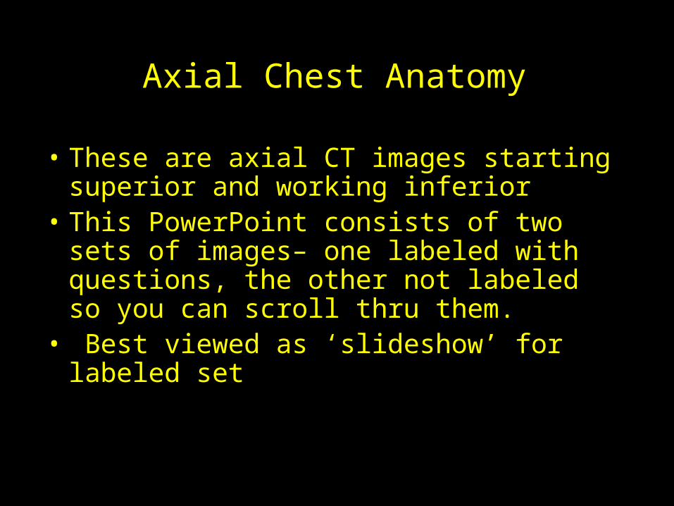
Axial Chest Anatomy
• These are axial CT images starting superior and working inferior
• This PowerPoint consists of two sets of images– one labeled with questions, the other not labeled so you can scroll thru them.
• Best viewed as ‘slideshow’ for labeled set
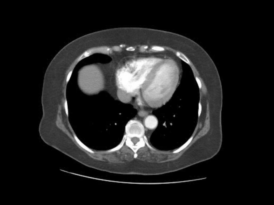
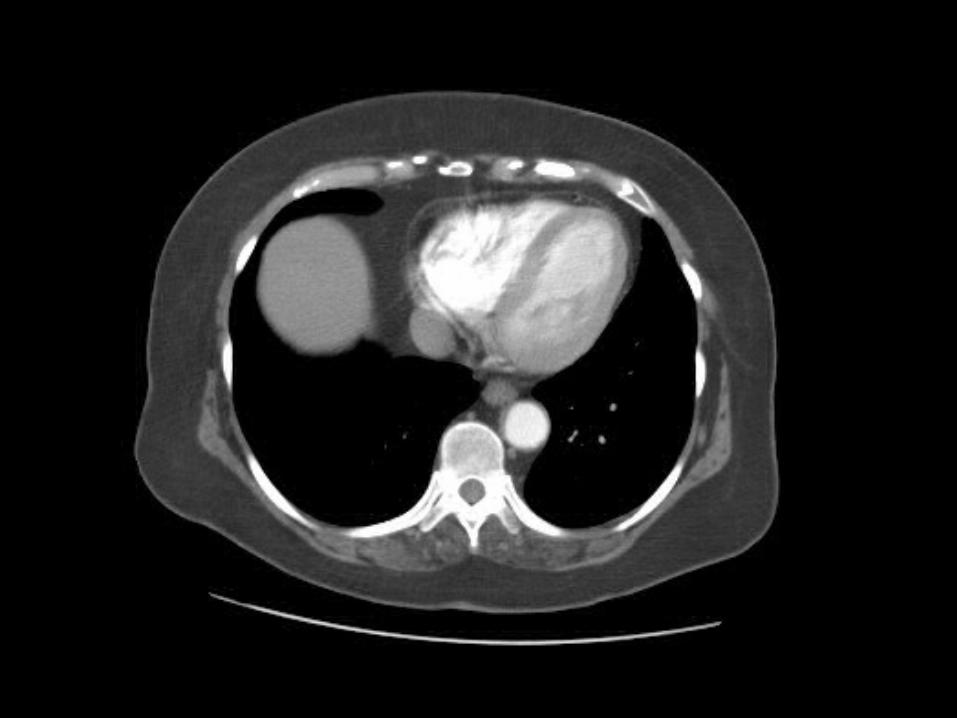
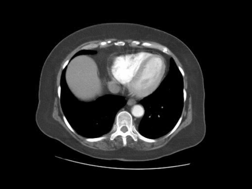
The following slide set is a collection of CT axial (transverse) 5mm slices from the lower neck to the upper abdomen. Soft tissue window settings were
selected to provide the best views of the mediastinal anatomy. Every few slides, outlines with a legend have been provided to label anatomical structures of interest.
Radiologists frequently page up and down on digital images to “follow” structures from their origin to their
termination. Try this with the unlabeled slide set. Use the labeled slides to help orient yourself while you page up
and down to try and identify various anatomical structures. It will be best to view these slides in the
‘slideshow’ mode.
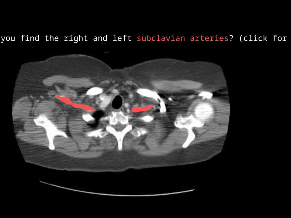
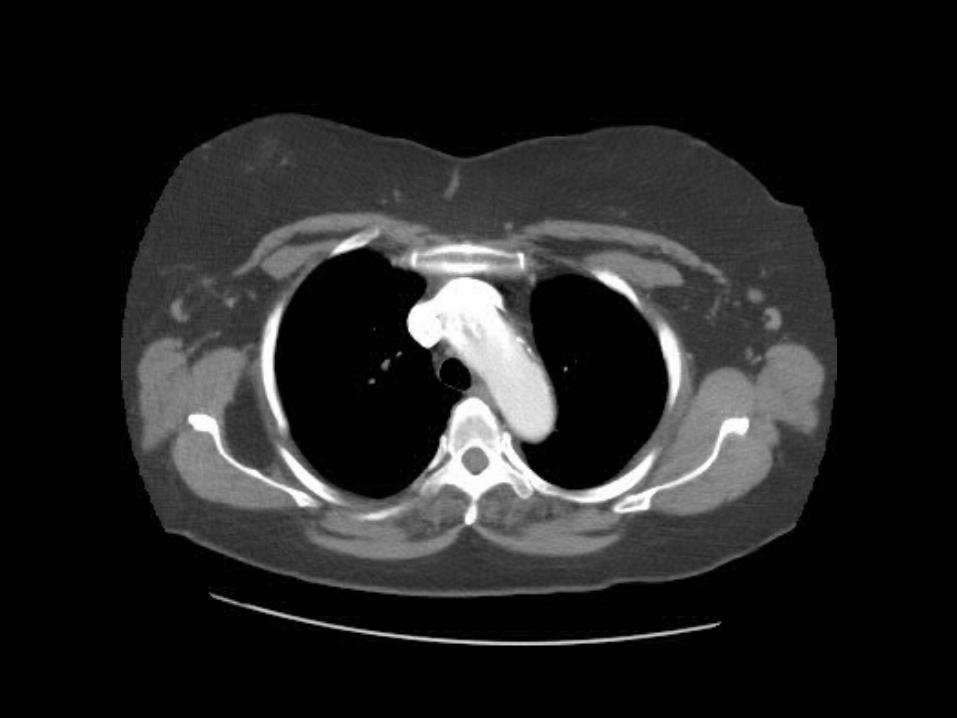
Can you find the right and left subclavian arteries? (click for answer)
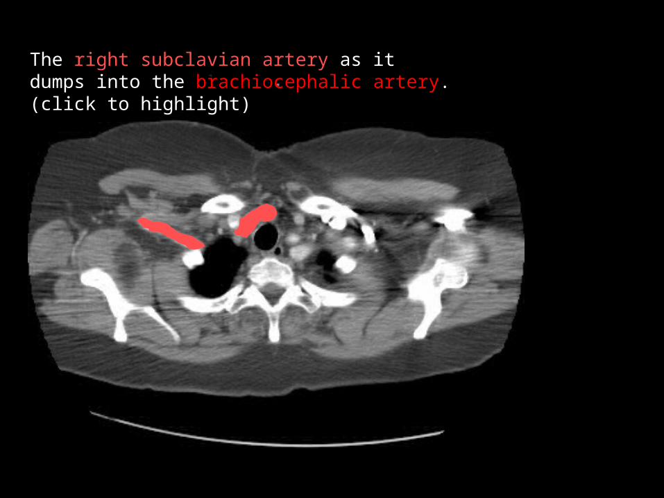
The right subclavian artery as itdumps into the brachiocephalic artery.(click to highlight)

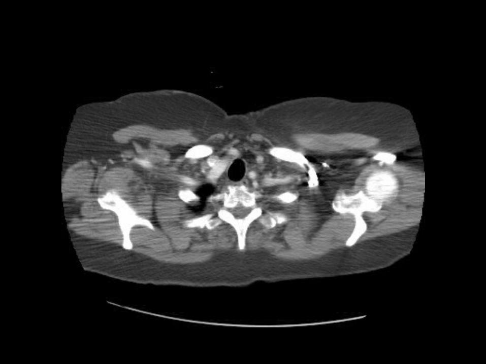
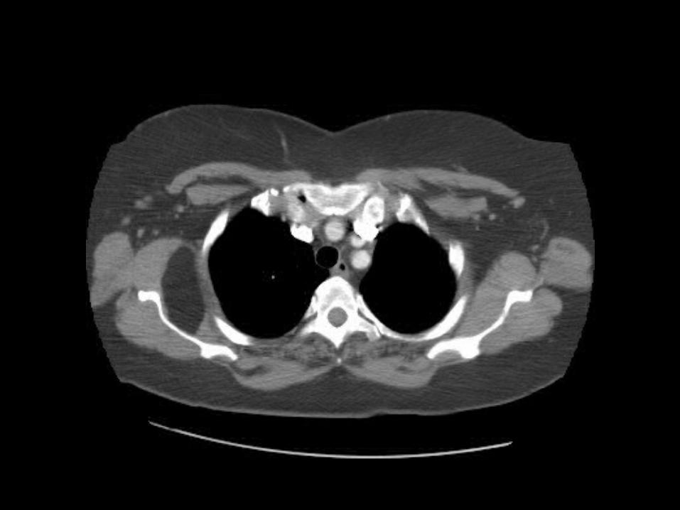
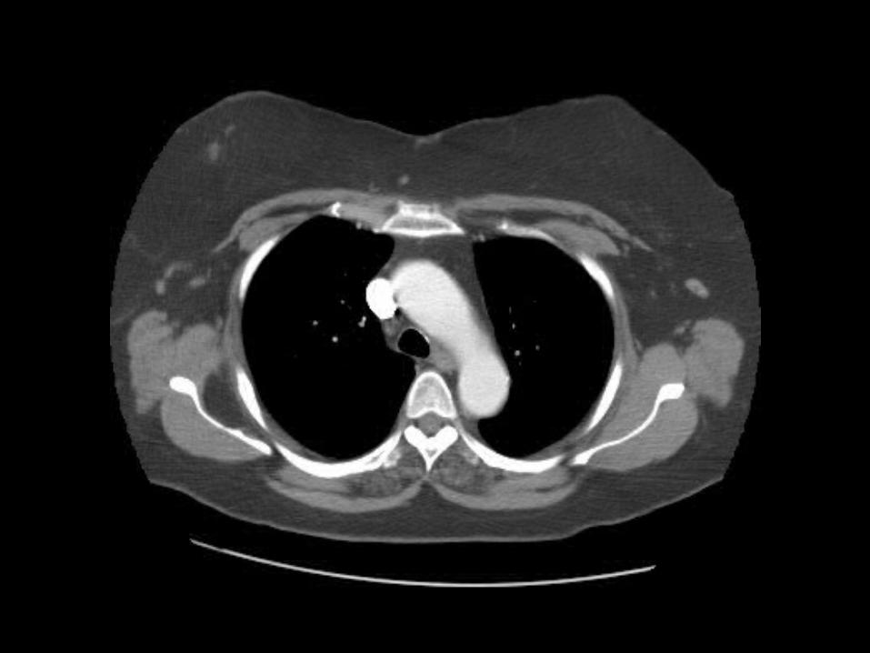
What are the 3 yellow air-filled (black) structures highlighted here? (click to highlight)
These are the Right lung apexTrachea and Esophagus.
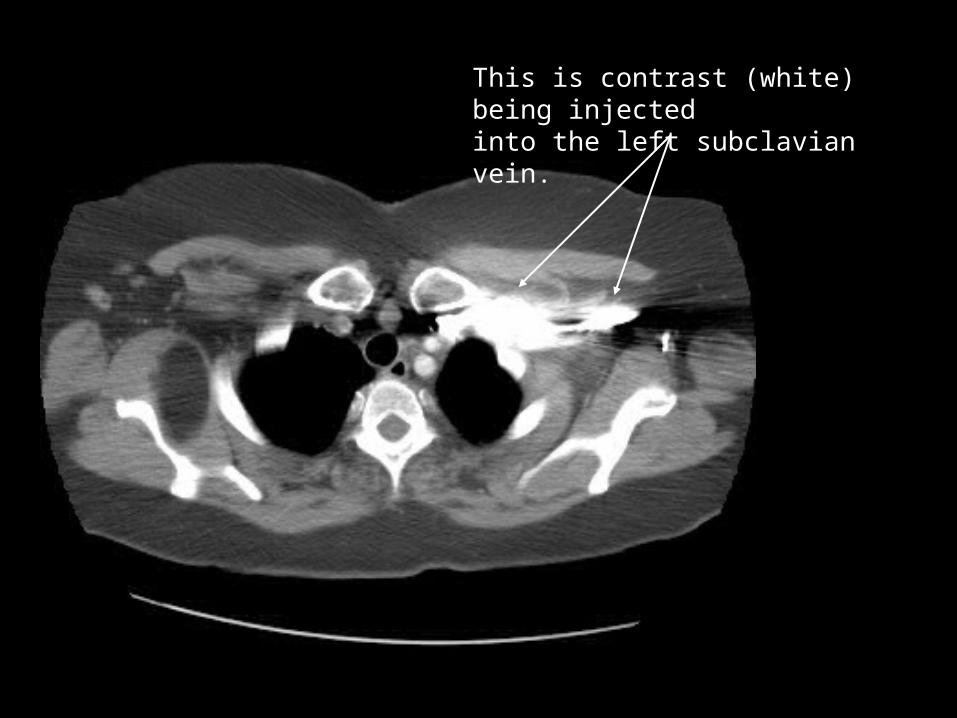
This is contrast (white) being injected into the left subclavian vein.
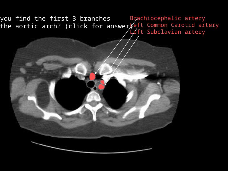
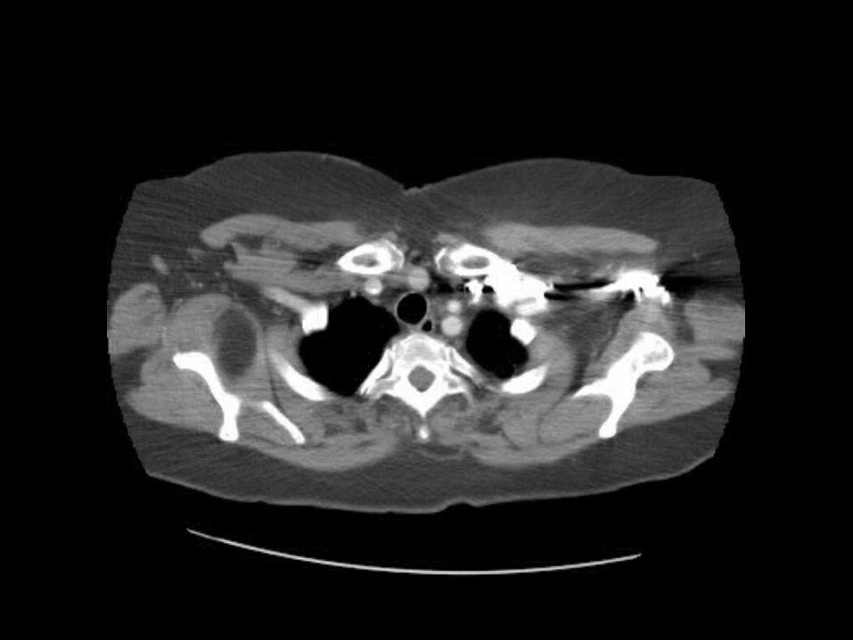
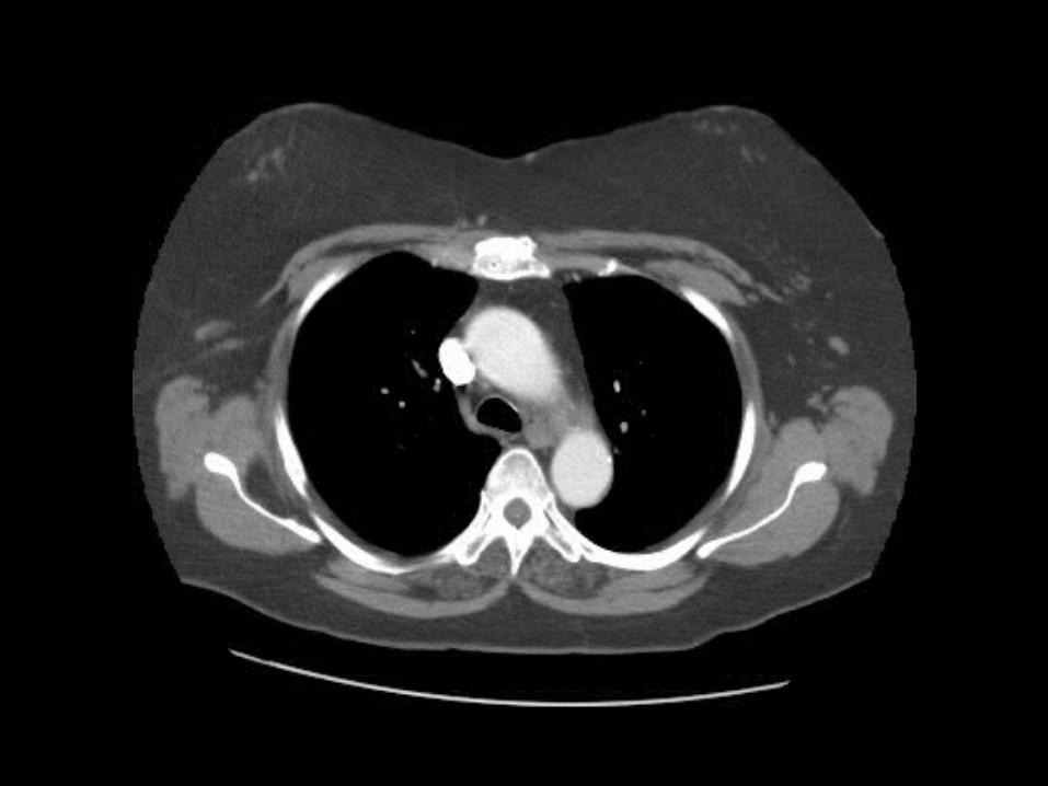
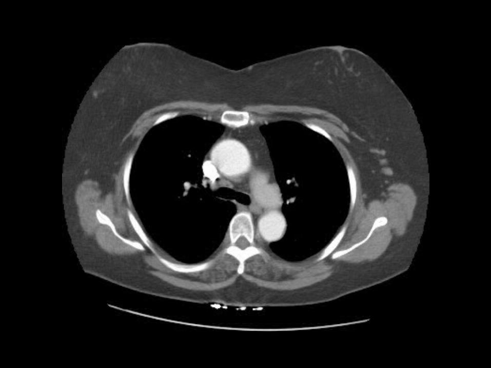
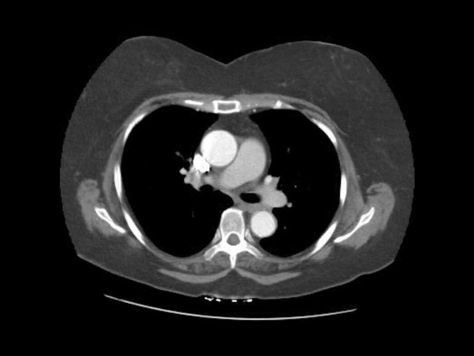
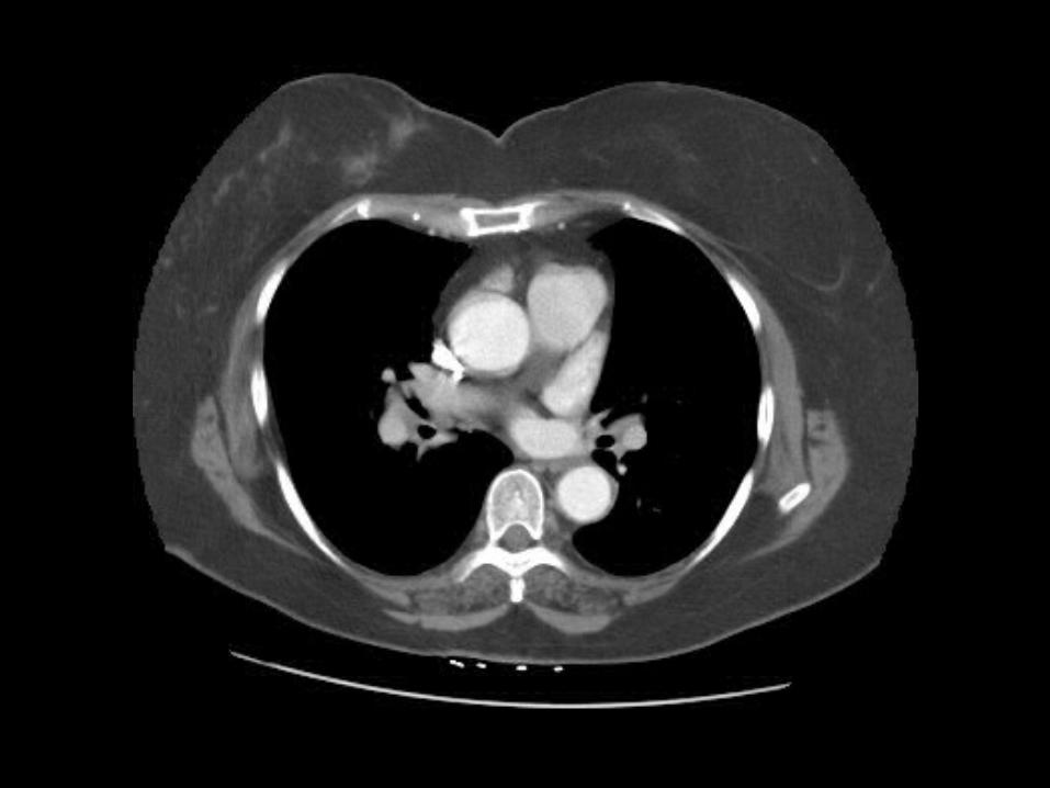
Can you find the first 3 branches off the aortic arch? (click for answer)
Brachiocephalic arteryLeft Common Carotid arteryLeft Subclavian artery
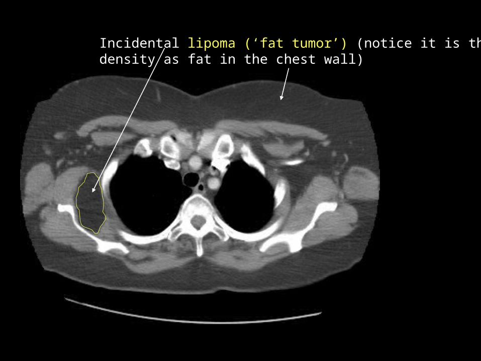
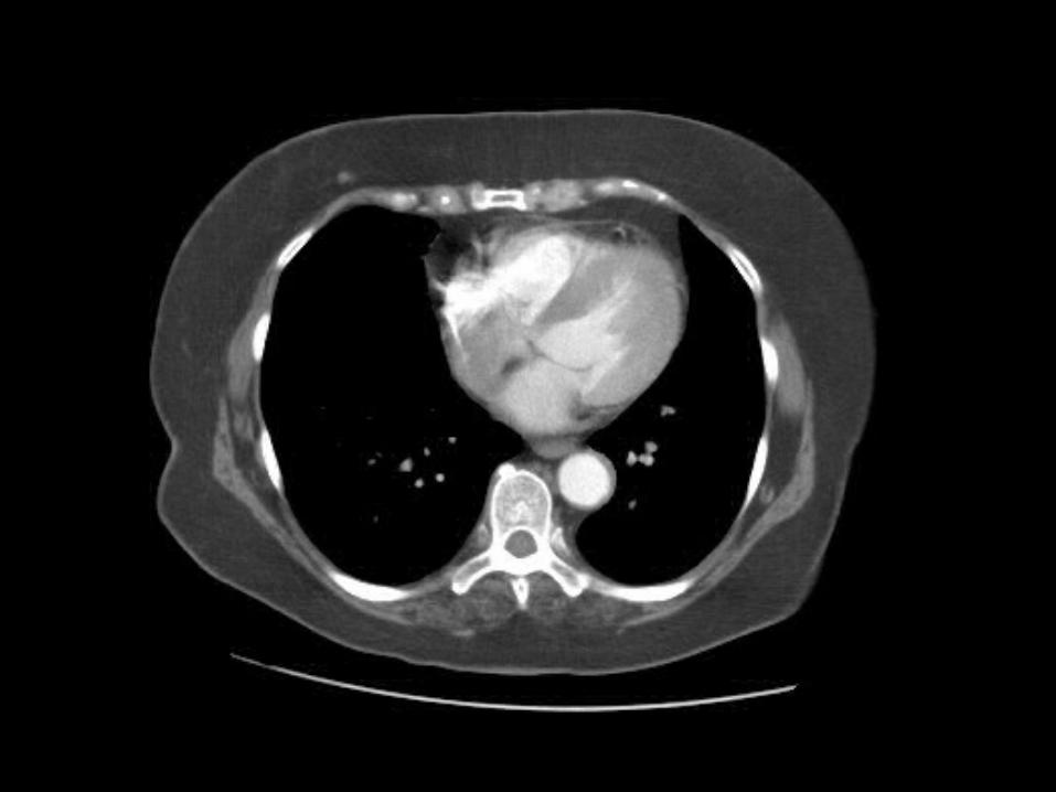
Incidental lipoma (‘fat tumor’) (notice it is the same density as fat in the chest wall)

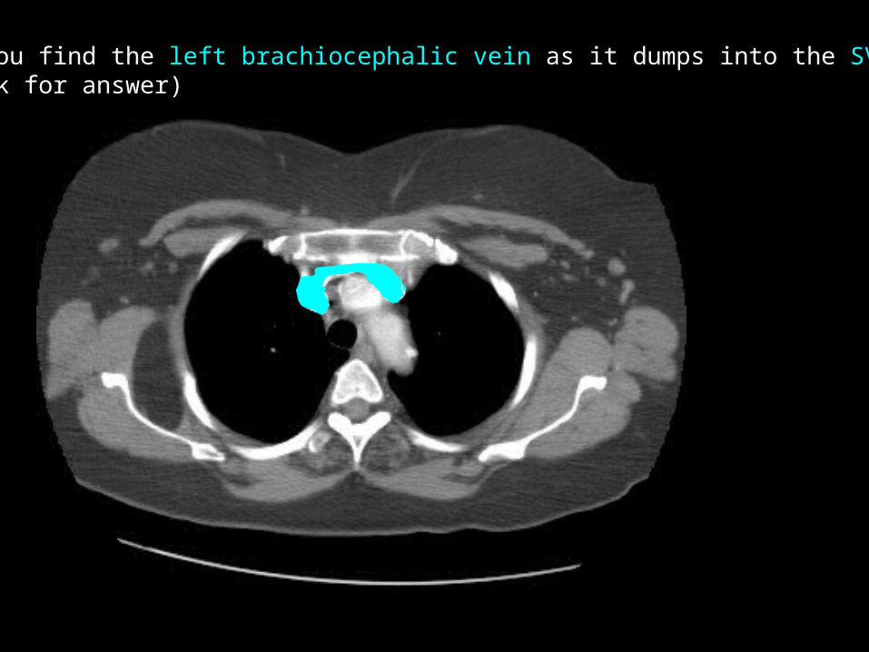
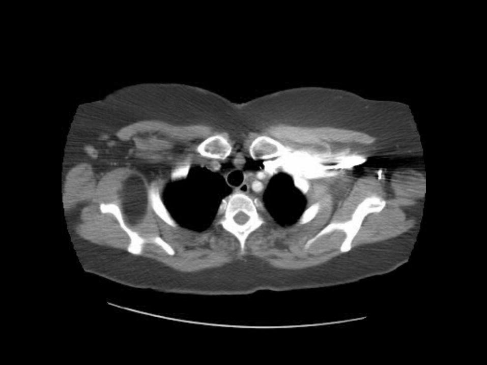
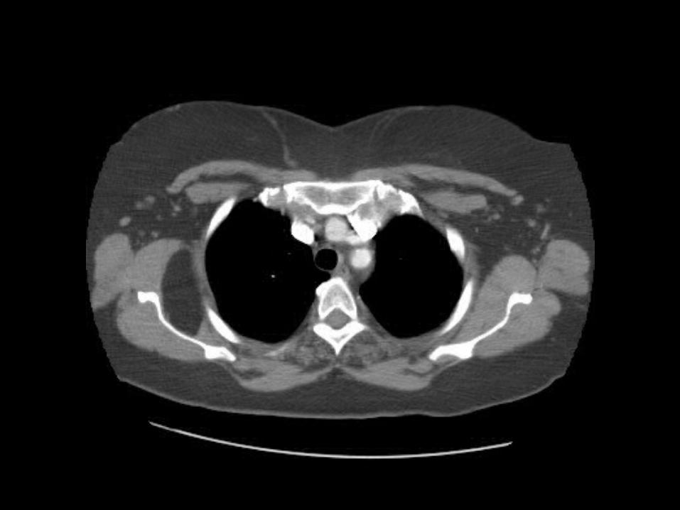
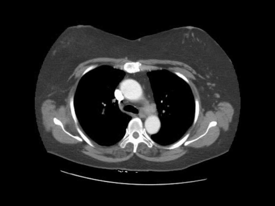
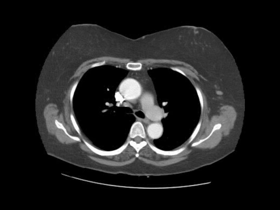
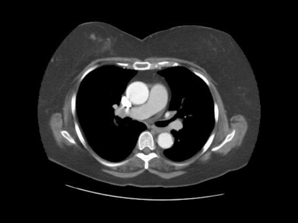
Can you find the left brachiocephalic vein as it dumps into the SVC?(click for answer)
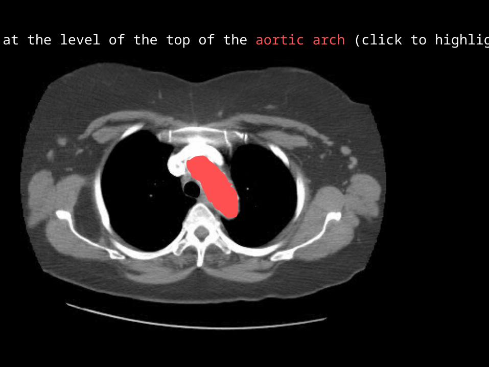
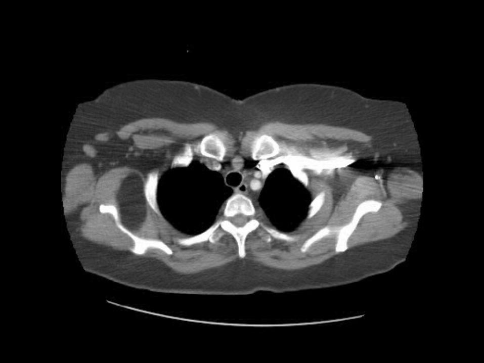
We are at the level of the top of the aortic arch (click to highlight)
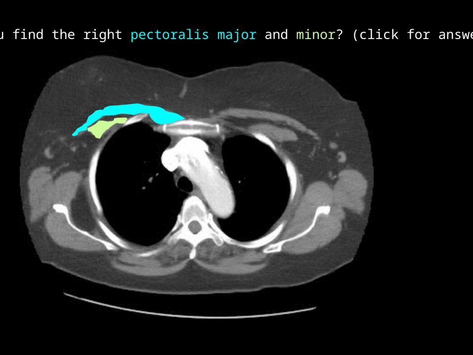
Can you find the right pectoralis major and minor? (click for answer)
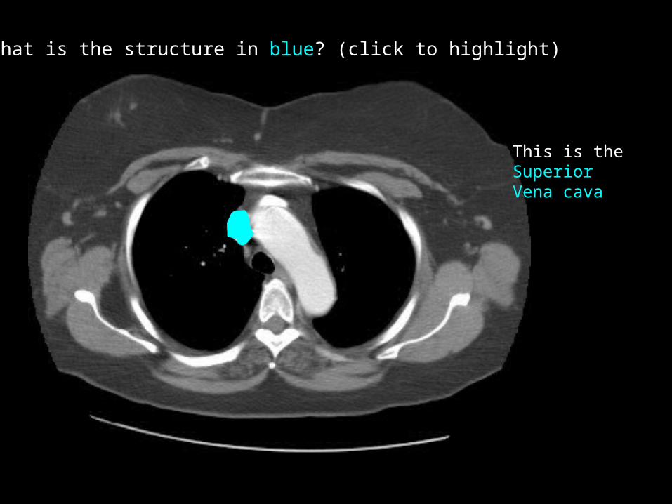
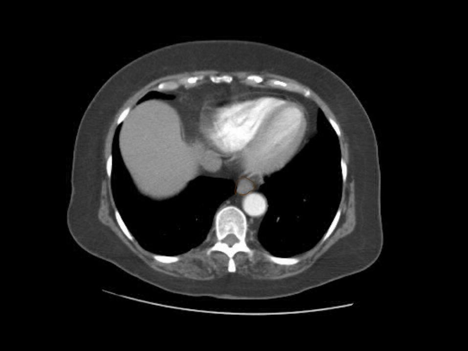
What is the structure in blue? (click to highlight)
This is the SuperiorVena cava
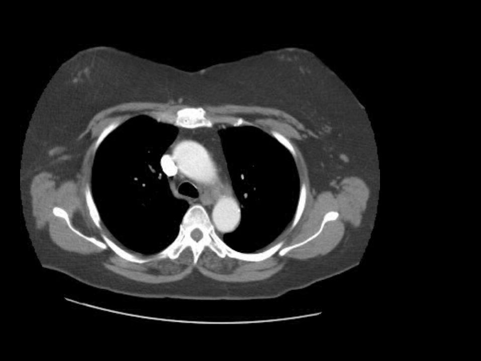

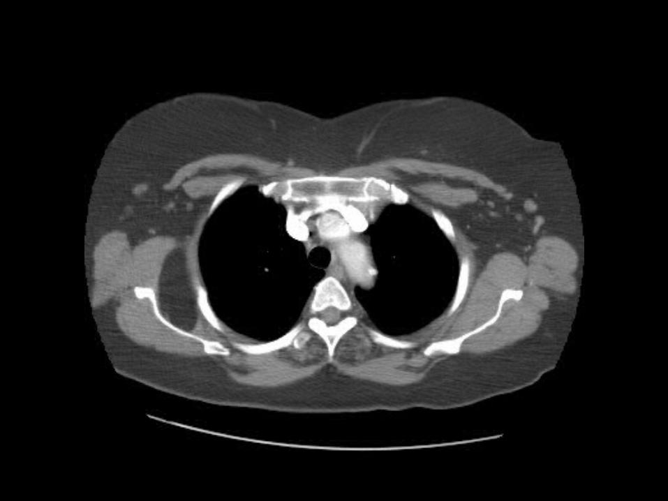
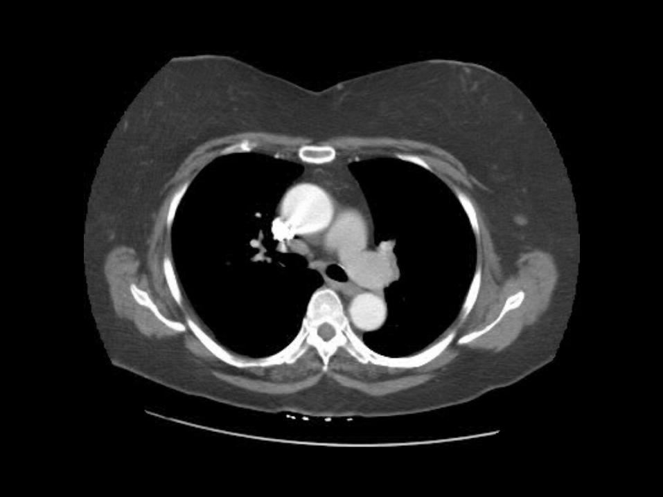
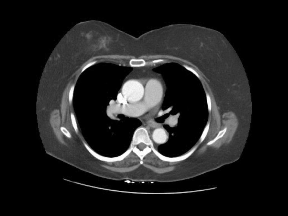
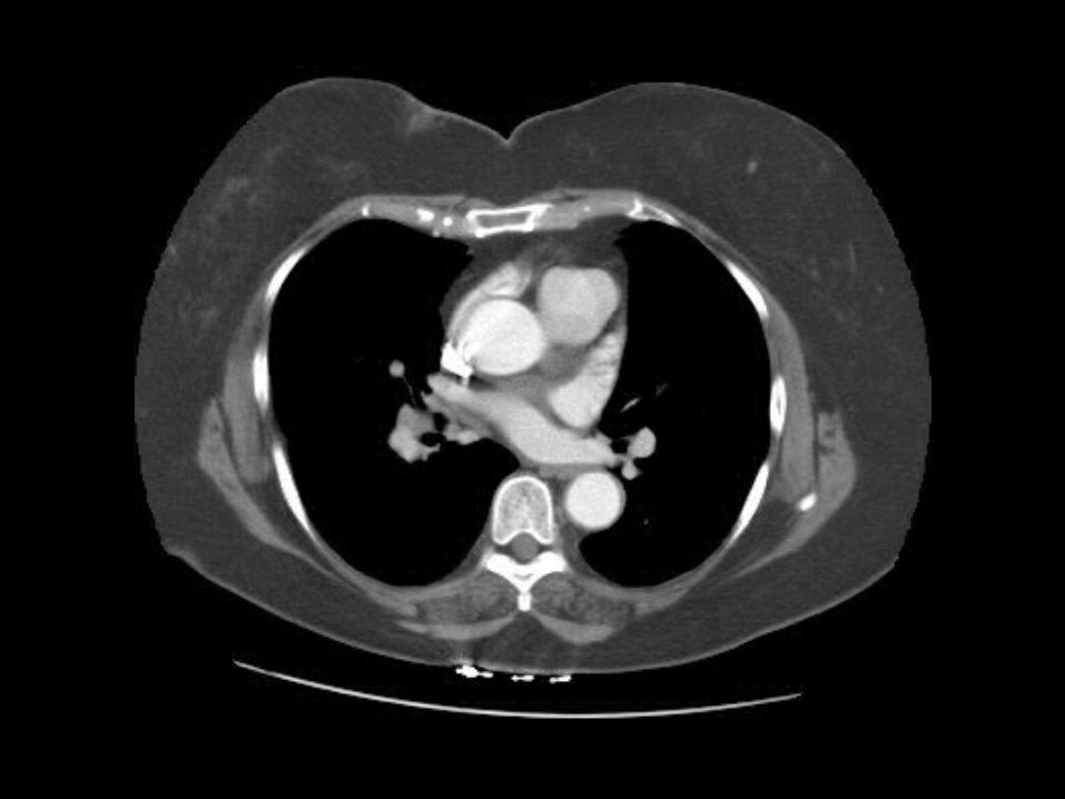
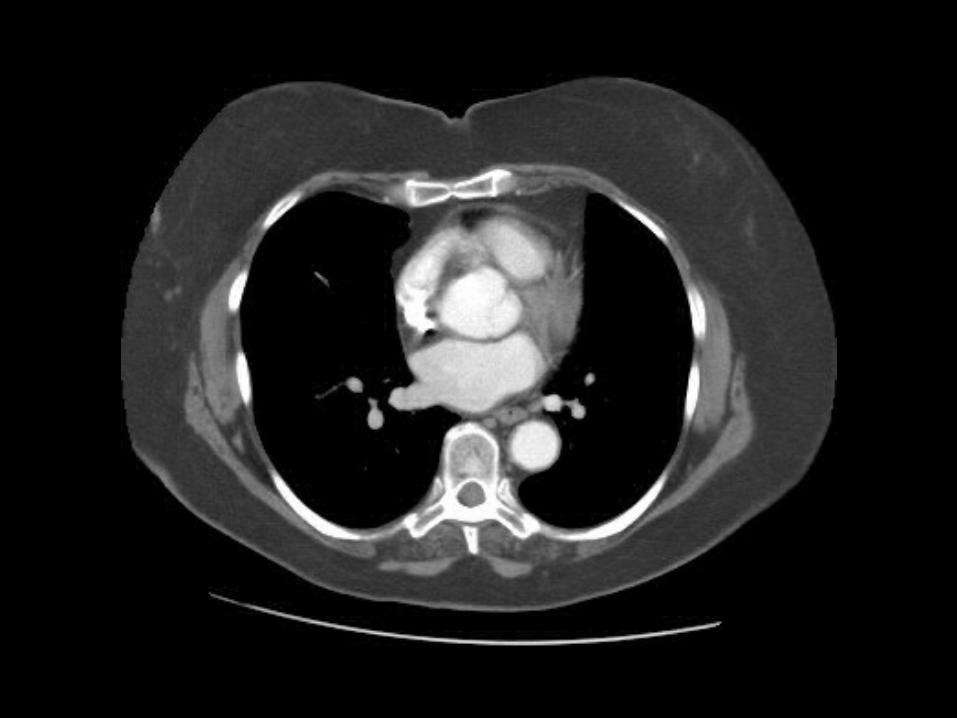
Can you find the azygous arch ? (where the azygous vein comes across to dump into the SVC) (click for answer)
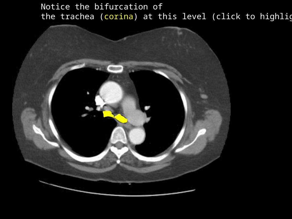
Notice the bifurcation ofthe trachea (corina) at this level (click to highlight)
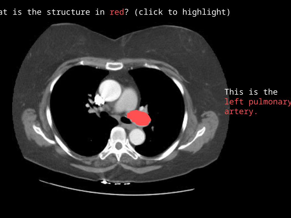
What is the structure in red? (click to highlight)
This is the left pulmonaryartery.


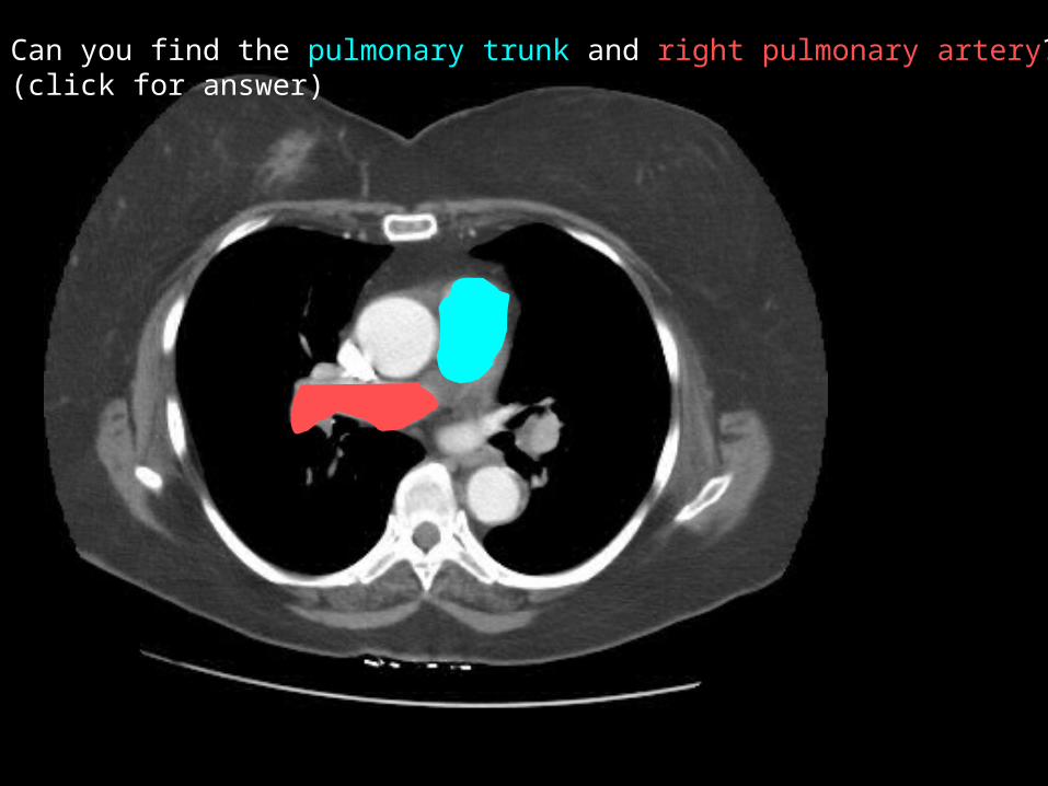
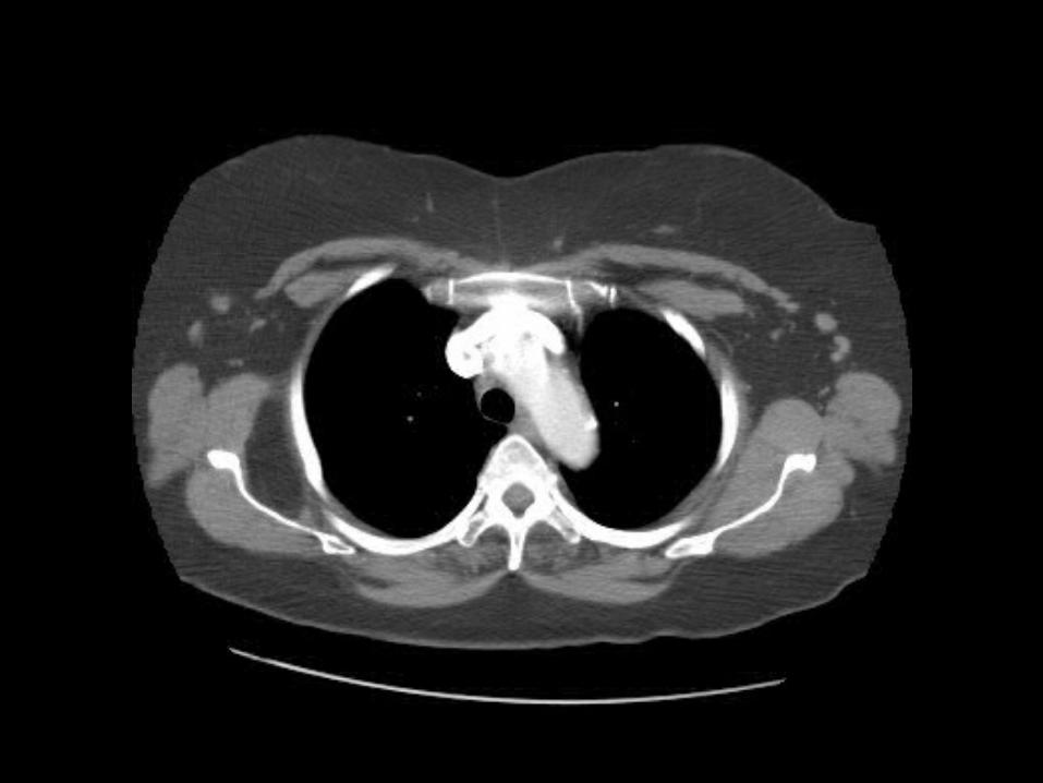
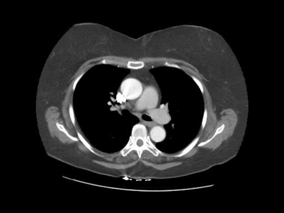
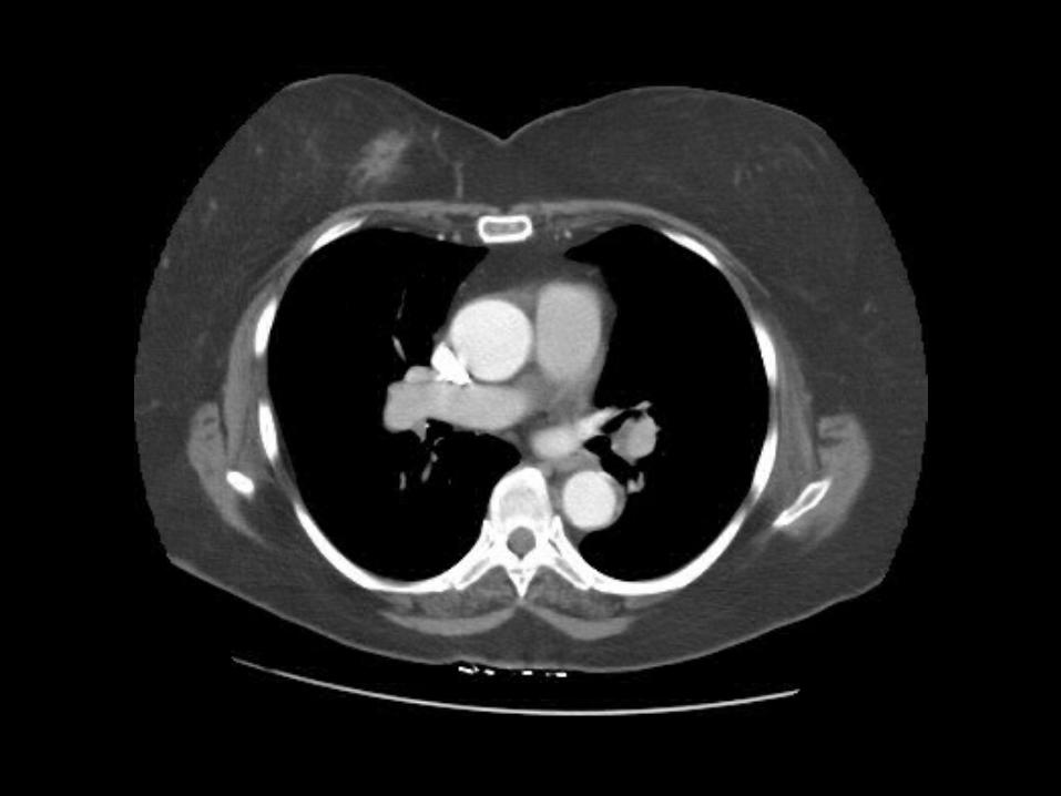
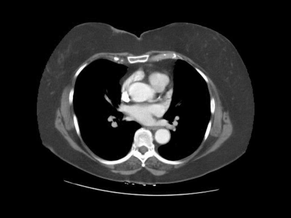
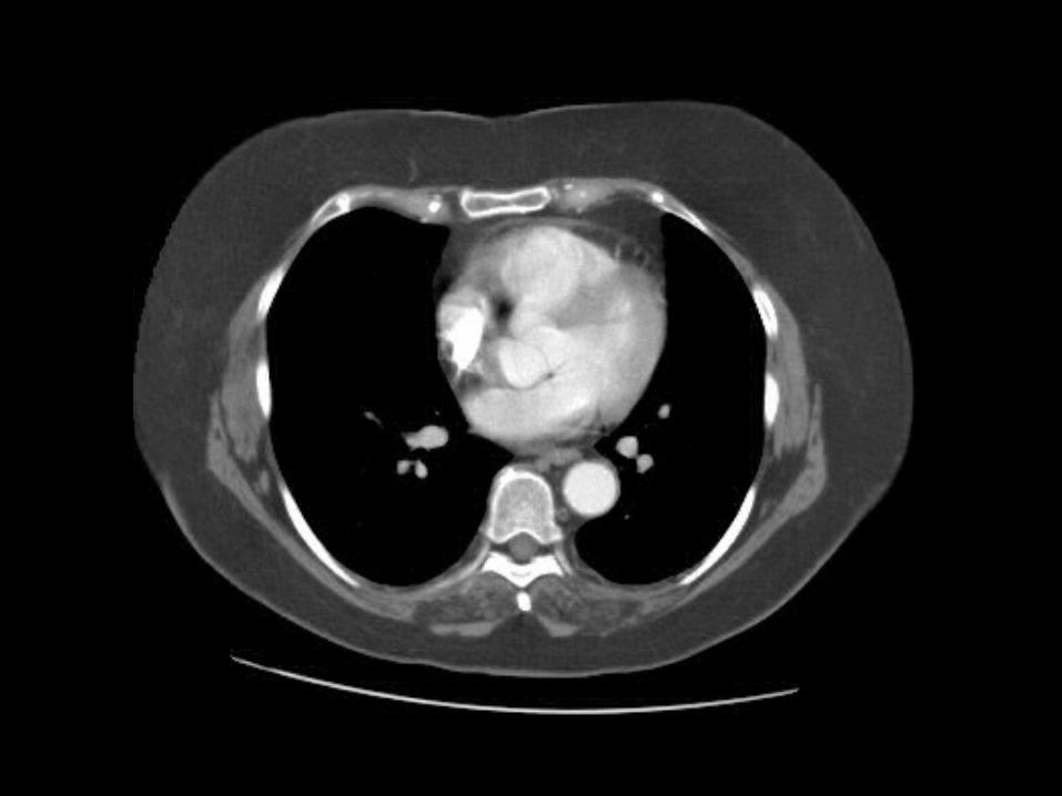
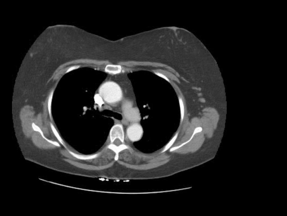
Can you find the pulmonary trunk and right pulmonary artery? (click for answer)
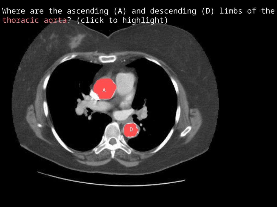
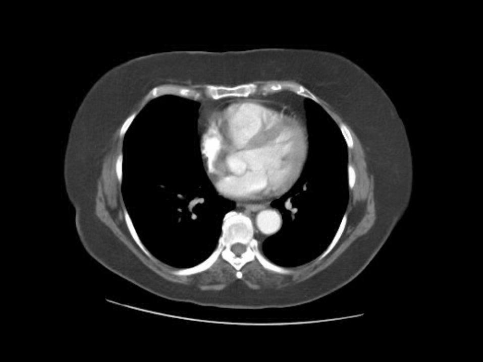
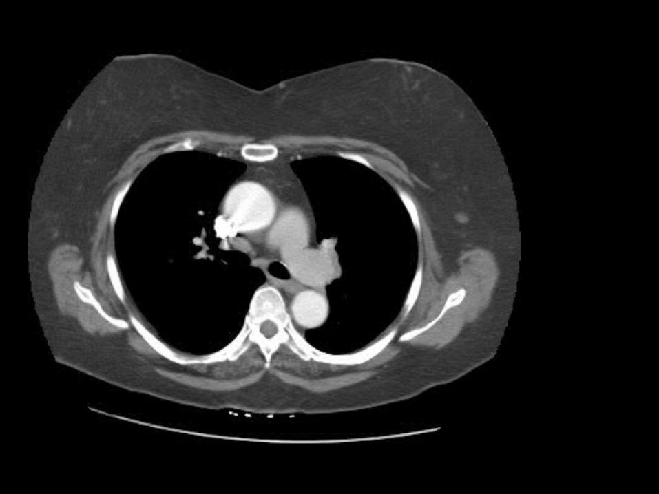
Where are the ascending (A) and descending (D) limbs of the thoracic aorta? (click to highlight)
A
D
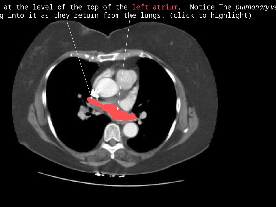
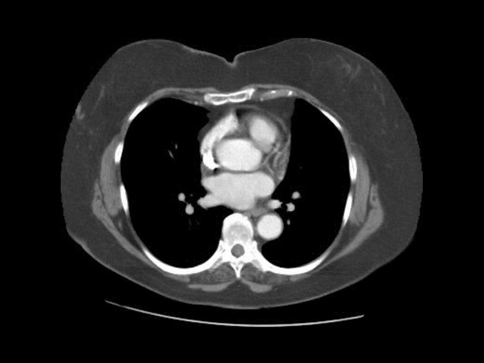
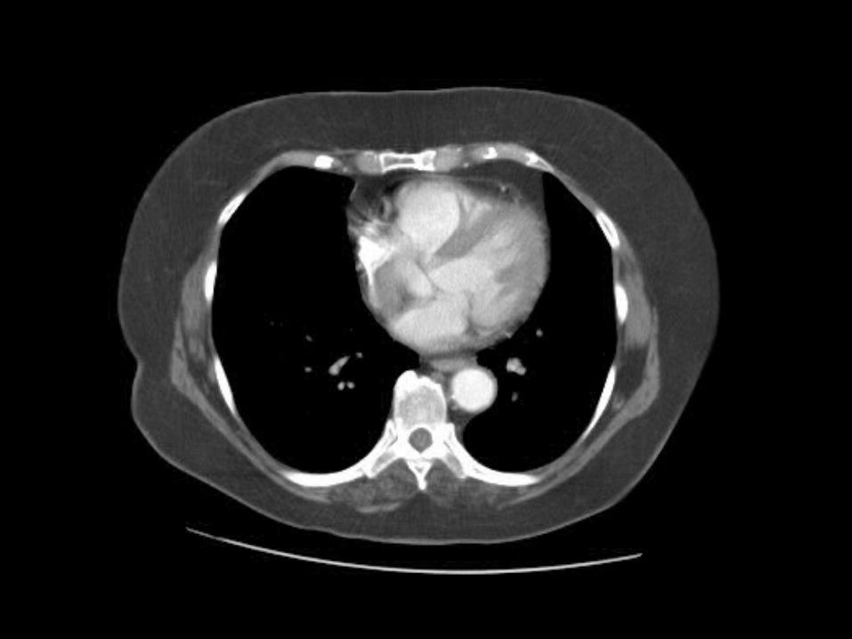
We are at the level of the top of the left atrium. Notice The pulmonary veins dumping into it as they return from the lungs. (click to highlight)

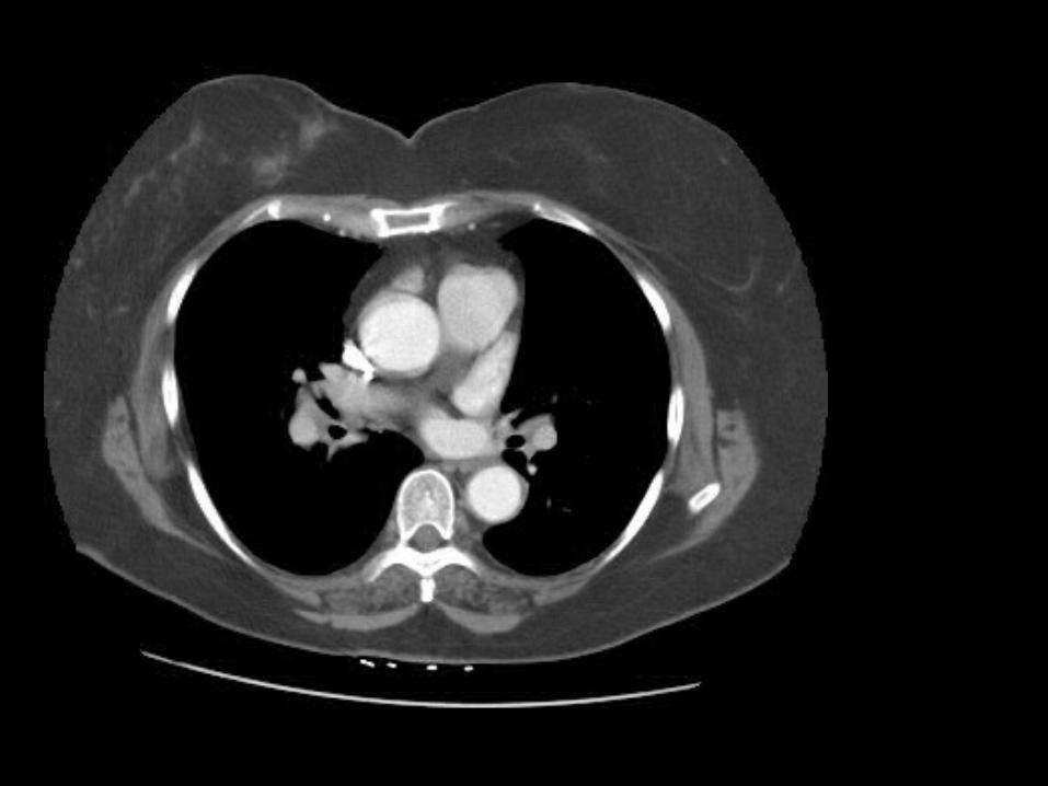
Can you find the left main coronary artery coming off the aorta? (click for answer)
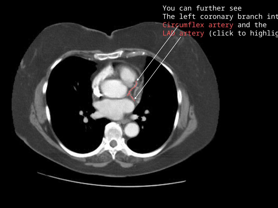
You can further seeThe left coronary branch into theCircumflex artery and theLAD artery (click to highlight)
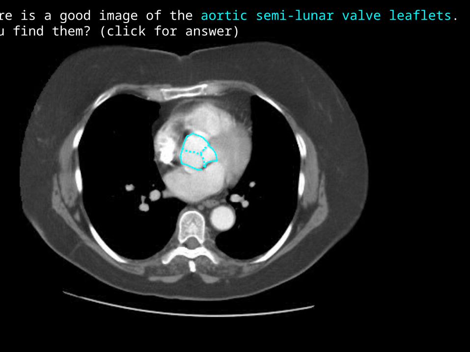
Here is a good image of the aortic semi-lunar valve leaflets. Can you find them? (click for answer)

Here is a good image of the left atrium dumping Into the left ventricle. Can you find it? (click to highlight)
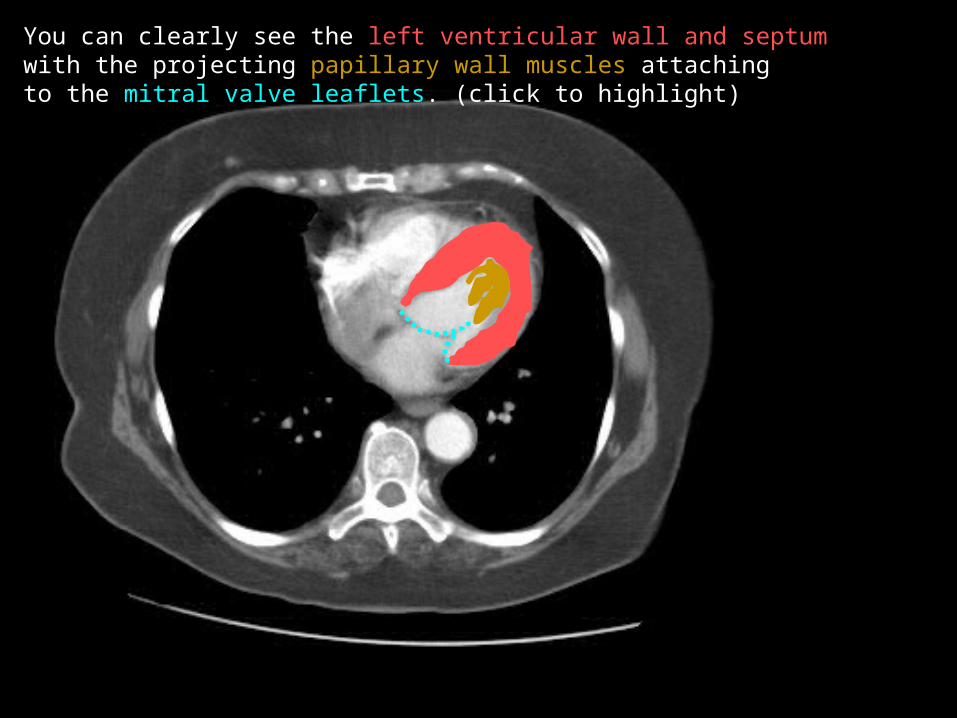
You can clearly see the left ventricular wall and septum with the projecting papillary wall muscles attachingto the mitral valve leaflets. (click to highlight)
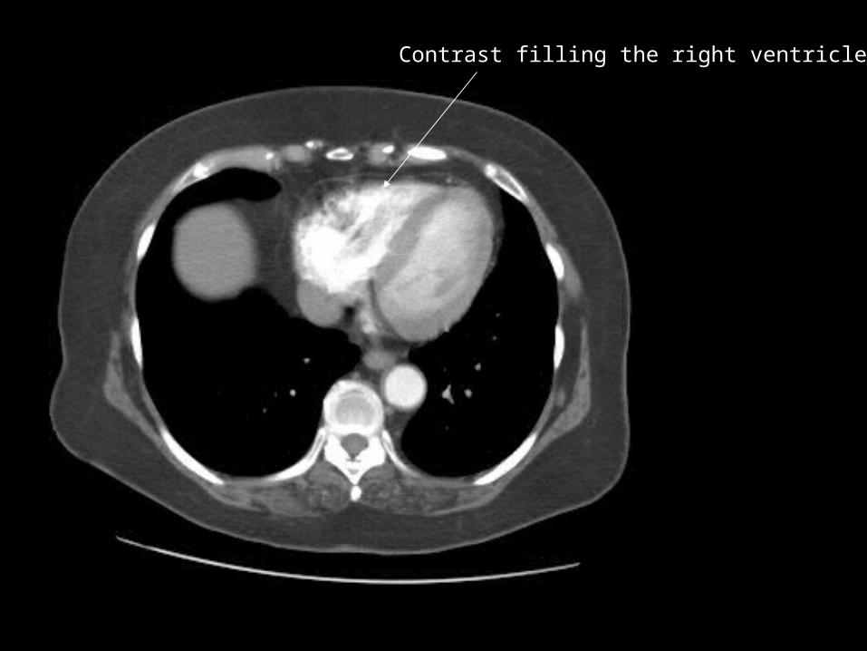
Contrast filling the right ventricle

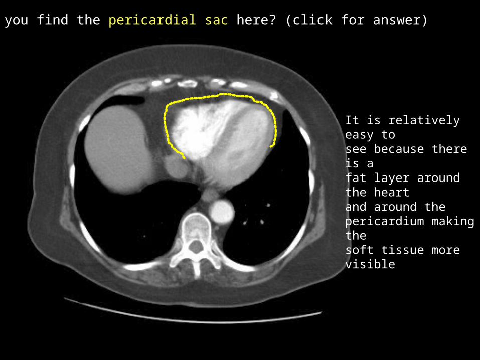
Can you find the pericardial sac here? (click for answer)
It is relatively easy tosee because there is a fat layer around the heartand around the pericardium making the soft tissue more visible
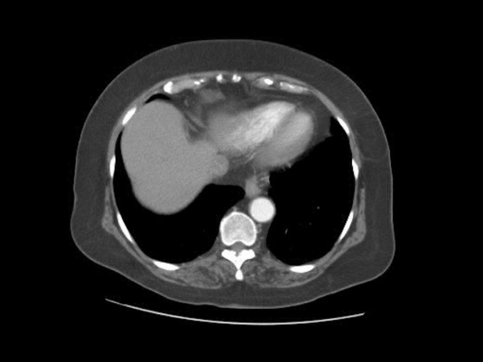
Unlabeled Axial Chest Set