Embed Size (px)
Citation preview
REVIEW Open Access
Auxotrophy to Xeno-DNA: an exploration ofcombinatorial mechanisms for a high-fidelity biosafety system for syntheticbiology applicationsChristopher M. Whitford1 , Saskia Dymek1, Denise Kerkhoff1, Camilla März1, Olga Schmidt1, Maximilian Edich1,Julian Droste1,2, Boas Pucker1,2,3 , Christian Rückert1,2 and Jörn Kalinowski1,2*
Abstract
Background: Biosafety is a key aspect in the international Genetically Engineered Machine (iGEM) competition,which offers student teams an amazing opportunity to pursue their own research projects in the field of SyntheticBiology. iGEM projects often involve the creation of genetically engineered bacterial strains. To minimize the risksassociated with bacterial release, a variety of biosafety systems were constructed, either to prevent survival ofbacteria outside the lab or to hinder horizontal or vertical gene transfer.
Main body: Physical containment methods such as bioreactors or microencapsulation are considered the firstsafety level. Additionally, various systems involving auxotrophies for both natural and synthetic compounds havebeen utilized by iGEM teams in recent years. Combinatorial systems comprising multiple auxotrophies have beenshown to reduced escape frequencies below the detection limit. Furthermore, a number of natural toxin-antitoxinsystems can be deployed to kill cells under certain conditions. Additionally, parts of naturally occurring toxin-antitoxin systems can be used for the construction of ‘kill switches’ controlled by synthetic regulatory modules,allowing control of cell survival. Kill switches prevent cell survival but do not completely degrade nucleic acids.To avoid horizontal gene transfer, multiple mechanisms to cleave nucleic acids can be employed, resulting in ‘self-destruction’ of cells. Changes in light or temperature conditions are powerful regulators of gene expression andcould serve as triggers for kill switches or self-destruction systems. Xenobiology-based containment usesapplications of Xeno-DNA, recoded codons and non-canonical amino acids to nullify the genetic informationof constructed cells for wild type organisms. A ‘minimal genome’ approach brings the opportunity to reducethe genome of a cell to only genes necessary for survival under lab conditions. Such cells are unlikely tosurvive in the natural environment and are thus considered safe hosts. If suitable for the desired application,a shift to cell-free systems based on Xeno-DNA may represent the ultimate biosafety system.
Conclusion: Here we describe different containment approaches in synthetic biology, ranging fromauxotrophies to minimal genomes, which can be combined to significantly improve reliability. Since the iGEMcompetition greatly increases the number of people involved in synthetic biology, we will focus especially onbiosafety systems developed and applied in the context of the iGEM competition.
Keywords: Kill switch, iGEM, Semantic containment, Physical containment, Auxotrophy, Escherichia coli,BioBrick, Genetic engineering
* Correspondence: [email protected];[email protected] for Biotechnology, Bielefeld University, 33615 Bielefeld, Germany2Faculty of Biology, Bielefeld University, Bielefeld, GermanyFull list of author information is available at the end of the article
© The Author(s). 2018 Open Access This article is distributed under the terms of the Creative Commons Attribution 4.0International License (http://creativecommons.org/licenses/by/4.0/), which permits unrestricted use, distribution, andreproduction in any medium, provided you give appropriate credit to the original author(s) and the source, provide a link tothe Creative Commons license, and indicate if changes were made. The Creative Commons Public Domain Dedication waiver(http://creativecommons.org/publicdomain/zero/1.0/) applies to the data made available in this article, unless otherwise stated.
Whitford et al. Journal of Biological Engineering (2018) 12:13 https://doi.org/10.1186/s13036-018-0105-8
BackgroundSafety approaches in synthetic biology are frequently dis-tinguished into mechanisms of biosecurity or biosafety.However, many methods can only be distinguished at atheoretical level into biosafety or biosecurity [1]. Here,we focus on biosafety, defined as the combination of allpreventive measures against accidental infection with, orrelease of, genetically engineered organisms into theenvironment [2]. Nevertheless, some of the presentedmechanisms can also contribute to biosecurity, definedas the protection of biological systems against anintended misuse [3].Recombinant DNA technology has been applied since
1973 to modify the genetic information of cells for scien-tific as well as for economic purposes [4]. However, thistechnology poses risks if applied carelessly [5] or withdeleterious intent [6–8]. This risk has long been recog-nized, and multiple safety regulations have been pro-posed to prevent harm to humans, animals, and theenvironment [9, 10].As Escherichia coli became a model organism in mo-
lecular biology and biotechnology, the development ofsafety strains was of high interest [11]. For example, E.coli K12 MG1655 [12, 13] and E. coli B derivatives likeREL606 and BL21 (DE3) are safety strains [14]commonly used for molecular cloning and heterologousgene expression [15–19]. Due to several mutations, thesestrains are no longer able to compete with wild typestrains within the human gut [5, 20]. More sophisticatedstrains have been developed, including dedicatedbiosafety approaches like the relA deletion in combinationwith a conditional phoA expression [21, 22], as describedin the next chapter. The combination of differentprecautions may have successfully prevented any accidentsinvolving genetically modified organisms (GMOs) duringthe last four decades [2, 23–25]. Today, the handling ofGMOs is strictly controlled for multiple reasons, includingecological and health considerations [2, 26, 27], but alsothe protection of intellectual property [28].However, the demand for novel biosafety systems is
still high, due to the spread of genetic engineering cap-abilities which in turn is facilitated by the growing num-ber of people involved in the field of synthetic biology.Students participating in the iGEM [29] competitionhave contributed significantly to the synthetic biologytoolbox [30–43]. The increased availability of genomesequences [12, 44] as well as an ever-increasing numberof sequenced bacterial genomes could enhance the dis-covery and implementation of innovative safety systems.We will describe general biosafety mechanisms alongwith advanced systems developed within this competi-tion. It is our intention to provide an overview of theapplied concepts as well as the completely implementedand characterized systems. Since the deletion of an
essential gene is probably the most basic mechanism, wewill start with the description of auxotrophies. Releasedcells with failing auxotrophy systems could be stoppedby the activation of kill switches. Activation of suchmechanism should kill the cell, but the nucleic acids arestill present, requiring a self-destruction of the geneticinformation to prevent horizontal gene transfer (HGT).Given that all known life on earth is based on the samebiochemistry, cells are composed of common materialslike proteins, sugars, lipids, and nucleic acids. Althoughthe sequence of nucleotides and amino acids varieswithin higher layer molecules, the basal molecules areshared across all species. A common genetic code allowsthe exchange of genetic information between differentspecies through HGT. In general, HGT might not bebeneficial for a specific species [45] but is assumed tomaintain an overall unity of life by preventing changesin the genetic code [46]. Therefore, central signal andmetabolic pathways are shared between distantly relatedspecies, while additional pathways are specific to certaintaxonomic groups. Interesting in terms of biosafety arerare cases, where replicating systems escaped this systemand modified their genetic code [47–49]. Modificationsto this universal genetic code pose a powerful biosafetymechanism [50]. Therefore, xenobiology-based contain-ment is another way to prevent the successful transfer ofinformation from another species. Sensors for light,temperature, pH, UV or other physical parameters canbe used as triggers for biosafety systems. Avoiding issuesof vertical gene transfer (VGT) through cell division, cellfree systems seem to be a well suited biosafety approach.In combination with xenobiology, cell free systems couldefficiently minimize the risk of HGT and VGT. Finally,we aim to describe the design of an optimized biosafetysystem by integration of different mechanisms, whichconsequently could be referred to as a synthetic bio-safety system [51].
AuxotrophyOne of the earliest methods applied for biocontainmentwas auxotrophy, introduced either by random mutationor targeted engineering [52]. In respect to biosafety,auxotrophic organisms are not able to synthesize one ormore compounds required for survival or replication, sothe missing components must be provided externally[53]. Therefore, the organism cannot survive outside acontrolled environment, such as a bioreactor. Researchon auxotrophic strains dates back to 1941, when Beadlyand Tatum characterized a strain of Neurospora sitophilawithout pyridoxine [54]. The first construction and ap-plication of an auxotroph E. coli strain for biosafety rea-sons was developed in 1977 by Curtiss and colleagues[52], who developed the strain χ1776, which requiresdiaminopimelic acid (DAP) and thymine or thymidine
Whitford et al. Journal of Biological Engineering (2018) 12:13 Page 2 of 28
for growth. Indeed, many prominent laboratory E. colistrains are auxotrophic mutants [55]. Over the years,auxotropy-based systems have been developed further andsuccessfully improved with regards to reducing the escapefrequency [53] (the escape frequency describes the prob-ability of an organism bypassing the containment mea-sures and is usually determined by quantification ofescape mutants found in a defined cell number [56]), withan increasing number of studies reporting escape frequen-cies (Table 2) below the detection limit (e.g. [57, 58]).Auxotrophy-based systems are manifold in nature, withdifferent hosts (e.g. E. coli or other bacteria, plants, orfungi) and various possible dependencies, most commonlyfor amino acids or vitamins.
Strains auxotrophic for proteinogenic amino acidsThe dependency of engineered organisms on one ormultiple naturally occurring amino acids has been usedin many studies to improve biosafety via containment orreplacement of problematic functions. A prime examplefor the latter is the substitution of antibiotic resistancemarkers on plasmids. These standard tools in geneticengineering traditionally contain an antibiotic resistancegene as a selection marker, which further ensures plasmidmaintenance. This is potentially hazardous, as theresistance genes could be released into the environment[59–61]. As an alternative to antibiotic selection markers,the auxotrophic E. coli strain M15 carries an inactive glyAgene, which is necessary for intracellular glycine synthesis[59]. The antibiotic-free expression system utilizes theM15 strain with a constructed plasmid providing the glyAgene and thereby preventing plasmid loss. A similar ap-proach was used in the modified E. coli strain JM83 auxo-trophic for proline and a complementing vector supplyingthe proBA genes necessary for the proline biosyntheticpathway [62]. Antibiotic resistance-free systems have alsobeen used as a safe method for vaccine production andare not limited to E. coli. An example is the cattle vaccineBrucella abortus mutant strain (RB51) with non-func-tional 3-isopropylmalate dehydrogenase (leuB), which isessential for biosynthesis of leucine [60]. By adding a plas-mid carrying the wild type leuB to the leucine-auxotrophicstrain, the B. abortus strain is able to survive and the cor-responding plasmid is maintained due to selective pres-sure. The resulting system is a biologically safe vaccinesystem, as the B. abortus mutant strain induces immuneresponses through a plasmid containing the gene encod-ing the desired antigen, but no antibiotic resistance gene,which could potentially spread to the microbial flora of ananimal. In 2012, a system was engineered that supportedthe usage of the duckweed Lemna for the production ofvaccines and therapeutics [63]. An avian influenza vaccineantigen was successfully expressed in the system contain-ing an isoleucine auxotroph strain of Lemna. Particularly,
threonine deaminase expression necessary for the isoleu-cine biosynthesis had been inactivated in a system con-taining an isoleucine auxotroph strain of Lemna, whichwas then supplemented with isoleucine to enable growth.
Strains auxotrophic for other natural componentsA different biosafety approach is to prevent an organismfrom building a cell wall by means of engineered auxot-rophy. For example, D-alanine is an essential componentof the peptidoglycan layer, since it is one of the mole-cules necessary for the cross-linkage of the polysacchar-ide chains [64, 65]. L-alanine can be converted toD-alanine by two different alanine racemases in E. coli,one being encoded by alr, the other one by dadX [66].In Corynebacteriaceae, only the alr gene is present,which was deleted in Corynebacterium glutamicum [65]and Mycobacterium smegmatis [67, 68]. While Tauch etal. [65] successfully created a D-alanine auxotroph andsubsequently used a plasmid-borne alr gene to replacean antibiotic resistance marker, Chacon et al. [67] foundthat M. smegmatis mutants with inserted inactivatedD-alanine racemase gene were still able to grow withoutsupplied D-alanine.Focusing on the aspartate semialdehyde dehydrogen-
ase (asd) gene, an antibiotic resistance gene free plasmidSalmonella enteritidis “ghost” (i.e. an empty cell enve-lope possessing intact bacterial surface structures and in-tegrated antigen proteins) has been developed as a safevaccine against infectious diseases in chicken [69]. Thestrategy was to delete the asd gene, which encodesaspartate ß-semialdehyde dehydrogenase (Asd), anenzyme located at the root of biosynthesis of theaspartate-derived amino acids lysine, threonine andmethionine. Asd is also involved in forming the precursorfor the production of diaminopimelic acid (DAP), whichin turn is necessary to build the cell wall structures inmost bacteria. As a complement, a plasmid carrying theasd gene and a lysis system (pJHL101) for production ofthe “ghost” was constructed. The resulting strain wastested as a vaccine in chickens and shown to elicitsubstantial immune responses [69].Additionally, an auxotrophic E. coli strain has been
engineered by inactivating the chromosomal quinolinicacid phosphoribosyltransferase (QAPRTase) gene, a keyenzyme in the nicotinamide adenine dinucleotide (NAD)synthesis pathway [61]. Thus, an antibiotic-free plasmidselection system has been created by complementing thesystem with a plasmid containing the QAPRTase gene ofthe mouse. The resulting antibiotic-free selection systemwas the first to use a vertebrate gene and the strain canbe maintained in complex (LB) or minimal media.Hirota et al. developed a novel biocontainment strat-
egy based on a dependency on phosphite [70]. By dis-rupting all endogenous Pi and organic Pi transporters
Whitford et al. Journal of Biological Engineering (2018) 12:13 Page 3 of 28
and by producing the transporter HtxBCDE, as well asHtxA and PtxD, the researchers engineered an E. colistrain dependent on phosphite or hypophosphite.HtxBCDE, a transporter from Pseudomonas stutzeriWM88, is capable of transporting phosphite and hypo-phosphite but not phosphate. Production of the hypo-phosphite dioxygenase HtxA and of the phosphitedehydrogenase PtxD allowed utilization of hypopho-sphite and phosphite as sole sources of Pi. When testedon non-permissive growth medium, no escape mutantswere detected after 21 days. An escape frequency of1.94 × 10− 13 was achieved, which is extremely low com-pared to other biocontainment approaches (Table 2).Examples of natural containment systems also include a
strategy to treat inflammatory bowel disease in humans bya genetically modified Lactococcus lactis strain. The strainwas altered by replacing thyA, the gene encoding thymidy-late synthase, which is essential for growth of the bacter-ium, with a human interleukin-10 gene [71]. The resultingstrain produces human interleukin-10, which is used as atherapeutic for inflammatory bowel disease. Furthermore,it is dependent on thymidine or thymine for survival, thusproviding a biologically contained system for clinical treat-ment in humans. The strain has been successfully testedin a clinical trial [72]. Specifically, no severe side effectswere detected in the patients, and the thyA-deficient or-ganism was found to be unable to replicate without propersupplementation.To increase performance, auxotrophic systems can be
combined to engineer strains with multiple auxotrophiesin order to further improve containment capability, suchas the synthetic E. coli auxotrophs with ligand-dependent essential genes (SLiDE) [73], which uses amutation approach demonstrated by the Karanicolas lab[74]. The resulting strains can only survive if they aresupplied with the low-cost ligand benzothiazole, enab-ling the growth through the gain-of function of an es-sential protein in which an aromatic amino acid residuewas mutated to a glycine. For further improved contain-ment, multiple SLiDE alleles were incorporated in E.coli. While escape frequencies (Table 2) for strains witha single SLiDE allele ranged between 8 × 10− 4 and 3 ×10− 9, incorporating two alleles resulted in improvedescape frequencies of up to 5 × 10− 10, and building inthree SLiDE alleles lead to undetectable escape frequen-cies below 3 × 10− 11 [74].Still, even combining several auxotrophies for natural
compounds required by the organisms cannot overcomea major drawback inherent in this approach: The neces-sary compounds can potentially occur in the naturalenvironment, compromising the safety mechanism.Hence, compounds which do not naturally occur in theenvironment have been considered as a complement forauxotrophs.
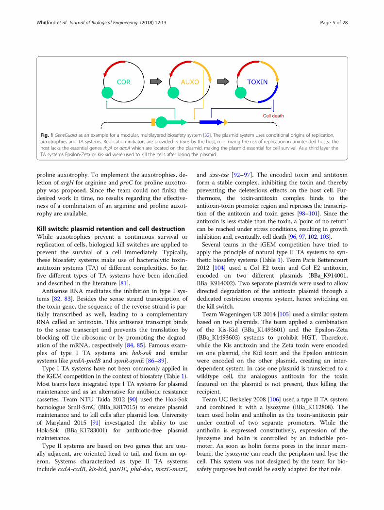
Evaluation of auxotrophic strains for biosafety purposesIn conclusion, auxotrophic GMOs are simple andcost-efficient biocontainment systems with a variety ofpossible applications. Complementing substances can beprovided via the growth media, and multiple auxotro-phies further reduce escape risks. However, auxotrophicsystems suffer from substantial drawbacks, such as pos-sible metabolic cross-feeding, toxic overexpression, andloss of function due to decreased selection pressure in aheterogeneous natural environment (for a summary see[75]). Therefore, instead of relying on an auxotrophysystem alone, they are often part of a complex multi-layered biosafety system, such as SafeGuard [76] or Gen-eGuard [32]. In the GeneGuard plasmid system (Fig. 1),auxotrophies based on translocation of the essentialgenes thyA or dapA to a plasmid location were com-bined with a conditional origin of replication andtoxin-antitoxin system to engineer a host-plasmid mu-tual dependency [32].
Auxotrophy systems in the iGEM competitionCompared to their prominence in research, the imple-mentations of auxotrophic strains in iGEM projects havebeen relatively rare, which is in part due to the fact thatthe iGEM foundation does not collect and provide suchgenetically modified strains, making it necessary for theiGEM teams to either create them themselves or obtainthem from other sources.A successful implementation of an auxotrophic strain
for a biosafety system had been accomplished by teamBielefeld-Germany 2013 [77]. The team created abiosafety system composed of three independent layers,one of which being a D-alanine auxotrophy, establishedby deleting both racemase genes, alr and dadX. Theteam showed that cells grown on media withoutD-alanine supplemented or not carrying a plasmid-bound alanine racemase (BBa_K1172901) were unable togrow. Bielefeld-CeBiTec 2014 [78] adapted the systemfor an antibiotic-free selection, hence eliminating theproblems resulting from extensive use of antibiotics inlaboratories. This underlines the multifaceted possibleapplications of auxotrophies.Other teams have conceptually considered auxotrophic
strains for their projects, whilst not implementing thesethemselves. Team BYU Provo 2014 [79], for example,worked with the bacterium Nitrosospira multiformis inthe context of wastewater treatment and identified serineas being abundant in the bioreactor/sedimentation tank,but not in the waterways. Therefore, it was proposed thatdeletion of serA from N. multiformis would result in a de-pendency for serine, hence making growth outside thebioreactor or sedimentation tank improbable.The biosafety system for Synechocystis sp. suggested by
team Amsterdam 2015 [80] is based on an arginine and
Whitford et al. Journal of Biological Engineering (2018) 12:13 Page 4 of 28
proline auxotrophy. To implement the auxotrophies, de-letion of argH for arginine and proC for proline auxotro-phy was proposed. Since the team could not finish thedesired work in time, no results regarding the effective-ness of a combination of an arginine and proline auxot-rophy are available.
Kill switch: plasmid retention and cell destructionWhile auxotrophies prevent a continuous survival orreplication of cells, biological kill switches are applied toprevent the survival of a cell immediately. Typically,these biosafety systems make use of bacteriolytic toxin-antitoxin systems (TA) of different complexities. So far,five different types of TA systems have been identifiedand described in the literature [81].Antisense RNA meditates the inhibition in type I sys-
tems [82, 83]. Besides the sense strand transcription ofthe toxin gene, the sequence of the reverse strand is par-tially transcribed as well, leading to a complementaryRNA called an antitoxin. This antisense transcript bindsto the sense transcript and prevents the translation byblocking off the ribosome or by promoting the degrad-ation of the mRNA, respectively [84, 85]. Famous exam-ples of type I TA systems are hok-sok and similarsystems like pndA-pndB and symR-symE [86–89].Type I TA systems have not been commonly applied in
the iGEM competition in the context of biosafety (Table 1).Most teams have integrated type I TA systems for plasmidmaintenance and as an alternative for antibiotic resistancecassettes. Team NTU Taida 2012 [90] used the Hok-Sokhomologue SrnB-SrnC (BBa_K817015) to ensure plasmidmaintenance and to kill cells after plasmid loss. Universityof Maryland 2015 [91] investigated the ability to useHok-Sok (BBa_K1783001) for antibiotic-free plasmidmaintenance.Type II systems are based on two genes that are usu-
ally adjacent, are oriented head to tail, and form an op-eron. Systems characterized as type II TA systemsinclude ccdA-ccdB, kis-kid, parDE, phd-doc, mazE-mazF,
and axe-txe [92–97]. The encoded toxin and antitoxinform a stable complex, inhibiting the toxin and therebypreventing the deleterious effects on the host cell. Fur-thermore, the toxin-antitoxin complex binds to theantitoxin-toxin promoter region and represses the transcrip-tion of the antitoxin and toxin genes [98–101]. Since theantitoxin is less stable than the toxin, a ‘point of no return’can be reached under stress conditions, resulting in growthinhibition and, eventually, cell death [96, 97, 102, 103].Several teams in the iGEM competition have tried to
apply the principle of natural type II TA systems to syn-thetic biosafety systems (Table 1). Team Paris Bettencourt2012 [104] used a Col E2 toxin and Col E2 antitoxin,encoded on two different plasmids (BBa_K914001,BBa_K914002). Two separate plasmids were used to allowdirected degradation of the antitoxin plasmid through adedicated restriction enzyme system, hence switching onthe kill switch.Team Wageningen UR 2014 [105] used a similar system
based on two plasmids. The team applied a combinationof the Kis-Kid (BBa_K1493601) and the Epsilon-Zeta(BBa_K1493603) systems to prohibit HGT. Therefore,while the Kis antitoxin and the Zeta toxin were encodedon one plasmid, the Kid toxin and the Epsilon antitoxinwere encoded on the other plasmid, creating an inter-dependent system. In case one plasmid is transferred to awildtype cell, the analogous antitoxin for the toxinfeatured on the plasmid is not present, thus killing therecipient.Team UC Berkeley 2008 [106] used a type II TA system
and combined it with a lysozyme (BBa_K112808). Theteam used holin and antiholin as the toxin-antitoxin pairunder control of two separate promoters. While theantiholin is expressed constitutively, expression of thelysozyme and holin is controlled by an inducible pro-moter. As soon as holin forms pores in the inner mem-brane, the lysozyme can reach the periplasm and lyse thecell. This system was not designed by the team for bio-safety purposes but could be easily adapted for that role.
Fig. 1 GeneGuard as an example for a modular, multilayered biosafety system [32]. The plasmid system uses conditional origins of replication,auxotrophies and TA systems. Replication initiators are provided in trans by the host, minimizing the risk of replication in unintended hosts. Thehost lacks the essential genes thyA or dapA which are located on the plasmid, making the plasmid essential for cell survival. As a third layer theTA systems Epsilon-Zeta or Kis-Kid were used to kill the cells after losing the plasmid
Whitford et al. Journal of Biological Engineering (2018) 12:13 Page 5 of 28
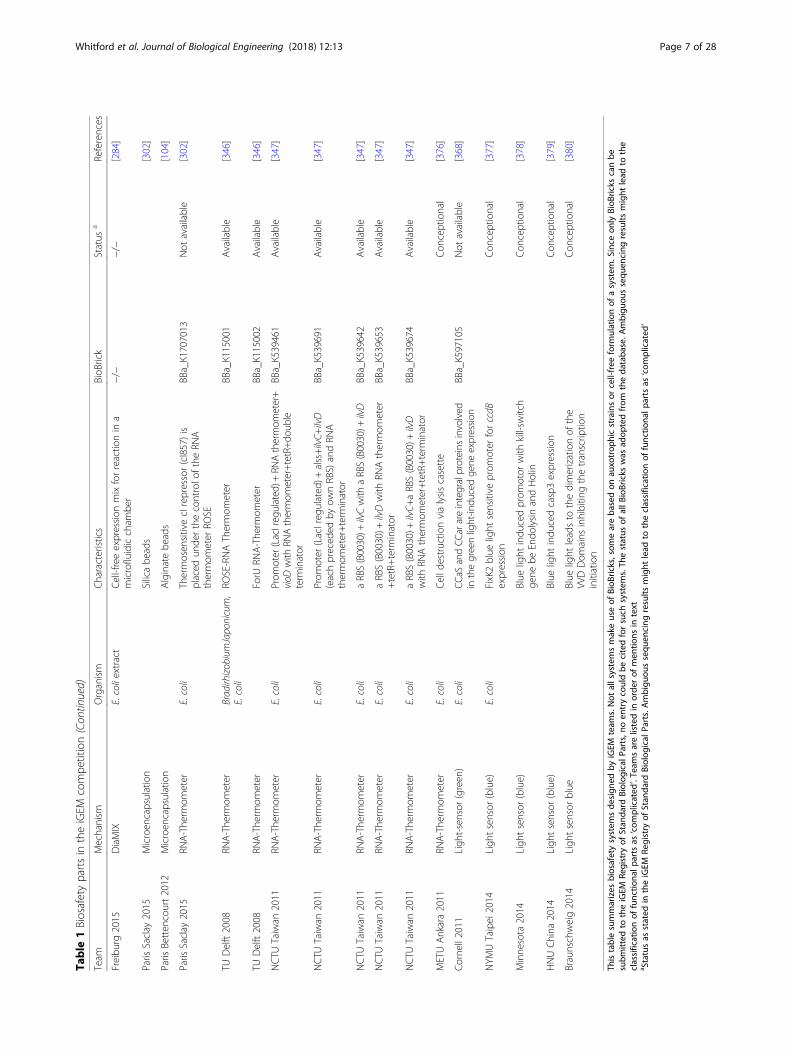
Table
1Biosafetypartsin
theiGEM
compe
tition
Team
Mechanism
Organism
Characteristics
BioB
rick
Status
aReferences
NTU
Taida2012
Type
ITA-system
E.coli
SrnB
-SrnCTA
-system,usedforplasmid
mainten
ance
BBa_K817015
Available
[90]
University
ofMaryland
2015
Type
ITA-system
E.coli
Hok-Sok
TA-system,antibiotic
free
plasmid
mainten
ance
BBa_K1783001
Not
available
[91]
Paris
Betten
court2012
Type
IITA
-system
E.coli
Col
E2TA
-system,located
ontw
oseparate
plasmids
BBa_K914001,Ba_K914002
Available,
complicated
[104]
Wagen
inge
nUR2014
Type
IITA
-system
E.coli
Com
binatio
nof
theKis-Kidandthe
Epsilon-Ze
taTA
-system
topreven
tho
rizon
talg
enetransfer,located
ontw
oseparate
plasmids
BBa_K1493601,
BBa_K1493603
Available,
complicated
[105]
UCBerkeley
2008
Type
IITA
-system
E.coli
Holin-antiholin
TA-system,cou
pled
expression
oflysozyme
BBa_K112808
Com
plicated
[106]
LMUMun
ich2012
Sporulationindu
cedkillswitch
B.subtilis
mazFun
dercontrolo
fPydfG,w
hich
isactivated
inge
rminatingcells
BBa_K823044
Available
[113]
TUEind
hoven2014
Timer-cou
pled
killswitch
E.coli
Oscillatingconcen
trationof
Spo0
AP
indu
cesexpression
ofatoxin
Con
ceptional
[114]
UCBerkley2007
RNase
E.coli
CDSof
Barnase,with
outstartcodo
nBBa_I716211
Available
[124]
BielefeldGermany2013
RNase
E.coli
CDSof
Barnase
BBa_K1172904
Available
[77]
UCLond
on2012
Sequ
ence
specificself-
destructionsystem
E.coli
threefoldactivebiolog
icalcontainm
ent
system
containing
EcoRI/EcoRI
methyltransferase,Holin/Anti-H
olin
Endo
lysin
andCo
licin-E3/Co
licin
Immun
ityE3
BBa_K729009,BBa_K729010
Not
available
[133]
TUMun
ich2013
Sequ
ence
unspecificself-
destructionsystem
Physcomitrellapatens
MatureNucleaseNucAfro
mStaphylococcus
aureus
(Therm
onuclease)
inRFC[25]
BBa_K1159105
Available
[138]
HKU
Hon
gkon
g2015
Sequ
ence
specificandun
specific
self-de
structionsystem
E.coli
CRISPR/Cas9containm
entde
vice
repressedby
arabinoseandtryptoph
anBBa_K1774000
Not
available
[160]
Harvard
2010
Gen
eticfence
E.coli
Barnase:ge
neforthege
netic
fence,
Barstar:Inhibitorof
barnase
−/−
Con
ceptional
[374]
Virginia2016
Dep
ende
ncyon
amod
ified
aminoacid
E.coli
N-carbo
benzyloxy(CBZ
)-cleavage
enzymeto
detach
theprotectin
ggrou
pfrim
aminoacids
BBa_K1879000
Not
available
[375]
Virginia2016
Dep
ende
ncyon
amod
ified
aminoacid
E.coli
Mutatnt
Leucyl-tRN
Asynthe
tase
−/−
Con
ceptional
[375]
Bielefeld-CeBiTec
2015
Cellfreeproteinsynthe
sis
E.coliextract
Mixture
ofam
inoacids,co
factors,cell
extract,NTPs,en
ergy
source,D
NA
template,Mg-
andK-glutam
atesolutio
ns,
nuclease
freewater
forcellfre
eprotein
synthe
sisin
microcentrifug
etube
sor
amulti-wellp
late
−/−
−/−
[280]
Whitford et al. Journal of Biological Engineering (2018) 12:13 Page 6 of 28
Table
1Biosafetypartsin
theiGEM
compe
tition(Con
tinued)
Team
Mechanism
Organism
Characteristics
BioB
rick
Status
aReferences
Freibu
rg2015
DiaMIX
E.coliextract
Cell-freeexpression
mixforreactio
nin
amicrofluidiccham
ber
−/−
−/−
[284]
Paris
Saclay
2015
Microen
capsulation
Silicabe
ads
[302]
Paris
Betten
court2012
Microen
capsulation
Alginatebe
ads
[104]
Paris
Saclay
2015
RNA-The
rmom
eter
E.coli
Thermosen
sitivecIrepressor(cI857)is
placed
unde
rthecontrolo
ftheRN
Athermom
eter
ROSE
BBa_K1707013
Not
available
[302]
TUDelft2008
RNA-The
rmom
eter
BradirhizobiumJaponicum,
E.coli
ROSE-RNAThermom
eter
BBa_K115001
Available
[346]
TUDelft2008
RNA-The
rmom
eter
ForU
RNA-The
rmom
eter
BBa_K115002
Available
[346]
NCTU
Taiwan
2011
RNA-The
rmom
eter
E.coli
Prom
oter
(LacIregulated)+
RNAthermom
eter+
vioD
with
RNAthermom
eter+tetR+do
uble
term
inator
BBa_K539461
Available
[347]
NCTU
Taiwan
2011
RNA-The
rmom
eter
E.coli
Prom
oter
(LacIreg
ulated
)+alss+ilvC+
ilvD
(eachpreced
edby
ownRBS)
andRN
Athermom
eter+term
inator
BBa_K539691
Available
[347]
NCTU
Taiwan
2011
RNA-The
rmom
eter
E.coli
aRBS(B0030)+
ilvCwith
aRBS(B0030)+
ilvD
BBa_K539642
Available
[347]
NCTU
Taiwan
2011
RNA-The
rmom
eter
E.coli
aRBS(B0030)+
ilvDwith
RNAthermom
eter
+tetR+term
inator
BBa_K539653
Available
[347]
NCTU
Taiwan
2011
RNA-The
rmom
eter
E.coli
aRBS(B0030)+
ilvC+
aRBS(B0030)+
ilvD
with
RNAthermom
eter+tetR+term
inator
BBa_K539674
Available
[347]
METUAnkara2011
RNA-The
rmom
eter
E.coli
Celld
estructio
nvialysiscasette
Con
ceptional
[376]
Corne
ll2011
Ligh
t-sensor
(green
)E.coli
CCaS
andCC
arareintegralproteins
involved
inthegreenlight-indu
cedgene
expressio
nBBa_K597105
Not
available
[368]
NYM
UTaipei2014
Ligh
tsensor
(blue)
E.coli
FixK2blue
light
sensitive
prom
oter
forccdB
expression
Con
ceptional
[377]
Minne
sota
2014
Ligh
tsensor
(blue)
Blue
light
indu
cedprom
otor
with
kill-sw
itch
gene
beEndo
lysinandHolin
Con
ceptional
[378]
HNUChina
2014
Ligh
tsensor
(blue)
Blue
light
indu
cedcasp3expression
Con
ceptional
[379]
Braunschweig2014
Ligh
tsensor
blue
Blue
light
leadsto
thedimerizationof
the
VVDDom
ains
inhibitin
gthetranscrip
tion
initiation
Con
ceptional
[380]
Thistablesummarizes
biosafetysystem
sde
sign
edby
iGEM
team
s.Not
allsystemsmakeuseof
BioB
ricks,som
eareba
sedon
auxotrop
hicstrainsor
cell-free
form
ulationof
asystem
.Since
only
BioB
ricks
canbe
subm
itted
totheiGEM
Registry
ofStan
dard
Biolog
ical
Parts,no
entrycouldbe
citedforsuch
system
s.Th
estatus
ofallB
ioBricks
was
adop
tedfrom
theda
taba
se.A
mbigu
oussequ
encing
results
might
lead
tothe
classificationof
functio
nalp
arts
as‘com
plicated
’.Team
sarelistedin
orde
rof
men
tions
intext
a Statusas
stated
intheiGEM
Registry
ofStan
dard
Biolog
ical
Parts.Ambigu
oussequ
encing
results
might
lead
totheclassificationof
functio
nalp
arts
as‘com
plicated
’
Whitford et al. Journal of Biological Engineering (2018) 12:13 Page 7 of 28
Type II TA systems have also been extensively appliedoutside of the iGEM competition. Stirling and colleaguesbuilt two evolutionary stable kill switches to control theenvironment in which a genetically engineered strain ofE. coli can survive [107]. Their “essentializer” kill switchis based on a bi-stable cI/Cro memory switch. Cell deathis induced by loss of the memory switch. The “cryo-death” kill switch was built around a cold-inducible pro-motor, allowing growth at 37 °C. At a temperature of22 °C and below, a survival ratio of less than 10− 5 wasreached (Table 2). Both kill switches were engineeredusing the type II TA system CcdB-CcdA [107].While type I and type II systems are based on interac-
tions of two components of the same kind to mediate in-hibition either via RNA or protein toxin-antitoxincomplexes, type III systems are based on the interactionof antitoxin RNA with the toxin protein [108, 109]. Toour knowledge, only one example for a type III TAsystem has been identified so far: ToxIN from Erwiniacarotovora subsp. atroseptica which acts as an abortiveinfection system [108, 110]. No iGEM team has appliedthis type III TA system as a biosafety system yet.Recently, type IV and V TA systems have been identi-
fied. In type IV systems like yeeU-yeeV, the antitoxin andtoxin do not form a complex. Instead, the antitoxin acts asan antagonist. While YeeV inhibits the assembly of FtsZand MreB filaments, YeeU promotes the reaction, hencecounteracting the toxicity of YeeV [111]. Type V TA systemslike ghoS-ghoT also function without formation of a toxin-antitoxin complex. GhoS possesses a sequence-specific
endoribonuclease activity, cleaving the GhoT mRNA,thereby inhibiting formation of the toxin protein [112].Many teams in the iGEM competition used toxins
under control of specific promoters without a correspond-ing antitoxin to create biosafety systems (Table 1). TeamLMU Munich 2012 [113] created a kill switch for Bacillussubtilis to kill germinating spores (BBa_K823044): theteam placed mazF under control of PydfG. This promoteris activated by the sigma factor of RNA polymerase ECF41which is produced during sporulation, thus the team cre-ated a sporulation-induced kill switch. Team TU Eindho-ven 2014 [114] proposed a timer-coupled kill switch basedon the oscillating concentration of phosphorylated Spo0Aprotein (Spo0AP). Once a certain concentration ofSpo0AP is reached, expression of a toxic gene under thecontrol of a Spo0AP-sensitive promoter will be induced,leading to cell death.The ribonuclease ba of Bacillus amyloliquefaciens
(barnase) and the corresponding inhibitor barstar aresometimes referred to as a toxin-antitoxin system whencombined [95]. Whilst the organization of barstar andbarnase genes differ from the organization of naturaltoxin and antitoxin systems, the combination of barnaseand barstar exhibits many similarities to naturalTA-systems. For biosafety applications, the inhibitorgene barstar is usually integrated in the chromosomeand constitutively expressed. Barnase is encoded on aplasmid, preferably under control of an inducible or re-pressible promoter. Furthermore, the fusion of thebarnase to secretion signals like that of PhoA allows the
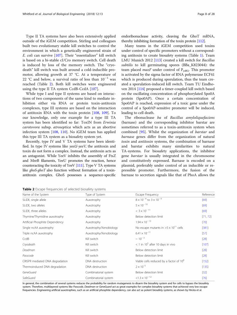
Table 2 Escape frequencies of selected biosafety systems
Name of the System Type of System Escape Frequency Reference
SLiDE, single allele Auxotrophy 8 × 10− 4 to 3 × 10− 9 [69]
SLiDE, two alleles Auxotrophy 5 × 10− 10 [69]
SLiDE, three alleles Auxotrophy < 3 × 10− 11 [69]
Thymine/Thymidine auxotrophy Auxotrophy Below detection limit [71, 72]
Artificial Phosphite Dependency Auxotrophy 1.94 × 10− 13 [70]
Single ncAA auxotrophy Auxotrophy/Xenobiology No escape mutants in >5 × 10¹¹ cells [381]
Triple ncAA auxotrophy Auxotrophy/Xenobiology 6.41 × 10⁻¹¹ [57]
CcdB Kill switch ~ 10− 3 [28]
Cryodeath Kill switch < 1 in 105 after 10 days in vivo [107]
Deadman Kill switch Below detection limit [28]
Passcode Kill switch Below detection limit [28]
CRISPR mediated DNA degradation DNA destruction Viable cells reduced by a factor of 108 [152]
Thermoinduced DNA degradation DNA destruction 2 × 10⁻⁵ [135]
GeneGuard Combinatorial system Below detection limit [32]
SafeGuard Combinatorial system <1.3 × 10− 12 [76]
In general, the combination of several systems reduces the probability for random mutagenesis to disarm the biosafety system and for cells to bypass the biosafetysystem. Therefore, multilayered systems like Passcode, Deadman or GeneGuard act as great examples for complex biosafety systems that achieved very low escapefrequencies. Engineering artificial auxotrophies, such as an artificial phosphite dependency, can also act as potent biosafety systems, as shown by Hirota et al.
Whitford et al. Journal of Biological Engineering (2018) 12:13 Page 8 of 28
secretion into the periplasm [115–121]. Secreted barnasecan be toxic to bacteria in the proximity of the produ-cing cells if they do not express the inhibitor barstar, butthe exact mechanism remains unknown [120, 121]. Incombination with another biosafety systems whichrepresses the expression of barnase through arabinose-inducible promoter like PBAD, the expression of barnasecan be induced if that biosafety mechanism fails, hencekilling the host cell [122, 123].Many iGEM teams employed a barnase system, but
only few used it solely for biosafety purposes. Team UCBerkley 2007 [124] was the first to use the barnase(BBa_I716211) under control of the promoter PBAD toinduce self-destruction.Team Bielefeld-Germany 2013 [77] improved the bar-
nase based biosafety system by using it in combinationwith a D-alanine auxotrophic strain (Δalr) as a two-partsystem (BBa_K1172904). The first part contains the re-pressor for the promoter of the second part (PBAD) andthe alanine racemase, both under the control of arhamnose-inducible promoter (PRha). The second partcontains the PBAD promoter and barnase itself. Shouldthe first promoter remain inactive due to auxotrophyfailures, barnase is expressed and will lead to cell death.Team Valencia UPV 2014 [125] aimed to develop a
biosafety module to prevent the spread of genetic
material in plant seeds. The concept was to use barnase(BBa_I716211) in combination with the tapetum-specificpromoter TA29. Due to time restrictions, this conceptcould not be tested.More complex kill switches like Deadman and Pass-
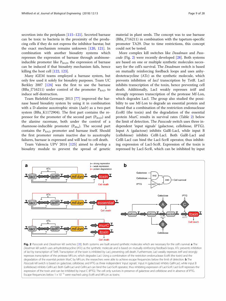
code (Fig. 2) were recently developed [28]. Both systemsare based on one or multiple synthetic molecules neces-sary for the cell’s survival. The Deadman switch is basedon mutually reinforcing feedback loops and uses anhy-drotetracycline (ATc) as the synthetic molecule, whichprevents inhibition of lacI transcription by TetR. LacIinhibits transcription of the toxin, hence preventing celldeath. Additionally, LacI weakly represses tetR andstrongly represses transcription of the protease Mf-Lon,which degrades LacI. The group also studied the possi-bility to use Mf-Lon to degrade an essential protein andfound that a combination of the restriction endonucleaseEcoRI (the toxin) and the degradation of the essentialprotein MurC results in survival rates (Table 2) belowthe limit of detection. The Passcode switch uses three in-dependent ‘input signals’ (galactose, cellobiose, IPTG).Input A (galactose) inhibits GalR-LacI, while input B(cellobiose) inhibits CelR-LacI. Both GalR-LacI andCelR-LacI can bind the LacI-ScrR operator, thus inhibit-ing expression of LacI-ScrR. Expression of the toxin isrepressed by LacI-ScrR, which can be inhibited by input
Fig. 2 Passcode and Deadman kill switches [28]. Both systems are built around synthetic molecules which are necessary for the cell’s survival. a TheDeadman kill switch uses anhydrotetracycline (ATc) as the synthetic molecule and is based on mutually reinforcing feedback loops. ATc prevents inhibitionof lacI by transcription of TetR. Transcription of the toxin is inhibited by LacI, preventing cell death. Furthermore, LacI weakly represses tetR and stronglyrepresses transcription of the protease Mf-Lon, which degrades LacI. Using a combination of the restriction endonuclease EcoRI (the toxin) and thedegradation of the essential protein MurC by Mf-Lon, the researchers were able to achieve escape frequencies below the limit of detection. b ThePasscode kill switch is based on galactose, cellobiose, and IPTG as three independent ‘input signals’. Input A (galactose) inhibits GalR-LacI, while input B(cellobiose) inhibits CelR-LacI. Both GalR-LacI and CelR-LacI can bind the LacI-ScrR operator, thus inhibiting expression of LacI-ScrR. LacI-ScrR represses theexpression of the toxin and can be inhibited by input C (IPTG). The cell only survives in presence of galactose and cellobiose and in absence of IPTG.Escape frequencies below 1 × 10− 6 were reached using EcoRI and Mf-Lon as toxins
Whitford et al. Journal of Biological Engineering (2018) 12:13 Page 9 of 28
C (IPTG). Therefore, the cell only survives in presenceof galactose and cellobiose and in absence of IPTG.Using again EcoRI and Mf-Lon as toxins, cell survival ra-tios below 1 × 10⁻⁶ were reached.
Kill switch: DNA destructionKill switch systems for GMOs are widely used as bio-safety systems. But, depending on the TA type (I to V)used, genetic information can potentially be released andspread through HGT [26, 32, 126, 127]. Therefore, gen-etic information has to be released, taken up by anotherorganism, maintained and expressed [26]. If the uptakeof this genetic information has an evolutionary advan-tage for the recipient cells, for example the informationfor an antibiotic resistance gene, it is likely to remain inthe genome. Thus, genetic incorporation can lead to re-sistance to antibiotics or other advantages for recipientcells. Adapting a combination of toxin-antitoxin systemsand self-destruction systems prevents the spread of re-combinant DNA by the degradation of nucleic acids,while the targeted cells are killed. Therefore, self-de-struction can be seen as a special case of a kill switchsystem. All systems based on destruction of nucleic acidscan be loosely subdivided into three systems dependingon the sequence specificity of used systems: (i) specificsystems are based on nucleases which hydrolyze nucleicacids at sequence specific sites known as restrictionsites, (ii) unspecific systems utilize nucleases whichhydrolyze DNA and are not sequence specific and (iii) athird system which is a combination of sequence specificand unspecific parts.Specific systems of self-destruction are toxin-antitoxin sys-
tems based on nucleases applied to kill cells with minor riskof DNA leakage. Usually, the nuclease (toxin) is encoded onthe plasmid, while the nuclease-methyltransferase (antitoxin)is encoded on the chromosome. Both nuclease and nuclease-methyltransferase compete for the same sequence-specificDNA recognition site. Thus, expression levels between thistoxin-antitoxin gene pairs need to be adjusted. The toxinis constitutively expressed and the expression of the anti-toxin is induced, for instance, by using an anhydro-tetra-cyclin-responsive promoter [76]. When expressed thenuclease-methyltransferase methylates certain nucleobasesat the recognition site, hence preventing cleavage of theDNA by the nuclease. This modification preventsdigestion of the cell’s own DNA. A known toxin-antitoxinsystem is EcoRI/EcoRI methyltransferase [128–132].Destruction of the genomic information is initiated byswitching off the expression of the antitoxin-methyl-transferase. Even if HGT occurs, the recipient cellsare unlikely to counteract the nuclease due to thelack of methyltransferase.The team University College London 2012 [133] used
the EcoRI/EcoRI methyltransferase system in their novel
threefold active biological containment system (Table 1)in combination with holin/anti-holin endolysin andcolicin-E3/colicin Immunity E3 (BBa_K729009, BBa_K729010). The aim was to minimize HGT via bacterialconjugation using this new system.At the end of the last century, genes encoding se-
quence specific restriction nucleases under different pro-moters were characterized for inducible degradation ofDNA [134]. Stephen Cuskey performed preliminary testswith EcoRI under control of an inducible promoter asmentioned by Molin et al. 1993 [134]. Results showedthat growth was reduced due to a high base level of geneexpression without induction. Double strand breakscaused by EcoRI at a high rate are responsible for slowgrowth, as the repair mechanisms are insufficient to fixthem [134].Nonspecific nucleases, which introduce single strand
breaks, combined with an inducible promoter, a ribo-some binding-site, and a start codon, appeared to be bet-ter candidates [134, 135]. Molin and Ahrenholtz bothtested the extracellular nuclease of Serratia marcescens[134, 135]. The nucA gene encodes a 266 amino acidlong polypeptide with a 21 amino acid long N-terminalleader peptide [136, 137]. In both cases, the nucA genewithout the leader peptide was under control of aninducible promoter lac and lambda pL, respectively[134, 135]. NucA is one example for an unspecificself-destruction system.The team TU Munich 2013 [138] used the thermonu-
clease NucA of Staphylococcus aureus (BBa_K1159105) todegrade DNA of a genetically modified moss (Table 1).The nuclease (BBa_K1159111) is bound to the membranewith a transmembrane domain (BBa_K1159315) and con-tains a TEV cleavage site as well as a SV40 nuclearlocalization signal (NLS) (BBa_K1159303). After PhyB(BBa_K801031) is activated with red light, it binds eitherprotein PIF3 (BBa_K1159103) or PIF6 (BBa_K1159104).This binding initiates the assembly of the N- andC-terminal split TEV protease which in turn cleaves theTEV cleavage site thus releasing the nuclease; this is trans-located into the nucleus where the DNA is degraded.Team NTU-LIHPAO-Taiwan 2015 [139] also used the
thermonuclease NucA of S. aureus (BBa_K1159105)under control of a lambda cl-regulated promoter(BBa_R0051) to degrade DNA, thus killing Lactobacilluscasei if the cl protein (BBa_C0051) concentration de-creased below a certain threshold. The expression of clis controlled by the lac-promoter and thereforeregulated by lactose and glucose. The whole system ishypothetically designed to inhibit HGT from L.casei tobacteria in the human gastrointestinal tract while con-trolling the proliferation of the cells.While sequence specific and unspecific systems may
be of use in general self-destruction, the expression
Whitford et al. Journal of Biological Engineering (2018) 12:13 Page 10 of 28
controls can be leaky leading to a baseline expressionwhich is damaging to the cells even if they are con-tained. Also, both systems are not able to target specificsites which encode vital enzymes and proteins and arethus inefficient at mediating cell death. A combinatorialsystem of sequence specific and unspecific parts can beadapted for high-efficiency, easily controllable biosafety.A self-destruction system which is based on sequence
specific and unspecific parts is the clustered regularlyinterspaced short palindromic repeats (CRISPR)/CRISPRassociated (Cas) system - the RNA-mediated adaptivedefense systems of bacteria [140–144] Shortly after itsdiscovery, it was adapted for genome and transcriptomeediting [145–149]. The sequence-specific CRISPR is usedto guide the sequence-unspecific Cas nuclease to its tar-get, thus making this system highly regulated withoutthe risk of uncontrollable cleavage. Employing the samemechanism on essential and non-essential genes enablescontrolled degradation to prevent HGT in the event ofan unintentional release [150–152]. In nature three typesof CRISPR/Cas systems are specified with variationsconcerning target and mechanism: type I systems cleaveand degrade DNA; type II systems solely cleave DNA;and type III systems cleave DNA and RNA [153]. Forthe purpose of self-destruction, the type I system is wellsuited and frequently employed [151, 152]. Type I andtype II systems are dependent on (i) CRISPR RNAspacer and target protospacer sequence complementarityas well as (ii) the protospacer-adjacent motif (PAM)[154–157]. By combining different Cas proteins andPAM sequences, a broad range of applications can beenabled influencing the kinetics of target degradation[151–153, 158]. Type III systems also require spacer-protospacer complementarity and specific sequences inthe neighborhood of the protospacer [159]. Specific ONand OFF states of expression should be defined whenemploying the CRISPR/Cas mechanism for degrad-ation of DNA to ensure induction of expression ofboth components in response to specific environmen-tal changes [152].Team HKU Hongkong 2015 [160] designed a CRISPR/
Cas9 system with a specific sgRNA (BBa_K1774000) totarget the DNA polymerase III alpha subunit (dnaE)thus inhibiting replication of the bacteria. The OFF statewas defined by the availability of arabinose and trypto-phan. Arabinose induced the expression of araC whichin turn induces the expression of cl under control of thePBAD promoter, thus inhibiting the PR promoter and theexpression of cas9. If available, tryptophan binds to a re-pressor which in turn blocks the trp promoter and thussgRNA expression. In the ON state, arabinose andtryptophan are not available, which mimics a possiblephysical containment breach. A lack of transcriptionalrepression results in the formation of Cas9 and the
sgRNA, which can destroy the gene of DNA polymeraseIII alpha subunit.Given that kill switches are prone to inactivating point
mutations [161], especially when constitutively expressed,researchers have developed new biosafety systems that donot harm the host, potentially minimizing the risk of unin-tended proliferation caused by mutagenesis. Jia and col-leagues developed an orthogonal ribosome biofirewall,consisting of an activation circuit and a degradation circuit[162]. The activation circuit, a genetic AND gate, utilizesan orthogonal ribosome to activate an encrypted pathwaybased on specific environmental inputs. The genes encod-ing the orthogonal ribosome can be degraded by the deg-radation circuit, a genetic NOT gate, based on a change ofthe environmental inputs. This elegant system not onlyminimizes the burden on the host, given that the toxinI-SceI is not constitutively expressed, but also makes ex-pression of the genes of interest dependent of the pres-ence of the orthogonal ribosome. Therefore, even if HGToccurs, expression of the genes of interest is prevent dueto the lack of an orthogonal ribosome. The plasmid con-taining the genes for the orthogonal ribosome is digestedin absence of specific environmental inputs. This workhighlights how conditional degradation of genetic infor-mation can be combined with genetic encryption to createan adaptable and tightly regulated biofirewall for microbialbiocontainment [162].
Xenobiology-based containmentThe term ‘xenobiology’ has experienced significant se-mantic shift over the last few decades [163–165].Kubyshkin et al. define xenobiology as an approach toexpand the framework of natural chemistries withnon-natural building blocks in living cells to accomplishartificial biodiversity [50]. Therefore, one key aspect ofxenobiology is the search for alternative chemistry fornucleic acids, proteins and other cellular componentsand functions. Xenobiological systems are also referredto as orthogonal systems or chemically modified organ-isms (CMOs) [50, 163]. Current biosafety systems aremeant to kill cells once they escape from the assignedenvironment, leaving their recombinant DNA freelyavailable in nature [166]. An orthogonal system preventsHGT [167] through transduction [168], conjugation, andtransformation, as reviewed by Davison [169]. As a con-sequence, wild type cells are unable to integrate andmaintain XNA into their genome and cannot handle theincorporation of ncAAs [170, 171]. Thus, XNA canpotentially become a powerful biosafety tool by prevent-ing HGT as it should not be read properly by wild typeDNA processing enzymes like DNA and RNA polymer-ases [164].The classification of xenobiology is not consistent.
Some categorize it along with trophic and semantic
Whitford et al. Journal of Biological Engineering (2018) 12:13 Page 11 of 28
containment for the prevention of metabolic and geneticexchange [171]. Giving this inconsistency, the followingchapters are categorized according to the componentsthat could be changed or alienated like the bases, back-bone, leaving group, codons and amino acids.
Synthetic bases – building up Xeno-DNADuring the emergence of recombinant DNA technology,a plasmid containing DNA of another species was de-scribed as Xeno-DNA (XNA) [20]; the modern percep-tion of XNA describes non-canonical DNA buildingblocks or substantial modifications of the natural struc-ture, such as alternative pairing nucleotides, modifiedsugars, or backbones [165, 172, 173]. XNA could be con-sidered a genetic firewall masking the encoded informa-tion from nature [163]. The main approach of designingXNA is to replace or extend the standard genetic codecomprising four naturally occurring nucleotides in theDNA. There are various sophisticated approaches toidentify potential replacements for the four canonicalbases [174–181]. Nevertheless, advances in XNA tech-nology have to fulfill some requirements to establishstable products in vivo [172, 182].First experiments extended the four nucleotide alpha-
bet by replacing thymine with 5-chlorouracil in E. coliover a period of 25 weeks [183, 184]. Other approachesexpanded the genetic alphabet by introducing the twoartificial bases dP (2-amino-8-(1′-β-D-2′-deoxyribofura-nosyl)-imidazol[1,2-α]-1,3,5-trizan-4(H)-one), and dZ(6-amino-5-nitro-3-(1′-β-D-2′-deoxyribofurano-syl)-2(1H)-pyridone) [185]. These artificial bases pairwith three hydrogen bonds but vary in the pattern ofdonor and acceptor groups. A Taq DNA polymerase wasmodified to accept the new ATCGPZ-DNA, resulting ina retention rate of 98.9% [186, 187]. Moreover, a T7RNA polymerase and a reverse transcriptase were devel-oped for an RNA product containing P and Z [188]. Thesix nucleotide genetic alphabet will lead to DNA with aB-form as well as an A-form, with the major groves be-ing 1 Å wider than the natural G:C pair [189].Interestingly, the concept of DNA can be extended be-
yond bases and pairing through hydrogen bonds. For ex-ample, pairings dependent on metal ion coordination[190–192] or hydrophobic interactions [193] wereexplored recently. Two promising candidates usinghydrophobic interactions are d5SICS – dMMO2 andd5SICS-dNaM, which allowed transcription [193]. Thefirst demonstration in E. coli was based on one plasmidencoding the nucleoside triphosphate transporter fordNaM and d5SICS and the other plasmid encoding a genesequence using the extended genetic code [194]. Uptakeof the synthetic bases as well as a stable plasmid replica-tion over 24 generations was demonstrated [194]. In 2017,the Romesberg group presented a new version of their
semi-synthetic organism. The most important advanceswere an optimized transporter with improved uptake ofunnatural triphosphates and better retention of XNA withdNaM-dTPT3. Furthermore, they used a CRISPR-Cas sys-tem to eliminate plasmids that lost the XNA [177].Besides expanding the alphabet of canonical DNA,
XNA provides the opportunity to change the generaltopology. For example, benzo homologation provides theopportunity to expand the physical DNA size. The benzoexpansion of pyrimidines to create dxT and dxC resultsin expanded DNA (xDNA), with the size increasingabout 2.4 Å and the helix becoming more thermallystable [195, 196]. Stable replication of a plasmid contain-ing up to eight xDNA bases in a GFP encoding sequenceas well as expression of the altered gfp gene was demon-strated in E. coli [197]. By further changing the vector ofextension, wide DNA (yDNA) can be obtained [198],although stable replication of this DNA type is problem-atic [199].Use of XNA often necessitates synthetic or evolved
proteins that allow for replication, transcription, andDNA packaging of the XNA. All presented examples de-pend on supplementation of the non-canonical nucleo-tides. This auxotrophy is a potent biosafety mechanism,which does not just prevent an uncontrolled growth ofthe engineered cells in the environment, but also pro-tects the encoded information from spreading throughHGT [163, 171].
Alternative XNAs: modifying backbone or leaving groupBesides the incorporation of non-canonical bases, exper-iments to engineer the DNA backbone by integratingsubstitutes for deoxyribose and ribose have been per-formed. For biosafety purposes, an altered backboneneeds to meet the requirement to build a functionalhelix that does not interact with natural replication en-zymes, instead requiring adjusted or even syntheticenzymes [172]. Some candidates for alternative back-bone chemistries have previously been investigated asreviewed by Herdewijn and Marlière [172]; these exam-ples include: hexitol nucleic acid (HNA) [200], threose(TNA) [201, 202], glycerol (GNA) [203], and cyclohex-ene (CeNA) [204]. A less complex method compared tothe substitution of the whole genomic backbone is utiliz-ing an orthogonal XNA episome which contains essen-tial genes [172].Besides the bases and the backbone, the third
potential target to design XNA is alternating theleaving group of NTPs by replacing the pyrophos-phate, such that they cannot be recognized by wildtype polymerases. Studied analogues of ATP aremethylene phosphonate, phosphoamidate [205], andthiophosphonate [206]. An alternative leaving groupneeds a high energy bond for the polymerization
Whitford et al. Journal of Biological Engineering (2018) 12:13 Page 12 of 28
process. Studies on L-aspartate and L-histidine linkednucleotidemonophosphates showed that aspartic acidphosphoramidate derivates are working substrates forthe HIV reverse transcriptase [207].
Amber codon and non-canonical amino acidsBy systematically expanding the approach of auxotro-phies based on xenobiotic compounds to utilizing awhole orthogonal genetic code, e.g. by means of xeno-biology, the spread of recombinant sequences can alsobe prevented. There are three stop codons in the geneticcode: ochre (UAA), opal (UGA) and amber (UAG) [208].In E. coli, the amber codon is least common, with justover 300 occurrences (depending on the E. coli strainused). In 2013, Lajoie and colleagues coined the termgenetically recoded organism (GRO) to describe organ-isms with an alternative genetic code [209]. Such GROshave been developed for enhanced biosafety comparedto natural amino acid auxotrophs by engineering organ-isms to become auxotrophic for non-canonical aminoacids (ncAAs) [58]. Rovner et al. constructed GROsbased on E. coli without any TAG codon and the possi-bility to terminate translation at the UAA and UAG co-dons. After recoding the TAG codon to a sense codonfor ncAAs by means of an orthogonal translation system(OTS), the recoded codon was incorporated into essen-tial genes of the organism, thus making it dependent onncAAs. Sixty variants of the auxotroph-GROs with vary-ing growth and containment rates have been isolated,with one strain containing three recoded TAG codonsmaintaining stable growth, and undetectable escape fre-quencies over the course of 1 week or 20 days on solidor in liquid media, respectively. Changing the ambercodon into a sense codon has been done multiple timesby various groups [210–217], but results in the mistrans-lation of all genes using this stop codon. Therefore, theChurch lab presented a GRO in which all 314 UAG stopcodons were replaced by UAA stop codons. Deletion ofrelease factor 1 (encoded by prfA), which recognizesUAG and UAA, then allows for recoding of the amberstop codon [218, 219]. Engineering a new aminoacyl-tRNA synthetase (aaRS) and corresponding tRNA leadsto an orthogonal translation machinery required to har-ness the potential of an amber-free strain [220]. Thenon-canonical amino acid (ncAA) L-4,4′-biphenylala-nine (BPA) had been integrated via the UAG stop codonof the GRO E. coli strain C321.ΔA by Mandell et al..This resulted in auxotrophs designed to be dependenton ncAAs [57]. Thus, the essential enzymes of the strainrequired BPA for core functions, such as translation.Additionally, in a proof-of-concept study, an E. colistrain BL21-AI (IY, lamB-immE3) containing the syn-thetic essential gene immE3 had been constructed, thetranslation of which depends on supplementation of the
medium with the non-canonical amino acid 3-iodo-L-tyrosine [221]. As an alternative to reassigning stopcodons, it is also possible to reassign sense codons toncAAs. In order to ensure proper functioning of the al-tered strain, rarely-used sense codons have been used,such as the codon AGG, which usually codes for argin-ine and has been reassigned to code for the ncAAL-homoarginine in E. coli [222]. Such an approach hasbeen explored with other sense codons, such as AUG[223], and as a combination of sense and stop codon re-assignment [224]. Reassigning single codons is not theonly way to incorporate ncAAs to the genetic code. In-stead, Hoesl and colleagues [225] have evolved culturesof E. coli to grow on a non-canonical amino acid alterna-tive to L-tryptophan (L-β-(thieno[3,2-b]pyrrolyl)alanine)in a long-term cultivation experiment. While cells werecapable of surviving in the total absence of L-tryptophan,they were still able to grow when L-tryptophan waspresent. In the future, further strain engineering mightprovide an evolutionary approach to altered strainsdependent on ncAAs.The resulting combination of trophic and semantic
containment constitutes a powerful biosafety system.While not preventing the transfer of genetic material inthe first place, the recoded DNA cannot be expressedafter an HGT event in natural organisms. Moreover,HGT of sequences encoding an aaRS and the corre-sponding tRNA are either lethal or very detrimental innatural, non-recoded organisms as they will lead to mis-translation of amber containing genes.In 2006, Wang et al. recoded the amber codon to imple-
ment a non-canonical amino acid. The source organism ofthe altered tRNA and aminoacyl tRNA synthetase fortyrosine was Methanococcus jannaschii [226]. After engin-eering, this system was able to insert O-methyl-L-tyrosinein a gene encoding the dihydrofolate reductase. Anotherexample, based on a translation switch controlled by theabsence of 3-iodo-L-tyrosine [221, 227], is described inthe section on auxotrophy-based systems.Shifty codes [228] are used to encode the same prod-
uct as expected in the wild type, but are based onquadruplets and orthogonal ribosomes. The evolution oforthogonal ribosomes translating a quadruplet codeprovides the amazing opportunity to assign 256 blankcodons. Neumann et al. [229] evolved a synthetic ribo-some (ribo-Q1), whose decoding fidelity was as high as inwild type ribosomes [230]. They tested the incorporationof two non-canonical amino acids, p-azido-L-phenylalan-ine (AzPhe) and N6-[(2-propinyloxy)carbonyl]-L-lysine)(CAK), encoded by a combination of a quadruplet codonAGGA and the amber codon UAG in E. coli. The proteinwas only completely synthesized when both non-canonical amino acids were encoded in the DNA[229, 231, 232].
Whitford et al. Journal of Biological Engineering (2018) 12:13 Page 13 of 28
Application of xenobiology-based containment in the iGEMcompetitionIn 2012, team Paris Bettencourt [104] worked on an ex-tensive biosafety project (Table 1). A semantic contain-ment part was based on an amber mutation in the geneconferring kanamycin resistance (BBa_P1003). Theobjective was to prevent expression of the antibiotic re-sistance gene in wild type bacteria cells after a HGTevent. Two parts were constructed to realize the ambersuppression in E. coli MG1655: BBa_K914000 encodingPlac-supD-T: tRNA amber suppressor and BBa_K914009:P1003*Ser133 encoding a kanamycin resistance gene withone amber mutation at a serine residue at position 133.The team demonstrated that the amber codon was ef-
fectively recoded. The growth rate and level of resistancewere not significantly decreased compared to the straincarrying the original kanamycin resistance gene as wellas the tRNA amber suppressor. However, the culturewithout the tRNA amber suppressor reached a higherOD600 value, because other amber stop codons on thechromosome were also suppressed. Interestingly, theyfound that a single amber mutation was quickly over-come by mutations, a problem that could be addressedby introducing a second amber mutation in the kanamy-cin resistance gene. Thus, amber codons within anti-biotic resistances are an effective way to prevent the easyspread of such resistances. However, further improve-ments of the system are needed to prevent the HGT ofthe amber suppressing tRNA.The team TU Darmstadt 2016 [233] combined auxo-
trophic incorporation of a non-canonical amino acid anda reporter for low levels of the ncAA (BBa_K1416000,BBa_K1976025) [234] designed by the team AustinTexas 2014 [235]. Amber codons were placed at the be-ginning of a Colicin E2 immunity protein [236] and themutated Zif23-GCN4 repressor (F4OMT), a dimericCis2His2 zinc finger protein [237]. In case of the ab-sence of the ncAA, both proteins cannot be translated,resulting in expression of the reporter system mVenus[238] under control of a Zif23-GCN4-controlled pro-moter and subsequent initiation of the suicide reaction.However, no results were reported on the expression ofthe reporter and the OMT-RS expression under a T7promoter (BBa_K525998).Team Bielefeld-CeBiTec 2017 worked on expanding the
genetic code with the unnatural base pair formed betweenisoguanosine (isoG) and 5-methyl-isocytosine (isoCm), andnon-canonical amino acids [239]. The team used CRISPR/Cas9 to retain the unnatural base pair in a specified se-quence. Furthermore, the algae transporter PtNTT2 wasused to facilitate uptake of the unnatural nucleoside tri-phosphates. Given that this transporter can also facilitatetransport of ATP, it may also be used to engineer an artifi-cial ATP auxotrophy. By recoding the amber codon, the
team incorporated ncAAs in a number of proteins. Culti-vations showed growth defects if the desired ncAA wasnot supplemented. The cells were still able to grow as theaaRS, with lower affinity, also incorporated endogenousamino acids. Therefore, engineering artificial ncAA auxot-rophies requires highly specific aaRS.
Minimal genomeSometimes referred to as the “holy grail” of synthetic biol-ogy [240–243] the minimal genome is defined as a set ofgenes which are essential for survival of the cell [244] inan environment containing all required supplements forlife [240]. The size of a minimal genome depends on thesurrounding environment [245]. Therefore, the sets of re-quired genes differ slightly as reviewed by Gil and col-leagues [246]. Most approaches rely on transposonmutagenesis [247] or antisense RNA [248, 249] to identifyessential genes [250]. Nevertheless, it is hard to determinethe minimal set of essential genes, since an essential func-tion might be encoded by two or more genes, thus result-ing in false negative assignments [245, 251].Minimal genomes can be constructed via “top down”
or “bottom up” approaches, with “top down” being thesystematic deletion of redundant genes, while “bottomup” describes the synthesis and assembly of a genomewith the minimal set of genes [252]. Numerous, sophisti-cated attempts were made to identify essential genes ofan organism in order to construct a minimal genome ofHaemophilus influenzae [253], H. influenza andStreptococcus pneumoniae [254], Mycoplasma genitalium[255, 256] S. aureus [249], Buchnera spp. [257], Saccha-romyces cerevisiae [258], Corynebacterium glutamicum[259] and E. coli [260–262].In M. genitalium, which had the smallest known gen-
ome at that time [263], global mutagenesis led to theidentification of 265 essential genes [256] which alignswell with the published predicted minimal genome con-sisting of 256 genes [264]. An 1.08 Mbp synthetic gen-ome sequence of M. mycoides was assembled andtransplanted into an M. capricolum recipient cell produ-cing M. mycoides JCVI-syn1.0 [265]. Latest research hasled to the development of a minimal genome of M.mycoides called JCVI-syn3.0 [245]. Based onJCVI-syn1.0, the genome reduction was achieved in2016 in the new synthetic 532 kbp genome ofJCVI-syn3.0 [245]. It contains the minimal set of essen-tial genes, although the function of 149 genes remainedunknown. Further reduction processes identified quasiessential genes, which were not required for survival,but contributed significantly to stable growth [245]. Cellswith a reduced genome showed a duplication time of180 min, which is substantially different to the duplica-tion time of 60 min of JCVI-syn1.0 with a gene set forrobust growth [245].
Whitford et al. Journal of Biological Engineering (2018) 12:13 Page 14 of 28
A bacterial cell with a minimal genome offers variousapplications in biosafety. The cells would depend oncomplex media as well as on stable conditions. There-fore, survival in a natural environment with fluctuationsin environmental conditions is improbable. However,HGT is still an issue in this scenario, which is why werecommend aiming for a combination with self-destruc-tion systems as described above.Since the iGEM competition is about the construction
of plasmid-based BioBricks, the development of a min-imal genome would be challenging for the teams. Still,teams like Alberta 2009 [266], ETH Zurich 2008 [267],and UESTC 2015 [268] developed concepts and imple-mented software to identify minimal gene sets based onbacterial genome sequences.
Cell-free systemsThe main concerns from a biosafety perspective are thepossible release of GMOs into the environment andHGT between engineered and wild type organisms.Cell-free protein synthesis (CFPS) represents a promis-ing possibility to eliminate most of these risks. The firstresearchers to show that disrupted bacterial cells can beused for in vitro protein synthesis were Gale and Folkesin 1954 [269]. In 1961, Nirenberg and Matthaei con-ducted pioneering work using E. coli cells and showedthat template RNA is a requirement for cell-free proteinsynthesis [270]. CFPS is feasible for a variety of applica-tions, including synthetic biology, vaccine productionand protein engineering [271–273].There are two main strategies for CFPS. The first,
older one is based on crude cell extracts from thedesired cells. While the necessary crude extracts are easyto prepare, fast energy depletion and degradation by pro-teases and nucleases pose two major problems [274–276].To counter those problems, the PURE (“protein synthesisusing recombinant elements”) system developed by Shi-mizu et al. 2001 [276] can be used. This cell-free system isbased on purified (His)-tagged translation factors and canbe programmed by natural mRNA. For biosafety reasons,it is important to effectively remove all living cells beforedeploying a CFPS system outside of the lab. While stand-ard methods for the preparation of cell-extracts arealready highly effective, they still do not provide acompletely cell-free extract [277, 278]. Protocols based onsterile filtration and lyophilization can provide a muchmore sterile extract, minimizing the risk of accidental re-lease of genetically modified organisms into the environ-ment [278]. To fully circumvent the risk of an unsterilecell-extract, the aforementioned PURE system can be de-ployed. This system is not only safer, but also has lowerenergy consumption relative to S30 cell extract systems(cell extracts cleared from heavier components by centri-fugation at 30,000 xg), with greater productivity. However,
Shimizu et al. could not top the productivity of400 μg/ml with a S30 extract when they first pub-lished their results [276, 279].Several iGEM teams used cell-free systems for their
projects. Bielefeld-CeBiTec 2015 (Table 1) used cell ex-tracts of E. coli KRX and ER2566 strains to producesfGFP as part of a paper-based biosensor [280]. Bothstrains feature low endogenous protease activity and achromosomally integrated T7 polymerase. To create acell extract, the team harvested the cells at mid-to-lateexponential growth phase and sonicated the cells asdescribed by Kwon and Jewett [281]. Using this system,the team successfully produced sfGFP in vitro on apaper strip.Teams Edinburgh 2015 and Exeter 2015 also tried to
build a biosensor using cell-free protein synthesis. Edin-burgh used E. coli BL21 to express the desired enzymesfused to cellulose-binding domains. The cells werefreeze-dried to obtain an extract containing the enzymeswhich were then immobilized on paper to create apaper-based drug testing biosensor [282]. Exeter 2015developed a biosensor for the detection of bovine tuber-culosis. The team used a commercially available S30cell-free kit to express GreenFET1J as a response to thetrigger RNA [283].Team Freiburg 2015 tried to build a microchip for
simultaneous detection of several infectious diseases[284]. The team used an E. coli lysate in a microfluidicchamber and expressed HA- and (His)-tagged GFP aswell as luciferase as a proof of concept. Their final goalwas to express disease-specific antigens like the Clostrid-ium tetani antigen, however even after optimization,antigen production could not be detected.In 2017, team Lethbridge worked on a standardized,
modular system for CFPS with their project ‘next vivo’[285]. The design of the system was based on standard-ized expression and purification of all proteins requiredfor transcription and translation, the ribosomes, as wellas the necessary tRNAs. The team aimed to overexpressall 38 essential proteins required for transcription andtranslation in E. coli BL21-Gold (DE3) to subsequentlypool and co-purify all components. The team success-fully overexpressed and purified key proteins requiredfor CFPS and succeeded in purifying tRNAPhe as a proofof concept. Additionally, the team hypothesized the useof a modified codon table as a biocontainment strategyfor CFPS.
Physical containmentWhile biological strategies have strengthened biocon-tainment, most strategies, especially in industry, are cen-tered around physical containment [56]. We definephysical containment as the separation of cells from theenvironment by means of physical materials e.g. the wall
Whitford et al. Journal of Biological Engineering (2018) 12:13 Page 15 of 28
of a bioreactor [23]. Physical containment acts as a pre-emptive strategy intended to prevent the release ofGMOs and includes the design of equipment as well asfacilities used [56]. Besides “classical” full containmentsystems like bioreactors, cell retainment systems whichallow the exchange of small molecules, e.g. micro encap-sulation, enable a broad range of applications [286–289].In theory, cells should secrete their product, e.g. neuro-transmitter [290, 291], continuously over a long-timeperiod. However, this mechanism requires expensiveviability controls and the development of mechanisms toprevent unintended release of bacteria from capsules[292]. Stability of encapsulation can be increased by che-lating compounds, anti-gelling cations like Na+ and Mg2+ or polymers [293, 294].The survival rate of cells in silica gels as encapsulating
substance [295–297] reached 55% living cells after 4weeks by adding glycerol as osmotic stabilizer [298]. Thespecific advantage of silica gels is their action as physicalbarriers between the cells, preventing cell aggregationand thus physical interaction between cells [298]. Al-though there is a mechanical and chemical stability ofsilica gels which ensure entrapment of cells, silica gelsare still not as stable as polymer gels [299–301].Since iGEM applications are often targeting environ-
mental problems or offer applications for daily use, thedevelopment of solutions for encapsulated cells is apressing need. Such physical containment strategieswere applied by Paris Saclay 2015 to embed bacteria insilica beads [302]. They developed a novel protocolbased on two publications [303, 304] which remains tobe tested.Paris Bettencourt 2012 worked on alginate beads for
application with E. coli, based on a study with S. cerevi-siae [305], using glutaraldehyde as a cross-linker [104].This compound is toxic to unprotected cells but can beused together with polyethyleneimine to stabilize thecells within the alginate beads. Blue white staining wasapplied to demonstrate the viability of encapsulated E.coli cells. The stabilized beads showed a 1000-foldreduced cell-release rate. This was demonstrated by cul-tivating cells entrapped by both types of beads for 24 hfollowed by streaking out the supernatant on agar platesto quantify the number of released cells.
SensorsIn a broader sense, physical containment can be under-stood also as the containment by physical parameterslike temperature or light. These parameters are suitableas they are usually well controlled in a bioreactor. Inaddition, a number of control systems can be found inmany organisms, as living cells must adapt to changes inthe temperature and light condition of their environ-ment. By sensing these (or other) environmental
conditions, release of bacterial cells from a controlledenvironment can be linked to other biosafety systems,like kill switches.
TemperatureDepending on the organism, release from the physicalcontainment might result in either a heat or cold shockwhich could be used to differentiate between growth ina temperature-controlled environment like a bioreactoror in a natural environment, respectively [306, 307].As many organisms encounter (sometimes drastic)
changes in temperature in nature, a wide variety of sen-sors and response systems have evolved in bacteria andother organisms. For example, over 100 proteins werepreviously described to be involved in the heat shockresponse of E. coli [308–310]. Most of these proteinsfunction as molecular chaperons or proteases, whichpreserve the protein structures, membrane homeostasisor nucleic acid topology [308–310]. On the other hand,cold shock proteins (CSPs) allow to tolerate low temper-atures by maintaining efficient translation of RNA andmembrane fluidity [311, 312]. Another group of proteinsare methyl-accepting chemotaxis proteins (MCPs) whichare involved in temperature-dependent changes in themovement of E. coli. Of these Tsr, Tar and Trg were de-scribed to detect heat shock temperatures while Tapdetected low temperatures [313–316].A more direct level of temperature-based control is re-
alized on the DNA and RNA levels. For example, tran-scription efficiency is dependent on the DNA topology,especially on the level of supercoiling [317–319] whichcan increase or decrease due to temperature stress [320].Another common control mechanism that has been rec-ognized is on the level of RNA in the form of ribos-witches. These elements are usually located within the5′-UTR of protein encoding genes [321] and controlprotein biosynthesis through temperature-mediatedstructural changes [322–324]. They usually consist oftemperature unstable hairpin loops [325], but more so-phisticated RNA structures like the pseudo knot struc-ture of the cspA transcript [326] are also known. Thetypical mode of action of these ‘RNA thermometers’ issequestration of the RBS by hairpin formation whichis released at elevated temperatures [327]. Besidecis-acting RNA molecules, at least one trans-actingRNA has been described: DsrA affects the expressionof other genes through modulation of translation ofrpoS, encoding the non-essential sigma factor of RNApolymerase in E. coli, at low temperatures by bindingto the rpoS transcript, releasing the otherwise seques-tered RBS [328–330].Promising work has been done on the integration of
riboswitches into biosafety systems regarding gene ex-pression [331, 332]. Dedicated non-coding RNA
Whitford et al. Journal of Biological Engineering (2018) 12:13 Page 16 of 28
molecules were frequently applied to measure tempera-tures in vivo [333–335]. Multiple examples oftemperature sensing RNA molecules integrated as keypart of a biosafety system were presented by iGEMteams during the last years (Table 1) [30, 31]. TeamParis Saclay 2015 applied the RNA-based temperaturesensor technology to set upper (42 °C) and lower (32 °C)temperature boundaries for growth of E. coli [302]. TheRNA molecule ROSE [336] from Bradyrhizobium japo-nicum [337] was proposed to control the expression ofthree essential genes in E. coli. Genes encoding thetRNA synthetases for alanine, tyrosine and methionineas well as the DNA polymerase III subunit delta mightbe good targets for conditional expression [262]. Otherpotential targets, which are essential, non-redundant andconstitutively expressed, include adenylate kinase (adk)[338], alanyl-tRNA synthethase [339], DNA polymeraseIII subunit delta (holB) [340], methionyl-tRNA synthe-tase (metG) [341], phosphoglycerate kinase (pgk) [342],and tyrosyl-tRNA synthetase (tyrS) [343].The native promoter sequences of three essential
genes were substituted with a repressor-controlled tetpromoter (Ptet) [344]. ROSE was applied to control theexpression of the TetR repressor to indirectly regulatethe expression of all three tRNA synthetases. SinceROSE inhibits translation of the controlled mRNA below30 °C [336], E. coli is only able to grow above thistemperature when the Tet repressor is translated. ThecI gene from bacteriophage λ was applied to achievean upper growth limit, which may be important forapplications involving thermotolerant or thermophilicbacteria. While an upper restrictive temperature isnot necessary for application in most environments,the concept of producing a stenoecious (insensitive toenvironmental factors) organism by synthetic biologyis very interesting.Another good example is the use of the RNA molecule
FourU from Salmonella entericaserovar TyphimuriumM556 [345] (BBa_K115002) by the iGEM teams TUDelft 2008 [346] as well as NCTU Formosa 2011 [347].Expression of essential genes is controlled by thistemperature-sensing RNA molecule, permitting the E.coli cells to grow only at 37 °C. Growth at lowertemperature is inhibited by the formation of a stableRNA hairpin structure which prevents the translation ofessential genes [345]. Growth at higher temperatures isnot possible, since the high temperature is detrimentalto E. coli. Functionality of this part was demonstratedvia Renilla reniformis luficerase [348] and GFP [349]respectively, serving as reporter genes.
Light-regulated gene expressionLight exposure is a major difference between the con-trolled environment within a bioreactor and the natural
environment. Therefore, light sensors [350, 351] mightbe applicable in microbial biosafety systems whencoupled to kill switches or other previously describedsystems.One way for perception of light in bacteria are LOV
photoreceptors [352] based on flavine nucleotides as chro-mophores [353]. A synthetic biology approach replacesthe oxygen-sensing module of the histidine kinase FixL[354] of B. japonicum [355] with the LOV photo sensormodule of YtvA from B. subtilis [356]. The resulting histi-dine kinase YF1 can be inhibited by blue light. The com-bination of the response regulator FixJ of B. japonicum[357] and YF1 forms a synthetic two component system.This system controls the FixK2 promoter [355] allowinglight-dependent repression of genes under PfixK2 control[352], realized on the plasmid pDusk [358, 359]. For bio-safety purposes, the opposite behavior is usually desired,requiring the use of an inverter. A ready to use sys-tem is available via the plasmid pDawn which uses aYf1-FixJ-downregulated cI as well as a cI-repressed PRupstream of the gene(s) of interest [358, 359]. Despitethe availability of BioBricks, only a few iGEM teamshave proposed to use this system for biosafety. Toour knowledge, no team has so far successfully usedthe system for this purpose.Phytochromes are another type of a membrane-bound
extracellular sensor for light detection which regulatetranscription via intracellular response regulators [360].A synthetic biology approach involves the fusion of thisphotoreceptor and an intracellular E. coli histidine kin-ase domain [361]. This system is based on theEnvZ-OmpR two component system of E. coli which isinvolved in porin expression upon osmotic shock [362].The chimera protein was constructed by replacing EnvZwith the phytochrome Cph1 of Synechocystis sp. PCC6803 [363, 364]. The light-sensing receptor part (phyco-cyanobilin) is not produced in E. coli wildtype. However,phycocyanobilin biosynthesis can be achieved in E. coliby introducing the genes ho1 (BBa_I15008) und pcyA(BBa_I15009) of Synechocystis sp. PCC6803 resulting inconversion of haem into phycocyanobilin [365, 366].The chimeric protein Cph8 (encoded by BBa_I15010) re-acts to light input with a strong response. This systemsallows expression of the target gene(s) in the dark to beinactivated by red light [361].The teams of Uppsala 2011 [367] and Cornell 2011
[368] conceptualized light-dependent biosafety systems.Green light was used by the Cornell team to trigger theexpression of a lysis cassette. As green light is absentunder normal cultivation conditions, this system shouldkill all cells outside a bioreactor. Cornell 2011 plannedthe combination of ccaS and ccaR on one plasmid incombination with the phycocyaobilin biosynthesis genesho1 and pcyA (BBa_K597105).
Whitford et al. Journal of Biological Engineering (2018) 12:13 Page 17 of 28
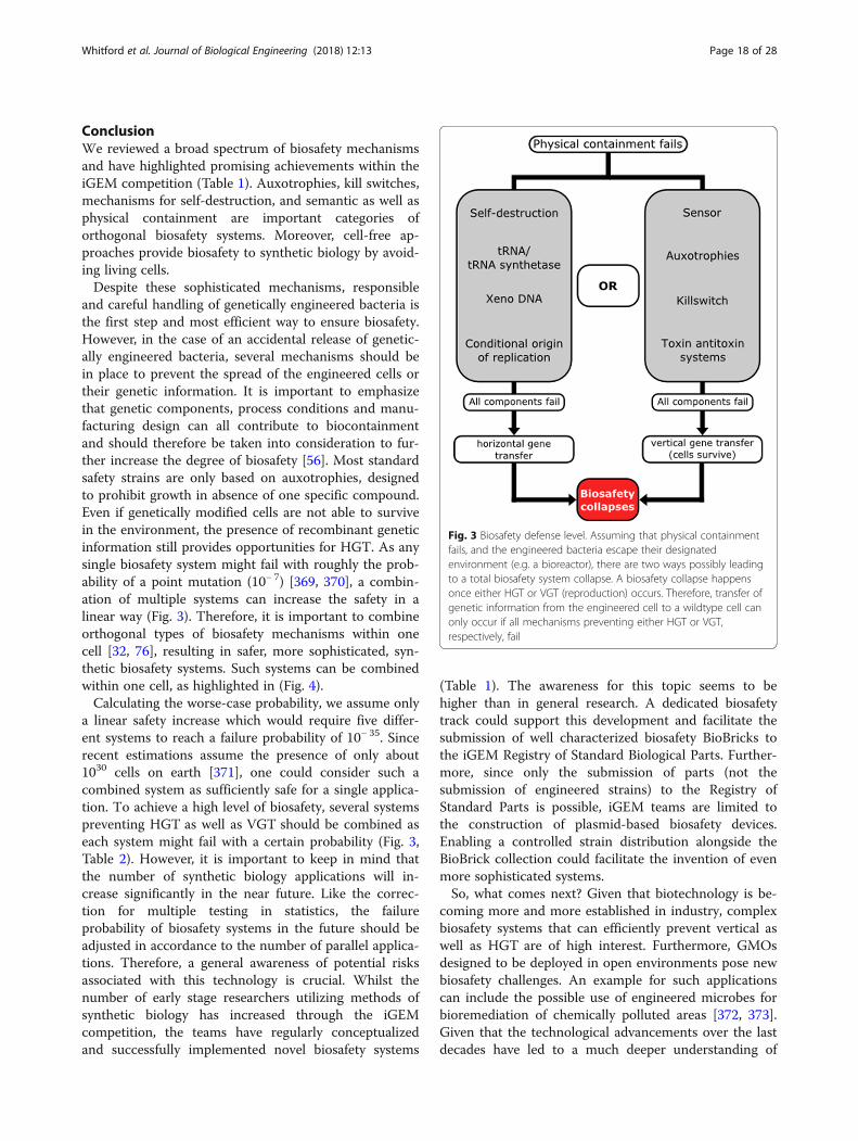
ConclusionWe reviewed a broad spectrum of biosafety mechanismsand have highlighted promising achievements within theiGEM competition (Table 1). Auxotrophies, kill switches,mechanisms for self-destruction, and semantic as well asphysical containment are important categories oforthogonal biosafety systems. Moreover, cell-free ap-proaches provide biosafety to synthetic biology by avoid-ing living cells.Despite these sophisticated mechanisms, responsible
and careful handling of genetically engineered bacteria isthe first step and most efficient way to ensure biosafety.However, in the case of an accidental release of genetic-ally engineered bacteria, several mechanisms should bein place to prevent the spread of the engineered cells ortheir genetic information. It is important to emphasizethat genetic components, process conditions and manu-facturing design can all contribute to biocontainmentand should therefore be taken into consideration to fur-ther increase the degree of biosafety [56]. Most standardsafety strains are only based on auxotrophies, designedto prohibit growth in absence of one specific compound.Even if genetically modified cells are not able to survivein the environment, the presence of recombinant geneticinformation still provides opportunities for HGT. As anysingle biosafety system might fail with roughly the prob-ability of a point mutation (10− 7) [369, 370], a combin-ation of multiple systems can increase the safety in alinear way (Fig. 3). Therefore, it is important to combineorthogonal types of biosafety mechanisms within onecell [32, 76], resulting in safer, more sophisticated, syn-thetic biosafety systems. Such systems can be combinedwithin one cell, as highlighted in (Fig. 4).Calculating the worse-case probability, we assume only
a linear safety increase which would require five differ-ent systems to reach a failure probability of 10− 35. Sincerecent estimations assume the presence of only about1030 cells on earth [371], one could consider such acombined system as sufficiently safe for a single applica-tion. To achieve a high level of biosafety, several systemspreventing HGT as well as VGT should be combined aseach system might fail with a certain probability (Fig. 3,Table 2). However, it is important to keep in mind thatthe number of synthetic biology applications will in-crease significantly in the near future. Like the correc-tion for multiple testing in statistics, the failureprobability of biosafety systems in the future should beadjusted in accordance to the number of parallel applica-tions. Therefore, a general awareness of potential risksassociated with this technology is crucial. Whilst thenumber of early stage researchers utilizing methods ofsynthetic biology has increased through the iGEMcompetition, the teams have regularly conceptualizedand successfully implemented novel biosafety systems
(Table 1). The awareness for this topic seems to behigher than in general research. A dedicated biosafetytrack could support this development and facilitate thesubmission of well characterized biosafety BioBricks tothe iGEM Registry of Standard Biological Parts. Further-more, since only the submission of parts (not thesubmission of engineered strains) to the Registry ofStandard Parts is possible, iGEM teams are limited tothe construction of plasmid-based biosafety devices.Enabling a controlled strain distribution alongside theBioBrick collection could facilitate the invention of evenmore sophisticated systems.So, what comes next? Given that biotechnology is be-
coming more and more established in industry, complexbiosafety systems that can efficiently prevent vertical aswell as HGT are of high interest. Furthermore, GMOsdesigned to be deployed in open environments pose newbiosafety challenges. An example for such applicationscan include the possible use of engineered microbes forbioremediation of chemically polluted areas [372, 373].Given that the technological advancements over the lastdecades have led to a much deeper understanding of
Fig. 3 Biosafety defense level. Assuming that physical containmentfails, and the engineered bacteria escape their designatedenvironment (e.g. a bioreactor), there are two ways possibly leadingto a total biosafety system collapse. A biosafety collapse happensonce either HGT or VGT (reproduction) occurs. Therefore, transfer ofgenetic information from the engineered cell to a wildtype cell canonly occur if all mechanisms preventing either HGT or VGT,respectively, fail
Whitford et al. Journal of Biological Engineering (2018) 12:13 Page 18 of 28
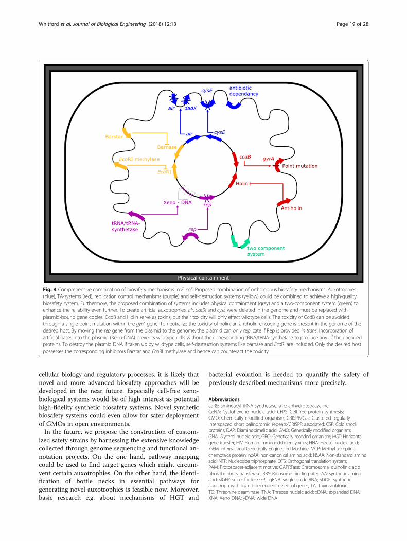
cellular biology and regulatory processes, it is likely thatnovel and more advanced biosafety approaches will bedeveloped in the near future. Especially cell-free xeno-biological systems would be of high interest as potentialhigh-fidelity synthetic biosafety systems. Novel syntheticbiosafety systems could even allow for safer deploymentof GMOs in open environments.In the future, we propose the construction of custom-
ized safety strains by harnessing the extensive knowledgecollected through genome sequencing and functional an-notation projects. On the one hand, pathway mappingcould be used to find target genes which might circum-vent certain auxotrophies. On the other hand, the identi-fication of bottle necks in essential pathways forgenerating novel auxotrophies is feasible now. Moreover,basic research e.g. about mechanisms of HGT and
bacterial evolution is needed to quantify the safety ofpreviously described mechanisms more precisely.
AbbreviationsaaRS: aminoacyl-tRNA synthetase; aTc: anhydrotetracycline;CeNA: Cyclohexene nucleic acid; CFPS: Cell-free protein synthesis;CMO: Chemically modified organism; CRISPR/Cas: Clustered regularlyinterspaced short palindromic repeats/CRISPR associated; CSP: Cold shockproteins; DAP: Diaminopimelic acid; GMO: Genetically modified organism;GNA: Glycerol nucleic acid; GRO: Genetically recoded organism; HGT: Horizontalgene transfer; HIV: Human immunodeficiency virus; HNA: Hexitol nucleic acid;iGEM: international Genetically Engineered Machine; MCP: Methyl-acceptingchemotaxis protein; ncAA: non-canonical amino acid; NSAA: Non-standard aminoacid; NTP: Nucleoside triphosphate; OTS: Orthogonal translation system;PAM: Protospacer-adjacent motive; QAPRTase: Chromosomal quinolinic acidphosphoribosyltransferase; RBS: Ribosome binding site; sAA: synthetic aminoacid; sfGFP: super folder GFP; sgRNA: single-guide RNA; SLiDE: Syntheticauxotroph with ligand-dependent essential genes; TA: Toxin-antitoxin;TD: Threonine deaminase; TNA: Threose nucleic acid; xDNA: expanded DNA;XNA: Xeno DNA; yDNA: wide DNA
Fig. 4 Comprehensive combination of biosafety mechanisms in E. coli. Proposed combination of orthologous biosafety mechanisms. Auxotrophies(blue), TA-systems (red), replication control mechanisms (purple) and self-destruction systems (yellow) could be combined to achieve a high-qualitybiosafety system. Furthermore, the proposed combination of systems includes physical containment (grey) and a two-component system (green) toenhance the reliability even further. To create artificial auxotrophies, alr, dadX and cysE were deleted in the genome and must be replaced withplasmid-bound gene copies. CcdB and Holin serve as toxins, but their toxicity will only effect wildtype cells. The toxicity of CcdB can be avoidedthrough a single point mutation within the gyrA gene. To neutralize the toxicity of holin, an antiholin-encoding gene is present in the genome of thedesired host. By moving the rep gene from the plasmid to the genome, the plasmid can only replicate if Rep is provided in trans. Incorporation ofartificial bases into the plasmid (Xeno-DNA) prevents wildtype cells without the corresponding tRNA/tRNA-synthetase to produce any of the encodedproteins. To destroy the plasmid DNA if taken up by wildtype cells, self-destruction systems like barnase and EcoRI are included. Only the desired hostpossesses the corresponding inhibitors Barstar and EcoRI methylase and hence can counteract the toxicity
Whitford et al. Journal of Biological Engineering (2018) 12:13 Page 19 of 28
AcknowledgementsWe are grateful to the entire iGEM team Bielefeld-CeBiTec 2017. We thankLara Makewita and Nathanael Walker-Hale for language editing and proofreading. We acknowledge the financial support of the German ResearchFoundation (DFG) and the Open Access Publication Fund of Bielefeld Universityfor the article processing charge.
Availability of data and materialsThe datasets described in this review are freely available at the Registry ofStandard Biological Parts, http://parts.igem.org/Main_Page.
Authors’ contributionsJD and BP designed research. SD, DK, CM, OS and CMW wrote the initialdraft. ME designed figures. CMW, JD, BP, CR and JK revised the manuscript.All authors read and approved the final manuscript.
Authors’ informationSD, DK, CM, OS, ME and CMW participated in iGEM 2017 in the team‘Bielefeld-CeBiTec’ focusing on biosafety aspects of the project. JD and BPare fascinated by synthetic biology and especially biosafety since theirparticipation in iGEM 2014. CR and JK are experts in synthetic biologysupervising iGEM teams in Bielefeld since 2010.
Ethics approval and consent to participateNot applicable.
Consent for publicationNot applicable.
Competing interestsThe authors declare that they have no competing interests.
Publisher’s NoteSpringer Nature remains neutral with regard to jurisdictional claims inpublished maps and institutional affiliations.
Author details1Center for Biotechnology, Bielefeld University, 33615 Bielefeld, Germany.2Faculty of Biology, Bielefeld University, Bielefeld, Germany. 3Present address:Evolution and Diversity, Department of Plant Sciences, University ofCambridge, Cambridge, UK.
Received: 17 January 2018 Accepted: 25 June 2018
References1. Bakanidze L, Imnadze P, Perkins D. Biosafety and biosecurity as essential
pillars of international health security and cross-cutting elements ofbiological nonproliferation. BMC Public Health. 2010;10(Suppl 1):S12. https://doi.org/10.1186/1471-2458-10-S1-S12.
2. Sewell DL. Laboratory-associated infections and biosafety. Clin MicrobiolRev. 1995;8:389–405.
3. Gaudioso J. Science and government: biosecurity and research: minimizingadverse impacts. Science. 2004;304:687. https://doi.org/10.1126/science.1096911.
4. Cohen SN. DNA cloning: a personal view after 40 years. Proc Natl Acad Sci.2013;110:15521–9. https://doi.org/10.1073/pnas.1313397110.
5. Curtiss III R, Alexander J, Alexander L, Clark-Curtiss O, Goldschmidt R, Hsu C,et al. Biological Containment and Cloning Vector-Transmissibility. J InfectDis. 1978;137:668–75. http://www.ncbi.nlm.nih.gov/pubmed/351084.
6. Miller S, Selgelid MJ. Ethical and philosophical consideration of the dual-usedilemma in the biological sciences. Dordrecht: Springer Netherlands; 2008.https://doi.org/10.1007/978-1-4020-8312-9.
7. Cenciarelli O, Gabbarini V, Pietropaoli S, Malizia A, Tamburrini A, LudoviciGM, et al. Viral bioterrorism: Learning the lesson of Ebola virus in WestAfrica 2013–2015. Virus Res. 2015;210:318–26. https://doi.org/10.1016/j.virusres.2015.09.002.
8. van Aken J, Hammond E. Genetic engineering and biological weapons.New technologies, desires and threats from biological research. EMBO Rep.2003;4:Spec No:S57–S60. https://doi.org/10.1038/sj.embor.embor860.
9. Singer M, Soll D. Guidelines for DNA hybrid molecules. Science. 1973;181:1114. https://doi.org/10.1126/science.181.4105.1114.
10. Berg P, Baltimore D, Nathans D, Boyer HW, Roblin R, Cohen SN, et al.Potential biohazards of recombinant DNA molecules. Science. 1974;185:303.
11. Molin S, Klemm P, Poulsen LK, Biehl H, Gerdes K, Andersson P. Conditionalsuicide system for containment of Bacteria and plasmids. Nat Biotechnol.1987;5:1315–8. https://doi.org/10.1038/nbt1287-1315.
12. Blattner FR, Plunkett G, Bloch CA, Perna NT, Burland V, Riley M, et al. Thecomplete genome sequence of Escherichia coli K-12. Science. 1997;277:1453–62. https://doi.org/10.1126/science.277.5331.1453.
13. Guyer MS, Reed RR, Steitz JA, Low KB. Identification of a sex-factor-affinitysite in E. Coli as γδ. Cold Spring Harb Symp Quant Biol. 1981;45:135–40.https://doi.org/10.1101/SQB.1981.045.01.022.
14. Daegelen P, Studier FW, Lenski RE, Cure S, Kim JF. Tracing ancestors and relativesof Escherichia coli B, and the derivation of B strains REL606 and BL21(DE3). J MolBiol. 2009;394:634–43. https://doi.org/10.1016/j.jmb.2009.09.022.
15. Hanahan D. Studies on transformation of Escherichia coli with plasmids.J Mol Biol. 1983;166:557–80. https://doi.org/10.1016/S0022-2836(83)80284-8.
16. Smith DB, Johnson KS. Single-step purification of polypeptides expressed inEscherichia coli as fusions with glutathione S-transferase. Gene. 1988;67:31–40.https://doi.org/10.1016/0378-1119(88)90005-4.
17. William Studier F, Rosenberg AH, Dunn JJ, Dubendorff JW. Use of T7 RNApolymerase to direct expression of cloned genes. Methods Enzymol. 1990;185(C):60–89. https://doi.org/10.1016/0076-6879(90)85008-C.
18. Tenover FC, Arbeit RD, Goering RV, Mickelsen PA, Murray BE, Persing DH, etal. Interpreting chromosomal DNA restriction patterns produced by pulsed-field gel electrophoresis: criteria for bacterial strain typing. J Clin Microbiol.1995;33:2233–9.
19. Datsenko KA, Wanner BL. One-step inactivation of chromosomal genes inEscherichia coli K-12 using PCR products. Proc Natl Acad Sci U S A. 2000;97:6640–5. https://doi.org/10.1073/pnas.120163297.
20. Anderson ES. Viability of and transfer of a plasmid from, E. coli K12 in thehuman intestine. Nature. 1975;255:502–4. https://doi.org/10.1038/255502a0.
21. Schweder T, Hofmann K, Hecker BM. Escherichia coli K12 relA strains as safe hostsfor expression of recombinant DNA. Appl Microbiol Biotechnol. 1995;42:718–23.
22. Schweder T, Schmidt I, Herrmann H, Neubauer P, Hecker M, Hofmann K. Anexpression vector system providing plasmid stability and conditional suicideof plasmid-containing cells. Appl Microbiol Biotechnol. 1992;38:91–3. http://www.ncbi.nlm.nih.gov/pubmed/1369014.
23. Kimman TG, Smit E, Klein MR. Evidence-based biosafety: a review of theprinciples and effectiveness of microbiological containment measures. ClinMicrobiol Rev. 2008;21:403–25. https://doi.org/10.1128/CMR.00014-08.
24. Andrup L, Nielsen BH, Kølvraa S. Biosafety considerations in industries withproduction methods based on the use of recombinant deoxyribonucleicacid. Scand J Work Environ Health. 1990;16:85–95. http://www.ncbi.nlm.nih.gov/pubmed/2191426
25. Walton S. Biosafety committees advise less but better DNA research oversight.Bioscience. 1981;31:14–5. https://doi.org/10.1093/bioscience/31.1.14.
26. Dröge M, Pühler A, Selbitschka W. Horizontal gene transfer as a biosafetyissue: a natural phenomenon of public concern. J Biotechnol. 1998;64:75–90.https://doi.org/10.1016/S0168-1656(98)00105-9.
27. Snow AA, Andow DA, Gepts P, Hallerman EM, Power A, Tiedje JM, et al.Genetically engineered organisms and the environment: current statusand recommendations. Ecol Appl. 2005;15:377–404. https://doi.org/10.1890/04-0539.
28. Chan CTY, Lee JW, Cameron DE, Bashor CJ, Collins JJ. “Deadman” and“passcode” microbial kill switches for bacterial containment. Nat Chem Biol.2015;12:82–6. https://doi.org/10.1038/nchembio.1979.
29. iGEM. http://igem.org/Main_Page. Accessed 25 Jun 2017.30. Guan Z, Schmidt M, Pei L, Wei W, Ma K. Biosafety Considerations of
Synthetic Biology in the International Genetically Engineered Machine(iGEM) Competition. Bioscience. 2013;63:25–34. https://doi.org/10.1525/bio.2013.63.1.7.
31. Vilanova C, Porcar M. iGEM 2.0—Refoundations for engineering biology.Nat Biotechnol. 2014;32:420–4. https://doi.org/10.1038/nbt.2899.
32. Wright O, Delmans M, Stan GB, Ellis T. GeneGuard: a modular plasmidsystem designed for biosafety. ACS Synth Biol. 2015;4:307–16.
33. Goodman C. Engineering ingenuity at iGEM. Nat Chem Biol. 2008;4:13.https://doi.org/10.1038/nchembio0108-13.
34. Smolke CD. Building outside of the box: iGEM and the BioBricks foundation.Nat Biotechnol. 2009;27:1099–102. https://doi.org/10.1038/nbt1209-1099.
Whitford et al. Journal of Biological Engineering (2018) 12:13 Page 20 of 28

35. Cooling MT, Rouilly V, Misirli G, Lawson J, Yu T, Hallinan J, et al. Standardvirtual biological parts: a repository of modular modeling components forsynthetic biology. Bioinformatics. 2010;26:925–31.
36. Cai Y, Wilson ML, Peccoud J. GenoCAD for iGEM: a grammatical approachto the design of standard-compliant constructs. Nucleic Acids Res. 2010;38:2637–44. https://doi.org/10.1093/nar/gkq086.
37. Chen YY, Galloway KE, Smolke CD. Synthetic biology: advancing biologicalfrontiers by building synthetic systems. Genome Biol. 2012;13:240.
38. Harger M, Zheng L, Moon A, Ager C, An JH, Choe C, et al. Expanding theproduct profile of a microbial alkane biosynthetic pathway. ACS Synth Biol.2013;2:59–62.
39. Radeck J, Kraft K, Bartels J, Cikovic T, Dürr F, Emenegger J, et al. The BacillusBioBrick box: generation and evaluation of essential genetic building blocksfor standardized work with Bacillus subtilis. J Biol Eng. 2013;7:29. https://doi.org/10.1186/1754-1611-7-29.
40. Chen R, Rishi HS, Potapov V, Yamada MR, Yeh VJ, Chow T, et al. A barcodingstrategy enabling higher-throughput library screening by microscopy. ACSSynth Biol. 2015;4:1205–16. https://doi.org/10.1021/acssynbio.5b00060.
41. Kelwick R, Bowater L, Yeoman KH, Bowater RP. Promoting microbiologyeducation through the iGEM synthetic biology competition. FEMS MicrobiolLett. 2015;362:fnv129. https://doi.org/10.1093/femsle/fnv129.
42. Storch M, Casini A, Mackrow B, Fleming T, Trewhitt H, Ellis T, et al. BASIC: anew biopart assembly standard for idempotent cloning provides accurate,single-tier DNA assembly for synthetic biology. ACS Synth Biol. 2015;4:781–7.https://doi.org/10.1021/sb500356d.
43. Jack BR, Leonard SP, Mishler DM, Renda BA, Leon D, Suárez GA, et al.Predicting the genetic stability of engineered DNA sequences with the EFMcalculator. ACS Synth Biol. 2014;4:939–43.
44. Hayashi K, Morooka N, Yamamoto Y, Fujita K, Isono K, Choi S, et al. Highlyaccurate genome sequences of Escherichia coli K-12 strains MG1655 andW3110. Mol Syst Biol. 2006;2 https://doi.org/10.1038/msb4100049.
45. Gogarten JP, Townsend JP. Horizontal gene transfer, genome innovationand evolution. Nat Rev Microbiol. 2005;3:679–87.
46. Kubyshkin V, Acevedo-Rocha CG, Budisa N. On universal coding events inprotein biogenesis. Biosystems. 2018;164:16–25. https://doi.org/10.1016/j.biosystems.2017.10.004.
47. Knight RD, Freeland SJ, Landweber LF. Rewiring the keyboard: Evolvabilityof the genetic code. Nat Rev Genet. 2001;2:49–58.
48. Yokobori SI, Suzuki T, Watanabe K. Genetic code variations in mitochondria: tRNAas a major determinant of genetic code plasticity. J Mol Evol. 2001;53:314–26.
49. Santos MAS, Moura G, Massey SE, Tuite MF. Driving change: the evolutionof alternative genetic codes. Trends Genet. 2004;20:95–102.
50. Kubyshkin V, Budisa N. Synthetic alienation of microbial organisms by usinggenetic code engineering: why and how? Biotechnol J. 2017;12:1600097.https://doi.org/10.1002/biot.201600097.
51. Simon AJ, Ellington AD. Recent advances in synthetic biosafety. F1000Res.2016;5:2118. https://doi.org/10.12688/f1000research.8365.1.
52. Curtiss IIIR, Inoue M, Pereira D, Hsu JC, Alexander L, Rock L. Constructionand use of safer bacterial host strains for recombinant DNA research. In:Scott WA, Werner R, editors. Molecular cloning of recombinant DNA.Academic press; 1977. p. 99–114.
53. Moe-Behrens GHG, Davis R, Haynes KA. Preparing synthetic biology for theworld. Front Microbiol. 2013;4:1–10.
54. Beadle GW, Tatum EL. Genetic control of biochemical reactions in Neurospora.Proc Natl Acad Sci. 1941;27:499–506. https://doi.org/10.1073/pnas.27.11.499.
55. E. coli Genetic Resources at Yale CGSC, The Coli Genetic Stock Center, List ofCommonly Requested Auxotrophic Strains. https://cgsc2.biology.yale.edu/Auxotrophs.php. Accessed 25 Jun 2017.
56. Torres L, Krüger A, Csibra E, Gianni E, Pinheiro VB. Synthetic biologyapproaches to biological containment: pre-emptively tackling potential risks.Essays Biochem. 2016;60:393–410. https://doi.org/10.1042/EBC20160013.
57. Mandell DJ, Lajoie MJ, Mee MT, Takeuchi R, Kuznetsov G, Norville JE, et al.Biocontainment of genetically modified organisms by synthetic proteindesign. Nature. 2015;518:55–60. https://doi.org/10.1038/nature14121.
58. Rovner AJ, Haimovich AD, Katz SR, Li Z, Grome MW, Gassaway BM, et al.Recoded organisms engineered to depend on synthetic amino acids.Nature. 2015;518:89–93. https://doi.org/10.1038/nature14095.
59. Vidal L, Pinsach J, Striedner G, Caminal G, Ferrer P. Development of anantibiotic-free plasmid selection system based on glycine auxotrophy forrecombinant protein overproduction in Escherichia coli. J Biotechnol. 2008;134:127–36. https://doi.org/10.1016/j.jbiotec.2008.01.011.
60. Rajasekaran P, Seleem MN, Contreras A, Purwantini E, Schurig GG,Sriranganathan N, et al. Brucella abortus strain RB51 leucine auxotroph as anenvironmentally safe vaccine for plasmid maintenance and antigenoverexpression. Appl Environ Microbiol. 2008;74:7051–5. https://doi.org/10.1128/AEM.01511-08.
61. Dong W-R, Xiang L-X, Shao J-Z. Novel antibiotic-free plasmid selectionsystem based on complementation of host Auxotrophy in the NAD DeNovo synthesis pathway. Appl Environ Microbiol. 2010;76:2295–303. https://doi.org/10.1128/AEM.02462-09.
62. Fiedler M, Skerra A. proBA complementation of an auxotrophic E. coli strainimproves plasmid stability and expression yield during fermenterproduction of a recombinant antibody fragment. Gene. 2001;274:111–8.https://doi.org/10.1016/S0378-1119(01)00629-1.
63. Nguyen LV, Cox KM, Ke JS, Peele CG, Dickey LF. Genetic engineering of aLemna isoleucine auxotroph. Transgenic Res. 2012;21:1071–83. https://doi.org/10.1007/s11248-012-9594-2.
64. Cava F, Lam H, de Pedro MA, Waldor MK. Emerging knowledge of regulatoryroles of d-amino acids in bacteria. Cell Mol Life Sci. 2011;68:817–31. https://doi.org/10.1007/s00018-010-0571-8.
65. Tauch A, Götker S, Pühler A, Kalinowski J, Thierbach G. The alanine racemasegene alr is an alternative to antibiotic resistance genes in cloning systems forindustrial Corynebacterium glutamicum strains. J Biotechnol. 2002;99:79–91.https://doi.org/10.1016/S0168-1656(02)00159-1.
66. Walsh CT. Enzymes in the D-alanine branch of bacterial cell wall peptidoglycanassembly. J Biol Chem. 1989;264:2393–6. http://www.ncbi.nlm.nih.gov/pubmed/2644260
67. Chacon O, Feng Z, Harris NB, Cáceres NE, Adams LG, Barletta RG.Mycobacterium smegmatis D-alanine racemase mutants are not dependenton D-alanine for growth. Antimicrob Agents Chemother. 2002;46:47–54.https://doi.org/10.1128/AAC.46.2.47-54.2002.
68. Milligan DL, Tran SL, Strych U, Cook GM, Krause KL. The alanineracemase of Mycobacterium smegmatis is essential for growth in theabsence of D-alanine. J Bacteriol. 2007;189:8381–6. https://doi.org/10.1128/JB.01201-07.
69. Jawale CV, Lee JH. Development of a biosafety enhanced and immunogenicSalmonella Enteritidis ghost using an antibiotic resistance gene free plasmidcarrying a bacteriophage lysis system. PLoS One. 2013;8:1–11.
70. Hirota R, Abe K, Katsuura Z, Noguchi R, Moribe S, Motomura K, et al. A novelbiocontainment strategy makes bacterial growth and survival dependent onPhosphite. Sci Rep. 2017;7:44748. https://doi.org/10.1038/srep44748.
71. Steidler L, Neirynck S, Huyghebaert N, Snoeck V, Vermeire A, Goddeeris B,et al. Biological containment of genetically modified Lactococcus lactis forintestinal delivery of human interleukin 10. Nat Biotechnol. 2003;21:785–9.https://doi.org/10.1038/nbt840.
72. Braat H, Rottiers P, Hommes DW, Huyghebaert N, Remaut E, Remon J, et al.A phase I trial with transgenic Bacteria expressing Interleukin-10 in Crohn’sdisease. Clin Gastroenterol Hepatol. 2006;4:754–9. https://doi.org/10.1016/j.cgh.2006.03.028.
73. Lopez G, Anderson JC. Synthetic Auxotrophs with ligand-dependentessential genes for a BL21(DE3) biosafety strain. ACS Synth Biol. 2015;4:1279–86. https://doi.org/10.1021/acssynbio.5b00085.
74. Deckert K, Budiardjo SJ, Brunner LC, Lovell S, Karanicolas J. Designingallosteric control into enzymes by chemical rescue of structure. J Am ChemSoc. 2012;134:10055–60.
75. Wright O, Stan G-B, Ellis T. Building-in biosafety for synthetic biology.Microbiology. 2013;159(Pt_7):1221–35. https://doi.org/10.1099/mic.0.066308-0.
76. Gallagher RR, Patel JR, Interiano AL, Rovner AJ, Isaacs FJ. Multilayeredgenetic safeguards limit growth of microorganisms to definedenvironments. Nucleic Acids Res. 2015;43:1945–54. https://doi.org/10.1093/nar/gku1378.
77. Bielefeld Germany 2013. http://2013.igem.org/Team:Bielefeld-Germany.Accessed 6 May 2017.
78. Bielefeld-CeBiTec 2014. http://2014.igem.org/Team:Bielefeld-CeBiTec.Accessed 22 Mar 2017.
79. BYU Provo 2014. http://2014.igem.org/Team:BYU_Provo. Accessed 22 Mar2017.
80. Amsterdam 2015. http://2015.igem.org/Team:Amsterdam. Accessed 22 Mar2017.
81. Unterholzner SJ, Poppenberger B, Rozhon W. Toxin-antitoxin systems:Biology, identification, and application. Mob Genet Elements. 2013;3:e26219.https://doi.org/10.4161/mge.26219.
Whitford et al. Journal of Biological Engineering (2018) 12:13 Page 21 of 28
82. Hayes F, Van Melderen L. Toxins-antitoxins: diversity, evolution and function.Crit Rev Biochem Mol Biol. 2011;46:386–408. https://doi.org/10.3109/10409238.2011.600437.
83. Gerdes K, Gultyaev AP, Franch T, Pedersen K, Mikkelsen ND. AntisenseRNA-regulated programmed cell death. Annu Rev Genet. 1997;31:1–31.https://doi.org/10.1146/annurev.genet.31.1.1.
84. Gerdes K, Wagner EGH. RNA antitoxins. Curr Opin Microbiol. 2007;10:117–24.85. Van Melderen L, Saavedra De Bast M. Bacterial Toxin–Antitoxin Systems:
More Than Selfish Entities? PLoS Genet. 2009;5:e1000437. https://doi.org/10.1371/journal.pgen.1000437.
86. Gerdes K, Rasmussen PB, Molin S. Unique type of plasmid maintenancefunction: postsegregational killing of plasmid-free cells. Proc Natl Acad Sci US A. 1986;83:3116–20.
87. Ohnishi Y, Akimoto S, I-Like R. Plasmids promote degradation of stableribonucleic acid in Escherichia coli. J Bacteriol. 1980;144:833–5.
88. Akimoto S, Ohnishi Y. R483 and F plasmid genes promoting RNA degradation:comparative restriction mapping. Microbiol Immunol. 1982;26:779–93.
89. Kawano M, Aravind L, Storz G. An antisense RNA controls synthesis of an SOS-induced toxin evolved from an antitoxin. Mol Microbiol. 2007;64:738–54.
90. NTU Taida 2012. http://2012.igem.org/Team:NTU-Taida. Accessed 16 Mar 2017.91. University of Maryland 2015. http://2015.igem.org/Team:UMaryland.
Accessed 16 Mar 2017.92. Roberts RC, Burioni R, Helinksi DR. Genetic characterization of the stabilizing
functions of a region of broad-host-range plasmid RK2. J Bacteriol. 1990;172:6204–16.
93. Bravo A, de Torrontegui G, Díaz R. Identification of components of a newstability system of plasmid R1, ParD, that is close to the origin of replicationof this plasmid. Mol Gen Genet MGG. 1987;210:101–10. https://doi.org/10.1007/BF00337764.
94. Ogura T, Hiraga S. Mini-F plasmid genes that couple host cell division toplasmid proliferation. Proc Natl Acad Sci. 1983;80:4784–8. https://doi.org/10.1073/pnas.80.15.4784.
95. Lehnherr H, Maguin E, Jafri S, Yarmolinsky MB. Plasmid addiction genes ofbacteriophage P1: doc, which causes cell death on curing of prophage, andphd, which prevents host death when prophage is retained. J Mol Biol.1993;233:414–28. https://doi.org/10.1006/jmbi.1993.1521.
96. Aizenman E, Engelberg-Kulka H, Glaser G. An Escherichia coli chromosomal“addiction module” regulated by 3′,5′-bispyrophosphate: a model forprogrammed bacterial cell death. Proc Natl Acad Sci. 1996;93:6059–63.https://doi.org/10.1073/pnas.93.12.6059.
97. Grady R, Hayes F. Axe-Txe, a broad-spectrum proteic toxin-antitoxin systemspecified by a multidrug-resistant, clinical isolate of Enterococcus faecium.Mol Microbiol. 2003;47:1419–32.
98. Zhang J, Zhang Y, Inouye M. Characterization of the interactions within themazEF addiction module of Escherichia coli. J Biol Chem. 2003;278:32300–6.
99. Oberer M, Zangger K, Prytulla S, Keller W. The anti-toxin ParD of plasmidRK2 consists of two structurally distinct moieties and belongs to the ribbon-helix-helix family of DNA-binding proteins. Biochem J. 2002;361(Pt 1):41–7.https://doi.org/10.1042/0264-6021:3610041.
100. Marianovsky I, Aizenman E, Engelberg-Kulka H, Glaser G. The regulation ofthe Escherichia coli mazEF promoter involves an unusual alternatingpalindrome. J Biol Chem. 2001;276:5975–84.
101. Magnuson R, Lehnherr H, Mukhopadhyay G, Yarmolinsky MB.Autoregulation of the plasmid addiction operon of bacteriophage P1. J BiolChem. 1996;271:18705–10.
102. Christensen SK, Maenhaut-Michel G, Mine N, Gottesman S, Gerdes K, VanMelderen L. Overproduction of the Lon protease triggers inhibition of translationin Escherichia coli: involvement of the yefM-yoeB toxin-antitoxin system. MolMicrobiol. 2004;51:1705–17. https://doi.org/10.1046/j.1365-2958.2003.03941.x.
103. Amitai S, Yassin Y, Engelberg-Kulka H. MazF-mediated cell death inEscherichia coli: a point of no return. J Bacteriol. 2004;186:8295–300. https://doi.org/10.1128/JB.186.24.8295-8300.2004.
104. Paris Bettencourt 2012. http://2012.igem.org/Team:Paris_Bettencourt.Accessed 16 Mar 2017.
105. Wageningen UR 2014. http://2014.igem.org/Team:Wageningen_UR.Accessed 16 Mar 2017.
106. UC Berkeley 2008. http://2008.igem.org/Team:UC_Berkeley. Accessed 16Mar 2017.
107. Stirling F, Bitzan L, O’Keefe S, Redfield E, Oliver JWK, Way J, et al. RationalDesign of Evolutionarily Stable Microbial Kill Switches. Mol Cell. 2017;68:686–697.e3. https://doi.org/10.1016/j.molcel.2017.10.033.
108. Fineran PC, Blower TR, Foulds IJ, Humphreys DP, Lilley KS, Salmond GPC.The phage abortive infection system, ToxIN, functions as a protein-RNAtoxin-antitoxin pair. Proc Natl Acad Sci U S A. 2009;106:894–9. https://doi.org/10.1073/pnas.0808832106.
109. Samson JE, Spinelli S, Cambillau C, Moineau S. Structure and activity ofAbiQ, a lactococcal endoribonuclease belonging to the type III toxin-antitoxin system. Mol Microbiol. 2013;87:756–68.
110. Blower TR, Fineran PC, Johnson MJ, Toth IK, Humphreys DP, Salmond GPC.Mutagenesis and functional characterization of the RNA and proteincomponents of the toxIN abortive infection and toxin-antitoxin locus ofErwinia. J Bacteriol. 2009;191:6029–39.
111. Masuda H, Tan Q, Awano N, Wu KP, Inouye M. YeeU enhances the bundlingof cytoskeletal polymers of MreB and FtsZ, antagonizing the CbtA (YeeV)toxicity in Escherichia coli. Mol Microbiol. 2012;84:979–89.
112. Wang X, Lord DM, Cheng H-Y, Osbourne DO, Hong SH, Sanchez-Torres V,et al. A new type V toxin-antitoxin system where mRNA for toxin GhoT iscleaved by antitoxin GhoS. Nat Chem Biol. 2012;8:855–61. https://doi.org/10.1038/nchembio.1062.
113. LMU Munich 2012. http://2012.igem.org/Team:LMU-Munich. Accessed 17Mar 2017.
114. TU Eindhoven 2014. http://2014.igem.org/Team:TU_Eindhoven. Accessed 17Mar 2017.
115. Inouye H, Barnes W, Beckwith J. Signal sequence of alkaline phosphatase ofEscherichia coli. J Bacteriol. 1982;149:434–9. http://www.ncbi.nlm.nih.gov/pubmed/7035431.
116. Hartley RW. Barnase and barstar. Expression of its cloned inhibitor permitsexpression of a cloned ribonuclease. J Mol Biol. 1988;202:913–5. https://doi.org/10.1016/0022-2836(88)90568-2.
117. Chaudhary VK, Xu YH, FitzGerald D, Adhya S, Pastan I. Role of domain II ofPseudomonas exotoxin in the secretion of proteins into the periplasm andmedium by Escherichia coli. Proc Natl Acad Sci U S A. 1988;85:2939–43.http://www.pubmedcentral.nih.gov/articlerender.fcgi?artid=280118&tool=pmcentrez&rendertype=abstract
118. Nesmeyanova MA, Tsfasman IM, Karamyshev AL, Suzina NE. Secretion of theoverproduced periplasmic PhoA protein into the medium andaccumulation of its precursor in phoA-transformed Escherichia coli strains:involvement of outer membrane vesicles. World J Microbiol Biotechnol.1991;7:394–406. https://doi.org/10.1007/BF00329408.
119. Ramos HJO, Souza EM, Soares-Ramos JRL, Pedrosa FO. A new system tocontrol the barnase expression by a NifA-dependent promoter. J Biotechnol.2005;118:9–16. https://doi.org/10.1016/j.jbiotec.2005.03.002.
120. Ramos HJO, Souza EM, Soares-Ramos JRL, Pedrosa FO. Antibiosis by Bacillusamyloliquefaciens ribonuclease barnase expressed in Escherichia coli againstsymbiotic and endophytic nitrogen-fixing bacteria. J Biotechnol. 2006;126:291–4. https://doi.org/10.1016/j.jbiotec.2006.04.020.
121. Coray DS, Kurenbach B, Heinemann JA. Exploring the parameters of post-segregational killing using heterologous expression of secreted toxinbarnase and antitoxin barstar in an Escherichia coli case study.Microbiology. 2017;163:122–30. https://doi.org/10.1099/mic.0.000395.
122. Guzman LM, Belin D, Carson MJ, Beckwith J. Tight regulation, modulation,and high-level expression by vectors containing the arabinose PBADpromoter. J Bacteriol. 1995;177:4121–30.
123. Siegele DA, Hu JC. Gene expression from plasmids containing the araBADpromoter at subsaturating inducer concentrations represents mixedpopulations. Proc Natl Acad Sci. 1997;94:8168–72. https://doi.org/10.1073/pnas.94.15.8168.
124. UC Berkeley 2007. http://2007.igem.org/Berkeley_UC. Accessed 23 Jun 2017.125. Valencia UPV 2014. http://2014.igem.org/Team:Valencia_UPV. Accessed 23
Jun 2017.126. Velkov VV. Stress-induced evolution and the biosafety of genetically
modified microorganisms released into the environment. J Biosci. 2001;26:667–83. https://doi.org/10.1007/BF02704764.
127. Thomas CM, Nielsen KM. Mechanisms of, and barriers to, horizontal genetransfer between Bacteria. Nat Rev Microbiol. 2005;3:711–21. https://doi.org/10.1038/nrmicro1234.
128. Greenes PJ, Gupta M, Boyer W, Francisco S, Francisco S, Brown WE, et al.Sequence analysis of the DNA encoding the DNA encoding the eco RIendonuclease and Methylase*. Biol Chem. 1980;25:2143–53.
129. Williams RJ. Restriction endonucleases: classification, properties, andapplications. Mol Biotechnol. 2003;23:225–43. https://doi.org/10.1385/MB:23:3:225.
Whitford et al. Journal of Biological Engineering (2018) 12:13 Page 22 of 28
130. Roberts RJ, Belfort M, Bestor T, Bhagwat AS, Bickle TA, Bitinaite J, et al. Anomenclature for restriction enzymes, DNA methyltransferases, homingendonucleases and their genes. Nucleic Acids Res. 2003;31:1805–12. https://doi.org/10.1093/nar/gkg274.
131. Roberts RJ. How restriction enzymes became the workhorses of molecularbiology. Proc Natl Acad Sci U S A. 2005;102:5905–8. https://doi.org/10.1073/pnas.0500923102.
132. Roberts RJ, Vincze T, Posfai J, Macelis D. REBASE - enzymes and genes for DNArestriction and modification. Nucleic Acids Res. 2007;35(Suppl. 1):2006–7.
133. University College London 2012. http://2012.igem.org/Team:University_College_London. Accessed 23 Jun 2017.
134. Molin S, Boe L, Jensen LB, Kristensen CS, Givskov M, Ramos JL, et al. Suicidalgenetic elements and their use in biological containment of Bacteria. AnnuRev Microbiol. 1993;47:139–66. https://doi.org/10.1146/annurev.mi.47.100193.001035.
135. Ahrenholtz I, Lorenz MG, Wackernagel W. A conditional suicide system inEscherichia coli based on the intracellular degradation of DNA. Appl EnvironMicrobiol. 1994;60:3746–51. http://www.ncbi.nlm.nih.gov/pubmed/7986048
136. Ball TK, Saurugger PN, Benedik MJ. The extracellular nuclease gene ofSerratia marcescens and its secretion from Escherichia coli. Gene. 1987;57:183–92. https://doi.org/10.1016/0378-1119(87)90121-1.
137. Biedermann K, Jepsen PK, Riise E, Svendsen I. Purification andcharacterization of a Serratia marcescens nuclease produced by Escherichiacoli. Carlsb Res Commun. 1989;54:17–27. http://www.ncbi.nlm.nih.gov/pubmed/2665765
138. TU Munich 2013. http://2013.igem.org/Team:TU-Munich. Accessed 23 Jun2017.
139. NTU LIHPAO Taiwan 2015. http://2015.igem.org/Team:NTU-LIHPAO-Taiwan.Accessed 23 Jun 2017.
140. Bhaya D, Davison M, Barrangou R. CRISPR-Cas Systems in Bacteria and Archaea:versatile small RNAs for adaptive defense and regulation. Annu Rev Genet.2011;45:273–97. https://doi.org/10.1146/annurev-genet-110410-132430.
141. Terns MP, Terns RM. CRISPR-based adaptive immune systems. Curr OpinMicrobiol. 2011;14:321–7.
142. Wiedenheft B, Sternberg SH, Doudna JA. RNA-guided genetic silencingsystems in bacteria and archaea. Nature. 2012;482:331–8. https://doi.org/10.1038/nature10886.
143. Gasiunas G, Barrangou R, Horvath P, Siksnys V. Cas9-crRNAribonucleoprotein complex mediates specific DNA cleavage for adaptiveimmunity in bacteria. Proc Natl Acad Sci. 2012;109:E2579–86. https://doi.org/10.1073/pnas.1208507109.
144. Jinek M, Chylinski K, Fonfara I, Hauer M, Doudna JA, Charpentier E. Aprogrammable dual-RNA-guided DNA endonuclease in adaptive bacterialimmunity. Science. 2012;337:816–21. https://doi.org/10.1126/science.1225829.
145. Jiang W, Bikard D, Cox D, Zhang F, Marraffini LA. RNA-guided editing ofbacterial genomes using CRISPR-Cas systems. Nat Biotechnol. 2013;31:233–9.https://doi.org/10.1038/nbt.2508.
146. Sander JD, Joung JK. CRISPR-Cas systems for editing, regulating and targetinggenomes. Nat Biotechnol. 2014;32:347–55. https://doi.org/10.1038/nbt.2842.
147. Ran FA, Hsu PD, Lin C, Gootenberg JS, Konermann S, Trevino AE, et al.Double nicking by RNA-guided CRISPR Cas9 for enhanced genome editingspecificity. Cell. 2013;154:1380–9. https://doi.org/10.1016/j.cell.2013.08.021.
148. Ran FA, Hsu PD, Wright J, Agarwala V, Scott DA, Zhang F. Genomeengineering using the CRISPR-Cas9 system. Nat Protoc. 2013;8:2281–308.https://doi.org/10.1038/nprot.2013.143.
149. Mei Y, Wang Y, Chen H, Sun ZS, Ju X-D. Recent progress in CRISPR/Cas9technology. J Genet Genomics. 2016;43:63–75. https://doi.org/10.1016/j.jgg.2016.01.001.
150. Marraffini LA, Sontheimer EJ. CRISPR interference limits horizontal genetransfer in staphylococci by targeting DNA. Science. 2008;322:1843–5.https://doi.org/10.1126/science.1165771.
151. A a G, Klumpe HE, Luo ML, Selle K, Barrangou R, Beisel CL. Programmableremoval of bacterial strains by use of genome-targeting CRISPR-Cas systems.MBio. 2014;5:e00928–13. https://doi.org/10.1128/mBio.00928-13.
152. Caliando BJ, Voigt CA. Targeted DNA degradation using a CRISPR devicestably carried in the host genome. Nat Commun. 2015;6:6989. https://doi.org/10.1038/ncomms7989.
153. Makarova KS, Haft DH, Barrangou R, Brouns SJJ, Charpentier E, Horvath P,et al. Evolution and classification of the CRISPR–Cas systems. Nat RevMicrobiol. 2011;9:467–77. https://doi.org/10.1038/nrmicro2577.
154. Semenova E, Jore MM, Datsenko KA, Semenova A, Westra ER, Wanner B,et al. Interference by clustered regularly interspaced short palindromicrepeat (CRISPR) RNA is governed by a seed sequence. Proc Natl Acad Sci US A. 2011;108:10098–103. https://doi.org/10.1073/pnas.1104144108.
155. Mojica FJM, Díez-Villaseñor C, García-Martínez J, Almendros C. Shortmotif sequences determine the targets of the prokaryotic CRISPRdefence system. Microbiology. 2009;155(Pt 3):733–40. https://doi.org/10.1099/mic.0.023960-0.
156. Wiedenheft B. In defense of phage: viral suppressors of CRISPR-mediatedadaptive immunity in bacteria. RNA Biol. 2013;10:886–90. https://doi.org/10.4161/rna.23591.
157. Jiang W, Bikard D, Cox D, Zhang F, Marraffini LA. RNA-guided editing ofbacterial genomes using CRISPR-Cas systems. Nat Biotechnol. 2013;31:233–9.
158. Niewoehner O, Jinek M, Doudna JA. Evolution of CRISPR RNA recognitionand processing by Cas6 endonucleases. Nucleic Acids Res. 2014;42:1341–53.https://doi.org/10.1093/nar/gkt922.
159. Marraffini LA, Sontheimer EJ. Self versus non-self discrimination duringCRISPR RNA-directed immunity. Nature. 2010;463:568–71. https://doi.org/10.1038/nature08703.
160. HKU Hongkong 2015. http://2015.igem.org/Team:Hong_Kong_HKU.Accessed 23 Jun 2017.
161. Bernard P, Couturier M. Cell killing by the F plasmid CcdB protein involvespoisoning of DNA-topoisomerase II complexes. J Mol Biol. 1992;226:735–45.https://doi.org/10.1016/0022-2836(92)90629-X.
162. Jia B, Qi H, Li BZ, Pan S, Liu D, Liu H, et al. Orthogonal ribosome biofirewall.ACS Synth Biol. 2017;6:2108–17.
163. Schmidt M. Xenobiology: a new form of life as the ultimate biosafety tool.BioEssays. 2010;32:322–31. https://doi.org/10.1002/bies.200900147.
164. Schmidt M, de Lorenzo V. Synthetic constructs in/for the environment:managing the interplay between natural and engineered biology. FEBS Lett.2012;586:2199–206. https://doi.org/10.1016/j.febslet.2012.02.022.
165. Pinheiro VB, Holliger P. The XNA world: progress towards replication andevolution of synthetic genetic polymers. Curr Opin Chem Biol. 2012;16:245–52.https://doi.org/10.1016/j.cbpa.2012.05.198.
166. Nielsen KM, Johnsen PJ, Bensasson D, Daffonchio D. Release andpersistence of extracellular DNA in the environment. Environ Biosaf Res.2007;6:37–53. https://doi.org/10.1051/ebr:2007031.
167. Plos K, Hull SI, Hull RA, Levin BR, Orskov I, Orskov F, et al. Distribution of theP-associated-pilus (pap) region among Escherichia coli from natural sources:evidence for horizontal gene transfer. Infect Immun. 1989;57:1604–11.http://www.ncbi.nlm.nih.gov/pubmed/2565294
168. de la Cruz F, Davies J. Horizontal gene transfer and the origin of species:lessons from bacteria. Trends Microbiol. 2000;8:128–33. https://doi.org/10.1016/S0966-842X(00)01703-0.
169. Davison J. Genetic exchange between Bacteria in the environment. Plasmid.1999;42:73–91. https://doi.org/10.1006/plas.1999.1421.
170. Acevedo-Rocha CG, Budisa N. On the road towards chemically modifiedorganisms endowed with a genetic firewall. Angew Chem Int Ed Engl.2011;50:6960–2. https://doi.org/10.1002/anie.201103010.
171. Marlière P. The farther, the safer: a manifesto for securely navigating syntheticspecies away from the old living world. Syst Synth Biol. 2009;3:77–84.https://doi.org/10.1007/s11693-009-9040-9.
172. Herdewijn P, Marlière P. Toward safe genetically modified organisms throughthe chemical diversification of nucleic acids. Chem Biodivers. 2009;6:791–808.https://doi.org/10.1002/cbdv.200900083.
173. Agostini F, Völler J-S, Koksch B, Acevedo-Rocha CG, Kubyshkin V, Budisa N.Biocatalysis with unnatural amino acids: enzymology meets Xenobiology. AngewChem Int Ed Engl. 2017;56:9680–703. https://doi.org/10.1002/anie.201610129.
174. Martinot TA, Benner SA. Artificial genetic systems: exploiting the“aromaticity” formalism to improve the tautomeric ratio for Isoguanosinederivatives. J Org Chem. 2004;69:3972–5. https://doi.org/10.1021/jo0497959.
175. Jiang D, Seela F. Oligonucleotide duplexes and multistrand assemblies with8-Aza-2′-deoxyisoguanosine: a fluorescent isoG d shape mimic expandingthe genetic alphabet and forming Ionophores. J Am Chem Soc. 2010;132:4016–24. https://doi.org/10.1021/ja910020n.
176. Kwok R. Chemical biology: DNA’s new alphabet. Nature. 2012;491:516–8.https://doi.org/10.1038/491516a.
177. Zhang Y, Lamb BM, Feldman AW, Zhou AX, Lavergne T, Li L, et al. Asemisynthetic organism engineered for the stable expansion of the geneticalphabet. Proc Natl Acad Sci. 2017;114:1317–22. https://doi.org/10.1073/pnas.1616443114.
Whitford et al. Journal of Biological Engineering (2018) 12:13 Page 23 of 28
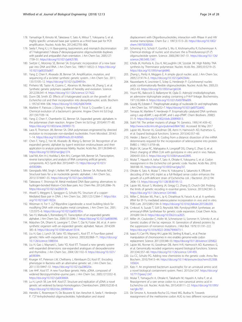
178. Yamashige R, Kimoto M, Takezawa Y, Sato A, Mitsui T, Yokoyama S, et al.Highly specific unnatural base pair systems as a third base pair for PCRamplification. Nucleic Acids Res. 2012;40:2793–806.
179. Seela F, Peng X, Li H. Base-pairing, tautomerism, and mismatch discriminationof 7-halogenated 7-deaza-2′-deoxyisoguanosine: oligonucleotide duplexeswith parallel and antiparallel chain orientation. J Am Chem Soc. 2005;127:7739–51. https://doi.org/10.1021/ja0425785.
180. Switzer C, Moroney SE, Benner SA. Enzymatic incorporation of a new basepair into DNA and RNA. J Am Chem Soc. 1989;111:8322–3. https://doi.org/10.1021/ja00203a067.
181. Yang Z, Chen F, Alvarado JB, Benner SA. Amplification, mutation, andsequencing of a six-letter synthetic genetic system. J Am Chem Soc. 2011;133:15105–12. https://doi.org/10.1021/ja204910n.
182. Pinheiro VB, Taylor AI, Cozens C, Abramov M, Renders M, Zhang S, et al.Synthetic genetic polymers capable of heredity and evolution. Science.2012;336:341–4. https://doi.org/10.1126/science.1217622.
183. Dunn DB, Smith JD. Effects of 5-halogenated uracils on the growth ofEscherichia coli and their incorporation into deoxyribonucleic acids. BiochemJ. 1957;67:494–506. https://doi.org/10.1042/bj0670494.
184. Marlière P, Patrouix J, Döring V, Herdewijn P, Tricot S, Cruveiller S, et al.Chemical evolution of a bacterium’s genome. Angew Chem Int Ed Engl.2011;50:7109–14.
185. Yang Z, Chen F, Chamberlin SG, Benner SA. Expanded genetic alphabets inthe polymerase chain reaction. Angew Chem Int Ed Engl. 2010;49:177–80.https://doi.org/10.1002/anie.200905173.
186. Laos R, Thomson JM, Benner SA. DNA polymerases engineered by directedevolution to incorporate non-standard nucleotides. Front Microbiol. 2014;5:1–14. https://doi.org/10.3389/fmicb.2014.00565.
187. Chen F, Yang Z, Yan M, Alvarado JB, Wang G, Benner SA. Recognition of anexpanded genetic alphabet by type-II restriction endonucleases and theirapplication to analyze polymerase fidelity. Nucleic Acids Res. 2011;39:3949–61.https://doi.org/10.1093/nar/gkq1274.
188. Leal NA, Kim H-J, Hoshika S, Kim M-J, Carrigan MA, Benner SA. Transcription,reverse transcription, and analysis of RNA containing artificial geneticcomponents. ACS Synth Biol. 2015;4:407–13. https://doi.org/10.1021/sb500268n.
189. Georgiadis MM, Singh I, Kellett WF, Hoshika S, Benner SA, Richards NGJ.Structural basis for a six nucleotide genetic alphabet. J Am Chem Soc.2015;137:6947–55. https://doi.org/10.1021/jacs.5b03482.
190. Takezawa Y, Shionoya M. Metal-mediated DNA Base pairing: alternatives tohydrogen-bonded Watson–Crick Base pairs. Acc Chem Res. 2012;45:2066–76.https://doi.org/10.1021/ar200313h.
191. Atwell S, Meggers E, Spraggon G, Schultz PG. Structure of a copper-Mediated Base pair in DNA. J Am Chem Soc. 2001;123:12364–7. https://doi.org/10.1021/ja011822e.
192. Weizman H, Tor Y. 2,2′-Bipyridine Ligandoside: a novel building block formodifying DNA with intra-duplex metal complexes. J Am Chem Soc. 2001;123:3375–6. https://doi.org/10.1021/ja005785n.
193. Seo YJ, Matsuda S, Romesberg FE. Transcription of an expanded geneticalphabet. J Am Chem Soc. 2009;131:5046–7. https://doi.org/10.1021/ja9006996.
194. Malyshev DA, Dhami K, Lavergne T, Chen T, Dai N, Foster JM, et al. A semi-synthetic organism with an expanded genetic alphabet. Nature. 2014;509:385–8. https://doi.org/10.1038/nature13314.
195. Liu H, Gao J, Lynch SR, Saito YD, Maynard L, Kool ET. A Four-Base pairedgenetic Helix with expanded size. Science. 2003;302:868–71. https://doi.org/10.1126/science.1088334.
196. Liu H, Gao J, Maynard L, Saito YD, Kool ET. Toward a new genetic systemwith expanded dimensions: size-expanded analogues of deoxyadenosineand thymidine. J Am Chem Soc. 2004;126:1102–9. https://doi.org/10.1021/ja038384r.
197. Krueger AT, Peterson LW, Chelliserry J, Kleinbaum DJ, Kool ET. Encodingphenotype in Bacteria with an alternative genetic set. J Am Chem Soc.2011;133:18447–51. https://doi.org/10.1021/ja208025e.
198. Lee AHF, Kool ET. A new Four-Base genetic Helix, yDNA, composed ofwidened Benzopyrimidine−purine pairs. J Am Chem Soc. 2005;127:3332–8.https://doi.org/10.1021/ja0430604.
199. Lu H, Lynch SR, Lee AHF, Kool ET. Structure and replication of yDNA: a novelgenetic set widened by benzo-homologation. Chembiochem. 2009;10:2530–8.https://doi.org/10.1002/cbic.200900434.
200. Hendrix C, Rosemeyer H, De Bouvere B, Van Aerschot A, Seela F, HerdewijnP. 1′,5′-Anhydrohexitol oligonucleotides: hybridisation and strand
displacement with Oligoribonucleotides, interaction with RNase H and HIVreverse transcriptase. Chem Eur J. 1997;3:1513–20. https://doi.org/10.1002/chem.19970030920.
201. Schoning K-U, Scholz P, Guntha S, Wu X, Krishnamurthy R, Eschenmoser A.Chemical etiology of nucleic acid structure: the α-Threofuranosyl-(3′-2′)oligonucleotide system. Science. 2000;290:1347–51. https://doi.org/10.1126/science.290.5495.1347.
202. Ichida JK, Horhota A, Zou K, McLaughlin LW, Szostak JW. High fidelity TNAsynthesis by Therminator polymerase. Nucleic Acids Res. 2005;33:5219–25.https://doi.org/10.1093/nar/gki840.
203. Zhang L, Peritz A, Meggers E. A simple glycol nucleic acid. J Am Chem Soc.2005;127:4174–5. https://doi.org/10.1021/ja042564z.
204. Nauwelaerts K, Lescrinier E, Sclep G, Herdewijn P. Cyclohexenyl nucleicacids: conformationally flexible oligonucleotides. Nucleic Acids Res. 2005;33:2452–63. https://doi.org/10.1093/nar/gki538.
205. Yount RG, Babcock D, Ballantyne W, Ojala D. Adenylyl imidiodiphosphate,an adenosine triphosphate analog containing a P-N-P linkage. Biochemistry.1971;10:2484–9. https://doi.org/10.1021/bi00789a009.
206. Goody RS, Eckstein F. Thiophosphate analogs of nucleoside Di- and triphosphates.J Am Chem Soc. 1971;93:6252–7. https://doi.org/10.1021/ja00752a042.
207. Terrazas M, Marlière P, Herdewijn P. Enzymatically catalyzed DNA synthesisusing L-asp-dGMP, L-asp-dCMP, and L-asp-dTMP. Chem Biodivers. 2008;5:31–9. https://doi.org/10.1002/cbdv.200890013.
208. Stahl FW. The amber mutants of phage T4. Genetics. 1995;141:439–42.http://www.ncbi.nlm.nih.gov/pubmed/8647382. Accessed 30 May 2017
209. Lajoie MJ, Rovner AJ, Goodman DB, Aerni H, Haimovich AD, Kuznetsov G,et al. Expand biological functions. Science. 2013;342:357–60.
210. Heider J, Baron C, Böck A. Coding from a distance: dissection of the mRNAdeterminants required for the incorporation of selenocysteine into protein.EMBO J. 1992;11:3759–66.
211. Blight SK, Larue RC, Mahapatra A, Longstaff DG, Chang E, Zhao G, et al.Direct charging of tRNA CUA with pyrrolysine in vitro and in vivo. Nature.2004;431:333–5. https://doi.org/10.1038/nature02895.
212. Mukai T, Hayashi A, Iraha F, Sato A, Ohtake K, Yokoyama S, et al. Codonreassignment in the Escherichia coli genetic code. Nucleic Acids Res. 2010;38:8188–95. https://doi.org/10.1093/nar/gkq707.
213. Ohtake K, Sato A, Mukai T, Hino N, Yokoyama S, Sakamoto K. Efficientdecoding of the UAG triplet as a full-fledged sense codon enhances thegrowth of a prfA-deficient strain of Escherichia coli. J Bacteriol. 2012;194:2606–13. https://doi.org/10.1128/JB.00195-12.
214. Lajoie MJ, Kosuri S, Mosberg JA, Gregg CJ, Zhang D, Church GM. Probingthe limits of genetic recoding in essential genes. Science. 2013;342:361–3.https://doi.org/10.1126/science.1241460.
215. Miller C, Bröcker MJ, Prat L, Ip K, Chirathivat N, Feiock A, et al. A synthetictRNA for EF-Tu mediated selenocysteine incorporation in vivo and in vitro.FEBS Lett. 2015;589:2194–9. https://doi.org/10.1016/j.febslet.2015.06.039.
216. Crnković A, Suzuki T, Söll D, Reynolds NM. Pyrrolysyl-tRNA Synthetase, anaminoacyl-tRNA Synthetase for genetic code expansion. Croat Chem Acta.2016;89:134–9. https://doi.org/10.5562/cca2825.
217. Miller JH, Coulondre C, Hofer M, Schmeissner U, Sommer H, Schmitz A, et al.Genetic studies of the lac repressor. IX. Generation of altered proteins bythe suppression of nonsence mutations. J Mol Biol. 1979;131:191–222.https://doi.org/10.1016/0022-2836(79)90073-1.
218. Isaacs FJ, Carr PA, Wang HH, Lajoie MJ, Sterling B, Kraal L, et al. Precisemanipulation of chromosomes in vivo enables genome-wide codonreplacement. Science. 2011;333:348–53. https://doi.org/10.1126/science.1205822.
219. Lajoie MJ, Rovner AJ, Goodman DB, Aerni H-R, Haimovich AD, Kuznetsov G,et al. Genomically recoded organisms expand biological functions. Science.2013;342:357–60. https://doi.org/10.1126/science.1241459.
220. Liu CC, Schultz PG. Adding new chemistries to the genetic code. Annu RevBiochem. 2010;79:413–44. https://doi.org/10.1146/annurev.biochem.052308.105824.
221. Kato Y. An engineered bacterium auxotrophic for an unnatural amino acid:a novel biological containment system. PeerJ. 2015;3:e1247. https://doi.org/10.7717/peerj.1247.
222. Mukai T, Yamaguchi A, Ohtake K, Takahashi M, Hayashi A, Iraha F, et al.Reassignment of a rare sense codon to a non-canonical amino acid inEscherichia coli. Nucleic Acids Res. 2015;43:8111–22. https://doi.org/10.1093/nar/gkv787.
223. De Simone A, Acevedo-Rocha CG, Hoesl MG, Budisa N. Towardsreassignment of the methionine codon AUG to two different noncanonical
Whitford et al. Journal of Biological Engineering (2018) 12:13 Page 24 of 28
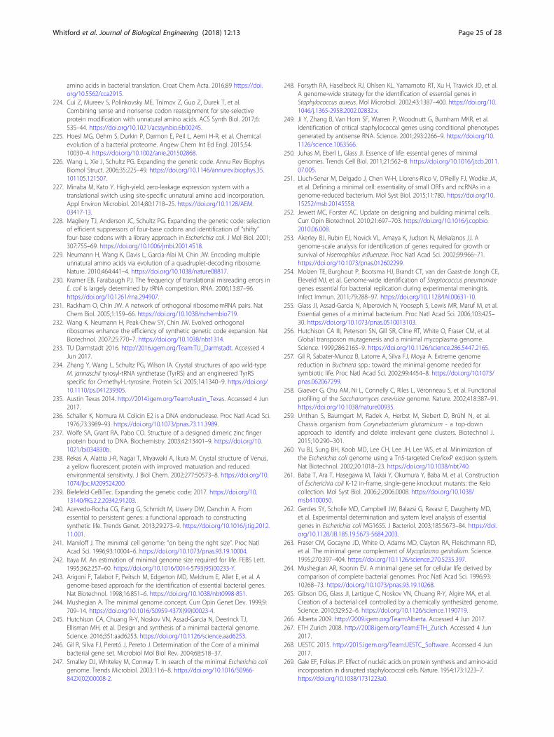
amino acids in bacterial translation. Croat Chem Acta. 2016;89 https://doi.org/10.5562/cca2915.
224. Cui Z, Mureev S, Polinkovsky ME, Tnimov Z, Guo Z, Durek T, et al.Combining sense and nonsense codon reassignment for site-selectiveprotein modification with unnatural amino acids. ACS Synth Biol. 2017;6:535–44. https://doi.org/10.1021/acssynbio.6b00245.
225. Hoesl MG, Oehm S, Durkin P, Darmon E, Peil L, Aerni H-R, et al. Chemicalevolution of a bacterial proteome. Angew Chem Int Ed Engl. 2015;54:10030–4. https://doi.org/10.1002/anie.201502868.
226. Wang L, Xie J, Schultz PG. Expanding the genetic code. Annu Rev BiophysBiomol Struct. 2006;35:225–49. https://doi.org/10.1146/annurev.biophys.35.101105.121507.
227. Minaba M, Kato Y. High-yield, zero-leakage expression system with atranslational switch using site-specific unnatural amino acid incorporation.Appl Environ Microbiol. 2014;80:1718–25. https://doi.org/10.1128/AEM.03417-13.
228. Magliery TJ, Anderson JC, Schultz PG. Expanding the genetic code: selectionof efficient suppressors of four-base codons and identification of “shifty”four-base codons with a library approach in Escherichia coli. J Mol Biol. 2001;307:755–69. https://doi.org/10.1006/jmbi.2001.4518.
229. Neumann H, Wang K, Davis L, Garcia-Alai M, Chin JW. Encoding multipleunnatural amino acids via evolution of a quadruplet-decoding ribosome.Nature. 2010;464:441–4. https://doi.org/10.1038/nature08817.
230. Kramer EB, Farabaugh PJ. The frequency of translational misreading errors inE. coli is largely determined by tRNA competition. RNA. 2006;13:87–96.https://doi.org/10.1261/rna.294907.
231. Rackham O, Chin JW. A network of orthogonal ribosome·mRNA pairs. NatChem Biol. 2005;1:159–66. https://doi.org/10.1038/nchembio719.
232. Wang K, Neumann H, Peak-Chew SY, Chin JW. Evolved orthogonalribosomes enhance the efficiency of synthetic genetic code expansion. NatBiotechnol. 2007;25:770–7. https://doi.org/10.1038/nbt1314.
233. TU Darmstadt 2016. http://2016.igem.org/Team:TU_Darmstadt. Accessed 4Jun 2017.
234. Zhang Y, Wang L, Schultz PG, Wilson IA. Crystal structures of apo wild-typeM. jannaschii tyrosyl-tRNA synthetase (TyrRS) and an engineered TyrRSspecific for O-methyl-L-tyrosine. Protein Sci. 2005;14:1340–9. https://doi.org/10.1110/ps.041239305.
235. Austin Texas 2014. http://2014.igem.org/Team:Austin_Texas. Accessed 4 Jun2017.
236. Schaller K, Nomura M. Colicin E2 is a DNA endonuclease. Proc Natl Acad Sci.1976;73:3989–93. https://doi.org/10.1073/pnas.73.11.3989.
237. Wolfe SA, Grant RA, Pabo CO. Structure of a designed dimeric zinc fingerprotein bound to DNA. Biochemistry. 2003;42:13401–9. https://doi.org/10.1021/bi034830b.
238. Rekas A, Alattia J-R, Nagai T, Miyawaki A, Ikura M. Crystal structure of Venus,a yellow fluorescent protein with improved maturation and reducedenvironmental sensitivity. J Biol Chem. 2002;277:50573–8. https://doi.org/10.1074/jbc.M209524200.
239. Bielefeld-CeBiTec. Expanding the genetic code; 2017. https://doi.org/10.13140/RG.2.2.20342.91203.
240. Acevedo-Rocha CG, Fang G, Schmidt M, Ussery DW, Danchin A. Fromessential to persistent genes: a functional approach to constructingsynthetic life. Trends Genet. 2013;29:273–9. https://doi.org/10.1016/j.tig.2012.11.001.
241. Maniloff J. The minimal cell genome: “on being the right size”. Proc NatlAcad Sci. 1996;93:10004–6. https://doi.org/10.1073/pnas.93.19.10004.
242. Itaya M. An estimation of minimal genome size required for life. FEBS Lett.1995;362:257–60. https://doi.org/10.1016/0014-5793(95)00233-Y.
243. Arigoni F, Talabot F, Peitsch M, Edgerton MD, Meldrum E, Allet E, et al. Agenome-based approach for the identification of essential bacterial genes.Nat Biotechnol. 1998;16:851–6. https://doi.org/10.1038/nbt0998-851.
244. Mushegian A. The minimal genome concept. Curr Opin Genet Dev. 1999;9:709–14. https://doi.org/10.1016/S0959-437X(99)00023-4.
245. Hutchison CA, Chuang R-Y, Noskov VN, Assad-Garcia N, Deerinck TJ,Ellisman MH, et al. Design and synthesis of a minimal bacterial genome.Science. 2016;351:aad6253. https://doi.org/10.1126/science.aad6253.
246. Gil R, Silva FJ, Peretó J, Pereto J. Determination of the Core of a minimalbacterial gene set. Microbiol Mol Biol Rev. 2004;68:518–37.
247. Smalley DJ, Whiteley M, Conway T. In search of the minimal Escherichia coligenome. Trends Microbiol. 2003;11:6–8. https://doi.org/10.1016/S0966-842X(02)00008-2.
248. Forsyth RA, Haselbeck RJ, Ohlsen KL, Yamamoto RT, Xu H, Trawick JD, et al.A genome-wide strategy for the identification of essential genes inStaphylococcus aureus. Mol Microbiol. 2002;43:1387–400. https://doi.org/10.1046/j.1365-2958.2002.02832.x.
249. Ji Y, Zhang B, Van Horn SF, Warren P, Woodnutt G, Burnham MKR, et al.Identification of critical staphylococcal genes using conditional phenotypesgenerated by antisense RNA. Science. 2001;293:2266–9. https://doi.org/10.1126/science.1063566.
250. Juhas M, Eberl L, Glass JI. Essence of life: essential genes of minimalgenomes. Trends Cell Biol. 2011;21:562–8. https://doi.org/10.1016/j.tcb.2011.07.005.
251. Lluch-Senar M, Delgado J, Chen W-H, Llorens-Rico V, O’Reilly FJ, Wodke JA,et al. Defining a minimal cell: essentiality of small ORFs and ncRNAs in agenome-reduced bacterium. Mol Syst Biol. 2015;11:780. https://doi.org/10.15252/msb.20145558.
252. Jewett MC, Forster AC. Update on designing and building minimal cells.Curr Opin Biotechnol. 2010;21:697–703. https://doi.org/10.1016/j.copbio.2010.06.008.
253. Akerley BJ, Rubin EJ, Novick VL, Amaya K, Judson N, Mekalanos JJ. Agenome-scale analysis for identification of genes required for growth orsurvival of Haemophilus influenzae. Proc Natl Acad Sci. 2002;99:966–71.https://doi.org/10.1073/pnas.012602299.
254. Molzen TE, Burghout P, Bootsma HJ, Brandt CT, van der Gaast-de Jongh CE,Eleveld MJ, et al. Genome-wide identification of Streptococcus pneumoniaegenes essential for bacterial replication during experimental meningitis.Infect Immun. 2011;79:288–97. https://doi.org/10.1128/IAI.00631-10.
255. Glass JI, Assad-Garcia N, Alperovich N, Yooseph S, Lewis MR, Maruf M, et al.Essential genes of a minimal bacterium. Proc Natl Acad Sci. 2006;103:425–30. https://doi.org/10.1073/pnas.0510013103.
256. Hutchison CA III, Peterson SN, Gill SR, Cline RT, White O, Fraser CM, et al.Global transposon mutagenesis and a minimal mycoplasma genome.Science. 1999;286:2165–9. https://doi.org/10.1126/science.286.5447.2165.
257. Gil R, Sabater-Munoz B, Latorre A, Silva FJ, Moya A. Extreme genomereduction in Buchnera spp.: toward the minimal genome needed forsymbiotic life. Proc Natl Acad Sci. 2002;99:4454–8. https://doi.org/10.1073/pnas.062067299.
258. Giaever G, Chu AM, Ni L, Connelly C, Riles L, Véronneau S, et al. Functionalprofiling of the Saccharomyces cerevisiae genome. Nature. 2002;418:387–91.https://doi.org/10.1038/nature00935.
259. Unthan S, Baumgart M, Radek A, Herbst M, Siebert D, Brühl N, et al.Chassis organism from Corynebacterium glutamicum - a top-downapproach to identify and delete irrelevant gene clusters. Biotechnol J.2015;10:290–301.
260. Yu BJ, Sung BH, Koob MD, Lee CH, Lee JH, Lee WS, et al. Minimization ofthe Escherichia coli genome using a Tn5-targeted Cre/loxP excision system.Nat Biotechnol. 2002;20:1018–23. https://doi.org/10.1038/nbt740.
261. Baba T, Ara T, Hasegawa M, Takai Y, Okumura Y, Baba M, et al. Constructionof Escherichia coli K-12 in-frame, single-gene knockout mutants: the Keiocollection. Mol Syst Biol. 2006;2:2006.0008. https://doi.org/10.1038/msb4100050.
262. Gerdes SY, Scholle MD, Campbell JW, Balazsi G, Ravasz E, Daugherty MD,et al. Experimental determination and system level analysis of essentialgenes in Escherichia coli MG1655. J Bacteriol. 2003;185:5673–84. https://doi.org/10.1128/JB.185.19.5673-5684.2003.
263. Fraser CM, Gocayne JD, White O, Adams MD, Clayton RA, Fleischmann RD,et al. The minimal gene complement of Mycoplasma genitalium. Science.1995;270:397–404. https://doi.org/10.1126/science.270.5235.397.
264. Mushegian AR, Koonin EV. A minimal gene set for cellular life derived bycomparison of complete bacterial genomes. Proc Natl Acad Sci. 1996;93:10268–73. https://doi.org/10.1073/pnas.93.19.10268.
265. Gibson DG, Glass JI, Lartigue C, Noskov VN, Chuang R-Y, Algire MA, et al.Creation of a bacterial cell controlled by a chemically synthesized genome.Science. 2010;329:52–6. https://doi.org/10.1126/science.1190719.
266. Alberta 2009. http://2009.igem.org/Team:Alberta. Accessed 4 Jun 2017.267. ETH Zurich 2008. http://2008.igem.org/Team:ETH_Zurich. Accessed 4 Jun
2017.268. UESTC 2015. http://2015.igem.org/Team:UESTC_Software. Accessed 4 Jun
2017.269. Gale EF, Folkes JP. Effect of nucleic acids on protein synthesis and amino-acid
incorporation in disrupted staphylococcal cells. Nature. 1954;173:1223–7.https://doi.org/10.1038/1731223a0.
Whitford et al. Journal of Biological Engineering (2018) 12:13 Page 25 of 28
270. Nirenberg MW, Matthaei JH. The dependence of cell-free protein synthesisin E. coli upon naturally occurring or synthetic polyribonucleotides. ProcNatl Acad Sci. 1961;47:1588–602. https://doi.org/10.1073/pnas.47.10.1588.
271. Hodgman CE, Jewett MC. Cell-free synthetic biology: thinking outside thecell. Metab Eng. 2012;14:261–9. https://doi.org/10.1016/j.ymben.2011.09.002.
272. Yang J, Kanter G, Voloshin A, Michel-Reydellet N, Velkeen H, Levy R, et al.Rapid expression of vaccine proteins for B-cell lymphoma in a cell-freesystem. Biotechnol Bioeng. 2005;89:503–11.
273. Smith MT, Wu JC, Varner CT, Bundy BC. Enhanced protein stability throughminimally invasive, direct, covalent, and site-specific immobilization.Biotechnol Prog. 2013;29:247–54.
274. Kitaoka Y, Nishimura N, Niwano M. Cooperativity of stabilized mRNA andenhanced translation activity in the cell-free system. J Biotechnol. 1996;48:1–8.
275. Matveev SV, Vinokurov LM, Shaloiko LA, Davies C, Matveeva EA, Alakhov YB.Effect of the ATP level on the overall protein biosynthesis rate in a wheatgerm cell-free system. Biochim Biophys Acta. 1996;1293:207–12.
276. Shimizu Y, Inoue A, Tomari Y, Suzuki T, Yokogawa T, Nishikawa K, et al. Cell-free translation reconstituted with purified components. Nat Biotechnol.2001;19:751–5. https://doi.org/10.1038/90802.
277. Shrestha P, Holland TM, Bundy BC. Streamlined extract preparation forEscherichia coli-based cell-free protein synthesis by sonication or beadvortex mixing. BioTechniques. 2012;53:163–74.
278. Smith MT, Bennett AM, Hunt JM, Bundy BC. Creating a completely “cell-free”system for protein synthesis. Biotechnol Prog. 2015;31:1716–9. https://doi.org/10.1002/btpr.2157.
279. Kim D-M, Kigawa T, Choi C-Y, Yokoyama S. A highly efficient cell-free proteinsynthesis system from Escherichia coli. Eur J Biochem. 1996;239:881–6. https://doi.org/10.1111/j.1432-1033.1996.0881u.x.
280. Bielefeld-CeBiTec 2015. http://2015.igem.org/Team:Bielefeld-CeBiTec.Accessed 21 Apr 2017.
281. Kwon Y-C, Jewett MC. High-throughput preparation methods of crudeextract for robust cell-free protein synthesis. Sci Rep. 2015;5:8663. https://doi.org/10.1038/srep08663.
282. Edinburgh 2015. http://2015.igem.org/Team:Edinburgh. Accessed 21 Apr 2017.283. Exeter 2015. http://2015.igem.org/Team:Exeter. Accessed 21 Apr 2017.284. Freiburg 2015. http://2015.igem.org/Team:Freiburg. Accessed 21 Apr 2017.285. iGEM Lethbridge 2017. http://2017.igem.org/Team:Lethbridge. Accessed 26
Mar 2017.286. Kailasapathy K. Microencapsulation of probiotic bacteria: technology and
potential applications. Curr Issues Intest Microbiol. 2002;3:39–48. http://www.ncbi.nlm.nih.gov/pubmed/12400637.
287. Tatiya PD, Hedaoo RK, Mahulikar PP, Gite VV. Novel polyurea microcapsulesusing dendritic functional monomer: synthesis, characterization, and its usein self-healing and anticorrosive polyurethane coatings. Ind Eng Chem Res.2013;52:1562–70.
288. Puddu M, Paunescu D, Stark WJ, Grass RN. Magnetically recoverable,thermostable, hydrophobic DNA/silica encapsulates and their application asinvisible oil tags. ACS Nano. 2014;8:2677–85.
289. Hedaoo RK, Gite VV. Renewable resource-based polymericmicroencapsulation of natural pesticide and its release study: analternative green approach. RSC Adv. 2014;4:18637. https://doi.org/10.1039/c4ra01558d.
290. Orive G, De Castro M, Ponce S, Hernández RM, Gascón AR, Bosch M, et al.Long-term expression of erythropoietin from myoblasts immobilized inbiocompatible and neovascularized microcapsules. Mol Ther. 2005;12:283–9.
291. Murua A, de Castro M, Orive G, Hernández RM, Pedraz JL. In vitro characterizationand in vivo functionality of erythropoietin-secreting cells immobilized in alginate-poly-L-lysine-alginate microcapsules. Biomacromolecules. 2007;8:3302–7.
292. Rathore S, Desai PM, Liew CV, Chan LW, Heng PWS. Microencapsulation ofmicrobial cells. J Food Eng. 2013;116:369–81. https://doi.org/10.1016/j.jfoodeng.2012.12.022.
293. Smidsrød O, Skjåk-Braek G. Alginate as immobilization matrix for cells. TrendsBiotechnol. 1990;8:71–8. https://doi.org/10.1016/0167-7799(90)90139-O.
294. John RP, Tyagi RD, Brar SK, Surampalli RY, Prévost D. Bio-encapsulation ofmicrobial cells for targeted agricultural delivery. Crit Rev Biotechnol.2011;31:211–26.
295. Avnir D, Braun S, Lev O, Ottolenghi M. Enzymes and other proteinsentrapped in sol-gel materials. Chem Mater. 1994;6:1605–14.
296. Zink JI, Yamanaka SA, Ellerby LM, Valentine JS, Nishida F, Dunn B.Biomolecular materials based on sol-gel encapsulated proteins - code: G1.J Sol-Gel Sci Technol. 1994;2:791–5.
297. Pankratov I, Lev O. Sol-gel derived renewable-surface biosensors. J ElectroanalChem. 1995;393:35–41.
298. Nassif N, Bouvet O, Noelle Rager M, Roux C, Coradin T, Livage J. Livingbacteria in silica gels. Nat Mater. 2002;1:42–4.
299. Dusseault J, Langlois G, Meunier MC, Ménard M, Perreault C, Hallé JP. Theeffect of covalent cross-links between the membrane components ofmicrocapsules on the dissemination of encapsulated malignant cells.Biomaterials. 2008;29:917–24.
300. Zimmermann H, Wählisch F, Baier C, Westhoff M, Reuss R, Zimmermann D,et al. Physical and biological properties of barium cross-linked alginatemembranes. Biomaterials. 2007;28:1327–45.
301. Shen F, Mazumder MAJ, Burke NAD, Stöver HDH, Potter MA. Mechanicallyenhanced microcapsules for cellular gene therapy. J Biomed Mater Res BAppl Biomater. 2009;90(B):350–61.
302. Paris Saclay 2015. http://2015.igem.org/Team:Paris_Saclay. Accessed 23 Jun2017.
303. Perullini M, Jobbágy M, Soler-Illia GJAA, Bilmes SA. Cell growth at cavitiescreated inside silica monoliths synthesized by sol-gel. Chem Mater. 2005;17:3806–8.
304. Perullini M, Orias F, Durrieu C, Jobbágy M, Bilmes SA. Co-encapsulation ofDaphnia magna and microalgae in silica matrices, a stepping stone towarda portable microcosm. Biotechnol Rep (Amst). 2014;4:147–50. https://doi.org/10.1016/j.btre.2014.10.002.
305. Birnbaum S, Pendleton R, Larsson PO, Mosbach K. Covalent stabilization ofalginate gel for the entrapment of living whole cells. Biotechnol Lett. 1981;3:393–400.
306. Yamamori T, Yura T. Genetic control of heat-shock protein synthesis and itsbearing on growth and thermal resistance in Escherichia coli K-12. Proc NatlAcad Sci U S A. 1982;79:860–4.
307. Fayet O, Ziegelhoffer T, Georgopoulos C. The groES and groEL heat shockgene products of Escherichia coli are essential for bacterial growth at alltemperatures. J Bacteriol. 1989;171:1379–85. http://www.pubmedcentral.nih.gov/articlerender.fcgi?artid=209756&tool=pmcentrez&rendertype=abstract
308. Zhao K, Liu M, Burgess RR. The global transcriptional response of Escherichiacolito induced σ32 protein involves σ32 regulon activation followed byinactivation and degradation of σ32 in vivo. J Biol Chem. 2005;280:17758–68. https://doi.org/10.1074/jbc.M500393200.
309. Nonaka G. Regulon and promoter analysis of the E. coli heat-shock factor,σ32, reveals a multifaceted cellular response to heat stress. Genes Dev. 2006;20:1776–89. https://doi.org/10.1101/gad.1428206.
310. Wade JT, Roa DC, Grainger DC, Hurd D, Busby SJW, Struhl K, et al. Extensivefunctional overlap between sigma factors in Escherichia coli. Nat Struct MolBiol. 2006;13:806–14.
311. Ermolenko DN, Makhatadze GI. Bacterial cold-shock proteins. Cell Mol LifeSci. 2002;59:1902–13.
312. Horn G, Hofweber R, Kremer W, Kalbitzer HR. Structure and function ofbacterial cold shock proteins. Cell Mol Life Sci. 2007;64:1457–70.
313. Mizuno T, Imae Y. Conditional inversion of the thermoresponse inEscherichia coli. J Bacteriol. 1984;159:360–7.
314. Maeda K, Imae Y. Thermosensory transduction in Escherichia coli: inhibitionof the thermoresponse by L-serine. Proc Natl Acad Sci U S A. 1979;76:91–5.http://www.pubmedcentral.nih.gov/articlerender.fcgi?artid=382882&tool=pmcentrez&rendertype=abstract
315. Salman H, Libchaber A. A concentration-dependent switch in the bacterialresponse to temperature. Nat Cell Biol. 2007;9:1098–100. https://doi.org/10.1038/ncb1632.
316. Nara T, Lee L, Imae Y. Thermosensing ability of Trg and tap chemoreceptorsin Escherichia coli. J Bacteriol. 1991;173:1120–4.
317. Pruss GJ, Drlicat K. DNA supercoiling and Prokatyotic transcriptionMinireview. Cell. 1988;56:521–3.
318. Dorman CJ, Corcoran CP. Bacterial DNA topology and infectious disease.Nucleic Acids Res. 2009;37:672–8.
319. Dorman CJ. Flexible response: DNA supercoiling, transcription and bacterialadaptation to environmental stress. Trends Microbiol. 1996;4:214–6.
320. López-García P, Forterre P. DNA topology and the thermal stress response, atale from mesophiles and hyperthermophiles. BioEssays. 2000;22:738–46.
321. Schlax PJ, Worhunsky DJ. Translational repression mechanisms inprokaryotes. Mol Microbiol. 2003;48:1157–69.
322. Morita MT, Tanaka Y, Kodama TS, Kyogoku Y, Yanagi H, Yura T. Translationalinduction of heat shock transcription factor σ32: evidence for a built-in RNAthermosensor. Genes Dev. 1999;13:655–65. https://doi.org/10.1101/gad.13.6.655.
Whitford et al. Journal of Biological Engineering (2018) 12:13 Page 26 of 28
323. Nocker A, Hausherr T, Balsiger S, Krstulovic NP, Hennecke H, Narberhaus F. AmRNA-based thermosensor controls expression of rhizobial heat shockgenes. Nucleic Acids Res. 2001;29:4800–7. https://doi.org/10.1093/nar/29.23.4800.
324. Johansson J, Mandin P, Renzoni A, Chiaruttini C, Springer M, Cossart P. AnRNA thermosensor controls expression of virulence genes in Listeriamonocytogenes. Cell. 2002;110:551–61. https://doi.org/10.1016/S0092-8674(02)00905-4.
325. Altuvia S, Kornitzer D, Teff D, Oppenheim AB. Alternative mRNA structuresof the cIII gene of bacteriophage λ determine the rate of its translationinitiation. J Mol Biol. 1989;210:265–80. https://doi.org/10.1016/0022-2836(89)90329-X.
326. Giuliodori AM, Di Pietro F, Marzi S, Masquida B, Wagner R, Romby P, et al.The cspA mRNA is a Thermosensor that modulates translation of the cold-shock protein CspA. Mol Cell. 2010;37:21–33. https://doi.org/10.1016/j.molcel.2009.11.033.
327. Narberhaus F, Waldminghaus T, Chowdhury S. RNA thermometers. FEMSMicrobiol Rev. 2006;30:3–16.
328. Sledjeski DD, Gupta A, Gottesman S. The small RNA, DsrA, is essential for thelow temperature expression of RpoS during exponential growth inEscherichia coli. EMBO J. 1996;15:3993–4000.
329. Repoila F, Gottesman S. Signal transduction cascade for regulation of RpoS:temperature regulation of DsrA. J Bacteriol. 2001;183:4012–23. https://doi.org/10.1128/JB.183.13.4012-4023.2001.
330. Repoila F, Majdalani N, Gottesman S. Small non-coding RNAs, co-ordinatorsof adaptation processes in Escherichia coli: the RpoS paradigm. MolMicrobiol. 2003;48:855–61.
331. Ketzer P, Kaufmann JK, Engelhardt S, Bossow S, von Kalle C, Hartig JS, et al.Artificial riboswitches for gene expression and replication control of DNAand RNA viruses. Proc Natl Acad Sci U S A. 2014;111:E554–62. https://doi.org/10.1073/pnas.1318563111.
332. Verhounig A, Karcher D, Bock R. Inducible gene expression from the plastidgenome by a synthetic riboswitch. Proc Natl Acad Sci. 2010;107:6204–9.https://doi.org/10.1073/pnas.0914423107.
333. Hurme R, Rhen M. Temperature sensing in bacterial gene regulation-what itall boils down to. Mol Microbiol. 1998;30:1–6. http://www.ncbi.nlm.nih.gov/pubmed/9786180
334. Nagai H, Yuzawa H, Yura T. Interplay of two cis-acting mRNA regions intranslational control of σ32 synthesis during the heat shock response ofEscherichia coli. Proc Natl Acad Sci U S A. 1991;88:10515–9. https://doi.org/10.1073/pnas.88.23.10515.
335. Craig EA, Gross CA. Is hsp70 the cellular thermometer? Trends Biochem Sci.1991;16 C:135–40.
336. Chowdhury S, Maris C, Allain FH-T, Narberhaus F. Molecular basis fortemperature sensing by an RNA thermometer. EMBO J. 2006;25:2487–97.
337. Narberhaus F, Weiglhofer W, Fischer HM, Hennecke H. The Bradyrhizobiumjaponicum rpoH1 gene encoding a sigma 32-like protein is part of a uniqueheat shock gene cluster together with groESL1 and three small heat shockgenes. J Bacteriol. 1996;178:5337–46. http://www.ncbi.nlm.nih.gov/pubmed/8808920.
338. Glaser M, Nulty W, Vagelos PR. Role of adenylate kinase in the regulation ofmacromolecular biosynthesis in a putative mutant of Escherichia colidefective in membrane phospholipid biosynthesis. J Bacteriol. 1975;123:128–36.
339. Böck A. Mutation affecting the charging reaction of alanyl-tRNA synthetasefrom Escherichia coli K 10. Arch Mikrobiol. 1969;68:165–78.
340. McHenry CS, Crow W. DNA polymerase III of Escherichia coli. Purification andidentification of subunits. J Biol Chem. 1979;254:1748–53. http://www.jbc.org/content/254/5/1748.abstract
341. Heinrikson RL, Hartley BS. Purification and properties of methionyl-transfer-ribonucleic acid synthetase from Escherichia coli. Biochem J. 1967;105:17–24.https://www.ncbi.nlm.nih.gov/pubmed/3283735.
342. Nellemann LJ, Holm F, Atlung T, Hansen FG. Cloning and characterization ofthe Escherichia coliphosphoglycerate kinase (pgk) gene. Gene. 1989;77:185–91. https://doi.org/10.1016/0378-1119(89)90373-9.
343. Calendar R, Berg P. Purification and physical characterization of Tyrosylribonucleic acid Synthetases from Escherichia coli and Bacillus subtilis.Biochemistry. 1966;5:1681–90. https://doi.org/10.1021/bi00869a033.
344. Aramaki H, Yagi N, Suzuki M. Residues important for the function of amultihelical DNA binding domain in the new transcription factor family ofcam and tet repressors. Protein Eng. 1995;8:1259–66.
345. Waldminghaus T, Heidrich N, Brantl S, Narberhaus F. FourU: a novel type ofRNA thermometer in Salmonella. Mol Microbiol. 2007;65:413–24.
346. TU Delft 2008. http://2008.igem.org/Team:TUDelft/Team. Accessed 23 Jun2017.
347. NCTU Taiwan 2011. http://2011.igem.org/Team:NCTU_Formosa. Accessed 23Jun 2017.
348. Cormier MJ, Hori K, Anderson JM. Bioluminescence in coelenterates.Biochim Biophys Acta. 1974;346:137–64. https://doi.org/10.1016/0304-4173(74)90007-X.
349. Cormack BP, Valdivia RH, Falkow S. FACS-optimized mutants of the greenfluorescent protein (GFP). Gene. 1996;173:33–8. https://doi.org/10.1016/0378-1119(95)00685-0.
350. Schwarz C, Bohne AV, Wang F, Cejudo FJ, Nickelsen J. An intermoleculardisulfide-based light switch for chloroplast psbD gene expression inChlamydomonas reinhardtii. Plant J. 2012;72:378–89.
351. Taylor BL, Zhulin IB. PAS domains: internal sensors of oxygen, redoxpotential, and light. Microbiol Mol Biol Rev. 1999;63:479–506.
352. Möglich A, Ayers RA, Moffat K. Design and signaling mechanism of light-regulated histidine kinases. J Mol Biol. 2009;385:1433–44. https://doi.org/10.1016/j.jmb.2008.12.017.
353. Crosson S, Rajagopal S, Moffat K. The LOV domain family: Photoresponsivesignaling modules coupled to diverse output domains. Biochemistry. 2003;42:2–10.
354. Gilles-Gonzalez MA, Ditta GS, Helinski DR. A haemoprotein with kinaseactivity encoded by the oxygen sensor of Rhizobium meliloti. Nature. 1991;350:170–2. https://doi.org/10.1038/350170a0.
355. Fischer HM. Genetic regulation of nitrogen fixation in rhizobia. MicrobiolRev. 1994;58:352–86. http://www.pubmedcentral.nih.gov/articlerender.fcgi?artid=372973&tool=pmcentrez&rendertype=abstract
356. Losi A, Polverini E, Quest B, Gärtner W. First evidence for phototropin-related blue-light receptors in prokaryotes. Biophys J. 2002;82:2627–34.
357. Gilles-Gonzalez MA, Gonzalez G, Perutz MF, Kiger L, Marden MC, Poyart C.Heme-based sensors, exemplified by the kinase FixL, are a new class ofheme protein with distinctive ligand binding and autoxidation.Biochemistry. 1994;33:8067–73. https://doi.org/10.1021/bi00192a011.
358. Ohlendorf R, Vidavski RR, Eldar A, Moffat K, Möglich A. From dusk till dawn:one-plasmid systems for light-regulated gene expression. J Mol Biol. 2012;416:534–42. https://doi.org/10.1016/j.jmb.2012.01.001.
359. Diensthuber RP, Ohlendorf R, Gleichmann T, Schubert R, Möglich A.Lichtregulierte Genexpression. BIOspektrum. 2013;19:149–51.
360. Yeh K. A cyanobacterial Phytochrome two-component light sensory system.Science. 1997;277:1505–8. https://doi.org/10.1126/science.277.5331.1505.
361. Levskaya A, Chevalier AA, Tabor JJ, Simpson ZB, Lavery LA, Levy M, et al.Synthetic biology: engineering Escherichia coli to see light. Nature. 2005;438:441–2. https://doi.org/10.1038/nature04405.
362. Utsumi R, Brissette RE, Rampersaud A, Forst SA, Oosawa K, Inouye M.Activation of bacterial porin gene expression by a chimeric signaltransducer in response to aspartate. Science. 1989;245:1246–9.
363. Kaneko T, Sato S, Kotani H, Tanaka A, Asamizu E, Nakamura Y, et al.Sequence analysis of the genome of the unicellular cyanobacteriumSynechocystis sp. strain PCC6803. II. Sequence determination of the entiregenome and assignment of potential protein-coding regions. DNA Res.1996;3:109–36.
364. Hughes J, Lamparter T, Mittmann F, Hartmann E, Gärtner W, Wilde A, et al.A prokaryotic phytochrome. Nature. 1997;386:663. https://doi.org/10.1038/386663a0.
365. Beale SI, Cornejo J. Biosynthesis of Phycobilins. J Biol Chem. 1991;266:22328–32.
366. Gambetta GA, Lagarias JC. Genetic engineering of phytochromebiosynthesis in bacteria. Proc Natl Acad Sci U S A. 2001;98:10566–71. https://doi.org/10.1073/pnas.191375198.
367. Uppsala 2011. http://2011.igem.org/Team:Uppsala-Sweden. Accessed 23 Jun2017.
368. Cornell 2011. http://2011.igem.org/Team:Cornell. Accessed 23 Jun 2017.369. Kunkel TA. DNA replication Fidelity. J Biol Chem. 2004;279:16895–8. https://
doi.org/10.1074/jbc.R400006200.370. Martinez JL, Baquero F. Mutation frequencies and antibiotic resistance.
Antimicrob Agents Chemother. 2000;44:1771–7. https://doi.org/10.1128/AAC.44.7.1771-1777.2000.
371. Microbiology by numbers. Nat Rev Microbiol. 2011;9:628–8. https://doi.org/10.1038/nrmicro2644.
Whitford et al. Journal of Biological Engineering (2018) 12:13 Page 27 of 28
372. Chakrabarty AM. Genetically-manipulated microorganisms and theirproducts in the oil service industries. Trends Biotechnol. 1985;3:32–9.
373. Rojo F, Pieper D, Engesser K, Knackmuss H, Timmis K. Assemblage of orthocleavage route for simultaneous degradation of chloro- andmethylaromatics. Science. 1987;238:1395–8. https://doi.org/10.1126/science.3479842.
374. Harvard 2010. http://2010.igem.org/Team:Harvard. Accessed 23 Jun 2017.375. Virginia 2016. http://2016.igem.org/Team:Virginia. Accessed 23 Jun 2017.376. METU Ankara 2011. http://2011.igem.org/Team:METU-Ankara. Accessed 23
Jun 2017.377. NYMU Taipei 2014. http://2014.igem.org/Team:NYMU-Taipei. Accessed 23
Jun 2017.378. Minnesota 2014. http://2014.igem.org/Team:Minnesota. Accessed 23 Jun
2017.379. HNU China 2014. http://2014.igem.org/Team:HNU_China. Accessed 23 Jun
2017.380. Braunschweig 2014. http://2014.igem.org/Team:Braunschweig. Accessed 23
Jun 2017.381. Tack DS, Ellefson JW, Thyer R, Wang B, Gollihar J, Forster MT, et al. Addicting
diverse bacteria to a noncanonical amino acid. Nat Chem Biol. 2016;12:138–40.https://doi.org/10.1038/nchembio.2002.
Whitford et al. Journal of Biological Engineering (2018) 12:13 Page 28 of 28