Embed Size (px)
Citation preview
Journ
alof
Cell
Scie
nce
Autophagy and microtubules – new story, old players
Rafah Mackeh1, Daniel Perdiz1, Severine Lorin1, Patrice Codogno2,* and Christian Pous1,3,*1UPRES EA4530, Universite Paris-Sud, Faculte de Pharmacie, 92296 Chatenay-Malabry, France2INSERM U845, Universite Paris Descartes, 75014 Paris, France3Biochimie-Hormonologie, AP-HP, Hopital Antoine Beclere, 92141 Clamart, France
*Authors for correspondence ([email protected]; [email protected])
Journal of Cell Science 126, 1071–1080� 2013. Published by The Company of Biologists Ltddoi: 10.1242/jcs.115626
SummaryBoth at a basal level and after induction (especially in response to nutrient starvation), the function of autophagy is to allow cells todegrade and recycle damaged organelles, proteins and other biological constituents. Here, we focus on the role microtubules have in
autophagosome formation, autophagosome transport across the cytoplasm and in the formation of autolysosomes. Recent insights intothe exact relationship between autophagy and microtubules now point to the importance of microtubule dynamics, tubulin post-translational modifications and microtubule motors in the autophagy process. Such factors regulate signaling pathways that converge tostimulate autophagosome formation. They also orchestrate the movements of pre-autophagosomal structures and autophagosomes or
more globally organize and localize immature and mature autophagosomes and lysosomes. Most of the factors that now appear to linkmicrotubules to autophagosome formation or to autophagosome dynamics and fate were identified initially without the notion thatsequestration, recruitment and/or interaction with microtubules contribute to their function. Spatial and temporal coordination of many
stages in the life of autophagosomes thus underlines the integrative role of microtubules and progressively reveals hidden parts of theautophagy machinery.
Key words: Microtubule, Macroautophagy, Phagocytosis, Phagophore, Cell signaling, Lysosome, Acetylation, Molecular motor
IntroductionAutophagy is a catabolic process that leads to the degradation of
cytoplasmic components by lysosomes. There are several types of
autophagy, such as macroautophagy, microautophagy and
chaperone-mediated autophagy (CMA) (for a review, see
Mizushima and Komatsu, 2011). Here, we will focus on
macroautophagy (hereafter referred to as autophagy). The initial
step of autophagy is the formation of a cup-shaped structure,
named the phagophore (Fig. 1A). In most cell types, autophagy
proceeds at a basal rate to ensure a level of quality control of the
cytoplasm and to maintain cellular homeostasis, by eliminating
long-lived or aggregated proteins and damaged organelles,
including mitochondria (for a review, see Yang and Klionsky,
2010). However, under several stress conditions (e.g. metabolic
stress, hypoxia, chemotherapy), autophagy is greatly induced to
cope with the stress and to fulfil the energetic or biosynthetic
demand, thereby allowing cell survival (for reviews, see Kroemer
et al., 2010; Singh and Cuervo, 2011; Yang and Klionsky, 2010).
Autophagy not only has an important physiological role in stress
management but is also involved in development and immunity
(reviewed by Deretic and Levine, 2009; Levine and Klionsky,
2004). Deregulation of autophagy leads to a wide range of
disorders, such as cancers, neurodegenerative disorders, heart and
liver diseases, myopathies and ageing (Ravikumar et al., 2010;
Rubinsztein et al., 2011).
The autophagosome membrane elongates to surround the
cytoplasmic material to be degraded (e.g. proteins, lipid droplets,
organelles and pathogens), before it expands, curves and then
closes to form a double-membrane vesicle. The sequestered
material is then degraded by acid hydrolases that reside in the
lysosome. The resulting degradation products (e.g. amino acids,
fatty acids and nucleotides) are exported through membrane
permeases to the cytoplasm, where they are recycled for the
biosynthesis of new macromolecules or the production of ATP
(Mehrpour et al., 2010; Yang and Klionsky, 2010).
Similar to most of the membrane-bound organelles and
vesicles, various aspects of autophagosome dynamics rely in
part on their interactions with the cytoskeleton and especially
with microtubules (MTs) (Monastyrska et al., 2009). Although
the involvement of MTs in many steps of autophagosome
dynamics has been a matter of considerable debate over the past
years, it appears that a consistent picture is now emerging in
which MTs have essential roles in coordinating and organizing
many crucial steps of autophagosome life. After providing a brief
overview of autophagy and of microtubule function and
dynamics, we will focus here on the roles that MTs and
microtubule molecular motors have in autophagosome formation,
their movements and their interaction with lysosomes. We will
also discuss recent advances that highlight the importance of
global organelle positioning and of MT acetylation in autophagy.
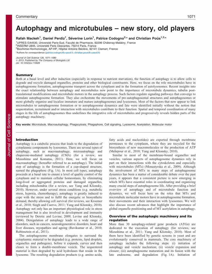
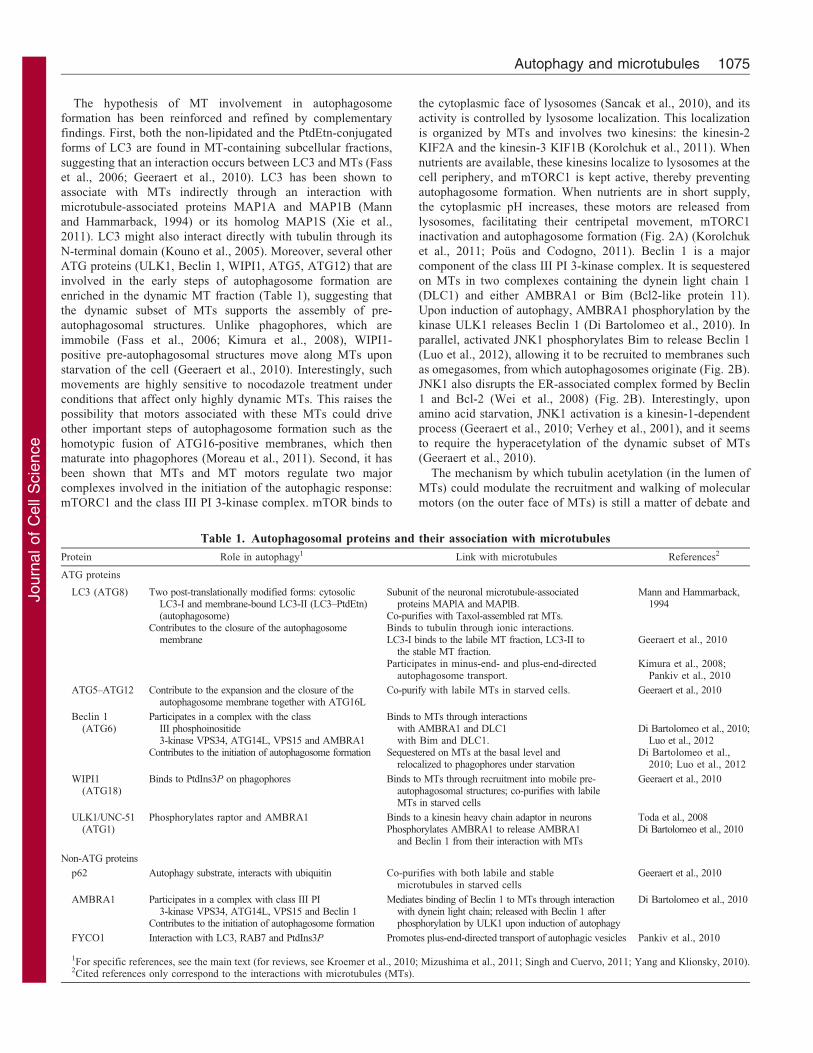
Overview of the autophagic machinery and itsregulationMore than 30 autophagy-related gene products (ATGs) are
dedicated to the execution of autophagy (for reviews, see
Mizushima et al., 2011; Yang and Klionsky, 2010). Most of
them have been identified by genetic screens in yeast and are
evolutionarily conserved in mammals. The core machinery of
autophagy includes the following steps: (i) initiation of
autophagy and vesicle nucleation; (ii) vesicle expansion and
closure; (iii) autophagosome maturation and its fusion with the
late endosome, and degradation (Fig. 1A). Initiation of
Commentary 1071
Journ
alof
Cell
Scie
nce
autophagy starts with the activation of the ULK1 complex (Atg1
complex in budding yeast) that contains the serine/threonine-
protein kinase ULK1, ATG13, focal adhesion kinase family
kinase-interacting protein of 200 kD (FIP200, also known as
RBCC1) and ATG101 (reviewed by Mizushima, 2010). Upon
induction of autophagy, this complex localizes to the site of
phagophore formation to regulate the nucleation machinery.
Phagophore nucleation is highly dependent on the production of
phosphatidylinositol 3-phosphate [PtdIns(3)P (PI3P)] by a
phosphoinositide 3-kinase (PI 3-kinase) complex that is
recruited to the phagophore membrane during the initiation
step. This complex comprises the class III PI 3-kinase Vps34
AMPK
mTOR
Lysosome
Rag-GTP
Energy(ATP)
TSC1 TSC2
Rheb
AKT
Amino acids Energy(Glucose)
Class I PI3K
Growth factors
DAPK JNK
Amino acids
Beclin1 Bcl-2 / Bcl-xL
B
Amino acids Fatty acids Nucleotides
Initiation Nucleation Elongation Closure
Maturation Degradation
LysosomeAutophagosomePhagophore
A
AutolysosomeULK1complex
Class III PI3Kcomplex Atg12–5-16 LC3–PE
mTOR
PI3P
PI3P
Atg1
01 Atg1
3
Atg10
Atg12 Atg7Atg16L
Atg3
Atg7
Ubiquitin-like conjugation systems
Class III PI3K complex
Atg12
Atg4
Atg5Atg5
LC3LC3
LC3Ptd-Etn
Gly
WIPI1/2
AMBRA1
Beclin1Atg14L
hVPS34 p150
FIP2
00
ULK
1
C
Atg5g12
At
Atg16L
LC3
LC3Atg9
Class III PI3Kcomplex
Phagophore formation
ULKcomplex
Fig. 1. See next page for legend.
Journal of Cell Science 126 (5)1072
Journ
alof
Cell
Scie
nce
(PK3C3), Beclin 1 (the mammalian ortholog of Atg6 in yeast),
ATG14, Vps15 and activating molecule in BECN1-regulated
autophagy protein 1 (AMBRA1) (Funderburk et al., 2010). The
production of PtdIns(3)P in the phagophore membrane allows the
recruitment of the WD repeat domain phosphoinositide-
interacting proteins WIPI1 and WIPI2 (the mammalian
orthologs of Atg18 in budding yeast) (Polson et al., 2010).
Both contribute to the expansion and the closure of the vesicle
in concert with two ubiquitin-like conjugation systems,
resulting in the ATG12–ATG5-ATG16L complex and the
phosphatidylethanolamine (PtdEtn) conjugate of microtubule-
associated protein 1 light chain 3 [LC3 (MLP3); the mammalian
ortholog of Atg8 in budding yeast] (Fujita et al., 2008; Hanada
et al., 2007; for a review, see Xie and Klionsky, 2007). The
transmembrane protein ATG9 also participates in the nucleation
and the expansion of the phagophore membrane by cycling
between different compartments and the phagophore (Orsi et al.,
2012) (Fig. 1C). Autophagosome maturation and its fusion with
the lysosome occur in the vicinity of the centrosome and depend
on several lysosomal membrane proteins, such as the small
GTPase Ras-related protein Rab7 and the transmembrane
lysosome-associated membrane glycoprotein 2 (LAMP2)
(Gutierrez et al., 2004; Jager et al., 2004; Tanaka et al., 2000).
Degradation of autophagosomal cargoes is then achieved by the
acid hydrolases and the cathepsin proteases that are present in the
lysosomal lumen.
Induction of autophagy is strictly regulated through upstream
signaling pathways that are governed by growth factors, amino
acids, glucose and the energy status (Fig. 1B). Most of these
pathways control the two initiation complexes ULK1 and PI 3-
kinase . Activity of the ULK1 complex is controlled by one of the
main players in autophagy regulation, the mammalian target of
rapamycin complex 1 (mTORC1), which comprises the mTORserine/threonine-protein kinase and auxiliary proteins. As long as
mTOR is activated, it inhibits autophagy by inhibitoryphosphorylation of ULK1 (for a review, see Yang andKlionsky, 2010). Under diverse stresses including amino acidstarvation, mTORC1 is inhibited, thereby promoting ULK1
complex activity and autophagy (Hosokawa et al., 2009; Junget al., 2009). One of the important pathways that regulatesmTORC1 is initiated by growth factors and involves the class I PI
3-kinase s and Akt serine/threonine-protein kinases (for a review,see Sengupta et al., 2010). Another pathway that controlsautophagy in response to amino acids involves the Rag
GTPase, which allows the recruitment of mTOR at thelysosomal membrane, where its direct activators reside (Sancaket al., 2010). Upon energy depletion, when the AMP:ATP ratiorises, AMP-activated protein kinase (AMPK) can also activate
the ULK1 complex by directly phosphorylating ULK1 or,indirectly, by inactivating mTORC1 (Alers et al., 2012; Kimet al., 2011). Activity of the PI 3-kinase complex is also
regulated, mainly through the interaction of Beclin 1 with theanti-apoptotic members of the Bcl-2 family (Kang et al., 2011).Interaction of Beclin 1 with Bcl-2 or Bcl-xL through its BH3
domain inhibits autophagy. Upon amino acid starvation,activation of c-Jun N-terminal kinase-1 [JNK1 (mitogen-activated protein kinase 8)] leads to Bcl-2 phosphorylation and
the release of Beclin 1, which in turn induces autophagy (Weiet al., 2008). Interaction between Beclin 1 and apoptosis regulatorBcl-2 or Bcl-xL can also be disrupted following thephosphorylation of Beclin 1 by death-associated protein kinase
(DAPK) (Zalckvar et al., 2009).
The origin of the autophagosomal membrane and the processof phagophore nucleation have long been an enigma. The
phagophore appears to expand by membrane addition rather thanby de novo lipid synthesis (for a review, see Yang and Klionsky,2010). Several organelles could serve as membrane donors for
autophagosome formation, such as the endoplasmic reticulum(ER), the Golgi complex, mitochondria, endosomes, the plasmamembrane or the nuclear envelope (for a review, see Mizushimaet al., 2011). Recent studies suggest that a specialized domain of
the ER called the omegasome (Axe et al., 2008) is a privilegedsite for phagophore biogenesis. The autophagosomal membraneelongates inside a cradle that is formed by ER membranes, which
acts as a template for the spherical shape of the autophagosome(Hayashi-Nishino et al., 2009; Yla-Anttila et al., 2009). As wediscuss below, many steps in the life cycle of autophagosomes
involve MTs and their associated molecular motors.
Microtubule dynamics and functionsMTs are hollow cylinders of ,25 nm in diameter that are formed
by polymerization of a–b tubulin dimers. In most mammaliancells, interphase MTs assemble at the centrosome and/or atmembrane MT-organizing centers (MTOCs) such as the Golgi
complex. c-Tubulin-containing ring complexes nucleate MTs andinteract with their minus-ends (for a review, see Kollman et al.,2011). MT plus-ends grow towards the cell periphery by
incorporating GTP-bound tubulin subunits. Growing MTsgenerally hydrolyze GTP in the inner regions of the polymer,yielding a GTP–tubulin cap (David-Pfeuty et al., 1977; Dimitrov
et al., 2008). Once this cap is lost at the cell periphery, MTsdisassemble and release GDP–tubulin into the cytosol. Thealternation between growth and shrinking phases of MTs that are
Fig. 1. The autophagic pathway and its regulation. (A) Autophagy
(specifically, macroautophagy) begins with the nucleation of an isolation
membrane, termed the phagophore, which surrounds a fraction of cytoplasm
to be degraded. After elongation and closure, the newly formed
autophagosome receives input from the endocytic pathway and ultimately
fuses with a lysosome, allowing the degradation of autophagic substrates.
The resultant macromolecules are exported to the cytosol and recycled for
ATP production and biosynthesis. (B) Autophagy is regulated by upstream
signaling, which integrates stimuli mediated by growth factors as well as the
sensing of available energy (ATP) and amino acids. These signaling
pathways converge on two initiation complexes – ULK1 and class III
phosphoinositide 3-kinase (PI3K). The kinase mTOR, a master regulator of
autophagy, integrates multiple signals and inhibits ULK1 by
phosphorylation. As two of its activators, Rag and Rheb, localize to the
lysosomal membrane, mTOR is activated at the lysosomal surface.
(C) Phagophore formation. Once activated, the ULK1 complex localizes at
sites of phagophore formation together with the class III PI3K complex
containing Beclin 1. Newly synthesized PtdIns(3)P then recruits the FYVE-
domain-containing proteins WIPI1 and WIPI2. They contribute to the
expansion and the closure of the autophagic vesicle in concert with ATG12–
ATG5–ATG16L and LC3–PtdEtn. The formation of these two complexes
involves a covalent linkage of ATG12 to ATG5 and of LC3 to PtdEtn by
ubiquitin-like conjugation systems. The latter involve the E1-like enzyme
ATG7, which activates ATG12 and LC3 (previously cleaved by ATG4), and
two distinct E2-like enzymes, ATG10 and ATG3, which transfer,
respectively, ATG12 to ATG5 and LC3 to PtdEtn. The ATG12–ATG5
conjugate then forms a complex with ATG16L and associates with the
phagophore, which is necessary for insertion of LC3–PtdEtn in the
membrane. ATG9-enriched vesicles might provide lipids to the phagophore
membrane, allowing its expansion.
Autophagy and microtubules 1073
Journ
alof
Cell
Scie
nce
separated by transitions that are termed either catastrophes (fromMT assembly or pausing to disassembly) and rescues (from
phases of MT shrinking or pausing to their re-growth) is calleddynamic instability (Mitchison and Kirschner, 1984). Such acontinuous remodelling operates independently for each MT andallows MTs to interact temporarily with cellular components, to
explore the intracellular space and to position organellesdynamically (for reviews, see Desai and Mitchison, 1997;Howard, 2006). For instance, MTs maintain the ER and
mitochondria that are dispersed throughout the cytoplasm,while they at the same time also maintain the Golgi complexand the endosomes that are clustered in the vicinity of the nucleus
(reviewed by Cole and Lippincott-Schwartz, 1995). MTs moveand position organelles by functioning as tracks, on which plus-end- (kinesins) or minus-end-directed (cytoplasmic dynein)molecular motors carry membranes, by exerting pushing and
pulling forces during their assembly and depolymerization or bysliding along each other in a way that is powered by molecularmotors (for a review, see Tolic-Nørrelykke, 2008). MTs could also
interact directly or indirectly with a wealth of proteins that might besequestered, released, assembled into complexes and/or transportedto organize and modulate signal transduction (for a review, see
Etienne-Manneville, 2010; Gundersen and Cook, 1999).
At a given time, not all the MT network of a cell is subjected toremodelling by dynamic instability. Indeed, a subpopulationcomprises long-lived polymers, which can persist even for an
entire interphase (so-called stable MTs) (Webster et al., 1987).MT stabilization, which is enhanced by cell confluence, mightoccur at the cell periphery, where MT plus-ends are capped with
proteins that prevent tubulin exchange (Bartolini et al., 2008;Infante et al., 2000; Palazzo et al., 2001). This stabilization is notstochastic and can result from specific cortical interactions
(reviewed by Gundersen et al., 2004) or from interactions withorganelles such as the Golgi complex (Chabin-Brion et al., 2001;Efimov et al., 2007; Miller et al., 2009). In addition, stable MTs
exhibit numerous tubulin post-translational modifications, suchas de-tyrosination, polyglutamylation or acetylation (for areview, see Janke and Bulinski, 2011). These modificationsmight prevent the binding of disassembling factors or modulate
MT functions by fine-tuning the binding of proteins to the MTsurface. Stable and dynamic MT subpopulations are functionallyspecialized in organizing signaling pathways or vesicle
trafficking. For example, dynamic MTs are involved inbasolateral-directed post-Golgi trafficking in epithelial cells, aswell as in transcytosis (Hunziker et al., 1990; Lafont et al., 1994;
Pous et al., 1998) or in caveolae-mediated pathogeninternalization (Guignot et al., 2001). Stable MTs participate inER-to-Golgi traffic (Mizuno and Singer, 1994), the Golgi-to-
plasma-membrane traffic (Cai et al., 2009) or in the recycling ofendosomes to the plasma membrane (Lin et al., 2002). StableMTs are also involved in alcohol-induced alterations in proteintraffic in hepatocytes (Joseph et al., 2008). Vesicular carriers are
thought to recognize the MT tracks they require throughinteraction with molecular motors, especially with kinesins (Caiet al., 2009). Indeed, the kinesin-1 KIF5C preferentially moves
on stable and post-translationally modified MTs, whereas thekinesin-2 KIF17 and the kinesin-3 KIF1A are not selective (Caiet al., 2009).
Various pharmacological MT-targeting agents (anti-polymerization drugs, such as nocodazole or vinca alkaloids,and MT-stabilizing drugs such as Taxol) and biochemical
procedures are used to identify the role the MT network has in
biological processes, including the specific involvement of
dynamic and stable MT subpopulations (see Box 1). An
indirect relationship between macroautophagy and MTs was
first proposed over 35 years ago (Amenta et al., 1977). This study
reported that the vinca alkaloids vincristine or vinblastine inhibit
the autophagic protein degradation occurring in response to
serum deprivation. Such a global response includes: first, the
formation of autophagosomes and, second, their fusion with
lysosomes and subsequent protein degradation. The following
sections address the roles of MTs and of their associated
molecular motors in these two steps of autophagy.
The role of microtubules and molecular motors inautophagosome formationThe role of MTs in autophagosome formation appears to be
different between basal and stress-induced autophagy. Under
basal conditions, several studies using nocodazole and Taxol
suggest that MTs do not participate in autophagosome formation
(Aplin et al., 1992; Kochl et al., 2006; Reunanen et al., 1988).
Upon amino acid starvation, disassembling MTs with high doses
of nocodazole prevents autophagosome formation, highlighting
the role of MTs in this step (Geeraert et al., 2010; Kochl et al.,
2006). In addition, MT stabilization by Taxol or by
submicromolar nocodazole concentrations has the same effect,
suggesting that MT dynamics are also important (Geeraert
et al., 2010; Kochl et al., 2006). In the above studies, the
functional importance of MTs in autophagosome formation
essentially relies on the use of tubulin-acting drugs and thus
might be subject to misinterpretations owing to possible side
effects. This is especially the case with the experiments
performed with vinca alkaloids, in which autophagosome
formation is enhanced both under basal or amino acid
starvation conditions (Kochl et al., 2006). This puzzling
behaviour might result from the fact that vinca alkaloids cause
tubulin precipitation into paracrystals. These structures, even if
they do not resemble functional MTs, could perhaps act as
molecular scaffolds that facilitate autophagosome formation.
Box 1. Tools to study microtubule subpopulations
Tubulin-binding drugs are used to identify the biological role of the
whole MT network or of dynamic and stable MT subsets. To
depolymerize the whole MT network, long-term treatments (within
hours) with high concentrations (micromolar) of anti-polymerizing
agents such as nocodazole or vinca alkaloids are used. They
trigger MT depolymerization as they prevent tubulin incorporation
into MTs but do not affect MT catastrophes and disassembly of
pre-existing MTs. Note that, in contrast to nocodazole, vinca
alkaloids cause tubulin precipitation into paracrystals. To analyze
the role of dynamic MTs, they can be depolymerized using short-
term treatment (usually ,5 minutes) with high concentrations of
the same drugs. Alternatively, dynamic MTs can be stabilized
using low concentrations (submicromolar, within hours) of these
agents. Indeed, a few molecules of vinca alkaloids bound to high-
affinity sites at the MT plus-end block MT dynamics. Similarly, the
incorporation of a few tubulin dimers bound to nocodazole in
growing MTs also suppresses their dynamics (for a review, see
Jordan and Wilson, 2004). Another possibility is to use Taxol
(paclitaxel), which binds to polymerized tubulin to stabilize MTs.
Journal of Cell Science 126 (5)1074
Journ
alof
Cell
Scie
nce
The hypothesis of MT involvement in autophagosome
formation has been reinforced and refined by complementary
findings. First, both the non-lipidated and the PtdEtn-conjugated
forms of LC3 are found in MT-containing subcellular fractions,
suggesting that an interaction occurs between LC3 and MTs (Fass
et al., 2006; Geeraert et al., 2010). LC3 has been shown to
associate with MTs indirectly through an interaction with
microtubule-associated proteins MAP1A and MAP1B (Mann
and Hammarback, 1994) or its homolog MAP1S (Xie et al.,
2011). LC3 might also interact directly with tubulin through its
N-terminal domain (Kouno et al., 2005). Moreover, several other
ATG proteins (ULK1, Beclin 1, WIPI1, ATG5, ATG12) that are
involved in the early steps of autophagosome formation are
enriched in the dynamic MT fraction (Table 1), suggesting that
the dynamic subset of MTs supports the assembly of pre-
autophagosomal structures. Unlike phagophores, which are
immobile (Fass et al., 2006; Kimura et al., 2008), WIPI1-
positive pre-autophagosomal structures move along MTs upon
starvation of the cell (Geeraert et al., 2010). Interestingly, such
movements are highly sensitive to nocodazole treatment under
conditions that affect only highly dynamic MTs. This raises the
possibility that motors associated with these MTs could drive
other important steps of autophagosome formation such as the
homotypic fusion of ATG16-positive membranes, which then
maturate into phagophores (Moreau et al., 2011). Second, it has
been shown that MTs and MT motors regulate two major
complexes involved in the initiation of the autophagic response:
mTORC1 and the class III PI 3-kinase complex. mTOR binds to
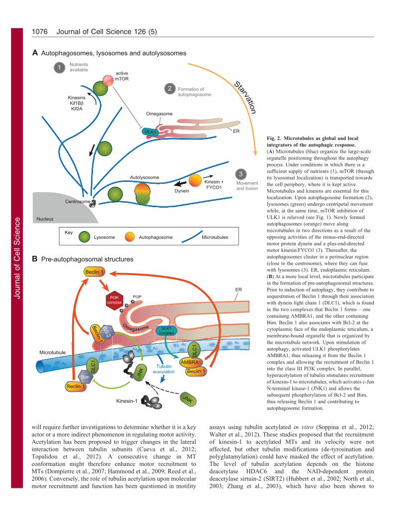
the cytoplasmic face of lysosomes (Sancak et al., 2010), and its
activity is controlled by lysosome localization. This localization
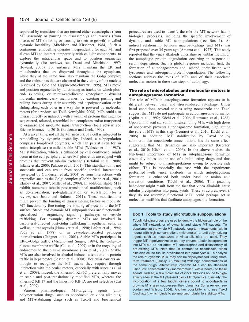
is organized by MTs and involves two kinesins: the kinesin-2
KIF2A and the kinesin-3 KIF1B (Korolchuk et al., 2011). When
nutrients are available, these kinesins localize to lysosomes at the
cell periphery, and mTORC1 is kept active, thereby preventing
autophagosome formation. When nutrients are in short supply,
the cytoplasmic pH increases, these motors are released from
lysosomes, facilitating their centripetal movement, mTORC1
inactivation and autophagosome formation (Fig. 2A) (Korolchuk
et al., 2011; Pous and Codogno, 2011). Beclin 1 is a major
component of the class III PI 3-kinase complex. It is sequestered
on MTs in two complexes containing the dynein light chain 1
(DLC1) and either AMBRA1 or Bim (Bcl2-like protein 11).
Upon induction of autophagy, AMBRA1 phosphorylation by the
kinase ULK1 releases Beclin 1 (Di Bartolomeo et al., 2010). In
parallel, activated JNK1 phosphorylates Bim to release Beclin 1
(Luo et al., 2012), allowing it to be recruited to membranes such
as omegasomes, from which autophagosomes originate (Fig. 2B).
JNK1 also disrupts the ER-associated complex formed by Beclin
1 and Bcl-2 (Wei et al., 2008) (Fig. 2B). Interestingly, upon
amino acid starvation, JNK1 activation is a kinesin-1-dependent
process (Geeraert et al., 2010; Verhey et al., 2001), and it seems
to require the hyperacetylation of the dynamic subset of MTs
(Geeraert et al., 2010).
The mechanism by which tubulin acetylation (in the lumen of
MTs) could modulate the recruitment and walking of molecular
motors (on the outer face of MTs) is still a matter of debate and
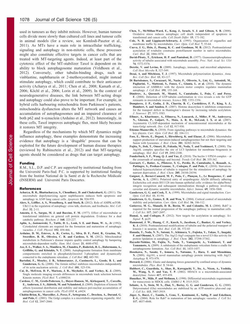
Table 1. Autophagosomal proteins and their association with microtubules
Protein Role in autophagy1 Link with microtubules References2
ATG proteins
LC3 (ATG8) Two post-translationally modified forms: cytosolicLC3-I and membrane-bound LC3-II (LC3–PtdEtn)(autophagosome)
Contributes to the closure of the autophagosomemembrane
Subunit of the neuronal microtubule-associatedproteins MAPlA and MAPlB.
Co-purifies with Taxol-assembled rat MTs.Binds to tubulin through ionic interactions.LC3-I binds to the labile MT fraction, LC3-II to
the stable MT fraction.Participates in minus-end- and plus-end-directed
autophagosome transport.
Mann and Hammarback,1994
Geeraert et al., 2010
Kimura et al., 2008;Pankiv et al., 2010
ATG5–ATG12 Contribute to the expansion and the closure of theautophagosome membrane together with ATG16L
Co-purify with labile MTs in starved cells. Geeraert et al., 2010
Beclin 1(ATG6)
Participates in a complex with the classIII phosphoinositide3-kinase VPS34, ATG14L, VPS15 and AMBRA1
Contributes to the initiation of autophagosome formation
Binds to MTs through interactionswith AMBRA1 and DLC1with Bim and DLC1.
Sequestered on MTs at the basal level andrelocalized to phagophores under starvation
Di Bartolomeo et al., 2010;Luo et al., 2012
Di Bartolomeo et al.,2010; Luo et al., 2012
WIPI1(ATG18)
Binds to PtdIns3P on phagophores Binds to MTs through recruitment into mobile pre-autophagosomal structures; co-purifies with labileMTs in starved cells
Geeraert et al., 2010
ULK1/UNC-51(ATG1)
Phosphorylates raptor and AMBRA1 Binds to a kinesin heavy chain adaptor in neuronsPhosphorylates AMBRA1 to release AMBRA1
and Beclin 1 from their interaction with MTs
Toda et al., 2008Di Bartolomeo et al., 2010
Non-ATG proteins
p62 Autophagy substrate, interacts with ubiquitin Co-purifies with both labile and stablemicrotubules in starved cells
Geeraert et al., 2010
AMBRA1 Participates in a complex with class III PI3-kinase VPS34, ATG14L, VPS15 and Beclin 1
Contributes to the initiation of autophagosome formation
Mediates binding of Beclin 1 to MTs through interactionwith dynein light chain; released with Beclin 1 afterphosphorylation by ULK1 upon induction of autophagy
Di Bartolomeo et al., 2010
FYCO1 Interaction with LC3, RAB7 and PtdIns3P Promotes plus-end-directed transport of autophagic vesicles Pankiv et al., 2010
1For specific references, see the main text (for reviews, see Kroemer et al., 2010; Mizushima et al., 2011; Singh and Cuervo, 2011; Yang and Klionsky, 2010).2Cited references only correspond to the interactions with microtubules (MTs).
Autophagy and microtubules 1075
Journ
alof
Cell
Scie
nce
will require further investigations to determine whether it is a key
actor or a more indirect phenomenon in regulating motor activity.
Acetylation has been proposed to trigger changes in the lateral
interaction between tubulin subunits (Cueva et al., 2012;
Topalidou et al., 2012). A consecutive change in MT
conformation might therefore enhance motor recruitment to
MTs (Dompierre et al., 2007; Hammond et al., 2009; Reed et al.,
2006). Conversely, the role of tubulin acetylation upon molecular
motor recruitment and function has been questioned in motility
assays using tubulin acetylated in vitro (Soppina et al., 2012;
Walter et al., 2012). These studies proposed that the recruitment
of kinesin-1 to acetylated MTs and its velocity were not
affected, but other tubulin modifications (de-tyrosination and
polyglutamylation) could have masked the effect of acetylation.
The level of tubulin acetylation depends on the histone
deacetylase HDAC6 and the NAD-dependent protein
deacetylase sirtuin-2 (SIRT2) (Hubbert et al., 2002; North et al.,
2003; Zhang et al., 2003), which have also been shown to
activemTOR
Lysosome Microtubules
Omegasome
Centrosome
Nucleus
KinesinsKif1BβKif2A
Formation ofautophagosome
Movementand fusion
Nutrientsavailable
Autolysosome
Autophagosome
Dynein
Kinesin +FYCO1
1
2
3
ULK1
A Autophagosomes, lysosomes and autolysosomes
Beclin 1
AMBRA1
Beclin 1
Beclin 1
Bim
Beclin 1Bcl-2
Be B
JNK
JNK
PI3Kcomplex
DLC
1
DLC
1
Microtubule
PI3P
Tubulinacetylation
Kinesin-1
J
eeececBcccl 2
JJ
aa
JN
aK aNK Beclin 1ationcetylaationcetyla n 11
Bimm
ececlin 1
DLC
1
1cl-2
1
B
clin clin
B
BecBec
Microtuubule
Becli
AMAulin
tiTubu
t l B li
MBRA
DLC
1
M
n 11
A1
Ac
Ac
Ac
Ac
AcAc Ac
ULK1complex
B Pre-autophagosomal structures
ER
Key
ER
Omegasome
Fig. 2. Microtubules as global and local
integrators of the autophagic response.
(A) Microtubules (blue) organize the large-scale
organelle positioning throughout the autophagy
process. Under conditions in which there is a
sufficient supply of nutrients (1), mTOR (through
its lysosomal localization) is transported towards
the cell periphery, where it is kept active.
Microtubules and kinesins are essential for this
localization. Upon autophagosome formation (2),
lysosomes (green) undergo centripetal movement
while, at the same time, mTOR inhibition of
ULK1 is relieved (see Fig. 1). Newly formed
autophagosomes (orange) move along
microtubules in two directions as a result of the
opposing activities of the minus-end-directed
motor protein dynein and a plus-end-directed
motor kinesin/FYCO1 (3). Thereafter, the
autophagosomes cluster in a perinuclear region
(close to the centrosome), where they can fuse
with lysosomes (3). ER, endoplasmic reticulum.
(B) At a more local level, microtubules participate
in the formation of pre-autophagosomal structures.
Prior to induction of autophagy, they contribute to
sequestration of Beclin 1 through their association
with dynein light chain 1 (DLC1), which is found
in the two complexes that Beclin 1 forms – one
containing AMBRA1, and the other containing
Bim. Beclin 1 also associates with Bcl-2 at the
cytoplasmic face of the endoplasmic reticulum, a
membrane-bound organelle that is organized by
the microtubule network. Upon stimulation of
autophagy, activated ULK1 phosphorylates
AMBRA1, thus releasing it from the Beclin 1
complex and allowing the recruitment of Beclin 1
into the class III PI3K complex. In parallel,
hyperacetylation of tubulin stimulates recruitment
of kinesin-1 to microtubules, which activates c-Jun
N-terminal kinase-1 (JNK1) and allows the
subsequent phosphorylation of Bcl-2 and Bim,
thus releasing Beclin 1 and contributing to
autophagosome formation.
Journal of Cell Science 126 (5)1076

Journ
alof
Cell
Scie
nce
regulate autophagy. SIRT2 inhibits this process (Zhao et al.,
2010), whereas HDAC6 functions as a scaffold that binds topolyubiquitylated proteins to allow the formation of aggresomesand binds to damaged mitochondria that are cleared by autophagy
(Kawaguchi et al., 2003; Lee et al., 2010; Pandey et al., 2007).The relationship between MTs and the function of SIRT2 inautophagy is not straightforward given its inhibitory activity, butHDAC6 sequestration in cytoplasmic regions engulfed by
autophagosomes could perhaps contribute to its inactivationand thus to the induction of tubulin acetylation. Whatever theexact roles deacetylases have in autophagosome formation, the
functional importance of tubulin acetylation will deserve furtherclarification.
Altogether, the above data support the idea that stress-inducedautophagosome formation involves MTs, and many proteins
involved in this process localize on the dynamic MT subset.Interestingly, the rate of autophagosome formation (,1/minute)(Fass et al., 2006) or the lifetime of omegasomes (,3 minutes)
(Ktistakis et al., 2011) is consistent with the life span of dynamicMTs (a few minutes). By contrast, mature autophagosomes existfor ,30 minutes before they fuse with lysosomes (Fass et al.,
2006; Ktistakis et al., 2011). This period fits well with the lifespan of stable MTs, along which they could get clustered near thenucleus and meet lysosomes, as discussed in the next section.
The role of microtubules and molecular motors inautolysosome formationThe role of MTs in the fusion of autophagosomes with lysosomes
has long been controversial, as most studies did not clearlydistinguish transport from fusion. The involvement of MTs inautolysosome formation was initially proposed based on theobservation that a complete disassembly of MTs inhibits the
colocalization of autophagosomes and lysosomes and/or proteindegradation by autophagy (Aplin et al., 1992; Kochl et al., 2006;Webb et al., 2004). The role of MTs in autolysosome formation is
likely to depend on stable MTs. Indeed, Taxol-mediated MTstabilization does not affect autophagosome and lysosome fusion,suggesting that MT dynamics are not involved in the gathering of
autophagosomes and lysosomes or in their fusion (Kochl et al.,2006). Also, under basal conditions, stable acetylated MTs seemto participate in autolysosome formation (Xie et al., 2010). Fassand colleagues precisely determined MT participation in
autophagosome-to-lysosome fusion events in CHO cells starvedof amino acids and confirmed that protein degradation isimpaired by MT disassembly. They also found that the lifetime
of autophagosomes does not change in the absence of MTs (Fasset al., 2006). The authors proposed that fusion would still occur inthe absence of MTs, but that nocodazole impairs lysosomal
degradation owing to a drop in protease transport to lysosomes,as shown in the early 1990s (Fass et al., 2006; Scheel et al.,1990). Taken together, these results suggest that MTs are
dispensable for the fusion between autophagosomes andlysosomes. However, a trafficking of autophagosomes alongMTs towards lysosomes is necessary to allow effective fusion, ashas been shown by Kimura and colleagues (Kimura et al., 2008)
using fluorescence recovery after photobleaching (FRAP) assays.
Once they have formed, autophagosomes move bi-directionally along MTs and finally concentrate around the
centrosome in the perinuclear region. The centripetal movementof autophagosomes is mediated by the MT minus-end-directedmotor dynein, as shown using an inhibitor of dynein ATPase
activity (Jahreiss et al., 2008). Furthermore, dominant-negative
mutations of the gene encoding dynein were shown to decreasethe autophagy-mediated clearance of a mutant form of a-synuclein, which causes familial Parkinson disease (Ravikumar
et al., 2005). In this study, the expression of dynein mutants alsoincreased the number of autophagosomes and impaired theirfusion with lysosomes. Dynein involvement was confirmed by adecrease in autophagosome movements in HeLa cells that
expressed GFP–LC3 and p50 dynamitin, which disrupts thedynein–dynactin complex and impairs centripetal organellemovements (Ravikumar et al., 2005). A similar reduction was
observed after blocking LC3 function by microinjecting a specificantibody during amino acid starvation (Kimura et al., 2008). LC3might participate in the recruitment of dynein to autophagosomes.
Indeed, LC3 can bind to Rab7 (Pankiv et al., 2010), which has beenshown to be involved in the recruitment of the dynein-associateddynactin subunit 1 protein (p150Glued) (Johansson et al., 2007) and
in autophagosome maturation (Gutierrez et al., 2004; Jager et al.,2004). Regarding the centrifugal movement of autophagosomes,kinesin-1 is involved under basal conditions (Cardoso et al., 2009;Geeraert et al., 2009), but the kinesin motor(s) involved in stress
conditions are still unknown. A possible functional link betweenautophagosomes and kinesins in stressed cells might involveFYVE and coiled-coil domain-containing protein 1 (FYCO1),
which contains a possible kinesin binding site and is recruited toautophagosomes by means of Rab7 and LC3 (Pankiv et al., 2010)(Fig. 2A).
Altogether, the importance of MTs in the formation of
autolysosomes mainly relies on their role in the localization ofautophagosomes and lysosomes in the juxta-nuclear region. Thisgathering most likely results from their ‘motorization’ by dynein
after inhibition of centrifugal trafficking that is mediated bykinesins. The emerging picture thus shows that the spatial controlof autophagosome location during their formation and movements
depends both on their compartmentalization between dynamic andstable MTs and on their subcellular positioning through thebalance between dynein and kinesin activities. These mechanisms
might thus prevent the premature fusion of immatureautophagosomes with lysosomes.
Conclusions and perspectivesIt is now clear that MTs are involved in different steps of
autophagy – that is, in the formation and motility ofautophagosomes, but not in their fusion with lysosomes. MTsmight serve as local suppliers or as scaffolds to promote the
interaction of proteins that are required during the early stages ofautophagy, such as Beclin 1 or AMBRA1. In addition, numerousstudies have demonstrated that MTs have a role in the motility of
autophagosomes. Here, we have discussed how two characteristicsof MTs are also important for autophagosome formation: thedynamics of MTs and the post-translational modification of
tubulin, in particular its acetylation. This adds a novel layer to thegrowing importance of acetylation in the overall regulation ofautophagy (Hamaı and Codogno, 2012). MT dynamics alsoprovide cells with a way to compartmentalize and organize, first,
the upstream signaling of autophagosome formation and, second,autophagosome biogenesis and trafficking. The relationshipbetween MTs and autophagy thus expands the repertoire of MT
function in terms of signaling and membrane trafficking.
Finally, the role of MTs in autophagy needs to be consideredfrom a therapeutic point of view. MT-targeting agents are being
Autophagy and microtubules 1077
Journ
alof
Cell
Scie
nce
used in tumours as they inhibit mitosis. However, human tumour
cells divide more slowly than cultured cell lines and tumour cells
in animal models (for a review, see Komlodi-Pasztor et al.,
2011). As MTs have a main role in intracellular trafficking,
signaling and autophagy in non-mitotic cells, these processes
might also constitute effective targets in cancer cells that are
treated with MT-targeting agents. Indeed, at least part of the
cytotoxic effect of the MT-stabilizer Taxol is dependent on its
ability to block autophagosome maturation (Veldhoen et al.,
2012). Conversely, other tubulin-binding drugs, such as
vinblastine, naphtharazin or 2-methoxyestradiol, might instead
stimulate autophagy, which could contribute to their anticancer
activity (Acharya et al., 2011; Chen et al., 2008; Kamath et al.,
2006; Kochl et al., 2006; Lorin et al., 2009). In the context of
neurodegenerative diseases, the link between MT-acting drugs
and autophagy could also prove to be important. For example, in
hybrid cells harbouring mitochondria from Parkinson’s patients,
mitochondria dysfunction alters the MT network and causes an
accumulation of autophagosomes and an impaired clearance of
both p62 and a-synuclein (Arduıno et al., 2012). Interestingly, in
these cells, Taxol improves autophagic flux, most likely because
it restores MT integrity.
Regardless of the mechanisms by which MT dynamics might
influence autophagy, these examples demonstrate the increasing
evidence that autophagy is a ‘drugable’ process that can be
exploited for the future development of human disease therapies
(reviewed by Rubinsztein et al., 2012) and that MT-targeting
agents should be considered as drugs that can target autophagy.
FundingR.M., S.L., D.P. and C.P. are supported by institutional funding fromthe Universite Paris-Sud. P.C. is supported by institutional fundingfrom the Institut National de la Sante et de la Recherche Medicale(INSERM) and Universite Paris Descartes.
ReferencesAcharya, B. R., Bhattacharyya, S., Choudhury, D. and Chakrabarti, G. (2011). The
microtubule depolymerizing agent naphthazarin induces both apoptosis and
autophagy in A549 lung cancer cells. Apoptosis 16, 924-939.
Alers, S., Loffler, A. S., Wesselborg, S. and Stork, B. (2012). Role of AMPK-mTOR-
Ulk1/2 in the regulation of autophagy: cross talk, shortcuts, and feedbacks. Mol. Cell.
Biol. 32, 2-11.
Amenta, J. S., Sargus, M. J. and Baccino, F. M. (1977). Effect of microtubular or
translational inhibitors on general cell protein degradation. Evidence for a dual
catabolic pathway. Biochem. J. 168, 223-227.
Aplin, A., Jasionowski, T., Tuttle, D. L., Lenk, S. E. and Dunn, W. A., Jr (1992).
Cytoskeletal elements are required for the formation and maturation of autophagic
vacuoles. J. Cell. Physiol. 152, 458-466.
Arduıno, D. M., Esteves, A. R., Cortes, L., Silva, D. F., Patel, B., Grazina, M.,
Swerdlow, R. H., Oliveira, C. R. and Cardoso, S. M. (2012). Mitochondrial
metabolism in Parkinson’s disease impairs quality control autophagy by hampering
microtubule-dependent traffic. Hum. Mol. Genet. 21, 4680-4702.
Axe, E. L., Walker, S. A., Manifava, M., Chandra, P., Roderick, H. L., Habermann, A.,
Griffiths, G. and Ktistakis, N. T. (2008). Autophagosome formation from membrane
compartments enriched in phosphatidylinositol 3-phosphate and dynamically
connected to the endoplasmic reticulum. J. Cell Biol. 182, 685-701.
Bartolini, F., Moseley, J. B., Schmoranzer, J., Cassimeris, L., Goode, B. L. and
Gundersen, G. G. (2008). The formin mDia2 stabilizes microtubules independently
of its actin nucleation activity. J. Cell Biol. 181, 523-536.
Cai, D., McEwen, D. P., Martens, J. R., Meyhofer, E. and Verhey, K. J. (2009).
Single molecule imaging reveals differences in microtubule track selection between
Kinesin motors. PLoS Biol. 7, e1000216.
Cardoso, C. M., Groth-Pedersen, L., Høyer-Hansen, M., Kirkegaard, T., Corcelle,
E., Andersen, J. S., Jaattela, M. and Nylandsted, J. (2009). Depletion of kinesin 5B
affects lysosomal distribution and stability and induces peri-nuclear accumulation of
autophagosomes in cancer cells. PLoS ONE 4, e4424.
Chabin-Brion, K., Marceiller, J., Perez, F., Settegrana, C., Drechou, A., Durand, G.
and Pous, C. (2001). The Golgi complex is a microtubule-organizing organelle. Mol.
Biol. Cell 12, 2047-2060.
Chen, Y., McMillan-Ward, E., Kong, J., Israels, S. J. and Gibson, S. B. (2008).Oxidative stress induces autophagic cell death independent of apoptosis intransformed and cancer cells. Cell Death Differ. 15, 171-182.
Cole, N. B. and Lippincott-Schwartz, J. (1995). Organization of organelles andmembrane traffic by microtubules. Curr. Opin. Cell Biol. 7, 55-64.
Cueva, J. G., Hsin, J., Huang, K. C. and Goodman, M. B. (2012). Posttranslationalacetylation of a-tubulin constrains protofilament number in native microtubules.Curr. Biol. 22, 1066-1074.
David-Pfeuty, T., Erickson, H. P. and Pantaloni, D. (1977). Guanosinetriphosphataseactivity of tubulin associated with microtubule assembly. Proc. Natl. Acad. Sci. USA
74, 5372-5376.
Deretic, V. and Levine, B. (2009). Autophagy, immunity, and microbial adaptations.Cell Host Microbe 5, 527-549.
Desai, A. and Mitchison, T. J. (1997). Microtubule polymerization dynamics. Annu.
Rev. Cell Dev. Biol. 13, 83-117.
Di Bartolomeo, S., Corazzari, M., Nazio, F., Oliverio, S., Lisi, G., Antonioli, M.,
Pagliarini, V., Matteoni, S., Fuoco, C., Giunta, L. et al. (2010). The dynamicinteraction of AMBRA1 with the dynein motor complex regulates mammalianautophagy. J. Cell Biol. 191, 155-168.
Dimitrov, A., Quesnoit, M., Moutel, S., Cantaloube, I., Pous, C. and Perez,F. (2008). Detection of GTP-tubulin conformation in vivo reveals a role for GTPremnants in microtubule rescues. Science 322, 1353-1356.
Dompierre, J. P., Godin, J. D., Charrin, B. C., Cordelieres, F. P., King, S. J.,
Humbert, S. and Saudou, F. (2007). Histone deacetylase 6 inhibition compensatesfor the transport deficit in Huntington’s disease by increasing tubulin acetylation.J. Neurosci. 27, 3571-3583.
Efimov, A., Kharitonov, A., Efimova, N., Loncarek, J., Miller, P. M., Andreyeva,N., Gleeson, P., Galjart, N., Maia, A. R. R., McLeod, I. X. et al. (2007).Asymmetric CLASP-dependent nucleation of noncentrosomal microtubules at thetrans-Golgi network. Dev. Cell 12, 917-930.
Etienne-Manneville, S. (2010). From signaling pathways to microtubule dynamics: thekey players. Curr. Opin. Cell Biol. 22, 104-111.
Fass, E., Shvets, E., Degani, I., Hirschberg, K. and Elazar, Z. (2006). Microtubulessupport production of starvation-induced autophagosomes but not their targeting andfusion with lysosomes. J. Biol. Chem. 281, 36303-36316.
Fujita, N., Itoh, T., Omori, H., Fukuda, M., Noda, T. and Yoshimori, T. (2008). TheAtg16L complex specifies the site of LC3 lipidation for membrane biogenesis inautophagy. Mol. Biol. Cell 19, 2092-2100.
Funderburk, S. F., Wang, Q. J. and Yue, Z. (2010). The beclin 1-VPS34 complex – atthe crossroads of autophagy and beyond. Trends Cell Biol. 20, 355-362.
Geeraert, C., Ratier, A., Pfisterer, S. G., Perdiz, D., Cantaloube, I., Rouault, A.,
Pattingre, S., Proikas-Cezanne, T., Codogno, P. and Pous, C. (2010). Starvation-induced hyperacetylation of tubulin is required for the stimulation of autophagy bynutrient deprivation. J. Biol. Chem. 285, 24184-24194.
Guignot, J., Bernet-Camard, M. F., Pous, C., Plancon, L., Le Bouguenec, C. andServin, A. L. (2001). Polarized entry of uropathogenic Afa/Dr diffusely adheringEscherichia coli strain IH11128 into human epithelial cells: evidence for alpha5beta1integrin recognition and subsequent internalization through a pathway involvingcaveolae and dynamic unstable microtubules. Infect. Immun. 69, 1856-1868.
Gundersen, G. G. and Cook, T. A. (1999). Microtubules and signal transduction. Curr.
Opin. Cell Biol. 11, 81-94.
Gundersen, G. G., Gomes, E. R. and Wen, Y. (2004). Cortical control of microtubulestability and polarization. Curr. Opin. Cell Biol. 16, 106-112.
Gutierrez, M. G., Munafo, D. B., Beron, W. and Colombo, M. I. (2004). Rab7 isrequired for the normal progression of the autophagic pathway in mammalian cells.J. Cell Sci. 117, 2687-2697.
Hamaı, A. and Codogno, P. (2012). New targets for acetylation in autophagy. Sci.
Signal. 5, pe29.
Hammond, J. W., Huang, C. F., Kaech, S., Jacobson, C., Banker, G. and Verhey,
K. J. (2010). Posttranslational modifications of tubulin and the polarized transport ofkinesin-1 in neurons. Mol. Biol. Cell. 21, 572-83
Hanada, T., Noda, N. N., Satomi, Y., Ichimura, Y., Fujioka, Y., Takao, T., Inagaki,
F. and Ohsumi, Y. (2007). The Atg12-Atg5 conjugate has a novel E3-like activity forprotein lipidation in autophagy. J. Biol. Chem. 282, 37298-37302.
Hayashi-Nishino, M., Fujita, N., Noda, T., Yamaguchi, A., Yoshimori, T. andYamamoto, A. (2009). A subdomain of the endoplasmic reticulum forms a cradle forautophagosome formation. Nat. Cell Biol. 11, 1433-1437.
Hosokawa, N., Sasaki, T., Iemura, S., Natsume, T., Hara, T. and Mizushima,N. (2009). Atg101, a novel mammalian autophagy protein interacting with Atg13.Autophagy 5, 973-979.
Howard, J. (2006). Elastic and damping forces generated by confined arrays of dynamicmicrotubules. Phys. Biol. 3, 54-66.
Hubbert, C., Guardiola, A., Shao, R., Kawaguchi, Y., Ito, A., Nixon, A., Yoshida,
M., Wang, X. F. and Yao, T. P. (2002). HDAC6 is a microtubule-associateddeacetylase. Nature 417, 455-458.
Hunziker, W., Male, P. and Mellman, I. (1990). Differential microtubule requirementsfor transcytosis in MDCK cells. EMBO J. 9, 3515-3525.
Infante, A. S., Stein, M. S., Zhai, Y., Borisy, G. G. and Gundersen, G. G. (2000).Detyrosinated (Glu) microtubules are stabilized by an ATP-sensitive plus-end cap.J. Cell Sci. 113, 3907-3919.
Jager, S., Bucci, C., Tanida, I., Ueno, T., Kominami, E., Saftig, P. and Eskelinen,E.-L. (2004). Role for Rab7 in maturation of late autophagic vacuoles. J. Cell Sci.
117, 4837-4848.
Journal of Cell Science 126 (5)1078
Journ
alof
Cell
Scie
nce
Jahreiss, L., Menzies, F. M. and Rubinsztein, D. C. (2008). The itinerary ofautophagosomes: from peripheral formation to kiss-and-run fusion with lysosomes.Traffic 9, 574-587.
Janke, C. and Bulinski, J. C. (2011). Post-translational regulation of the microtubulecytoskeleton: mechanisms and functions. Nat. Rev. Mol. Cell Biol. 12, 773-786.
Johansson, M., Rocha, N., Zwart, W., Jordens, I., Janssen, L., Kuijl, C., Olkkonen,V. M. and Neefjes, J. (2007). Activation of endosomal dynein motors by stepwiseassembly of Rab7-RILP-p150Glued, ORP1L, and the receptor betalll spectrin. J. Cell
Biol. 176, 459-471.
Jordan, M. A. and Wilson, L. (2004). Microtubules as a target for anticancer drugs.Nat. Rev. Cancer 4, 253-265.
Joseph, R. A., Shepard, B. D., Kannarkat, G. T., Rutledge, T. M., Tuma, D. J. and
Tuma, P. L. (2008). Microtubule acetylation and stability may explain alcohol-induced alterations in hepatic protein trafficking. Hepatology 47, 1745-1753.
Jung, C. H., Jun, C. B., Ro, S. H., Kim, Y. M., Otto, N. M., Cao, J., Kundu, M. and
Kim, D. H. (2009). ULK-Atg13-FIP200 complexes mediate mTOR signaling to theautophagy machinery. Mol. Biol. Cell 20, 1992-2003.
Kamath, K., Okouneva, T., Larson, G., Panda, D., Wilson, L. and Jordan, M. A.(2006). 2-Methoxyestradiol suppresses microtubule dynamics and arrests mitosiswithout depolymerizing microtubules. Mol. Cancer Ther. 5, 2225-2233.
Kang, R., Zeh, H. J., Lotze, M. T. and Tang, D. (2011). The Beclin 1 networkregulates autophagy and apoptosis. Cell Death Differ. 18, 571-580.
Kawaguchi, Y., Kovacs, J. J., McLaurin, A., Vance, J. M., Ito, A. and Yao, T. P.
(2003). The deacetylase HDAC6 regulates aggresome formation and cell viability inresponse to misfolded protein stress. Cell 115, 727-738.
Kim, J., Kundu, M., Viollet, B. and Guan, K. L. (2011). AMPK and mTOR regulateautophagy through direct phosphorylation of Ulk1. Nat. Cell Biol. 13, 132-141.
Kimura, S., Noda, T. and Yoshimori, T. (2008). Dynein-dependent movement ofautophagosomes mediates efficient encounters with lysosomes. Cell Struct. Funct. 33,109-122.
Kochl, R., Hu, X. W., Chan, E. Y. and Tooze, S. A. (2006). Microtubules facilitateautophagosome formation and fusion of autophagosomes with endosomes. Traffic 7,129-145.
Kollman, J. M., Merdes, A., Mourey, L. and Agard, D. A. (2011). Microtubulenucleation by c-tubulin complexes. Nat. Rev. Mol. Cell Biol. 12, 709-721.
Komlodi-Pasztor, E., Sackett, D., Wilkerson, J. and Fojo, T. (2011). Mitosis is not akey target of microtubule agents in patient tumors. Nat. Rev. Clin. Oncol. 8, 244-250.
Korolchuk, V. I., Saiki, S., Lichtenberg, M., Siddiqi, F. H., Roberts, E. A., Imarisio,
S., Jahreiss, L., Sarkar, S., Futter, M., Menzies, F. M. et al. (2011). Lysosomalpositioning coordinates cellular nutrient responses. Nat. Cell Biol. 13, 453-460.
Kouno, T., Mizuguchi, M., Tanida, I., Ueno, T., Kanematsu, T., Mori, Y., Shinoda,
H., Hirata, M., Kominami, E. and Kawano, K. (2005). Solution structure ofmicrotubule-associated protein light chain 3 and identification of its functionalsubdomains. J. Biol. Chem. 280, 24610-24617.
Kroemer, G., Marino, G. and Levine, B. (2010). Autophagy and the integrated stressresponse. Mol. Cell 40, 280-293.
Ktistakis, N. T., Andrews, S. and Long, J. (2011). What is the advantage of a transientprecursor in autophagosome biogenesis? Autophagy 7, 118-122.
Lafont, F., Burkhardt, J. K. and Simons, K. (1994). Involvement of microtubulemotors in basolateral and apical transport in kidney cells. Nature 372, 801-803.
Lee, J. Y., Nagano, Y., Taylor, J. P., Lim, K. L. and Yao, T. P. (2010). Disease-causing mutations in parkin impair mitochondrial ubiquitination, aggregation, andHDAC6-dependent mitophagy. J. Cell Biol. 189, 671-679.
Levine, B. and Klionsky, D. J. (2004). Development by self-digestion: molecularmechanisms and biological functions of autophagy. Dev. Cell 6, 463-477.
Lin, S. X., Gundersen, G. G. and Maxfield, F. R. (2002). Export from pericentriolarendocytic recycling compartment to cell surface depends on stable, detyrosinated(glu) microtubules and kinesin. Mol. Biol. Cell 13, 96-109.
Lorin, S., Borges, A., Ribeiro Dos Santos, L., Souquere, S., Pierron, G., Ryan,
K. M., Codogno, P. and Djavaheri-Mergny, M. (2009). c-Jun NH2-terminal kinaseactivation is essential for DRAM-dependent induction of autophagy and apoptosis in2-methoxyestradiol-treated Ewing sarcoma cells. Cancer Res. 69, 6924-6931.
Luo, S., Garcia-Arencibia, M., Zhao, R., Puri, C., Toh, P. P. C., Sadiq, O. andRubinsztein, D. C. (2012). Bim inhibits autophagy by recruiting Beclin 1 tomicrotubules. Mol. Cell 47, 359-370.
Mann, S. S. and Hammarback, J. A. (1994). Molecular characterization of light chain 3. Amicrotubule binding subunit of MAP1A and MAP1B. J. Biol. Chem. 269, 11492-11497.
Mehrpour, M., Esclatine, A., Beau, I. and Codogno, P. (2010). Overview ofmacroautophagy regulation in mammalian cells. Cell Res. 20, 748-762.
Miller, P. M., Folkmann, A. W., Maia, A. R. R., Efimova, N., Efimov, A. and
Kaverina, I. (2009). Golgi-derived CLASP-dependent microtubules control Golgiorganization and polarized trafficking in motile cells. Nat. Cell Biol. 11, 1069-1080.
Mitchison, T. and Kirschner, M. (1984). Dynamic instability of microtubule growth.Nature 312, 237-242.
Mizuno, M. and Singer, S. J. (1994). A possible role for stable microtubules inintracellular transport from the endoplasmic reticulum to the Golgi apparatus. J. Cell
Sci. 107, 1321-1331.
Mizushima, N. (2010). The role of the Atg1/ULK1 complex in autophagy regulation.Curr. Opin. Cell Biol. 22, 132-139.
Mizushima, N. and Komatsu, M. (2011). Autophagy: renovation of cells and tissues.Cell 147, 728-741.
Mizushima, N., Yoshimori, T. and Ohsumi, Y. (2011). The role of Atg proteins inautophagosome formation. Annu. Rev. Cell Dev. Biol. 27, 107-132.
Monastyrska, I., Rieter, E., Klionsky, D. J. and Reggiori, F. (2009). Multiple roles ofthe cytoskeleton in autophagy. Biol. Rev. Camb. Philos. Soc. 84, 431-448.
Moreau, K., Ravikumar, B., Renna, M., Puri, C. and Rubinsztein, D. C. (2011).Autophagosome precursor maturation requires homotypic fusion. Cell 146, 303-317.
North, B. J., Marshall, B. L., Borra, M. T., Denu, J. M. and Verdin, E. (2003). Thehuman Sir2 ortholog, SIRT2, is an NAD+-dependent tubulin deacetylase. Mol. Cell
11, 437-444.
Orsi, A., Razi, M., Dooley, H. C., Robinson, D., Weston, A. E., Collinson, L. M. andTooze, S. A. (2012). Dynamic and transient interactions of Atg9 with autophago-somes, but not membrane integration, are required for autophagy. Mol. Biol. Cell 23,1860-1873.
Palazzo, A. F., Cook, T. A., Alberts, A. S. and Gundersen, G. G. (2001). mDiamediates Rho-regulated formation and orientation of stable microtubules. Nat. Cell
Biol. 3, 723-729.
Pandey, U. B., Nie, Z., Batlevi, Y., McCray, B. A., Ritson, G. P., Nedelsky, N. B.,
Schwartz, S. L., DiProspero, N. A., Knight, M. A., Schuldiner, O. et al. (2007).HDAC6 rescues neurodegeneration and provides an essential link between autophagyand the UPS. Nature 447, 859-863.
Pankiv, S., Alemu, E. A., Brech, A., Bruun, J. A., Lamark, T., Overvatn, A., Bjørkøy, G.
and Johansen, T. (2010). FYCO1 is a Rab7 effector that binds to LC3 and PI3P to mediatemicrotubule plus end-directed vesicle transport. J. Cell Biol. 188, 253-269.
Polson, H. E., de Lartigue, J., Rigden, D. J., Reedijk, M., Urbe, S., Clague, M. J. and
Tooze, S. A. (2010). Mammalian Atg18 (WIPI2) localizes to omegasome-anchoredphagophores and positively regulates LC3 lipidation. Autophagy 6, 506-522.
Pous, C. and Codogno, P. (2011). Lysosome positioning coordinates mTORC1 activityand autophagy. Nat. Cell Biol. 13, 342-344.
Pous, C., Chabin, K., Drechou, A., Barbot, L., Phung-Koskas, T., Settegrana, C.,
Bourguet-Kondracki, M. L., Maurice, M., Cassio, D., Guyot, M. et al. (1998).Functional specialization of stable and dynamic microtubules in protein traffic inWIF-B cells. J. Cell Biol. 142, 153-165.
Ravikumar, B., Acevedo-Arozena, A., Imarisio, S., Berger, Z., Vacher, C., O’Kane,C. J., Brown, S. D. and Rubinsztein, D. C. (2005). Dynein mutations impairautophagic clearance of aggregate-prone proteins. Nat. Genet. 37, 771-776.
Ravikumar, B., Sarkar, S., Davies, J. E., Futter, M., Garcia-Arencibia, M., Green-Thompson, Z. W., Jimenez-Sanchez, M., Korolchuk, V. I., Lichtenberg, M., Luo,
S. et al. (2010). Regulation of mammalian autophagy in physiology andpathophysiology. Physiol. Rev. 90, 1383-1435.
Reed, N. A., Cai, D., Blasius, T. L., Jih, G. T., Meyhofer, E., Gaertig, J. and Verhey,
K. J. (2006). Microtubule acetylation promotes kinesin-1 binding and transport. Curr.
Biol. 16, 2166-2172.
Reunanen, H., Marttinen, M. and Hirsimaki, P. (1988). Effects of griseofulvin andnocodazole on the accumulation of autophagic vacuoles in Ehrlich ascites tumor cells.Exp. Mol. Pathol. 48, 97-102.
Rubinsztein, D. C., Marino, G. and Kroemer, G. (2011). Autophagy and aging. Cell
146, 682-695.
Rubinsztein, D. C., Codogno, P. and Levine, B. (2012). Autophagy modulation as apotential therapeutic target for diverse diseases. Nat. Rev. Drug Discov. 11, 709-730.
Sancak, Y., Bar-Peled, L., Zoncu, R., Markhard, A. L., Nada, S. and Sabatini, D. M.
(2010). Ragulator-Rag complex targets mTORC1 to the lysosomal surface and isnecessary for its activation by amino acids. Cell 141, 290-303.
Scheel, J., Matteoni, R., Ludwig, T., Hoflack, B. and Kreis, T. E. (1990). Microtubuledepolymerization inhibits transport of cathepsin D from the Golgi apparatus tolysosomes. J. Cell Sci. 96, 711-720.
Sengupta, S., Peterson, T. R. and Sabatini, D. M. (2010). Regulation of the mTORcomplex 1 pathway by nutrients, growth factors, and stress. Mol. Cell 40, 310-322.
Singh, R. and Cuervo, A. M. (2011). Autophagy in the cellular energetic balance. Cell
Metab. 13, 495-504.
Soppina, V., Herbstman, J. F., Skiniotis, G. and Verhey, K. J. (2012). Luminallocalization of a-tubulin K40 acetylation by cryo-EM analysis of fab-labeledmicrotubules. PLoS ONE 7, e48204.
Tanaka, Y., Guhde, G., Suter, A., Eskelinen, E. L., Hartmann, D., Lullmann-Rauch,
R., Janssen, P. M., Blanz, J., von Figura, K. and Saftig, P. (2000). Accumulation ofautophagic vacuoles and cardiomyopathy in LAMP-2-deficient mice. Nature 406,902-906.
Toda, H., Mochizuki, H., Flores, R., 3rd, Josowitz, R., Krasieva, T. B., Lamorte,V. J., Suzuki, E., Gindhart, J. G., Furukubo-Tokunaga, K. and Tomoda,
T. (2008). UNC-51/ATG1 kinase regulates axonal transport by mediating motor-cargo assembly. Genes Dev. 22, 3292-3307.
Tolic-Nørrelykke, I. M. (2008). Push-me-pull-you: how microtubules organize the cellinterior. Eur. Biophys. J. 37, 1271-1278.
Topalidou, I., Keller, C., Kalebic, N., Nguyen, K. C. Q., Somhegyi, H., Politi, K. A.,Heppenstall, P., Hall, D. H. and Chalfie, M. (2012). Genetically separable functionsof the MEC-17 tubulin acetyltransferase affect microtubule organization. Curr. Biol.
22, 1057-1065.
Veldhoen, R. A., Banman, S. L., Hemmerling, D. R., Odsen, R., Simmen, T.,
Simmonds, A. J., Underhill, D. A. and Goping, I. S. (2013). The chemotherapeuticagent paclitaxel inhibits autophagy through two distinct mechanisms that regulateapoptosis. Oncogene 32, 736-746.
Verhey, K. J., Meyer, D., Deehan, R., Blenis, J., Schnapp, B. J., Rapoport, T. A. and
Margolis, B. (2001). Cargo of kinesin identified as JIP scaffolding proteins andassociated signaling molecules. J. Cell Biol. 152, 959-970.
Walter, W. J., Beranek, V., Fischermeier, E. and Diez, S. (2012). Tubulin acetylationalone does not affect kinesin-1 velocity and run length in vitro. PLoS ONE 7, e42218.
Autophagy and microtubules 1079
Journ
alof
Cell
Scie
nce
Webb, J. L., Ravikumar, B. and Rubinsztein, D. C. (2004). Microtubuledisruption inhibits autophagosome-lysosome fusion: implications for studyingthe roles of aggresomes in polyglutamine diseases. Int. J. Biochem. Cell Biol. 36,2541-2550.
Webster, D. R., Gundersen, G. G., Bulinski, J. C. and Borisy, G. G. (1987).Differential turnover of tyrosinated and detyrosinated microtubules. Proc. Natl. Acad.
Sci. USA 84, 9040-9044.Wei, Y., Pattingre, S., Sinha, S., Bassik, M. and Levine, B. (2008). JNK1-mediated
phosphorylation of Bcl-2 regulates starvation-induced autophagy. Mol. Cell 30, 678-688.
Xie, Z. and Klionsky, D. J. (2007). Autophagosome formation: core machinery andadaptations. Nat. Cell Biol. 9, 1102-1109.
Xie, R., Nguyen, S., McKeehan, W. L. and Liu, L. (2010). Acetylatedmicrotubules are required for fusion of autophagosomes with lysosomes. BMC
Cell Biol. 11, 89.Xie, R., Nguyen, S., McKeehan, K., Wang, F., McKeehan, W. L. and Liu, L. (2011).
Microtubule-associated protein 1S (MAP1S) bridges autophagic components with
microtubules and mitochondria to affect autophagosomal biogenesis and degradation.J. Biol. Chem. 286, 10367-10377.
Yang, Z. and Klionsky, D. J. (2010). Mammalian autophagy: core molecular machineryand signaling regulation. Curr. Opin. Cell Biol. 22, 124-131.
Yla-Anttila, P., Vihinen, H., Jokitalo, E. and Eskelinen, E. L. (2009). 3D tomographyreveals connections between the phagophore and endoplasmic reticulum. Autophagy
5, 1180-1185.Zalckvar, E., Berissi, H., Mizrachy, L., Idelchuk, Y., Koren, I., Eisenstein, M.,
Sabanay, H., Pinkas-Kramarski, R. and Kimchi, A. (2009). DAP-kinase-mediatedphosphorylation on the BH3 domain of beclin 1 promotes dissociation of beclin 1from Bcl-XL and induction of autophagy. EMBO Rep. 10, 285-292.
Zhang, Y., Li, N., Caron, C., Matthias, G., Hess, D., Khochbin, S. and Matthias, P.
(2003). HDAC-6 interacts with and deacetylates tubulin and microtubules in vivo.EMBO J. 22, 1168-1179.
Zhao, Y., Yang, J., Liao, W., Liu, X., Zhang, H., Wang, S., Wang, D., Feng, J., Yu,L. and Zhu, W. G. (2010). Cytosolic FoxO1 is essential for the induction ofautophagy and tumour suppressor activity. Nat. Cell Biol. 12, 665-675.
Journal of Cell Science 126 (5)1080