Embed Size (px)
Citation preview
} w���������� ������������ !"#$%&'()+,-./012345<yA| Faculty of Informatics
Masaryk University
Czech Republic
Automated Image Analysis
in Fluorescence Microscopy
Habilitation Thesis
Collection of Articles
Petr Matula
2012


Abstract
After completion of human genome project in 2003 a signi�cant e�ort is put into researchof structure and especially function of proteins in both cells and whole organisms. Themost suitable technique to carry out this research in particular in living specimens is�uorescent microscopy. There are two main directions in �uorescence microscopy: (1)high-resolution imaging focused on observing details, typically in multi-dimensional data,and (2) high-throughput imaging focused on analyzing massive image data sets. Bothdirections raise challenges for automated image analysis.
The main goal of this thesis is to summarize my principal achievements in the area ofautomated image analysis in �uorescence microscopy and to show context and relationsbetween the works. The achievements have been obtained in three �elds: (1) automationof confocal microscopy, (2) automation of micro-axial tomography, and (3) image anal-ysis in high-content screening. From the methodological point of view, this comprisesnovel methods for (1) point-based image registration of 3D confocal images of living cellsand micro-axial tomography data, (2) chromatic aberration correction using polynomialfunctions, (3) object tracking with split and merge events based on combining optimalassignment problem and maximal �ows, (4) cell nucleus segmentation based on a combi-nation of a gradient thresholding scheme and mathematical morphology operators suitablefor high-content screening, (5) quanti�cation of the level of assembly or disassembly of cel-lular phenotypes based on mathematical morphology, and (6) spot detection in 3D confocaldata based on extrema dynamics measurements. All presented methods have successfullybeen applied in real biological problems.
The thesis is written as a commentary to a collection of 14 peer-reviewed journalarticles and 4 peer-reviewed conference papers. My percentage contribution for each paperis estimated and included in the thesis as well as a detailed description of my work.My percentage contribution to the papers ranges from 3% to 70%. Average percentagecontribution to the papers is about 30%.
i

ii
Abstrakt
Po té, co byl v roce 2003 dokon£en projekt sekvenace lidského genomu, je dal²ím velkýmvýzkumným cílem pochopení vztahu struktury a funkce jednotlivých protein· jak nabun¥£né úrovni tak i na úrovni celých organizm·. Nejvhodn¥j²í technikou k provád¥nítohoto výzkumu, a to zejména na ºivých bu¬kách, je �uorescen£ní mikroskopie. Zde lzehistoricky vypozorovat dva základní sm¥ry vývoje: (1) mikroskopie s vysokým rozli²ením,která typicky produkuje vícedimenzionální obrazy a (2) tzv. �high-throughput� mikrosko-pie (HTM), která produkuje velké mnoºství obraz·. V obou p°ípadech se jedná o velkémnoºství dat, a proto je nutné pouºívat automatickou analýzu obrazu.
Hlavním cílem této práce je sumarizovat nejd·leºit¥j²í výsledky mojí v¥decké prácev oblasti automatizované analýzy obraz· po°ízených �uorescen£ními mikroskopy a ukázatkontext práce a vztah mezi výsledky. Nejd·leºit¥j²ích výsledk· jsem dosáhl v t¥chto t°echoblastech: (1) automatizace konfokální mikroskopie, (2) automatizace mikro-axiální tomo-gra�e a (3) obrazová analýza v oblasti HTM. Z metodologického pohledu se jedná o tytonové metody: pro (1) registraci 3D konfokálních obraz· ºivých bun¥k a pro registraci datz mikro-axiálního tomografu, které jsou zaloºeny na extrakci významných bod·, (2) ko-rekci barevné vady s vyuºitím polynomiálních funkcí, (3) sledování pohybu d¥lících sea spojujících se objekt· pomocí kombinace optimálního p°i°azení v bipartitních grafecha maximálního toku v grafu, (4) segmentaci bun¥£ných jader, která je zaloºena na kombi-naci gradientního prahování a metod matematické morfologie, jenº je vhodná pro screeningpomocí HTM, (5) kvanti�kaci míry kompaktnosti nebo difuzivity struktur uvnit° bu¬kys vyuºitím matematické morfologie a (6) detekci malých objekt· ve 3D konfokálních dat-ech, která je zaloºena na m¥°ení dynamiky extrém· v obraze. V²echny popsané metodybyly úsp¥²n¥ pouºity v reálných biologických aplikacích.
Práce je koncipována jako soubor £trnácti £asopiseckých a £ty° sborníkových prací,které pro²ly °ádným recenzním °ízením a byly publikovány na mezinárodním fóru. U kaºdépráce je uveden procentuální odhad mého podílu na jejím vytvo°ení v£etn¥ detailníhopopisu vlastního p°ísp¥vku. M·j podíl se pohybuje v rozmezí od 3% do 70%. Pr·m¥rnýpodíl na jeden p°ísp¥vek je okolo 30%.
iii

iv
Acknowledgments
My warmest thanks are due to all co-authors of the included papers. It was a real pleasureto work with them on the exciting problems. In particular, I want to thank Michal Kozubekand Karl Rohr. Michal is the group leader of the Center of Biomedical Image Analysis(CBIA) at the Faculty of Informatics, who gave me the opportunity to work in his group,where I have learned a lot especially in my early scienti�c career. Karl is the group leader ofthe Biomedical Computer Vision (BMCV) Group at the German Cancer Research Centerand the University of Heidelberg, who o�ered me a post-doc position in his group, whereI got opportunity to work on many cutting edge life science problems, and who signi�cantlyin�uenced my way of thinking and working. I want also thank all my current or formercolleagues from CBIA and BMCV groups. Last but not least, I would like to thank myfamily and especially my wife for her never-ending support.
In Fontainebleau, April 2012 Petr Matula
v

vi
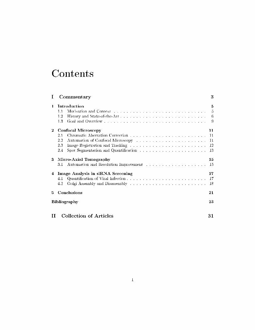
Contents
I Commentary 3
1 Introduction 51.1 Motivation and Context . . . . . . . . . . . . . . . . . . . . . . . . . . . . . 51.2 History and State-of-the-Art . . . . . . . . . . . . . . . . . . . . . . . . . . . 61.3 Goal and Overview . . . . . . . . . . . . . . . . . . . . . . . . . . . . . . . . 9
2 Confocal Microscopy 112.1 Chromatic Aberration Correction . . . . . . . . . . . . . . . . . . . . . . . . 112.2 Automation of Confocal Microscopy . . . . . . . . . . . . . . . . . . . . . . 112.3 Image Registration and Tracking . . . . . . . . . . . . . . . . . . . . . . . . 122.4 Spot Segmentation and Quanti�cation . . . . . . . . . . . . . . . . . . . . . 13
3 Micro-Axial Tomography 153.1 Automation and Resolution Improvement . . . . . . . . . . . . . . . . . . . 15
4 Image Analysis in siRNA Screening 174.1 Quanti�cation of Viral Infection . . . . . . . . . . . . . . . . . . . . . . . . . 174.2 Golgi Assembly and Disassembly . . . . . . . . . . . . . . . . . . . . . . . . 18
5 Conclusions 21
Bibliography 23
II Collection of Articles 31
1

2 CONTENTS

Part I
Commentary
3

Chapter 1
Introduction
The presented habilitation thesis consists of a collection of fourteen peer-reviewed journalpapers and four peer-reviewed conference papers. This introduction presents the overallmotivation, the context of the work, and the state of the art in the area. The next chaptersprovide a brief overview of the methods and results in my main research and development�elds together with the description of my contributions and they also highlight the relationsamong individual papers.
1.1 Motivation and Context
The human genome project has identi�ed approximately 20,000�25,000 human genes(DNA coding sequences). Each gene codes a unique protein1 that performs a speci�cfunction in a cell. The cell is the smallest functional unit of life. However, the function ofa vast majority of proteins is unknown2. The desire to understand the role of genes andproteins on the cellular level as well as on the higher levels such as tissues, organs andwhole organisms is the current driving force of many life sciences. For example, build-ing mathematical models that describe structure and function of biological systems bymeans of their reverse engineering3 is the main topic of an interdisciplinary �eld calledsystems biology [35, 37, 13]. In order to draw relevant biological conclusions and to buildsound mathematical models a close interdisciplinary collaboration of experts from manydi�erent �elds is necessary, in particular molecular and computational biology, biophysics,bio-informatics, statistics, mathematics, and computer science.
Cytometry is a technologically oriented science focused on obtaining measurementson molecular or cellular targets by means of �ow or image cytometry [85, 82]. While�ow cytometry [74] is suitable for obtaining a large number of basic measurements (such
1Proteins are the most important macromolecules in living systems and serve crucial functions inessentially all biological processes [3]. They function as catalysts, they transport and store other moleculessuch as oxygen or other proteins, they provide mechanical support and immune protection, they generatemovement, they transmit nerve impulses, and they control growth and di�erentiation.
2We know the letters, we know the words, but we do not understand the meaning.3i.e. systematic perturbation of the biological system (biologically, genetically, or chemically); monitor-
ing the gene, protein, and informational pathway responses; integrating these data for the mathematicalmodeling.
5
6 CHAPTER 1. INTRODUCTION
as cell size, presence or absence of proteins of interest, DNA content) on cells in liquidsuspensions, image cytometry is used for advanced measurements (such as morphologicalmeasurements or spatial and dynamic relations of proteins and genes) based on microscopy.Electron as well as optical microscopy are widely used for morphological studies, but opticalmicroscopy is a unique technique used in live cell imaging.
Fluorescence microscopy is a technique based on optical microscopy, which is of a crit-ical importance in modern life sciences. It uses �uorochromes, which are chemical com-pounds that can re-emit light upon light excitation, for speci�c visualization of objects ofinterest. This allows in combination with staining techniques (such as FISH�FluorescenceIn Situ Hybridization4 and/or immuno�uorescence5) or genetic modi�cations6 highly spe-ci�c observation and measurement of �uorescent signal even on a single molecule level inboth �xed and live cells.
1.2 History and State-of-the-Art
There has been two main parallel trends in the development of �uorescence microscopy: (1)resolution improvement to obtain images containing more details and (2) automation toacquire and analyze more data. There are many resolution improvement techniques suchas confocal microscopy, 4Pi microscopy, structured illumination microscopy, micro-axialtomography7, see [30, 33] for a review. The most wide-spread resolution improvementtechnique is confocal microscopy [66] where the out-of-focus light is blocked before thedetector8 and 3D optical sections of a sample can be obtained. The �rst automatedmicroscopes appeared in late 1970s and became commercially available to biologists in late1990s when the �rst high-content screening platform was built [25, 83]. At that time when�uorescence staining techniques matured and automation components were commerciallyavailable, research groups began to automate �uorescent microscopes to perform speci�ctasks, e.g., FISH-dot counting [60, 63, 40]. One of them was built by Michal Kozubek et al.at the Institute of Biophysics in Brno [40]. This automated microscope has been extendedby a Nipkow disk based confocal unit to permit combination of wide-�eld9 and confocalmodes [39]. While developing this combined automated microscope many di�erent imageanalysis problems appeared. I worked especially on the correction of chromatic aberrations(Section 2.1). Later, a new microscope supporting high-resolution live cell imaging wasconstructed [42], and I have worked on image analysis problems related to object trackingand image registration (Section 2.3). Nowadays, automated microscopes are o�ered by themain microscope manufactures and wide-�eld as well as confocal screening platforms10 are
4Visualization of DNA sequences�genes5Visualization of antibodies�proteins6Virtually any protein can be modi�ed to exhibit �uorescence without changing its primary function.
The invention and development of green �uorescent protein (GFP) was awarded the Nobel Prize forchemistry in 2008 to Osamu Shimomura, Martin Chal�e, and Robert Y. Tsien.
7Several results included in this thesis relate to this resolution improvement technique (see Section 3.1).8Pinholes in CLSM (Confocal Laser Scanning Microscopy) or spinning disks in Nipkow-disk confocal
microscopy.9non-confocal
10A screening platform di�ers from an automated microscope in the integration and automation of thewhole analytical process. Often, the screening platforms produce and analyze a larger number of imagesat lower resolution than automated microscopes.

1.2. HISTORY AND STATE-OF-THE-ART 7
commercially available [28, 84]. The commercially available screening platforms are buildfor certain types of assays often designed to meet the needs of drug discovery [4], but theyalso typically provide an option to save the raw images and to analyze them in an externalsoftware package. External software packages11 and/or involvement of image analysisexperts is necessary, in particular, for specialized and complex problems often related tolive cell imaging [90].
From the point of view of image analysis there exists no universal solution that couldbe applied to any biological problem. Based on own experience, even a small change in thebiological protocol or in the image acquisition process can dramatically change the imageanalysis problem to solve. Similarly, small misunderstanding of the biological problemby an image analysis expert or misunderstanding of the limitations and assumptions ofimage analysis method by a biologist especially in the results interpretation phase canhave serious consequences. Therefore close interaction, good communication between allengaged persons, and good problem understanding are crucially important to �nd the rightsolution. Nevertheless, there are some typical problems that appear often in the �eld,for example, cell nucleus segmentation, whole cell segmentation, or small spot detection.However it is very di�cult to compare di�erent methods unless they are tested on thesame data and under the same conditions and even then it can be di�cult to decide whatapproach is better than the other without knowing the real application (see e.g., [77]).
Recently, several survey papers have been published discussing typical or commonimage analysis methods in automated �uorescence microscopy [2, 31, 70]. In the sequel,a brief overview of the areas related to the main topics of the thesis are provided, namelyimage correction, segmentation, classi�cation, registration, and tracking.
Image correction It is common to correct images before any further processing, but itshould be done with care and only if one knows the source of distortion. The most commontype of correction is the correction of inhomogeneities in the �uorescence signal [47, 58].Multi-spectral images should be corrected for chromatic shifts. We have developed ane�cient algorithm to address this problem (Section 2.1). There is also z-scaling presentin practically all 3D images caused by refractive index mismatch [18]. In general, it isnot possible to correct this distortion without additional information. We have proposeda solution in micro-axial tomography based on precise point-based image registration,where images from di�erent views are available (Section 3.1).
Segmentation In the area of image segmentation, the most popular methods in the �eldare thresholding [65, 73], watershed [86], mathematical morphology operators [71, 79], vari-ational approaches [59], level-sets [49, 72, 64], Markov random �elds [24], and graph cuts[75, 5]. The selection of the right approach very depends on the application problem andobjects of interest. Typical problems are 2D or 3D cell nucleus segmentation approached bygraph-cuts [15], k-means and level-sets [12], watershed with model-based merging [46, 45],level-sets [88], deformable templates [23], or multi-scale techniques [29]. We have proposed
11Among the most popular publicly available software packages among biologists are ImageJ(http://rsbweb.nih.gov/ij/) [1] and CellPro�ler [8, 44].
8 CHAPTER 1. INTRODUCTION
a method for 2D cell nucleus segmentation suitable for siRNA screening12, which is su-perior to other commonly used methods in high-content screening especially for clusterednuclei (Section 4.1).
Another common segmentation problem is 3D spot detection [77]. This is very commonin FISH studies where colocalizations of two targets are sought, because they indicategenetic translocations [68, 87]. For the spot detection, we have developed an approachbased on HMAX-transform from mathematical morphology [79] which yielded very stableand reproducible results for the detection of endoplasmic reticulum exit sites in massive3D confocal data sets (Section 2.4).
Classi�cation Classi�cations in �uorescence microscopy are mostly related to machinelearning approaches whose ultimate goal is to fully automatically learn di�erent localiza-tion patterns of di�erent proteins and to relate the localization patterns to protein function[10]. In general, classi�cations are often used to distinguish di�erent cell phenotypes13
[61, 32, 26, 11, 67]. We have developed an approach for cell classi�cation as infected andnon-infected by maximizing the di�erence between positive and negative controls (Section4.1) and a measure for distinguishing compact and di�use phenotypes (assembled anddisassembled Golgi complex, Section 4.2) based on mathematical morphology operations.
Registration Image registration is similarly as image segmentation very broad researcharea [34, 92]. We have developed a rigid point-based registration approach for the elim-ination of the global movement of a living cell (Section 2.3). We have applied a similarapproach to the alignment of micro-axial tomography data (Section 3.1). The main dif-ference from the methodological point of view between these two approaches lied in thepoint pattern matching phase and the class of considered transformations. Whereas theclass of transformations was a composition of 3D rotation and 3D translation in the formercase, we have additionally searched for z-scale factor in the latter case. The point pat-tern matching problem was solved by a 3D generalization of an invariant14 point patternmatching problem in the former case and by a reduction of the matching problem to anoptimal assignment problem in bipartite graphs in the latter case. Note that elastic imageregistration has also been applied in �uorescence microscopy [17, 36, 16, 89, 20].
Tracking Tracking is usually necessary when dealing with live specimens [9, 21, 57].In general probabilistic approaches overcome deterministic approaches in particle track-ing [27]. Parametric active contours [91] and level-set-based tracking [19] were used totrack the whole cells. We have developed a tracking approach considering appearance,disappearance, splitting, and merging of objects based on bipartite graph matching andmaximum �ows [42] (Section 2.3). In [48, 54], we have tracked the cells by a method basedon agglomerative clustering (Section 4.2).
12siRNAs are short double-stranded RNA molecules which permit systematic knockout of related genesleading to silencing of the related protein. Each gene is typically attacked by several (often 3) di�erentsiRNAs. siRNA screening is common reverse engineering approach to discover gene/protein function [7, 14]
13cell phenotype = physical characteristics of the cell = how it can be seen14invariant to rotation, translation, scaling and missing or extra points
1.3. GOAL AND OVERVIEW 9
1.3 Goal and Overview
The goal of the thesis is to concisely show my main results in the area of automated imageanalysis in �uorescence microscopy. The most natural way how to classify the results isto use the type of microscopy as the main criterion, because it highly correlates (1) to themain projects I have been working on, (2) to the type of image data, and (3) to the keyimage analysis problems I tackled. The main results are summarized in the next threechapters. Note that related work and relation to the big picture was shown in the previoussection.
Chapter 2: Confocal MicroscopyInput images: Mostly multi-spectral 3D imagesMain problems: image registration, tracking of sub-cellular structures, point patternmatching, 3D small object segmentation and quanti�cation
Chapter 3: Micro-Axial TomographyInput images: Tilted 3D gray-scale imagesMain problems: image registration, point pattern matching, image fusion, resolutionimprovement
Chapter 4: Image Analysis in siRNA ScreeningInput images: Many 2D multi-spectral imagesMain problems: cell nucleus segmentation, whole cell segmentation, quanti�cation,classi�cation, tracking of cell nuclei, quality control

10 CHAPTER 1. INTRODUCTION
Chapter 2
Confocal Microscopy
In the area of confocal microscopy I have been working on several problems.
2.1 Chromatic Aberration Correction
Together with Michal Kozubek we have developed a method for the correction of chro-matic aberrations [41]. The method is quick, precise, corrects lateral as well as axialaberrations, and is suitable especially for �uorescence microscopy where a limited numberof �uorochromes are observed. It is based on 3D localization of �uorescent beads in 3Dimages and approximation of chromatic shifts by �rst and second order polynomials. Themethod has been in use in our laboratory since its invention and correction surfaces havebeen regularly computed and stored into a database for all microscopes maintained by ourlaboratory.
[41] M. Kozubek and Pe. Matula. An e�cient algorithm for measurement and correc-tion of chromatic aberrations in �uorescence microscopy. Journal of Microscopy,200(3):206�217, 2000.
My contribution (30%): Design, development, and evaluation of the correction phaseof the algorithm. Preparation of �gures 3-7, collaboration on text writing. (fulltexton page ??)
2.2 Automation of Confocal Microscopy
We have developed an automated system which combines confocal and wide-�eld acquisi-tion modes to provide high-resolution and high-throughput measurements at the same timeon FISH stained cells [39]. The system was an extension of a previous high-resolution cy-tometry instrument primary developed by Michal Kozubek [40] and it was called HRCM-2(High-Resolution CytoMetry instrument). Later, we were developing a client/server-basedsolution for image cytometry [50]. During the development �rst versions of two open source
11
12 CHAPTER 2. CONFOCAL MICROSCOPY
libraries (i3dlib1 and v3dlib2) were created. The former, i3dlib, is a C++ template libraryfor image representation and processing. The latter, v3dlib, is a C++ template libraryfor orthogonal visualization of 3D images. Both libraries are used in image acquisitionand image analysis software called Acquiarium3 [51], which has been developed in ourlaboratory as a software solution for high-resolution cytometry.
[39] M. Kozubek, S. Kozubek, E. Luká²ová, E. Bártová, M. Skalníková, Pa. Matula,Pe. Matula, P. Jirsová, A. Cafourková, and I. Koutná. Combined confocal andwide-�eld high-resolution cytometry of �uorescent in situ hybridization-stained cells.Cytometry, 45(1):1�12, 2001.
My contribution (10%): Design, development, evaluation, and testing of severalparts of the system (most notably chromatic aberration correction module, but alsocollaboration on, e.g., switching between confocal and wide-�eld modes or manualsegmentation of nuclei). (fulltext on page ??)
[50] Pa. Matula, Pe. Matula, M. Kozubek, and P. Mejzlík. High-Resolution CytometryNetwork Project: Client/Server System for 3D Optical Microscope Data Storage andAnalysis. In: Proc. of 2nd International Symposium on 3D Data Processing, Visu-alization, and Transmission (3DPVI 2004), Thessaloniki, Greece, 580�583, 2004.
My contribution (20%): Collaboration on the design and development of the system.Collaboration on text writing. (fulltext on page ??)
[51] Pa. Matula, M. Ma²ka, O. Dan¥k, Pe. Matula, M. Kozubek. Acquiarium: FreeSoftware for the Acquisition and Analysis of 3D Images of Cells in FluorescenceMicroscopy. In: Proc. of IEEE International Symposium on Biomedical Imaging�From Nano to Macro (ISBI'09), 1138�1141, 2009.
My contribution (10%): A member of the team involved in the design of the initialversions of Acquiarium and an author of some of its parts related to data visualization(v3dlib). Collaboration on text writing. (fulltext on page ??)
2.3 Image Registration and Tracking
We have extended the HRCM-2 system to permit high-resolution live cell imaging [42]. Asthe main contributions we have described a novel image acquisition strategies suitable forlive cell studies and a novel tracking strategy based on graph theory to track sub-cellularobjects. This tracking strategy was used in a real biological problem to track and analyzemotion of HP1 foci [62].
We have developed a fast point-based registration method for the suppression of theglobal cellular movement [55]. It is based on extraction of signi�cant intracellular objectsand solving a point pattern matching problem and orthogonal Procrustes problem to �nd
1http://cbia.fi.muni.cz/user_dirs/i3dlib_doc/i3dcore/index.html2http://cbia.fi.muni.cz/projects/viewing-3d-images-with-v3dlib.html3http://cbia.fi.muni.cz/projects/acquiarium.html
2.4. SPOT SEGMENTATION AND QUANTIFICATION 13
the optimal composition of 3D translation and rotation in least-squares manner. Therobustness of the method was evaluated on generated data and two real applications werepresented.
[42] M. Kozubek, Pe. Matula, Pa. Matula, and S. Kozubek. Automated acquisitionand processing of multidimensional image data in confocal in vivo microscopy. Mi-croscopy Research and Technique, 64(2):164�175, 2004.
My contribution (30%): Design, development, and evaluation of a fast image regis-tration and object tracking method based on graph theory. The algorithm developedin our previous paper [52] has been generalized to object tracking in case of appear-ance, disappearance, splitting and merging events. Collaboration on writing thepaper. (fulltext on page ??)
[62] V. Ond°ej, S. Kozubek, E. Luká²ová, M. Falk, Pa. Matula, Pe. Matula, and M. Ko-zubek. Directional motion of foreign plasmid DNA to nuclear HP1 foci. ChromosomeResearch, 14(5):515�514, 2006.
My contribution (10%): The algorithm developed in [42] has been applied to solvea real biological problem. The images were processed and analyzed in a close collab-oration with the biologists. Collaboration on writing the paper. (fulltext on page??)
[55] Pe. Matula, Pa. Matula, M. Kozubek, and V. Dvo°ák. Fast point-based 3-D align-ment of live cells. IEEE Transactions on Image Processing, 15(8):2388�2396, 2006.
My contribution (70%): Design and development of the fast point-based registra-tion algorithm for eliminating global movement in 3D confocal microscopy images.Collaboration on evaluations and comparisons to alternative approaches. Writingthe paper. (fulltext on page ??)
2.4 Spot Segmentation and Quanti�cation
We have worked on segmentation of small sub-cellular objects and estimating their spatialdistribution [76]. The main objective was to decide if the objects are uniformly distributedinside cell nucleus or have signi�cantly higher or lower density in the center. This wasachieved by an approach based on top-hat �ltering and computer simulations.
In collaboration with EMBL Heidelberg, we have worked on the quanti�cation of thenumber of endoplasmic reticulum exit sites (ERES) in time series of 3D confocal �uores-cence images [56]. The approach comprises: (1) Semi-automatic tracking of cells in max-imum intensity projections, (2) 3D cell segmentation, (3) Segmentation of ERES (smalldot-like objects), and (4) quanti�cation of their number over time with respect to di�erentphases of cell cycle. A novel method for 3D detection of small dots based on mathematicalmorphology has been developed.
[76] M. Skalníková, E. Bártová, V. Ulman, Pe. Matula, D. Svoboda, A. Harni£arová,M. Kozubek, and S. Kozubek. Distinct patterns of histone methylation and acety-
14 CHAPTER 2. CONFOCAL MICROSCOPY
lation in human interphase nuclei. Physiological Research, 56(6):797�806, 2007.
My contribution (10%): Collaboration on image analysis and simulation parts ofthe work and collaboration on writing its relevant parts. (fulltext on page ??)
[56] Pe. Matula, F. Verissimo, S. Wörz, R. Eils, R. Pepperkok, and K. Rohr. Quan-ti�cation of Fluorescent Spots in Time Series of 3-D Confocal Microscopy Imagesof Endoplasmic Reticulum Exit Sites Based on the HMAX Transform. In: Proc.of the Conference on Medical Imaging 2010�Biomedical Applications in Molecular,Structural, and Functional Imaging, Proceedings of SPIE, San Diego, USA, 7626:7pages, 2010.
My contribution (70%): Design and development of the whole image analysis ap-proach and its evaluation. Writing the paper. (fulltext on page ??)
Chapter 3
Micro-Axial Tomography
One of the major limitations of most optical systems for �uorescence imaging is the spatialanisotropy of resolution1. To overcome this limitation a resolution improvement techniquecalled micro-axial tomography based on tilting devices has been proposed [6]. The resolu-tion improvement is achieved by acquisition of a set of tilted views of the sample, alignmentof the tilted views, and fusion of the aligned images.
3.1 Automation and Resolution Improvement
We have worked on the automation of micro-axial tomography [43]. A strategy for auto-mated acquisition and image processing has been developed. As an important part of thee�ort, we have developed (1) a point-based registration method to precisely align tilted3D images of observed objects and (2) two image fusion procedures based on fusion eitherin spatial or frequency domain, which lead to resolution improvement [52]. The mainadvantages of the proposed method are high speed and high precision and accuracy. Theprecise registration method has been used to calculate real angular steps of the rotationmotor, to correct z-scale distortion of images caused by refractive index mismatch [52] andto improve distance measurements between objects [80].
[43] M. Kozubek, M. Skalníková, Pe. Matula, E. Bártová, J. Rauch, F. Neuhaus, H. Eipel,and M. Hausmann. Automated micro-axial tomography of cell nuclei after speci�clabelling by �uorescence in situ hybridisation. Micron, 33, 7-8:655�665, 2002.
My contribution (10%): Collaboration on the automation of micro-axial tomogra-phy, in particular design and development of image registration and image fusionmodules. (fulltext on page ??)
[52] Pe. Matula, M. Kozubek, F. Staier, and M. Hausmann. Precise 3D image align-ment in micro-axial tomography. Journal of Microscopy, 209:126�142, 2003.
1i.e., di�erent resolution in all spatial directions, which is caused by di�raction and which is equal forconfocal microscopy to about 0.25µm in the lateral direction (in focal plane) and about 0.6µm in theaxial direction (along optical axis).
15

16 CHAPTER 3. MICRO-AXIAL TOMOGRAPHY
My contribution (70%): Design, development, and evaluation of a two-phase point-based registration method, in which point matching problem is reduced to an optimalassignment problem in weighted bipartite graphs. Writing the paper. (fulltext onpage ??)
[80] F. Staier, H. Eipel, Pe. Matula, A. V. Evsikov, M. Kozubek, C. Cremer, andM. Hausmann. Micro-axial tomography: A miniaturized, versatile stage device toovercome resolution anisotropy in �uorescence light microscopy, Review of Scienti�cInstruments, 82(9):8 pages, 2011.
My contribution (10%): Alignment of the images used in the work. Discussions ondistance measurements. Revisions of the text. (fulltext on page ??)
Chapter 4
Image Analysis in siRNA
Screening
I have been working on several projects related to siRNA screening in the BiomedicalComputer Vision Group, which is a part of the University of Heidelberg and GermanCancer Research Center (DKFZ), Heidelberg.
4.1 Quanti�cation of Viral Infection
We have developed a single-cell-based image analysis approach for the quanti�cation ofviral infection using cell arrays [22] and high-throughput microscopy [53]. A central issueis e�cient, robust, and automated single-cell-based analysis of massive image datasets.For the segmentation of cell nuclei we have developed a novel, gradient-based thresholdingscheme combined with mathematical morphology operations which does not require sub-sequent post-processing steps for separation of clustered nuclei and which yielded betterresults than other commonly used approaches1. The approach has been used in screeningfor human kinases2, which are involved in virus entry and replication of hepatitis C anddengue viruses. One kinase screen based on cell arrays comprises around 20,000 images.Up today, we have analyzed around 106 images in several di�erent projects. Because itis not possible to see all images we have developed also methods to check image quality(e.g., detection of out-of-focus images) [53].
A very important feature of the developed system is that it works on the single celllevel and therefore spatial relations between cells can be studied [81]. This is importantbecause it is known that virus infectivity depends on the density of cells [78]. The spatiallocalization of segmented cells can also be used for improved signal normalization [38].
The statistical analysis based on the single-cell-based normalization [38] was used todetect 13 di�erent kinases involved in hepatitis C virus entry and replication [69]. Thepaper [69] is one of the main results of the VIROQUANT project I worked on in Ger-
1Comparison results obtained based on the same data2Enzymes involved in energy transfer in cells coded by 719 genes, which were attacked by 2157 siRNAs
(3 siRNAs per gene).
17
18 CHAPTER 4. IMAGE ANALYSIS IN SIRNA SCREENING
many during my post-doc stay. It is included to present importance of our image analysisapproach and in particular importance of interdisciplinary research. Expertise and collab-oration of many di�erent people was necessary to obtain this highly cited paper3.
[53] Pe. Matula, A. Kumar, I. Wörz, H. Er�e, R. Bartenschlager, R. Eils, and K. Rohr.Single-Cell-Based Image Analysis of High-Throughput Cell Array Screens for Quan-ti�cation of Viral Infection. Cytometry Part A, 75A(4):309�318, 2009.
My contribution (70%): Design and development of the whole image analysis ap-proach, its evaluation and comparison to other approaches. Writing the paper. (full-text on page ??)
[81] A. Suratanee, I. Rebhan, Pe. Matula, A. Kumar, L. Kaderali, K. Rohr, R. Barten-schlager, R. Eils, and R. König. Detecting host factors involved in virus infection byobserving the clustering of infected cells in siRNA screening images. Bioinformatics,26(18):i653�i658, 2010.
My contribution (10%): Complete image analysis of hepatitis C and dengue virusdata. Help with writing image analysis parts of the paper. (fulltext on page ??)
[69] S. Reiss, I. Rebhan, P. Backes, I. Romero-Brey, H. Er�e, Pe. Matula, L. Kader-ali, M. Poenisch, H. Blankenburg, M. S. Hiet, T. Longerich, S. Diehl, F. Ramirez,T. Balla, K. Rohr, A. Kaul, S. Buhler, R. Pepperkok, T. Lengauer, M. Albrecht,R. Eils, P. Schirmacher, V. Lohmann, and R. Bartenschlager. Recruitment and Ac-tivation of a Lipid Kinase by Hepatitis C Virus NS5A Is Essential for Integrity ofthe Membranous Replication Compartment. Cell Host & Microbe, 9(1):32�45, 2011.
My contribution (3%): Single-cell-based image analysis of approximately 40,000 im-ages (two full kinase screens) and quanti�cation of �uorescence signal in cells infectedby hepatitis C virus. Image analysis approach described in [53] was primary devel-oped to detect interesting kinases for further investigation in this work. (fulltext onpage ??)
[38] B. Knapp, I. Rebhan, A. Kumar, Pe. Matula; N. A. Kiani, M. Binder, H. Er�e,K. Rohr, R. Eils, R. Bartenschlager, and L. Kaderali. Normalizing for individualcell population context in the analysis of high-content cellular screens. BMC Bioin-formatics, 12:14 pages, 2011.
My contribution (10%): Image analysis of hepatitis C and dengue virus data. Writ-ing image analysis related sections. (fulltext on page ??)
4.2 Golgi Assembly and Disassembly
We have developed a live cell assay to study tra�cking between endoplasmic reticulum andGolgi complex [48]. The key part of the live cell assay is its image analysis part [54]. Image
3The paper has been cited 17 times in 14 months.
4.2. GOLGI ASSEMBLY AND DISASSEMBLY 19
analysis comprises: (1) nucleus detection, (2) nucleus tracking, and (3) quanti�cationof Golgi complex localization patterns for single cell nuclei over time. We developed anew measure based on mathematical morphology for the quanti�cation of the level ofassembly or disassembly of the Golgi complex. It turned out that the new measure issigni�cantly better than a previous frequently used measure to distinguish compact anddi�use phenotypes.
[48] T. Lisauskas, Pe. Matula, C. Claas, S. Reusing, S. Wiemann, H. Er�e, L. Lehmann,P. Fischer, R. Eils, K. Rohr, B. Storrie, and V. Starkuviene. Live-Cell Assays toIdentify Regulators of ER-to-Golgi Tra�cking. Tra�c, 13(3):416�432, 2012.
My contribution (20%): Development of the image analysis approach used in thedescribed live-cell assay. Image analysis of all data. Writing image analysis relatedsections. (fulltext on page ??)
[54] Pe. Matula, T. Lisauskas, V. Starkuviene, and K. Rohr. Quanti�cation of GolgiComplex Assembly and Disassembly in Live Cell Fluorescence Microscopy Images.In Proceedings of Workshop on Microscopic Image Analysis with Applications in Bi-ology. Heidelberg, Germany, 4 pages, 2011.
My contribution (70%): Design and development of the whole image analysis ap-proach and its evaluation. Writing the paper. (fulltext on page ??)

20 CHAPTER 4. IMAGE ANALYSIS IN SIRNA SCREENING
Chapter 5
Conclusions
In this habilitation thesis I have presented a commentary to my work related to imageanalysis in �uorescence microscopy. The main contributions to the �eld are novel meth-ods/algorithms for (1) point-based image registration of 3D confocal images and axialtomography data [55, 52], (2) chromatic aberration correction [41], (3) object trackingwith split and merge events [43], (4) cell nucleus segmentation based on a combination ofa gradient thresholding scheme and mathematical morphology operators suitable for high-content screening [53], (5) quanti�cation of the level of assembly or disassembly of cellularphenotypes [54, 48], and (6) spot detection in 3D confocal data based on extrema dynam-ics measurements [56]. All proposed methods have successfully been applied for solvingreal biological or technical problems. This is presented by including relevant papers, whichdemonstrate practical usability of the developed methods [39, 43, 62, 76, 81, 38, 69, 80, 48].
21

22 CHAPTER 5. CONCLUSIONS
Bibliography
[1] M. D. Abràmo�, P. J. Magalhães, and S. J. Ram. Image Processing with ImageJ.Biophotonics International, 11(7):36�42, 2004.
[2] W. M. Ahmed, S. J. Leavesley, B. Rajwa, M. N. Ayyaz, A. Ghafoor, and J. P. Robin-son. State of the Art in Information Extraction and Quantitative Analysis for Mul-timodality Biomolecular Imaging. Proceedings of the IEEE, 96(3):512�531, 2008.
[3] J. M. Berg, J. L. Tymoczko, and L. Stryer. Biochemistry. New York: W. H. Freeman,5th edition, 2002.
[4] M. Bickle. The beautiful cell: high-content screening in drug discovery. Analyticaland Bioanalytical Chemistry, 398(1):219�26, 2010.
[5] Y. Boykov, O. Veksler, and R. Zabih. Fast approximate energy minimization via graphcuts. IEEE Transactions on Pattern Analysis and Machine Intelligence, 23(11):1222�1239, 2001.
[6] J. Bradl, M. Hausmann, B. Schneider, B. Rinke, and C. Cremer. A versatile 2π-tiltingdevice for �uorescence microscopes. Journal of Microscopy, 176(3):211�221, 1994.
[7] P. Brodin and T. Christophe. High-content screening in infectious diseases. CurrentOpinion in Chemical Biology, 15(4):534�539, 2011.
[8] A. E. Carpenter, T. R. Jones, M. R. Lamprecht, C. Clarke, I. H. Kang, O. Friman,D. a Guertin, J. H. Chang, R. a Lindquist, J. Mo�at, P. Golland, and D. M. Sabatini.CellPro�ler: image analysis software for identifying and quantifying cell phenotypes.Genome Biology, 7(10):R100, 2006.
[9] M. K. Cheezum, W. F. Walker, and W. H. Guilford. Quantitative comparison ofalgorithms for tracking single �uorescent particles. Biophysical Journal, 81(4):2378�2388, October 2001.
[10] X. Chen, M. Velliste, and R. F. Murphy. Automated interpretation of subcellularpatterns in �uorescence microscope images for location proteomics. Cytometry PartA, 69(7):631�640, 2006.
[11] X. Chen, X. Zhou, and S. T. C. Wong. Automated segmentation, classi�cation, andtracking of cancer cell nuclei in time-lapse microscopy. IEEE Transactions on Bio-medical Engineering, 53(4):762�766, 2006.
23

24 BIBLIOGRAPHY
[12] R. Chinta and M. Wasser. Three-dimensional segmentation of nuclei and mitoticchromosomes for the study of cell divisions in live Drosophila embryos. CytometryPart A, 81(1):52�64, 2012.
[13] F. S. Collins, E. D. Green, A. E. Guttmacher, and M. S. Guyer. A vision for thefuture of genomics research. Nature, 422(6934):835�847, 2003.
[14] C. Conrad and D. W. Gerlich. Automated microscopy for high-content RNAi screen-ing. The Journal of Cell Biology, 188(4):453�461, 2010.
[15] O. Dan¥k, Pa. Matula, M. Ma²ka, and M. Kozubek. Smooth Chan-Vese Segmentationvia Graph Cuts. Pattern Recognition Letters, March:in press, 2012.
[16] W. H. De Vos, G. H. Joss, W. Ha�mans, R. a Hoebe, E. M. M. Manders, and P. VanOostveldt. Four-dimensional telomere analysis in recordings of living human cells ac-quired with controlled light exposure microscopy. Journal of Microscopy, 238(3):254�264, 2010.
[17] J. De Vylder, W. H. De Vos, E. M. Manders, and W. Philips. 2D mapping of stronglydeformable cell nuclei-based on contour matching. Cytometry Part A, 79(7):580�8,2011.
[18] A. Diaspro, F. Federici, and M. Robello. In�uence of Refractive-Index Mismatch inHigh-Resolution Three-Dimensional Confocal Microscopy. Applied Optics, 41(4):685�690, 2002.
[19] O. Dzyubachyk, W. a van Cappellen, J. Essers, W. J. Niessen, and E. Meijering.Advanced level-set-based cell tracking in time-lapse �uorescence microscopy. IEEETransactions on Medical Imaging, 29(3):852�867, 2010.
[20] J. P. Eichorst, S. Lu, J. Xu, and Y. Wang. Di�erential RhoA dynamics in migratoryand stationary cells measured by FRET and automated image analysis. PloS One,3(12):e4082, 2008.
[21] R. Eils and C. Athale. Computational imaging in cell biology. The Journal of CellBiology, 161(3):477�481, 2003.
[22] H. Er�e, J. C. Simpson, P. I. H. Bastiaens, and R. Pepperkok. siRNA cell arrays forhigh-content screening microscopy. Biotechniques, 37(3):454�462, 2004.
[23] A. Garrido, N. Pe, and H. D. Blanca. Applying deformable templates for cell imagesegmentation. Pattern Recognition, 33:821�832, 2000.
[24] S. Geman and D. Geman. Stochastic Relaxation, Gibbs Distributions, and theBayesian Restoration of Images. IEEE Transactions on Pattern Analysis and MachineIntelligence, PAMI-6(6):721�741, 1984.
[25] K. A. Giuliano, R. L. De Biasio, R. T. Dunlay, A. Gough, J. M. Volosky, J. Zock,G. N. Pavlakis, and D. L. Taylor. High-Content Screening: A New Approach to EasingKey Bottlenecks in the Drug Discovery Process. Journal of Biomolecular Screening,2(4):249�259, 1997.
BIBLIOGRAPHY 25
[26] E. Glory and R. F. Murphy. Automated subcellular location determination and high-throughput microscopy. Developmental Cell, 12(1):7�16, 2007.
[27] W. J. Godinez, M. Lampe, S. Wörz, B. Müller, R. Eils, and K. Rohr. Deterministicand probabilistic approaches for tracking virus particles in time-lapse �uorescencemicroscopy image sequences. Medical Image Analysis, 13(2):325�342, 2009.
[28] A. H. Gough and P. A. Johnston. Requirements, Features, and Performance of HighContent Screening Platforms. In D. L. Taylor, J. R. Haskins, and K. A. Giuliano,editors, High Content Screening: A Powerful Approach to Systems Cell Biology andDrug Discovery, volume 356, pages 41�61. Springer, 2006.
[29] P. R. Gudla, K. Nandy, J. Collins, K. J. Meaburn, T. Misteli, and S. J. Lockett. Ahigh-throughput system for segmenting nuclei using multiscale techniques. CytometryPart A, 73(5):451�466, 2008.
[30] M. G. Gustafsson. Extended resolution �uorescence microscopy. Current Opinion inStructural Biology, 9(5):627�634, 1999.
[31] N. Hamilton. Quanti�cation and its applications in �uorescent microscopy imaging.Tra�c, 10(8):951�961, 2009.
[32] N. Harder, F. Mora-Bermúdez, W. J. Godinez, A. Wünsche, R. Eils, J. Ellenberg,and K. Rohr. Automatic analysis of dividing cells in live cell movies to detect mitoticdelays and correlate phenotypes in time. Genome Research, 19(11):2113�2124, 2009.
[33] R. Heintzmann and G. Ficz. Breaking the resolution limit in light microscopy. Brief-ings in Functional Genomics & Proteomics, 5(4):289�301, 2006.
[34] D. L. Hill, P. G. Batchelor, M. Holden, and D. J. Hawkes. Medical image registration.Physics in Medicine and Biology, 46(3):R1�R45, 2001.
[35] T. Ideker, T. Galitski, and L. Hood. A new approach to decoding life: systems biology.Annual Review of Genomics and Human Genetics, 2:343�372, 2001.
[36] I.-H. Kim, Y.-C. M Chen, D. L. Spector, R. Eils, and K. Rohr. Nonrigid registrationof 2-D and 3-D dynamic cell nuclei images for improved classi�cation of subcellularparticle motion. IEEE Transactions on Image Processing, 20(4):1011�1022, 2011.
[37] H. Kitano. Systems biology: a brief overview. Science, 295(5560):1662�1664, 2002.
[38] B. Knapp, I. Rebhan, A. Kumar, Pe. Matula, N. A. Kiani, M. Binder, H. Er�e,K. Rohr, R. Eils, R. Bartenschlager, and L. Kaderali. Normalizing for individual cellpopulation context in the analysis of high-content cellular screens. BMC Bioinfor-matics, 12(485), 2011.
[39] M. Kozubek, S. Kozubek, E. Luká²ová, E. Bártová, M. Skalníková, Pa. Matula, Pe.Matula, P. Jirsová, A. Cafourková, and I. Koutná. Combined confocal and wide-�eldhigh-resolution cytometry of �uorescent in situ hybridization-stained cells. Cytometry,45(1):1�12, 2001.
26 BIBLIOGRAPHY
[40] M. Kozubek, S. Kozubek, E. Luká²ová, A. Mare£ková, E. Bártová, M. Skalníková,and A. Jergová. High-resolution cytometry of FISH dots in interphase cell nuclei.Cytometry, 36(4):279�293, 1999.
[41] M. Kozubek and Pe. Matula. An e�cient algorithm for measurement and correction ofchromatic aberrations in �uorescence microscopy. Journal of Microscopy, 200(3):206�217, 2000.
[42] M. Kozubek, Pe. Matula, Pa. Matula, and S. Kozubek. Automated acquisition andprocessing of multidimensional image data in confocal in vivo microscopy. MicroscopyResearch and Technique, 64(2):164�175, 2004.
[43] M. Kozubek, M. Skalníková, Pe. Matula, E. Bártová, J. Rauch, F. Neuhaus, H. Eipel,and M. Hausmann. Automated microaxial tomography of cell nuclei after speci�clabelling by �uorescence in situ hybridisation. Micron, 33(7-8):655�665, 2002.
[44] M. Lamprecht, D. Sabatini, and A. Carpenter. CellPro�lerTM
: free, versatile softwarefor automated biological image analysis. Biotechniques, 42(1):71�75, 2007.
[45] G. Lin, U. Adiga, K. Olson, J. F. Guzowski, C. a Barnes, and B. Roysam. A hybrid3D watershed algorithm incorporating gradient cues and object models for automaticsegmentation of nuclei in confocal image stacks. Cytometry Part A, 56(1):23�36, 2003.
[46] G. Lin, M. K. Chawla, K. Olson, C. a Barnes, J. F. Guzowski, C. Bjornsson, W. Shain,and B. Roysam. A multi-model approach to simultaneous segmentation and classi�-cation of heterogeneous populations of cell nuclei in 3D confocal microscope images.Cytometry Part A, 71(9):724�736, 2007.
[47] J. Lindblad and E. Bengtsson. A Comparison of Methods for Estimation of IntensityNonuniformities in 2D and 3D Microscope Images of Fluorescence Stained Cells. InScandinavian Conference on Image Analysis, pages 254�271, Bergen, Norway, 2001.
[48] T. Lisauskas, Pe. Matula, C. Claas, S. Reusing, S. Wiemann, H. Er�e, L. Lehmann,P. Fischer, R. Eils, K. Rohr, B. Storrie, and V. Starkuviene. Live-Cell Assays toIdentify Regulators of ER-to-Golgi Tra�cking. Tra�c, 13(3):416�432, 2012.
[49] R. Malladi, J. A. Sethian, and B. C. Vemuri. Shape modeling with front propaga-tion: a level set approach. IEEE Transactions on Pattern Analysis and MachineIntelligence, 17(2):158�175, 1995.
[50] Pa. Matula, Pe. Matula, M. Kozubek, and P. Mejzlík. High-resolution cytometrynetwork project: client/server system for 3D optical microscope data storage andanalysis. In 2nd International Symposium on 3D Data Processing, Visualization, andTransmission (3DPVT'04), pages 580�583. Los Alamitos: IEEE Computer Society,2004.
[51] Pa. Matula, M. Ma²ka, O. Dan¥k, Pe. Matula, and M. Kozubek. Acquiarium: Freesoftware for the acquisition and analysis of 3d images of cells in �uorescence mi-croscopy. In International Symposium on Biomedical Imaging, pages 1138�1141, 2009.
BIBLIOGRAPHY 27
[52] Pe. Matula, M. Kozubek, F. Staier, and M. Hausmann. Precise 3D image alignmentin micro-axial tomography. Journal of Microscopy, 209(Pt 2):126�142, 2003.
[53] Pe. Matula, A. Kumar, I. Wörz, H. Er�e, R. Bartenschlager, R. Eils, and K. Rohr.Single-cell-based image analysis of high-throughput cell array screens for quanti�ca-tion of viral infection. Cytometry Part A, 75(4):309�318, 2009.
[54] Pe. Matula, T. Lisauskas, V. Starkuviene, and K. Rohr. Quanti�cation of GolgiComplex Assembly and Disassembly in Live Cell Fluorescence Microscopy Images.In Workshop on Microscopic Image Analysis with Applications in Biology, pages 1�5,Heidelberg, Germany, 2011.
[55] Pe. Matula, Pa. Matula, M. Kozubek, and V. Dvorák. Fast point-based 3-D alignmentof live cells. IEEE Transactions on Image Processing, 15(8):2388�2396, 2006.
[56] Pe. Matula, F. Verissimo, S. Wörz, R. Eils, R. Pepperkok, and K. Rohr. Quanti�cationof �uorescent spots in time series of 3D confocal microscopy images of endoplasmicreticulum exit sites based on the HMAX transform. In R. C. Molthen and J. B.Weaver, editors, Medical Imaging 2010: Biomedical Applications in Molecular, Struc-tural, and Functional Imaging, volume 42 of Proceedings of SPIE, pages 1�7, SanDiego, USA, 2010.
[57] E. Meijering, I. Smal, and G. Danuser. Tracking in molecular bioimaging. IEEESignal Processing Magazine, 23(3):46�53, 2006.
[58] J. Michálek, M. �apek, and L. Kubínová. Compensation of inhomogeneous �uores-cence signal distribution in 2D images acquired by confocal microscopy. MicroscopyResearch and Technique, 74:831�838, 2010.
[59] D. Mumford and J. Shah. Boundary detection by minimizing functionals. In Proceed-ings of IEEE Conference on Computer Vision and Pattern Recognition, volume 17,pages 22�26. IEEE, 1985.
[60] H. Netten, I. T. Young, L. J. van Vliet, H. J. Tanke, H. Vroljik, and W. C. Sloos.FISH and chips: automation of �uorescent dot counting in interphase cell nuclei.Cytometry, 28(1):1�10, 1997.
[61] B. Neumann, T. Walter, J.-K. Hériché, J. Bulkescher, H. Er�e, C. Conrad, P. Rogers,I. Poser, M. Held, U. Liebel, C. Cetin, F. Sieckmann, G. Pau, R. Kabbe, A. Wün-sche, V. Satagopam, M. H. A. Schmitz, C. Chapuis, D. W. Gerlich, R. Schneider,R. Eils, W. Huber, J.-M. Peters, A. A. Hyman, R. Durbin, R. Pepperkok, and J. El-lenberg. Phenotypic pro�ling of the human genome by time-lapse microscopy revealscell division genes. Nature, 464(7289):721�727, 2010.
[62] V. Ond°ej, S. Kozubek, E. Luká²ová, M. Falk, Pa. Matula, Pe. Matula, and M. Ko-zubek. Directional motion of foreign plasmid DNA to nuclear HP1 foci. ChromosomeResearch, 14(5):505�514, 2006.
[63] C. Ortiz de Solórzano, A. Santos, I. Vallcorba, J. M. García-Sagredo, and F. del Pozo.Automated FISH spot counting in interphase nuclei: statistical validation and datacorrection. Cytometry, 31(2):93�99, 1998.
28 BIBLIOGRAPHY
[64] S. Osher and R. P. Fedkiw. Level Set Methods and Dynamic Implicit Surfaces.Springer-Verlag, New York, 2002.
[65] N. Otsu. A threshold selection method from gray-level histograms. IEEE Transactionson Systems, Man and Cybernetics, 8:62�66, 1978.
[66] J. Pawley, editor. Handbook of Biological Confocal Microscopy. Springer, 3rd edition,2006.
[67] Z. E. Perlman, M. D. Slack, Y. Feng, T. J. Mitchison, L. F. Wu, and S. J.Altschuler. Multidimensional Drug Pro�ling By Automated Microscopy. Science,306(5699):1194�1198, 2004.
[68] F. Raimondo, M. a Gavrielides, G. Karayannopoulou, K. Lyroudia, I. Pitas, andI. Kostopoulos. Automated evaluation of Her-2/neu status in breast tissue from�uorescent in situ hybridization images. IEEE Transactions on Image Processing,14(9):1288�1299, 2005.
[69] S. Reiss, I. Rebhan, P. Backes, I. Romero-Brey, H. Er�e, Pe. Matula, L. Kader-ali, M. Poenisch, H. Blankenburg, M.-S. Hiet, T. Longerich, S. Diehl, F. Ramirez,T. Balla, K. Rohr, A. Kaul, S. Bühler, R. Pepperkok, T. Lengauer, M. Albrecht,R. Eils, P. Schirmacher, V. Lohmann, and R. Bartenschlager. Recruitment and ac-tivation of a lipid kinase by hepatitis C virus NS5A is essential for integrity of themembranous replication compartment. Cell Host & Microbe, 9(1):32�45, 2011.
[70] J. Rittscher. Characterization of Biological Processes through Automated ImageAnalysis. Annual Review of Biomedical Engineering, 12:315�344, 2010.
[71] J. Serra. Image Analysis and Mathematical Morphology. Academic Press, Inc., Or-lando, FL, USA, 1983.
[72] J. A. Sethian. Level Set Methods and Fast Marching Methods. Cambridge UniversityPress, 2nd edition, 1999.
[73] M. Sezgin and B. Sankur. Survey over image thresholding techniques and quantitativeperformance evaluation. Journal of Electronic Imaging, 13(1):146�168, 2004.
[74] H. M. Shapiro. Practical Flow Cytometry. Wiley-Liss, New York, 4th edition, 2003.
[75] J. Shi and J. Malik. Normalized cuts and image segmentation. Pattern Analysis andMachine Intelligence,, 22(8):888�905, 2000.
[76] M. Skalníková, E. Bártová, V. Ulman, Pe. Matula, D. Svoboda, A. Harni£arová,M. Kozubek, and S. Kozubek. Distinct patterns of histone methylation and acetylationin human interphase nuclei. Physiological Research, 56(6):797�806, 2007.
[77] I. Smal, M. Loog, W. Niessen, and E. Meijering. Quantitative comparison of spot de-tection methods in �uorescence microscopy. IEEE Transactions on Medical Imaging,29(2):282�301, 2010.
BIBLIOGRAPHY 29
[78] B. Snijder, R. Sacher, P. Rämö, E.-M. Damm, P. Liberali, and L. Pelkmans. Pop-ulation context determines cell-to-cell variability in endocytosis and virus infection.Nature, 461(7263):520�523, 2009.
[79] P. Soille. Morphological Image Analysis: Principles and Applications. Springer-VerlagBerlin / Heidelberg, 2nd edition, 2003.
[80] F. Staier, H. Eipel, Pe. Matula, A. V. Evsikov, M. Kozubek, C. Cremer, and M. Haus-mann. Micro axial tomography: a miniaturized, versatile stage device to overcomeresolution anisotropy in �uorescence light microscopy. The Review of Scienti�c In-struments, 82(9):093701, 2011.
[81] A. Suratanee, I. Rebhan, Pe. Matula, A. Kumar, L. Kaderali, K. Rohr, R. Barten-schlager, R. Eils, and R. König. Detecting host factors involved in virus infection byobserving the clustering of infected cells in siRNA screening images. Bioinformatics,26(18):i653�i658, 2010.
[82] A. Tárnok. Slide-based cytometry for cytomics�a minireview. Cytometry Part A,69(7):555�562, 2006.
[83] D. L. Taylor. Past, present, and future of high content screening and the �eld ofcellomics. In D. L. Taylor, J. R. Haskins, and K. A. Giuliano, editors, High ContentScreening: A Powerful Approach to Systems Cell Biology and Drug Discovery, volume356, pages 3�18. Springer, 2007.
[84] N. Thomas. High-content screening: a decade of evolution. Journal of BiomolecularScreening, 15(1):1�9, 2010.
[85] G. Valet. Cytomics: an entry to biomedical cell systems biology. Cytometry Part A,63(2):67�68, 2005.
[86] L. Vincent and P. Soille. Watersheds in digital spaces: an e�cient algorithm basedon immersion simulations. IEEE Transactions on Pattern Analysis and MachineIntelligence, 13(6):583�598, 1991.
[87] S. Wörz, P. Sander, M. Pfannmöller, R. J. Rieker, S. Joos, G. Mechtersheimer,P. Boukamp, P. Lichter, and K. Rohr. 3D geometry-based quanti�cation of colo-calizations in multichannel 3D microscopy images of human soft tissue tumors. IEEETransactions on Medical Imaging, 29(8):1474�1484, 2010.
[88] G. Xiong, X. Zhou, and L. Ji. Automated Segmentation of Drosophila RNAi Fluo-rescence Cellular Images Using Deformable Models. IEEE Transactions on Circuitsand Systems I: Regular Papers, 53(11):2415�2424, 2006.
[89] S. Yang, D. Kohler, K. Teller, T. Cremer, P. Le Baccon, E. Heard, R. Eils, andK. Rohr. Nonrigid registration of 3-d multichannel microscopy images of cell nuclei.IEEE Transactions on Image Processing, 17(4):493�499, 2008.
[90] X. Zhou and S. T. C. Wong. Informatics challenges of high-throughput microscopy.IEEE Signal Processing Magazine, 23(3):63�72, 2006.

30 BIBLIOGRAPHY
[91] C. Zimmer, E. Labruyère, V. Meas-Yedid, N. Guillén, and J.-C. Olivo-Marin. Seg-mentation and tracking of migrating cells in videomicroscopy with parametric activecontours: a tool for cell-based drug testing. IEEE Transactions on Medical Imaging,21(10):1212�1221, 2002.
[92] B. Zitová and J. Flusser. Image registration methods: a survey. Image and VisionComputing, 21(11):977�1000, 2003.

Part II
Collection of Articles
31


33
This part is not publicly available to avoid copyright infringements.