Embed Size (px)
Citation preview

This article appeared in a journal published by Elsevier. The attachedcopy is furnished to the author for internal non-commercial researchand education use, including for instruction at the authors institution
and sharing with colleagues.
Other uses, including reproduction and distribution, or selling orlicensing copies, or posting to personal, institutional or third party
websites are prohibited.
In most cases authors are permitted to post their version of thearticle (e.g. in Word or Tex form) to their personal website orinstitutional repository. Authors requiring further information
regarding Elsevier’s archiving and manuscript policies areencouraged to visit:
http://www.elsevier.com/copyright

Author's personal copy
Neural Networks 24 (2011) 575–591
Contents lists available at ScienceDirect
Neural Networks
journal homepage: www.elsevier.com/locate/neunet
2011 Special Issue
Computational cognitive models of prefrontal-striatal-hippocampal interactionsin Parkinson’s disease and schizophreniaAhmed A. Moustafa ∗, Mark A. GluckCenter for Molecular and Behavioral Neuroscience, Rutgers University-Newark, USA
a r t i c l e i n f o
Keywords:Parkinson’s diseaseDeficit and nondeficit schizophreniaDepressionDopaminePrefrontal cortexHippocampal regionBasal gangliaSubtypes of brain disorders
a b s t r a c t
Disruption to different components of the prefrontal cortex, basal ganglia, and hippocampal circuits leadsto various psychiatric and neurological disorders including Parkinson’s disease (PD) and schizophrenia.Medications used to treat these disorders (such as levodopa, dopamine agonists, antipsychotics, amongothers) affect the prefrontal-striatal-hippocampal circuits in a complex fashion. We have built modelsof prefrontal-striatal and striatal-hippocampal interactions which simulate cognitive dysfunction inPD and schizophrenia. In these models, we argue that the basal ganglia is key for stimulus-responselearning, the hippocampus for stimulus-stimulus representational learning, and the prefrontal cortex forstimulus selection during learning about multidimensional stimuli. In our models, PD is associated withreduced dopamine levels in the basal ganglia and prefrontal cortex. In contrast, the cognitive deficits inschizophrenia are associated primarily with hippocampal dysfunction, while the occurrence of negativesymptoms is associated with frontostriatal deficits in a subset of patients. In this paper, we review ourpast models and provide new simulation results for both PD and schizophrenia. We also describe anextendedmodel that includes simulation of the different functional role of D1 andD2 dopamine receptorsin the basal ganglia and prefrontal cortex, a dissociation we argue is essential for understanding the non-uniform effects of levodopa, dopamine agonists, and antipsychotics on cognition. Motivated by clinicaland physiological data, we discuss model limitations and challenges to be addressed in future models ofthese brain disorders.
Published by Elsevier Ltd
1. Introduction
The prefrontal cortex, basal ganglia, and hippocampal regionhave been implicated in many psychiatric and neurologicaldisorders, including Parkinson’s disease (PD) and schizophrenia.In addition to the motor and psychiatric disorders, PD andschizophrenia are also associated with cognitive dysfunction (Abi-Dargham et al., 2002; Bodi et al., 2009b; Cohen, Barch, Carter,& Servan-Schreiber, 1999; Cools, Barker, Sahakian, & Robbins,2001; Frank, Seeberger, & O’Reilly, 2004; Owen et al., 1993). Wedescribe here our computational–neuropsychological approachto understand how disruption to different components of theprefrontal–striatal–hippocampal system gives rise to the patternof cognitive deficits seen in PD and schizophrenia.
One goal of our theoreticalwork is to explain existing neuropsy-chological results. For example, the Moustafa and Gluck modelsimulates prefrontal–striatal interactions, along with dopamin-ergic manipulation, in learning and attention in medicated and
∗ Corresponding address: Center for Molecular and Behavioral Neuroscience,Rutgers University, 197University Ave., Room209, Newark, New Jersey 07102, USA.Tel.: +1 973 353 1080X3228; fax: +1 973 353 1272.
E-mail address: [email protected] (A.A. Moustafa).
unmedicated PD patients (Moustafa & Gluck, 2010). This modelexplains to key results. First, medicated PD patients are moreimpaired at feedback learning than unmedicated PD patients, asshown in empirical studies (Jahanshahi, Wilkinson, Gahir, Dhar-minda, & Lagnado, 2009). Second, medicated PD patients showenhanced attentional learning performance, as reported by Coolset al. (2001). We have also simulated the hippocampal–striatalinteractions in cognition, and have shown how damage to thebasal ganglia and/or hippocampus leads to cognitive deficits inPD and schizophrenia (Moustafa, Keri, Herzallah, Myers, & Gluck,2010; Moustafa, Myers, & Gluck, 2009). Our Moustafa et al. (2010)model simulates performance in a learning- and-transfer gener-alization task (Keri, Nagy, Kelemen, Myers, & Gluck, 2005b; My-ers, Shohamy, Gluck, Grossman, Kluger et al., 2003a; Weiler, Belle-baum, Brune, Juckel, & Daum, 2009), known as ‘‘acquired equiv-alence’’ in which prior training to treat two stimuli as equivalentincreases generalization between them, even if those stimuli aresuperficially very dissimilar. In line with empirical results (Myers,Shohamy, Gluck, Grossman, Kluger et al., 2003a), this model ex-plains why PD patients are worse at feedback learning, but betterat transfer generalization, than schizophrenic patients.
PD and schizophrenia share many similarities at both the neu-ral and cognitive levels. PD and schizophrenia are examples of
0893-6080/$ – see front matter. Published by Elsevier Ltddoi:10.1016/j.neunet.2011.02.006
Author's personal copy
576 A.A. Moustafa, M.A. Gluck / Neural Networks 24 (2011) 575–591
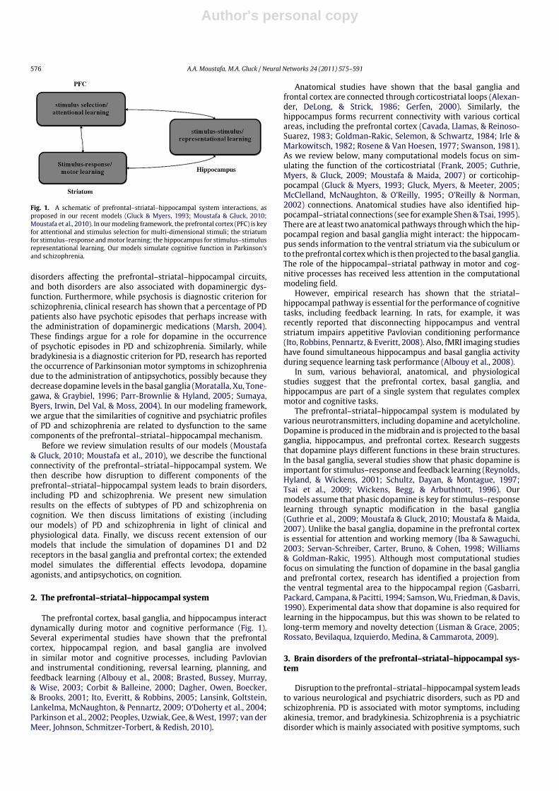
Fig. 1. A schematic of prefrontal–striatal–hippocampal system interactions, asproposed in our recent models (Gluck & Myers, 1993; Moustafa & Gluck, 2010;Moustafa et al., 2010). In ourmodeling framework, the prefrontal cortex (PFC) is keyfor attentional and stimulus selection for multi-dimensional stimuli; the striatumfor stimulus–response andmotor learning; the hippocampus for stimulus–stimulusrepresentational learning. Our models simulate cognitive function in Parkinson’sand schizophrenia.
disorders affecting the prefrontal–striatal–hippocampal circuits,and both disorders are also associated with dopaminergic dys-function. Furthermore, while psychosis is diagnostic criterion forschizophrenia, clinical research has shown that a percentage of PDpatients also have psychotic episodes that perhaps increase withthe administration of dopaminergic medications (Marsh, 2004).These findings argue for a role for dopamine in the occurrenceof psychotic episodes in PD and schizophrenia. Similarly, whilebradykinesia is a diagnostic criterion for PD, research has reportedthe occurrence of Parkinsonian motor symptoms in schizophreniadue to the administration of antipsychotics, possibly because theydecrease dopamine levels in the basal ganglia (Moratalla, Xu, Tone-gawa, & Graybiel, 1996; Parr-Brownlie & Hyland, 2005; Sumaya,Byers, Irwin, Del Val, & Moss, 2004). In our modeling framework,we argue that the similarities of cognitive and psychiatric profilesof PD and schizophrenia are related to dysfunction to the samecomponents of the prefrontal–striatal–hippocampal mechanism.
Before we review simulation results of our models (Moustafa& Gluck, 2010; Moustafa et al., 2010), we describe the functionalconnectivity of the prefrontal–striatal–hippocampal system. Wethen describe how disruption to different components of theprefrontal–striatal–hippocampal system leads to brain disorders,including PD and schizophrenia. We present new simulationresults on the effects of subtypes of PD and schizophrenia oncognition. We then discuss limitations of existing (includingour models) of PD and schizophrenia in light of clinical andphysiological data. Finally, we discuss recent extension of ourmodels that include the simulation of dopamines D1 and D2receptors in the basal ganglia and prefrontal cortex; the extendedmodel simulates the differential effects levodopa, dopamineagonists, and antipsychotics, on cognition.
2. The prefrontal–striatal–hippocampal system
The prefrontal cortex, basal ganglia, and hippocampus interactdynamically during motor and cognitive performance (Fig. 1).Several experimental studies have shown that the prefrontalcortex, hippocampal region, and basal ganglia are involvedin similar motor and cognitive processes, including Pavlovianand instrumental conditioning, reversal learning, planning, andfeedback learning (Albouy et al., 2008; Brasted, Bussey, Murray,& Wise, 2003; Corbit & Balleine, 2000; Dagher, Owen, Boecker,& Brooks, 2001; Ito, Everitt, & Robbins, 2005; Lansink, Goltstein,Lankelma, McNaughton, & Pennartz, 2009; O’Doherty et al., 2004;Parkinson et al., 2002; Peoples, Uzwiak, Gee, &West, 1997; van derMeer, Johnson, Schmitzer-Torbert, & Redish, 2010).
Anatomical studies have shown that the basal ganglia andfrontal cortex are connected through corticostriatal loops (Alexan-der, DeLong, & Strick, 1986; Gerfen, 2000). Similarly, thehippocampus forms recurrent connectivity with various corticalareas, including the prefrontal cortex (Cavada, Llamas, & Reinoso-Suarez, 1983; Goldman-Rakic, Selemon, & Schwartz, 1984; Irle &Markowitsch, 1982; Rosene & Van Hoesen, 1977; Swanson, 1981).As we review below, many computational models focus on sim-ulating the function of the corticostriatal (Frank, 2005; Guthrie,Myers, & Gluck, 2009; Moustafa & Maida, 2007) or corticohip-pocampal (Gluck & Myers, 1993; Gluck, Myers, & Meeter, 2005;McClelland, McNaughton, & O’Reilly, 1995; O’Reilly & Norman,2002) connections. Anatomical studies have also identified hip-pocampal–striatal connections (see for example Shen&Tsai, 1995).There are at least two anatomical pathways throughwhich the hip-pocampal region and basal ganglia might interact: the hippocam-pus sends information to the ventral striatum via the subiculum orto theprefrontal cortexwhich is thenprojected to the basal ganglia.The role of the hippocampal–striatal pathway in motor and cog-nitive processes has received less attention in the computationalmodeling field.
However, empirical research has shown that the striatal–hippocampal pathway is essential for the performance of cognitivetasks, including feedback learning. In rats, for example, it wasrecently reported that disconnecting hippocampus and ventralstriatum impairs appetitive Pavlovian conditioning performance(Ito, Robbins, Pennartz, & Everitt, 2008). Also, fMRI imaging studieshave found simultaneous hippocampus and basal ganglia activityduring sequence learning task performance (Albouy et al., 2008).
In sum, various behavioral, anatomical, and physiologicalstudies suggest that the prefrontal cortex, basal ganglia, andhippocampus are part of a single system that regulates complexmotor and cognitive tasks.
The prefrontal–striatal–hippocampal system is modulated byvarious neurotransmitters, including dopamine and acetylcholine.Dopamine is produced in themidbrain and is projected to the basalganglia, hippocampus, and prefrontal cortex. Research suggeststhat dopamine plays different functions in these brain structures.In the basal ganglia, several studies show that phasic dopamine isimportant for stimulus–response and feedback learning (Reynolds,Hyland, & Wickens, 2001; Schultz, Dayan, & Montague, 1997;Tsai et al., 2009; Wickens, Begg, & Arbuthnott, 1996). Ourmodels assume that phasic dopamine is key for stimulus–responselearning through synaptic modification in the basal ganglia(Guthrie et al., 2009; Moustafa & Gluck, 2010; Moustafa & Maida,2007). Unlike the basal ganglia, dopamine in the prefrontal cortexis essential for attention and working memory (Iba & Sawaguchi,2003; Servan-Schreiber, Carter, Bruno, & Cohen, 1998; Williams& Goldman-Rakic, 1995). Although most computational studiesfocus on simulating the function of dopamine in the basal gangliaand prefrontal cortex, research has identified a projection fromthe ventral tegmental area to the hippocampal region (Gasbarri,Packard, Campana, & Pacitti, 1994; Samson,Wu, Friedman, &Davis,1990). Experimental data show that dopamine is also required forlearning in the hippocampus, but this was shown to be related tolong-term memory and novelty detection (Lisman & Grace, 2005;Rossato, Bevilaqua, Izquierdo, Medina, & Cammarota, 2009).
3. Brain disorders of the prefrontal–striatal–hippocampal sys-tem
Disruption to theprefrontal–striatal–hippocampal system leadsto various neurological and psychiatric disorders, such as PD andschizophrenia. PD is associated with motor symptoms, includingakinesia, tremor, and bradykinesia. Schizophrenia is a psychiatricdisorder which is mainly associated with positive symptoms, such

Author's personal copy
A.A. Moustafa, M.A. Gluck / Neural Networks 24 (2011) 575–591 577
as delusions and hallucinations. Nondeficit schizophrenia ismainlyassociated with the occurrence of positive symptoms, while deficitschizophrenia is associated with positive symptoms and severenegative symptoms.
We use computational and empirical methods to study howPD and schizophrenia affect the prefrontal–striatal–hippocampalsystem. While PD typically impacts the frontostriatal circuitry,schizophrenia (particularly the ‘‘nondeficit’’ type) impairs hip-pocampal function (Buchanan et al., 1993; Farkas et al., 2008;Heckers et al., 1999; Polgar et al., 2010; Tamminga et al., 1992).
Parkinson’s disease is a neurodegenerative disorder associatedwith reduced dopamine levels in the basal ganglia (Jellinger,1999; Kish, Shannak, & Hornykiewicz, 1988). Many studies havealso shown that PD is associated with decreased dopamine inthe prefrontal cortex (Cutsuridis & Perantonis, 2006; Dagher &Robbins, 2009; Diaconescu, Menon, Jensen, Kapur, & McIntosh,2010; Fera et al., 2007; Lanoue, Dumitriu, Myers, & Soghomonian,2010; Prediger et al., 2006; Tadaiesky et al., 2008; Williams-Gray, Hampshire, Barker, & Owen, 2008). In addition to motordysfunction, PD patients show impairment performing variouscognitive tasks such as feedback learning (Bodi et al., 2009b; Franket al., 2004) and reversal learning (Cools et al., 2001). PD is oftenassociated with psychiatric symptoms, such as depression andpsychosis, which has been shown to be associated with furtherdopamine reduction and impaired hippocampal function (Bruck,Kurki, Kaasinen, Vahlberg, & Rinne, 2004; Walter, Skoloudik, &Berg, 2010).
Dopamine medications, including levodopa and D2 agonists,are used to treat motor symptoms of PD (tremor, akinesia, andbradykinesia), but can either enhance or impair cognitive function(Cools et al., 2001; Feigin et al., 2003; Frank et al., 2004; Swainsonet al., 2000). For example, various studies show that dopaminemedications and agents impair stimulus–response learning inboth PD patients (Gotham, Brown, & Marsden, 1988; Jahanshahiet al., 2009; Shohamy, Myers, Geghman, Sage, & Gluck, 2006) andhealthy subjects (Breitenstein et al., 2006; Pizzagalli et al., 2007).In combination with dopamine medications, PD patients are oftenprescribed anticholinergic medications, such as Trihexyphenidyl,which are muscarinic receptor antagonists. The hippocampalregion has a large density of these receptors (Levey, 1993); thus,it is possible that the administration of anticholinergic antagonistsinterferes with hippocampal function. In agreement with this andother studies (Ehrt, Broich, Larsen, Ballard, & Aarsland, 2010;Meco et al., 1984; Pondal, Del Ser, & Bermejo, 1996), we haverecently shown that the administration of anticholinergics toPD patients impairs performance in hippocampal-based tasks(Herzallah, Moustafa, Misk, Myers, & Gluck, 2010).
Unlike PD, schizophrenia is typically associated with hip-pocampal dysfunction (Bogerts, Meertz, & Schonfeldt-Bausch,1985; Goldman & Mitchell, 2004; Grace, 2010; Heckers, 2001;Keri, 2008; Weinberger, 1999). Schizophrenic patients also showdeclarative memory deficits, which suggest hippocampal-regiondysfunction (Aleman, Hijman, de Haan, & Kahn, 1999; Cir-illo & Seidman, 2003). Lesioning the hippocampus in animalsis also used as a model of schizophrenia (Tseng, Chambers,& Lipska, 2009). In addition, Rametti et al. (2009) reporteddecreased hippocampal activity in schizophrenic patients per-forming declarative memory tasks. Research at our laboratoryhave shown that schizophrenic patients’ performance on thelearning-and-transfer acquired equivalence task is very simi-lar to the performance of patients with mild Alzheimer’s dis-ease, hippocampal atrophy, and hypoxia (Bodi, Csibri, Myers,Gluck, & Keri, 2009a; Myers et al., 2008; Myers, Shohamy, Gluck,Grossman, Kluger et al., 2003a; Myers, Shohamy, Gluck, Gross-man, Onlaor et al., 2003b), suggesting a common hippocampaldysfunction in all these patient groups. Though debatable, it is
argued that the basal ganglia is generally intact in nondeficitschizophrenia (Okubo et al., 1997). For example, Bogerts et al.(1985) reported a decrease in hippocampal size but an intact basalganglia structure in schizophrenic patients as seen in structuralbrain imaging.
Schizophrenia is often accompanied by negative symptoms,such as apathy and emotional withdrawal. Schizophrenic patientswith severe negative symptoms, known as deficit schizophrenia,usually perform worse than nondeficit schizophrenic patients inmultiple cognitive domains (Cascella et al., 2008). Using structuralbrain imaging, Buchanan et al. (1993)found that deficit, butnot nondeficit, schizophrenia is associated with striatal damage.Supporting the role of prefrontal–striatal system in the occurrenceof severe negative symptoms in schizophrenia (van Veelen, Vink,Ramsey, & Kahn, 2010), we have shown that that negativesymptoms in schizophrenia are associated with feedback learningimpairment (Farkas et al., 2008; Keri, 2008; Polgar et al., 2010),a behavioral task that typically recruits the frontostriatal system(Houk, 1995b).
Specifically, empirical studies that measured protein functionassociated with dopamine receptors implicate D1 receptors in thestriatum for the occurrence of negative symptoms in schizophrenia(Monteleone, Di Lieto, Martiadis, Bartoli, & Maj, 2002). Similarly,using a PET scan, Okubo et al. (1997) have found that that thedistribution of D1 receptors in the prefrontal cortex correlateswith the severity of negative symptoms in schizophrenic patients(for similar finding, see Abi-Dargham, 2003). These findings arein agreement with why antipsychotics (D2 antagonists) usuallydo not treat D1-mediated negative symptoms of schizophrenia(Rueter et al., 2004). Notably, computational modeling andempirical work suggest that D1 agonists can be used to treatnegative symptoms and cognitive deficits in schizophrenia (Loh,Rolls, & Deco, 2007; Roberts, Seymour, Schmidt, Williams, &Castner, 2010). D1 receptors are activated by phasic dopamine(Dalley et al., 2005; Goto&Grace, 2008; Richfield, Penney, & Young,1989; Zweifel et al., 2009), which has been shown to be essentialfor learning in basal ganglia (Reynolds et al., 2001; Schultz et al.,1997; Tsai et al., 2009). Thus, impaired D1 receptor function inschizophrenic patients with severe negative symptoms explainsfeedback learning deficits in these patients, as we have found inour studies (Farkas et al., 2008).
Antipsychotics are used to treat psychiatric deficits, includ-ing positive symptoms, in schizophrenia, possibly by blocking D2receptors in the striatum or hippocampus (Rueter et al., 2004). Un-like PD, very few studies have studied the effects of medicationson cognition and symptoms severity in schizophrenia. This is per-haps due to the fact that withdrawal of medications in schizophre-nia (unlike PD) is rarely done as it might have major effectson patients’ health and life. Beside antipsychotics, schizophrenicpatients are often prescribed anticholinergic medications; inter-estingly, like in PD, recent research has also shown that anti-cholinergics impair cognition, particularly working and long-termmemory, in schizophrenic patients (Vinogradov et al., 2009). Weprovide here a summary of empirical findings on the effects ofParkinson’s disease, schizophrenia, and associated medications onthe prefrontal cortex, hippocampus, and basal ganglia in Table 1.
4. Cognitive dysfunction in PD and schizophrenic patients
Various behavioral tasks have been used to assess the natureof cognitive deficits in PD and schizophrenic patients, includingfeedback learning, reversal learning, and transfer generalization.We simulate cognitive performance in many of these tasks andbriefly review here relevant empirical results on how PD andschizophrenia affect cognition. Finally, we present simulationresults these behavioral tasks.

Author's personal copy
578 A.A. Moustafa, M.A. Gluck / Neural Networks 24 (2011) 575–591
Table 1A summary of empirical results of the effects of Parkinson’s disease and schizophrenia, as well as associated medications, on the prefrontal cortex, hippocampus, and basalganglia., that our models simulate. Our modeling framework simulates many of the excising empirical data shown here, but also see model limitations and future directions.Abbreviations: DA, dopamine.
Basal ganglia Prefrontal cortex Hippocampus
Unmedicated PD Reduced DA Reduced DAMedicated PD Increased DA Increased and perhaps overdosed with DANondeficit schizophrenia Damaged and DA dysregulationDeficit schizophrenia Dysregulation of D1 receptors Dysregulation of D1 receptors Damaged and DA dysregulationPD depression Further reduced DA DamagedPD on anticholinergics Reduced DA Reduced DA DamagedAntipsychotics Dysregulation of D1 receptors Dysregulation of D1 receptors
Feedback learning: In feedback learning tasks, subjects learnto associate the presentation of different stimuli with differentresponses, based on corrective feedback. Many experimentalstudies have shown that PD patients are impaired at feedbacklearning (Jahanshahi et al., 2009; Shohamy et al., 2006). In contrast,schizophrenic patients are generally intact on feedback learningtasks (Gomar et al., 2011; Keri et al., 2005a, 2005b; Leeson et al.,2009; Somlai, Moustafa, Keri, Myers, & Gluck, 2010). For example,Jahanshahi et al. (2009) have shown that medicated PD patientsare more impaired than unmedicated PD patients at the ‘‘weatherprediction’’ task, a multi-cue probabilistic categorization task, andboth patient groups are more impaired than healthy controls. Inthe weather prediction task, subjects classify patterns composedof sets of two to four cards as being predictive of rain versussunshine (Fera et al., 2005; Gluck, Shohamy, & Myers, 2002;Knowlton, Mangels, & Squire, 1996; Knowlton, Squire, & Gluck,1994; Shohamy, Myers, Onlaor, & Gluck, 2004). In other studiesusing the weather prediction task, it was found that schizophrenicpatients show intact feedback learning (Keri et al., 2005a, 2000;Weickert et al., 2002).
We have also found that schizophrenia patients show intactfeedback learning performance in a different feedback learningtask that tests learning from positive or negative feedback (Somlaiet al., 2010). This is, however, in contrast to another studyin which Waltz, Frank, Robinson, and Gold (2007) found thatschizophrenic patients are impaired at learning from reward butnot from punishment. The inconsistent results in the literature areperhaps because existing empirical studies test different subtypesof schizophrenic patients. Supporting this hypothesis, in our ownprior studies (Farkas et al., 2008; Polgar et al., 2008), we havefound that deficit, but not nondeficit, schizophrenic patients showfeedback learning impairments (Farkas et al., 2008; Polgar et al.,2008). The correlation between feedback learning and the severityof negative symptoms in schizophrenia was also reported in theliterature (Murray et al., 2008).
Reversal: In reversal learning tasks, subjects initially learnto associate different stimuli with different responses, andsubsequently learn to associate the same stimuli with the oppositeresponses (i.e., reversal). Various studies show that the basalganglia, hippocampus, and prefrontal cortex are important forreversal learning performance (Clatworthy et al., 2009; Coolset al., 2001; Cools & Frank, 2009; McDonald, Ko, & Hong, 2002;Mitchell, Rhodes, Pine, & Blair, 2008). For example, Pasupathyand Miller (2005) recorded from both the striatum and prefrontalcortex while a monkey performed a reversal task. They found that,within a trial, the striatum increased its activation before that ofprefrontal cortex neurons, suggesting that both basal ganglia andprefrontal cortex are engaged during reversal learning processes.Cools et al. (2001) found that medicated PD patients are moreimpaired at reversal learning than unmedicated PD patients (alsosee Swainson et al., 2000). Furthermore, Jentsch, Olausson, De LaGarza, and Taylor (2002) found that the administration of cocaine,which is a dopamine agonist, to monkeys lead to impairmentperforming reversal learning tasks. Similar results were found
with the administration of quinpirole (a dopamine agonist) to rats(Boulougouris, Castane, & Robbins, 2009). It has been argued thatdopamine medications overdose the prefrontal cortex and thusimpair performance in reversal tasks (Cools et al., 2001). In linewith this hypothesis, our model demonstrates that an increasein dopamine levels in the prefrontal cortex impairs reversalperformance. Similarly, empirical studies show that schizophrenicpatients are impaired at reversal learning (Leeson et al., 2009),possibly due to prefrontal dysfunction (McKirdy et al., 2009).
Learning-and-transfer: The Rutgers learning-and-transfer ‘‘ac-quired equivalence’’ task (Myers, Shohamy, Gluck, Grossman,Kluger et al., 2003a) is used to test the contributions of the basalganglia and hippocampus to cognition. This task has two phases:acquisition and transfer. In the acquisition phase, subjects (andalso themodel) learn to associate two stimuli, while in the transfergeneralization phase, subjects learn that cues become equivalentwhen theywere previously associatedwith the same response. Thetransfer generalization phase includes two types of trials: reten-tion and reversal. Retention trials are trials that were previouslypresented in the learning phase, while transfer trials include novelcombinations of stimuli (see Myers, Shohamy, Gluck, Grossman,Kluger et al., 2003a). Several neuropsychological studies from ourlaboratory have argued that the associative learning and transfergeneralization processes rely on different neural structures (Keri,2008; Myers et al., 2008): initial associative learning relies on theintegrity of the basal ganglia, whereas transfer generalization re-lies on the integrity of the hippocampal region. Specifically, wehave shown that PD patients are more impaired at learning thanschizophrenic patients, while the opposite is true for the transfergeneralization phase: schizophrenic patients are more impairedthan PD patients at transfer generalization (Keri et al., 2005b; My-ers, Shohamy, Gluck, Grossman, Kluger et al., 2003a). Interestingly,Shohamy et al. (2010) found that the administration of antipsy-chotics to schizophrenic patients ameliorates their transfer gener-alization impairment on the same task.
In our models, different brain areas play different computa-tional roles. In agreement with empirical (Dusek & Eichenbaum,1997) andmodeling (Cutsuridis &Wennekers, 2009)work, the hip-pocampus in our models is important for stimulus–stimulus rep-resentational learning (Gluck et al., 2005; Moustafa & Gluck, 2010;Moustafa et al., 2010). The hippocampus sends recoded informa-tion of input stimuli to the basal ganglia and prefrontal cortex forfurther processing. In ourmodels, the basal ganglia is key for stimu-lus–response and reinforcement learning, in linewith empirical re-sults (Schultz et al., 1997; Tsai et al., 2009). Unlike the basal ganglia,the prefrontal cortex in our models is essential for stimulus selec-tion during learning aboutmultidimensional stimuli, in agreementwith physiological studies (Iba & Sawaguchi, 2003). Importantly,our simulations explain how interactions among these brain areasgive rise to complex cognitive and motor functions.
Our computational models also aim at understanding howdamage to different components with the prefrontal–striatal–hippocampal system leads to cognitive dysfunction in PD andschizophrenia. Below, we provide simulation results of how our

Author's personal copy
A.A. Moustafa, M.A. Gluck / Neural Networks 24 (2011) 575–591 579
1 2 3 4
Block Number
Per
cent
age
Cor
rect
Weather Prediction performance
HC
PD off
PD on
0.580.6
0.620.640.660.68
0.70.720.740.76
Weather Prediction performance
Per
cent
agte
Cor
rect
PD off
PD on
HC
1 2 3 4
Block Number
0.45
0.5
0.55
0.6
0.65
0.7
0.75
0.8
(a) Experiment. (b) Model.
Fig. 2. Weather prediction task performance in medicated and unmedicated PD patients. Each block here is 50 trials. (a) Figure is adapted from Jahanshahi et al. (2009).(b): Simulation results are qualitatively similar to the results of Jahanshahi et al. (2009).
prefrontal–striatal and striatal–hippocampal models (Moustafa &Gluck, 2010; Moustafa et al., 2010) simulate feedback learning,reversal, and learning-and-transfer acquired equivalence in PD andschizophrenia. We start by describing simulation results of theeffects of prefrontal–striatal dysfunction in PD patients, and thenpresent simulation results of the effects of striatal–hippocampaldysfunction in PD and schizophrenia. We also present simulationresults of the effects of PD depression, the administration ofanticholinergics to PD, and nondeficit vs. deficit schizophrenia oncognition.
5. Simulation of prefrontal–striatal interactions in PD
We have recently designed a prefrontal–striatal model thatsimulates the effects of Parkinson’s disease and dopaminemedications on learning and attention (Moustafa & Gluck, 2010).Unlike prior models of Parkinson’s disease, we have simulatedthe effects of prefrontal dopamine in attentional learning, andhow this cognitive function is affected by Parkinson’s diseaseand dopamine medications. In this model, we have simulated thedifferential effects of phasic and tonic dopamine on motor andcognitive processes, and how phasic and tonic dopamine might beaffected by Parkinson’s disease and dopamine medications. Phasicdopamine in our model is key for learning, while tonic dopamineis essential for the initiation of motor responses. The Moustafaand Gluck (2010) model argues that PD is associated with reducedphasic and tonic dopamine levels, while dopamine medicationsincrease tonic dopamine and further decrease phasic dopaminesignaling. Our simulation results show that unmedicated andmedicated PD patients are impaired the weather prediction task(Fig. 2(a)), as found in empirical results (Figure 2a, Jahanshahi et al.,2009).
The Moustafa and Gluck model also simulates performancein probabilistic reversal learning tasks. This task consists of twophases: acquisition and reversal. The acquisition phase involvesprobabilistic classification of stimuli. On each trial of this phase,the model learns to select one of two stimuli (see Cools et al.,2001, for task details). One stimulus is designated as the correctstimulus, which is associated with 80% of positive feedback (and20% negative feedback). The other stimulus is designated theopposite ratio of reinforcement. As in Cools et al. (2001), this phasehas 40 trials. The second phase is the reversal phase in whichreinforcement contingencies are reversed so that the previouslyincorrect stimulus is now correct and vice versa. As in the initiallearning phase, the reversal phase has 40 trials. Following Coolset al. (2001), the learning criterion of any of the phases in oursimulations is correct responses in eight trials. In addition tosimulating the probabilistic reversal task, we further ran themodelon the exact same task but by increasing number of trials in the
reversal phase to test extended learning on reversal performance.We assume that each run of the model corresponds to a differentsubject (each simulation run has different initial random values).
In simulating the original reversal task as published, we foundthat many of the simulation runs of the medicated PD network didnot reach criterion performance in the reversal phase (Fig. 3(b)),which is qualitatively similar to the empirical results of Cools et al.(Fig. 3(a)). In other words, the model accounts for the findingthat medicated PD patients are more impaired at the reversalphase than unmedicated PD patients and controls. In the model,dopamine medications impair performance in the reversal phase.In the beginning of the reversal phase, themodel receives negativefeedback, and because of an increase of tonic dopamine in theprefrontal cortex, the model shifts attention to the other cueinstead of learning to reverse responses. This in turn led to anincrease number of errors in the reversal phase in many of thesimulation runs of the medicated PD patients network. This delayscorrect reversal learning, and thus explainsmedicated PD patients’impaired performance in this phase. In the extended reversal task,we found that many of the runs of the medicated PD patientsnetwork were able to reach performance criterion in the reversalphase (Fig. 3(c)). Themodel here shows that impaired performanceinmedicated PDpatients in the original reversal task as reported byCools et al. (2001) is perhaps due to the use of a fewnumber of trialsin the reversal phase,which did not allowpatients to learn the task.In sum, our prefrontal–striatal model accounts for the findingsthat medicated and medicated PD patients are more impairedthan healthy controls at feedback learning, and that medicatedPD patients are more impaired than unmedicated PD patients atreversal learning.
6. Simulation of striatal–hippocampal interactions in PD andschizophrenia
The Moustafa and Gluck (2010) model does not have a hip-pocampal module and is limited to the simulation of the frontos-triatal connectivity in cognition.We have recently investigated theinteractions among the basal ganglia and hippocampus in cogni-tion Moustafa et al. (2010). The interaction of the hippocampusand the basal ganglia in cognition is not addressed by most ex-isting models. The Moustafa et al. (2010) model integrates ourearlier models of basal ganglia and hippocampal-region function,and their modulation by dopamine and acetylcholine. Like theMoustafa and Gluck (2010) model, the basal ganglia is key forreinforcement learning, motivated by dopamine signals comingfrom the ventral tegmental area and substantia nigra pars com-pacta (Reynolds et al., 2001; Wickens et al., 1996). In the Moustafaet al. (2010) model, the hippocampal region is required for stimu-lus–stimulus representation learning, as argued in our prior mod-els (Gluck, Allen, Myers, & Thompson, 2001). In the Moustafa et al.

Author's personal copy
580 A.A. Moustafa, M.A. Gluck / Neural Networks 24 (2011) 575–591
Reversal
Per
cent
age
of s
ubje
cts
Pas
sing
PD on
Acquisition Reversal
PD off
40
50
60
70
80
90
100
Per
cent
age
of s
imul
atio
n ru
ns P
assi
ng
HC
Acquisition Reversal
PD off
PD on
40
50
60
70
80
90
100
(a) Experiment. (b) Model.
PD off
HC
PD on
Per
cent
age
of s
imul
atio
n ru
ns P
assi
ng
40
50
60
70
80
90
100
Acquisition Reversal
(c) Model (extended reversal training).
Fig. 3. PD performance in the probabilistic reversal task using the prefrontal–striatal model. (a) Experimental results from Cools et al. (2001). (b) Modeling results of theoriginal reversal task. (c) Modeling results in the extended reversal learning tasks (see text). Increasing number of training trials of the reversal phase shows that PD patientscan learn the reversal task. Prefrontal dysfunction explains reversal deficits in PD patients.
(2010) model, the hippocampal region preprocesses input infor-mation and projects coded information to the basal ganglia for fur-ther computational processing. This model simulates performancein the learning-and-transfer ‘‘acquired equivalence’’ task describedabove (Myers, Shohamy, Gluck, Grossman, Kluger et al., 2003a).
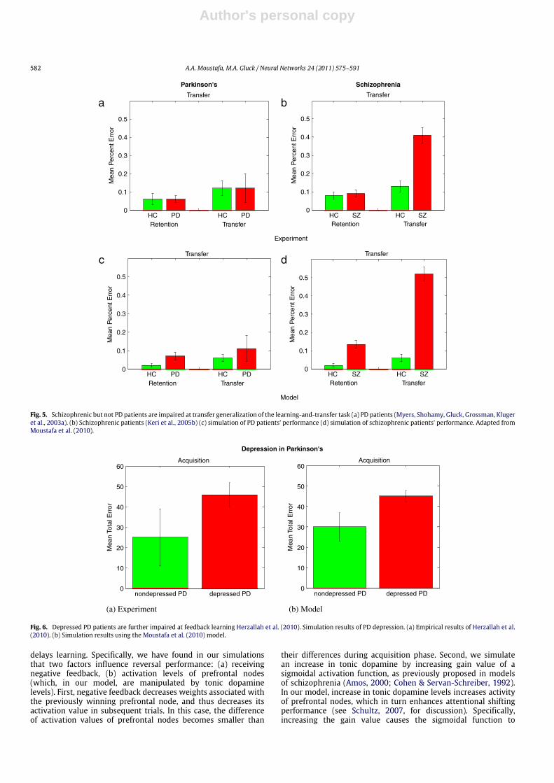
The Moustafa et al. (2010) model has been applied to simulatethe effects of Parkinson’s disease and schizophrenia on learningand transfer generalization. We present here simulation results interms of number of errors in the acquisition and transfer (includingretention and transfer trials) phases. Consistent with the empiricalresults (Fig. 5(a)), the model shows that simulating a loss ofdopamine function in the basal ganglia module, as in Parkinson’sdisease, leads to slow acquisition learning but intact transfergeneralization (Fig. 4(c)). Damaging the hippocampal module inthe model, as in schizophrenia, did not interfere with acquisition(Fig. 4(d)), which is line with empirical results (Fig. 4(b)). Incontrast to feedback learning, our simulation results show thatschizophrenic patients are impaired, but PD patients show intactperformance, at the transfer generalization phase (Fig. 5(c) and(d)),which is in agreementwith empirical results (Fig. 5(a) and (b)).In sum, our striatal–hippocampal model explains why PD patientsare worse at learning, but better at transfer generalization, thanschizophrenic patients.
7. Simulation of the effects of depression and anticholinergicson cognition in PD
Our earlier simulation results were limited to the simulation ofstandard PD and schizophrenia that are not associated with any
additional symptoms, such as depression or negative symptoms.As shown above, subtypes of PD and schizophrenic patients havedifferent cognitive profiles. In a recent neuropsychological study,we have tested the effects of depression and anticholinergicmedications on learning and transfer generalization in PD patients(Herzallah et al., 2010). We have found that depressed PD patientsare more impaired than nondepressed patients at feedbacklearning (Fig. 6(a)). In contrast, we have found that PD patientson anticholinergics are more impaired at transfer generalizationthan patients who are not on anticholinergics (Fig. 7(a)). Empiricalstudies have shown that PD depression (and major depression)is associated with further reduced dopamine levels than innondepressed PD patients (Walter et al., 2010). Accordingly, wesimulate depression in PD by further decreasing the learning rateparameter in the basal gangliamodule. As in our empirical findings(Fig. 6(a)), our simulations show a qualitatively similar patternof results (Fig. 6(b)). Furthermore, empirical research has shownthat anticholinergics impair hippocampal function (Ehrt et al.,2010; Meco et al., 1984; Pondal et al., 1996). By removing thehippocampal module from the Moustafa et al. (2010) model, weshow that the simulations of PD patients on anticholinergics aremore impaired at transfer generalization than patients not onanticholinergics (Fig. 7(b)).
In sum, usingparallel neuropsychological-computationalmeth-ods, we show here that depression in PD patients is associatedwith impaired feedback learning, while the administration of anti-cholinergics to PD patients impairs transfer generalization.

Author's personal copy
A.A. Moustafa, M.A. Gluck / Neural Networks 24 (2011) 575–591 581
Parkinson's Schizophrenia
Acquisition
Mea
n To
tal E
rror
Mea
n To
tal E
rror
HC PD
Acquisition
SZHC
Experiment
0
5
10
15
20
25
30
35
0
10
20
30
40
Acquisition
Mea
n To
tal E
rror
Mea
n To
tal E
rror
HC PD
Acquisition
SZHC
Model
0
10
20
30
40
0
10
20
30
40
50
a b
c d
Fig. 4. PD patients but not schizophrenic patients are impaired at feedback learning. (a) PD patients (Myers, Shohamy, Gluck, Grossman, Kluger et al., 2003a). (b)Schizophrenic patients (Keri et al., 2005b) (c) simulation of PD patients’ performance (d) simulation of schizophrenic patients’ performance. Adapted from Moustafa et al.(2010). Adapted from Moustafa et al. (2010). Abbreviations: HC, healthy controls; PD, Parkinson’s patients; SZ; schizophrenia.
8. Simulationof the effects of deficit andnondeficit schizophre-nia on cognition
As mentioned above, various clinical studies have shownthat deficit and nondeficit schizophrenic patients have differentcognitive profiles.
At our laboratory, we have found that while deficit schizo-phrenic patients are impaired at both the learning and transferphases of the acquired equivalence task, nondeficit schizophrenicpatients only are impaired at the transfer phase of this task (Farkaset al., 2008). Research suggests that nondeficit schizophrenia is as-sociated with hippocampal dysfunction, while deficit schizophre-nia is associated with both hippocampal and striatal dysfunction(Buchanan et al., 1993; Heckers et al., 1999; Polgar et al., 2010;Tamminga et al., 1992). In agreement with experimental resultsshowing that deficit schizophrenia is associated with hippocam-pal damage and dysfunction of D1 receptors in the basal gan-glia (Monteleone et al., 2002), we simulate deficit schizophreniaby removing the hippocampus module and decreasing learningrate parameter in the striatal module in our striatal–hippocampalmodel (Moustafa et al., 2010). In agreement with empirical find-ings (Farkas et al., 2008), our simulations show that both deficit andnondeficit schizophrenic patients are impaired at transfer general-ization, while deficit schizophrenic patients are more impaired atlearning than nondeficit schizophrenic patients (Fig. 8(a) and (b)).
9. Discussion
We have built computational models of frontal–striatal(Moustafa & Gluck, 2010) and striatal–hippocampal interactions(Moustafa et al., 2010) to simulate cognitive performance in PD andschizophrenia. PD and schizophrenia affect the prefrontal–striatal–hippocampal system in different ways: PD is associated withfronto-striatal dysfunction, while nondeficit schizophrenia is asso-ciated with hippocampal dysfunction.
Our computational models simulate performance in a range ofcognitive tasks, including feedback learning, reversal learning, andlearning-and-transfer ‘‘acquired equivalence’’. Various empiricalstudies show that dopamine medications and agents impairstimulus–response learning performance in both PD patients(Gotham et al., 1988; Jahanshahi et al., 2009; Shohamy et al.,2006) and healthy controls (Breitenstein et al., 2006; Santessoet al., 2009). The Moustafa and Gluck (2010) model shows thata decreasing learning rate (due to increase of dopamine levelsin the basal ganglia and prefrontal cortex) leads to impairmentin performing the weather prediction task, in line with empiricalresults (Jahanshahi et al., 2009). The Moustafa and Gluck (2010)model also simulates the findings that medicated PD patientsare impaired at performing reversal tasks, due to increase ofdopamine levels in the prefrontal cortex. Simulation results showthat during the reversal phase, increase of dopamine levelsin the prefrontal cortex made the model shifts attention todifferent stimuli instead of learning to reverse responses, which
Author's personal copy
582 A.A. Moustafa, M.A. Gluck / Neural Networks 24 (2011) 575–591
Parkinson's Schizophrenia
Transfer
Mea
n P
erce
nt E
rror
PDRetention
HC
Transfer
Experiment
PDTransfer
HC SZRetention
HC SZTransfer
HC0
0.1
0.2
0.3
0.4
0.5
Mea
n P
erce
nt E
rror
0
0.1
0.2
0.3
0.4
0.5
Transfer
Mea
n P
erce
nt E
rror
PDRetentionHC
Transfer
Model
PDTransfer
HC SZRetentionHC SZ
TransferHC
Mea
n P
erce
nt E
rror
0
0.1
0.2
0.3
0.4
0.5
a b
c d
0
0.1
0.2
0.3
0.4
0.5
Fig. 5. Schizophrenic but not PD patients are impaired at transfer generalization of the learning-and-transfer task (a) PD patients (Myers, Shohamy, Gluck, Grossman, Klugeret al., 2003a). (b) Schizophrenic patients (Keri et al., 2005b) (c) simulation of PD patients’ performance (d) simulation of schizophrenic patients’ performance. Adapted fromMoustafa et al. (2010).
Depression in Parkinson's
Acquisition
Mea
n To
tal E
rror
nondepressed PD
(a) Experiment (b) Model
0
10
20
30
40
50
60
depressed PD
Mea
n To
tal E
rror
nondepressed PD0
10
20
30
40
50
60
depressed PD
Acquisition
Fig. 6. Depressed PD patients are further impaired at feedback learning Herzallah et al. (2010). Simulation results of PD depression. (a) Empirical results of Herzallah et al.(2010). (b) Simulation results using the Moustafa et al. (2010) model.
delays learning. Specifically, we have found in our simulationsthat two factors influence reversal performance: (a) receivingnegative feedback, (b) activation levels of prefrontal nodes(which, in our model, are manipulated by tonic dopaminelevels). First, negative feedback decreases weights associated withthe previously winning prefrontal node, and thus decreases itsactivation value in subsequent trials. In this case, the differenceof activation values of prefrontal nodes becomes smaller than
their differences during acquisition phase. Second, we simulatean increase in tonic dopamine by increasing gain value of asigmoidal activation function, as previously proposed in modelsof schizophrenia (Amos, 2000; Cohen & Servan-Schreiber, 1992).In our model, increase in tonic dopamine levels increases activityof prefrontal nodes, which in turn enhances attentional shiftingperformance (see Schultz, 2007, for discussion). Specifically,increasing the gain value causes the sigmoidal function to

Author's personal copy
A.A. Moustafa, M.A. Gluck / Neural Networks 24 (2011) 575–591 583
Parkinson's and anticholinergics
Transfer
Mea
n P
erce
nt E
rror
not Anti-Ach Anti-Ach not Anti-Ach Anti-AchRetention Transfer
(a) Experiment
(b) Model
Transfer
0
0.2
0.4
0.6
0.8
1
1.2
Mea
n P
erce
nt E
rror
not Anti-Ach Anti-Ach not Anti-Ach Anti-AchRetention Transfer
0
0.2
0.4
0.6
0.8
1
1.2
Fig. 7. PD patients on anticholinergics are impaired at generalization than PDpatients not on anticholinergics. (a) Empirical results of Herzallah et al. (2010).(b) Simulation results using the Moustafa et al. (2010) model. Abbreviations: Anti-Ach refers to PD patients on anticholinergics, while ‘‘not Anti-Ach’’ refers to PDpatients not on anticholinergics.
decrease the difference between the representations of inputs. Inother words, increasing the gain value increases the competitivedynamics in the simulated prefrontal cortex and the likelihood toshift to a new dimension. From these two points, we have found
that that an increase in tonic dopamine levels, as in medicated PDsimulations) increase activity and competition among prefrontalnodes, which in turn enhance selecting different stimuli followingnegative feedback. This delays reversal learning since reversallearning is acquired more quickly if the model (and subjects) justlearned to reverse motor responses (in the basal ganglia module),and not shift attention to other irrelevant cues (in the prefrontalmodule).
Interestingly, the same mechanism of reversal performancein our model also explains enhanced attentional performancein medicated PD patients as we showed in our earlier work(Moustafa & Gluck, 2010) and as reported experimentally (Coolset al., 2001; Swainson et al., 2000). Furthermore, the Moustafaand Gluck (2010) model shows that increasing the number oftraining trials in the reversal phase enhances performance ofmedicated PD patients. We conclude that impaired performanceof medicated PD patients in the reversal task in the Cools et al.(2001)study is perhaps due to the use of a low number of trials inthe reversal phase. Future experimental research should confirm(or disconfirm) model prediction.
Our models also simulate cognitive dysfunction in subtypesof PD and schizophrenia. In agreement with existing results(Herzallah et al., 2010), our simulation results show that depressedPD patients are more impaired than nondepressed patients onfeedback learning. We also simulate the effects of anticholinergicson hippocampal function. Our simulation results show thatthe administration of anticholinergics to PD patients impairhippocampal function, and thus impair transfer generalization, inline with our empirical findings (Herzallah et al., 2010).
Furthermore, research has shown that deficit and nondeficitschizophrenia are associated with different neural and cognitivedysfunction. While nondeficit schizophrenia is associated withhippocampal dysfunction, deficit schizophrenia is additionallyassociated with frontostriatal dysfunction (Buchanan et al., 1993;Farkas et al., 2008; Heckers et al., 1999; Polgar et al., 2008,2010; Tamminga et al., 1992). Our models also simulate cognitivedysfunction in deficit and nondeficit schizophrenic patients. As inour empirical findings (Farkas et al., 2008), our simulations showthat while both deficit and nondeficit schizophrenic patients areimpaired at transfer generalization, deficit schizophrenic patientsonly are impaired at feedback learning. Unlike other models ofschizophrenia (reviewed below), our Moustafa et al. (2010) modelsimulates the effects of negative symptoms on cognition. In linewith empirical results, we assumed that negative symptoms are
Deficit vs. nondeficit schizophrenia
Acquisition
Mea
n To
tal E
rror
Non-deficit Non-deficit
Transfer
Mea
n P
erce
nt E
rror
(a) acquisition (b) transfer
0
5
10
15
20
25
deficit deficit Non-deficit deficitRetention Transfer
0
0.1
0.2
0.3
0.4
0.5
0.6
0.7
Fig. 8. Simulation of the effects of deficit and nondeficit schizophrenia on learning and transfer ‘‘acquired equivalence’’ task. Farkas et al. (2008) have found that nondeficitschizophrenic patients are impaired at the transfer generalization phase, but deficit schizophrenic patients are impaired at acquisition and transfer phases. (a) Simulationresults of the effects of deficit and nondeficit schizophrenia on learning. (b) Simulation results of the effects of deficit and nondeficit schizophrenia on transfer generalization.

Author's personal copy
584 A.A. Moustafa, M.A. Gluck / Neural Networks 24 (2011) 575–591
associated with disregulation of striatal and frontal D1 receptors,and thus associated with impaired learning, as reported in manystudies (Murray et al., 2008). One limitation of this model is itdoes not have a prefrontal module and thus does not simulate theeffect of deficit and nondeficit schizophrenia on working memory.For example, Polgar et al. (2010) have recently shown that deficitschizophrenic patients are more impaired at working memorythan nondeficit patients. This is perhaps due to more severeprefrontal dysfunction in deficit schizophrenic patients, which hasbeen reported in the literature (Okubo et al., 1997).
9.1. Existing models of PD and schizophrenia
Below, we review how existingmodels of PD and schizophreniarelate prefrontal, hippocampal, or striatal dysfunction to cognitiveand psychiatric deficits in these patients.
There are very fewmodels that simulate cognitive performancein both PD and schizophrenia (Amos, 2000; Monchi, Taylor, &Dagher, 2000; Moustafa et al., 2010). Amos (2000) simulatesperformance in the Wisconsin Card Sorting Task in Parkinson’sdisease, Huntington’s disease, and schizophrenic patients, usingmathematical techniques similar to those used by Moustafa andGluck (2010). The Amos model showed that decreasing activityof the prefrontal cortex explains working memory deficits inschizophrenia, and it also shows that decreasing activity inbasal ganglia and prefrontal cortex modules simulates impairedperformance in Parkinson’s disease patients. In the Amos model,dopamine reduction (as in PD) was simulated by decreasing thegain parameters of the sigmoidal activation function and lesioningwas simulated by decreasing the output of neurons representingthe lesioned area. As in our model, Amos argues that prefrontalcortex maintains the sorting rule (card, color, or shape) in workingmemory. The sensory association cortex encoded representationsof input stimuli and the striatum integrated cortical informationand decided what action to perform. Feedback to prefrontal cortexfrom the basal ganglia informed prefrontal cortex whether tomaintain or change the sorting rule (not modeled). The modelsimulated the occurrence of perseverative and random responsesin prefrontal cortex-damaged and PD patients. Unlike our models(Moustafa & Gluck, 2010; Moustafa et al., 2009, 2010), the Amosmodel is not a learningmodel and does not simulate the role of thehippocampus in cognition.
Monchi and colleagues (Monchi et al., 2000) proposed a modelthat simulates the role of frontostriatal loops in working memoryin PD and schizophrenic patients. This model argues that PD isassociated with basal ganglia dysfunction, while schizophrenia isassociated with both basal ganglia and frontal dysfunction. Themodel successfully accounts for various data on the effects of PDand schizophrenia on working memory. Specifically, Monchi et al.(2000) simulate PD by decreasing values of weights connectingprefrontal cortex and striatal units. Like Amos (2000) model,Monchi et al. did not simulate the role of dopamine in learning anddoes not incorporate a hippocampal module.
Most of the existing models focus on simulating the role ofthe frontostriatal and/or dopaminergic dysfunction in PD patients.Existing models of schizophrenia simulate the effects of frontal orhippocampal dysfunction on cognition and psychosis. Below, wereview these models, starting by models of PD.
9.1.1. Models of frontostriatal dysfunction in Parkinson’s diseaseWe here review computational models that address the ef-
fects of PD on cognition. Most, if not all, existing models of PD fo-cus on simulating the functional contribution of the basal gangliaand/or prefrontal cortex to motor and cognitive processes (Amos,2000; Frank, 2005; Guthrie et al., 2009; Moustafa & Gluck, 2010;
Moustafa et al., 2009). The most common framework for simu-lating the role of the basal ganglia in feedback learning is theactor-critic model (Berns & Sejnowski, 1995; Houk, 1995a; Joel,Niv, & Ruppin, 2002; Redish, Jensen, Johnson, & Kurth-Nelson,2007; Suri, Bargas, & Arbib, 2001; Suri & Schultz, 1998). Thesemodels assume that there are two different systems respon-sible for reinforcement-based stimulus–response associations:(a) critic (which is responsible for reward-prediction learning) and(b) actor (which is responsible for stimulus–response learning)(Barto, 1995). These systems are interrelated: the critic sends a re-inforcement signal to the actor to either increase the likelihood ofselecting the action it has justmade if it has desirable consequencesor not to select the action just made if it does not have desirableconsequences. The critic, on the other hand, is not informed aboutwhat action the actor has made. However, it is informed aboutwhether the action made had rewarding consequences. Based onexisting actor-critic models, Moustafa and Gluck (2010) simulatethe effects of PD and dopaminemedications on stimulus–responselearning; our model assumes that dopamine medications increasetonic dopamine levels in the basal ganglia and prefrontal cortex.This in turn reduces phasic signaling of dopamine cells, and thusimpairs learning (for similar assumptions, see Breitenstein et al.,2006).
The Moustafa and Gluck (2010) model also simulates perfor-mance in reversal learning tasks. Reversal learning is arguablymore complex than stimulus–response learning, and there arefewer models of reversal learning than stimulus–response learn-ing. Frank (2005) proposed a model that simulates performancein probabilistic reversal tasks. Unlike our model, Frank (2005) as-sumes that reversal deficits inmedicated PDpatients are due to im-paired learning in the basal ganglia indirect pathway (whichwedidnot incorporate in the model). A more recent model by Frank andClaus (2006) incorporates the orbitofrontal cortex and simulatesperformance in reversal tasks. Assuming that dopamine medica-tions might perhaps overdose and thus impair the function of theorbitofrontal cortex (as argued by Cools et al., 2001), the Frank andClaus model can readily simulate reversal learning performance inmedicated PD patients. Unlike the Frank (2005) model, we showthat reversal deficits in medicated PD patients are perhaps due toprefrontal dysfunction, as originally argued by Cools et al. (2001).Although we provide an alternative interpretation of existing dataon how dopamine medications might affect reversal learning per-formance, the Frank (2005)model and ourmodel are plausible, andprovide different predictions.
As similar to our model, Suri and Schultz (1999) proposeda model that simulates performance in delayed-response tasks.In these tasks, a stimulus is presented to the subject (e.g., Aor B), and after a delay period in which this stimulus is nolonger present, the subject must select a motor response (e.g., R1or R2) depending on which stimulus was presented before thedelay. As in our model, this model incorporated an actor-criticarchitecture and was trained using the temporal difference (TD)algorithm. Like our models, Suri and Schulz assume that thestriatum subserved motor responses, and that lateral connectivityof striatal neurons, simulated by a winner-take-all network,subserve action selection. In a neurophysiological study, Schultzet al. (1997) found dopamine phasic signals in healthy monkeysare associated with the presentation of rewarding stimuli duringearly trials of an instrumental conditioning task, but then time shiftto the presentation of reward predicting stimuli in late trainingtrials. Building on this finding, Suri and Schultz specifically arguethat in PD, the dopamine reward signal is always associatedwith the time of the primary reward, whether predicted or not,and thus does not time shift to the presentation of rewardpredicting stimuli. This concept is known as the unconditionalreinforcement signal. Training the model using an unconditional

Author's personal copy
A.A. Moustafa, M.A. Gluck / Neural Networks 24 (2011) 575–591 585
reinforcement signal, Suri and Schultz (1999) found that themodelgenerated perseverative responses comparable to those found inPD patients. These results suggest that inappropriate time shiftingof the dopamine phasic signal can explain the occurrence ofperseverative responses in PD (for similar results, see Moustafa& Maida, 2007). Interestingly, a recent fMRI study supports thehypothesis of inappropriate time shifting of phasic dopaminesignaling to the time of unconditioned stimuli in PD patients(Schott et al., 2007). The Suri and Schultz (1999) model did notsimulate the effects of dopamine medications on cognition in PDpatients.
At our laboratory, we have also built a computational model ofprefrontal cortex and basal ganglia interactions during sequencelearning performance in medicated and unmedicated PD patients(Guthrie et al., 2009). Like the Moustafa and Gluck (2010) model,Guthrie et al. assume that PD is associated with decreasedphasic and tonic dopamine signaling, while dopaminemedicationsincrease tonic dopamine and further decrease the phasic signalingof dopamine cells. Unlike the Moustafa and Gluck (2010) model,the Guthrie model assumes that PD and dopamine medicationsmainly affect the basal ganglia, though experimental studies foundevidence that dopamine medications do increase dopamine levelsin the prefrontal cortex Carey, Pinheiro-Carrera, Dai, Tomaz, andHuston (1995); Dagher and Robbins (2009) and Diaconescu et al.(2010).
9.1.2. Models of prefrontal and hippocampal dysfunction in schizophre-nia
Most existing models of schizophrenia simulate the contribu-tion of either the prefrontal cortex (Cohen & Servan-Schreiber,1992; Rolls, Loh, Deco, &Winterer, 2008; Wang, 2006), hippocam-pal region (Chen, 1995; Lisman, Pi, Zhang, & Otmakhova, 2010;Siekmeier, Hasselmo, Howard, & Coyle, 2007; Talamini & Meeter,2009), or dopamine (Schmajuk, 2005) to cognitive dysfunctionand/or the occurrence of psychotic episodes.
For example, Talamini andMeeter (2009) argue contextual pro-cessing deficits in schizophrenic patients are due to hippocampalandmedial temporal lobe dysfunction. A recent empirical study bythe same group confirmed model hypothesis (Talamini, de Haan,Nieman, Linszen, & Meeter, 2010). Similarly, Hasselmo and col-leagues have simulated the role of the hippocampus, and partic-ularly area CA1, in contextual processes (Siekmeier et al., 2007).Unlike the Talamini and Meeter (2009) model, Siekmeier et al.(2007) focus on simulating the function of NMDA receptors in con-textual processing. Like these models, our striatal–hippocampalmodel also argues that hippocampal damage underlies some ofthe cognitive deficits observed in schizophrenic patients. For ex-ample, Moustafa et al. (2009) shows that hippocampal damage,as in schizophrenia, impairs contextual shifting. Furthermore, theMoustafa et al. (2010) model shows that transfer generaliza-tion deficits in schizophrenic patients are due to hippocampaldysfunction.
Other existing models relate cognitive dysfunction in schizo-phrenia to prefrontal damage. For example, Cohen and colleaguesargue that cognitive andworkingmemory deficits in schizophrenicpatients are caused by a dysfunction to prefrontal dopamine (Co-hen, Braver, & O’Reilly, 1996). Specifically, Cohen and colleaguesargue that that schizophrenia is associated with decreased pha-sic dopamine and increased tonic dopamine in the prefrontal cor-tex (Braver & Cohen, 1999). In sum, these models suggest thatdifferent neural deficits underlie dissociable cognitive dysfunc-tion in schizophrenic patients: while working memory dysfunc-tion is caused by prefrontal damage, contextual learning andtransfer generalization deficits are caused by hippocampal damagein schizophrenia.
Interestingly, like cognitive dysfunction, existing models alsorelate the occurrence of psychotic episodes in schizophreniato either hippocampal, prefrontal, or dopaminergic dysfunction(Chen, 1995; Corlett et al., 2007; Lisman et al., 2010; Rolls et al.,2008). There are at least three theories regarding how neuraldysfunction leads to psychotic symptoms in schizophrenia.
(1) Rolls et al. (2008) argue that prefrontal dysfunction isthe mechanism underlying the occurrence of psychoticsymptoms in schizophrenic patients. Rolls et al. argue thatimpaired working memory leads to aberrant maintenanceof information in working memory. Rolls et al. argue thatpsychosis corresponds to the maintenance of a large amountof irrelevant information in working memory. Interestingly, amore recent model by Rolls and colleagues argue that corticalGABA dysfunction is responsible for psychotic episodes inschizophrenia (Rolls & Deco, 2010).
(2) In contract, Fletcher and colleagues (Corlett et al., 2007;Fletcher & Frith, 2008) argue that psychosis is related todisrupted prediction error signaling in the prefrontal cortex. Inother words, Corlett et al. argue that disrupted error predictionlearning leads to the reinforcement of random information thatare not generally linked to reward. Frank (2008) has similarlyargued that aberrant prediction error learning is responsiblefor positive symptoms in schizophrenia. In line with theseideas, McDannald and Schoenbaum (2009) have designed anew reward-based learning paradigm to test psychosis in rats.McDannald and Schoenbaum argue that psychosis is relatedto the aberrant association of neutral stimuli with rewardingstimuli, such that in schizophrenic animals, these neutralstimuli will function as rewarding stimuli. We are not awareof any simulation model that relates reward processes topsychotic episodes in schizophrenia.
(3) In contrast to the dopaminergic and prefrontal accounts forthe occurrence of psychosis in schizophrenia, some arguethat psychotic symptoms in schizophrenia are caused byhippocampal damage (Chen, 1995; Grace, 2010; Lismanet al., 2010). Chen (1995) proposed a one-layer attractornetwork which addresses how hippocampal dysfunction inschizophrenia leads to psychotic symptoms. This modelassumes that psychosis is related to aberrant retrieval ofinformation from memory. The Chen model shows that anincrease of correlation of encoding inputs in the hippocampusinterferes with retrieval processes, such that the model willretrieve wrong information at the wrong time. Chen arguesthat retrieval of wrong information from the hippocampus’slong-term memory store corresponds to psychotic symptoms.Lisman et al. (2010) have provided an alternative theory tohow hippocampal dysfunction leads to psychotic episodes.Like the Siekmeier et al. (2007) model, Lisman et al. focus onsimulating the role of NMDA dysfunction in schizophrenia.Lisman et al. argue that NMDA dysfunction increases theactivity of CA1, which in turn increase firing of dopamine,and thus causes psychosis. Similarly, Grace (2010), argue thathippocampal damage is responsible for increased dopaminelevels in schizophrenia. Our computational models did notsimulate the occurrence of psychotic episodes in schizophrenia(Moustafa et al., 2010).
Future models should simulate both cognitive and psychiatricdysfunction in schizophrenia within a single framework. Benefitof such models is to study correlations between psychiatric(e.g., psychosis, apathy, delusion) and cognitive (e.g., learning,working memory, cognitive control) variables in schizophrenia, asis the case in clinical studies (Murray et al., 2008).
Author's personal copy
586 A.A. Moustafa, M.A. Gluck / Neural Networks 24 (2011) 575–591
9.1.3. Models of prefrontal–striatal–hippocampal interactionsFew theoretical and simulation models have addressed the
interactions among the prefrontal cortex, basal ganglia, andhippocampus, but did not simulate performance in neurological orpsychiatric disorders (Grace, 2008; Hazy, Frank, & O’Reilly, 2006,2007; Turnock & Becker, 2007). Hazy and colleagues (Hazy et al.,2006, 2007) proposed a model simulating roles of the prefrontalcortex, basal ganglia, and hippocampus in working memory andexecutive control. This model integrates features from existingmodels of the hippocampal models (O’Reilly & Norman, 2002) andbasal ganglia models (O’Reilly & Frank, 2006), but also extendsthese models by providing a detailed model of phasic dopaminefiring patterns (for similar ideas on development of phasicdopamine responses, see Brown, Bullock, & Grossberg, 1999).Like Frank, Loughry, and O’Reilly (2001) model, the Hazy et al.model assumes the basal ganglia is key for gating for perceptualinformation into working memory (for similar ideas, see Moustafa&Maida, 2007). Like ourmodel (Moustafa & Gluck, 2010), the Hazymodel incorporates data showing that the prefrontal cortex is keyfor maintenance of information in working memory. Unlike theO’Reilly and Frank (2006) model, the Hazy model also simulatesthe role of the hippocampus in working memory.
In another physiologically-inspired theoretical model, Grace(2008) argued that the ventral striatum is controlled by eitherthe prefrontal and hippocampal system, such that increase ofdopamine levels shifts the control from the prefrontal cortexto the hippocampus, and a decrease in dopamine levels shiftsthe control to the prefrontal system (Grace, 2008). The Gracemodel is not a simulation model, and it is not clear how itmight relate to motor and cognitive processes. Recently, Turnockand Becker (2007) proposed a simulation model incorporatingGrace’s theoretical ideas, and further simulate performance inconditioning paradigms. Although the Turnock and Becker andHazy models did not simulate performance in patient populations,they provide insights in the nature of interactions of the prefrontalcortex, basal ganglia, and hippocampus.
9.1.4. Future directions: simulation of the effects of levodopa,dopamine agonists, and antipsychotics
Although there are frontostriatal models of the effects ofdopamine medications on cognition in PD (Frank, 2005; Guthrieet al., 2009; Moustafa & Gluck, 2010), none of these models ad-dressed dissociable effects of the different dopamine medications,such as levodopa or non-ergot dopamine agonists, on cognition inPD patients. In addition, we are not aware of any computationalmodel that simulates the effects of antipsychotics on cognition inschizophrenic patients. Below, we explain how an extension of ourmodels can help understand the effects of levodopa, dopamine ag-onists, and antipsychotics on motoric, psychiatric, and cognitiveprocesses.
Because levodopa, dopamine agonists, and antipsychoticstarget selective dopamine receptors, the simulation of differentdopamine receptors in the basal ganglia and prefrontal cortex isessential for understanding the effects of these medications onmotor and cognitive processes. Dopamines D1 and D2 receptorsare often expressed in different neurons in the basal ganglia,hippocampus, and prefrontal cortex (Gerfen, 1992; Hopf, Cascini,Gordon, Diamond, & Bonci, 2003). One limitation of our models(Moustafa et al., 2010, 2009) is they do not simulate the functionalroles of the different dopamine receptors in the basal gangliaand prefrontal cortex. Some existing models have addressed thefunction of dopamine receptors in striatum (Frank, 2005) andprefrontal cortex (Cohen, Braver, & Brown, 2002). Frank (2005)argued that D1 receptors in the basal ganglia are key learning frompositive feedback, while D2 receptors are important for learningfrom negative feedback. The Frank model does not incorporate
the different dopamine receptors in the prefrontal cortex, anddoes not simulate differential effects of levodopa vs. dopamineagonists on cognition. In contrast, Cohen et al. (2002) argued thatD1 and D2 receptors in the prefrontal cortex play different roles,such that prefrontal D1 receptors are important for maintenanceof information in working memory, while prefrontal D2 receptorsare important for learning. Cohen et al. (2002) did not addresshow disruption to prefrontal dopamine receptors might relate topsychiatric or cognitive function in schizophrenia.
Physiological and behavioral studies have pointed out thatlevodopa and dopamine agonists work differently on dopaminereceptors. Most of the commonly used non-ergot dopamineagonists, such as pramipexole and ropinirole, have a high affinityfor D2 receptors, while levodopa is a dopamine precursor, takenup by dopamine cells and converted into dopamine; thus, itacts on both D1 and D2 dopamine receptors. Behaviorally,neuropsychological studies have shown that unlike dopamineagonist, the administration of levodopa to healthy subjects andPD patients enhances learning (Beeler et al., 2010; Floel et al.,2008; Graef et al., 2010) and working memory (Costa et al., 2003;Fernandez-Ruiz, Doudet, & Aigner, 1999; Pascual-Sedano et al.,2008). In a new computational model, we have simulated thedifferential effects of levodopa and dopamine agonists on brainand cognition (Moustafa &Gluck, under review). Thismodel arguesthat the dissociable effects of levodopa and dopamine agonistson cognition are related to their affinity to different dopaminereceptors. Because D1 receptors are associated with learning,working memory, our model provides a mechanistic account forhow levodopa (but not dopamine agonists) enhances learning andworking memory.
Future models should address the effects of levodopa anddopamine agonists on impulse control disorders and levodopa-induced dyskinesia. For example, experimental studies haveshown that dopamine agonists (and to a lesser extent levodopa)lead to impulse control disorders in PD patients (Weintraub, 2008).Ourmodel suggests that impulse control disordersmight be causedby an overstimulation of D2 receptors, which leads to a decreasein phasic dopamine firing. This, in turn, leads to impaired rewardlearning (Ray & Strafella, 2010), and, consequently, to repetitivereward-seeking behavior. Furthermore, the chronic administra-tion of levodopa to PD patients leads to dyskinesia, a phenomenonknown as levodopa-induced dyskinesia. In our model, this is be-cause of levodopa’s higher affinity for D1 receptors, which over-stimulates motor learning, and thus eventually leads to dyskinesia(Sammut et al., 2006). Unlike levodopa, dopamine agonists haveweaker affinity to D1 receptors, and are consequently less associ-ated with dyskinesia. Future models should explicitly simulate theeffects of levodopa and dopamine agonists on impulse control dis-orders and dyskinesia in PD patients.
Simulating the function of dopamine D1 and D2 receptors inthe basal ganglia and prefrontal cortex will also help understandthe effects of antipsychotics on cognition in schizophrenic patients.Empirical research have found that although antipsychotics de-crease psychotic episodes, theymight cause (a) Parkinsonism (phe-nomenon known as neuroleptic-induced Parkinsonism (Moratallaet al., 1996; Parr-Brownlie & Hyland, 2005; Sumaya et al., 2004)and (b) may not alleviate impairment in some cognitive functionsin schizophrenic patients (Holmes et al., 2005).
In ourmodeling framework, antipsychotics cause Parkinsonismbecause they inhibit D2 receptors. While D1 receptors are overex-pressed in the basal ganglia direct pathway, D2 receptors are moreabundant in basal ganglia indirect pathway (Gerfen, 1992), whichis essential for initiating motor responses (Gerfen, 2000; Kitagawaet al., 2009;Matsukawaet al., 2007; Tremblay et al., 2009). Thus, in-hibiting D2 receptors increases the inhibitory function of the basalganglia indirect pathway, which in turn attenuates the initiation of

Author's personal copy
A.A. Moustafa, M.A. Gluck / Neural Networks 24 (2011) 575–591 587
motor responses. Interestingly, first-generation (typical) antipsy-chotics, such as haloperidol, have higher affinity to D2 receptorsand more associated with Parkinsonism than second-generation(atypical) antipsychotics, such as clozapine (Abi-Dargham & Laru-elle, 2005; Joy, Adams, & Lawrie, 2006; Juckel et al., 2006). Buildingon the same theory, a further inhibition of D2 receptors by first-generation antipsychotics will further attenuates the initiation ofmotor responses, and thus explain why they are more associatedwith Parkinsonism than second-generation antipsychotics.
Furthermore, our model suggests that cognitive and workingmemory deficits in both medicated (Holmes et al., 2005) andunmedicated (van Veelen et al., 2010) schizophrenic patients maybe due to dysfunction of D1 receptors in the prefrontal cortex(Abi-Dargham, 2003), which have shown to be important forworking memory processes (Abi-Dargham et al., 2002; Castner& Goldman-Rakic, 2004; McNab et al., 2009; Sawaguchi, 2001;Williams & Goldman-Rakic, 1995). Antipsychotics mainly targetD2 receptors and thus do not affect D1-mediated workingmemory. In addition, unlike second-generation antipsychotics,first-generation antipsychotics are more associated with impairedfeedback learning (Bedard et al., 2000; Paquet et al., 2004; Purdon,Woodward, Lindborg, & Stip, 2003; Scherer et al., 2004). Inour modeling framework, the administration of first-generationantipsychotics should further inhibit D2 receptors and thusdecrease dopamine levels in the basal ganglia, which has beenshown to be essential for feedback learning (Reynolds et al.,2001; Schultz et al., 1997; Tsai et al., 2009). A further decreasein basal ganglia dopamine by first-generation antipsychoticswill further impair learning, and thus explain why they aremore associated with impaired feedback learning than second-generation antipsychotics. In line with this theory, Zirnheld et al.(2004) have found that the administration of haloperidol to healthysubjects impairs learning. Futuremodels should explicitly simulatethe differential effects of first vs. second generation antipsychoticson psychiatric and cognitive function in schizophrenia.
9.2. Conclusion
Computational modeling has become an increasingly usefultool for understanding the complex linkages between brain andcognition. Our computational models incorporate the findingsthat the basal ganglia is key for stimulus–response learning(for similar ideas, also see Doya, 2000; Suri & Schultz, 1999),the hippocampus for stimulus–stimulus representational learning,and the prefrontal cortex for stimulus selection in learning (Glucket al., 2005; Moustafa et al., 2010, 2009; Moustafa & Gluck,2010). Our simulation results show how interactions amongthese brain systems explain cognitive performance in PD andschizophrenia, including feedback learning, reversal learning, andlearning-and-transfer ‘‘acquired equivalence’’. Our models, forexample, explain why PD patients show intact feedback learning,while schizophrenic patients show impaired feedback learning.
We also show that the simulation of function roles of thedifferent dopamines D1 and D2 receptors in the prefrontal cortexand basal ganglia will explain the effects of levodopa, dopamineagonists, and antipsychotics on motoric, psychiatric, and cognitiveprocesses. Futurework is still needed to build on such priormodelsto simulate the dissociable effects of thesemedications on impulsecontrol disorders, psychosis, dyskinesia, and Parkinsonism.
In contrast to other approaches that focus on simulatingthe role of a single brain area to the occurrence of motoricand cognitive dysfunction in brain disorders (Rolls & Deco,2010), we adopt a systems-level approach to study how damageto different components of the prefrontal–striatal–hippocampalmechanism relates to cognitive dysfunction in subtypes of PD andschizophrenia and their associated medications.
Acknowledgement
Bachmann-Strauss Dystonia and Parkinson Foundation/DekkerFoundation Award; NINDS Udall Center of Excellence in Parkin-son’s Research. Funding Source Grant to MAG from NSF CognitiveNeuroscience Program Grant # 0718153.
References
Abi-Dargham, A. (2003). Probing cortical dopamine function in schizophrenia:whatcan D1 receptors tell us?World Psychiatry, 2(3), 166–171.
Abi-Dargham, A., & Laruelle, M. (2005). Mechanisms of action of second generationantipsychotic drugs in schizophrenia: insights from brain imaging studies.European Psychiatry, 20(1), 15–27.
Abi-Dargham, A., Mawlawi, O., Lombardo, I., Gil, R., Martinez, D., Huang, Y.,et al. (2002). Prefrontal dopamine D1 receptors and working memory inschizophrenia. Journal of Neuroscience, 22(9), 3708–3719.
Albouy, G., Sterpenich, V., Balteau, E., Vandewalle, G., Desseilles, M., Dang-Vu, T.,et al. (2008). Both the hippocampus and striatum are involved in consolidationof motor sequence memory. Neuron, 58(2), 261–272.
Aleman, A., Hijman, R., de Haan, E. H., & Kahn, R. S. (1999). Memory impairmentin schizophrenia: a meta-analysis. American Journal of Psychiatry, 156(9),1358–1366.
Alexander, G. E., DeLong, M. R., & Strick, P. L. (1986). Parallel organization offunctionally segregated circuits linking basal ganglia and cortex. Annual Reviewof Neuroscience, 9, 357–381.
Amos, A. (2000). A computational model of information processing in the frontalcortex and basal ganglia. Journal of Cognitive Neuroscience, 12(3), 505–519.
Barto, A. G. (1995). Adaptive critics and the basal ganglia. In J. C. Houk, J. L. Davis, &D. G. Beiser (Eds.), Models of information processing in the basal ganglia.Cambridge, Mass: MIT Press, pp. xii, 382 p..
Bedard, M. A., Scherer, H., Stip, E., Cohen, H., Rodriguez, J. P., & Richer, F. (2000).Procedural learning in schizophrenia: further consideration on the deleteriouseffect of neuroleptics. Brain and Cognition, 43(1–3), 31–39.
Beeler, J. A., Cao, Z. F., Kheirbek, M. A., Ding, Y., Koranda, J., Murakami, M., et al.(2010). Dopamine-dependent motor learning: insight into levodopa’s long-duration response. Annals of Neurology, 67(5), 639–647.
Berns, G. S., & Sejnowski, T. J. (1995). How the basal ganglia make decisions. InA. Damasio, H. Damasio, & Y. Christen (Eds.), The neurobiology of decisionmaking .Springer-Verlag.
Bodi, N., Csibri, E., Myers, C. E., Gluck, M. A., & Keri, S. (2009a). Associativelearning, acquired equivalence, and flexible generalization of knowledge inmildAlzheimer disease. Cognitive & Behavioral Neurology, 22(2), 89–94.
Bodi, N., Keri, S., Nagy, H., Moustafa, A., Myers, C. E., Daw, N., et al. (2009b). Reward-learning and the novelty-seeking personality: a between- and within-subjectsstudy of the effects of dopamine agonists on young Parkinson’s patients. Brain,132(9), 2385–2395.
Bogerts, B., Meertz, E., & Schonfeldt-Bausch, R. (1985). Basal ganglia and limbicsystem pathology in schizophrenia. A morphometric study of brain volume andshrinkage. Archives of General Psychiatry, 42(8), 784–791.
Boulougouris, V., Castane, A., & Robbins, T. W. (2009). Dopamine D2/D3 receptoragonist quinpirole impairs spatial reversal learning in rats: investigation ofD3 receptor involvement in persistent behavior. Psychopharmacology (Berlin),202(4), 611–620.
Brasted, P. J., Bussey, T. J., Murray, E. A., &Wise, S. P. (2003). Role of the hippocampalsystem in associative learning beyond the spatial domain. Brain, 126(Pt. 5),1202–1223.
Braver, T. S., & Cohen, J. D. (1999). Dopamine, cognitive control, and schizophrenia:the gating model. Progress in Brain Research, 121, 327–349.
Breitenstein, C., Korsukewitz, C., Floel, A., Kretzschmar, T., Diederich, K., & Knecht, S.(2006). Tonic dopaminergic stimulation impairs associative learning in healthysubjects. Neuropsychopharmacology, 31(11), 2552–2564.
Brown, J., Bullock, D., & Grossberg, S. (1999). How the basal ganglia useparallel excitatory and inhibitory learning pathways to selectively respond tounexpected rewarding cues. Journal of Neuroscience, 19(23), 10502–10511.
Bruck, A., Kurki, T., Kaasinen, V., Vahlberg, T., & Rinne, J. O. (2004). Hippocampal andprefrontal atrophy in patients with early non-demented Parkinson’s disease isrelated to cognitive impairment. Journal of Neurology, Neurosurgery&Psychiatry,75(10), 1467–1469.
Buchanan, R. W., Breier, A., Kirkpatrick, B., Elkashef, A., Munson, R. C., Gellad, F.,et al. (1993). Structural abnormalities in deficit and nondeficit schizophrenia.American Journal of Psychiatry, 150(1), 59–65.
Carey, R. J., Pinheiro-Carrera, M., Dai, H., Tomaz, C., & Huston, J. P. (1995). L-DOPAand psychosis: evidence for L-DOPA-induced increases in prefrontal cortexdopamine and in serum corticosterone. Biological Psychiatry, 38(10), 669–676.
Cascella, N. G., Testa, S. M., Meyer, S. M., Rao, V. A., Diaz-Asper, C. M., Pearlson,G. D., et al. (2008). Neuropsychological impairment in deficit vs. non-deficitschizophrenia. Journal of Psychiatric Research, 42(11), 930–937.
Castner, S. A., & Goldman-Rakic, P. S. (2004). Enhancement of working memory inaged monkeys by a sensitizing regimen of dopamine D1 receptor stimulation.Journal of Neuroscience, 24(6), 1446–1450.
Cavada, C., Llamas, A., & Reinoso-Suarez, F. (1983). Allocortical afferent connectionsof the prefrontal cortex in the cat. Brain Research, 260(1), 117–120.

Author's personal copy
588 A.A. Moustafa, M.A. Gluck / Neural Networks 24 (2011) 575–591
Chen, E. Y. (1995). A neural network model of cortical information processingin schizophrenia. II—role of hippocampal–cortical interaction: a review and amodel. Canadian Journal of Psychiatry, 40(1), 21–26.
Cirillo, M. A., & Seidman, L. J. (2003). Verbal declarative memory dysfunction inschizophrenia: from clinical assessment to genetics and brain mechanisms.Neuropsychology Review, 13(2), 43–77.
Clatworthy, P. L., Lewis, S. J., Brichard, L., Hong, Y. T., Izquierdo, D., Clark, L.,et al. (2009). Dopamine release in dissociable striatal subregions predictsthe different effects of oral methylphenidate on reversal learning and spatialworking memory. Journal of Neuroscience, 29(15), 4690–4696.
Cohen, J. D., Barch, D. M., Carter, C., & Servan-Schreiber, D. (1999). Context-processing deficits in schizophrenia: converging evidence from three theo-retically motivated cognitive tasks. Journal of Abnormal Psychology, 108(1),120–133.
Cohen, J. D., Braver, T. S., & Brown, J. W. (2002). Computational perspectives ondopamine function in prefrontal cortex. Current Opinion in Neurobiology, 12(2),223–229.
Cohen, J. D., Braver, T. S., & O’Reilly, R. C. (1996). A computational approach toprefrontal cortex, cognitive control and schizophrenia: recent developmentsand current challenges. Philosophical Transactions of the Royal Society, Series B(Biological Sciences), 351(1346), 1515–1527.
Cohen, J. D., & Servan-Schreiber, D. (1992). Context, cortex, and dopamine: aconnectionist approach to behavior and biology in schizophrenia. PsychologicalReview, 99(1), 45–77.
Cools, R., Barker, R. A., Sahakian, B. J., & Robbins, T. W. (2001). Enhanced or impairedcognitive function in Parkinson’s disease as a function of dopaminergicmedication and task demands. Cerebral Cortex, 11(12), 1136–1143.
Cools, R., & Frank, M. J. (2009). Striatal dopamine predicts outcome-specific reversallearning and its sensitivity to dopaminergic drug administration. Journal ofNeuroscience.
Corbit, L. H., & Balleine, B. W. (2000). The role of the hippocampus in instrumentalconditioning. Journal of Neuroscience, 20(11), 4233–4239.
Corlett, P. R., Murray, G. K., Honey, G. D., Aitken, M. R., Shanks, D. R., Robbins, T.W., et al. (2007). Disrupted prediction-error signal in psychosis: evidence for anassociative account of delusions. Brain, 130(Pt. 9), 2387–2400.
Costa, A., Peppe, A., Dell’Agnello, G., Carlesimo, G. A., Murri, L., Bonuccelli, U.,et al. (2003). Dopaminergic modulation of visual–spatial working memory inParkinson’s disease. Dementia and Geriatric Cognitive Disorders, 15(2), 55–66.
Cutsuridis, V., & Perantonis, S. (2006). A neural network model of Parkinson’sdisease bradykinesia. Neural Networks, 19(4), 354–374.
Cutsuridis, V., & Wennekers, T. (2009). Hippocampus, microcircuits and associativememory. Neural Networks, 22(8), 1120–1128.
Dagher, A., Owen, A. M., Boecker, H., & Brooks, D. J. (2001). The role of the striatumand hippocampus in planning: a PET activation study in Parkinson’s disease.Brain, 124(Pt. 5), 1020–1032.
Dagher, A., & Robbins, T. W. (2009). Personality, addiction, dopamine: insights fromParkinson’s disease. Neuron, 61(4), 502–510.
Dalley, J. W., Laane, K., Theobald, D. E., Armstrong, H. C., Corlett, P. R., Chudasama,Y., et al. (2005). Time-limited modulation of appetitive Pavlovian memory byD1 and NMDA receptors in the nucleus accumbens. Proceedings of the NationalAcademy of Sciences, 102(17), 6189–6194.
Diaconescu, A. O., Menon, M., Jensen, J., Kapur, S., & McIntosh, A. R. (2010).Dopamine-induced changes in neural network patterns supporting aversiveconditioning. Brain Research, 1313, 143–161.
Doya, K. (2000). Complementary roles of basal ganglia and cerebellum in learningand motor control. Current Opinion in Neurobiology, 10(6), 732–739.
Dusek, J. A., & Eichenbaum, H. (1997). The hippocampus and memory for orderlystimulus relations. Proceedings of the National Academy of Sciences, 94(13),7109–7114.
Ehrt, U., Broich, K., Larsen, J. P., Ballard, C., & Aarsland, D. (2010). Use of drugs withanticholinergic effect and impact on cognition in Parkinson’s disease: a cohortstudy. Journal of Neurology, Neurosurgery & Psychiatry, 81(2), 160–165.
Farkas, M., Polgar, P., Kelemen, O., Rethelyi, J., Bitter, I., Myers, C. E., et al. (2008).Associative learning in deficit and nondeficit schizophrenia. Neuroreport , 19(1),55–58.
Feigin, A., Ghilardi, M. F., Carbon, M., Edwards, C., Fukuda, M., Dhawan, V., et al.(2003). Effects of levodopa on motor sequence learning in Parkinson’s disease.Neurology, 60(11), 1744–1749.
Fera, F., Nicoletti, G., Cerasa, A., Romeo, N., Gallo, O., Gioia, M. C., et al. (2007).Dopaminergic modulation of cognitive interference after pharmacologicalwashout in Parkinson’s disease. Brain Research Bulletin, 74(1–3), 75–83.
Fera, F., Weickert, T. W., Goldberg, T. E., Tessitore, A., Hariri, A., Das, S., et al. (2005).Neural mechanisms underlying probabilistic category learning in normal aging.Journal of Neuroscience, 25(49), 11340–11348.
Fernandez-Ruiz, J., Doudet, D., & Aigner, T. G. (1999). Spatial memory improvementby levodopa in parkinsonian MPTP-treated monkeys. Psychopharmacology(Berlin), 147(1), 104–107.
Fletcher, P. C., & Frith, C. D. (2008). Perceiving is believing: a Bayesian approachto explaining the positive symptoms of schizophrenia. Nature ReviewsNeuroscience.
Floel, A., Vomhof, P., Lorenzen, A., Roesser, N., Breitenstein, C., & Knecht, S. (2008).Levodopa improves skilled hand functions in the elderly. European Journal ofNeuroscience, 27(5), 1301–1307.
Frank, M. J. (2005). Dynamic dopamine modulation in the basal ganglia: a neuro-computational account of cognitive deficits in medicated and nonmedicatedParkinsonism. Journal of Cognitive Neuroscience, 17(1), 51–72.
Frank, M. J. (2008). Schizophrenia: a computational reinforcement learningperspective. Schizophrenia Bulletin, 34(6), 1008–1011.
Frank, M. J., & Claus, E. D. (2006). Anatomy of a decision: striato-orbitofrontal inter-actions in reinforcement learning, decision making, and reversal. PsychologicalReview, 113(2), 300–326.
Frank, M. J., Loughry, B., & O’Reilly, R. C. (2001). Interactions between frontalcortex and basal ganglia inworkingmemory: a computationalmodel. Cognitive,Affective & Behavioral Neuroscience, 1(2), 137–160.
Frank, M. J., Seeberger, L. C., & O’Reilly, R. C. (2004). By carrot or by stick: cognitivereinforcement learning in parkinsonism. Science, 306(5703), 1940–1943.
Gasbarri, A., Packard, M. G., Campana, E., & Pacitti, C. (1994). Anterograde andretrograde tracing of projections from the ventral tegmental area to thehippocampal formation in the rat. Brain Research Bulletin, 33(4), 445–452.
Gerfen, C. R. (1992). The neostriatal mosaic: multiple levels of compartmentalorganization in the basal ganglia. Annual Review of Neuroscience, 15, 285–320.
Gerfen, C. R. (2000). Molecular effects of dopamine on striatal-projection pathways.Trends in Neurosciences, 23(10 Suppl.), S64–S70.
Gluck,M. A., Allen,M. T., Myers, C. E., & Thompson, R. F. (2001). Cerebellar substratesfor error correction inmotor conditioning.Neurobiology of Learning andMemory,76(3), 314–341.
Gluck, M. A., & Myers, C. E. (1993). Hippocampal mediation of stimulusrepresentation: a computational theory. Hippocampus, 3(4), 491–516.
Gluck, M. A., Myers, C., & Meeter, M. (2005). Cortico–hippocampal interaction andadaptive stimulus representation: a neurocomputational theory of associativelearning and memory. Neural Networks, 18(9), 1265–1279.
Gluck, M. A., Shohamy, D., & Myers, C. (2002). How do people solve the ‘‘weatherprediction’’ task?: individual variability in strategies for probabilistic categorylearning. Learning & Memory, 9(6), 408–418.
Goldman, M. B., & Mitchell, C. P. (2004). What is the functional significanceof hippocampal pathology in schizophrenia? Schizophrenia Bulletin, 30(2),367–392.
Goldman-Rakic, P. S., Selemon, L. D., & Schwartz, M. L. (1984). Dual pathwaysconnecting the dorsolateral prefrontal cortex with the hippocampal formationand parahippocampal cortex in the rhesus monkey. Neuroscience, 12(3),719–743.
Gomar, J. J., Pomarol-Clotet, E., Sarro, S., Salvador, R., Myers, C. E., & McKenna,P. J. (2011). Procedural learning in schizophrenia: reconciling the discrepantfindings. Biological Psychiatry, 69(1), 49–54.
Gotham, A. M., Brown, R. G., & Marsden, C. D. (1988). Frontal’ cognitive functionin patients with Parkinson’s disease ‘on’ and ‘off’ levodopa. Brain, 111(Pt. 2),299–321.
Goto, Y., & Grace, (2008). Dopamine modulation of hippocampal–prefrontalcortical interaction drives memory-guided behavior. Cereberal Cortex, 18(6),1407–1414.
Grace, (2008). Physiology of the normal and dopamine–depleted basal ganglia:insights into levodopa pharmacotherapy. Movement Disorders, 23(Suppl. 3),S560–S569.
Grace, (2010). Dopamine system dysregulation by the ventral subiculum as thecommon pathophysiological basis for schizophrenia psychosis, psychostimu-lant abuse, and stress. Neurotoxicity Research, 18(3–4), 367–376.
Graef, S., Biele, Guido, Krugel, Lea K., Marzinzik, Frank, Wahl, Michael, Wotka,Johann, et al. (2010). Differential influence of levodopa on reward-basedlearning in Parkinson’s disease. Frontiers in Human Neuroscience.
Guthrie,M.,Myers, C. E., & Gluck,M. A. (2009). A neurocomputationalmodel of tonicand phasic dopamine in action selection: a comparison with cognitive deficitsin Parkinson’s disease. Behavioural Brain Research.
Hazy, T. E., Frank, M. J., & O’Reilly, R. C. (2006). Banishing the homunculus: makingworking memory work. Neuroscience, 139(1), 105–118.
Hazy, T. E., Frank, M. J., & O’Reilly, R. C. (2007). Towards an executive withouta homunculus: computational models of the prefrontal cortex/basal gangliasystem. Philosophical Transactions of the Royal Society Series B (BiologicalSciences).
Heckers, S. (2001). Neuroimaging studies of the hippocampus in schizophrenia.Hippocampus, 11(5), 520–528.
Heckers, S., Goff, D., Schacter, D. L., Savage, C. R., Fischman, A. J., Alpert, N. M.,et al. (1999). Functional imaging of memory retrieval in deficit vs. nondeficitschizophrenia. Archives of General Psychiatry, 56(12), 1117–1123.
Herzallah, M., Moustafa, , Misk, A., Myers, C. E., & Gluck, M. A. (2010). Depressionimpairs acquisition, while anticholinergics impair transfer generalization inParkinson’s patients. Cognitive and Behavioral Neurology.
Holmes, A. J., MacDonald, A., 3rd, Carter, C. S., Barch, D. M., Andrew Stenger,V., & Cohen, J. D. (2005). Prefrontal functioning during context processing inschizophrenia and major depression: an event-related fMRI study. Schizophre-nia Research, 76(2–3), 199–206.
Hopf, F. W., Cascini, M. G., Gordon, A. S., Diamond, I., & Bonci, A. (2003). Cooperativeactivation of dopamine D1 and D2 receptors increases spike firing of nucleusaccumbens neurons via G-protein betagamma subunits. Journal of Neuroscience,23(12), 5079–5087.
Houk, J. C. (1995a). Information processing in modular circuits linking basal gangliaand cerebral cortex. In J. C. Houk, J. L. Davis, & D. G. Beiser (Eds.), Modelsof information processing in the basal ganglia. Cambridge, Mass: MIT Press,pp. xii, 382 p.
Houk, J. C. (1995b). Amodel of how the basal ganglia generate and use neural signalsthat predict reinforcement. In J. C. Houk, J. L. Davis, & D. G. Beiser (Eds.),Modelsof information processing in the basal ganglia. Cambridge, Mass: MIT Press, pp.xii, 382 p.

Author's personal copy
A.A. Moustafa, M.A. Gluck / Neural Networks 24 (2011) 575–591 589
Iba, M., & Sawaguchi, T. (2003). Involvement of the dorsolateral prefrontal cortexof monkeys in visuospatial target selection. Journal of Neurophysiology, 89(1),587–599.
Irle, E., & Markowitsch, H. J. (1982). Widespread cortical projections of thehippocampal formation in the cat. Neuroscience, 7(11), 2637–2647.
Ito, R., Everitt, B. J., & Robbins, T. W. (2005). The hippocampus and appetitive Pavlo-vian conditioning: effects of excitotoxic hippocampal lesions on conditioned lo-comotor activity and autoshaping. Hippocampus, 15(6), 713–721.
Ito, R., Robbins, T. W., Pennartz, C. M., & Everitt, B. J. (2008). Functional interactionbetween the hippocampus and nucleus accumbens shell is necessary for theacquisition of appetitive spatial context conditioning. Journal of Neuroscience,28(27), 6950–6959.
Jahanshahi, M., Wilkinson, L., Gahir, H., Dharminda, A., & Lagnado, D. A. (2009).Medication impairs probabilistic classification learning in Parkinson’s disease.Neuropsychologia.
Jellinger, K. A. (1999). Post mortem studies in Parkinson’s disease—is it possible todetect brain areas for specific symptoms? Journal of Neural Transmission, 56,1–29.
Jentsch, J. D., Olausson, P., De La Garza, R., 2nd, & Taylor, J. R. (2002).Impairments of reversal learning and response perseveration after repeated,intermittent cocaine administrations to monkeys. Neuropsychopharmacology,26(2), 183–190.
Joel, D., Niv, Y., & Ruppin, E. (2002). Actor-critic models of the basal ganglia:new anatomical and computational perspectives. Neural Networks, 15(4–6),535–547.
Joy, C. B., Adams, C. E., & Lawrie, S. M. (2006). Haloperidol versus placebo forschizophrenia. Cochrane Database of Systematic Reviews, 4, CD003082.
Juckel, G., Schlagenhauf, F., Koslowski, M., Filonov, D., Wustenberg, T., Villringer, A.,et al. (2006). Dysfunction of ventral striatal reward prediction in schizophrenicpatients treated with typical, not atypical, neuroleptics. Psychopharmacology(Berlin), 187(2), 222–228.
Keri, S. (2008). Interactivememory systems and category learning in schizophrenia.Neuroscience & Biobehavioral Reviews, 32(2), 206–218.
Keri, S., Juhasz, A., Rimanoczy, A., Szekeres, G., Kelemen, O., Cimmer, C., et al.(2005a). Habit learning and the genetics of the dopamine D3 receptor: evidencefrompatientswith schizophrenia and healthy controls. Behavioral Neuroscience,119(3), 687–693.
Keri, S., Kelemen, O., Szekeres, G., Bagoczky, N., Erdelyi, R., Antal, A., et al. (2000).Schizophrenics know more than they can tell: probabilistic classificationlearning in schizophrenia. Psychological Medicine, 30(1), 149–155.
Keri, S., Nagy, O., Kelemen, O., Myers, C. E., & Gluck, M. A. (2005b). Dissociationbetween medial temporal lobe and basal ganglia memory systems inschizophrenia. Schizophrenia Research, 77(2–3), 321–328.
Kish, S. J., Shannak, K., & Hornykiewicz, O. (1988). Uneven pattern of dopamine lossin the striatumof patientswith idiopathic Parkinson’s disease. Pathophysiologicand clinical implications. New England Journal of Medicine, 318(14), 876–880.
Kitagawa, K., Kitamura, Y., Miyazaki, T., Miyaoka, J., Kawasaki, H., Asanuma,M., et al.(2009). Effects of pramipexole on the duration of immobility during the forcedswim test in normal and ACTH-treated rats. Naunyn-Schmiedeberg’s Archives ofPharmacology, 380(1), 59–66.
Knowlton, B. J., Mangels, J. A., & Squire, L. R. (1996). A neostriatal habit learningsystem in humans. Science, 273(5280), 1399–1402.
Knowlton, B. J., Squire, L. R., & Gluck, M. A. (1994). Probabilistic classificationlearning in amnesia. Learning & Memory, 1(2), 106–120.
Lanoue, A. C., Dumitriu, A., Myers, R. H., & Soghomonian, J. J. (2010). Decreasedglutamic acid decarboxylase mRNA expression in prefrontal cortex inParkinson’s disease. Experimental Neurology.
Lansink, C. S., Goltstein, P. M., Lankelma, J. V., McNaughton, B. L., & Pennartz,C. M. (2009). Hippocampus leads ventral striatum in replay of place-rewardinformation. PLoS Biology, 7(8), e1000173.
Leeson, V. C., Robbins, T. W., Matheson, E., Hutton, S. B., Ron, M. A., Barnes, T. R.,et al. (2009). Discrimination learning, reversal, and set-shifting in first-episodeschizophrenia: stability over six years and specific associationswithmedicationtype and disorganization syndrome. Biological Psychiatry, 66(6), 586–593.
Levey, A. I. (1993). Immunological localization of m1–m5 muscarinic acetylcholinereceptors in peripheral tissues and brain. Life Sciences, 52(5–6), 441–448.
Lisman, J. E., & Grace, (2005). The hippocampal-VTA loop: controlling the entry ofinformation into long-term memory. Neuron, 46(5), 703–713.
Lisman, J. E., Pi, H. J., Zhang, Y., & Otmakhova, N. A. (2010). A tha-lamo–hippocampal–ventral tegmental area loop may produce the positivefeedback that underlies the psychotic break in schizophrenia. BiologicalPsychiatry, 68(1), 17–24.
Loh, M., Rolls, E. T., & Deco, G. (2007). A dynamical systems hypothesis ofschizophrenia. PLoS Computational Biology, 3(11), e228.
Marsh, L. (2004). Psychosis in Parkinson’s disease. Current Treatment Options inNeurology, 6(3), 181–189.
Matsukawa, N., Maki, M., Yasuhara, T., Hara, K., Yu, G., Xu, L., et al. (2007). Over-expression of D2/D3 receptors increases efficacy of ropinirole in chronically 6-OHDA-lesioned Parkinsonian rats. Brain Research, 1160, 113–123.
McClelland, J. L., McNaughton, B. L., & O’Reilly, R. C. (1995). Why there arecomplementary learning systems in the hippocampus and neocortex: insightsfrom the successes and failures of connectionist models of learning andmemory. Psychological Review, 102(3), 419–457.
McDannald, M., & Schoenbaum, G. (2009). Toward a model of impaired realitytesting in rats. Schizophrenia Bulletin, 35(4), 664–667.
McDonald, R. J., Ko, C. H., & Hong, N. S. (2002). Attenuation of context-specificinhibition on reversal learning of a stimulus-response task in rats withneurotoxic hippocampal damage. Behavioral Brain Research, 136(1), 113–126.
McKirdy, J., Sussmann, J. E., Hall, J., Lawrie, S. M., Johnstone, E. C., & McIntosh, A. M.(2009). Set shifting and reversal learning in patients with bipolar disorder orschizophrenia. Psychological Medicine, 39(8), 1289–1293.
McNab, F., Varrone, A., Farde, L., Jucaite, A., Bystritsky, P., Forssberg, H., et al. (2009).Changes in cortical dopamine D1 receptor binding associated with cognitivetraining. Science, 323(5915), 800–802.
Meco, G., Casacchia, M., Lazzari, R., Franzese, A., Castellana, F., Carta, A., et al. (1984).Mental impairment in Parkinson’s disease. The role of anticholinergic drugs.Acta Psychiatrica Belgica, 84(4), 325–335.
Mitchell, D. G., Rhodes, R. A., Pine, D. S., & Blair, R. J. (2008). The contributionof ventrolateral and dorsolateral prefrontal cortex to response reversal.Behavioural Brain Research, 187(1), 80–87.
Monchi, O., Taylor, J. G., & Dagher, A. (2000). A neural model of working memoryprocesses in normal subjects, Parkinson’s disease and schizophrenia for fMRIdesign and predictions. Neural Networks, 13(8–9), 953–973.
Monteleone, P., Di Lieto, A., Martiadis, V., Bartoli, L., & Maj, M. (2002). Correlationsbetween negative symptoms and peripheral G protein levels in mononuclearleukocytes of deficit and nondeficit schizophrenics. Preliminary results.European archives of psychiatry and clinical neuroscience, 252(5), 214–218.
Moratalla, R., Xu, M., Tonegawa, S., & Graybiel, A. M. (1996). Cellular responses topsychomotor stimulant and neuroleptic drugs are abnormal inmice lacking theD1 dopamine receptor. Proceedings of the National Academy of Sciences, 93(25),14928–14933.
Moustafa, A. A., & Gluck, M. A. (Under review). Levodopa vs. dopamine agonistseffects on cognition: a computational hypothesis.
Moustafa, A. A., & Gluck, M. A. (2010). Neurocomputational model of dopamineand prefrontal–striatal interactions during multicue category learning byParkinson’s patients. Journal of Cognitive Neuroscience.
Moustafa, A. A., Keri, S., Herzallah, M. M., Myers, C. E., & Gluck, M. A. (2010). Aneural model of hippocampal–striatal interactions in associative learning andtransfer generalization in various neurological and psychiatric patients. Brainand Cognition, 74(2), 132–144.
Moustafa, , & Maida, A. S. (2007). Using TD learning to simulate working memoryperformance in a model of the prefrontal cortex and basal ganglia. CognitiveSystems Research, 8, 262–281.
Moustafa, , Myers, C. E., & Gluck, M. A. (2009). A neurocomputational model ofclassical conditioning phenomena: a putative role for the hippocampal regionin associative learning. Brain Research, 1276, 180–195.
Murray, G. K., Cheng, F., Clark, L., Barnett, J. H., Blackwell, A. D., Fletcher, P. C.,et al. (2008). Reinforcement and reversal learning in first-episode psychosis.Schizophrenia Bulletin, 34(5), 848–855.
Myers, C. E., Hopkins, R. O., DeLuca, J., Moore, N. B., Wolansky, L. J., Sumner, J.M., et al. (2008). Learning and generalization deficits in patients with memoryimpairments due to anterior communicating artery aneurysm rupture orhypoxic brain injury. Neuropsychology, 22(5), 681–686.
Myers, C. E., Shohamy, D., Gluck, M. A., Grossman, S., Kluger, A., Ferris, S.,et al. (2003a). Dissociating hippocampal versus basal ganglia contributions tolearning and transfer. Journal of Cognitive Neuroscience, 15(2), 185–193.
Myers, C. E., Shohamy, D., Gluck, M. A., Grossman, S., Onlaor, S., & Kapur, N. (2003b).Dissociating medial temporal and basal ganglia memory systems with a latentlearning task. Neuropsychologia, 41(14), 1919–1928.
O’Doherty, J., Dayan, P., Schultz, J., Deichmann, R., Friston, K., & Dolan, R. J. (2004).Dissociable roles of ventral and dorsal striatum in instrumental conditioning.Science, 304(5669), 452–454.
Okubo, Y., Suhara, T., Suzuki, K., Kobayashi, K., Inoue, O., Terasaki, O., et al. (1997).Decreased prefrontal dopamine D1 receptors in schizophrenia revealed by PET.Nature, 385(6617), 634–636.
O’Reilly, R. C., & Frank,M. J. (2006).Makingworkingmemorywork: a computationalmodel of learning in the prefrontal cortex andbasal ganglia.Neural Computation,18(2), 283–328.
O’Reilly, R. C., & Norman, K. A. (2002). Hippocampal and neocortical contributions tomemory: advances in the complementary learning systems framework. Trendsin Cognitive Sciences, 6(12), 505–510.
Owen, A.M., Roberts, A. C., Hodges, J. R., Summers, B. A., Polkey, C. E., & Robbins, T.W.(1993). Contrastingmechanisms of impaired attentional set-shifting in patientswith frontal lobe damage or Parkinson’s disease. Brain, 116(Pt. 5), 1159–1175.
Parkinson, J. A., Dalley, J. W., Cardinal, R. N., Bamford, A., Fehnert, B., Lachenal, G.,et al. (2002). Nucleus accumbens dopamine depletion impairs both acquisitionand performance of appetitive Pavlovian approach behaviour: implicationsfor mesoaccumbens dopamine function. Behavioural Brain Research, 137(1–2),149–163.
Parr-Brownlie, L. C., & Hyland, B. I. (2005). Bradykinesia induced by dopamine D2receptor blockade is associated with reduced motor cortex activity in the rat.Journal of Neuroscience, 25(24), 5700–5709.
Pascual-Sedano, B., Kulisevsky, J., Barbanoj, M., Garcia-Sanchez, C., Campolongo, A.,Gironell, A., et al. (2008). Levodopa and executive performance in Parkinson’sdisease: a randomized study. Journal of International Neuropsychological Society,14(5), 832–841.
Pasupathy, A., & Miller, E. K. (2005). Different time courses of learning-relatedactivity in the prefrontal cortex and striatum. Nature, 433(7028), 873–876.
Paquet, F., Soucy, J. P., Stip, E., Levesque, M., Elie, A., & Bedard, M. A. (2004).Comparison between olanzapine and haloperidol on procedural learning andthe relationship with striatal D2 receptor occupancy in schizophrenia. Journalof Neuropsychiatry Clinical Neuroscience, 16(1), 47–56.
Author's personal copy
590 A.A. Moustafa, M.A. Gluck / Neural Networks 24 (2011) 575–591
Peoples, L. L., Uzwiak, A. J., Gee, F., & West, M. O. (1997). Operant behavior duringsessions of intravenous cocaine infusion is necessary and sufficient for phasicfiring of single nucleus accumbens neurons. Brain Research, 757(2), 280–284.
Pizzagalli, D. A., Evins, A. E., Schetter, E. C., Frank, M. J., Pajtas, P. E., Santesso, D. L.,et al. (2007). Single dose of a dopamine agonist impairs reinforcement learningin humans: behavioral evidence from a laboratory-based measure of rewardresponsiveness. Psychopharmacology (Berlin).
Polgar, P., Farkas, M., Nagy, O., Kelemen, O., Rethelyi, J., Bitter, I., et al. (2008). How tofind the way out from four rooms? The learning of ‘‘chaining’’ associations mayshed light on the neuropsychology of the deficit syndrome of schizophrenia.Schizophrenia Research, 99(1–3), 200–207.
Polgar, P., Rethelyi, J. M., Balint, S., Komlosi, S., Czobor, P., & Bitter, I. (2010).Executive function in deficit schizophrenia: what do the dimensions of theWisconsin card sorting test tell us? Schizophrenia Research, 122(1–3), 85–93.
Pondal, M., Del Ser, T., & Bermejo, F. (1996). Anticholinergic therapy and dementiain patients with Parkinson’s disease. Journal of Neurology, 243(7), 543–546.
Prediger, R. D., Batista, L. C., Medeiros, R., Pandolfo, P., Florio, J. C., & Takahashi,R. N. (2006). The risk is in the air: intranasal administration of MPTP to ratsreproducing clinical features of Parkinson’s disease. Experimental Neurology,202(2), 391–403.
Purdon, S. E., Woodward, N., Lindborg, S. R., & Stip, E. (2003). Procedural learningin schizophrenia after 6 months of double-blind treatment with olanzapine,risperidone, and haloperidol. Psychopharmacology (Berl), 169(3–4), 390–397.
Rametti, G., Junque, C., Vendrell, P., Catalan, R., Penades, R., Bargallo, N., et al.(2009). Hippocampal underactivation in an fMRI study of word and facememory recognition in schizophrenia. European Archive of Psychiatry andClinical Neuroscience, 259(4), 203–211.
Ray, N., & Strafella, A. P. (2010). Dopamine, reward, and frontostriatal circuitryin impulse control disorders in Parkinson’s disease: insights from functionalimaging. Clinical EEG and Neuroscience, 41(2), 87–93.
Redish, A. D., Jensen, S., Johnson, A., & Kurth-Nelson, Z. (2007). Reconcilingreinforcement learning models with behavioral extinction and renewal:implications for addiction, relapse, and problemgambling. Psychological Review,114(3), 784–805.
Reynolds, J. N., Hyland, B. I., &Wickens, J. R. (2001). A cellularmechanism of reward-related learning. Nature, 413(6851), 67–70.
Richfield, E. K., Penney, J. B., & Young, A. B. (1989). Anatomical and affinity statecomparisons between dopamine D1 and D2 receptors in the rat central nervoussystem. Neuroscience, 30(3), 767–777.
Roberts, B. M., Seymour, P. A., Schmidt, C. J., Williams, G. V., & Castner, S. A. (2010).Amelioration of ketamine-induced working memory deficits by dopamine D1receptor agonists. Psychopharmacology (Berlin), 210(3), 407–418.
Rolls, E. T., & Deco, G. (2010). A computational neuroscience approach toschizophrenia and its onset. Neuroscience & Biobehavioral Reviews.
Rolls, E. T., Loh, M., Deco, G., & Winterer, G. (2008). Computational modelsof schizophrenia and dopamine modulation in the prefrontal cortex. NatureReviews Neuroscience, 9(9), 696–709.
Rosene, D. L., & Van Hoesen, G. W. (1977). Hippocampal efferents reach widespreadareas of cerebral cortex and amygdala in the rhesusmonkey. Science, 198(4314),315–317.
Rossato, J. I., Bevilaqua, L. R., Izquierdo, I., Medina, J. H., & Cammarota, M.(2009). Dopamine controls persistence of long-term memory storage. Science,325(5943), 1017–1020.
Rueter, L. E., Ballard, M. E., Gallagher, K. B., Basso, A. M., Curzon, P., & Kohlhaas, K.L. (2004). Chronic low dose risperidone and clozapine alleviate positive but notnegative symptoms in the rat neonatal ventral hippocampal lesion model ofschizophrenia. Psychopharmacology (Berlin), 176(3–4), 312–319.
Sammut, S., Dec, A., Mitchell, D., Linardakis, J., Ortiguela, M., & West, A. R. (2006).Phasic dopaminergic transmission increases NO efflux in the rat dorsal striatumvia a neuronal NOS and a dopamine D(1/5) receptor-dependent mechanism.Neuropsychopharmacology, 31(3), 493–505.
Samson, Y., Wu, J. J., Friedman, A. H., & Davis, J. N. (1990). Catecholaminergicinnervation of the hippocampus in the cynomolgus monkey. Journal ofComparative Neurology, 298(2), 250–263.
Santesso, D. L., Evins, A. E., Frank, M. J., Schetter, E. C., Bogdan, R., & Pizzagalli,D. A. (2009). Single dose of a dopamine agonist impairs reinforcementlearning in humans: evidence from event-related potentials and computationalmodeling of striatal–cortical function.Human BrainMapping , 30(7), 1963–1976.
Sawaguchi, T. (2001). The effects of dopamine and its antagonists on directionaldelay-period activity of prefrontal neurons in monkeys during an oculomotordelayed-response task. Neuroscience Research, 41(2), 115–128.
Scherer, H., Bedard, M. A., Stip, E., Paquet, F., Richer, F., Beriault, M., et al. (2004).Procedural learning in schizophrenia can reflect the pharmacologic propertiesof the antipsychotic treatments. Cognitive & Behavioral Neurology, 17(1), 32–40.
Schmajuk, N. (2005). Brain-behaviour relationships in latent inhibition: a compu-tational model. Neuroscience & Biobehavioral Reviews, 29(6), 1001–1020.
Schott, B. H., Niehaus, L.,Wittmann, B. C., Schutze, H., Seidenbecher, C. I., Heinze,H. J.,et al. (2007). Ageing and early-stage Parkinson’s disease affect separable neuralmechanisms of mesolimbic reward processing. Brain, 130(Pt. 9), 2412–2424.
Schultz, W. (2007). Multiple dopamine functions at different time courses. AnnualReview of Neuroscience, 30, 259–288.
Schultz,W., Dayan, P., &Montague, P. R. (1997). A neural substrate of prediction andreward. Science, 275(5306), 1593–1599.
Servan-Schreiber, D., Carter, C. S., Bruno, R. M., & Cohen, J. D. (1998). Dopamine andthemechanisms of cognition: part II. D-amphetamine effects in human subjectsperforming a selective attention task. Biological Psychiatry, 43(10), 723–729.
Shen, A. Y., & Tsai, C. T. (1995). Neural connection from hippocampus to nucleusaccumbens and the subpallidal area and their contribution to locomotoractivity. Chinese Journal of Physiology, 38(2), 111–116.
Shohamy, D., Mihalakos, P., Chin, R., Thomas, B., Wagner, A. D., & Tamminga, C.(2010). Learning and generalization in schizophrenia: effects of disease andantipsychotic drug treatment. Biological Psychiatry, 67(10), 926–932.
Shohamy, D., Myers, C. E., Geghman, K. D., Sage, J., & Gluck, M. A. (2006).L-dopa impairs learning, but spares generalization, in Parkinson’s disease.Neuropsychologia, 44(5), 774–784.
Shohamy, D., Myers, C. E., Onlaor, S., & Gluck, M. A. (2004). Role of the basal gangliain category learning: howdopatientswith Parkinson’s disease learn? BehavioralNeuroscience, 118(4), 676–686.
Siekmeier, P. J., Hasselmo, M. E., Howard, M. W., & Coyle, J. (2007). Modelingof context-dependent retrieval in hippocampal region CA1: implications forcognitive function in schizophrenia. Schizophrenia Research, 89(1–3), 177–190.
Somlai, Z., Moustafa, , Keri, S., Myers, C. E., & Gluck,M. A. (2010). General functioningpredicts reward and punishment learning in schizophrenia. SchizophreniaResearch.
Sumaya, I. C., Byers, D. M., Irwin, L. N., Del Val, S., & Moss, D. E. (2004).Circadian-dependent effect of melatonin on dopaminergic D2 antagonist-induced hypokinesia and agonist-induced stereotypies in rats. Pharmacology,Biochemistry, & Behavior , 78(4), 727–733.
Suri, R. E., Bargas, J., & Arbib, M. A. (2001). Modeling functions of striatal dopaminemodulation in learning and planning. Neuroscience, 103(1), 65–85.
Suri, R. E., & Schultz, W. (1998). Learning of sequential movements by neuralnetwork model with dopamine-like reinforcement signal. Experimental BrainResearch, 121(3), 350–354.
Suri, R. E., & Schultz, W. (1999). A neural network model with dopamine-likereinforcement signal that learns a spatial delayed response task. Neuroscience,91(3), 871–890.
Swainson, R., Rogers, R. D., Sahakian, B. J., Summers, B. A., Polkey, C. E., & Robbins,T. W. (2000). Probabilistic learning and reversal deficits in patients withParkinson’s disease or frontal or temporal lobe lesions: possible adverse effectsof dopaminergic medication. Neuropsychologia, 38(5), 596–612.
Swanson, L. W. (1981). A direct projection from Ammon’s horn to prefrontal cortexin the rat. Brain Research, 217(1), 150–154.
Tadaiesky, M. T., Dombrowski, P. A., Figueiredo, C. P., Cargnin-Ferreira, E., DaCunha, C., & Takahashi, R. N. (2008). Emotional, cognitive and neurochemicalalterations in a premotor stage model of Parkinson’s disease. Neuroscience,156(4), 830–840.
Talamini, L. M., de Haan, L., Nieman, D. H., Linszen, D. H., & Meeter, M. (2010).Reduced context effects on retrieval in first-episode schizophrenia. PLoS One,5(4), e10356.
Talamini, L. M., & Meeter, M. (2009). Dominance of objects over context in amediotemporal lobe model of schizophrenia. PLoS One, 4(8), e6505.
Tamminga, C. A., Thaker, G. K., Buchanan, R., Kirkpatrick, B., Alphs, L. D., Chase,T. N., et al. (1992). Limbic system abnormalities identified in schizophreniausing positron emission tomography with fluorodeoxyglucose and neocorticalalterations with deficit syndrome. Archives of General Psychiatry, 49(7),522–530.
Tremblay, P. L., Bedard, M. A., Levesque, M., Chebli, M., Parent, M., Courtemanche,R., et al. (2009). Motor sequence learning in primate: role of the D2 receptorin movement chunking during consolidation. Behavioral Brain Research, 198(1),231–239.
Tsai, H. C., Zhang, F., Adamantidis, A., Stuber, G. D., Bonci, A., de Lecea, L., et al. (2009).Phasic firing in dopaminergic neurons is sufficient for behavioral conditioning.Science, 324(5930), 1080–1084.
Tseng, K. Y., Chambers, R. A., & Lipska, B. K. (2009). The neonatal ventralhippocampal lesion as a heuristic neurodevelopmental model of schizophrenia.Behavioral Brain Research, 204(2), 295–305.
Turnock, M., & Becker, S. (2007). A neural network model of hippocam-pal–striatal–prefrontal interactions in contextual conditioning. Brain Research.
van der Meer, M. A., Johnson, A., Schmitzer-Torbert, N. C., & Redish, A. D.(2010). Triple dissociation of information processing in dorsal striatum, ventralstriatum, and hippocampus on a learned spatial decision task. Neuron, 67(1),25–32.
van Veelen, N. M., Vink, M., Ramsey, N. F., & Kahn, R. S. (2010). Left dorsolateralprefrontal cortex dysfunction inmedication-naive schizophrenia. SchizophreniaResearch, 123(1), 22–29.
Vinogradov, S., Fisher, M., Warm, H., Holland, C., Kirshner, M. A., & Pollock, B.G. (2009). The cognitive cost of anticholinergic burden: decreased responseto cognitive training in schizophrenia. American Journal of Psychiatry, 166(9),1055–1062.
Walter, U., Skoloudik, D., & Berg, D. (2010). Transcranial sonography findings relatedto non-motor features of Parkinson’s disease. Journal of the Neurological Sciences,289(1–2), 123–127.
Waltz, J. A., Frank,M. J., Robinson, B.M., & Gold, J. M. (2007). Selective reinforcementlearning deficits in schizophrenia support predictions from computationalmodels of striatal–cortical dysfunction. Biological Psychiatry, 62(7), 756–764.
Wang, X. J. (2006). Toward a prefrontal microcircuit model for cognitive deficits inschizophrenia. Pharmacopsychiatry, 39(Suppl. 1), S80–S87.
Weickert, T. W., Terrazas, A., Bigelow, L. B., Malley, J. D., Hyde, T., Egan, M. F., et al.(2002). Habit and skill learning in schizophrenia: evidence of normal striatalprocessing with abnormal cortical input. Learning & Memory, 9(6), 430–442.
Weiler, J. A., Bellebaum, C., Brune, M., Juckel, G., & Daum, I. (2009). Impairment ofprobabilistic reward-based learning in schizophrenia. Neuropsychology, 23(5),571–580.

Author's personal copy
A.A. Moustafa, M.A. Gluck / Neural Networks 24 (2011) 575–591 591
Weinberger, D. R. (1999). Cell biology of the hippocampal formation in schizophre-nia. Biological Psychiatry, 45(4), 395–402.
Weintraub, D. (2008). Dopamine and impulse control disorders in Parkinson’sdisease. Annals in Neurology, 64(Suppl. 2), S93–S100.
Wickens, J. R., Begg, A. J., & Arbuthnott, G. W. (1996). Dopamine reversesthe depression of rat corticostriatal synapses which normally follows high-frequency stimulation of cortex in vitro. Neuroscience, 70(1), 1–5.
Williams, G. V., & Goldman-Rakic, P. S. (1995). Modulation of memory fields bydopamine D1 receptors in prefrontal cortex. Nature, 376(6541), 572–575.
Williams-Gray, C. H., Hampshire, A., Barker, R. A., & Owen, A. M. (2008). Attentional
control in Parkinson’s disease is dependent on COMT val 158 met genotype.Brain, 131(Pt. 2), 397–408.
Zirnheld, P. J., Carroll, C. A., Kieffaber, P. D., O’Donnell, B. F., Shekhar, A., & Hetrick,W. P. (2004). Haloperidol impairs learning and error-related negativity inhumans. Journal of Cognitive Neuroscience, 16(6), 1098–1112.
Zweifel, L. S., Parker, J. G., Lobb, C. J., Rainwater, A.,Wall, V. Z., Fadok, J. P., et al. (2009).Disruption of NMDAR-dependent burst firing by dopamine neurons providesselective assessment of phasic dopamine-dependent behavior. Proceedings ofthe National Academy of Sciences, 106(18), 7281–7288.


![arXiv:1504.02251v2 [math.PR] 5 Jun 2016 · Nic12, Nic13a, Nic13b, Nic14], Sarnak and Wigman [SW15], Cammarota, Marinucci and Wigman [CMW15, CW15], Douglas, Shi man, and Zelditch [DSZ04,](https://img.pdfslide.us/doc/110x75/60175e48ce898f23c83672cd/arxiv150402251v2-mathpr-5-jun-2016-nic12-nic13a-nic13b-nic14-sarnak-and.jpg)

![FEDERAL COURT OF AUSTRALIA2020... · 2020. 5. 20. · FEDERAL COURT OF AUSTRALIA WorkPac Pty Ltd v Rossato [2020] FCAFC 84 File number: QUD 724 of 2018 Judges: BROMBERG, WHITE AND](https://img.pdfslide.us/doc/110x75/5ff369f521acbc2b1f248078/federal-court-of-australia-2020-2020-5-20-federal-court-of-australia-workpac.jpg)