Embed Size (px)
Citation preview

This article appeared in a journal published by Elsevier. The attachedcopy is furnished to the author for internal non-commercial researchand education use, including for instruction at the authors institution
and sharing with colleagues.
Other uses, including reproduction and distribution, or selling orlicensing copies, or posting to personal, institutional or third party
websites are prohibited.
In most cases authors are permitted to post their version of thearticle (e.g. in Word or Tex form) to their personal website orinstitutional repository. Authors requiring further information
regarding Elsevier’s archiving and manuscript policies areencouraged to visit:
http://www.elsevier.com/authorsrights

Author's personal copy
Neuroscience and Biobehavioral Reviews 41 (2014) 3–15
Contents lists available at ScienceDirect
Neuroscience and Biobehavioral Reviews
jou rn al h om epage: www.elsev ier .com/ locate /neubiorev
Review
Sensory substitution: Closing the gap between basic researchand widespread practical visual rehabilitation�
Shachar Maidenbauma, Sami Abbouda, Amir Amedia,b,∗
a Department of Medical Neurobiology, The Institute for Medical Research Israel-Canada, Faculty of Medicine, The Hebrew University of Jerusalem,Jerusalem 91220, Israelb The Edmond and Lily Safra Center for Brain Sciences (ELSC), The Hebrew University of Jerusalem, Jerusalem 91220, Israel
a r t i c l e i n f o
Article history:Received 9 February 2013Received in revised form 6 October 2013Accepted 8 November 2013
Keywords:Sensory substitution (SSDs)Visual rehabilitationVisual plasticityBlind
a b s t r a c t
Sensory substitution devices (SSDs) have come a long way since first developed for visual rehabilitation.They have produced exciting experimental results, and have furthered our understanding of the humanbrain. Unfortunately, they are still not used for practical visual rehabilitation, and are currently consideredas reserved primarily for experiments in controlled settings.
Over the past decade, our understanding of the neural mechanisms behind visual restoration haschanged as a result of converging evidence, much of which was gathered with SSDs. This evidence sug-gests that the brain is more than a pure sensory-machine but rather is a highly flexible task-machine, i.e.,brain regions can maintain or regain their function in vision even with input from other senses.
This complements a recent set of more promising behavioral achievements using SSDs and new promis-ing technologies and tools.
All these changes strongly suggest that the time has come to revive the focus on practical visual rehabil-itation with SSDs and we chart several key steps in this direction such as training protocols and self-traintools.
© 2014 The Authors. Published by Elsevier Ltd. All rights reserved.
Contents
1. Introduction . . . . . . . . . . . . . . . . . . . . . . . . . . . . . . . . . . . . . . . . . . . . . . . . . . . . . . . . . . . . . . . . . . . . . . . . . . . . . . . . . . . . . . . . . . . . . . . . . . . . . . . . . . . . . . . . . . . . . . . . . . . . . . . . . . . . . . . . . . . . 42. The challenge of blindness, and visual rehabilitation approaches . . . . . . . . . . . . . . . . . . . . . . . . . . . . . . . . . . . . . . . . . . . . . . . . . . . . . . . . . . . . . . . . . . . . . . . . . . . . . . . . . . . . 4
2.1. Goals of visual rehabilitation . . . . . . . . . . . . . . . . . . . . . . . . . . . . . . . . . . . . . . . . . . . . . . . . . . . . . . . . . . . . . . . . . . . . . . . . . . . . . . . . . . . . . . . . . . . . . . . . . . . . . . . . . . . . . . . . . . . 42.2. Current and near-future invasive methodologies . . . . . . . . . . . . . . . . . . . . . . . . . . . . . . . . . . . . . . . . . . . . . . . . . . . . . . . . . . . . . . . . . . . . . . . . . . . . . . . . . . . . . . . . . . . . . 42.3. Sensory substitution devices (SSDs) . . . . . . . . . . . . . . . . . . . . . . . . . . . . . . . . . . . . . . . . . . . . . . . . . . . . . . . . . . . . . . . . . . . . . . . . . . . . . . . . . . . . . . . . . . . . . . . . . . . . . . . . . . . 42.4. Can using SSDs be considered “seeing”? . . . . . . . . . . . . . . . . . . . . . . . . . . . . . . . . . . . . . . . . . . . . . . . . . . . . . . . . . . . . . . . . . . . . . . . . . . . . . . . . . . . . . . . . . . . . . . . . . . . . . . . 5
3. Difficulties with using SSDs for visual rehabilitation. . . . . . . . . . . . . . . . . . . . . . . . . . . . . . . . . . . . . . . . . . . . . . . . . . . . . . . . . . . . . . . . . . . . . . . . . . . . . . . . . . . . . . . . . . . . . . . . . . 53.1. Why haven’t these devices been widely adopted? . . . . . . . . . . . . . . . . . . . . . . . . . . . . . . . . . . . . . . . . . . . . . . . . . . . . . . . . . . . . . . . . . . . . . . . . . . . . . . . . . . . . . . . . . . . . 53.2. Problems with the SSDs themselves . . . . . . . . . . . . . . . . . . . . . . . . . . . . . . . . . . . . . . . . . . . . . . . . . . . . . . . . . . . . . . . . . . . . . . . . . . . . . . . . . . . . . . . . . . . . . . . . . . . . . . . . . . . 53.3. General limitations on visual rehabilitation . . . . . . . . . . . . . . . . . . . . . . . . . . . . . . . . . . . . . . . . . . . . . . . . . . . . . . . . . . . . . . . . . . . . . . . . . . . . . . . . . . . . . . . . . . . . . . . . . . . 53.4. Theoretical neurobiological basis for pessimism concerning visual rehabilitation . . . . . . . . . . . . . . . . . . . . . . . . . . . . . . . . . . . . . . . . . . . . . . . . . . . . . . . . . . . 6
4. The basis for some optimism for visual rehabilitation . . . . . . . . . . . . . . . . . . . . . . . . . . . . . . . . . . . . . . . . . . . . . . . . . . . . . . . . . . . . . . . . . . . . . . . . . . . . . . . . . . . . . . . . . . . . . . . . 64.1. A different view of brain organization and re-organization . . . . . . . . . . . . . . . . . . . . . . . . . . . . . . . . . . . . . . . . . . . . . . . . . . . . . . . . . . . . . . . . . . . . . . . . . . . . . . . . . . . 64.2. A neurobiological basis for optimism for visual rehabilitation . . . . . . . . . . . . . . . . . . . . . . . . . . . . . . . . . . . . . . . . . . . . . . . . . . . . . . . . . . . . . . . . . . . . . . . . . . . . . . . . 84.3. The substituted sense shares sensory space . . . . . . . . . . . . . . . . . . . . . . . . . . . . . . . . . . . . . . . . . . . . . . . . . . . . . . . . . . . . . . . . . . . . . . . . . . . . . . . . . . . . . . . . . . . . . . . . . . . 84.4. Additional new behavioral results . . . . . . . . . . . . . . . . . . . . . . . . . . . . . . . . . . . . . . . . . . . . . . . . . . . . . . . . . . . . . . . . . . . . . . . . . . . . . . . . . . . . . . . . . . . . . . . . . . . . . . . . . . . . . 84.5. Insights from other types of visual restoration . . . . . . . . . . . . . . . . . . . . . . . . . . . . . . . . . . . . . . . . . . . . . . . . . . . . . . . . . . . . . . . . . . . . . . . . . . . . . . . . . . . . . . . . . . . . . . . . 9
5. Outlining several future practical steps . . . . . . . . . . . . . . . . . . . . . . . . . . . . . . . . . . . . . . . . . . . . . . . . . . . . . . . . . . . . . . . . . . . . . . . . . . . . . . . . . . . . . . . . . . . . . . . . . . . . . . . . . . . . . . 10
� This is an open-access article distributed under the terms of the Creative Commons Attribution-NonCommercial-ShareAlike License, which permits non-commercial use,distribution, and reproduction in any medium, provided the original author and source are credited.
∗ Corresponding author at: Department of Medical Neurobiology – The Institute for Medical Research Israel-Canada, Faculty of Medicine, The Hebrew University ofJerusalem, Jerusalem 91220, Israel. Tel.: +972 2 675 7259; fax: +972 2 675 8602.
E-mail address: [email protected] (A. Amedi).URL: http://www.brain.huji.ac.il/ (A. Amedi).
0149-7634/$ – see front matter © 2014 The Authors. Published by Elsevier Ltd. All rights reserved.http://dx.doi.org/10.1016/j.neubiorev.2013.11.007
Author's personal copy
4 S. Maidenbaum et al. / Neuroscience and Biobehavioral Reviews 41 (2014) 3–15
5.1. The potential of future devices and technological advances . . . . . . . . . . . . . . . . . . . . . . . . . . . . . . . . . . . . . . . . . . . . . . . . . . . . . . . . . . . . . . . . . . . . . . . . . . . . . . . . . 105.2. The importance of training . . . . . . . . . . . . . . . . . . . . . . . . . . . . . . . . . . . . . . . . . . . . . . . . . . . . . . . . . . . . . . . . . . . . . . . . . . . . . . . . . . . . . . . . . . . . . . . . . . . . . . . . . . . . . . . . . . . 115.3. The importance of online and virtual training. . . . . . . . . . . . . . . . . . . . . . . . . . . . . . . . . . . . . . . . . . . . . . . . . . . . . . . . . . . . . . . . . . . . . . . . . . . . . . . . . . . . . . . . . . . . . . . . 115.4. Taking the step to simplified environments and consequently the real-world . . . . . . . . . . . . . . . . . . . . . . . . . . . . . . . . . . . . . . . . . . . . . . . . . . . . . . . . . . . . . . 125.5. The importance of active sensing . . . . . . . . . . . . . . . . . . . . . . . . . . . . . . . . . . . . . . . . . . . . . . . . . . . . . . . . . . . . . . . . . . . . . . . . . . . . . . . . . . . . . . . . . . . . . . . . . . . . . . . . . . . . . 125.6. Augmenting retinal prostheses and residual vision – a combined vision-rehabilitation device (VRD) . . . . . . . . . . . . . . . . . . . . . . . . . . . . . . . . . . . . . 12
6. Conclusion and future directions . . . . . . . . . . . . . . . . . . . . . . . . . . . . . . . . . . . . . . . . . . . . . . . . . . . . . . . . . . . . . . . . . . . . . . . . . . . . . . . . . . . . . . . . . . . . . . . . . . . . . . . . . . . . . . . . . . . . . 13Acknowledgments . . . . . . . . . . . . . . . . . . . . . . . . . . . . . . . . . . . . . . . . . . . . . . . . . . . . . . . . . . . . . . . . . . . . . . . . . . . . . . . . . . . . . . . . . . . . . . . . . . . . . . . . . . . . . . . . . . . . . . . . . . . . . . . . . . . . 13References . . . . . . . . . . . . . . . . . . . . . . . . . . . . . . . . . . . . . . . . . . . . . . . . . . . . . . . . . . . . . . . . . . . . . . . . . . . . . . . . . . . . . . . . . . . . . . . . . . . . . . . . . . . . . . . . . . . . . . . . . . . . . . . . . . . . . . . . . . . . 13
1. Introduction
In this review we describe approaches to using sensory sub-stitution devices (SSDs) to help the visually impaired. Section 2introduces the problem of visual rehabilitation in general, attemptsto deal with this problem, and in particular experiments involv-ing sensory substitution devices (SSDs). Section 3 briefly discussesthe reasons for the limited adoption of SSDs. Section 4 presentsrecent theoretical, practical and technological advances. Section 5puts forward some practical steps to bridge the gap between theuse of SSDs for research and their applicability for practical visualrehabilitation in everyday use by the blind community.
2. The challenge of blindness, and visual rehabilitationapproaches
In this section we describe the goals of visual rehabilitation (Sec-tion 2.1), current and near-future approaches (Section 2.2) andsensory substitution devices (Section 2.3), and explore whether“seeing” via sensory substitution devices counts as vision (Section2.4).
2.1. Goals of visual rehabilitation
Over 285,000,000 people worldwide are affected by severevisual impairments, of whom nearly 40 million are blind. This con-stitutes both a clinical and scientific challenge to develop effectivevisual rehabilitation techniques (WHO, 2012). These visual impair-ments arise from a wide variety of etiologies, and in many casesrequire completely different types of treatment. Additionally, thevast majority of the visually impaired live in developing countriesand in harsh economic conditions, such that any comprehensivesolution must be both relatively cheap and easily available (Heldet al., 2011; WHO, 2012).
2.2. Current and near-future invasive methodologies
There are a number of current approaches to visual rehabilita-tion (see Striem-Amit et al., 2011 for recent reviews of these andother methods). Invasive approaches aim at physically replacing orrestoring the function of the peripheral visual system, for instanceby using artificial retinal prostheses (Ahuja et al., 2011; Chaderet al., 2009; Collignon et al., 2011a; Djilas et al., 2011; Humayunet al., 2012; Rizzo III, 2011; Wang et al., 2012; Zrenner et al., 2011),gene therapy (Busskamp et al., 2010) or transplantation of pho-toreceptors (Yang et al., 2010). However, while in the long termthese solutions hold great promise, they still face huge hurdles interms of technical capabilities, ability to customize to specific eti-ologies (the type and severity of visual deterioration and the siteof the lesion along the visual pathways), are extremely expensive,and only provide very low-resolution end-result sight (Humayunet al., 2012). In addition, even these limited results still require avery long and arduous visual rehabilitation process.
2.3. Sensory substitution devices (SSDs)
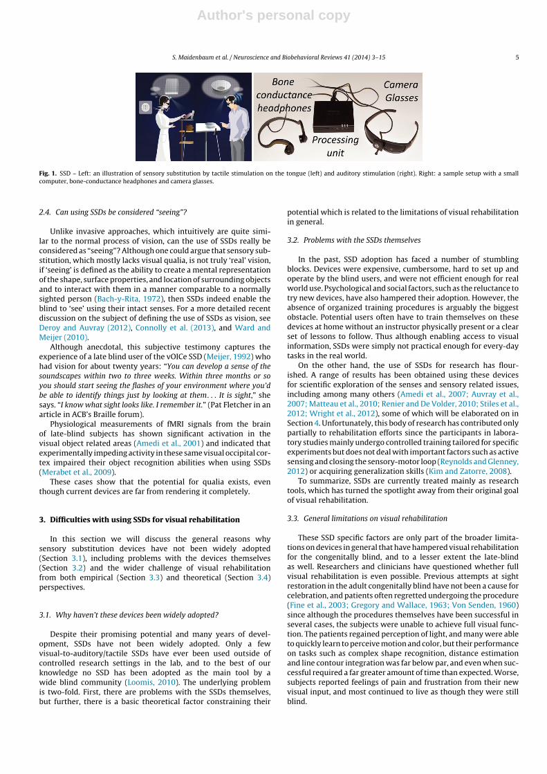
A different approach, known as Sensory Substitution, is designedto convey visual information to the visually impaired by sys-tematically substituting visual information into one of theirintact senses. Sensory substitution devices (SSDs) are non-invasivehuman–machine interfaces which, in the case of the blind, trans-form visual information into auditory or tactile representationsusing a predetermined transformation algorithm (see Fig. 1 forillustration).
The first such structured substitution system is probably Braillereading. This technique, developed originally by Barbier as a meansfor writing and reading in the dark for the French military in theNapoleonic era, was later revised by Louis Braille to enable theblind to read by substituting visual letters with tactile ones. Thiswas further developed in the early 1950s with the development ofautomatic text-to-braille converters such as the Optacon (Goldishand Taylor, 1974).
A highly interesting effort which is often neglected historically,was the Elektroftalm that attempted to electronically transform avisual image into auditory (late 1890s) and tactile (1950s) stim-ulation (Starkiewicz and Kuliszewski, 1965) using one or severalsensors.
These early attempts led to the more organized and method-ological attempts of Paul Bach-y-Rita in the 1970s, whichpositioned him as the pioneer of the extensive use of sensory sub-stitution for research. Bach-y-Rita focused on tactile devices andspecifically a prototype device he named the “Tactile Vision Sen-sory Substitution” (TVSS) which blind users could use for tasks suchas recognizing large letters, catch a ball tossed at them and so on(Bach-y-Rita, 1972).
The work of Bach-y-Rita suggested that these devices couldserve as stand-alone aids for limited daily use, providing other-wise non-existing visual capabilities such as perception of shape,color and location. Additionally, as SSDs are relatively low-cost theycould be made accessible to the majority of the world’s visuallyimpaired population, who as mentioned above primarily reside indeveloping countries and have limited access to advanced medicaltreatment (Held et al., 2011; WHO, 2012).
SSDs have enormous potential for non-invasive rehabilitationfor the majority of the blind. In over 95% of all cases of blind-ness, the problem is not in the visual/occipital parts of the brainbut rather in the eye, retina or the visual pathways (WHO, 2012).In addition, in the subset of cases where the visual pathwaysbetween the ganglion cells and the visual cortex is damaged,approaches that repair the retina would not be able to conveythe information from there to the brain, leaving SSDs as themain potential therapeutic approach. However, despite of sev-eral decades of research, the use of SSDs has hardly exploitedthis vast potential. Before we explore the reasons for this rela-tive failure, and how this might be remedied in the near futurebased on recent theoretical practical and technological advances,it is worth inquiring how ‘seeing’ using SSDs compares to naturalvision.
Author's personal copy
S. Maidenbaum et al. / Neuroscience and Biobehavioral Reviews 41 (2014) 3–15 5
Fig. 1. SSD – Left: an illustration of sensory substitution by tactile stimulation on the tongue (left) and auditory stimulation (right). Right: a sample setup with a smallcomputer, bone-conductance headphones and camera glasses.
2.4. Can using SSDs be considered “seeing”?
Unlike invasive approaches, which intuitively are quite simi-lar to the normal process of vision, can the use of SSDs really beconsidered as “seeing”? Although one could argue that sensory sub-stitution, which mostly lacks visual qualia, is not truly ‘real’ vision,if ‘seeing’ is defined as the ability to create a mental representationof the shape, surface properties, and location of surrounding objectsand to interact with them in a manner comparable to a normallysighted person (Bach-y-Rita, 1972), then SSDs indeed enable theblind to ‘see’ using their intact senses. For a more detailed recentdiscussion on the subject of defining the use of SSDs as vision, seeDeroy and Auvray (2012), Connolly et al. (2013), and Ward andMeijer (2010).
Although anecdotal, this subjective testimony captures theexperience of a late blind user of the vOICe SSD (Meijer, 1992) whohad vision for about twenty years: “You can develop a sense of thesoundscapes within two to three weeks. Within three months or soyou should start seeing the flashes of your environment where you’dbe able to identify things just by looking at them. . . It is sight,” shesays. “I know what sight looks like. I remember it.” (Pat Fletcher in anarticle in ACB’s Braille forum).
Physiological measurements of fMRI signals from the brainof late-blind subjects has shown significant activation in thevisual object related areas (Amedi et al., 2001) and indicated thatexperimentally impeding activity in these same visual occipital cor-tex impaired their object recognition abilities when using SSDs(Merabet et al., 2009).
These cases show that the potential for qualia exists, eventhough current devices are far from rendering it completely.
3. Difficulties with using SSDs for visual rehabilitation
In this section we will discuss the general reasons whysensory substitution devices have not been widely adopted(Section 3.1), including problems with the devices themselves(Section 3.2) and the wider challenge of visual rehabilitationfrom both empirical (Section 3.3) and theoretical (Section 3.4)perspectives.
3.1. Why haven’t these devices been widely adopted?
Despite their promising potential and many years of devel-opment, SSDs have not been widely adopted. Only a fewvisual-to-auditory/tactile SSDs have ever been used outside ofcontrolled research settings in the lab, and to the best of ourknowledge no SSD has been adopted as the main tool by awide blind community (Loomis, 2010). The underlying problemis two-fold. First, there are problems with the SSDs themselves,but further, there is a basic theoretical factor constraining their
potential which is related to the limitations of visual rehabilitationin general.
3.2. Problems with the SSDs themselves
In the past, SSD adoption has faced a number of stumblingblocks. Devices were expensive, cumbersome, hard to set up andoperate by the blind users, and were not efficient enough for realworld use. Psychological and social factors, such as the reluctance totry new devices, have also hampered their adoption. However, theabsence of organized training procedures is arguably the biggestobstacle. Potential users often have to train themselves on thesedevices at home without an instructor physically present or a clearset of lessons to follow. Thus although enabling access to visualinformation, SSDs were simply not practical enough for every-daytasks in the real world.
On the other hand, the use of SSDs for research has flour-ished. A range of results has been obtained using these devicesfor scientific exploration of the senses and sensory related issues,including among many others (Amedi et al., 2007; Auvray et al.,2007; Matteau et al., 2010; Renier and De Volder, 2010; Stiles et al.,2012; Wright et al., 2012), some of which will be elaborated on inSection 4. Unfortunately, this body of research has contributed onlypartially to rehabilitation efforts since the participants in labora-tory studies mainly undergo controlled training tailored for specificexperiments but does not deal with important factors such as activesensing and closing the sensory-motor loop (Reynolds and Glenney,2012) or acquiring generalization skills (Kim and Zatorre, 2008).
To summarize, SSDs are currently treated mainly as researchtools, which has turned the spotlight away from their original goalof visual rehabilitation.
3.3. General limitations on visual rehabilitation
These SSD specific factors are only part of the broader limita-tions on devices in general that have hampered visual rehabilitationfor the congenitally blind, and to a lesser extent the late-blindas well. Researchers and clinicians have questioned whether fullvisual rehabilitation is even possible. Previous attempts at sightrestoration in the adult congenitally blind have not been a cause forcelebration, and patients often regretted undergoing the procedure(Fine et al., 2003; Gregory and Wallace, 1963; Von Senden, 1960)since although the procedures themselves have been successful inseveral cases, the subjects were unable to achieve full visual func-tion. The patients regained perception of light, and many were ableto quickly learn to perceive motion and color, but their performanceon tasks such as complex shape recognition, distance estimationand line contour integration was far below par, and even when suc-cessful required a far greater amount of time than expected. Worse,subjects reported feelings of pain and frustration from their newvisual input, and most continued to live as though they were stillblind.
Author's personal copy
6 S. Maidenbaum et al. / Neuroscience and Biobehavioral Reviews 41 (2014) 3–15
3.4. Theoretical neurobiological basis for pessimism concerningvisual rehabilitation
Why haven’t these treatments been successful? Why can’t thesubjects learn to see? To answer these questions we will take a stepback, and look at the larger neuroscience picture of how our brainprocesses sensory information, and at recent alternative models inthis field, many of which derive from the findings mentioned in Sec-tion 3.2 and acquired using SSDs, before returning to our discussionof the practical usage of SSDs.
In traditional neuroscience, the common view is that the humanbrain is divided into the “visual cortex”, the “auditory cortex”and so on according to the sensory modality that elicits it, andinto higher-order multisensory areas integrating information fromthese unimodal cortices (the sensory division-of-labor principle;Zeki, 1978). The vast majority of textbooks emphasize this organi-zational principle explicitly and implicitly. However, over the pastdecade there have been a growing number of articles suggestingthat this view may not be fully accurate, a point which will be elab-orated on further in this review in Section 4.1 (Amedi et al., 2001,2007; Merabet and Pascual-Leone, 2010; Pascual-Leone et al., 2005;Pascual-Leone and Hamilton, 2001; Pascual-Leone et al., 2011;Reich et al., 2011a; Renier et al., 2013).
It is well established that the ‘visual’ cortex of the blind becomesplastically recruited to process other modalities and even cognitivetasks such as language and memory (reviewed also in Frasnelli et al.,2011; Merabet and Pascual-Leone, 2010). Many of these changesstart to occur within days following the onset of blindness (Pascual-Leone et al., 2005), and therefore affect not only the congenitallyblind but also, though probably to a different extent, early andlate blind individuals (Cohen et al., 1999; Lacey and Sathian, 2008;Sathian, 2005).
This plasticity may nevertheless be a double-edged sword. Onthe one hand, it helps the blind to better cope with blindness bysupporting compensatory capabilities (Amedi et al., 2003, 2004;Bedny et al., 2011; Gougoux et al., 2005; Rauschecker, 1995; Roderand Rosler, 2003) but at the same time, it may interfere with sightrestoration efforts by altering the visual cortex’s original functions.This means that attempts of rehabilitation pose risks not only whenthey fail, but also when they succeed. Even if we could somehowcause a plastic reorganization of the vision-deprived occipital cor-tex for processing the visual input, it might damage the use of thesesame areas for tasks customarily tapped for this purpose, poten-tially blocking habits and skills the individual has learned to relyon or impairing functions for which these areas were dedicated,such as memory. This problem impinges not only on the congeni-tally blind but to a lesser extent on the late blind as well, when theirformerly “visual” areas are plastically partially recruited for othernew tasks (Sathian and Lacey, 2007).
Another though not mutually exclusive mechanism often usedto explain the failure of medical restoration cases in the congeni-tally blind is the existence of “critical periods” in early childhood inwhich the brain is particularly plastic, during which lack of visionmay prevent the proper functional specialization and developmentof many of these visual regions. The watershed works of Hubel andWiesel (1970) on the visual system of cats made this view one ofthe most basic tenets in visual research; namely that the visual sys-tem cannot regain function if infants cannot see during their firstyears of life.
This may account for some of the failures of the medical casesmentioned above. In these cases of failed sight rehabilitation, itseems as if the gained visual input was made available to a brainthat was wholly unpracticed at analyzing and interpreting thisinput, and the visual experience gained at this stage without super-vised explicit training, and in contrast to normal development, maycome too little or too late.
In conclusion, the main explanations for sight restoration fail-ures have to do with missing “critical periods” required for thedevelopment of visual areas in the brain, and/or the plastic recruit-ment of the occipital cortex for other, non-visual tasks in lateand early blindness. Together, these features may impede the re-emergence of visual abilities in the newly sighted in adulthood, andrender attempts at visual restoration very difficult and limited.
4. The basis for some optimism for visual rehabilitation
In this section we discuss the shift from pessimism to poten-tial hope for visual rehabilitation. We review changes in theoriesof brain organization (Section 4.1) and their implications for visualrehabilitation (Section 4.2). We then review recent results indicat-ing that the substituted information is integrated into a sharedsensory framework (Section 4.3) and discuss recent behavioralachievements using SSDs (Section 4.4). Finally, we explore newresults in the field of visual rehabilitation using other rehabilitationapproaches and their potential for visual rehabilitation (Section4.5).
4.1. A different view of brain organization and re-organization
It is well established that the visual cortex in most sightedhumans has a hierarchical organization and is comprised of dif-ferent functional areas, each processing different aspects of vision.For example the most fundamental large-scale division of labor ofthe visual cortex is between two functional processing streams forobjects, the dorsal ‘how and where is it?’ and the ventral ‘whatis it?’ streams. Moreover, even within the stream specialized forprocessing object identity, different functional areas show prefer-ential activation for different object categories. For example, theFusiform Face Area (FFA) shows preference for processing faces, theMiddle Temporal gyrus (MT) for visual motion, and the Visual WordForm Area (VWFA) for script reading and visual representation oflanguage.
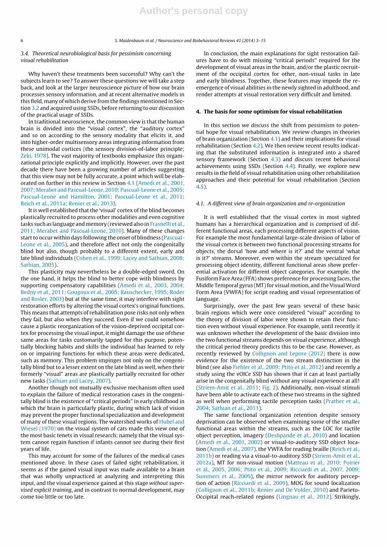
Surprisingly, over the past few years several of these basicbrain regions which were once considered “visual” according tothe theory of division of labor were shown to retain their func-tion even without visual experience. For example, until recently itwas unknown whether the development of the basic division intothe two functional streams depends on visual experience, althoughthe critical period theory predicts this to be the case. However, asrecently reviewed by Collignon and Lepore (2012) there is nowevidence for the existence of the two stream distinction in theblind (see also Fiehler et al., 2009; Ptito et al., 2012) and recently astudy using the vOICe SSD has shown that it can at least partiallyarise in the congenitally blind without any visual experience at all!(Striem-Amit et al., 2011; Fig. 2). Additionally, non-visual stimulihave been able to activate each of these two streams in the sightedas well when performing tactile perception tasks (Prather et al.,2004; Sathian et al., 2011).
The same functional organization retention despite sensorydeprivation can be observed when examining some of the smallerfunctional areas within the streams, such as the LOC for tactileobject perception, imagery (Deshpande et al., 2010) and location(Amedi et al., 2001, 2002) or visual-to-auditory SSD object loca-tion (Amedi et al., 2007), the VWFA for reading braille (Reich et al.,2011b) or reading via a visual-to-auditory SSD (Striem-Amit et al.,2012a), MT for non-visual motion (Matteau et al., 2010; Poirieret al., 2005, 2006; Ptito et al., 2009; Ricciardi et al., 2007, 2009;Summers et al., 2009), the mirror network for auditory percep-tion of action (Ricciardi et al., 2009), MOG for sound localization(Collignon et al., 2011b; Renier and De Volder, 2010) and Parieto-Occipital reach-related regions (Lingnau et al., 2012). Strikingly,

Author's personal copy
S. Maidenbaum et al. / Neuroscience and Biobehavioral Reviews 41 (2014) 3–15 7
Fig. 2. “Visual” ventral and dorsal streams – fMRI map of the dorsal/ventral visual pathway division of labor in adult congenitally blind participants using SSDs (Striem-Amitet al., 2011).
even listening to sound echoes can activate the visual rather thanthe auditory cortex in blind echolocation experts (Thaler et al.,2011).
The LOC, MT and VWFA provide the most detailed examples (seeFig. 3). LOC, the lateral occipital cortex, was first shown to be acti-vated in both tactile object recognition (TOR) and visual objectionrecognition in the sighted, suggesting that part of LOC (LOtv) might
Fig. 3. Meta-modal research – Top: results obtained using SSD as sensory input. Left:LOC activated for SSD object recognition (Adapted from Amedi et al. (2007)). Right:VWFA activated for SSD letter reading (Adapted from Striem-Amit et al. (2012a)).Bottom: results obtained using tactile sensing as sensory input. Left: LOC activatedbilaterally for left hand tactile object recognition (Adapted from Amedi et al. (2010)).Right: VWFA activated for Braille reading (Adapted from Reich et al. (2011a)).
actually be a sensory independent task operator for decipheringthe geometrical shape of 2D and 3D objects (Amedi et al., 2001).In particular, the LOtv was activated by visual and tactile shaperecognition tasks but not by object recognition by sound usingassociations (Amedi et al., 2002). This claim was paralleled by thetheoretical framework suggested that same year by Pascual-Leoneand Hamilton of the brain as a metamodal operator (Pascual-Leoneand Hamilton, 2001). Other groups soon showed that vision andtouch share shape information within the LOC (James et al., 2002).Further research into the multisensory nature of LOC revealed itsactivation during mental imagery (Zhang et al., 2004) and recogni-tion of familiar tactile objects (Lacey et al., 2010) and activation fortactile shape over texture (Stilla and Sathian, 2008). More recentresearch has confirmed these findings by showing peak activa-tion in the LOC for TOR without visual experience (Amedi et al.,2010) and by findings on the retrieval of shape information in thesighted, late blind and congenitally blind using visual-to-auditorySSDs (Amedi et al., 2007, 2010; Lacey et al., 2009).
Several different experimental approaches have shown that theMT or Middle Temporal gyrus, also known as V5, the area that pro-cesses visual motion is activated for tactile motion in the absenceof vision (Ptito et al., 2009) and electro-tactile motion is perceivedon the tongue via the TDU SSD (Matteau et al., 2010).
Similarly the VWFA, a ventral visual area that processes visualwritten language in the sighted, is used for reading Braille, which

Author's personal copy
8 S. Maidenbaum et al. / Neuroscience and Biobehavioral Reviews 41 (2014) 3–15
is a tactile process, and is also the location for the peak of selectiveactivation to Braille words in the congenitally blind (Reich et al.,2011b). These results were further expanded by Striem-Amit et al.(2012a) who showed that the VFWA is activated when using thevOICe SSD for reading regular letters via an auditory soundscape.The VWFA was also shown to have exactly the same selectivity forletter strings vs. ANY other category in both vision and visual-to-auditory SSDs even in subjects who have never seen.
All of these results contribute to a growing body of evidenceaccumulating over the last decade that challenges the canonicalview of the sensory-specific brain. This evidence demonstrates thatin both sighted and blind individuals the occipital visual cortex isnot purely visual and that its functional specialization is indepen-dent of visual input (reviewed in Reich et al., 2011a; Ricciardi andPietrini, 2011; and detailed below), despite showing a clear prefer-ence for the visual modality. This in turn has led to the hypothesisthat the brain is task-oriented and sensory-modality independent(Reich et al., 2011a,b; Striem-Amit et al., 2011), or in other words a“task machine”. Thus although the brain regions each show a pref-erence for a specific modality or set thereof, they can still performtheir specific task if they receive relevant information, regardless ofthe sensory input channel through which this information reachedit.
Furthermore, as discussed above, recent evidence from the fullycongenitally blind has shown that in some cases the same special-ization emerges even without any visual experience or memories(Amedi et al., 2007; Collignon et al., 2011b; Fiehler et al., 2009;Mahon et al., 2009; Matteau et al., 2010; Ptito et al., 2009; Reichet al., 2011b; Striem-Amit et al., 2012a,b), and can occur rapidlyonce the brain is trained to interpret the relevant information,suggesting that cortical functional specialization can be attributedat least partially to innately determined constraints (Striem-Amitet al., 2012b). Support for the task-machine brain hypothesis comesfrom findings on the auditory cortex in the deaf animals as well(Lomber et al., 2010) as well as in at least two anecdotal singlecase studies testing causality by disrupting the activity in theseregions in the blind (Hamilton et al., 2000; Merabet et al., 2009).For reviews on this topic see (Bavelier and Hirshorn, 2010; Dormaland Collignon, 2011; Reich et al., 2011a).
This view of brain organization is consistent with severalsimilar theories, such as the Metamodal (Pascual-Leone andHamilton, 2001) and Supramodal (Kupers et al., 2011; Pietrini et al.,2004; Ricciardi and Pietrini, 2011) theories of brain organization.Although the specific differences in definition between these the-ories are beyond the scope of this review, all three offer a similarpositive potential for visual rehabilitation.
These findings suggest that while indeed showing some pref-erence for information from a specific sense, most higher ordervisual areas might be task-based and not sensory-based. However,it is worthwhile noting that there is still considerable controversywhether early retinotopic areas such as V1 which are linked directlyto the sensory organs of one modality, also behave as task machinesor whether this is limited to regions more distant from directinput modalities. It is clear though that these area do indeed showcrossmodal organization and plasticity, which makes it especiallyimportant to continue to test whether this task specific metamodalorganization can occur in them after training.
An additional bias inherent to these experiments is that theyhave mostly been conducted on populations of the congenitallyblind. It is thus unclear how these data relate to the late blind. Thecause for this may have been the attempt to avoid confounds suchas visual imagery, and the difference caused by possible criticalperiods of development which the late-blind have experienced butthe congenitally blind have not. However, while the findings fromcomparative studies indeed tend to indicate differences in resultsbetween the sighted, congenitally blind and late-blind, they all still
exhibit the basic existence of task based neural activation (Thaleret al., 2011; Amedi et al., 2007; Renier and De Volder, 2010). On theother hand, several conflicting results suggest that for the late blindthe preservation of functional specialization for other senses mayonly be on a level comparable to the results from sighted subjects(Dormal et al., 2012).
4.2. A neurobiological basis for optimism for visual rehabilitation
If the hypothesis of the highly flexible task-oriented sensory-independent/metamodal/supramodal brain is borne out, theabsence of visual experience should not limit the task-specialization of the visual system, despite its recruitment forvarious functions in the blind, and the visual cortex of the blind maystill be able to retain its functional properties using other sensory-modalities. This is very encouraging with regard to the potentialfor visual rehabilitation, and may form the theoretical basis for thenew empirical evidence of success in rehabilitation which will bediscussed below.
Note that many of these results were achieved using SSDs, andin particular after training using SSDs, as part of the research resultsmentioned in Section 3.2. These results also suggest that SSD train-ing might be useful for shaping the brain to interpret input comingfrom other SSDs and devices, a point we elaborate upon in Sections5.2 and 5.6.
4.3. The substituted sense shares sensory space
Adding to this optimism, Ward & Wright have recently used anaudio–visual mismatch paradigm conveyed by visual informationand visual-to-auditory sensory substitution information to showthe existence of a shared mental visual workspace between them(Wright et al., 2012). Similarly, it has recently been shown (Levy-Tzedek et al., 2012b) that information acquired through SSDs canbe integrated into the shared multisensory perceptual grasp of ourenvironment using a rotation-reaching task using information fromvision and from a vision-to-auditory SSD. These results hint at amental transfer suggestive of a shared sensory representation.
4.4. Additional new behavioral results
But if the suggestions of the previous two sections are true, whyhave the blind been unable to regain visual function using previoustypes of visual rehabilitation? The surprising answer is that withtime, and to a certain very limited extent, they actually were able toregain more visual functions than previously expected. While stillvery far from “normal” vision, these achievements offer the userspractical tools and skills for dealing with a wide variety of otherwiseinaccessible visual-based tasks. In this section we discuss this claimin the light of recent results with SSDs, and then in the next sectionbriefly report similar recent evidence using other forms of visualrehabilitation.
A small number of individuals who have used SSDs extensivelyhave been able to acquire some practical every-day life skills, andadvanced visual functions such as depth perception (Ward andMeijer, 2010) and recent behavioral results with SSDs on largernumbers of subjects who had less experience with the devices havesurpassed by far the theoretical limit set by previous theories.
In recent experiments the performance of some blind users(Striem-Amit and Amedi, 2012) even exceeded the threshold forthe World Health Organization (WHO) definition of blindness onthe Snellen acuity test, showing that at least “legally” the subjectsare no longer functionally fully blind (see Fig. 4 for more examples),but rather on par with the severely visually impaired. These resultsare consistent with findings on blindfolded participants with noprior SSD experience (Haigh et al., 2013).

Author's personal copy
S. Maidenbaum et al. / Neuroscience and Biobehavioral Reviews 41 (2014) 3–15 9
Fig. 4. Examples of behavioral tasks performed using SSDs.
Furthermore, it has recently been shown (Striem-Amit et al.,2012a) with a small number of congenitally blind individuals that70 h of training with the vOICe SSD not only enable the blind to betaught to read using the device, but also enables them to discrimi-nate a wide set of categories (letters, textures, faces, houses, objects,body shapes, geometrical shapes), and perform difficult tasks suchas recognizing facial expressions.
Even after only brief training, a small number of congenitallyblind individuals have been able to recognize patterns (Poirieret al., 2007), perform motion discrimination and tracking tasks(Chekhchoukh et al., 2011; Ptito et al., 2009), extract depth cuesand estimate object distance (Renier and De Volder, 2010), matchsocks by color (Bologna et al., 2009), and recognize. Blind users haveperformed basic navigational tasks such as walking down a corri-dor and opening doors, detecting and avoiding obstacles duringeffective navigation within a human-sized obstacle course (Chebatet al., 2011), walking along a colored line (Bologna et al., 2009), rec-ognizing different virtual routes (Kupers et al., 2010) and locations,objects and landmarks encountered on the way while navigatingreal-world streets such as buildings, crosswalks, fences and trees.They have used SSDs for finding an object in a room, differentiatingbetween different types of fruit, and locating light sources (Amediet al., 2007; Bologna et al., 2010; Capalbo and Glenney, 2009;Durette et al., 2008; Reynolds and Glenney, 2009). The blind userswere even able to perform “eye”-hand coordination such as locat-ing targets, pointing to them and reaching for them (Levy-Tzedeket al., 2012a), placing rings on a cone, winning a game of miniaturebowling, recognizing an unoccupied chair, etc. (Maidenbaum andAmedi, 2012).
While there is relatively little published research on thelate blind, their behavioral achievements using SSDs seem tomatch those attained by the congenitally blind, and indeedsome of the best achievements were made by members ofthis group (Maidenbaum and Amedi, 2012; Ward and Meijer,2010).
Nevertheless these behavioral achievements have mostly beenobtained in lab settings as part of research programs focusedon obtaining answers to specific experimental questions and notdealing with visual rehabilitation. Thus, they can be seen as a proof-of-concept that far better results could be achieved with a programdedicated to rehabilitation.
It should also be emphasized that these results are of coursestill far from that which can be obtained with full vision, but are farbetter than previously expected, and show that SSDs have concretepractical uses.
4.5. Insights from other types of visual restoration
These results are complemented by recent reports from stud-ies using other types of visual restoration. Researchers at the“Prakash” (Sanskrit for “Light”) project in India that aims to restorevision through conventional treatment generally unavailable inthe third world such as cataract removal and corneal transplants,have reported behavioral results which seem far more promisingthan expected for these patients, including adults. While resultsimmediately after surgery are typical of the 20th century outcomesdescribed in Section 3.3 that involve gaining some basic visualskills but failing to gain others, follow-up exams conducted sev-eral months later note a significant improvement in many of theseskills over time, such as the ability to count 3D objects, line contourintegration, name an object in a noisy image, count overlappingobjects, etc., all of which provide a significantly more optimisticoutlook (Held et al., 2011; Ostrovsky et al., 2009), and challenge thestrict view that critical periods are completely irreversible (Thomas,2011).
Specifically, in a case-study conducted in 2006, Sinha reported(Ostrovsky et al., 2006) that SRD, a patient who was blind untilthe age of 12 and whose initial performance seemingly echoed thepessimistic view, had mastered a wide variety of visual tasks 20years later, enabling her to function independently as a person withlow-vision. Thus, the combination of time and training may enablethe newly sighted to learn to see beyond the level once thoughtpossible.
Another advancing field with similar results is the use of retinalimplants. Patients implanted with such a prosthesis have beenable to perform tasks such as reading letters, locating objects,etc. beyond the expected ability from the pessimistic theory,though still at a level far from satisfactory (Dagnelie, 2012), and todate there are very few congenitally blind individuals with suchimplants.
It should be noted that in both of these cases success was limitedand time consuming. While there has been no direct comparison

Author's personal copy
10 S. Maidenbaum et al. / Neuroscience and Biobehavioral Reviews 41 (2014) 3–15
Fig. 5. Practical visual rehabilitation using SSDs: from pessimism to potential hope.
between these subjects and SSD users, initial comparison of thereported results indicate that SSDs may currently hold the upperhand both in terms of accomplishments and in terms of the timeneeded to achieve this milestone. As elaborated on in Section 5.6we believe that a synergistic attempt which would use SSDs withthese patients before and after they undergo surgery could increasetheir success rate significantly.
5. Outlining several future practical steps
There are several additional factors which we believe have thepotential to change the current status of SSD usage in clinical sett-ings. These include technological advances that will make thesedevices both more user friendly and upgrade their capabilities(Section 5.1), advances in training programs (Section 5.2), har-nessing the power of the web, through the use of virtual (Section5.3) and real-world simplified and safe environments to preparefor real world experiences (Section 5.4), making better use ofthe advantages offered by active sensing (Section 5.5) and finallysynergistically combining SSDs and other methods of visual reha-bilitation (Section 5.6). These factors are summarized in Fig. 5.
5.1. The potential of future devices and technological advances
We believe that the recent achievements detailed in Section 4.4are only the first step toward fulfilling the potential of these devices,and that current technological advances will play a great part in thisprocess.
Recent technological advances are continuously contributing tothe three main modules which comprise an SSD. These includeinput sensors capturing visual information, a processing unit thatextracts data and generates the representation, and an outputhuman machine interface that portrays the data to the blind user.Newly available input sensors offer SSD users new parameters suchas depth and color information. For example, depth informationcan be captured using stereo imaging (Akhter et al., 2011), 3D IRcameras such as Microsoft Kinect (Zöllner et al., 2011) and TOFimagining (Callaghan and Mahony, 2010; Zeng, 2012) and color viastandard cameras (Deville et al., 2009; Levy-Tzedek et al., 2012b).In addition, as a general trend, higher quality cameras are becomingcheaper, smaller and more widely available due to their integrationinto smartphones (Akhter et al., 2011; Blum et al., 2012), including
depth cameras (Firth, 2013). Initial results using color by SSDs suchas EyeMusic (Levy-Tzedek et al., 2012a,b) or SeeColOr (Bolognaet al., 2009) now enable tasks ranging from simple color discrim-ination, to tasks which require more complex usage of the colorinformation such as reaching for the correct object, following theright line or even matching socks.
On the processing unit front, two main trends are worth mention-ing. The software trend includes the integration of computer visionalgorithms (HVSS; Tan et al., 2010; Zeng, 2012) and machine learn-ing techniques (Zeng, 2012) to filter the captured signals, whichhelps reduce the mental load, and enables the extraction of impor-tant objects and signs (Lescal et al., 2013; Tanveer et al., 2012; Zeng,2012) as well as their simplification for user convenience. This soft-ware can be adapted from external packages, by taking advantageof research in other fields such as image processing and computervision, especially when given additional visual information aboutthe scene such as depth maps. In addition, there are new attempts tobetter refine the transformation algorithms based on recent litera-ture in psychophysics to increase intuitiveness (Stiles and Shimojo,2013) and computational tools such as genetic algorithms (Wrightand Ward, 2013).
The hardware trend is mainly affected by the flourishing processof component miniaturization and the fact that smartphones offer-ing a simple programming API are starting to be very widespread.This provides developers with a powerful yet small and elegantmobile computing platform that can be adapted to veteran SSDsby combining them with smartphone implementations (the vOICe,the EyeMusic) or new SSDs that have smartphones as their mainplatform (the HVSS). Moreover, improvements in user accessibility(such as native speech and hand gesture input APIs) make thesedevices easier to operate by blind users without any sighted aid.New platforms such as Google Glass, which consists of a glasses-mounted augmented reality platform, and Or-Cam, which offersaugmented reality information to the visually impaired, could alsoserve as a dedicated unobtrusive platform for future SSD develop-ment.
The output human machine interface is the module that is cur-rently the furthest from meeting expectations, especially for tactileSSDs. That said, better haptic feedback is being produced in manyprojects (among many others see Zeng, 2012; Schorr et al., 2013;Visell, 2009). In one interesting project, electrical actuators, simi-lar to those used in the expensive TDU for tongue stimulation, have

Author's personal copy
S. Maidenbaum et al. / Neuroscience and Biobehavioral Reviews 41 (2014) 3–15 11
been recreated into a low resolution open-source SSD, thus increas-ing their accessibility (Dublon and Paradiso, 2012). Two interestingprojects currently under development which hold potential forfuture interfaces are the Senseg (2012) and the Disney TeslaTouch(Bau et al., 2010), which attempt to augment touch screens byenabling the user to haptically sense the visual information dis-played on them, which increases accessibility to parameters suchas contour, texture and basic 3D information.
In the auditory domain, veteran SSDs use headphones whichhave become cheaper, less obtrusive and of better quality. It isworthwhile mentioning on this front the availability of bone-conductance headphones which keep the ears free to receive otherauditory input. There has been greater use of stereo channels tomap objects on each side of the image (Tan et al., 2010) and direc-tional sound for better x-axis mapping, and in some cases even fulllocation information (Bujacz et al., 2012). The main hurdle facingauditory SSDs is the auditory pleasantness of the cues, which whileimproving, are still far from satisfactory, and remain a major reasonwhy users do not adopt these devices in everyday life (Wright andWard, 2013).
Moreover, some SSDs are opting for a multisensory approachwhere the visual data is portrayed in both the auditory and tac-tile modalities in order to create a redundant representation whichcould potentially lead to higher performance (Stein and Meredith,1993; Zeng, 2012) or provide the most appropriate information tothe optimal channel by scenario recognition.
As new technologies emerge and technologies in the proto-type phases mature, we believe that SSDs will have better visualprocessing algorithms and output interfaces, smaller apparatusesand at lower prices, thus removing many of the obstacles towidespread use of these devices have faced. However, we believethat without a concentrated effort to take these developments fromthe lab to the clinic, and without proper training and coordinationwith a large community of blind individuals, all these advances willremain more of a curiosity that attracts media exposure but remainsin use mainly in labs or by a very small number of people, as hasbeen the fate of many SSDs developed over the last ten years.
5.2. The importance of training
The profound importance of training is one of the clearestemerging and relatively new insights from working with patientsusing all types of visual rehabilitation including SSDs (Reynoldsand Glenney, 2012; Striem-Amit et al., 2012b; Ward and Meijer,2010), retinal implants (Dagnelie, 2012) and sight restoring surgery(Ostrovsky et al., 2006; Putzar et al., 2012; Xu et al., 2012).
Seeing is not as intuitive as is customarily believed, and the pro-cess of learning to process unconventional visual information canbe long and difficult. The lack of basic understanding of visual prin-ciples taken for granted by the sighted is obvious after minutes oftalking to any blind person. These are especially important for thecongenitally and very-early blind who simply do not have theseconcepts, but is also true for the late blind trying to cope with atyp-ical and degraded input such as input arriving from a prosthesis orfrom an SSDs. Concepts such as the fact that shading changes imageseem foreign to them need to be learned, since shadows do nothave an intuitive correlate in other modalities. The concept of visualperspective makes no sense to someone who is used to relying onauditory representations. Even the concept of size, and especiallythe fact that perceived size changes with distance, is odd despite theauditory corollary of signal strength. Transparent objects like win-dows or light bulbs baffle our subjects and the concept of mirrorsevoke fear, especially the idea of seeing themselves (Maidenbaumand Amedi, 2012). For the congenitally blind, some visual perceptssuch as color which have no other sensory correlate require spe-cial focus when taught. The late blind, on the other hand, may have
a better idea of what the visual information they are receiving issupposed to look like, but the transformation can be difficult, espe-cially for visual rules they used subconsciously while still sighted.All of these rules and concepts need to be taught and explained.
Thus subjects who have received serious longitudinal trainingnot only learn to use the devices far better, but are able to bet-ter process the visual information and learn visual principles. Thislive guidance is also important on an emotional level when dealingwith concepts such as how others see you, and on a motivationallevel, especially when at first the stimuli are confusing and theirinterpretation is tiring.
There are several reports in the literature documenting howsubjects who struggled to learn to see, and received patientand long-term support from people around them; such as SRD(Ostrovsky et al., 2006) and PF (Ward and Meijer, 2010). Whenworking with our subjects, these improvements and the progressfrom one training session to the next are plain to see. Furthermore,when the training is structured and focused our subjects learn bet-ter and faster. Thus we believe that the creation and disseminationof such longitudinal training programs could significantly enhancethe potential outcomes of visual rehabilitation.
To be maximally effective, we suggest that this training shouldinclude several rigorous first steps in which the basic functions ofthe device should be learned in a supervised fashion, followed bya series of more dynamic lessons utilizing tools such as dedicatedvirtual environments, games, active sensing and real environmenttraining (see below). These lessons should incorporate a gradualincrease in level of difficulty from static stimuli in the first hoursto dynamic virtual games, and finally to basic real world tasks.They should also include dedicated modules for important objectcategories such as faces and home objects, and specific real-lifetasks such as navigation and object location. Some of these mod-ules should be an early part of the training process, to give users afeeling of accomplishment and the way they can acquire practicalskills with the device. While there have already been some attemptsat long-term training programs (Striem-Amit et al., 2012b), webelieve that such a program will require a long period of refinement,similar to the evolution of training programs for the traditionalwhite-cane during the twentieth century (Welsh, 1981).
5.3. The importance of online and virtual training
One of the key problems with the above-mentioned trainingprograms is their cost and the effort involved in them; e.g., thepersonnel needed for such training efforts. We suggest that even-tually current Orientation & Mobility resources could be turned tothis goal but even they are currently insufficient, especially in thedeveloping world in which most of the blind reside.
A possible solution could be to incorporate online and virtualtraining to the training protocols, as are currently in developmentfor devices such as the white cane (Lahav, 2012) and novel devicessuch as the EyeCane (Maidenbaum et al., 2012, 2013). In a recentstudy, a virtual environment was shown to be as efficient in teach-ing the spatial layout of a building as more classic sessions withinstructors (Merabet et al., 2012). The blind and visually impairedcould connect to this online training from the comfort and safetyof their own homes, using cheap smartphones or cheap comput-ers. This will help them gain experience both in using the SSDsin various scenarios, and in practicing visual principles in dedi-cated lessons which would also be applicable to the newly visuallyrestored.
Another important aspect of virtual training is the ability toadd gamification elements, which can both increase user moti-vation, and enable implicit feedback to make sure the user isusing the SSD correctly; for instance when presented with threeobjects, instead of describing the stimuli to an instructor which is

Author's personal copy
12 S. Maidenbaum et al. / Neuroscience and Biobehavioral Reviews 41 (2014) 3–15
Fig. 6. SSDs and VRD – Left: the three different layers of SSDs: an input sensory device, a processing unit and an output human machine interface. Right: a sample VRDcombining three output modalities conveying the captured visual information using skin stimulation, auditory output and a retinal prosthesis. Copyright © 2013 SecondSight Medical Products, Inc.
manpower-intensive and requires explicit explanations, oranswering a long series of questions about the stimuli, there couldbe a game where the user must bring object 1 to object 2 whileavoiding object 3 – a task that requires perception and recognitionof all three objects and their spatial properties achieving the sameresult implicitly.
5.4. Taking the step to simplified environments and consequentlythe real-world
Another important step involves bridging the gap between usingSSDs in controlled conditions and in real world settings which arefar noisier and more difficult to interpret. This level of noise, causedby the many other objects in a visually rich real environment, ishighly confusing, and can produce sensory overload in the user. Wesuggest this gap can be bridged by a combination of several differenttools. These include (1) dedicated real-world “safe environments”for training, similar to those currently used in classic orientationand mobility training. In these environments SSD users will stillexperience the sensory overload, but because they are safer, theycan learn to deal with the excess information and focus their atten-tion on relevant details, similar to the way children learn to filtertheir senses when growing up. (2) Since virtual environments areeasier and cheaper to create, dedicated virtual environments canaugment the real ones described above. These environments can bedesigned to increase gradually in difficulty. This may lead to a bet-ter learning curve, simulate a wider variety of environments, andlet users practice from their own homes. (3) Input-simplificationalgorithms that will filter out a large part of the noise using tech-niques such as cartoonization or a depth camera to filter out inputbeyond a certain range.
5.5. The importance of active sensing
As mentioned above, most tasks performed with SSDs havebeen highly controlled laboratory trials. While such trials enableclearer results, they are often static, and participants thus losethe advantages of closing the sensory-motor loop (Held, 1965).Using SSDs with an active sensory-motor loop should significantlyimprove behavioral results. For example Ahissar et al. have shownthat human subjects learning to use “whiskers” enter an iterativemotor-sensory process that gradually converges on stable percepts(Horev et al., 2011; Mitchinson et al., 2007). Subjects who used theEyeMusic dynamically were able to accurately reach for an objectperceived via the SSD, just as they were able to reach for it visu-ally (Levy-Tzedek et al., 2012a). This information was even shared
between the visual and substituted information (Levy-Tzedek et al.,2012b). The advantages of a more active sensory-motor paradigmwere also shown to boost SSD training (Reynolds and Glenney,2012).
5.6. Augmenting retinal prostheses and residual vision – acombined vision-rehabilitation device (VRD)
The recent advances in other approaches to visual restorationprovide interesting opportunities. We recently suggested (Reichet al., 2011a) a post-operation system combining an SSD with reti-nal prostheses (“Vision rehabilitation device”; VRD, illustrated inFig. 6). This system will include a camera consistently capturingimages of the surroundings and a processing unit. This unit con-verts the visual information into (i) an auditory SSD representationand (ii) a neural stimulation conveyed by the prostheses’ elec-trodes. Information about the surroundings would thus be receivedin parallel from the prostheses as well as from the SSD. In such adevice, the SSD would serve as a “sensory interpreter” providingexplanatory input for the visual signal arriving from the invasivedevice. This dual synchronous information is expected to signifi-cantly increase the rate of rehabilitation. At a later stage the SSDcan be used to provide input beyond the maximal capabilities ofthe prostheses or capabilities it does not possess such as addingcolor information via EyeMusic (Levy-Tzedek et al., 2012a). Thisconcept is also applicable to cases of late sight-restoration such asthe “Prakash” project participants (Ostrovsky et al., 2009).
This concept could be further expanded by augmenting theresidual vision of low vision users, who will use their residual visionand not the prosthesis, with the visual parameters provided by theSSD. This possibility is supported by the subjective descriptionsof CC, a woman who has residual eyesight and uses a visual-to-auditory SSD on a daily basis claims that her SSD perception andher visual perception share a single space, but that the SSD pro-vides greater detail such that a more complex perception of thescenery is generated (Ward and Meijer, 2010). The findings men-tioned in Section 4.3 that the substituted sense has a shared spacewith other senses, also make it possible to use the location informa-tion in motion even without awareness (Levy-Tzedek et al., 2012b;Wright et al., 2012). Tactile-Sight, a recent example of such a devicefor low-vision users, augments low vision with depth information(Cancar et al., 2013).
In a more futuristic view, the integration of special input sensorsto the VRD may further expand visual augmentation, as demon-strated in a recent interesting study by Alsberg (2012) who attacheda hyperspectral camera to the vOICe SSD, creating an SSD for

Author's personal copy
S. Maidenbaum et al. / Neuroscience and Biobehavioral Reviews 41 (2014) 3–15 13
sonifying chemical compounds, which in turn enabled him touse audition to distinguish identical looking chemical substances(sucrose vs. potato powder).
This approach, however, has its own drawbacks. For exam-ple, such information might cause sensory overload, competitionbetween the senses, or even simply tire users faster. However,we suggest that the advantages should outweigh these potentialdisadvantages, especially when balanced correctly in a structuredtraining program.
6. Conclusion and future directions
We have presented a variety of reasons to believe SSDs nowhave the potential to be re-applied successfully for visual rehabili-tation based on recent theoretical advances and current optimisticbehavioral results. We believe that the steps toward this impor-tant goal must include the creation of structured training programs,dedicated virtual and real world training environments, the devel-opment of the next generation of SSDs using state-of-the-arttechnology, and most importantly, the promotion of scientificefforts directly aimed at optimizing the visual rehabilitation pro-cess using SSDs, which has taken the back seat in the last fewdecades in favor of their use for research.
It should be made clear that the potential beneficiaries are notyet fully able to use these devices in the real world and that thedevices themselves are not yet fully ripe for full use. Rather, we wishto highlight the growing possibility for such use in the near future,both as a stand alone approach but primarily when combined withother approaches such as retinal implants (VRD) or with computervision algorithms. We believe that these devices could be exploitedfor much more practical use if efforts are made in this directionand have outlined several important factors we believe need to befurther developed as the next steps toward this goal.
Acknowledgments
This work was supported by a European Research Council grantto A.A. (grant number 310809), The Charitable Gatsby Foundation,The James S. McDonnell Foundation scholar award (to AA; grantnumber 220020284), The Israel Science Foundation (grant numberISF 1684/08), The Edmond and Lily Safra Center for Brain Sciences(ELSC) Vision center grant (to AA); SA was supported by a scholar-ship from the Israel Ministry of Science.
References
Ahuja, A., Dorn, J., Caspi, A., McMahon, M., Dagnelie, G., Dacruz, L., Stanga, P.,Humayun, M., Greenberg, R., 2011. Blind subjects implanted with the ArgusII retinal prosthesis are able to improve performance in a spatial-motor task.British Journal of Ophthalmology 95, 539.
Akhter, S., Mirsalahuddin, J., Marquina, F., Islam, S., Sareen, S., 2011. A Smartphone-based haptic vision substitution system for the blind. In: IEEE 2011 – 37th AnnualNortheast Bioengineering Conference (NEBEC), pp. 1–2.
Alsberg, B.K., 2012. Is sensing spatially distributed chemical information usingsensory substitution with hyperspectral imaging possible? Chemometrics andIntelligent Laboratory Systems 114, 24–29.
Amedi, A., Floel, A., Knecht, S., Zohary, E., Cohen, L.G., 2004. Transcranial magneticstimulation of the occipital pole interferes with verbal processing in blind sub-jects. Nature neuroscience 7 (11), 1266–1270.
Amedi, A., Jacobson, G., Hendler, T., Malach, R., Zohary, E., 2002. Convergence of visualand tactile shape processing in the human lateral occipital complex. CerebralCortex 12, 1202–1212.
Amedi, A., Malach, R., Hendler, T., Peled, S., Zohary, E., 2001. Visuo-haptic object-related activation in the ventral visual pathway. Nature Neuroscience 4,324–330.
Amedi, A., Raz, N., Azulay, H., Malach, R., Zohary, E., 2010. Cortical activity during tac-tile exploration of objects in blind and sighted humans. Restorative Neurologyand Neuroscience 28, 143–156.
Amedi, A., Raz, N., Pianka, P., Malach, R., Zohary, E., 2003. Early ‘visual’cortex activa-tion correlates with superior verbal memory performance in the blind. Natureneuroscience 6 (7), 758–766.
Amedi, A., Stern, W.M., Camprodon, J.A., Bermpohl, F., Merabet, L., Rotman, S.,Hemond, C., Meijer, P., Pascual-Leone, A., 2007. Shape conveyed by visual-to-auditory sensory substitution activates the lateral occipital complex. NatureNeuroscience 10, 687–689.
Auvray, M., Hanneton, S., O Regan, J.K., 2007. Learning to perceive with a visuo-auditory substitution system: localisation and object recognition with ThevOICe. Perception – London 36, 416.
Bach-y-Rita, P., 1972. Brain Mechanisms in Sensory Substitution. Academic Press.Bau, O., Poupyrev, I., Israr, A., Harrison, C.,2010. TeslaTouch: electrovibration
for touch surfaces. In: Proceedings of the 23rd Annual ACM Symposiumon User Interface Software and Technology. ACM, New York, NY, USA,pp. 283–292.
Bavelier, D., Hirshorn, E.A., 2010. I see where you’re hearing: how cross-modalplasticity may exploit homologous brain structures. Nature Neuroscience 13,1309.
Bedny, M., Pascual-Leone, A., Dodell-Feder, D., Fedorenko, E., Saxe, R., 2011. Languageprocessing in the occipital cortex of congenitally blind adults. Proceedings of theNational Academy of Sciences USA 108, 4429–4434.
Blum, J.R., Bouchard, M., Cooperstock, J.R., 2012. What’s around me? Spatializedaudio augmented reality for blind users with a Smartphone. Mobile and Ubiq-uitous Systems: Computing, Networking, and Services, 49–62.
Bologna, G., Deville, B., Pun, T., 2010. Sonification of color and depth in a mobilityaid for blind people. In: Proceedings of the 16th International Conference onAuditory Display (ICAD2010), Washington, DC, USA.
Bologna, G., Deville, B., Pun, T., 2009. Blind navigation along a sinuous path by meansof the See ColOr interface. In: Bioinspired Applications in Artificial and NaturalComputation. Springer, Berlin Heidelberg, pp. 235–243.
Bujacz, M., Skulimowski, P., Strumillo, P., 2012. Naviton – a prototype mobility aidfor auditory presentation of three-dimensional scenes to the visually impaired.Journal of the Audio Engineering Society 60, 696–708.
Busskamp, V., Duebel, J., Balya, D., Fradot, M., Viney, T.J., Siegert, S., Groner, A.C.,Cabuy, E., Forster, V., Seeliger, M., 2010. Genetic reactivation of cone pho-toreceptors restores visual responses in retinitis pigmentosa. Science 329,413.
Callaghan, K., Mahony, M., 2010. An obstacle segmentation and classification sys-tem for the visually impaired. In: IEEE 2010 – International Symposium onOptomechatronic Technologies (ISOT), pp. 1–6.
Cancar, L., Díaz, A., Barrientos, A., Travieso, D., Jacobs, D., 2013. Tactile-sight: asensory substitution device based on distance-related vibrotactile flow. Inter-national Journal of Advanced Robotic Systems 14, 21.
Capalbo, Z., Glenney, B., 2009. Hearing color: radical pluralistic realism and SSDs. In:AP-CAP 2009, October 1–2, Tokyo, Japan.
Chader, G.J., Weiland, J., Humayun, M.S., 2009. Artificial vision: needs, functioning,and testing of a retinal electronic prosthesis. Progress in Brain Research 175,317–332.
Chebat, D.R., Schneider, F.C., Kupers, R., Ptito, M., 2011. Navigation with a sen-sory substitution device in congenitally blind individuals. NeuroReport 22,342.
Chekhchoukh, A., Vuillerme, N., Glade, N., 2011. Vision substitution and movingobjects tracking in 2 and 3 dimensions via vectorial electro-stimulation ofthe tongue. In: Actes de ASSISTH 2011, 2ème Conférence internationale surl’Accessibilité et les Systèmes de Suppléance aux personnes en situaTions deHandicaps.
Cohen, L.G., Weeks, R.A., Sadato, N., Celnik, P., Ishii, K., Hallett, M., 1999. Period ofsusceptibility for cross-modal plasticity in the blind. Annals of Neurology 45,451–460.
Collignon, D., Lepore, 2012. Building the brain in the dark: functional and spe-cific crossmodal reorganization in the occipital cortex of blind individuals. In:Steeves, H. (Ed.), Plasticity in Sensory Systems. Cambridge Press.
Collignon, O., Champoux, F., Voss, P., Lepore, F., 2011a. Sensory rehabilitation in theplastic brain. Progress in Brain Research 191, 211.
Collignon, O., Vandewalle, G., Voss, P., Albouy, G., Charbonneau, G., Lassonde, M.,Lepore, F., 2011b. Functional specialization for auditory–spatial processing inthe occipital cortex of congenitally blind humans. Proceedings of the NationalAcademy of Sciences 108, 4435.
Connolly, K., Acosta Navas, D., Baysan, U., Paulsberg, J., Suarez, D., 2013. SensorySubstitution and Augmentation Workshop Report.
Dagnelie, G., 2012. Retinal implants: emergence of a multidisciplinary field. CurrentOpinion in Neurology 25, 67–75.
Deroy, O., Auvray, M., 2012. Beyond vision: the vertical integration of sensory substi-tution devices. In: Matthen, M., Stokes, D. (Eds.), Perception and its Modalities.Oxford University Press.
Deshpande, G., Hu, X., Lacey, S., Stilla, R., Sathian, K., 2010. Object familiarity mod-ulates effective connectivity during haptic shape perception. NeuroImage 49,1991–2000.
Deville, B., Bologna, G., Vinckenbosch, M., Pun, T., 2009. See color: Seeing colourswith an orchestra. In: Human Machine Interaction. Springer, Berlin Heidelberg,pp. 251–279.
Djilas, M., Ol s, C., Lorach, H., Bendali, A., Gardin, D., Dubus, J., Lissorgues-Bazin, E.,Rousseau, G., Benosman, L., Ieng, R.S., 2011. Three-dimensional electrode arraysfor retinal prostheses: modeling, geometry optimization and experimental vali-dation. Journal of Neural Engineering 8, 046020.
Dormal, G., Collignon, O., 2011. Functional selectivity in sensory-deprived cortices.Journal of Neurophysiology 105, 2627.
Dormal, G., Lepore, F., Collignon, O., 2012. Plasticity of the dorsal “spatial” stream invisually deprived individuals. Neural Plasticity.

Author's personal copy
14 S. Maidenbaum et al. / Neuroscience and Biobehavioral Reviews 41 (2014) 3–15
Dublon, G., Paradiso, J.A., 2012. Tongueduino: hackable, high-bandwidth sensoryaugmentation. In: Proceedings of the 2012 ACM Annual Conference ExtendedAbstracts on Human Factors in Computing Systems Extended Abstract, ACM, pp.1453–1454.
Durette, B., Louveton, N., Alleysson, D., Rault, H.J., 2008. Visuo-auditory sensorysubstitution for mobility assistance: testing TheVIBE. In: Computer Vision Appli-cations for the Visually Impaired (CVAVI 2008).
Fiehler, K., Burke, M., Bien, S., Röder, B., Rösler, F., 2009. The human dorsal actioncontrol system develops in the absence of vision. Cerebral Cortex 19, 1–12.
Fine, I., Wade, A.R., Brewer, A.A., May, M.G., Goodman, D.F., Boynton, G.M., Wandell,B.A., MacLeod, D.I.A., 2003. Long-term deprivation affects visual perception andcortex. Nature Neuroscience 6, 915–916.
Firth, N., 2013. Kinect sensor leaps into every day life. New Scientist 217, 22.Frasnelli, J., Collignon, O., Voss, P., Lepore, F., 2011. Crossmodal plasticity in sensory
loss. Progress in Brain Research 191, 233–249.Goldish, L.H., Taylor, H.E., 1974. The Optacon: a valuable device for blind persons.
New Outlook for the Blind 68, 49–56.Gougoux, F., Zatorre, R.J., Lassonde, M., Voss, P., Lepore, F., 2005. A functional
neuroimaging study of sound localization: visual cortex activity predicts per-formance in early-blind individuals. PLoS Biology 3, e27.
Gregory, R.L., Wallace, J.G., 1963. Recovery from Early Blindness. Heffer.Haigh, A., Brown, D.J., Meijer, P., Proulx, M.J., 2013. How well do you see what
you hear? The acuity of visual-to-auditory sensory substitution. Frontiers inPsychology, 4.
Hamilton, R., Keenan, J.P., Catala, M., Pascual-Leone, A., 2000. Alexia for Braillefollowing bilateral occipital stroke in an early blind woman. NeuroReport 11,237–240.
Held, R., 1965. Plasticity in Sensory-Motor Systems. Scientific American.Held, R., Ostrovsky, Y., de Gelder, B., Gandhi, T., Ganesh, S., Mathur, U., Sinha, P.,
2011. The newly sighted fail to match seen with felt. Nature Neuroscience 14,551–553.
Horev, G., Saig, A., Knutsen, P.M., Pietr, M., Yu, C., Ahissar, E., 2011. Motor–sensoryconvergence in object localization: a comparative study in rats and humans.Philosophical Transactions of the Royal Society B: Biological Sciences 366 (1581),3070–3076.
Hubel, D., Wiesel, T.N., 1970. The period of susceptibility to the physiological effectsof unilateral eye closure in kittens. Journal of Physiology 206, 419.
Humayun, M.S., Dorn, J.D., da Cruz, L., Dagnelie, G., Sahel, J.A., Stanga, P.E., Cideciyan,A.V., Duncan, J.L., Eliott, D., Filley, E., 2012. Interim results from the internationaltrial of second sight’s visual prosthesis. Ophthalmology.
James, T.W., Humphrey, G.K., Gati, J.S., Servos, P., Menon, R.S., Goodale, M.A., 2002.Haptic study of three-dimensional objects activates extrastriate visual areas.Neuropsychologia 40, 1706–1714.
Kim, J.K., Zatorre, R.J., 2008. Generalized learning of visual-to-auditory substitutionin sighted individuals. Brain Research 1242, 263–275.
Kupers, R., Chebat, D.R., Madsen, K.H., Paulson, O.B., Ptito, M., 2010. Neural correlatesof virtual route recognition in congenital blindness. Proceedings of the NationalAcademy of Sciences 107, 12716.
Kupers, R., Pietrini, P., Ricciardi, E., Ptito, M., 2011. The nature of consciousness inthe visually deprived brain. Frontiers in Psychology 2, 19.
Lacey, S., Flueckiger, P., Stilla, R., Lava, M., Sathian, K., 2010. Object familiaritymodulates the relationship between visual object imagery and haptic shapeperception. NeuroImage 49, 1977–1990.
Lacey, S., Sathian, K., 2008. Haptically evoked activation of visual cortex. In: Grun-wald, M. (Ed.), Human Haptic Perception: Basics and Applications. Springer, pp.251–257.
Lacey, S., Tal, N., Amedi, A., Sathian, K., 2009. A putative model of multisensory objectrepresentation. Brain Topography 21, 269–274.
Lahav, O., 2012. Improving orientation and mobility skills through virtual environ-ment for people who are blind: past research and future potential. In: ICDVRAT2012, Laval.
Lescal, D., Rouat, J., Voix, J., 2013. Sensorial substitution system from vision to audi-tion using transparent digital earplugs. In: Proceedings of Meetings on Acoustics,p. p040014.
Levy-Tzedek, S., Hanassy, S., Abboud, S., Maidenbaum, S., Amedi, A., 2012a. Fast,accurate reaching movements with a visual-to-auditory sensory substitutiondevice. Restorative Neurology and Neuroscience 30, 313–323.
Levy-Tzedek, S., Novick, I., Arbel, R., Abboud, S., Maidenbaum, S., Vaadia, E., Amedi,A., 2012b. Cross-sensory transfer of sensory-motor information: visuomotorlearning affects performance on an audiomotor task, using sensory-substitution.Scientific Reports 2, 949.
Lingnau, A., Strnad, L., He, C., Fabbri, S., Han, Z., Bi, Y., Caramazza, A., 2012. Cross-modal plasticity preserves functional specialization in posterior parietal cortex.Cerebral Cortex.
Lomber, S.G., Meredith, M.A., Kral, A., 2010. Cross-modal plasticity in specific audi-tory cortices underlies visual compensations in the deaf. Nature Neuroscience13, 1421–1427.
Loomis, J.M., 2010. Sensory substitution for orientation and mobility: whatprogress are we making? Perceiving to move and moving to perceive: con-trol of locomotion by students with vision loss. In: Wiener, W.R., Welsh,R.L., Blasch, B.B. (Eds.), Foundations of Orientation and Mobility. AFB Press,pp. 7–10.
Mahon, B.Z., Anzellotti, S., Schwarzbach, J., Zampini, M., Caramazza, A., 2009.Category-specific organization in the human brain does not require visual expe-rience. Neuron 63, 397–405.
Maidenbaum, S., Amedi, A., 2012. Applying plasticity to visual rehabilitation in adult-hood. In: Jennifer Steeves, L.R.H. (Ed.), Plasticity in Sensory Systems. CambridgeUniversity Press.
Maidenbaum, S., Arbel, R., Abboud, S., Chebat, D., Levy-Tzedek, S., Amedi, A., 2012.Virtual 3D shape and orientation discrimination using point distance informa-tion. In: Proceedings of the 9th Intl Conf. Disability, Virtual Reality & AssociatedTechnologies, 10–12 September, Laval, France.
Maidenbaum, S., Levy-Tzedek, S., Chebat, D.-R., Amedi, A., 2013. Increasing accessi-bility to the blind of virtual environments, using a virtual mobility aid based onthe blind. PloS One 8, e72555.
Matteau, I., Kupers, R., Ricciardi, E., Pietrini, P., Ptito, M., 2010. Beyond visual, auraland haptic movement perception: hMT+ is activated by electrotactile motionstimulation of the tongue in sighted and in congenitally blind individuals. BrainResearch Bulletin 82, 264–270.
Meijer, P.B.L., 1992. An experimental system for auditory image representations.IEEE Transactions on Biomedical Engineering 39, 112–121.
Merabet, L.B., Battelli, L., Obretenova, S., Maguire, S., Meijer, P., Pascual-Leone, A.,2009. Functional recruitment of visual cortex for sound encoded object identi-fication in the blind. NeuroReport 20, 132–138.
Merabet, L.B., Connors, E.C., Halko, M.A., Sánchez, J., 2012. Teaching the blind to findtheir way by playing video games. PloS One 7, e44958.
Merabet, L.B., Pascual-Leone, A., 2010. Neural reorganization following sensory loss:the opportunity of change. Nature Reviews Neuroscience 11, 44–52.
Mitchinson, B., Martin, C.J., Grant, R.A., Prescott, T.J., 2007. Feedback control inactive sensing: rat exploratory whisking is modulated by environmental contact.Proceedings of the Royal Society B: Biological Sciences 274 (1613), 1035–1041.
Ostrovsky, Y., Andalman, A., Sinha, P., 2006. Vision following extended congenitalblindness. Psychological Science 17, 1009.
Ostrovsky, Y., Meyers, E., Ganesh, S., Mathur, U., Sinha, P., 2009. Visual parsing afterrecovery from blindness. Psychological Science 20, 1484–1491.
Pascual-Leone, A., Amedi, A., Fregni, F., Merabet, L.B., 2005. The plastic human braincortex. Annual Review of Neuroscience 28, 377–401.
Pascual-Leone, A., Hamilton, R., 2001. The metamodal organization of the brain.Progress in Brain Research 134, 427–445.
Pascual-Leone, A., Obretenova, S., Merabet, L.B., 2011. Paradoxical effects of sensoryloss. In: Kapur, N. (Ed.), Paradoxical Brain. Cambridge University Press, p. 14.
Pietrini, P., Furey, M.L., Ricciardi, E., Gobbini, M.I., Wu, W.-H.C., Cohen, L., Guazzelli,M., Haxby, J.V., 2004. Beyond sensory images: object-based representation inthe human ventral pathway. Proceedings of the National Academy of Sciencesof the United States of America 101, 5658–5663.
Poirier, C., Collignon, O., DeVolder, A.G., Renier, L., Vanlierde, A., Tranduy, D.,Scheiber, C., 2005. Specific activation of the V5 brain area by auditory motionprocessing: an fMRI study. Cognitive Brain Research 25, 650–658.
Poirier, C., Collignon, O., Scheiber, C., Renier, L., Vanlierde, A., Tranduy, D., Veraart, C.,De Volder, A., 2006. Auditory motion perception activates visual motion areasin early blind subjects. NeuroImage 31, 279–285.
Poirier, C., De Volder, A., Tranduy, D., Scheiber, C., 2007. Pattern recognition usinga device substituting audition for vision in blindfolded sighted subjects. Neu-ropsychologia 45, 1108–1121.
Prather, S., Votaw, J.R., Sathian, K., 2004. Task-specific recruitment of dorsal andventral visual areas during tactile perception. Neuropsychologia 42, 1079–1087.
Ptito, M., Matteau, I., Gjedde, A., Kupers, R., 2009. Recruitment of the middle temporalarea by tactile motion in congenital blindness. NeuroReport 20, 543–547.
Ptito, M., Matteau, I., Zhi Wang, A., Paulson, O.B., Siebner, H.R., Kupers, R., 2012.Crossmodal recruitment of the ventral visual stream in congenital blindness.Neural Plasticity, 2012.
Putzar, L., Gondan, M., Röder, B., 2012. Basic Multisensory functions can be acquiredafter congenital visual pattern deprivation in humans. Developmental Neu-ropsychology 37, 697–711.
Rauschecker, J.P., 1995. Compensatory plasticity and sensory substitution in thecerebral cortex. Trends in Neuroscience 18, 36–43.
Reich, L., Maidenbaum, S., Amedi, A., 2011a. The brain as a flexible task machine:implications for visual rehabilitation using noninvasive vs. invasive approaches.Current Opinion in Neurology 25, 86–95.
Reich, L., Szwed, M., Cohen, L., Amedi, A., 2011b. A ventral visual stream readingcenter independent of visual experience. Current Biology 21, 363–368.
Renier, L., De Volder, A.G., 2010. Vision substitution and depth perception: earlyblind subjects experience visual perspective through their ears. Disability &Rehabilitation: Assistive Technology 5, 175–183.
Renier, L., De Volder, A.G., Rauschecker, J.P., 2013. Cortical plasticity and pre-served function in early blindness. Neuroscience & Biobehavioral Reviews,http://dx.doi.org/10.1016/j.neubiorev.2013.01.025.
Reynolds, Z., Glenney, B., 2009. Interactive training for sensory substitution devices.In: AP-CAP 2009, October 1–2, Tokyo, Japan.
Reynolds, Z., Glenney, B., 2012. When sensory substitution devices strike back: aninteractive training paradigm. Philosophy Study 432.
Ricciardi, E., Bonino, D., Sani, L., Vecchi, T., Guazzelli, M., Haxby, J.V., Fadiga, L.,Pietrini, P., 2009. Do we really need vision? How blind people “see” the actionsof others. Journal of Neuroscience 29, 9719–9724.
Ricciardi, E., Pietrini, P., 2011. New light from the dark: what blindness can teach usabout brain function. Current Opinion in Neurology 24, 357–363.
Ricciardi, E., Vanello, N., Sani, L., Gentili, C., Scilingo, E.P., Landini, L., Guazzelli, M.,Bicchi, A., Haxby, J.V., Pietrini, P., 2007. The effect of visual experience on thedevelopment of functional architecture in hMT+. Cerebral Cortex 17, 2933–2939.
Rizzo III, J.F., 2011. Update on retinal prosthetic research: the Boston retinal implantproject. Journal of Neuro-Ophthalmology 31, 160.

Author's personal copy
S. Maidenbaum et al. / Neuroscience and Biobehavioral Reviews 41 (2014) 3–15 15
Roder, B., Rosler, F., 2003. Memory for environmental sounds in sighted, congenitallyblind and late blind adults: evidence for cross-modal compensation. Interna-tional Journal of Psychophysiology 50, 27–39.
Sathian, K., 2005. Visual cortical activity during tactile perception in the sighted andthe visually deprived. Developmental Psychobiology 46, 279–286.
Sathian, K., Lacey, S., 2007. Journeying beyond classical somatosensory cortex. Cana-dian Journal of Experimental Psychology 61, 254.
Sathian, K., Lacey, S., Stilla, R., Gibson, G.O., Deshpande, G., Hu, X., LaConte, S., Glielmi,C., 2011. Dual pathways for haptic and visual perception of spatial and textureinformation. NeuroImage 57, 462–475.
Schorr, S.B., Quek, Z.F., Romano, R.Y., Nisky, I., Provancher, W.R., Okamura, A.M., 2013.Sensory substitution via cutaneous skin stretch feedback. In: IEEE InternationalConference on Robotics and Automation.
Senseg Corporation, http://www.senseg.com/, accessed 26/11/2014.Starkiewicz, W., Kuliszewski, T.,1965. Progress report on the elektroftalm mobility
aid. In: Proceedings of the Rotterdam Mobility Research Conference. AmericanFoundation for the Blind, New York, pp. 27–38.
Stein, B.E., Meredith, M.A., 1993. The Merging of the Senses. The MIT Press.Stiles, N., Chib, V., Shimojo, S., 2012. Behavioral and fMRI measures of “visual”
processing with a sensory substitution device. Journal of Vision 12, 703.Stiles, N., Shimojo, S., 2013. Exploiting crossmodal correspondences to make audi-
tory sensory substitution interpretation effortless. Journal of Vision 13, 869.Stilla, R., Sathian, K., 2008. Selective visuo-haptic processing of shape and texture.
Human Brain Mapping 29, 1123–1138.Striem-Amit, E., Bubic, A., Amedi, A., 2011. Neurophysiological mechanisms under-
lying plastic changes and rehabilitation following sensory loss in blindness anddeafness. In: Murray, M.M., Wallace, M.T. (Eds.), Frontiers in the Neural Bases ofMultisensory Processes. Taylor and Francis, Oxford, UK.
Striem-Amit, E., Cohen, L., Dehaene, S., Amedi, A., 2012a. Reading with sounds: sen-sory substitution selectively activates the visual word form area in the blind.Neuron 76, 640–652.
Striem-Amit, E., Dakwar, O., Reich, L., Amedi, A., 2012b. The large-scale organiza-tion of “visual” streams emerges without visual experience. Cerebral Cortex 22,1698–1709.
Striem-Amit, E.G., Amedi, M.A., 2012. ‘Visual’ acuity of the congeni-tally blind using visual-to-auditory sensory substitution. PloS One,http://dx.doi.org/10.1371/journal.pone.0033136.
Summers, I.R., Francis, S.T., Bowtell, R.W., McGlone, F.P., Clemence, M., 2009.A functional-magnetic-resonance-imaging investigation of cortical activationfrom moving vibrotactile stimuli on the fingertip. Journal of the Acoustical Soci-ety of America 125, 1033.
Tan, S.S., Maul, T.H.B., Mennie, N.R., Mitchell, P., 2010. Swiping with luminophon-ics. In: IEEE Conference on Cybernetics and Intelligent Systems (CIS), pp. 52–57.
Tanveer, M.I., Anam, A., Rahman, A., Ghosh, S., Yeasin, M., 2012. FEPS: a sensory sub-stitution system for the blind to perceive facial expressions. In: Proceedings of
the 14th International ACM SIGACCESS Conference on Computers and Accessi-bility, ACM, pp. 207–208.
Thaler, L., Arnott, S.R., Goodale, M.A., 2011. Neural correlates of natural humanecholocation in early and late blind echolocation experts. PloS One 6, e20162.
Thomas, S., 2011. Project Prakash: challenging the critical period: association ofresearch in vision and ophthalmology national meeting. Yale Journal of Biologyand Medicine 84, 483.
Visell, Y., 2009. Tactile sensory substitution: models for enaction in HCI. Interactingwith Computers 21, 38–53.
Von Senden, M., Trans. P. Heath. 1960. (Space and sight: the perception of space andshape in the congenitally blind before and after operation). Methuen, London.
Wang, L., Mathieson, K., Kamins, T., Loudin, J., Galambos, L., Goetz, G., Sher, A.,Mandel, Y., Huie, P., Lavinsky, D., 2012. Photovoltaic retinal prosthesis: implantfabrication and performance. Journal of Neural Engineering 9, 046014.
Ward, J., Meijer, P., 2010. Visual experiences in the blind induced by an auditorysensory substitution device. Consciousness and Cognition 19, 492–500.
Welsh, R., 1981. Foundations of orientation and mobility. American Foundation forthe Blind, New York.
WHO, 2012. Visual impairment and blindness – key facts and world demographics(Fact Sheet N◦282). UN, UN official report.
Wright, T., Ward, J., 2013. The evolution of a visual-to-auditory sensory substitutiondevice using interactive genetic algorithms. The Quarterly Journal of Experimen-tal Psychology 66 (8), 1620–1638.
Wright, T.D., Ward, J., Simonon, S., Margolis, A., 2012. Wheres Wally? Audio-visualmismatch directs ocular saccades in sensory substitution. Seeing and Perceiving25, 61- 61.
Xu, J., Yu, L., Rowland, B.A., Stanford, T.R., Stein, B.E., 2012. Incorporating cross-modalstatistics in the development and maintenance of multisensory integration.Journal of Neuroscience 32, 2287–2298.
Yang, Y., Mohand-Said, S., veillard, L., Fontaine, T., Simonutti, V., Sahel, M.J.A., 2010.Transplantation of photoreceptor and total neural retina preserves cone functionin P23H rhodopsin transgenic rat. PloS One 5, e13469.
Zeki, S., 1978. Uniformity and diversity of structure and function in rhesus monkeyprestriate visual cortex. Journal of Physiology 277, 273.
Zeng, L., 2012. Non-visual 2D representation of obstacles. ACM SIGACCESS Accessi-bility and Computing, 49–54.
Zhang, M., Weisser, V.D., Stilla, R., Prather, S., Sathian, K., 2004. Multisensory cor-tical processing of object shape and its relation to mental imagery. Cognitive,Affective, & Behavioral Neuroscience 4, 251–259.
Zöllner, M., Huber, S., Jetter, H.C., Reiterer, H., 2011. NAVI – a proof-of-concept ofa mobile navigational aid for visually impaired based on the Microsoft Kinect.Human–Computer Interaction – INTERACT 2011, 584–587.
Zrenner, E., Bartz-Schmidt, K.U., Benav, H., Besch, D., Bruckmann, A., Gabel, V.P.,Gekeler, F., Greppmaier, U., Harscher, A., Kibbel, S., 2011. Subretinal elec-tronic chips allow blind patients to read letters and combine them to words.Proceedings of the Royal Society B: Biological Sciences 278, 1489–1497.