Embed Size (px)
Citation preview

AUA/SUO Non-Muscle Invasive Bladder Cancer Guidelines
and What I Think is Important
Sam S. Chang, MD, MBAPatricia and Rodes Hart Professor of Urologic Surgery
Vanderbilt Medical Center


Bladder Cancer - 2016
survival
600,000 men and women are living with
bladder cancer
Annual cost $4B

TURBT• “Focal”• “Targeted”• “Personalized”• “Single-port”• “Natural orifice”• “Scope”• “No scalpel”
technique• “Minimally
invasive”—ok maybe not this one

Goals of Therapy
• Appropriate, aggressive therapy for high-risk tumors:• TO PREVENT TUMOR PROGRESSION• TO SAVE LIVES
• Modified, and perhaps reduced therapy for low-risk patients who don’t need aggressive therapy.
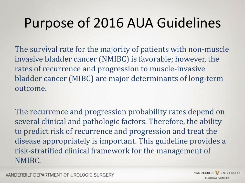
Purpose of 2016 AUA Guidelines
The survival rate for the majority of patients with non-muscle invasive bladder cancer (NMIBC) is favorable; however, the rates of recurrence and progression to muscle-invasive bladder cancer (MIBC) are major determinants of long-term outcome.
The recurrence and progression probability rates depend on several clinical and pathologic factors. Therefore, the ability to predict risk of recurrence and progression and treat the disease appropriately is important. This guideline provides a risk-stratified clinical framework for the management of NMIBC.

Prognosis
• The survival prognosis for patients with NMIBC is relatively favorable, with the cancer-specific survival (CSS) in high-grade disease ranging from approximately 70-85% at 10 years and a much higher rate for low-grade disease.
• The rates of recurrence and progression to MIBC are important surrogate endpoints for prognosis in NMIBC, as these are major determinants of long-term outcome.
• Risk stratification in NMIBC aids personalized treatment decisions and surveillance strategies as opposed to a generalized ‘one-size fits all’ approach

Low Risk Intermediate Risk High RiskLGa solitary Ta ≤ 3cm
Recurrence within 1 year, LG Ta
HG T1
PUNLMPb Solitary LG Ta > 3cm Any recurrent, HG Ta
LG Ta, multifocal HG Ta, >3cm (or multifocal)
HGc Ta, ≤ 3cm Any CISd
LG T1 Any BCG failure in HG patient
Any variant histologyAny LVIe
Any HG prostatic urethral involvement
aLG = low grade; bPUNLMP = papillary urothelial neoplasm of low malignant potential; cHG = high grade; dCIS=carcinoma in situ; eLVI = lymphovascular invasion
AUA Risk Stratification Table

Guidelines: Diagnosis1. At the time of resection of suspected bladder cancer, a clinician
should perform a thorough cystoscopic examination of a patient’s entire urethra and bladder that evaluates and documents tumor size, location, configuration, number, and mucosal abnormalities. (Clinical Principle)
2. At initial diagnosis of a patient with bladder cancer, a clinician should perform complete visual resection of the bladder tumor(s), when technically feasible. (Clinical Principle)
Incomplete TURBT is likely a significant contributing factor to early bladder cancer recurrences, as tumors are seen at first surveillance cystoscopy in up to 45% of patients
Brausi 2002

Contemporary Surgical Points That I Like to Emphasize
• Repeat TURBT within 6 weeks for high risk patients (include Ta, high grade; T1).
• Evaluate the urethra, especially in patients with CIS and/or suspected invasive disease.
• Utilize technology judiciously.
• Realize no substitute for experience.

Evaluate the Prostatic Urethra
• Up to 40% of patients may have involvement of prostatic urethra in patients with invasive disease and can be easily missed
• Important predictor of invasive disease
Wood D et al, J Urol, 1989Huguet J, et al. Eur Urol. 2005;48:53-59

Biopsies of the Prostatic Urethra
• TUR biopsies at 5 and 7 o’clock adjacent to the verumontanum – to help detect CIS
• WHY?– Highest concentration of prostatic ducts—where
CIS most likely to be found– TUR provides a full-thickness evaluation to rule
out invasion; can evaluate mucosa, ducts and stroma

Contemporary Surgical Points That I Like to Emphasize
• Repeat TURBT within 6 weeks for high risk patients (include Ta, high grade; T1).
• Evaluate the urethra, especially in patients with CIS and/or suspected invasive disease.
• Utilize technology judiciously.
• Realize no substitute for experience.

Use Technology Judiciously
• Utilize High-Definition imaging: it makes a difference– 5x increase in resolution compared to standard– Three-chip camera has better contrast and light intensity
• Mono-polar vs Bipolar: do what you are most comfortable with as data is equivocal
• EXCEPT that better pathology specimens are obtained with bipolar resection

Monopolar vs Bipolar Electrocautery
• Randomized, single center trial examined 147 patients and found– NO DIFFERENCE in:
• Bleeding• Obturator reflex• Operative time• Hyponatremia• Complications
– The only advantage was decreased cautery artifact with BIPOLAR
Venkatramani et al, J Urol, June 2014
Mild cautery artifact Severe cautery artifact

Contemporary Surgical Points That I Like to Emphasize
• Repeat TURBT within 6 weeks for high risk patients (include Ta, high grade; T1).
• Evaluate the urethra, especially in patients with CIS and/or suspected invasive disease.
• Utilize technology judiciously.
• Realize no substitute for experience.

Surgical Experience Makes a Difference
• When compared to experienced surgeons, younger surgeons – more likely to leave residual tumor (OR 7.3)– more likely to lack muscle in the specimen (OR 8.5)
• When detrusor muscle present: – the residual tumor rate was 20%
• BUT when detrusor muscle was absent: – the residual tumor rate was 51%
Huang J et al, Urol Int 89 (2012)

Guidelines: Diagnosis
3. A clinician should perform upper urinary tract imaging as a component of the initial evaluation of a patient with bladder cancer. (Clinical Principle)
4. In a patient with a history of NMIBC with normal cystoscopy and positive cytology, a clinician should consider prostatic urethral biopsies and upper tract imaging, as well as enhanced cystoscopic techniques (blue light cystoscopy, when available), ureteroscopy, or random bladder biopsies. (Expert Opinion)

Guidelines: Risk Stratification5. At the time of each occurrence/recurrence, a clinician
should assign a clinical stage and classify a patient accordingly as “low-,” “intermediate-,” or “high-risk.”(Moderate Recommendation; Evidence Strength: Grade C)
• EORTC/CUETO Model Tumor size, tumor focality, grade, stage, recurrence pattern, number
• AUA/SUO Additions Lymphovascular invasion, prostatic urethral involvement, variant histology, poor response to BCG

Guidelines: Variant Histology6. An experienced genitourinary pathologist should review the
pathology of a patient with any doubt in regards to variant or suspected variant histology (e.g., micropapillary, nested, plasmacytoid, neuroendocrine, sarcomatoid), extensive squamous or glandular differentiation, or the presence/absence of lymphovascular invasion. (Moderate Recommendation; Evidence Strength: Grade C)
7. If a bladder sparing approach is being considered in a patient with variant histology, then a clinician should perform a restaging TURBT within four to six weeks of the initial TURBT. (Expert Opinion)
8. Due to the high rate of upstaging associated with variant histology, a clinician should consider offering initial radical cystectomy. (Expert Opinion)

9. In surveillance of NMIBC, a clinician should not use urinary biomarkers in place of cystoscopic evaluation. (Strong Recommendation; Evidence Strength: Grade B)
NMP22® Protein-based; identifies nuclear matrix protein involved in the mitotic apparatus
BTA® Protein-based; identifies a basement membrane antigen related to complement factor H
UroVysion® FISH
Cell-based; identifies altered copy numbers of specific chromosomes using fluorescent probes
ImmunoCyt™
Cell-based; identifies three cell surface glycoproteins
Cxbladder™
Cell-based; identifies the presence of five mRNA fragments
Direct comparisons between markers are difficult, and given the uncertainty in sensitivity, these tests cannot be use to replace cystoscopy.
Tomasini 2013; O’Sullivan 2012
Guidelines: Urine Markers

10. In a patient with a history of low-risk cancer and a normal cystoscopy, a clinician should not routinely use a urinary biomarker or cytology during surveillance. (Expert Opinion)
11. In a patient with NMIBC, a clinician may use biomarkers to assess response to intravesicalBCG (UroVysion® FISH) and adjudicate equivocal cytology (UroVysion® FISH and ImmunoCyt™). (Expert Opinion)
Sarosdy 2002
Guidelines: Urine Markers

12.In a patient with non-muscle invasive disease who underwent an incomplete initial resection (not all visible tumor treated), a clinician should perform repeat transurethral resection or endoscopic treatment of all remaining tumor if technically feasible. (Strong Recommendation; Evidence Strength: Grade B)
13.In a patient with high-risk, high-grade Ta tumors, a clinician should consider performing repeat transurethral resection of the primary tumor site within six weeks of the initial TURBT. (Moderate Recommendation; Evidence Strength: Grade C)
14.In a patient with T1 disease, a clinician should perform repeat transurethral resection of the primary tumor site to include muscularis propria within six weeks of the initial TURBT. (Strong Recommendation; Evidence Strength: Grade B)
Guidelines: Repeat TURBT

15.In a patient with suspected or known low- or intermediate-risk bladder cancer, a clinician should consider administration of a single postoperative instillation of intravesical chemotherapy (e.g., mitomycin C or epirubicin) within 24 hours of TURBT. In a patient with a suspected perforation or extensive resection, a clinician should not use postoperative chemotherapy. (Moderate Recommendation; Evidence Strength: Grade B)
Sylvester 2004
Guidelines: Intravesical Therapy

16.In a low-risk patient, a clinician should not administer induction intravesical therapy. (Moderate Recommendation; Strength of Evidence Grade C)
17.In an intermediate-risk patient a clinician should consider administration of a six week course of induction intravesical chemotherapy or immunotherapy. (Moderate Recommendation; Evidence Strength: Grade B)
18.In a high-risk patient with newly diagnosed CIS, high-grade T1, or high-risk Ta urothelial carcinoma, a clinician should administer a six-week induction course of BCG. (Strong Recommendation; Evidence Strength: Grade B)
Guidelines: Intravesical Therapy

There is insufficient evidence to recommend one strain of BCG• Several small studies suggest that different strains may have different
efficacies
There is insufficient evidence to prescribe a particular BCG strength • EORTC 30962 recommends full dose for three years for high-risk
patients• For lower-risk patients, no difference in recurrence free survival
between full or 1/3 dose at 1 or 3 years
There is insufficient evidence to recommend using BCG in combination with other intravesical agents (AT THIS TIME…)• Several ongoing trials are currently examining synergistic
combinationsRentsch 2014; Oddens 2013; Houghton 2013
Guidelines: BCG

19.In an intermediate-risk patient who completely responds to an induction course of intravesical chemotherapy, a clinician may utilize maintenance therapy. (Conditional Recommendation; Evidence Strength: Grade C)
20.In an intermediate-risk patient who completely responds to induction BCG, a clinician should consider maintenance BCG for one year, as tolerated. (Moderate Recommendation; Evidence Strength: Grade C)
21.In a high-risk patient who completely responds to induction BCG, a clinician should continue maintenance BCG for three years, as tolerated. (Moderate Recommendation; Evidence Strength: Grade B)
Guidelines: Intravesical Therapy

EORTC-GU Cancers Group: Conclusions
–No differences in progression or survival rates in any of the arms
–No difference in morbidity between 1/3 and full dose arms
–“The benefit of the two additional years of maintenance should be weighed against its added costs and inconvenience”
Oddens et al, Eur Urol, 2013

22.In an intermediate- or high-risk patient with persistent or recurrent disease or positive cytology following intravesical therapy, a clinician should consider performing prostatic urethral biopsy and an upper tract evaluation prior to administration of additional intravesical therapy. (Conditional Recommendation; Evidence Strength: Grade C)
23.In an intermediate- or high-risk patient with persistent or recurrent Ta or CIS disease after a single course of induction intravesical BCG, a clinician should offer a second course of BCG. (Moderate Recommendation; Strength of Evidence C)
24.In a patient fit for surgery with high-grade T1 disease after a single course of induction intravesical BCG, a clinician should offer radical cystectomy. (Moderate Recommendation; Evidence Strength: Grade C)
Guidelines: BCG Relapse and Salvage Tx

My Take On BCG
• For high risk non-muscle invasive cancer, the treatment of choice at this time is: induction BCG + maintenance BCG
• The true response to BCG is not known until 6 month mark, regardless of induction or induction x 2 (6+6), or induction and maintenance (6+3)
• Options other than BCG are currently limited and/or their true success rate is unknown

Sanofi Pasteur will stop producing its version of the bladder-cancer drug treatment BCG, sold as TheraCys/ImmuCyst, in mid-2017, the company said in a statement Nov. 18.
Sanofi hasn't supplied BCG in the U.S. since 2012; the company said it will continue to supply doses to Canada, France and the U.K., where the product is currently available, until the end of 2018.
Despite taking significant steps to try to correct problems at its Toronto manufacturing plant, Sanofi's efforts "cannot guarantee the long-term continuity and reliable supply of the product,"

So Even More Than Ever We Need BCG Alternatives…

FDA and AUA/SUO Formulate Trial Design Goals

FDA Defines BCG UNRESPONSIVE
• Recurrent/persistent high-grade urothelialcarcinoma after completion of at least induction and one cycle maintenance BCG (5+2) for high grade Ta/T1 or CIS
• Never achieved CR or recurred within 6 months of last BCG dose
• T1 High grade at first evaluation after induction BCG

Switch Focus to T1 Tumors

T1 tumors
A B

Sfakianos JP et al, J Urology, February 2014
Re-TURBT Decreases Recurrence and Progression
Single TURBT
Repeat TURBT
Repeat TURBT
Single TURBT
Recurrence
Progression

And We May Even Begin to Understand T1 Even Better
• Combining – Depth of T1 invasion – Impact of Lymphovascular Invasion
• Recent T1 evaluation– Re-TURBT improved recurrence-free survival
(p < 0.001)– 1 mm cut off predicted progression (p < 0.04)
Patriarca C, et al. Diagn Pathol 2016 Jan 20;11(1):6

Impact of LVI on T1• Small study of 116 T1 patients • LVI in multivariate analysis
– In all patients, associated with stage progression (HR = 4.0)
– In patients treated with BCG, associated with
• Tumor recurrence (HR = 2.19)• Stage progression (HR = 3.76)
Fukumoto K, et al BMC Urol. 2016 Jan 19;16(1):5. doi: 10.1186/s12894-016-0122-1.

Impact of LVI on T1:If LVI negative—higher recurrence free and progression free
survival rate
Fukumoto K, et al BMC Urol. 2016 Jan 19;16(1):5. doi: 10.1186/s12894-016-0122-1

For Certain High Risk, Non Muscle Invasive Bladder Cancer: The Most Definitive Therapy

T1: Timely Cystectomy• I like the term “timely” cystectomy.• I guess I am the only one who likes it as every
one else uses the term “early” cystectomy.• The challenge of these patients:
– 30-53% progress to MIBC or metastatic disease.– Many patients have received prior intravesical therapy
with variable response.– Understaging: Re-TUR discovers muscle invasive disease in
approximately 20% of patients– If residual T1 disease, 82% progressed to MIBC by 5 yrs.

Chang Nomogram for Initial, Timely Cystectomy
• Absolute requirement
• No Brainers
• Not discussed but should undergo cystectomy:
• Patients with risk factors ; with the more they have, the harder to encourage to move to cystectomy
• Operative candidate
• Unresectable tumors
• Histologic variants like micropapillary, nested variant
• Residual T1 at repeat resection• Histopathologic features
– LVI– Depth of invasion (anatomic/extent)– CIS– Volume, size

Contemporary Update:New Ways to Evaluate the Bladder

Narrow Band ImagingTM (NBI)
Cauberg EC et al. Urol 76: 658, 2010
• Olympus Optical imaging technology enhances visibility of vessels on mucosa.
• Filters the white light into specific light wavelengths that penetrate only surface of human tissue and are absorbed by hemoglobin.
• Bluish light enhances superficial capillary network (brown)
• Greenish light enhances deeper vessel visibility: vessels are greenish-blue (cyan)

ALSO NBICan Be Used in Upper Tract

Narrow-band Imaging for Upper Urinary Tract Transitional-Cell CA
• Significant improvement in cancer detection and changes in management in 23% of patients. – Additional tumor detection
(14%) – Extended limits of ablation (9%)– 11% - traditional white light
imaging MISSED the cancer
Traxer, J Endourol 2011;25(1):19–23;

CysviewTM (Blue Light Cystoscopy)

U.S. Pivotal Study – Recurrence Results
• Tumor recurrence rates over 9 months were – 47% with Cysview– 56% with white light
(p=0.026)
• Relative reduction in recurrence rate was 16%
1.Stenzl A et al. J Urol 2010; 184: 1907-1914.2.Burger M et al. EAU 2012.

5 Different Trials: Cysview Detected Significantly More Ta, T1 and CIS tumors than white light
Tumour type Both methods, n (%) Cysview only, n (%) RR (CI) in favor of Cysview P value
Total Ta 1247 (81.6%) 202 (13.2%) 2.136 (1.578-2.890) <0.001
Ta initial 477 (88.7%) 49 (9.1%) 3.992 (1.833-7.040) <0.001
Ta recurrent 770 (77.7%) 153 (15.4%) 1.970 (1.399-2.775) <0.001
High risk 440 (80.0%) 73 (13.3%) 1.867 (1.186-2.939) 0.007
Intermediate risk
649 (80.7%) 123 (15.3%) 3.132 (1.992-4.926) <0.001
Low risk 158 (90.3%) 6 (3.4%) 0.702 (0.244-2.021) 0.512
Total T1 308 (85.3%) 34 (9.4%) 1.807 (0.957-3.412) 0.068
Initial 200 (85.8%) 26 (11.2%) 3.978 (1.617-9.785) 0.003
Recurrent 108 (84.4%) 8 (6.3%) 0.689 (0.247-1.922) 0.477
Total CIS 281 (55.1%) 203 (39.8%) 10.949 (6.808-17.610) <0.001
Initial 119 (55.3%) 91 (42.3%) 20.651 (7.766-54.909) <0.001
Recurrent 162 (54.9%) 112 (38.0%) 7.581 (4.364-13.168) <0.001
Witjes JA, et al, Eur Urol, 2014

Cysview Results in Cost Savings
• Cost Modeling Study from Sweden• For all patients
– Year 1, the added cost of Cysview was 0.86% higher
– Year 2, Cysview resulted in 1.5% cost savings– Year 5, Cysview results in 5.7% cost savings
Rose JB et al, BJU Intl, 2015

30.In a patient with NMIBC, a clinician should offer blue light cystoscopy at the time of TURBT, if available, to increase detection and decrease recurrence. (Moderate Recommendation; Evidence Strength: Grade B)
31. In a patient with NMIBC, a clinician may consider use of NBI to increase detection and decrease recurrence. (Conditional Recommendation; Evidence Strength: Grade C)
Guidelines: Enhanced Cystoscopy

32.After completion of the initial evaluation and treatment of a patient with NMIBC, a clinician should perform the first surveillance cystoscopy within three to four months. (Expert Opinion)
33.For a low-risk patient whose first surveillance cystoscopy is negative for tumor, a clinician should perform subsequent surveillance cystoscopy six to nine months later, and then annually thereafter; surveillance after five years in the absence of recurrence should be based on shared-decision making between the patient and clinician. (Moderate Recommendation; Evidence Strength: Grade C)
34.In an asymptomatic patient with a history of low-risk NMIBC, a clinician should not perform routine surveillance upper tract imaging. (Expert Opinion)
35.In a patient with a history of low-grade Ta disease and a noted sub-centimeter papillary tumor(s), a clinician may consider in-office fulguration as an alternative to resection under anesthesia. (Expert Opinion)
Guidelines: Surveillance and Follow-up

Let’s Switch Gears:
Are We Doing TOO Much for Low Risk Cancer?

Low Risk Cancer
• Should we be doing LESS Treatment:
• In Office Fulguration may be the best treatment • Recent Markov analysis (with certain
assumptions) evaluated low-grade, recurrent bladder cancer patients– Increased effectiveness– Decreased cost– Decreased patient therapeutic burden
Al Awamhii B et al, Urology (85) 2, 2015

Low Risk Cancer• In office fulguration: well tolerated technique
with little pain and minimal bleeding complications– Holmium:YAG laser with a 365- or 200-nm fiber– Settings: 0.6–0.8 J energy and rate of 10–15 Hz– Normal saline solution = irrigation fluid– Local anesthetic: INSTILLAGELTM
• Results– All patients had pain scores between 0-2– 4 patients on warfarin were treated safely– No patient-related complications
Wong, KA et al, BJU Int 2013 (112)

Low Risk Cancer
• Should we be doing even LESS Treatment: Active surveillance
• Pubmed search• pTa low grade disease• Evaluated progression, metastases, cost,
anesthetic risks of TURBT: and found no significant worsening of disease parameters
Tiu A et al, Urol Oncol, 2014

35. For an intermediate-risk patient whose first surveillance cystoscopy is negative for tumor, a clinician should perform subsequent cystoscopy with cytology every 3-6 months for 2 years, then 6-12 months for years 3 and 4, and then annually thereafter. (Expert Opinion)
36. For a high-risk patient whose first surveillance cystoscopy is negative for tumor, a clinician should perform subsequent cystoscopy with cytology every three to four months for two years, then six months for years three and four, and then annually thereafter. (Expert Opinion)
37. For an intermediate- or high-risk patient, a clinician should consider performing surveillance upper tract imaging at one to two year intervals. (Expert Opinion)
Guidelines: Surveillance and Follow-up

The future of NMIBC will likely be driven forward by science, novel technologies, new therapeutics and clinical trials. •Bladder Cancer Genome Atlas Project•Novel agents to improve BCG efficacy or manage BCG failures•New technologies
• A Study of Blue Light Flexible Cystoscopy With Cysview in the Detection of Bladder Cancer in the Surveillance Setting-- NCT02560584
• Transurethral Resection of Bladder Tumors Using PK Button Vaporization Electrode or Monopolar Loop Electrocautery in Treating Patients with Bladder Cancer-- NCT01567462
•Therapeutic Trials in Surgery/Radiation• Intracorporeal or Extracorporeal Urinary Diversion during Robotic Assisted
Radical Cystectomy in Reducing Complications in Patients with Bladder Cancer--NCT02252393
• Radiation Therapy and Cisplatin or Mitomycin and Fluorouracil in Treating Patients with Non-Muscle Invasive Stage I Bladder Cancer-- NCT00981656
•Surveillance• Surveillance Guidelines in Monitoring Patients with Non-muscle Invasive Bladder
Cancer-- NCT02298998
Future Research


Non-muscle Invasive Bladder Cancer Panel
Sam S. Chang, MD, MBA (Chair)James McKiernan, MD (Vice Chair)Peter Clark, MD (PGC Rep)Diane Zipursky Quale (Patient Adv)Stephen Boorjian, MDSiamak Daneshmand, MDBadrinath Konety, MDRaj Pruthi, MDChad Ritch, MDJohn Seigne, MDEila Skinner, MDNorm Smith, MD
The Agency for Healthcare Research and Quality
Oregon Health & Science UniversityRoger Chou, MDJessica Griffin
AUA StaffErin KirkbyAbid Khan
Acknowledgments
![Cancer Care Ontario Bladder Cancer Guideline: An ... · (AUA) “Treatment of Non-Metastatic Muscle-Invasive Bladder Cancer: AUA/ ASCO/ ASTRO/ SUO Guideline” [1]. TARGET POPULATION](https://img.pdfslide.us/doc/110x75/5f02dfc67e708231d406703f/cancer-care-ontario-bladder-cancer-guideline-an-aua-aoetreatment-of-non-metastatic.jpg)