Embed Size (px)
Citation preview

Atrial natriuretic peptide regulates adipose tissueaccumulation in adult atriaNadine Suffeea, Thomas Moore-Morrisb, Patrick Farahmandc, Catherine Rücker-Martind, Gilles Dilaniana, Magali Fradete,Daigo Sawakif, Geneviève Derumeauxf, Pascal LePrincea,c, Karine Clémenta,e, Isabelle Dugaila, Michel Puceatb,and Stéphane N. Hatema,e,1
aFaculté de Médecine, Université Pierre et Marie Curie, Sorbonne University, INSERM UMR_S1166, Paris, France; bFaculté de Médecine La Timone, UniversitéAix-Marseille, INSERM UMR_910, Marseille, France; cDepartment of Cardiology, Pitié-Salpêtrière Hospital, Assistance Publique-Hôpitaux de Paris, Paris,France; dINSERM UMR_S999, Laboratory of Excellence in Research on Medication and Innovative Therapeutics, Institut Paris-Sud d’InnovationThérapeutique Centre Chirurgical Marie Lanelongue, 92350 Le Plessis-Robinson, France; eInstitute of Cardiometabolism and Nutrition, Paris, France; andfINSERM U_955, Département Hospitalo-Universitaire Ageing-Thorax-Vessels-Blood, Département de Physiologie, Hôpital Henri Mondor, AssistancePublique-Hôpitaux de Paris, Creteil, France
Edited by Christine E. Seidman, Howard Hughes Medical Institute, Harvard Medical School, Boston, MA, and approved December 13, 2016 (received for reviewJuly 6, 2016)
The abundance of epicardial adipose tissue (EAT) is associated withatrial fibrillation (AF), the most frequent cardiac arrhythmia. How-ever, both the origin and the factors involved in EAT expansion areunknown. Here, we found that adult human atrial epicardial cellswere highly adipogenic through an epithelial–mesenchymal transi-tion both in vitro and in vivo. In a genetic lineage tracing theWT1CreERT2+/−RosatdT+/− mouse model subjected to a high-fat diet,adipocytes of atrial EAT derived from a subset of epicardial progen-itors. Atrial myocardium secretome induces the adipogenic differen-tiation of adult mesenchymal epicardium-derived cells by modulatingthe balance between mesenchymal Wingless-type Mouse MammaryTumor Virus integration site family, member 10B (Wnt10b)/β-cateninand adipogenic ERK/MAPK signaling pathways. The adipogenic prop-erty of the atrial secretome was enhanced in AF patients. The atrialnatriuretic peptide secreted by atrial myocytes is a major adipogenicfactor operating at a low concentration by binding to its natriureticpeptide receptor A (NPRA) receptor and, in turn, by activating a cGMP-dependent pathway. Hence, our data indicate cross-talk between EATexpansion and mechanical function of the atrial myocardium.
epicardial adipose tissue | epicardial progenitors | atrial natriureticpeptide | cGMP
The adipose tissue (AT) that accumulates at the surface andunderneath the epicardium, referred as to “epicardial adi-
pose tissue” (EAT), is a common histological component of theatrial myocardium. Anatomically, EAT is considered to be a specificvisceral AT depot responsible for the local delivery of fatty acid andadipokine substrates that can freely diffuse into the neighboringmyocardium (1). However, the recognition that EAT can be asso-ciated with the risk and severity of atrial fibrillation (AF), the mostfrequent cardiac arrhythmia in clinical practice, suggests that thisvisceral fat tissue is not always harmless or beneficial (2–4). Forinstance, EAT can promote fibrosis of the neighboring atrial myo-cardium, the substrate of AF, by secreting cytokines such as ActivinA (5) or by favoring fibrosis of the atrial subepicardium (6).Therefore understanding the origin of atrial EAT and identifyingthe factors regulating its local development is of major interest.The outer mesothelial layer of the heart, the epicardium, con-
tains a number of multipotent progenitor cells that can undergo anepithelial-to-mesenchymal transition (EMT) process, giving riseto multipotent mesenchymal epicardium-derived cells (EPDCs)(7–10). Recently it has been shown that epicardial-derived cellsdifferentiate into adipocytes during early postnatal development,contributing to physiological fat or EAT accumulation in theatrial–ventricular groove (10). Whether this epicardial-to-fat tran-sition can occur in the adult heart is being actively investigated. Inadult mice overexpressing the adipogenic transcriptional factorperoxisome proliferator-activated receptor-γ (PPARγ) and aftermassive acute myocardial injury, some adipocytes derived from theepicardium were detected in the ventricle (10, 11). Protein factors
such as Insulin-like growth factor 1 receptor (IGF1R) govern theformation of ventricular EAT after acute myocardial infarction (12).Here we provide evidence that atrial EAT derives from adult
atrial epicardial cells via EMT. Furthermore, we reveal that alow dose of atrial natriuretic peptide (ANP) secreted by atrialmyocytes promotes adipogenic differentiation of adult EPDCs(aEPDCs) by binding to the NPRA type receptor and activatingcGMP signaling. This ANP-mediated cross-talk between themyocardium and the epicardium reveals a link between EAT ex-pansion and the mechanical properties of the atrial myocardium.
ResultsAdult Atrial Epicardium Is Adipogenic. Because EAT is closely asso-ciated with the epicardium, we first investigated whether humanatrial epicardium contained adipocyte progenitors (Table S1). Inthe epicardial area of human atria, where AT depots were com-monly observed (Fig. 1 A and B), some cells expressed Wilm’stumor 1 (WT1) and Insulin gene enhancer protein 1 (Islet-1),respectively, two markers of epicardial and myocardial progenitors(Fig. 1C andD) (7, 13, 14). However, there was no clear relationshipbetween the presence of these progenitor cells and the clinical his-tory of donors (Table S1). In the epicardial layer, some WT1+ andIslet-1+ cells also expressed mesenchymal markers such as the cy-toskeleton proteins vimentin and α-smooth muscle actin (α-SMA),suggesting that they might have been derived from EMT of the adult
Significance
Atrial fibrillation is the most frequent cardiac arrhythmia and is amajor cause of stroke. Recently, it has been shown that theadipose tissue that accumulates at the surface of the heartcontributes to the pathogenesis of atrial fibrillation by favoringfibrosis of the neighboring myocardium. However, the cellularorigin of adult cardiac fat tissue is unknown. Here, we show thatresident progenitor cells of the external layer of the heart, re-ferred to as the “epicardium,” are a source of adipocytes throughan epithelial-to-mesenchymal transition process. The atrial na-triuretic peptide, which is secreted by atrial myocytes, is a potentfactor in the differentiation of epicardial progenitors in adipo-cytes. Our data uncover cross-talk between myocardial me-chanical properties and adipose tissue expansion.
Author contributions: N.S., T.M.-M., I.D., M.P., and S.N.H. designed research; N.S., T.M.-M.,P.F., C.R.-M., G. Dilanian, M.F., and D.S. performed research; N.S. and T.M.-M. contributednew reagents/analytic tools; N.S., T.M.-M., P.F., C.R.-M., G. Dilanian,M.F., D.S., G. Derumeaux,P.L., K.C., I.D., M.P., and S.N.H. analyzed data; and N.S., T.M.-M., G. Derumeaux, P.L., I.D.,M.P., and S.N.H. wrote the paper.
The authors declare no conflict of interest.
This article is a PNAS Direct Submission.1To whom correspondence should be addressed. Email: [email protected].
This article contains supporting information online at www.pnas.org/lookup/suppl/doi:10.1073/pnas.1610968114/-/DCSupplemental.
www.pnas.org/cgi/doi/10.1073/pnas.1610968114 PNAS | Published online January 16, 2017 | E771–E780
MED
ICALSC
IENCE
SPN
ASPL
US

epicardium and had migrated outside the epicardial layer (Fig. 1 Dand E) (15). Vimentin-positive cells also were seen in the vicinityof mature adipocytes in subepicardium (Fig. 1F). WT1+ cellscoexpressing the preadipocyte factor 1 (Pref-1, encoded by thegene DLK1) were present in the epicardium (Fig. 1G). They couldrepresent resident preadipocytes, in keeping with the observationof a number of WT1+/Pref-1+ cells in the epicardium of atria ofembryonic mice (Fig. S1).Next, we investigated the adipogenic capacity of atrial
aEPDCs by harvesting and culturing epicardial cells from sam-ples of human right atria obtained from 35 patients undergoingcardiac surgery (Table S1). After 2 d in culture, primary epi-cardial cells migrated out of the explants and showed the char-acteristics typical of epicardial progenitors including squamousmorphology and the expression of epicardial progenitor markerssuch as WT1, Transcription factor 21 (TCF21), and Connexin-43(Fig. S2A). They expressed high levels of WT1 and T-box tran-scription factor 18 (TBX18) (another epicardium progenitormarker) transcripts compared with cardiac myocytes (Fig. S2B).After the first passage and until passages 5–6, primary cellsspontaneously acquired and retained a mesenchymal stem cell(MSC) morphology and expressed several mesenchymal pro-teins, including matrix proteins such as fibronectin and collagen-1,the cytoskeleton proteins α-SMA, vimentin, Wingless-type MouseMammary Tumor Virus integration site family, member 10B(Wnt10b), the membrane protein N-cadherin, CD105, and thenuclear protein Snail (Fig. 1H). Mesenchymal proteins such asCD44, CD105, and the cell surface antigen Stro-1, but not theendothelial markers CD31 and CD34, were present at the plasmamembrane of aEPDCs as evidenced by flow cytometry (Fig.1I). The mesenchymal transition of primary epicardial cellsalso was supported by the expression of EMT-related genes,including Snail family transcriptional repressor (SNAIL) (Fig. 1J)
(7, 16). Finally, confirming their mesenchymal characteristics, wefound that aEPDCs had an osteogenic and chondrogenic potentialequivalent to that of human MSCs (Fig. 1K and Fig. S3) (17).We next examined the adipogenic capacity of aEPDCs by using
two distinct culture media known to induce massive adipogenicdifferentiation of humanMSCs (17, 18). Indeed, after 21 d, a subsetof aEPDCs showed lipid accumulation as revealed by Oil Red Ostaining (Fig. 2 A and B). In addition, both proteins and mRNAtranscripts encoding Adipokine, Perilipin-1, CCAAT/Enhancer bind-ing protein-α (C/EBPα), and PPARγ, the two last genes being mastertranscriptional regulators of adipocyte differentiation (10, 16, 19),were induced in aEPDCs (Fig. 2 C–E) (Table S2). Between days 7and 30 of culture in adipogenic medium, flow cytometry revealedPref-1 down-regulation and Perilipin-1 up-regulation in aEPDCs,indicating a shift from pre- to mature adipocytes (Fig. 2F). Ofnote, there was marked heterogeneity in the percentage of adi-pocytes (from 0 to 95%) derived from aEPDCs of the differentdonors. Furthermore, aging was associated with a high adipo-genic capacity of aEPDCs (r2 = 0.24, P = 0.027), whereas leftventricular dysfunction (ejection fraction <45%) was associatedwith reduced adipogenic capacity (r2 = 0.21, P = 0.046).Next we examined the epicardial contribution to EAT for-
mation in situ. We first defined the optimal conditions forreproducing EAT accumulation in mouse hearts. We examinedwhether obesity induced by a high-fat diet (HFD) could be as-sociated with atrial fat deposition. We found that the left atria ofadult C57BL/6 mice maintained on an obesogenic HFD for aprolonged period exhibited a clear EAT accumulation, mainly inthe epicardial layer, which we did not observe in mice fed anormal diet (Fig. 3 A and B). Although we did not first observesubepicardial fat, as seen in human patients, after 4 months cellscoexpressing WT1 and Pref-1 were detected both at the peri-cardial and myocardial faces of the sup-epicardium of obese
Fig. 1. Human atrial epicardial progenitor cellshave mesenchymal properties. (A) Masson’s tri-chrome staining of 7-μm-thick sections of humanatrial tissue (n = 26). (Scale bar, 100 μm.) (B–G) Im-munofluorescence staining in human atrial tissuesections (n = 8) for Perilipin-1 (B), WT1 (C), vimentinand Islet-1 (D), α-SMA and WT1 (E), vimentin andPerilipin-1 (F), and Pref-1 and WT1 (G). (Scale bars,20 μm.) Insets show Masson’s trichrome staining.(Scale bars, 10 μm.) (H) Immunofluorescence stainingof aEPDCs for fibronectin, α-SMA, N-cadherin, CD105,vimentin, Wnt10b, collagen-1, Snail, and DAPI (n =10). (Scale bars, 10 μm.) (I) Flow cytometry of aEPDCsfor CD44, CD105, CD31, CD34, and Stro-1 markers (n =10). Specific isotype controls are shown in gray.(J) Quantitative PCR (qPCR) analysis for Col1a1,TGFβ, SOX9, PDGFRα, and SNAIL in aEPDCs. Dataare expressed as the fold change relative to unpas-saged aEPDCs (n = 15) and represent the mean ± SEMof independent experiments. *P < 0.05, **P < 0.01,one-way ANOVA and Bonferroni’s post hoc test.(K) Bright-field images of aEPDC-derived chondrocytesor osteocytes revealed by alizarin red or Alcian bluestaining, respectively (n = 3). (Scale bars, 20 μm.)
E772 | www.pnas.org/cgi/doi/10.1073/pnas.1610968114 Suffee et al.

mice fed an HFD (Fig. 3B). Furthermore, the expression ofadipocyte gene markers was up-regulated in atria (Fig. 3C). Al-though the mice fed an HFD showed no evidence of cardiopathyon echocardiography imaging, they were more susceptible to AFthan lean animals, as indicated by the increased percentage ofmice that developed AF in response to burst pacing and by alonger duration of AF episodes (Fig. S4).To establish whether adult epicardium is adipogenic in vivo,
we performed genetic lineage tracing of adult epicardial cells inWT1CreERT2+/−RosatdT+/− mice subjected to the adipogenic nu-tritional protocol described above. Adult epicardial cells werespecifically and irreversibly labeled following tamoxifen inductionin young adults. The HFD-induced EAT deposition was repro-duced successfully in WT1CreERT2+/−RosatdT+/− adult mice, whichaccumulated AT in the sup-epicardium after 4 mo of HFD (Fig.3D). In mice subjected to HFD, but not in control mice fed anormal diet, we observed tandem dimer Tomato (tdT+) aEPDCsin the left atrial sup-epicardium. Furthermore, a subset of thesetdT+ aEPDCs coexpressed the adipocyte marker Perilipin-1, sug-gesting that they were adipocytes derived directly from adult epi-cardium (Fig. 3E). Expression of PPARγ also was observed in tdT+
cells that exhibited fully mature adipocyte morphology within theepicardial adipose layer (Fig. 3F).
Next, to establish the adipogenic potential of mouse atrial epicar-dium, epicardial cells were harvested from WT1CreERT2+/−RosatdT+/−atria as described for human epicardium. At day 1, tdT+ cellsmigrated from atrial tissue onto the culture dish, showed typicalsquamous morphology (Fig. 3G), and expressed the nuclearmarker TCF21 attesting their epicardial origin (Fig. 3H) (20).These tdT+ cells underwent EMT spontaneously, acquiring amesenchymal morphology as observed at day 3 (Fig. 3G). Themesenchymal transition was confirmed by the up-regulation ofmatrix (collagen-1), cytoskeleton (α-SMA, vimentin), and mem-brane (CD90/thymus cell antigen-1) proteins, as well as the nu-clear marker Snail (Fig. 3H). From the second passage, tdT+
murine aEPDCs exposed to adipogenic medium for 21 d showedlipid droplet accumulation, indicating they had undergone adi-pogenic differentiation (Fig. 3 I and J). Taken together, theseresults indicate that during obesity-induced EAT accumulation,epicardial progenitors undergo EMT and contribute directly toadipocyte accumulation.
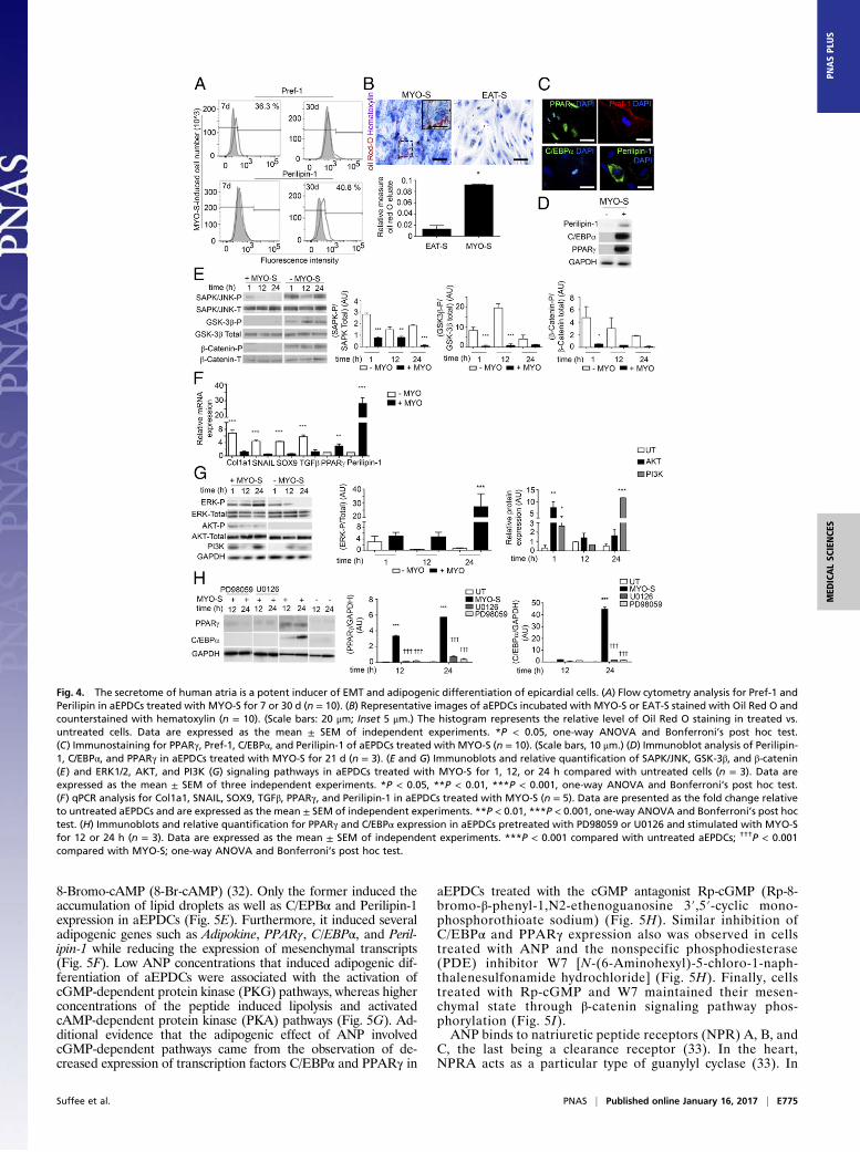
Atrial Myocardium Secretes Adipogenic Factors. A previous studyreporting that atrial myocardium can secrete adipogenic factors(21) prompted us to examine the impact of either EAT ormyocardial (MYO) secretomes (-S), derived from patient ex-plants (Table S1) on human aEPDCs in culture. Between days 7and 30, MYO-S but not EAT-S induced a shift from Pref-1+
preadipocytes to mature adipocytes containing Oil Red-O–stained lipid droplets and expressing Perilipin-1 (Fig. 4 A–D andF), PPARγ, and C/EBPα (Fig. 4 C, D, and F). There was sig-nificant variability in the adipogenic effect of atrial secretomes.Compared with secretomes from patients without AF, atrialsecretomes from AF patients had a more pronounced adipogeniccapacity on both aEPDCs and MSCs (Fig. S5).We next examined the kinases involved in aEPDC adipo-
genesis. After 24 h of culture in the presence of MYO-S, thepromesenchymal Wnt/β-catenin/Glycogen synthase kinase 3(GSK-3β) and Stress-activated protein kinase (SAPK)/JNKMAPK signaling pathways (22, 23) were suppressed (Fig. 4E).These results were in agreement with the observation of thesuppression of mesenchymal genes Collagen 1α (Col1a1),SNAIL, Sex-determining region box 9 (SOX9), and TGFβ andthe parallel expression of adipogenic genes by aEPDCs stimu-lated with MYO-S for 21 d (Fig. 4F). Next, we found that ERK/MAPK and PI3K/AKT, which are part of the adipogenic sig-naling pathways (22, 23), remained activated in aEPDCs (Fig.4G). In contrast, PD98059 and U0126, specific antagonists ofERK/MAPK and ERK2, respectively (19, 22, 23), suppressed theexpression of C/EBPα and decreased the expression of PPARγinduced by MYO-S (Fig. 4H). Of note, neither antagonist had aneffect on untreated aEPDCs. These results indicate that MYO-Scontains adipogenic factors that specifically regulate signalingpathways governing epicardial cell fate.
ANP Mediates the Adipogenic Effects of Atrial Myocardial SecretomeThrough cGMP Signaling. Using a protein screening assay, wefound that a number of growth factors, cytokines, and metal-loproteases were present in both EAT-S and MYO-S (Fig. S6).However, only FGF-7, Bone morphogenic protein 4 (BMP-4),and ANP were detected exclusively in MYO-S (Fig. 5 A and B).First, we tested the soluble proteins FGF-7 and BMP-4, whichdid not induce adipogenesis of aEPDCs (24). Second, the in-volvement of ANP in the adipogenic effect of MYO-S was testedfirst by culturing human aEPDCs in the presence of increasingconcentrations (1, 10, 100, and 10 000 pM) of human recombi-nant ANP. After 21 d in culture, 62 ± 0.3% of cells incubatedwith 10 pM of ANP contained lipid droplets stained with OilRed O (Fig. 5C) and expressed Perilipin-1 (Fig. 5D). Higherconcentrations of human recombinant ANP (1 and 10 nM) didnot further increase the percentage of aEPDC-derived adipo-cytes but instead reduced cell lipid content, suggesting a lipolyticeffect (Fig. 5 C and D). Of note, the adipogenic effect of ANPwas observed in the range of the peptide concentration found in
Fig. 2. Human EPDCs have the capacity to differentiate into adipocytes invitro. (A) Bright-field images of aEPDCs treated with adipogenic medium 1or medium 2 or with basal medium stained with Oil Red O to identify lipiddroplet formation and counterstained with hematoxylin (n = 35). (Scale bars,20 μm.) (B) Histogram showing Oil Red O content (A490) (n = 35). Datarepresent values relative to untreated aEPDCs and are expressed as mean ±SEM of independent experiments, *P < 0.05, **P < 0.01, one-way ANOVAand Bonferroni’s post hoc test. (C and D) Expression of adipokine, C/EBPα,PPARγ, and Perilipin-1 in aEPDCs treated with adipogenic medium 1 (C) ormedium 2 (D) (n = 10). Data represent the fold change relative to untreatedaEPDCs and are expressed as the mean ± SEM of independent experiments.*P < 0.05, **P < 0.01, ***P < 0.001, one-way ANOVA and Bonferroni’s posthoc test. (E) Immunofluorescence staining for C/EBPα and PPARγ in aEPDCstreated with adipogenic medium 1. (Scale bars, 20 μm.) (F) Histograms showaEPDC Pref-1 and Perilipin-1 signal intensity analyzed by flow cytometryfollowing treatment with adipogenic medium 2 for 7 or 30 d (n = 6). Isotypecontrols are shown in gray.
Suffee et al. PNAS | Published online January 16, 2017 | E773
MED
ICALSC
IENCE
SPN
ASPL
US
MYO-S (25, 26). Screening the supernatant of adipocytes de-rived from aEPDCs revealed a number of proteins belonging tothe inflammatory protein family (TNF-α and interleukins) involvedin extracellular matrix turnover [tissue inhibitor of metal-loproteinase 1 and 2 (TIMP-1 and -2)] or expressed by mature
adipocytes [BMP-4, Pre-B-cell–enhancing factor-related pro-tein (PBEF), leptin, and Pref-1] (Fig. S7) (27, 28).ANP regulates the balance between the cGMP and cAMP
signaling pathways (29–31) that was examined using two permeantanalogs of cyclic nucleotides, 8-Bromo-cGMP (8-Br-cGMP) and
Fig. 3. Obesity induces atrial epicardium-to-fat transition in mice. (A and D) Masson’s trichrome staining of 7-μm-thick sections of left atrial tissue (LA) incontrol mice (n = 10) (A) and WT1CreERT2+/−;tdT+/− mice (n = 8) (D), both fed an HFD. (Scale bar: 60 μm in A, 20 μm in D, 10 μm in Insets). (B) Immunoflu-orescence staining for Perilipin-1 in mice fed an HFD (n = 5), Pref-1 and WT1 (Inset) in mice fed a normal diet (n = 5). (Scale bars, 10 μm.) (C) qPCR analysis ofPerilipin-1, PPARγ, and C/EBPα expression in control mice fed a normal diet (n = 5). Data are represented as the fold change in mice fed an HFD relative to micefed a normal diet and are expressed as the mean ± SEM of five independent experiments, *P < 0.05, ***P < 0.001, one-way ANOVA and Bonferroni’s post hoctest. (E and F) Immunostaining for Perilipin-1 (E) or PPARγ (F) in atria of WT1CreERT2+/−;tdT+/− mice fed an HFD (n = 8). Arrows indicate signal colocalization ineach enlargement (E′, E′′, and E′′′) of epicardium-derived adipocytes. (Scale bars, 20 μm.) (G) Overlapped phase-contrast and fluorescence images of aEPDCsmigrating from a WT1CreERT2+/−; tdT+/− atrial explant at day 1 and day 3 in culture (n = 5). (Scale bars, 200 μm.) (H) WT1CreERT2+/−;tdT+/− aEPDCs immu-nostained for TCF21, Snail, CD90, vimentin, α-SMA, and collagen-1 (n = 5). (Scale bars, 50 μm.) (I and J) Bright-field, fluorescence, and overlapped fields (I) andOil Red O/hematoxylin staining (J) of WT1CreERT2+/−;tdT+/− aEPDCs induced by adipogenic medium 1 for 21 d (n = 5). (Scale bars, 200 μm.) The histogram in Jrepresents Oil Red O elution A490 compared with untreated aEPDCs (n = 5). Data are expressed as the mean ± SEM of five independent experiments. **P <0.01, one-way ANOVA and Bonferroni’s post hoc test. AU, arbitrary units; LA, left atria; LV, left ventricle; UT, untreated.
E774 | www.pnas.org/cgi/doi/10.1073/pnas.1610968114 Suffee et al.
8-Bromo-cAMP (8-Br-cAMP) (32). Only the former induced theaccumulation of lipid droplets as well as C/EPBα and Perilipin-1expression in aEPDCs (Fig. 5E). Furthermore, it induced severaladipogenic genes such as Adipokine, PPARγ, C/EBPα, and Peril-ipin-1 while reducing the expression of mesenchymal transcripts(Fig. 5F). Low ANP concentrations that induced adipogenic dif-ferentiation of aEPDCs were associated with the activation ofcGMP-dependent protein kinase (PKG) pathways, whereas higherconcentrations of the peptide induced lipolysis and activatedcAMP-dependent protein kinase (PKA) pathways (Fig. 5G). Ad-ditional evidence that the adipogenic effect of ANP involvedcGMP-dependent pathways came from the observation of de-creased expression of transcription factors C/EBPα and PPARγ in
aEPDCs treated with the cGMP antagonist Rp-cGMP (Rp-8-bromo-β-phenyl-1,N2-ethenoguanosine 3′,5′-cyclic mono-phosphorothioate sodium) (Fig. 5H). Similar inhibition ofC/EBPα and PPARγ expression also was observed in cellstreated with ANP and the nonspecific phosphodiesterase(PDE) inhibitor W7 [N-(6-Aminohexyl)-5-chloro-1-naph-thalenesulfonamide hydrochloride] (Fig. 5H). Finally, cellstreated with Rp-cGMP and W7 maintained their mesen-chymal state through β-catenin signaling pathway phos-phorylation (Fig. 5I).ANP binds to natriuretic peptide receptors (NPR) A, B, and
C, the last being a clearance receptor (33). In the heart,NPRA acts as a particular type of guanylyl cyclase (33). In
Fig. 4. The secretome of human atria is a potent inducer of EMT and adipogenic differentiation of epicardial cells. (A) Flow cytometry analysis for Pref-1 andPerilipin in aEPDCs treated with MYO-S for 7 or 30 d (n = 10). (B) Representative images of aEPDCs incubated with MYO-S or EAT-S stained with Oil Red O andcounterstained with hematoxylin (n = 10). (Scale bars: 20 μm; Inset 5 μm.) The histogram represents the relative level of Oil Red O staining in treated vs.untreated cells. Data are expressed as the mean ± SEM of independent experiments. *P < 0.05, one-way ANOVA and Bonferroni’s post hoc test.(C) Immunostaining for PPARγ, Pref-1, C/EBPα, and Perilipin-1 of aEPDCs treated with MYO-S (n = 10). (Scale bars, 10 μm.) (D) Immunoblot analysis of Perilipin-1, C/EBPα, and PPARγ in aEPDCs treated with MYO-S for 21 d (n = 3). (E and G) Immunoblots and relative quantification of SAPK/JNK, GSK-3β, and β-catenin(E) and ERK1/2, AKT, and PI3K (G) signaling pathways in aEPDCs treated with MYO-S for 1, 12, or 24 h compared with untreated cells (n = 3). Data areexpressed as the mean ± SEM of three independent experiments. *P < 0.05, **P < 0.01, ***P < 0.001, one-way ANOVA and Bonferroni’s post hoc test.(F) qPCR analysis for Col1a1, SNAIL, SOX9, TGFβ, PPARγ, and Perilipin-1 in aEPDCs treated with MYO-S (n = 5). Data are presented as the fold change relativeto untreated aEPDCs and are expressed as the mean ± SEM of independent experiments. **P < 0.01, ***P < 0.001, one-way ANOVA and Bonferroni’s post hoctest. (H) Immunoblots and relative quantification for PPARγ and C/EBPα expression in aEPDCs pretreated with PD98059 or U0126 and stimulated with MYO-Sfor 12 or 24 h (n = 3). Data are expressed as the mean ± SEM of independent experiments. ***P < 0.001 compared with untreated aEPDCs; †††P < 0.001compared with MYO-S; one-way ANOVA and Bonferroni’s post hoc test.
Suffee et al. PNAS | Published online January 16, 2017 | E775
MED
ICALSC
IENCE
SPN
ASPL
US

addition to their classical expression in atrial adipocytes andmyocytes, NPRA and NPRC also were detected in aEPDCs inthe subepicardium of human atrial sections (Fig. 6A). Of note,a subset of Pref-1+ cells coexpressing NPRA was detected inthe epicardial layer and also in the subepicardium (Fig. 6B).
Furthermore, NPRA was expressed in aEPDCs in vitro at boththe mRNA and protein levels (Fig. 6 C–E).The involvement of NPRA in the adipogenic effect of ANP on
aEPDCs was studied using a specific NPRA antagonist, HS-8140(34, 35). After 21 d of incubation, HS-8140 decreased PPARγ and
Fig. 5. The natriuretic peptide induces adipogenic differentiation of adult EPDCs. (A) Screening of proteins present in MYO-S (n = 8) or EAT-S (n = 8). (B) ANPlevels in secretome measured by an ELISA assay (MYO-S, n = 15; EAT-S, n = 10). Data are expressed as mean ± SEM; †††P < 0.001 compared with EAT-S; ‡‡‡P <0.001 compared with untreated (UT) cells, one-way ANOVA and Bonferroni’s post hoc test. (C) Bright-field images of aEPDCs treated with ANP and stainedwith Oil Red O/hematoxylin (n = 4). (Scale bars: 20 μm; Insets 5 μm.) The histogram represents the Oil Red O content of the cultures (Oil Red O elution at A490nm). Data are expressed as the mean ± SEM of independent experiments. ***P < 0.001 compared with untreated aEPDCs, one-way ANOVA and Bonferroni’spost hoc test. (D) Immunoblot and relative quantification of Perilipin-1 vs. GAPDH expression in aEPDCs treated with ANP for 21 d (n = 4). Vertical line in-dicates splicing between blots. Data are expressed as the mean ± SEM of independent experiments. **P < 0.01, ***P < 0.001 compared with untreatedaEPDCs, one-way ANOVA and Bonferroni’s post hoc test. (E) Oil Red O/hematoxylin staining and C/EBPα and Perilipin-1 immunostaining of aEPDCs treatedwith 8-Br-cGMP or 8-Br-cAMP (n = 3). (Scale bars: 20 μm; Inset 5 μm.) (F) qPCR analysis of adipogenic genes and mesenchymal transcripts from aEPDCs treatedwith 8-Br-cGMP for 21 d (n = 4). Data are presented as the fold change relative to untreated aEPDCs and are expressed as the mean ± SEM of independentexperiments. *P < 0.05, ***P < 0.001, one-way ANOVA and Bonferroni’s post hoc test. (G–I) Immunoblots and relative quantification of PKA and PKG (G),C/EBPα and PPARγ (H), and β-catenin (I) expression in aEPDCs pretreated with W7 or Rp-GMPc (H and I) and treated with ANP (G–I). Data are expressed as themean ± SEM of independent experiments. *P < 0.05, **P < 0.01, ***P < 0.001 compared with untreated aEPDCs. ††P < 0.01, †††P < 0.001 compared withaEPDCs treated with ANP, one-way ANOVA and Bonferroni’s post hoc test.
E776 | www.pnas.org/cgi/doi/10.1073/pnas.1610968114 Suffee et al.

C/EBPα expression in human aEPDCs treated with ANP (Fig.6F). Conversely, NPRC inhibition by HS-3134 had no effect onaEPDC differentiation (Fig. 6F). The inhibition of NPRA by HS-8140 under ANP stimulation was associated with the maintainedexpression of Snail and Wnt10b, indicating the repression of adi-pogenesis (Fig. 6G) (16, 36). These results were in agreement withthe persistence of SAPK/JNK and β-catenin/GSK-3β pathwayactivation in ANP-induced aEPDCs treated with HS-8140 (Fig. 6H–J). In MYO-S culture conditions, HS-8140 repressed aEPDC,C/EBPα, and PPARγ expression (Fig. 6K). Taken together theseresults indicate that the ANP/NPRA axis regulates the differen-tiation of aEPDCs into adipocytes.
Atrial myocytes are the source of ANP in the adult heart andsecrete ANP when maintained in primary culture (37, 38).Therefore, to establish the involvement of ANP in the adipogeniceffect of atrial secretome further, isolated human atrial myocyteswere maintained in primary culture conditions and plated on theupper chamber of Transwell dishes to induce adipogenic differ-entiation of confluent aEPDCs in the lower chamber (Fig. 6L).Indeed, after 7-d culture in the presence of human atrial myocytes,aEPDCs displayed an adipocyte phenotype, with the accumulationof lipid droplets stained by Oil Red O and Perilipin-1 expression(Fig. 6L) and the up-regulation of adipogenic genes C/EBPα,PPARγ, and Perilipin-1 (Fig. 6M).
Fig. 6. The NPRA type receptor is involved in the adipogenic effect of ANP. (A and B) Immunofluorescence staining for NPRA, NPRC (A) and costaining forPref-1 and NPRA (B) in 7-μm-thick sections of human atrial tissue (n = 6). (Scale bars, 10 μm.) (C ) NPRA and NPRC expression in aEPDCs analyzed by Westernblot (n = 3). Data are expressed as the mean ± SEM of three independent experiments. *P < 0.05, unpaired t test. (D) Adult EPDCs expression for NPRA andNPRC analyzed by immunofluorescence (n = 4) (Scale bars, 20 μm.) (E ) Expression for NPRA and NPRC analyzed by flow cytometry in aEPDCs (n = 5). (F andK) aEPDCs pretreated with NPRA antagonist (HS-8140) or NPRC antagonist (HS-3134) and then treated with ANP (F) (n = 3) or MYO-S (K) (n = 3) for 21d were assessed by immunoblots. Relative quantification for C/EBPα and PPARγ is compared with untreated cells (F and K ) (mean ± SEM of independentexperiments *P < 0.05; ***P < 0.001; ††P < 0.01, †††P < 0.001 compared with human EPDCs treated with ANP (F ) or MYO-S (K ); one-way ANOVA andBonferroni’s post hoc test). (G–J) aEPDCs pretreated with NPRA antagonist (HS-8140) (n = 3) and treated with ANP (n = 3) for 21 d were assessed byimmunoblots. Relative quantification of Wnt10b and Snail (G), SAPK/JNK (H), β-Catenin (I), and GSK3β (J) are compared with untreated cells (mean ± SEMof independent experiments; *P < 0.05; **P < 0.01; ***P < 0.001; one-way ANOVA and Bonferroni’s post hoc test). Vertical lines indicates splicing be-tween blots. (L, Left) Diagram representing the Transwell system. (Right) Oil Red O/hematoxylin staining and Perilipin-1 immunostaining of aEPDCcocultured with myocytes (n = 3). (Scale bars: 20 μm; Inset 5 μm.) (M ) qPCR analysis of C/EBPα, PPARγ, and Perilipin-1 expression in aEPDCs (n = 4)cocultured with myocytes (n = 3) for 7 d. qPCR data are presented as the fold change relative to aEPDCs cultured without myocytes. Data are expressed asthe mean ± SEM of independent experiments. *P < 0.05, **P < 0.01, compared with human EPDCs in normal culture; one-way ANOVA and Bonferroni’spost hoc test.
Suffee et al. PNAS | Published online January 16, 2017 | E777
MED
ICALSC
IENCE
SPN
ASPL
US

DiscussionEAT is now considered an important determinant of the pro-gression of the substrate of AF (21). Here we showed that theatrial epicardium is a source of adipocytes that can contribute tothe accumulation of EAT in adult atria and that myocardial ANPis a trigger of this process.The capacity of epicardial cells to undergo EMT, to migrate,
and to differentiate into smooth muscle cells or myofibroblasts iswell established (7, 9, 20, 39), but their adipogenic potential inadults has remained controversial (9). In the present study wefound that human and mouse adult EPDCs show a strong po-tential to differentiate into adipocytes in vitro. Moreover, both inhuman atria and in a murine genetic lineage tracing model, weprovide evidence that adult atrial epicardial cells undergo EMTand differentiate into adipocytes. A similar phenomenon of mes-enchymal transformation and adipogenic differentiation of epi-cardial progenitors has been reported in the atrioventricular canalduring embryonic development (11, 40). Of note, the endocar-dium has been shown to contribute to cardiac adipocytes duringdevelopment, but adult endocardial-to-fat transition has not yetbeen reported (41).The precise contribution of de novo EMT of epicardial pro-
genitors at the adult stage to EAT expansion is difficult to eval-uate. Additional mechanisms could be the recruitment ofdevelopmentally derived undifferentiated mesenchymal cells(EPDCs) maintained in a latent state in subepicardial layer (42).Resident and committed adipocyte progenitor cells in the atrialepicardial layer also could participate in EAT expansion. Indeed,WT1+ mesothelial cells originating in the lateral plate mesodermcan give rise to several visceral fat depots as well as to the epi-cardium and sup-epicardial adipocytes (12, 40). These early me-sodermal WT1+ cells, although contributing to adipogenesis, couldnot be labeled by the tomato in our experiments, the recombinasebeing induced at the adult stage. Interestingly, we found strongand specific expression of WT1+/Pref-1+ in most epicardial cellsduring development as early as embryonic day (E)12.5. In partic-ular, Pref-1 expression was high in atrial epicardial cells before andfollowing epicardial EMT, suggesting that the atrial epicardiumhas adipogenic potential throughout cardiac development. Theobservation of WT1+ cells in both sup- and subepicardial ATsuggests that both fat depots have a common cellular origin.However, the mechanisms regulating their respective expansionmight be distinct; for instance, subepicardial fat infiltration couldrequire chemotactic factors to drive the migration of epicardialprogenitors. We found that the adipogenic capacity of aEPDCsvaries among patients and could depend on clinical conditionssuch as aging, left ventricular dysfunction, or AF. This variation isin agreement with the current idea that adipose depots are acommon component of the atrial myocardium that could regulatethe metabolic or oxidative status of neighboring myocardium.However, various clinical factors could regulate its expansion (6,43). Under various AF-associated clinical conditions, this adi-pose depot can become deleterious, e.g., can favor myocardialfibrosis and the development of AF substrate (43).We found that the secretome of atrial myocardium is a potent
inducer of epicardial cell adipogenesis by activating key kinasesreported to regulate adipocyte formation (19, 22, 23, 36). Thisactivation is supported by the observation that the atrial secre-tome suppressed the mesenchymal signaling pathways Wnt/β-catenin/GSK-3β (19, 36) and SAPK/JNK MAPK (22, 23),whereas it stimulated ERK/MAPK and PI3K/AKT which regu-late the two adipogenic transcription factors C/EBPα andPPARγ. The effect of the atrial secretome is mediated primarilyby ANP; other potential adipogenic factors secreted by themyocardium such FGF-7 and BMP-4 did not induce adipogenicdifferentiation of aEPDCs. The natriuretic peptide binds to theNPRA receptor, activating cGMP-dependent signaling pathwaysand the recruitment of the transcriptional adipogenic factorsPPARγ and C/EBPα. PDE enzymes that regulate the balancebetween intracellular cGMP and cAMP levels are involved in theadipogenic effect of ANP, as indicated by the down-regulation of
C/EBPα and PPARγ expression and the maintenance of a mes-enchymal state of aEPDCs incubated with a PDE inhibitor.Distinct PDE enzymes such as PDE3, PDE5, and PDE11 havebeen reported to be involved in the transition from pre- to ma-ture adipocytes (44). Previous studies, mainly conducted with3T3-L1 preadipocyte cell lines, have reported that cGMP-dependent signaling can regulate adipogenesis (45, 46). Thesestudies notably reported activation of guanylyl cyclase-B byC-type natriuretic peptide or the inhibition of PDEs using3-isobutyl-1-methylxanthine (IBMX) (45, 46). Furthermore,during embryonic development, the regulation by the NPRA/cGMP signaling pathway of the balance between proliferationand differentiation of cardiac progenitor cells is essential forcardiac growth (47). Therefore, the NPRA/cGMP signalingpathway appears to be a critical regulatory node for the effect ofANP on cardiac tissue homeostasis.The adipogenic effect of ANP was observed after prolonged
incubation of aEPDCs with a low peptide concentration, whereasthe lipolytic effect was observed at a high peptide concentrationand following short exposure. This observation suggests that adi-pogenic versus lipolytic effects could depend on the route of ANPsecretion. Indeed, there is a regulated secretory pathway for ANPthat is activated in response to atrial stretch and which results inthe transient release of mature peptides stored in intracellulargranules. There also is a constitutive pathway, without interveningsecretion stimuli, in which the hormone is secreted after synthesis(48, 49), as observed in hypertrophied myocardium (50). Duringpermanent AF characterized by a certain degree of myocardialhypertrophy, constitutive release of ANP is activated, resulting inlocal accumulation of the peptide; this local accumulation couldfavor the adipogenic effects of ANP and in turn the expansion ofEAT, as observed in this clinical setting (51).Natriuretic peptides are known to regulate AT metabolism in a
dose-dependent manner (52, 53). Acutely, they can stimulatehuman fat cell lipolysis through PKG (30, 54) and were shownmore recently to turn on the expression of the thermogenic ma-chinery toward the browning of white adipocytes (55). Our presentdata identify a further role for natriuretic peptides in epicardial fatformation, indicating that these molecules could regulate a con-tinuum from the recruitment of fat cell progenitors to theirfunctional role in the release of fatty acids. In addition to natri-uretic peptides, other factors produced in different pathologicalcontexts could drive the transition of epicardium progenitors tofat. For instance, IGF1R could induce the aEPDCs-derived adi-pocytes following myocardial infarction (12). Moreover, rapidatrial beating in pigs and permanent AF in humans are associatedwith the expression of several genes able to regulate AT accu-mulation in human and pig atrial myocardium, a phenomenonattributed to an insufficient supply of oxygen and nutriments (11).Another example of cross-talk between AT and the atrial myo-cardium comes from the observation that human EAT secretesadipokines that regulate the oxidative status of the atrial myo-cardium (56). EAT expansion also could be caused by nutrientexcess in which epicardium becomes engorged with lipids resultingfrom the overwhelmed capacity of s.c. AT to clear excess triglyc-erides. This possibility is indicated by our observation that in mice,sustained HFD pressure is necessary to reveal fat accumulation inthe atria, whereas massive adipose infiltration of the posterior wallof left atria is easily described in sheep with moderate obesity (57).In summary, we show that adult atrial epicardium-to-fat tran-
sition contributes to atrial EAT, and we provide evidence that thisprocess is driven by ANP secreted by the myocardium. Our resultssupport the idea that EAT accumulation in adult atria is a slowprocess that could occur in response to chronic alterations of atrialmyocardium workload and metabolic conditions. Cardiac AT is asource of free fatty acids, the preferred metabolic substrates ofcardiomyocytes, and its accumulation in diseased atrial myocar-dium could be part of an adaptive process. However, the downsideof this process is the risk of progression of the substrate of AFbecause of the role played by EAT in atrial fibrosis (5, 6).
E778 | www.pnas.org/cgi/doi/10.1073/pnas.1610968114 Suffee et al.

Therefore, the epicardium-to-fat transition could be an early stepin the formation of the substrate of AF.
MethodsStudy Approval. All animal experiments conform to the Guide for the Careand Use of Laboratory Animals, according to Directive 2010/63/EU of theEuropean Parliament and were approved by the local committee of animalcare (agreement A751315).
Human tissue samples were obtained from patients undergoing cardio-vascular surgery as a bridge or for valvulopathy. Data and samples wereobtained in accordance with French Law Huriet-Sérusclat and with the ap-proval of the Ethical Committee (Comité de Protection des Personne Ile-deFrance VI) of Pitié-Salpêtrière Hospital, and informed consent to the researchwas obtained from each patient. The use of personal treatment data nec-essary for the research was reported to the National Commission for DataProtection and Liberties (CNIL-France) under the Data Protection Actnumber 78-17.
Mice. Eight-week-old male mice were used for all mouse studies and weremaintained under a 12-h light/12-h dark cycle at constant temperature (23 °C)with free access to food and water. WT1CreERT2+/− and Rosa26tdTomato+/+ miceon a C57BL/6J background were purchased from Jackson Laboratories.WT1CreERT2+/− mice were bred with Rosa26tdT+/+ mice. To trace the lineageand follow WT1+ epicardial progenitors, the male offspring were injectedwith tamoxifen to induce the recombinase at the adult stage (5 wk) aspreviously described (58). WT1CreERT2+/−RosatdT+/− transgenic mice (n = 10)and C57BL/6J wild-type mice (n = 30) (23–28 g; purchased from Janvier Labora-tories-CERJ) were fed an HFD (60% fat) (D12492i; Research Diets, Inc.) (n = 20) or anormal diet (4% fat) (n = 20) for 4 mo. The hearts were removed, perfusedthrough the aorta with PBS, fixed in 4% (wt/vol) paraformaldehyde (PFA) over-night, dehydrated overnight in 7, 14, and 25% (wt/vol) sucrose, embedded inOptimum Cutting Temperature (O.C.T.) compound, frozen, and sectioned (7 μm).
Human Tissue. Appendage samples of human atrial tissue were obtained forsecretome study (n = 29), histological study (n = 26), and isolation of epicardialprogenitor cells (n = 35). Samples of epicardial fat tissue (n = 22) were dedi-cated for secretome study. The subjects’ clinical data are provided in Table S1.
Masson’s Trichrome Staining. Human atrial appendage samples (n = 26) werefixed and embedded in paraffin as previously described (6). Frozen 7-μm-thick sections of mouse atria or 7-μm-thick paraffin-embedded sections ofhuman atria (n = 26) were stained with Masson’s trichrome according tothe manufacturer’s instructions (Sigma-Aldrich). Images were acquiredwith a Nikon DS-Ri1 camera coupled to an Eclipse-Ti Nikon microscope andNis-Element software (Nikon France S.A.) and were analyzed with ImageJ software.
Human Secretomes. Human atrial tissue (MYO) (n = 29) or EAT (n = 22) wasplaced at 37 °C, 5% CO2, in DMEM supplemented with 1% penicillin-strep-tomycin. After 24 h, conditioned medium (secretome, -S) was removed andconserved at −80 °C. The subjects’ clinical data are provided in Table S1.
Protein Screening Assay. Human atrial tissue secretome (MYO-S) (n = 8), EATsecretome (EAT-S) (n = 5), or aEDPC-derived adipocyte supernatant (n = 4)was screened with a cytokine array (catalog no. 126-AAH-CYT-2000-4; Tebu-Bio) and an Adipokine array (R&D Systems) according to the manufacturers’instructions. ELISA of the ANP level was performed in EAT-S (n = 22) or MYO-S(n = 29) according to the manufacturer’s instructions (R&D Systems).
Ex Vivo Culture of aEPDCs. Stripped layers of atrial epicardium were firstincubated in six-well plates for 2 d in DMEM (Thermo Fisher Scientific)supplemented with 10% (vol/vol) FCS and 1% penicillin-streptomycin (Sigma-Aldrich). After spontaneous migration of aEPDCs, tissue was removed, andcells were cultured in 1:1 DMEM andM199medium (Thermo Fisher Scientific)supplemented with 10% (vol/vol) FCS, 1% penicillin-streptomycin, and basic
FGF (bFGF) (10 ng/mL) at 37 °C, 5% CO2. The medium was changed every 2 d.Subjects’ clinical data are provided in Table S1.
Human MSCs. Adipose-derived stem cells (R7788-115) from human AT wereisolated and cultured according to the manufacturer’s instructions (ThermoFisher Scientific).
Isolation of Human Myocytes. Myocyte isolation and culture from human atrialtissue were performed as previously described (59). Briefly, cell dissociation wasachieved by enzymatic steps using collagenase (type IV) and protease (type XXIV)(Sigma-Aldrich). Isolated myocytes were cultured in DMEM (Thermo Fisher Sci-entific) supplemented with 10% (vol/vol) FCS (Sigma-Aldrich), nonessentialamino acids, 1 nM insulin, and antibiotics (100 IU/mL penicillin and 0.1 μg/mLstreptomycin) (Thermo Fisher Scientific). To inhibit fibroblast proliferation, 10 μMcytosine β-D-arabinofuranoside (Sigma-Aldrich) was added to the myocyte cul-ture. After 7 d in these culture conditions, human atrial myocytes underwent amarked growth and dedifferentiation process, as previously characterized (59).
Coculture Assay.HumanaEPDCswere cultured at a density of 1.106 cells/mL in thelower chamber of a six-plate Transwell system, and myocytes were incubated ata final density of 50 × 103/mL in the upper, laminin-coated (10 μg/mL) (ThermoFisher Scientific) chamber (Verfilco). Coculture was maintained for 7 d at 37 °C,5% CO2. The medium was changed once each week.
Differentiation of aEPDCs. Human or WT1CreERT2+/−RosatdT+/− transgenic mouseaEPDCs were incubated in adipogenic basal medium composed of DMEM/F12(Invitrogen) supplemented with insulin (5 μg/mL), ascorbic acid (200 μM), 10%(vol/vol) FCS (Sigma-Aldrich), bicarbonate (14 mM), and 1% penicillin-strep-tomycin (Thermo Fisher Scientific). To induce adipocyte differentiation, theadipogenic basal medium was supplemented with MYO-S (1/100), ANP (1, 10,100 pM or 10 nM) (Sigma-Aldrich), 8-Br-cGMP (10 nM) (Sigma-Aldrich), 8-Br-cAMP (10 nM) (Sigma-Aldrich), FGF-7 (10 μM) (Sigma-Aldrich), or BMP-4 (50 ng/mL)(Sigma-Aldrich) for 7 or 21 d. Two positive controls were used. The first wasadipogenic medium 1 composed of adipogenic basal medium supplementedwith biotin (8 μM), dexamethasone (1 μM) (Sigma-Aldrich), 3,3′,5-triiodo-l-thyronine (T3) (1 nM) (Sigma-Aldrich), and IBMX (500 μM) (Sigma-Aldrich) for21 d (21, 60). The second control was adipogenic medium 2 in which aEPDCswere cultured according to the manufacturer’s instructions (Thermo FisherScientific). All media were changed twice each week.
Oil Red Staining. MSCs (n = 3) or aEPDC-derived adipocytes (n = 31) were fixedwith 4% (wt/vol) PFA, incubated with 60% (vol/vol) isopropanol, and stained with3% (vol/vol) Oil RedO (Sigma-Aldrich). Then the cell nuclei were counterstainedwithhematoxylin (Sigma-Aldrich). Oil-red O staining then was eluted with isopropanoland quantified using spectrophotometry at 492 nm. All images were acquired witha Nikon DS-Ri1 camera coupled to an Eclipse-Ti Nikon microscope and Nis-Elementsoftware (Nikon France S.A.) and were analyzed with Image J software.
Statistics. Data are expressed as means ± SEM. Differences were investigatedusing the appropriate t test or one-way ANOVA and a Bonferroni post hocanalysis and were considered significant at P < 0.05. Statistical analysis wasperformed with GraphPad Prism 6.0 (GraphPad Software, Inc.).
ACKNOWLEDGMENTS. We thank the Leducq Foundation for its continuoussupport of our research. This work was supported by the French National Agencythrough the national program Investissements d’Avenir Grant ANR-10-IAHU-05(to N.S., M.F., K.C., and S.N.H.) and though the Recherche Hospital-Universitaire-Cardiac & Skeletal Muscle Alteration in Relation to Metabolic Diseases and Age-ing: Role of Adipose Tissue (RHU-CARMMA) Grant ANR-15-RHUS-0003 and theFondation de La Recherche Medicale (to N.S., T.M.-M., M.P., and S.N.H.). Thisproject received funding from the European Union’s Horizon 2020 Research andInnovation Programme under Grant 633193 “CATCH ME” and from FondationLeducq “Structural Alterations in the Myocardium and the Substrate for CardiacFibrillation” (N.S. and S.N.H.). N.S. was supported by European Union ProgramHorizon 2020 (CATCH ME). T.M.-M. was supported by the Leducq Foundation(SHAPEHEART) and the William Harvey International Translational ResearchAcademy European Union COFUND Program.
1. Iacobellis G (2015) Local and systemic effects of the multifaceted epicardial adipose
tissue depot. Nat Rev Endocrinol 11(6):363–371.2. Al Chekakie MO, et al. (2010) Pericardial fat is independently associated with human
atrial fibrillation. J Am Coll Cardiol 56(10):784–788.3. Thanassoulis G, et al. (2010) Pericardial fat is associated with prevalent atrial fibril-
lation: The Framingham Heart Study. Circ Arrhythm Electrophysiol 3(4):345–350.4. Wong CX, et al. (2011) Pericardial fat is associated with atrial fibrillation severity and
ablation outcome. J Am Coll Cardiol 57(17):1745–1751.
5. Venteclef N, et al. (2015) Human epicardial adipose tissue induces fibrosis of the
atrial myocardium through the secretion of adipo-fibrokines. Eur Heart J 36(13):
795a–805a.6. Haemers P, et al. (2015) Atrial fibrillation is associated with the fibrotic remodelling of
adipose tissue in the subepicardium of human and sheep atria. Eur Heart J ehv625
10.1093/eurheartj/ehv625.7. Chong JJH, et al. (2011) Adult cardiac-resident MSC-like stem cells with a proepicardial
origin. Cell Stem Cell 9(6):527–540.
Suffee et al. PNAS | Published online January 16, 2017 | E779
MED
ICALSC
IENCE
SPN
ASPL
US

8. Bollini S, et al. (2014) Re-activated adult epicardial progenitor cells are a heteroge-neous population molecularly distinct from their embryonic counterparts. Stem CellsDev 23(15):1719–1730.
9. van Tuyn J, et al. (2007) Epicardial cells of human adults can undergo an epithelial-to-mesenchymal transition and obtain characteristics of smooth muscle cells in vitro.Stem Cells 25(2):271–278.
10. Yamaguchi Y, et al. (2015) Adipogenesis and epicardial adipose tissue: A novel fate ofthe epicardium induced by mesenchymal transformation and PPARγ activation. ProcNatl Acad Sci USA 112(7):2070–2075.
11. Liu Q, et al. (2014) Epicardium-to-fat transition in injured heart. Cell Res 24(11):1367–1369.
12. Zangi L, et al. (2016) An IGF1R-dependent pathway drives epicardial adipose tissueformation after myocardial injury. Circulation CIRCULATIONAHA.116.022064 10.1161/CIRCULATIONAHA.116.022064.
13. Sun Y, et al. (2007) Islet 1 is expressed in distinct cardiovascular lineages, includingpacemaker and coronary vascular cells. Dev Biol 304(1):286–296.
14. Zhou B, et al. (2008) Epicardial progenitors contribute to the cardiomyocyte lineage inthe developing heart. Nature 454(7200):109–113.
15. Singh MK, Epstein JA (2012) Epicardium-derived cardiac mesenchymal stem cells:Expanding the outer limit of heart repair. Circ Res 110(7):904–906.
16. Lee YH, et al. (2013) Transcription factor Snail is a novel regulator of adipocyte dif-ferentiation via inhibiting the expression of peroxisome proliferator-activated re-ceptor γ. Cell Mol Life Sci 70(20):3959–3971.
17. Pittenger MF, et al. (1999) Multilineage potential of adult human mesenchymal stemcells. Science 284(5411):143–147.
18. Jiang C, et al. (2015) HIF-1A and C/EBPs transcriptionally regulate adipogenic differ-entiation of bone marrow-derived MSCs in hypoxia. Stem Cell Res Ther 6:21.
19. Tang Q-Q, et al. (2005) Sequential phosphorylation of CCAAT enhancer-bindingprotein beta by MAPK and glycogen synthase kinase 3beta is required for adipo-genesis. Proc Natl Acad Sci USA 102(28):9766–9771.
20. Asli NS, Xaymardan M, Harvey RP (2014) Epicardial origin of resident mesenchymalstem cells in the adult mammalian heart. J Dev Biol 2(2):117–137.
21. Chilukoti RK, et al. (2015) Atrial fibrillation and rapid acute pacing regulate adipo-cyte/adipositas-related gene expression in the atria. Int J Cardiol 187:604–613.
22. Bost F, Aouadi M, Caron L, Binétruy B (2005) The role of MAPKs in adipocyte dif-ferentiation and obesity. Biochimie 87(1):51–56.
23. Prusty D, Park B-H, Davis KE, Farmer SR (2002) Activation of MEK/ERK signalingpromotes adipogenesis by enhancing peroxisome proliferator-activated receptorgamma (PPARgamma ) and C/EBPalpha gene expression during the differentiation of3T3-L1 preadipocytes. J Biol Chem 277(48):46226–46232.
24. Zhang T, Guan H, Yang K (2010) Keratinocyte growth factor promotes preadipocyteproliferation via an autocrine mechanism. J Cell Biochem 109(4):737–746.
25. Tsukamoto O, et al. (2009) Natriuretic peptides enhance the production of adipo-nectin in human adipocytes and in patients with chronic heart failure. J Am CollCardiol 53(22):2070–2077.
26. Souza SC, et al. (2011) Atrial natriuretic peptide regulates lipid mobilization andoxygen consumption in human adipocytes by activating AMPK. Biochem Biophys ResCommun 410(3):398–403.
27. Pisani DF, et al. (2015) Visfatin expression analysis in association with recruitment andactivation of human and rodent brown and brite adipocytes. Adipocyte 5(2):186–195.
28. Liangpunsakul S, et al. (2014) Increasing serum pre-adipocyte factor-1 (Pref-1) cor-relates with decreased body fat, increased free fatty acids, and level of recent alcoholconsumption in excessive alcohol drinkers. Alcohol 48(8):795–800.
29. Zhang X, et al. (2010) Sildenafil promotes adipogenesis through a PKG pathway.Biochem Biophys Res Commun 396(4):1054–1059.
30. Moro C, Klimcakova E, Lafontan M, Berlan M, Galitzky J (2007) Phosphodiesterase-5Aand neutral endopeptidase activities in human adipocytes do not control atrial na-triuretic peptide-mediated lipolysis. Br J Pharmacol 152(7):1102–1110.
31. Ikoma-Seki K, et al. (2015) Role of LRP1 and ERK and cAMP signaling pathways inlactoferrin-induced lipolysis in mature rat adipocytes. PLoS One 10(10):e0141378.
32. Hemmrich K, et al. (2010) Nitric oxide and downstream second messenger cGMP andcAMP enhance adipogenesis in primary human preadipocytes. Cytotherapy 12(4):547–553.
33. Pandey KN (2014) Guanylyl cyclase/natriuretic peptide receptor-A signaling antago-nizes phosphoinositide hydrolysis, Ca(2+) release, and activation of protein kinase C.Front Mol Neurosci 7:75.
34. Moro C, et al. (2004) Functional and pharmacological characterization of the natri-uretic peptide-dependent lipolytic pathway in human fat cells. J Pharmacol Exp Ther308(3):984–992.
35. Houshmand F, Faghihi M, Zahediasl S (2015) Role of atrial natriuretic Peptide inoxytocin induced cardioprotection. Heart Lung Circ 24(1):86–93.
36. Ross SE, et al. (2000) Inhibition of adipogenesis by Wnt signaling. Science 289(5481):950–953.
37. Dietz JR (2005) Mechanisms of atrial natriuretic peptide secretion from the atrium.Cardiovasc Res 68(1):8–17.
38. de Bold AJ, de Bold MLK (2005) Determinants of natriuretic peptide production bythe heart: Basic and clinical implications. J Investig Med 53(7):371–377.
39. Zhou B, et al. (2011) Adult mouse epicardium modulates myocardial injury by se-creting paracrine factors. J Clin Invest 121(5):1894–1904.
40. Chau Y-Y, et al. (2014) Visceral and subcutaneous fat have different origins and ev-idence supports a mesothelial source. Nat Cell Biol 16(4):367–375.
41. Zhang H, et al. (2016) Endocardium contributes to cardiac fat. Circ Res 118(2):254–265.42. Ruiz-Villalba A, Ziogas A, Ehrbar M, Pérez-Pomares JM (2013) Characterization of
epicardial-derived cardiac interstitial cells: Differentiation and mobilization of heartfibroblast progenitors. PLoS One 8(1):e53694.
43. Hatem SN, Sanders P (2014) Epicardial adipose tissue and atrial fibrillation. CardiovascRes 102(2):205–213.
44. Armani A, Marzolla V, Rosano GMC, Fabbri A, Caprio M (2011) Phosphodiesterasetype 5 (PDE5) in the adipocyte: A novel player in fat metabolism? Trends EndocrinolMetab 22(10):404–411.
45. Katafuchi T, Garbers DL, Albanesi JP (2010) CNP/GC-B system: A new regulator ofadipogenesis. Peptides 31(10):1906–1911.
46. Mitschke MM, et al. (2013) Increased cGMP promotes healthy expansion andbrowning of white adipose tissue. FASEB J 27(4):1621–1630.
47. Hotchkiss A, et al. (2015) Atrial natriuretic peptide inhibits cell cycle activity of em-bryonic cardiac progenitor cells via its NPRA receptor signaling axis. Am J Physiol CellPhysiol 308(7):C557–C569.
48. Thibault G, Amiri F, Garcia R (1999) Regulation of natriuretic peptide secretion by theheart. Annu Rev Physiol 61:193–217.
49. Ogawa T, Vatta M, Bruneau BG, de Bold AJ (1999) Characterization of natriureticpeptide production by adult heart atria. Am J Physiol 276(6 Pt 2):H1977–H1986.
50. Ruskoaho H, Vakkuri O, Arjamaa O, Vuolteenaho O, Leppäluoto J (1989) Pressorhormones regulate atrial-stretch-induced release of atrial natriuretic peptide in thepithed rat. Circ Res 64(3):482–492.
51. Roy D, et al. (1987) Atrial natriuretic factor during atrial fibrillation and supraven-tricular tachycardia. J Am Coll Cardiol 9(3):509–514.
52. Nishikimi T, et al. (2009) Stimulatory and Inhibitory regulation of lipolysis by the NPR-A/cGMP/PKG and NPR-C/G(i) pathways in rat cultured adipocytes. Regul Pept 153(1-3):56–63.
53. Lafontan M, et al. (2008) Control of lipolysis by natriuretic peptides and cyclic GMP.Trends Endocrinol Metab 19(4):130–137.
54. Sengenès C, Berlan M, De Glisezinski I, Lafontan M, Galitzky J (2000) Natriureticpeptides: A new lipolytic pathway in human adipocytes. FASEB J 14(10):1345–1351.
55. Bordicchia M, et al. (2012) Cardiac natriuretic peptides act via p38 MAPK to induce the brownfat thermogenic program in mouse and human adipocytes. J Clin Invest 122(3):1022–1036.
56. Antonopoulos AS, et al. (2016) Mutual regulation of epicardial adipose tissue andmyocardial redox state by PPAR-γ/adiponectin signalling. Circ Res 118(5):842–855.
57. Mahajan R, et al. (2015) Electrophysiological, electroanatomical, and structural re-modeling of the atria as consequences of sustained obesity. J Am Coll Cardiol 66(1):1–11.
58. Moore-Morris T, et al. (2014) Resident fibroblast lineages mediate pressure overload-induced cardiac fibrosis. J Clin Invest 124(7):2921–2934.
59. Rücker-Martin C, Pecker F, Godreau D, Hatem SN (2002) Dedifferentiation of atrialmyocytes during atrial fibrillation: Role of fibroblast proliferation in vitro. CardiovascRes 55(1):38–52.
60. Elsen M, et al. (2014) BMP4 and BMP7 induce the white-to-brown transitionof primary human adipose stem cells. Am J Physiol Cell Physiol 306(5):C431–C440.
E780 | www.pnas.org/cgi/doi/10.1073/pnas.1610968114 Suffee et al.