Embed Size (px)
DESCRIPTION
af managemen
Citation preview
European Heart Journal (1998) 19, 1294–1320 Article No. hj981050
Working Group Report
Atrial fibrillation: current knowledge and recommendations for management
S. Levy, G. Breithardt, R. W. F. Campbell, A. J. Camm, J.-C. Daubert, M. Allessie, E. Aliot, A. Capucci, F. Cosio, H. Crijns, L. Jordaens, R. N. W. Hauer, F. Lombardi
and B. Lu deritz on behalf of the Working Group on Arrhythmias of the European Society of Cardiology
Introduction
Atrial fibrillation, a commonly encountered arrhythmia, has in recent years, been the subject of increased interest and intensive clinical research. There is also increasing awareness that atrial fibrillation is a major cause of embolic events which in 75% of cases are complicated by cerebrovascular accidents[1,2]. Atrial fibrillation is often associated with heart disease but a significant proportion of patients (about 30%) have no detectable heart disease[3]. Symptoms, occasionally disabling, haemodynamic impairment and a decrease in life expectancy are among the untoward effects of atrial fibrillation, resulting in an important morbidity, mortality and an increased cost for the health care provider[4].
The Working Group of Arrhythmias of the European Society of Cardiology created a Study Group on Atrial Fibrillation in order to establish recommendations for the better management of this arrhythmia and to promote multicentre studies. The purpose of this paper is to briefly outline the state of our knowledge on the clinical presentation, the causes, the mechanisms and therapeutic approaches currently available and to propose recommendations for management. Although atrial flutter can coexist with atrial fibrillation, it is considered a different arrhythmia and will not be covered in the present paper.
Key words: Atrial fibrillation, thromboembolism, antiarrhythmic agents, anticoagulant therapy.
Manuscript submitted 16 February 1998, and accepted 12 March 1998.
This Report represents the opinion of the Study Group on Atrial Fibrillation and does not necessarily reflect the opinion of the ESC.
Correspondence: Samuel Levy MD, FESC, Division of Cardiology, Hopital Nord, Marseille, 13015 France.
0195-668X/98/091294+27 $18.00/0
Clinical manifestations and classification of atrial fibrillation
Definition
In atrial fibrillation, there is a complete absence of coordinated atrial systole. This arrhythmia is characterized on the ECG by the absence of consistent P waves before each QRS complex; instead there are rapid oscillations of ‘f’ waves which vary in size, shape, and timing and there is usually an irregular ventricular rate[5]. However, the presence of fixed RR intervals is possible and should prompt consideration of the presence of idioventricular or idiojunctional rhythms associated with conduction system disease or drug therapy. The RR may also be regular in ventricularly-paced patients and the diagnosis in this circumstance may require temporary pacemaker inhibition in order to visualize underlying atrial fibrillatory activity.
Symptoms associated with atrial fibrillation
Atrial fibrillation may be symptomatic or asymptomatic. Both symptomatic and asymptomatic episodes of atrial fibrillation may occur in the same patient[6]. Asymptomatic atrial fibrillation may be discovered incidentally during cardiac auscultation, 12-lead ECG recording or ambulatory ECG recordings undertaken for reasons unrelated to this arrhythmia. The duration of atrial fibrillation may be unknown if not correlated with symptoms. Atrial fibrillation may present for the first time with an embolic complication or aggravation of congestive heart failure related to underlying heart disease. In most patients, atrial fibrillation is symptomatic and represents the most common arrhythmia responsible for hospitalizations according to a recent study[7]. The complaints associated with atrial fibrillation include rapid beating of the heart often perceived as palpitations (at rest and/or precipitated by exercise or emotion),
� 1998 The European Society of Cardiology
Task Force Report 1295
chest pain, shortness of breath on exertion, fatigue and dizziness. Atrial fibrillation may be associated with inappropriate chronotropic ventricular response. In some patients, an uncontrolled rapid heart rate may induce ventricular cardiomyopathy[8]. In the absence of an accessory atrioventricular connection, the ventricular response during atrial fibrillation depends on atrioventricular node conduction properties and concealed conduction (see section on rate control). Syncope is a rare but serious complication of atrial fibrillation and is particularly common in patients with sinus node dysfunction or obstructive structural heart disease, such as hypertrophic cardiomyopathy. Symptoms vary with the ventricular rate, the underlying functional status of the heart and the duration of atrial fibrillation. In some patients, irregularity of heart rate seems to play an important role in the genesis of symptoms.
Nomenclature
The nomenclature of atrial fibrillation will be covered as a separate issue as there is, at present, no general agreement on the terminology to be used. In current literature, atrial fibrillation is generally subdivided into two forms: paroxysmal and chronic. The term chronic is either used to categorize the history of atrial fibrillation or to describe the last episode. In this report it will be used to describe atrial fibrillation in which the episodes last for several days or years. The term acute may describe an episode of atrial fibrillation related to an acute curable cause[9] and is also used to describe an attack of atrial fibrillation[10]. Chronic (or established or permanent) atrial fibrillation may be the final stage of paroxysmal atrial fibrillation or may represent the initial aspect in a significant proportion of patients. Provided the patient is symptomatic, the differentiation of paroxysmal from chronic atrial fibrillation is based on the history given by the patient and/or the electrocardiographic documentation of recurrent episodes (in the paroxysmal form) and the duration of the last episode of atrial fibrillation. In some cases, no history is available, particularly in asymptomatic or mildly symptomatic patients, and the term recent onset or recently discovered atrial fibrillation is used. The latter is particularly appropriate for atrial fibrillation of unknown duration. In the paroxysmal (recurrent) form of atrial fibrillation, the episodes are generally self-terminating or persistent. The latter may require urgent medical, pharmacological or electrical, intervention. The term permanent implies that atrial fibrillation has been present for a long time, that cardioversion has not been indicated or that one or several attempts have failed to restore sinus rhythm. There is no agreement on the time frame used to characterize various forms of atrial fibrillation. A period of 7 days, has been proposed as a cut-off point to differentiate paroxysmal (<7 days) from chronic atrial fibrillation (>7days)[9]. In the paroxysmal form, an episode lasting more than 48 h may be called persistent. This time frame represents the duration beyond which
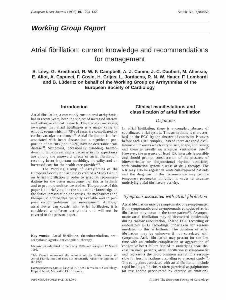
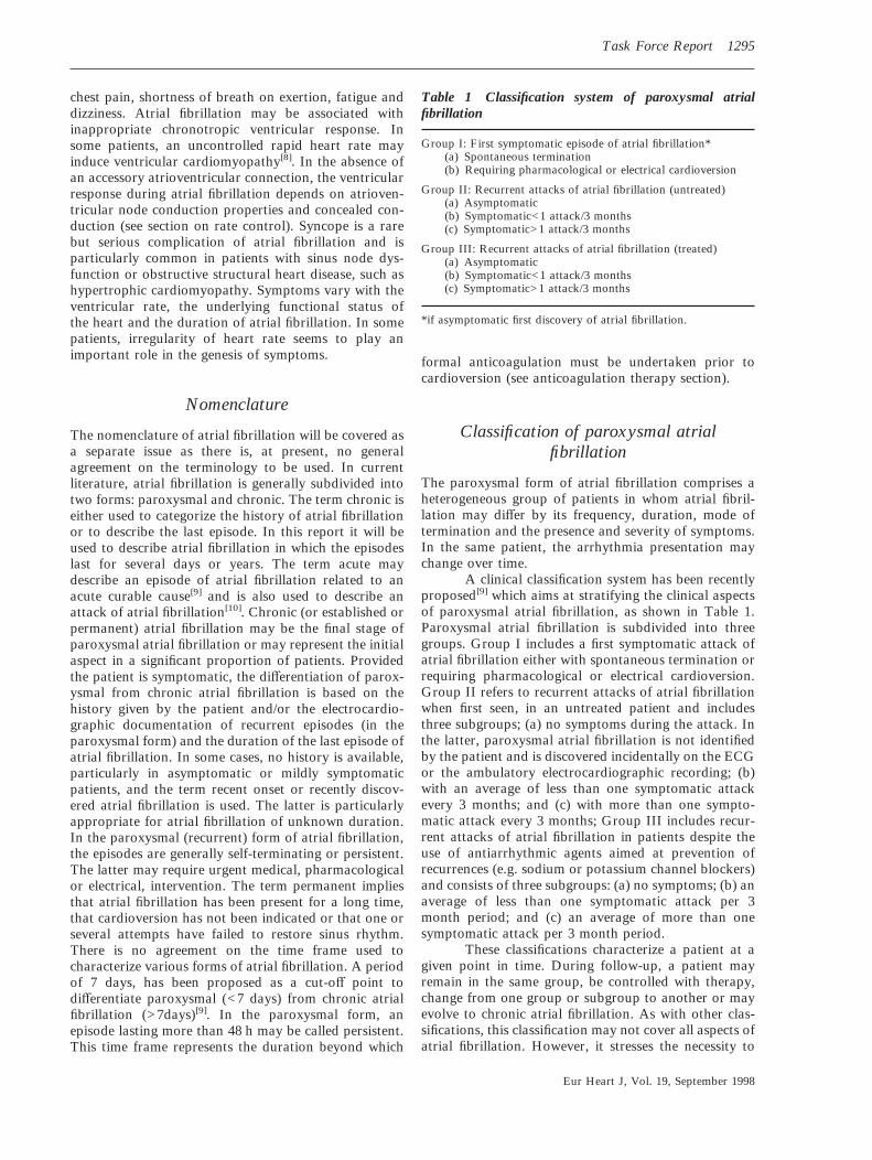
Table 1 Classification system of paroxysmal atrial fibrillation
Group I: First symptomatic episode of atrial fibrillation* (a) Spontaneous termination (b) Requiring pharmacological or electrical cardioversion
Group II: Recurrent attacks of atrial fibrillation (untreated) (a) Asymptomatic (b) Symptomatic<1 attack/3 months (c) Symptomatic>1 attack/3 months
Group III: Recurrent attacks of atrial fibrillation (treated) (a) Asymptomatic (b) Symptomatic<1 attack/3 months (c) Symptomatic>1 attack/3 months
*if asymptomatic first discovery of atrial fibrillation.
formal anticoagulation must be undertaken prior to cardioversion (see anticoagulation therapy section).
Classification of paroxysmal atrial fibrillation
The paroxysmal form of atrial fibrillation comprises a heterogeneous group of patients in whom atrial fibrillation may differ by its frequency, duration, mode of termination and the presence and severity of symptoms. In the same patient, the arrhythmia presentation may change over time.
A clinical classification system has been recently proposed[9] which aims at stratifying the clinical aspects of paroxysmal atrial fibrillation, as shown in Table 1. Paroxysmal atrial fibrillation is subdivided into three groups. Group I includes a first symptomatic attack of atrial fibrillation either with spontaneous termination or requiring pharmacological or electrical cardioversion. Group II refers to recurrent attacks of atrial fibrillation when first seen, in an untreated patient and includes three subgroups; (a) no symptoms during the attack. In the latter, paroxysmal atrial fibrillation is not identified by the patient and is discovered incidentally on the ECG or the ambulatory electrocardiographic recording; (b) with an average of less than one symptomatic attack every 3 months; and (c) with more than one symptomatic attack every 3 months; Group III includes recurrent attacks of atrial fibrillation in patients despite the use of antiarrhythmic agents aimed at prevention of recurrences (e.g. sodium or potassium channel blockers) and consists of three subgroups: (a) no symptoms; (b) an average of less than one symptomatic attack per 3 month period; and (c) an average of more than one symptomatic attack per 3 month period.
These classifications characterize a patient at a given point in time. During follow-up, a patient may remain in the same group, be controlled with therapy, change from one group or subgroup to another or may evolve to chronic atrial fibrillation. As with other classifications, this classification may not cover all aspects of atrial fibrillation. However, it stresses the necessity to
Eur Heart J, Vol. 19, September 1998
1296 S. Levy et al.
better define the characteristics of a patient population of atrial fibrillation at a given time, e.g. before inclusion in a study.
Epidemiology of atrial fibrillation
The Framingham Study still represents the major source of data on the incidence and prevalence of atrial fibrillation in the general population[4,11–13]. In 1982, the incidence of atrial fibrillation in subjects over 22 years was 2%, being slightly more common in men (2·2%) than in women (1·7%). The prevalence was 0·5% for subjects in the age range 50–59 years and 8·8% in subjects in the age range 80–89 years. The more recent Cardiovascular Health Study on Americans older than 65 years reported a prevalence of about 5%[14,15]. An important observation resulting from the biennial examinations in the Framingham Study is that the prevalence of atrial fibrillation has increased, particularly in men, from 1950 to 1980 (unpublished data, P.A. Wolf 1995).
Cardiovascular risk factors associated with atrial fibrillation include diabetes and left ventricular hypertrophy. Atrial fibrillation occurs commonly in rheumatic heart disease, hypertension, coronary artery disease and in cardiac failure. Statistical analysis has demonstrated that the best predictors for atrial fibrillation are stroke, heart failure, rheumatic heart disease and hypertension in men whereas in women, the only two significant predictors are heart failure and rheumatic heart disease[4,11]. In 30% of cases, atrial fibrillation occurs in the absence of organic heart disease (lone atrial fibrillation)[16].
A two-fold mortality risk has been reported in patients with atrial fibrillation compared to controls. Stroke is the most important cause of mortality and occurs in 1·5% of patients aged 50–59 years and in 30% of patients aged 80–89 years. An increased risk of stroke has been reported in lone atrial fibrillation only in patients older than 60 years[11,16].
Causes of atrial fibrillation
Atrial fibrillation may be related to acute causes and may not recur should the cause disappear or be cured. The term ‘transient’ atrial fibrillation is often applied to this situation but is confusing as it is also used to describe paroxysmal atrial fibrillation. The acute causes of atrial fibrillation include acute alcoholic intake (‘holiday heart syndrome’), electrocution, acute myocardial infarction, acute pericarditis, acute myocarditis, pulmonary embolism, hyperthyroidism and acute pulmonary disease. Atrial fibrillation is a common complication of cardiac surgery (e.g. coronary bypass surgery, mitral valvulotomy and valve replacement) or thoracic surgery. In some patients, particularly the young, atrial fibrillation may be related to the presence of another supraventricular tachycardia. The successful treatment of the
cause or of the acute episode may result in the disappearance of the arrhythmia and no recurrence[17]. In approximately 80% of patients, atrial fibrillation is associated with organic heart disease including valvular heart disease (mostly mitral valve disease), coronary artery disease, hypertension particularly if left ventricular hypertrophy is present[18,19], hypertrophic or dilated cardiomyopathy[20,21] or congenital heart disease and, particularly in adults, atrial septal defect. The long list of possible aetiologies includes restrictive cardiomyopathies (e.g. cardiac amyloidosis, haemochromatosis and endomyocardial fibrosis), cardiac tumours and constrictive pericarditis. Other heart diseases, such as mitral valve prolapse (without mitral regurgitation), calcification of the mitral annulus and idiopathic dilation of the right atrium have been reported to be associated with a higher incidence of atrial fibrillation. The relationship between these findings and the arrhythmia are still unclear. Although atrial fibrillation may occur in the absence of detectable organic heart disease[22], in a number of patients, underlying heart disease may appear as time progresses reducing the incidence of lone atrial fibrillation in the elderly. However the development of heart disease in the elderly may be coincidental i.e. not related to the cause of atrial fibrillation. The term ‘idiopathic atrial fibrillation’ suggests the absence of any detectable aetiology including heart disease, hyperthyroidism, chronic obstructive lung disease, overt sinus node dysfunction, phaeochromocytoma and overt or concealed pre-excitation (Wolff–Parkinson–White syndrome) to mention only a few of the possible causes of atrial fibrillation. In every instance of recently discovered atrial fibrillation, hyperthyroidism should be ruled out.
Atrial fibrillation may be associated with a variety of arrhythmias including atrioventricular reentrant tachycardias, atrioventricular nodal re-entrant tachycardias or atrial tachycardia. A cure for junctional re-entrant tachycardias may result in curing atrial fibrillation in selected patients in whom the associated arrhythmia triggers atrial fibrillation.
Recently, Brugada et al.[24] described a family of 26 members in which 10 members had atrial fibrillation segregating as an autonomal dominant disease. A mutation on chromosome 10 was identified as the possible genetic cause of atrial fibrillation. These findings raise the question to what extent are genetic defects risk factors for the development of atrial fibrillation.
Recommendations
A careful history should be taken from the patient with atrial fibrillation to determine the presence and type of symptoms, the circumstances of their occurrence, the type of atrial fibrillation (for example paroxysmal, chronic or recent onset), the frequency and duration of symptomatic atrial fibrillation episodes, the date of the first episode, the duration of the current or last episode and previous drug treatment including dosage, duration
Eur Heart J, Vol. 19, September 1998
Task Force Report 1297
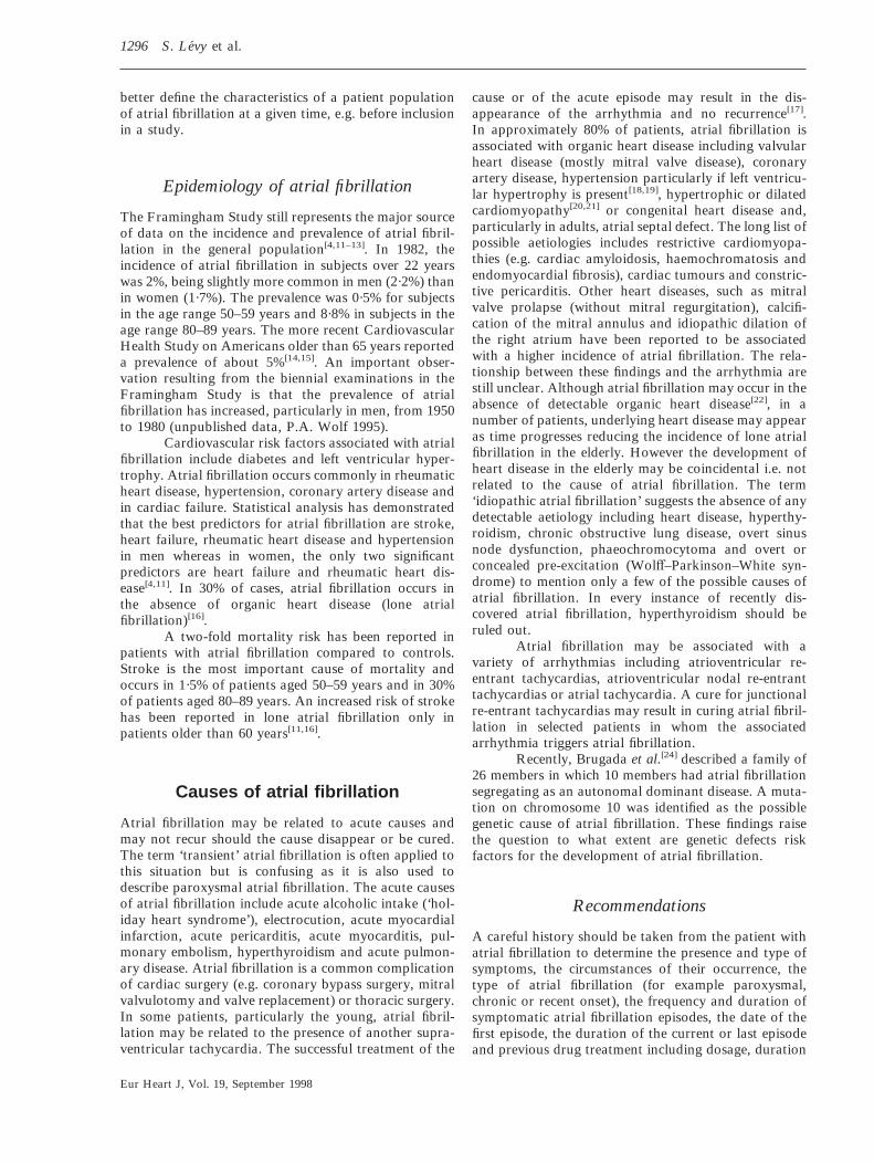
Table 2 Minimum work-up of the patient with atrial fibrillation
(1) History and physical examination 1.1 Define the presence and nature of symptoms 1.2 Define the clinical type of atrial fibrillation: paroxysmal, chronic or recent onset 1.3 Define the onset of the first symptomatic attack and/or date of discovery of atrial fibrillation. 1.4 Define the frequency, duration (shortest and longest episodes), precipitating factors and modes of termination (self-terminating
versus persistent) of symptomatic episodes. 1.5 Define the presence of an underlying heart disease or other possible identifiable cause (e.g. alcohol consumption, diabetes,
hyperthyroidism) which could be cured.
(2) Electrocardiogram 2.1 Left ventricular hypertrophy 2.2 Duration and morphology of the P waves in sinus rhythm 2.3 Evidence of repolarisation changes, bundle branch block, old myocardial infarction and other abnormality.
(3) Echocardiogram (M mode and bidimentional) 3.1 Evidence and type of underlying heart disease 3.2 Size of the left atrium 3.3 Left ventricular size and function 3.3 Left ventricular hypertrophy 3.4 Intracavitary thrombus (poor sensitivity).
(4) Thyroid test function If first discovery of atrial fibrillation, if the ventricular rate is difficult to control or if amiodarone has been used in the past.
of administration, effect of the drug and recurrence rate of symptoms. If the patient complains of angina, one should carefully establish whether this occurs only during attacks of atrial fibrillation or may occur independently of the arrhythmia. The latter would strongly suggest the presence of coronary artery disease. In recently discovered atrial fibrillation, the minimally acceptable work-up should include a 12-lead ECG, estimation of the basal thyroid-stimulating hormone level which if not suppressed, excludes hyperthyroidism with a high probability (Table 2). When appropriate, serum electrolytes should be checked. The presence of underlying heart disease should be detected by physical examination, M-mode and two-dimensional echocardiography evaluating left ventricular function, the size of the left atrium and the presence of intracardiac thrombus. In selected patients, documentation of atrial fibrillation may require 24 h ambulatory electrocardiographic recordings, the use of event recorders or exercise testing. Such tests may also be useful in some cases to evaluate the role of autonomic nervous system in atrial fibrillation initiation.
Mechanisms of atrial fibrillation
The mechanism of atrial fibrillation has been the subject of speculation for many years. Two theories have been advanced: enhanced automaticity involving one or more foci firing rapidly or re-entry involving one or more circuits[25,26]. The focal origin is supported by experimental models of aconitine-induced atrial fibrillation and by pacing-induced atrial fibrillation. Recent studies[26]
support the multiple re-entrant wavelet hypothesis of Moe et al.[25] and the concept that atrial fibrillation may involve a critical number of re-entrant circuits. Recently, it has been shown that rapidly firing atrial foci may be
responsible for atrial fibrillation, at least in a selected group of patients[27]. Ablation of such foci may result in the cure of atrial fibrillation. Despite recent advances, many questions on the mechanism of atrial fibrillation remain unanswered. Thus, in a recent model of experimental atrial fibrillation, a mixed form of functional and anatomical re-entry was found with a consistent involvement of Bachman’s bundle[28].
Mapping of atrial fibrillation
There is substantial evidence showing that most atrial fibrillation are related to re-entry. Unlike other arrhythmias in which a single circuit is generally identified, atrial fibrillation can involve a minimum of five or more circuits[29]. Thus, smaller circuits (the product of conduction velocity and refractoriness) on larger atria favour the development of atrial fibrillation. Using mapping techniques during atrial fibrillation in patients undergoing surgery for the Wolff–Parkinson–White syndrome, three patterns of induced atrial fibrillation were identified[28]. Type I showed single wavefronts propagating across the right atrium. Type II showed one or two wavefronts and type III multiple activation wavelets propagating in different directions. This study demonstrated that a number of re-entrant circuits with different dimensions account for these different types of atrial fibrillation. More recently, the role played by anisotropy in atrial fibrillation, possibly related to the orientation of atrial fibres and to the presence of pectinate muscles within the atria was investigated[30]. Using video imaging and an original experimental and an epicardial optical mapping technique, heterogenous breakthrough patterns over the epicardium, wave collisions and incomplete re-entry were found.
Eur Heart J, Vol. 19, September 1998
1298 S. Levy et al.
Development of atrial fibrillation in an experimental model
Atrial fibrillation has a tendency for self-perpetuation. Pharmacological and electrical cardioversion of atrial fibrillation have a higher success rate when atrial fibrillation has been present for less than 24 h[31]. The presence of atrial fibrillation for longer periods adversely affects the ability to restore and maintain sinus rhythm. These clinical observations have recently been substantiated by experimental studies. Wijfells et al.[32] used a goat model in which an automatic fibrillator detected spontaneous termination of induced atrial fibrillation and re-induced it by delivering a burst of electrical stimuli. They noted that initially, electrically-induced atrial fibrillation terminated spontaneously but following repetitive inductions, progressively longer episodes occurred and finally sustained atrial fibrillation with an increase in atrial rate ensued (‘atrial fibrillation begets atrial fibrillation’)[32]. The increasing propensity for long-lasting atrial fibrillation was related to progressive shortening of effective refractory periods with increasing duration of episodes, a phenomenon known as ‘electrophysiologic remodelling’. These observations are in agreement with previous clinical observations by Attuel et al.[33] who showed that the atrial effective refractory period is short in atrial fibrillation patients and that a lack of physiological rate adaptation of the atrial effective refractory period exists, particularly at slow heart rates, in patients with paroxysmal atrial fibrillation. These observations were confirmed by recordings of action potentials in isolated tissue from fibrillating atria[33]. A good correlation was found between the atrial fibrillation intervals (FF intervals) and atrial functional refractoriness[35]. The duration of atrial monophasic action potentials in atrial fibrillation patients was found to be short following cardioversion and correlated with the instability of sinus rhythm[36]. Decreased inward current through L-type calcium channels is likely to play a major role in action potential shortenings[37].
Factors involved in the mechanism of atrial fibrillation in man
Studies in man[38–40] have shown that increased inhomogeneity of refractory periods and conduction velocity is present in atrial fibrillation patients. Increased dispersion of refractoriness has been considered to be one of the major factors linked to inducibility and persistence of atrial fibrillation[36]. Slowing of conduction is also involved in the genesis of atrial fibrillation, particularly in diseased hearts[40]. Structural changes in atrial tissue may be one of the underlying factors for dispersion of refractoriness in atrial fibrillation. Other factors involved in the induction or maintenance of atrial fibrillation include premature beats, the interaction with the autonomic nervous system, atrial stretch[41], anisotropic conduction[42], and the ageing process. Among the
anatomical and histological changes that take place in atrial fibrillation, interruption of sympathetic and parasympathetic fibres may cause supersensitivity to catecholamines and acetylcholine, and contribute to increased dispersion of refractoriness[41].
Another important concept is that a critical mass of atrial tissue is necessary for atrial fibrillation to be sustained. This is supported by the success of the operative Maze procedure[44] and of catheter ablation of the atrial myocardium, both in experimental atrial fibrillation and in humans, which aim at reducing the mass of contiguous atrial tissue to the degree that it is no longer able to sustain atrial fibrillation. The maintenance of atrial fibrillation, once it is initiated, may involve the size of the atria and the distance between depolarization wavefronts. The concept of the wavelength, i.e. the product of effective refractory period and conduction velocity, was recently introduced[26]. The length of the electrical wave should not exceed the length of the path since it would otherwise extinguish itself. For an electrical impulse to propagate around an area of block, slow conduction must be present to allow the fibres located ahead to recover and become excitable again. A short refractory period or/and slow conduction will shorten excitation wave length, and thus, maintain reentry.
Aside from the changes related to the underlying heart disease, structural changes are present in atrial fibrillation including fibrosis, necrosis, fat and amyloid infiltration and inflammation[45]. Histological examination of atrial tissue of patients with atrial fibrillation has shown patchy fibrosis resulting in juxtaposition of diseased atrial fibres with normal atrial fibres which may account for inhomogeneity in atrial refractoriness[46,47]. Fibrosis may be a reaction to an inflammatory or degenerative process which is difficult to detect. The sinus node may also be involved by fibrosis or by fatty infiltration. Atrial fibre hypertrophy has also been described as a major and sometimes the sole histological change in atrial fibrillation patients[46]. Infiltration of the atrial myocardium may be suspected in amyloidosis, sarcoidosis or haemochromatosis. Histological changes were recently reported in lone atrial fibrillation[47] biopsy specimens and were consistent with myocarditis in 66% of patients. Other anatomical predisposing factors to atrial fibrillation include atrial hypertrophy and atrial dilatation. However, in most patients with atrial fibrillation, it is seldom possible to identify the underlying anatomical process responsible for the arrhythmia.
The role of the autonomic nervous system in the initiation of atrial fibrillation has been emphasized by Coumel et al.[48]. Atrial fibrillation may result from increased vagal tone (vagally-induced atrial fibrillation) and may occur during the night or after meals particularly in male patients without organic heart disease. In contrast, some patients may have atrial fibrillation induced by exercise, emotion and isoproterenol infusion (catecholamine-induced atrial fibrillation). In some patients one or the other mechanism may be predominant. However, frequently no clear distinction can be made between the various components based on a single
Eur Heart J, Vol. 19, September 1998
Task Force Report 1299
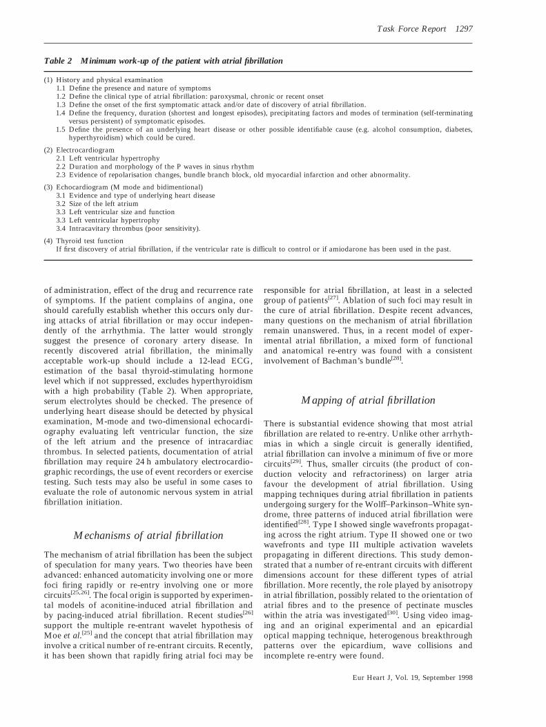
Table 3 Prevention of recurrences of atrial fibrillation following cardioversion
� Mechanism of arrhythmia: random reentry � Vulnerable parameters: Atrial refractoriness
Sympathetic or parasympathetic tone Premature atrial contractions or premature ventricular contractions (to be suppressed)
Therapeutic choice: (1) Increase atrial refractoriness (2) Decrease sympathetic orparasympatheti tone (3) Decrease ectopic activity
Targets: (1) Sodium channels or/and potassium channels (2) Beta-adrenergic receptors, muscarinic receptors (3) Decrease automaticity
Drugs: (1) Sodium and potassium channel blockers (2) Beta-antagonists, muscarinic antagonists (3) Sodium and potassium channel blockers
history. The mode of initiation of atrial fibrillation may also differ in the same patient over time. Premature beats play an important role as the initiating event in most cases of atrial fibrillation[48].
Heart rate variability has been used recently to study the role of the autonomic nervous system in atrial fibrillation patients. Signs of vagal predominance have been observed in patients with vagally-mediated atrial fibrillation and signs of sympathetic predominance were detected in approximately half of the patients with suspected cathecholamine-induced atrial fibrillation[49]. Experimental work in dogs has shown that vagal denervation of the atria could prevent induction of atrial fibrillation[8].
Clinical Implications
A better understanding of the mechanisms of atrial fibrillation would be an important step for future progress in defining appropriate therapy. The finding that atrial refractory periods are short in patients with atrial fibrillation has prompted the use of antiarrhythmic agents which prolong atrial refractoriness. According to the Sicilian Gambit approach[50], the target should be sodium channels or potassium channels (Table 3). In order to increase atrial refractoriness, sodium channel blockers such as quinidine, procainamide, disopyramide, propafenone or flecainide or antiarrhythmic agents whose major effect is to block potassium channels. Quinidine and disopiramide work by their effect on potassium channels. Flecainide works by blocking conduction. Agents which prolong action potential duration such as amiodarone or sotalol, are also appropriate antiarrhythmic agents. Some agents, such as ibutilide prevent the inactivation of the slow inward sodium current.
Electrophysiological and possibly structural changes take place within the first 24 h following atrial fibrillation, resulting in an electrical ‘remodelling’ and shortening of the refractory periods in the atria. In order to prevent or reverse these changes, prompt cardioversion of atrial fibrillation seems desirable. it may result in a higher success rate in restoration of sinus rhythm and
possibly in prevention of atrial fibrillation recurrences. This new concept suggested by animal experiments remains[34] to be proven in man and may have important clinical implications.
Prevention of embolic complications and indications of anticoagulation in
atrial fibrillation
Evaluation of embolic risk
Prevention of embolic complications is one of the major end-points of the therapeutic strategy of atrial fibrillation. The embolic risk is related to the presence and nature of underlying heart disease[11,13]. According to the Framingham study, there is a 5·6-fold embolic risk in non-rheumatic atrial fibrillation as compared to controls and a 17·6-fold risk in atrial fibrillation of rheumatic origin[4]. This is also consistent with the Reikjavik[51] and the Whitehall[52] studies which showed that the overall embolic risk is seven-fold higher when atrial fibrillation is present. Non-rheumatic atrial fibrillation is held responsible for a large percentage of strokes, being present in about 15–20% of cerebrovascular accidents of ischaemic origin[52,53]. There is no general agreement as to whether paroxysmal atrial fibrillation or chronic atrial fibrillation are associated with differences in embolic risk, although some data suggest that chronic atrial fibrillation carries a higher risk (6% per year) than paroxysmal atrial fibrillation (2–3% per year)[54]. Analysis of pooled data from five randomized controlled trials showed that the type of atrial fibrillation (paroxysmal or chronic) and the length of time the patient was in atrial fibrillation, had no discernible effect on stroke rate. The embolic risk seems to be highest after the onset of atrial fibrillation, during the first year and early after conversion to sinus rhythm. Cerebrovascular accidents associated with atrial fibrillation occur in a higher percentage in the elderly. They were found to represent 6·7% of the total number of cerebrovascular accidents in the 50-to59-year-old group and 36·2% in the 80-to-89-year-old
Eur Heart J, Vol. 19, September 1998
1300 S. Levy et al.
group[12]. Patients with a history of embolic stroke are at a higher risk of recurrence. Other clinical variables that were found to increase embolic risk, are a history of hypertension, coronary artery disease or congestive heart failure and cardiomegaly on chest roentgenogram and duration of atrial fibrillation for more than 1 year[52]. In the pooled data set, independent risk factors for stroke identified with multivariant analysis were increasing age, previous stroke or transient ischaemic attack, left atrial size, history of hypertension and diabetes. Transoesophageal echocardiographic findings such as left atrial thrombi, spontaneous echo contrast, ‘low-flow’ left atrial appendage and altered left ventricular function were suggested to be associated with an increased embolic risk[57–59]. Several studies have estimated the risk of embolic events in non-rheumatic atrial fibrillation to be around 5% per year, being lower in younger patients[55].
Controlled trials on prevention with warfarin or aspirin
Large trials have been conducted in recent years on the primary prevention of embolic events in patients with atrial fibrillation[60–66]. The primary end-point was to evaluate whether warfarin or aspirin reduced systemic embolism. The results of these trials were concordant showing a risk reduction ranging from 44% to 81%[59–62]
with warfarin. Anticoagulation was also effective in the secondary prevention of embolic complications in patients with non-rheumatic atrial fibrillation who had a recent cerebrovascular accident[65–67]. Warfarin reduced the incidence of cerebrovascular accident from 12% (placebo group) to 4% (warfarin). However, the use of warfarin was associated with an increased risk of bleeding. This risk was higher when the international normalized ratio was above 4. The Boston Area Anticoagulation Trial (BAATAF)[61] has shown that warfarin was effective at international normalized ratio levels (between 2 and 3) which were not associated with an increased risk of haemorrhage. This study[61] also showed that the use of warfarin was associated with a decreased mortality.
There have been several randomized trials comparing the efficacy of warfarin to that of aspirin. In the Atrial Fibrillation Aspirin and Anticoagulation (AFASAK) trial[58], patients on low-dose aspirin (75 mg . day�1) did not do better than patients on placebo whereas in the Stroke Prevention in Atrial Fibrillation (SPAF) trial[60], the use of aspirin at higher does (325 mg . day�1), was associated with some benefit. In the SPAF II trial[68], warfarin and aspirin resulted in a low-risk of stroke (1·3% and 1·9%, respectively) in patients under 75 years of age. Minor haemorrhagic events were higher in the warfarin group (20%) than in the aspirin group (11%) in patients over age 75. Major bleeding in the elderly was 5% per year in the warfarin group as compared with 1·6% in the aspirin group.
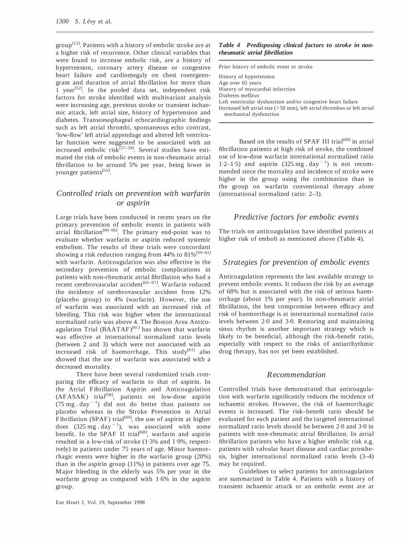
Table 4 Predisposing clinical factors to stroke in nonrheumatic atrial fibrillation
Prior history of embolic event or stroke
History of hypertensionAge over 65 yearsHistory of myocardial infarctionDiabetes mellitusLeft ventricular dysfunction and/or congestive heart failureIncreased left atrial size (>50 mm), left atrial thrombus or left atrial
mechanical dysfunction
Based on the results of SPAF III trial[68] in atrial fibrillation patients at high risk of stroke, the combined use of low-dose warfarin international normalized ratio 1·2–1·5) and aspirin (325 mg . day�1) is not recommended since the mortality and incidence of stroke were higher in the group using the combination than in the group on warfarin conventional therapy alone (international normalized ratio: 2–3).
Predictive factors for embolic events
The trials on anticoagulation have identified patients at higher risk of emboli as mentioned above (Table 4).
Strategies for prevention of embolic events
Anticoagulation represents the last available strategy to prevent embolic events. It reduces the risk by an average of 68% but is associated with the risk of serious haemorrhage (about 1% per year). In non-rheumatic atrial fibrillation, the best compromise between efficacy and risk of haemorrhage is at international normalized ratio levels between 2·0 and 3·0. Restoring and maintaining sinus rhythm is another important strategy which is likely to be beneficial, although the risk-benefit ratio, especially with respect to the risks of antiarrhythmic drug therapy, has not yet been established.
Recommendation
Controlled trials have demonstrated that anticoagulation with warfarin significantly reduces the incidence of ischaemic strokes. However, the risk of haemorrhagic events is increased. The risk–benefit ratio should be evaluated for each patient and the targeted international normalized ratio levels should be between 2·0 and 3·0 in patients with non-rheumatic atrial fibrillation. In atrial fibrillation patients who have a higher embolic risk e.g. patients with valvular heart disease and cardiac prosthesis, higher international normalized ratio levels (3–4) may be required.
Guidelines to select patients for anticoagulation are summarized in Table 4. Patients with a history of transient ischaemic attack or an embolic event are at
Eur Heart J, Vol. 19, September 1998
Task Force Report 1301
even a higher risk of recurrence and should be anticoagulated. Anticoagulation should be considered in patients with chronic atrial fibrillation in whom cardioversion failed or is not indicated, particularly if factors predisposing to stroke are present. The presence of a thrombus in the left heart cavities or of left atrial spontaneous contrast echos is another indication for anticoagulation. In patients with paroxysmal atrial fibrillation, the indication for anticoagulation should also be based on the presence and type of underlying heart disease and of other predisposing factors. The decision to anticoagulate has to be taken on an individual basis.
The results of trials utilizing aspirin are not convincing. Therefore, aspirin should only be used when anticoagulation is needed but contra-indication to warfarin is present or when the risk of stroke is low, for example in a patient younger than 60 years with no evidence of heart disease. Other coagulant or antiplatelet strategies have not yet been evaluated in patients with atrial fibrillation.
Conversion of atrial fibrillation to sinus rhythm
Restoring sinus rhythm has several potential benefits: relief of patient symptoms, improved hemodynamic status and possibly reduced embolic risk. Spontaneous conversion to sinus rhythm is observed in up to 48% of patients with paroxysmal atrial fibrillation and patients with recent onset (within 24 h) atrial fibrillation[69–71]. The duration of atrial fibrillation is the most important determinant of spontaneous conversion to sinus rhythm, with a decreasing tendency to spontaneous conversion with increasing duration of atrial fibrillation. The decision whether and when to cardiovert atrial fibrillation pharmacologically or electrically remains an important clinical problem, for which sufficient data are not yet available from large studies.
Pharmacological cardioversion
Pharmacological therapy aimed at restoring sinus rhythm may play a role in patients with atrial fibrillation of less than 48 h duration since the conversion rate falls dramatically when the arrhythmia has lasted longer. When atrial fibrillation exceeds 48 h duration, the current recommendation is to provide anticoagulant therapy for 3 weeks before attempting arrhythmia conversion to prevent embolic events[72]. If atrial fibrillation persists more than 48 h, the efficacy of pharmacological cardioversion decreases and electrical cardioversion is the most likely therapy to be successful. In a hospital setting, it is recommended to start heparin therapy immediately upon admission of a patient with recent onset atrial fibrillation, since the duration of the arrhythmia cannot be predicted.
In recent onset atrial fibrillation of less than 48 h, several antiarrhythmic agents have been proposed for restoration of sinus rhythm. In evaluating efficacy, the high spontaneous conversion rate of recent onset atrial fibrillation and the time needed for cardioversion must be taken into account[73]. Placebo-controlled data are therefore needed to confirm the efficacy of a given treatment. The antiarrhythmic efficacy must be evaluated within a few hours according to the pharmacokinetics and the pharmacodynamics of the drug itself[73]. Overall efficacy evaluated at 24 h following drug administration is potentially inaccurate and misleading since it might include in both the drug-limb and placebo, a high proportion of patients with spontaneous cardioversion.
Atrial fibrillation may profoundly affect the quality of life of the patients due to its abrupt onset and frequent recurrences. It has been suggested but not yet proven that an effective treatment administered within a short period of time after the onset of atrial fibrillation may reduce the number and duration of the attacks, may shorten the duration of hospitalizations and be cost-effective.
Since atrial fibrillation is not a life-threatening arrhythmia, every treatment offered should be safe and should not have deleterious effects. Several drugs influence the electrophysiological atrial substrate such as quinidine, procainamide, disopyramide, propafenone, felcainide, cibenzoline, amiodarone, sotalol and others. Digoxin was the favoured drug to terminate atrial fibrillation until controlled studies[69–71] demonstrated that it was no better than placebo. However, in almost all previous positive but uncontrolled studies, digoxin was used for atrial fibrillation termination in patients with congestive heart failure, a setting in which the drug may restore sinus rhythm indirectly by improving the haemodynamic status through its positive inotropic effect rather than by a direct electrophysiological effect.
A review of the literature on episodic treatment aimed at restoring sinus rhythm in recent onset atrial fibrillation, showed that there is no placebo-controlled study demonstrating the safety and efficacy of the use of intravenous or oral quinidine, procainamide or disopyramide. Open studies and placebo-controlled studies using intravenous flecainide or intravenous propafenone have shown that these agents were able to restore sinus rhythm within hours in up to 81% of patients[74–77]. One of the advantages of the pharmacological cardioversion of atrial fibrillation using the intravenous route is that it is performed under medical supervision and electrocardiographic monitoring.
Oral flecainide and propafenone have proved useful in the acute termination of recent onset atrial fibrillation[78–80] and in long-term therapy[81]. A single oral loading dose of 600 mg of propafenone or 300 mg of flecainide was studied in placebo-controlled groups with success rates of 50% at 3 h and 70–80% at 8 h for both drugs within a mean of 2 to 3 h after administration[78–80]. The treatment of atrial fibrillation with class IC antiarrhythmic drugs may be complicated by conversion of atrial fibrillation into atrial flutter or
Eur Heart J, Vol. 19, September 1998
1302 S. Levy et al.
tachycardia which may lead to a fast ventricular response (2:1 or 1:1 atrioventricular conduction). This has been reported in up to 5% of patients on long-term treatment[81]. Combination therapy with beta-blocker is advised in order to protect the ventricle from this effect. It is still a matter of controversy whether the slight beta-blocking effect of propafenone could be useful in preventing this complication[82,83]. No instance of atrial flutter with 1:1 atrioventricular conduction, however, has been reported so far in several hundred patients treated with an acute oral dosing with either drug[73,78–80]. Class IC drugs should not be administered in patients with heart failure, low ejection fraction or major conduction disturbances.
A bolus of oral amiodarone is frequently used for acute termination of recent onset atrial fibrillation (15 mg . kg�1) although no controlled study is available on the efficacy and safety of such treatment. A recent study[83] showed that 600 mg . day�1 of amiodarone converted about 20% of patients, who failed serial cardioversion and alternative drugs, with no adverse effect. Intravenous amiodarone has been used in the treatment of recent onset atrial fibrillation with reported efficacy rates ranging from 25 to 83%[85,86]. It is generally used in patients with atrial fibrillation and acute myocardial infarction or with left ventricular dysfunction in whom class IC drugs are contraindicated. Ibutilide, a class III antiarrhythmic agent, has been approved in the U.S.A. for intravenous termination of atrial fibrillation[87]. Ventricular proarrhythmia, torsades de pointes or sustained ventricular tachycardia, ranged from 1% to 4%[88]. Dofetilide, a class III antiarrhythmic agent, has shown a good efficacy in the termination of atrial fibrillation[88]. It has been recently reported that it did not affect mortality in patients with heart failure and depressed systolic function and in post-myocardial infarction patients at high risk of sudden death. Torsades de pointes is a potential risk of the use of class III antiarrhythmic agents.
When atrial fibrillation is secondary to hyperthyroidism, cardioversion should be delayed until thyroid function has returned to normal. Atrial fibrillation complicating cardiac surgery is common and tends to be a self-limited phenomenon. Calcium antagonists and betablockers have been used in atrial fibrillation following cardiac surgery but their role remains to be defined in this setting.
Recommendations
Pharmacological cardioversion of recent onset atrial fibrillation requires a careful consideration of the clinical setting and knowledge of the pharmacological properties of the antiarrhythmic drugs to be used. For restoration of sinus rhythm, class IC drugs administered either orally or intravenously seem efficient and safe in patients without underlying heart disease. In patients with ischaemic heart disease, low left ventricular ejection fraction, heart failure or major conduction disturbance,
class IC drugs should be avoided for restoring sinus rhythm. In any event, in atrial fibrillation occurring in patients with acute myocardial infarction, with low ejection fraction or after cardiac surgery, the role of antiarrhythmic therapy for restoration of sinus rhythm remains to be ascertained.
Electrical cardioversion
Electrical cardioversion may be indicated in patients with an episode of persistent (non-self terminating) atrial fibrillation associated with haemodynamic deterioration, either after failure of pharmacological cardioversion or as first line therapy[89–94]. External (transthoracic) direct current electrical cardioversion remains the technique of choice for restoring sinus rhythm in patients with chronic atrial fibrillation[89]. One or several[2–5] shocks may be delivered during the same session. Technical aspects concerning external direct current cardioversion[90,91] deserve important consideration including electrode size and position of electrodes (antero-apical versus antero-posterior), transthoracic impedance (which may be influenced by pressure on electrodes, conductive electrode gel, previous shocks), output waveform (most available external defibrillators use a monophasic truncated exponential waveform) and stored energy (50–400 J). The recommended initial energy is 200 J as 75% or more patients are successfully cardioverted with this energy[31]. Higher energies (360 J) are needed if a 200 J shock fails to restore sinus rhythm. Proper R wave synchronization is essential to avoid the rate occurrence of shock-induced ventricular fibrillation.
Success rates with external cardioversion range from 65% to 90%[89–95] provided that the abovementioned technical conditions are met. There are more secondary failures (early recurrences) than primary failures. The immediate success rates for external cardioversion predominantly depend on the duration of the arrhythmia. Other factors which may affect the success rates of external cardioversion include the weight of the patient and the presence of pulmonary disease[92] which may affect transthoracic impedance. The size of the left atrium is more related to the maintenance of sinus rhythm than to the immediate success rate. There is little evidence at present on the effect of antiarrhythmic agents on energy requirements, and the success rates of cardioversion.
Electrical external cardioversion is a simple, efficient and safe technique provided it is performed under proper anticoagulation, the patient is properly prepared, and the shocks are R-wave synchronized. Complications are rare but include systemic embolism, ventricular extrasystoles, non-sustained or sustained ventricular arrhythmias, sinus bradycardia, hypotension, pulmonary oedema and transient ST-T segment elevation. Restoration of sinus rhythm may unmask sinus node dysfunction or advanced atrioventricular block. If atrioventricular block or sinus dysfunction are suspected, prophylactic temporary ventricular pacing is recommended.
Eur Heart J, Vol. 19, September 1998
Task Force Report 1303
A technique of high energy (200 J or 300 J) electrical direct current internal cardioversion was shown to be particularly useful in patients who failed both external and pharmacological conversion[95,96]. The technique uses the proximal electrode of a quadripolar catheter as a cathode and a backplate as anode. In high energy internal cardioversion, barotrauma (stretch in cardiac structures) may play a role. In a study which randomized external versus internal cardioversion[95], the latter proved to be superior with no more recurrences over long-term follow-up. This technique may be useful in obese patients and in patients with chronic obstructive lung disease. More recently, a technique for low-energy (<20 J) cardioversion of atrial fibrillation using biphasic shocks and two large surface electrode catheters positioned in the right atrium (cathode) and the coronary sinus (anode) has been described[97–101]. An electrode-catheter in the left pulmonary artery may be used as an alternative after failure to catheterize the coronary sinus or as first choice[98]. Low-energy cardioversion restored sinus rhythm in 70–89% of various subsets of patients with atrial fibrillation including patients who failed external cardioversion and atrial fibrillation complicating diagnostic or ablation procedures[102]. This technique which does not require general anaesthesia, is under intensive evaluation and may also be useful for pre-implantation testing of patients in whom an atrial defibrillator is considered. For internal cardioversion, both proper R wave synchronization and delivery or shocks following RR intervals �500 ms are essential in order to avoid ventricular proarrhythmia. In the two cases of ventricular proarrhythmia so far reported[101,103], one or both of these important technical precautions were neglected.
Anticoagulation for restoration of sinus rhythm
In patients with atrial fibrillation lasting 48 h or more, oral anticoagulation for a minimum of 3 weeks before and one month after cardioversion is recommended. There is little good evidence for these specific recommendations, however, the risk of embolic event ranges from 1% to 5·3%[104] in non-anticoagulated patients. These embolic complications were initially attributed to dislodgement of pre-existing intracardiac thrombi. There is evidence showing that anticoagulation prior to cardioversion results in resolution of left atrial thrombi and reduction of embolic complications following cardioversion of atrial fibrillation[104]. As transoesophageal echocardiography can detect atrial thrombi with a high sensitivity, it was initially suggested that transoesophageal echocardiography could obviate the need for anticoagulation[105]. Reports of embolic events despite absence of thrombus at transoesophageal echocardiography showed that this was not the case[106]. More recently, it was shown that cardioversion of atrial fibrillation may be associated with transient mechanical
dysfunction of the left atrium, a phenomenon described as ‘atrial stunning’[107]. The left atrial appendage in which contractile function may be impaired, has been suggested as a source of emboli[108]. Following electrical cardioversion, spontaneous echo contrast developed or increased in 35% of patients[107]. The available data on the relationship between the mode of cardioversion and the worsening of the left atrial appendage function following cardioversion of atrial fibrillation, are conflicting. The ‘stunning’ phenomenon has also been observed in spontaneous cardioversion and is likely to occur with pharmacological cardioversion as well. The ‘stunning’ may be more related to the previous duration of atrial fibrillation than to the cardioversion process itself.
An interesting approach combines transoesophageal echocardiography and pre-transoesophageal echocardiography heparin in patients with atrial fibrillation longer than 48 h, a strategy which reduces duration of hospitalization and may be cost-effective[109]. In the absence of intracardiac thrombus, early cardioversion is performed. When a thrombus is detected, anticoagulation is undertaken for 6 weeks or more and transoesophageal echocardiography repeated. If the thrombus resolves, direct current cardioversion is performed. As a rule, persistence of a thrombus constitutes a contra-indication to cardioversion.
The use of warfarin is still needed for a minimum of 1 month after successful cardioversion as resumption of mechanical atrial function may be delayed as late as 3 weeks after sinus rhythm has been restored. The superiority of the systematic use of transoesophageal echocardiography prior to cardioversion has not yet been established. The answer may be given by the ongoing ACUTE (Assessment of Cardioversion Using Transesophageal Echocardiography study), the pilot of which showed that the transoesophageal echocardiography approach allows identification of patients who can safely undergo cardioversion without prior prolonged anticoagulation[109].
Recommendations
Restoration of sinus rhythm is a desirable end-point in patients with persistent (non-self terminating) paroxysmal atrial fibrillation and selected patients with chronic atrial fibrillation. Electrical cardioversion is the technique of choice provided there is no temporary contraindication such as digitalis toxicity, hypokalaemia, acute infectious or inflammatory diseases or non-compensated heart failure. External cardioversion may be hazardous in patients with severely depressed left ventricular function because of the risk of pulmonary oedema. Anticoagulation is recommended for 3 weeks before cardioversion in patients with atrial fibrillation of 48 h or more duration and for a minimum of 4 weeks afterwards. As general anaesthesia is required for external cardioversion, contra-indications to general anaesthesia should be ruled out. Another approach, using transoesophageal echocardiography and
Eur Heart J, Vol. 19, September 1998
1304 S. Levy et al.
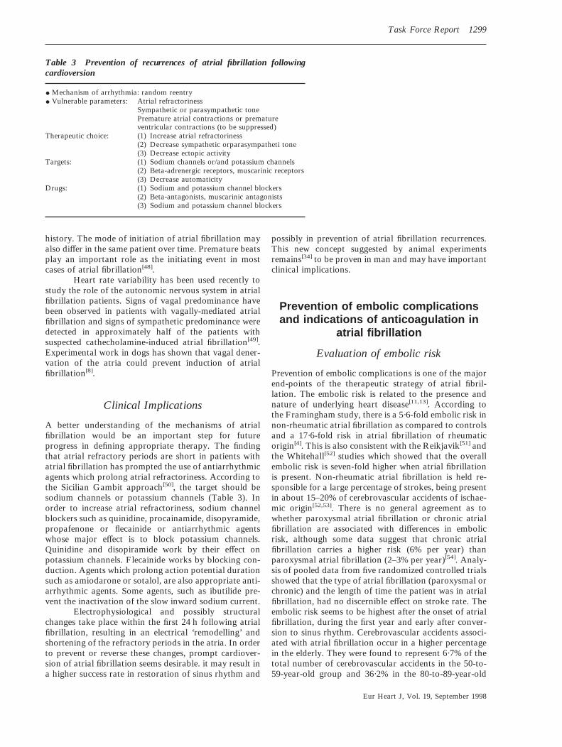
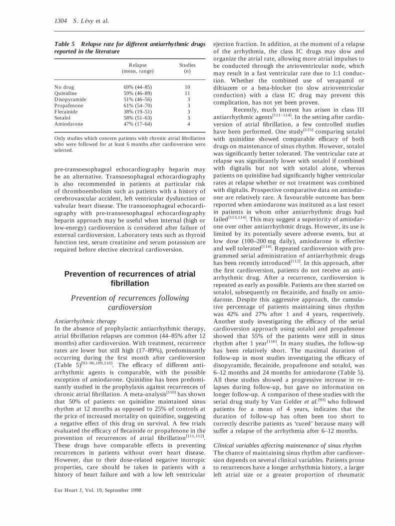
Table 5 Relapse rate for different antiarrhythmic drugs reported in the literature
Relapse Studies (mean, range) (n)
No drug 69% (44–85) 10 Quinidine 59% (46–89) 11 Disopyramide 51% (46–56) 3 Propafenone 61% (54–70) 3 Flecainide 38% (19–51) 3 Sotalol 58% (51–63) 3 Amiodarone 47% (17–64) 4
Only studies which concern patients with chronic atrial fibrillation who were followed for at least 6 months after cardioversion were selected.
pre-transoesophageal echocardiography heparin may be an alternative. Transoesophageal echocardiography is also recommended in patients at particular risk of thromboembolism such as patients with a history of cerebrovascular accident, left ventricular dysfunction or valvular heart disease. The transoesophageal echocardiography with pre-transoesophageal echocardiography heparin approach may be useful when internal (high or low-energy) cardioversion is considered after failure of external cardioversion. Laboratory tests such as thyroid function test, serum creatinine and serum potassium are required before elective electrical cardioversion.
Prevention of recurrences of atrial fibrillation
Prevention of recurrences following cardioversion
Antiarrhythmic therapy In the absence of prophylactic antiarrhythmic therapy, atrial fibrillation relapses are common (44–85% after 12 months) after cardioversion. With treatment, recurrence rates are lower but still high (17–89%), predominantly occurring during the first month after cardioversion (Table 5)[93–96,109,110]. The efficacy of different antiarrhythmic agents is comparable, with the possible exception of amiodarone. Quinidine has been predominantly studied in the prophylaxis against recurrences of chronic atrial fibrillation. A meta-analysis[110] has shown that 50% of patients on quinidine maintained sinus rhythm at 12 months as opposed to 25% of controls at the price of increased mortality on quinidine, suggesting a negative effect of this drug on survival. A few trials evaluated the efficacy of flecainide or propafenone in the prevention of recurrences of atrial fibrillation[111,112]. These drugs have comparable effects in preventing recurrences in patients without overt heart disease. However, due to their dose-related negative inotropic properties, care should be taken in patients with a history of heart failure and with a low left ventricular
ejection fraction. In addition, at the moment of a relapse of the arrhythmia, the class IC drugs may slow and organize the atrial rate, allowing more atrial impulses to be conducted through the atrioventricular node, which may result in a fast ventricular rate due to 1:1 conduction. Whether the combined use of verapamil or diltiazem or a beta-blocker (to slow atrioventricular conduction) with a class IC drug may prevent this complication, has not yet been proven.
Recently, much interest has arisen in class III antiarrhythmic agents[111–114]. In the setting after cardioversion of atrial fibrillation, a few controlled studies have been performed. One study[115] comparing sotalol with quinidine showed comparable efficacy of both drugs on maintenance of sinus rhythm. However, sotalol was significantly better tolerated. The ventricular rate at relapse was significantly lower with sotalol if combined with digitalis but not with sotalol alone, whereas patients on quinidine had significantly higher ventricular rates at relapse whether or not treatment was combined with digitalis. Prospective comparative data on amiodarone are relatively rare. A favourable outcome has been reported when amiodarone was instituted as a last resort in patients in whom other antiarrhythmic drugs had failed[113,114]. This may suggest a superiority of amiodarone over other antiarrhythmic drugs. However, its use is limited by its potentially severe adverse events, but at low dose (100–200 mg daily), amiodarone is effective and well tolerated[114]. Repeated cardioversion with programmed serial administration of antiarrhythmic drugs has been recently introduced[112]. In this approach, after the first cardioversion, patients do not receive an antiarrhythmic drug. After a recurrence, cardioversion is repeated as early as possible. Patients are then started on sotalol, subsequently on flecainide, and finally on amiodarone. Despite this aggressive approach, the cumulative percentage of patients maintaining sinus rhythm was 42% and 27% after 1 and 4 years, respectively. Another study investigating the efficacy of the serial cardioversion approach using sotalol and propafenone showed that 55% of the patients were still in sinus rhythm after 1 year[116]. In many studies, the follow-up has been relatively short. The maximal duration of follow-up in most studies investigating the efficacy of disopyramide, flecainide, propafenone and sotalol, was 6–12 months and 24 months for amiodarone (Table 5). All these studies showed a progressive increase in relapses during follow-up, but gave no information on longer follow-up. A comparison of these studies with the serial drug study by Van Gelder et al.[93] who followed patients for a mean of 4 years, indicates that the duration of follow-up has often been too short to correctly describe patients as ‘cured’ because many will suffer a relapse of the arrhythmia after 6–12 months.
Clinical variables affecting maintenance of sinus rhythm The chance of maintaining sinus rhythm after cardioversion depends on several clinical variables. Patients prone to recurrences have a longer arrhythmia history, a larger left atrial size or a greater proportion of rheumatic
Eur Heart J, Vol. 19, September 1998

Task Force Report 1305
mitral valve disease than those without. In addition, a low functional capacity may predict a poor arrhythmia prognosis. Due to the important role of the duration of the arrhythmia, restoration of sinus rhythm should be achieved quickly. An arrhythmia duration of only 3 months has been shown to impair the success of the serial cardioversion approach[93,116]. Remodelling of the atria during atrial fibrillation may lead to persistent morphological changes which may develop within a relatively short time period, and to tachycardia-induced electrical changes that may be more easily reversible. This suggests that cardioversion should be performed as early as possible after onset of the arrhythmia. Efficacy of cardioversion may be enhanced by patient counselling by explaining the symptoms suggestive of arrhythmia recurrence. Nevertheless, this approach still needs to be substantiated by a prospective randomized trial.
Clinical trials on atrial fibrillation A number of clinical trials on pharmacological therapy of atrial fibrillation are ongoing including PAFAC and PIAF in Germany, RACE, MEDCAR and VERDICT in The Netherlands, and AFFIRM and the U.S. Veterans Administration study in the U.S.A.[117]. The results of these trials and some of the protocols have not yet been published.
Prevention of recurrences of paroxysmal atrial fibrillation
In paroxysmal atrial fibrillation (defined as attacks lasting less than 7 days), the majority of attacks are self-terminating within 48 h. Others will have episodes which persist long enough to require cardioversion. The ideal end-point of therapy is to prevent attacks of atrial fibrillation but it may be satisfactory to reduce the frequency of occurrence and/or the duration of episodes. Another option is to slow heart rate during atrial fibrillation episodes, thereby alleviating patient symptoms. The superiority of one or the other approach, as mentioned above, has not yet been established[117].
To prevent recurrences of atrial fibrillation, one may choose to suppress the trigger mechanism (premature atrial depolarizations) or to change the atrial substrate by modifying atrial refractoriness, which may be another vulnerable parameter[118].
In which patients and when to use antiarrhythmic agents?
In the classification system described above[9], three clinical aspects of paroxysmal atrial fibrillation were identified in such a way as to have implications for therapy. In the first attack of atrial fibrillation (group I), the likelihood of recurrences cannot be determined, and therefore, pharmacological treatment for prevention of recurrences may not be justified. Recurrent atrial
fibrillation (group II) may be asymptomatic, and recorded by Holter monitoring (IIA), or symptomatic and categorized according to the frequency of attacks (subgroups IIB or IIC). In asymptomatic paroxysmal atrial fibrillation (group IIA), the role of pharmacological treatment to prevent recurrences and stroke has not been established. In group IIB (infrequent symptomatic episodes), episodic treatment to terminate or slow the heart rate of the attack may be an acceptable option as opposed to long-term prophylaxis of recurrences. In group IIC (frequent symptomatic episodes), prevention of recurrences with sodium or potassium-channel blockers may be warranted. Atrial fibrillation resistant to antiarrhythmic drug therapy (group III) may lead to further investigations to uncover a possible mechanism, to the use of agents which have an effect on the atrioventricular node (e.g. calcium blockers, beta-blockers, digitalis) in order to slow the ventricular rate or to consider non-pharmacological therapy including atrioventricular node modification or ablation, surgery (Maze procedure), or implantable devices.
The evaluation of antiarrhythmic therapy in patients with paroxysmal atrial fibrillation is difficult because as previously stated, patients vary and the pattern of the arrhythmia (frequency, duration and mode of termination of attacks) may change over time. An interesting approach was reported by Anderson et al.[119] in patients with symptomatic paroxysmal atrial fibrillation using trans-telephonic monitoring. They randomized the patients to the largest tolerated dose of flecainide or placebo and evaluated the time to first recurrence and the interval between attacks. These two parameters were found to be significantly prolonged by flecainide. Similar results were also reported by Pietersen et al.[120] in a placebo-controlled study and by Clementy et al.[121] in a large cohort of patients. Propafenone was found to be effective in reducing the rate of recurrences of atrial fibrillation attacks in placebo-controlled studies[122,123] and to be as effective as sotalol in preventing recurrences of atrial fibrillation[124]. No controlled studies on the long-term efficacy and safety of sodium channel blockers (class I agents) in paroxysmal atrial fibrillation is presently available.
Proarrhythmic effects of antiarrhythmic agents
The decision to start antiarrhythmic drug therapy should consider the risk of recurrence on the one hand, and the risk of severe side-effects, e.g. ventricular proarrhythmia, on the other. The most severe proarrhythmic effect in atrial fibrillation patients is new onset ventricular fibrillation, ventricular tachycardia or torsade de pointes. Whereas class IA and class III antiarrhythmic drugs predominantly cause polymorphic ventricular tachycardia or torsades de pointes[125,126], class IC drugs usually induce monomorphic ventricular tachycardia, mostly in the setting of poor left ventricular
Eur Heart J, Vol. 19, September 1998
1306 S. Levy et al.
function[127,129]. A challenging clinical problem is the prediction and recognition of proarrhythmia early after initiation of drug therapy as well as the prevention of late out-of-hospital proarrhythmia during chronic treatment. Electrocardiographic signs, potentially useful in the prediction of proarrhythmia with class IA and III agents, include acute and excessive QT prolongation, pause-related TU wave changes and increased QT dispersion. Torsades de pointes may occur especially if there is a pre-existing QT prolongation, and the risk is enhanced by bradycardia (e.g. occurring after sudden conversion of ‘rapid’ atrial fibrillation) and hypokalaemia. Late proarrhythmia may occur after addition of drugs, like diuretics or during intercurrent bradycardia. Ventricular proarrhythmia with class IC drugs is more common in patients with a history of sustained ventricular tachycardia and in those with structural heart disease receiving a high dose[129]. It is important to note that in contrast to the quinidine-like drugs, ventricular proarrhythmia or sudden death is virtually absent in patients without overt heart disease treated with a class IC antiarrhythmic drug. Torsades de pointes are a potential proarrhythmic complication of sotalol particularly in patients with ventricular hypertrophy, cardiomegaly heart failure and of female gender[126]. Amiodarone has a low proarrhythmia rate[113,114].
Recommendations
Following repeated cardioversion of atrial fibrillation, prophylactic antiarrhythmic therapy using sodium or potassium channel blockers should be considered. The indication for therapy and selection of the antiarrhythmic agent should take into account the presence and nature of underlying heart disease, a history of congestive heart failure or myocardial infarction, left ventricular function, mode of initiation of atrial fibrillation, and frequency and duration of episodes (see proposed management strategy). The risk–benefit ratio related to the use of antiarrhythmic agents should be considered in every patient.
Rate control during atrial fibrillation
Rate control may be indicated in patients who failed antiarrhythmic therapy aimed at preventing recurrences or as an alternative therapy to the maintenance of sinus rhythm. Although the results of studies comparing these two strategies (maintenance of sinus rhythm versus rate control) are not yet available[117], it seems likely that restoring and maintaining sinus rhythm should be highly desirable.
Determinants of ventricular rate during atrial fibrillation
Heart rate during Af is determined by the structure and electrical properties of the atrioventricular node which
exhibits concealed conduction characteristics[130–134]. Fast ventricular rates cause symptoms because stroke volume is impaired, and may be responsible for heart failure in different forms. Sympathetic drive and vagal tone will modify conduction, and hence, ventricular rate[133,134]. In the absence of atrioventricular node or His-Purkinje system impairment of conduction, ventricular rate in atrial fibrillation should be considered as ‘uncontrolled’. The presence of accessory pathways with antegrade conduction (as in the Wolff–Parkinson–White syndrome) will also modify the ventricular rate. Although symptoms are often related to fast ventricular rates, irregularity of heart action may also play an important role in some patients[130].
Definition and criteria for rate control
Rate control during atrial fibrillation is poorly defined. It can be judged from clinical symptoms but also from ECG criteria. Haemodynamic data, e.g. based on echocardiographic measurements, are rare[135]. It can be assumed that ‘controlled heart rate’ during atrial fibrillation at rest does not imply an appropriate rate during exercise. Inappropriate fast heart rates may occur during even minor exercise in patients with atrial fibrillation, even when resting heart rates are ‘controlled’[133,134]. Criteria for rate control may also vary according to age. Rate control is (arbitrarily) considered to be present when heart rate ranges between 60 to 80 beats . min�1
at rest, and between 90 to 115 beats . min�1 during moderate exercise[134,135]. It is useful to consider heart rate trends on Holter recording in order to assess rate control, as proposed by Atwood et al.[136]. Exercise testing can be used to analyse heart rate at submaximal and maximal exercise. Furthermore, heart rate variability during atrial fibrillation provides additional insight in the heart rate control, and may bring independent information on survival[137–139].
Haemodynamic consequences of rapid heart rate
Rapid heart rate in atrial fibrillation may have an untoward effect on cardiac function resulting in a tachycardia-induced cardiomyopathy which may be reversed after control of heart rate[140,144]. Activation of neurohumoral vasoconstrictors and secretion of atrial natriuretic peptide may occur in atrial fibrillation. The loss of the atrial contribution to ventricular filling may also contribute to the hemodynamic changes that take place in atrial fibrillation.
Interventions for rate control
Control of heart rate may be achieved with pharmacological intervention or with atrioventricular node
Eur Heart J, Vol. 19, September 1998
Task Force Report 1307
modification or ablation using radiofrequency current (see ablation section). In the absence of antegradely conducting accessory pathways, drugs are needed which depress atrioventricular conduction. The effective refractory period of the atrioventricular node correlates very well with ventricular rates during atrial fibrillation, and drugs prolonging the atrioventricular node effective refractory period should be effective. Cholinergic activity is another pharmacological determinant that is sought in a drug[143]. In paroxysmal atrial fibrillation patients, sinus bradycardia and heart block may occur in certain conditions, and particularly in the elderly as an unwanted effect of pharmacological intervention especially with glycosides or calcium channel antagonists.
Digoxin
Digoxin is generally considered to be effective for rate control in atrial fibrillation, particularly when congestive heart failure is present[144]. This has not been proven for other atrial fibrillation patients. However, digoxin does not control the ventricular rate during exercise[142] and this is not related to inadequate serum digoxin levels[143]. Both findings are related to an indirect vagomimetic effect of glycosides. Recent data on ventricular rate in converters and non-converters (in a group with recent onset atrial fibrillation, without overt heart failure) supports the idea that digoxin indeed lowers the ventricular rate[70]. The effect is visible early after initiation of digoxin therapy, and could be explained by an early vagotonic effect on the atrioventricular node. However, the level of rate control with digoxin is not impressive. It takes about 6 to 12 h to achieve rates >100 beats . min�1[72]. Therefore, additional drugs to control atrioventricular nodal conduction were often prescribed in recent trials designed to convert atrial fibrillation with dioxin[142]. A controlled study has not found digoxin to be effective in the prevention of recurrences of atrial fibrillation[145].
Non-dihydropyridine calcium antagonists
The most commonly used agents are verapamil and diltiazem. Intravenously, both drugs are effective in emergency settings[146,147]. The negative inoptropic effect of oral calcium antagonists require their cautious use in patients with heart failure. Calcium antagonists are preferred to beta-blockers in patients with chronic obstructive pulmonary disease. It has been shown that exercise duration increases when verapamil or diltiazem are used to control heart rate[148]. However, it has been suggested that calcium antagonists might prolong paroxysms, making chronic use in paroxysmal atrial fibrillation less desirable. This contrasts with other investigational data showing that electrical remodeling is prevented by calcium channel blockers, making its use for rate control acceptable[148].
Beta-receptor blockers
Beta-receptor blockers are also used to control heart rate in atrial fibrillation patients[149–153]. Intravenous beta-blockade with propanolol, atenolol, metoprolol or esmolol can be of value in specific settings[153]. Chronic beta-blockade is a safe therapy, and will prevent the effects of excessive sympathetic tone. Atenolol provided better control of exercise-induced tachycardia than digoxin alone[150]. Pindolol with digoxin offered better rate protection than digoxin alone or combined with verapamil during exercise, and did not affect resting heart rate to the same extent[155]. Xamoterol, having more intrinsic sympathetic activity than pindolol, offered more rate control than digoxin and placebo during exercise, and avoided pauses and slow heart rates[151]. It tended to improve exercise duration and made patients less symptomatic. In chronic atrial fibrillation, it is also better in this respect than verapamil. In another study, xamoterol and atenolol were compared and exercise performance was lower with atenolol[150]. Beta-blockers should be used with caution in patients with heart failure. Clonidine (having anti-adrenergic properties) can be an alternative for hypertensive patients, as it reduces standing heart rates by 15–20%[153].
Other antiarrhythmic agents
Sodium and potassium channel blockers (primarily used for conversion to sinus rhythm or for maintenance of sinus rhythm) are not advocated for rate control. Class IA agents (quinidine and disopyramide) may even improve atrioventricular conduction, thereby facilitating fast rates, due to anticholinergic effects. Propafenone may control heart rate when paroxysms of atrial fibrillation recur, due to its effect on the atrioventricular node. Nevertheless, if atrial rhythm becomes more regular and slower, faster atrioventricular conduction may occur. In one study, dl-Sotalol was better than quinidine in controlling ventricular rate[115].
Amiodarone, which has both sympatholytic and calcium antagonistic properties, depresses atrioventricular conduction and is effective in controlling ventricular rate. However, it has not been properly investigated in this indication. It should not be used as a first line agent in this indication because of its side-effect spectrum.
New antiarrhythmic drugs (dofetilide, ibutilide) are effective for acute conversion of atrial flutter and atrial fibrillation[87,88] but are not effective as rate control agents.
Special considerations for the Wolff–Parkinson–White syndrome
The intravenous use of agents that slow atrioventricular nodal conduction and particularly calcium antagonists are contra-indicated in Wolff–Parkinson–White
Eur Heart J, Vol. 19, September 1998
1308 S. Levy et al.
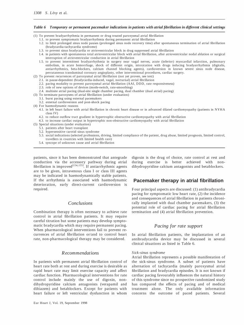
Table 6 Temporary or permanent pacemaker indications in patients with atrial fibrillation in different clinical settings
(1) To prevent bradyarrhythmia in permanent or drug treated paroxysmal atrial fibrillation 1.1. to prevent symptomatic bradyarrhythmia during permanent atrial fibrillation 1.2. to limit prolonged sinus node pauses (prolonged sinus node recovery time) after spontaneous termination of atrial fibrillation
(bradycardia-tachycardia syndrome) 1.3. to prevent sinus bradycardia or atrioventricular block in drug suppressed atrial fibrillation 1.4. in patients with spontaneous total atrioventricular block with atrial fibrillation, after atrioventricular nodal ablation or surgical
interruption of atrioventricular conduction in atrial fibrillation 1.5. to prevent intermittent bradyarrhythmia in surgery near vagal nerves, acute (inferior) myocardial infarction, pulmonary
embolism, in acute hemorrhage, shock of different origin, intoxication with drugs inducing bradyarrhythmia (digitalis, antiarrhythmics, beta-blockers, calcium channel blocking agents), cardioversion in known severe sinus node disease, percutaneous transluminal coronary angioplasty, other interventional procedures, cardiac surgery.
(2) To prevent recurrences of paroxysmal atrial fibrillation (not yet proven, see text) 2.1. in pause-dependent (bradycardia-induced, vagal, nocturnal) atrial fibrillation 2.2. pacing modality to prevent paroxysmal atrial fibrillation (AAI, DDD, rate responsiveness) 2.3. role of new options of devices (mode-switch, rate-smoothing) 2.4. multisite atrial pacing (dual-site single chamber pacing, dual chamber (dual atrial) pacing)
(3) To terminate paroxysms of atrial fibrillation (under evaluation) 3.1. burst pacing using external pacemakers 3.2. external cardioversion and post-shock pacing
(4) For haemodynamic reasons 4.1. in left heart failure with atrial fibrillation in chronic heart disease or in advanced dilated cardiomyopathy (patients in NYHA
class IV) 4.2. to reduce outflow tract gradient in hypertrophic obstructive cardiomyopathy with atrial fibrillation 4.3. to increase cardiac output in hypertrophic non-obstructive cardiomyopathy with atrial fibrillation
(5) Special situations (under evaluation) 5.1. patients after heart transplant 5.2. hypersensitive carotid sinus syndrome 5.3. social indications (selected professions, driving, limited compliance of the patient, drug abuse, limited prognosis, limited control,
travellers in countries with limited health care) 5.4. syncope of unknown cause and atrial fibrillation
patients, since it has been demonstrated that antegrade conduction via the accessory pathway during atrial fibrillation is improved[154,155]. If antiarrhythmic agents are to be given, intravenous class I or class III agents may be indicated in haemodynamically stable patients. If the arrhythmia is associated with haemodynamic deterioration, early direct-current cardioversion is required.
Conclusions
Combination therapy is often necessary to achieve rate control in atrial fibrillation patients. It may require careful titration but some patients may develop symptomatic bradycardia which may require permanent pacing. When pharmacological interventions fail to prevent recurrences of atrial fibrillation or/and to control heart rate, non-pharmacological therapy may be considered.
Recommendations
In patients with permanent atrial fibrillation control of heart rate both at rest and during exercise is desirable as rapid heart rate may limit exercise capacity and affect cardiac function. Pharmacological interventions for rate control include mainly the use of digoxin, nondihydropyridine calcium antagonists (verapamil and diltiazem) and betablockers. Except for patients with heart failure or left ventricular dysfunction in whom
digoxin is the drug of choice, rate control at rest and during exercise is better achieved with nondihydropyridine calcium antagonists and betablockers.
Pacemaker therapy in atrial fibrillation
Four principal aspects are discussed: (1) antibradycardia pacing for symptomatic low heart rate, (2) the incidence and consequences of atrial fibrillation in patients chronically implanted with dual chamber pacemakers, (3) the potential role of cardiac pacing for atrial fibrillation termination and (4) atrial fibrillation prevention.
Pacing for rate support
In atrial fibrillation patients, the implantation of an antibradycardia device may be discussed in several clinical situations as listed in Table 6.
Sick-sinus syndrome Atrial fibrillation represents a possible manifestation of the sick-sinus syndrome. A subset of patients have alternation of tachycardia (mainly paroxysmal atrial fibrillation and bradycardia episodes. It is not known if cardiac pacing favourably influences the natural history of this syndrome since no prospective randomized study has compared the effects of pacing and of medical treatment alone. The only available information concerns the outcome of paced patients. Several
Eur Heart J, Vol. 19, September 1998
Task Force Report 1309
non-randomized studies have compared the long-term effects of atrial and of ventricular pacing modes[156–160]. Results suggest a significant decrease in the incidence of evolution towards chronic atrial fibrillation with atrial pacing. After pooling the results of these different studies, the average annual incidence of chronic atrial fibrillation was 1·5% in atrially paced patients and 12% in the ventricularly paced groups. Some studies also showed a significant reduction in the incidence of thrombo-embolic events and a lower mortality rate[157]. These preliminary results were confirmed by a recently published prospective randomized study[161] which showed a trend for a lower incidence of atrial fibrillation in the atrially paced group. The incidence of chronic atrial fibrillation in patients surviving more than 1 year was 7% compared to 13% (ns) in the ventricular group. Interestingly, the incidence of thrombo-embolic events was significantly (P=0·0083) lower 5·5% vs 17·4%) in the atrially paced group. Finally, there was no significant difference in the mortality rate between the two groups. These data strongly suggest pacing the atrium when permanent pacing is indicated in patients with the ‘brady-tachy syndrome’, because of a lower incidence of thromboembolism and atrial fibrillation.
Chronic atrial fibrillation with symptomatic bradycardia related to atrioventricular block or to the concomitant use of necessary atrioventricular nodal depressant drugs may require pacing for rate support. A VVIR pacing mode is indicated.
Chronic atrial fibrillation with symptomatic chronotropic incompetence during exercise represents another possible indication. More than 50% of chronic atrial fibrillation patients have an abnormal response of heart rate during exercise. In some patients, the abnormal behaviour consists of an inappropriate bradycardia, resulting in a significant limitation in exercise capacity in the absence of drugs that slow heart rate. This situation may require the implantation of a rate-adaptive VVIR pacemaker even in the absence of significant bradycardia at rest[162].
Atrioventricular block resulting from atrioventricular nodal ablation or from attempted atrioventricular nodal modification for control of ventricular rate in patients with chronic or paroxysmal atrial fibrillation requires permanent pacing[163,164]. In the case of permanent atrial fibrillation, the choice of the pacing mode is clearly VVIR. In the case of paroxysmal atrial fibrillation, there is a general consensus to implant a rateresponsible DDDR pacemaker and to program a DDIR or a DDDR (with mode-switch and/or fallback algorithms) pacing mode. However, there is no definite evidence that the dual-chamber pacing mode will significantly contribute to the prevention of paroxysmal atrial fibrillation recurrences.
Atrial fibrillation and dual-chamber pacing
The incidence of atrial tachyarrhythmias and especially of atrial fibrillation, is generally underestimated in
patients chronically paced in a dual-chamber mode. Data from pacemaker and defibrillator Holter functions have shown an incidence of sustained atrial tachyarrhythmias. In 265 unselected patients, Guarrigue et al.[165] observed a 48·8% incidence of sustained atrial tachyarrhythmias (one or more episodes) during a follow-up ranging from 3 to 41 months. The relative incidence was 43·5% in patients without atrial tachyarrhythmias documented prior to implant and 64·7% in patients with previously documented atrial tachyarrhythmias. Most of the episodes were asymptomatic and lasted less than 24 h. A major consequence of these observations is a potentially high risk of pacemaker mediated tachycardias, by tracking the paced ventricular rate up to the programmed upper rate limit. This may result in severe symptoms especially in patients with structural heart disease and poor left ventricular function. To prevent this risk, most of the recent DDD/ DDDR pacemakers are equipped with algorithms for ventricular protection (fallback, dual-demand, mode switching). Such algorithms should be used in patients with electrically unstable atria.
Pacing for termination of atrial fibrillation
Despite the clear evidence of a reentrant mechanism in atrial fibrillation and the strong presumption of a (short) excitable gap, there is no evidence today that rapid burst atrial pacing reproducibly terminates atrial fibrillation in humans.
Pacing for prevention of atrial fibrillation
Several mechanisms have been postulated which might suggest a potential role of cardiac pacing in atrial fibrillation prevention. Atrial pacing may prevent the arrhythmogenic effect of bradycardia and of marked variation of heart rate causing dispersion of refractoriness. This may particularly apply in arrhythmia related to/or associated with bradycardia, such as vagallymediated atrial fibrillation[48]. Atrial fibrillation may be prevented by increasing the coupling interval of the initiating premature beat in the abnormal substrate. This result may be achieved either by pre-exciting the zone where reentry occurs, or by pacing at one or more sites which are opposite to the activation of the premature beat. This is the rationale for multi-site atrial pacing in atrial fibrillation prevention. Atrial fibrillation may also be prevented by suppressing the premature beats. Such an effect could be achieved by overdrive suppression of automatic foci.
The clinical relevance of theses concepts remains to be proven by prospective randomized trials, several of which are currently ongoing. Permanent atrial pacing has been shown to prevent atrial fibrillation recurrences and to reduce the need for antiarrhythmic drugs in a majority of patients. Patients with major atrial
Eur Heart J, Vol. 19, September 1998
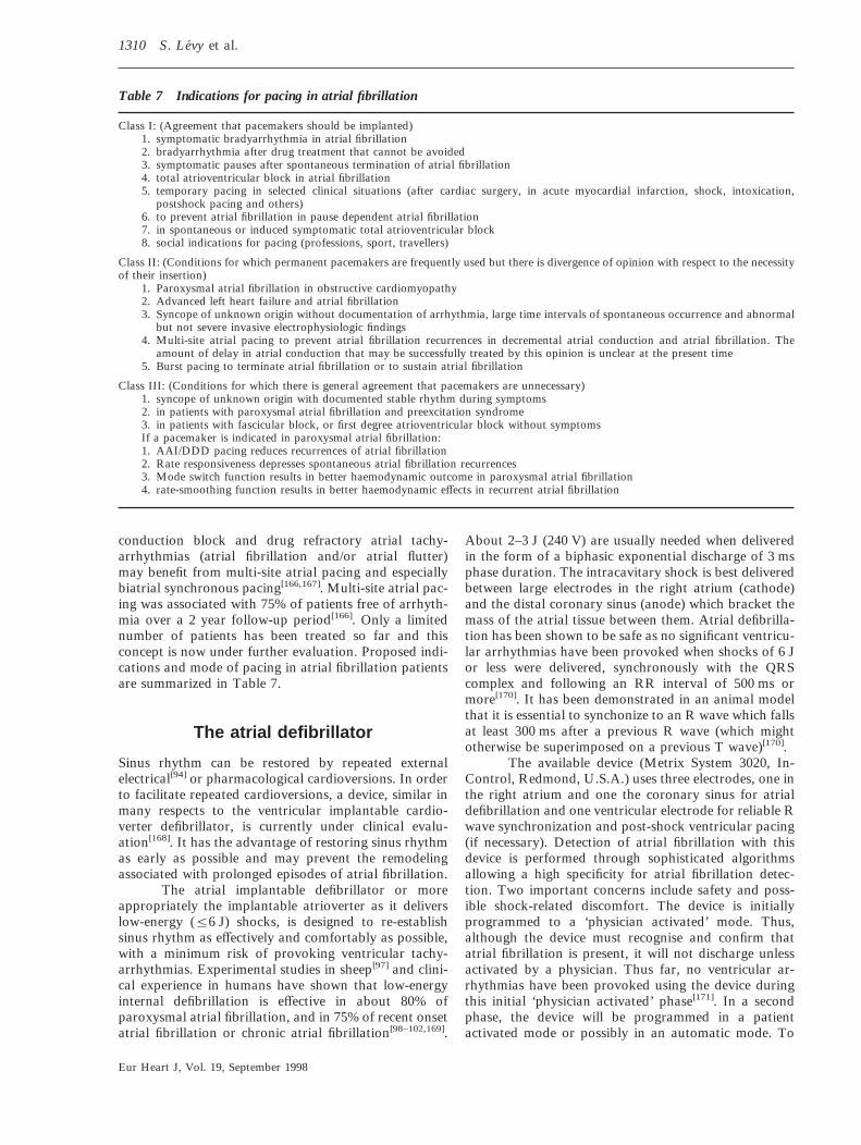
1310 S. Levy et al.
Table 7 Indications for pacing in atrial fibrillation
Class I: (Agreement that pacemakers should be implanted) 1. symptomatic bradyarrhythmia in atrial fibrillation 2. bradyarrhythmia after drug treatment that cannot be avoided 3. symptomatic pauses after spontaneous termination of atrial fibrillation 4. total atrioventricular block in atrial fibrillation 5. temporary pacing in selected clinical situations (after cardiac surgery, in acute myocardial infarction, shock, intoxication,
postshock pacing and others) 6. to prevent atrial fibrillation in pause dependent atrial fibrillation 7. in spontaneous or induced symptomatic total atrioventricular block 8. social indications for pacing (professions, sport, travellers)
Class II: (Conditions for which permanent pacemakers are frequently used but there is divergence of opinion with respect to the necessity of their insertion)
1. Paroxysmal atrial fibrillation in obstructive cardiomyopathy 2. Advanced left heart failure and atrial fibrillation 3. Syncope of unknown origin without documentation of arrhythmia, large time intervals of spontaneous occurrence and abnormal
but not severe invasive electrophysiologic findings 4. Multi-site atrial pacing to prevent atrial fibrillation recurrences in decremental atrial conduction and atrial fibrillation. The
amount of delay in atrial conduction that may be successfully treated by this opinion is unclear at the present time 5. Burst pacing to terminate atrial fibrillation or to sustain atrial fibrillation
Class III: (Conditions for which there is general agreement that pacemakers are unnecessary) 1. syncope of unknown origin with documented stable rhythm during symptoms 2. in patients with paroxysmal atrial fibrillation and preexcitation syndrome 3. in patients with fascicular block, or first degree atrioventricular block without symptoms If a pacemaker is indicated in paroxysmal atrial fibrillation: 1. AAI/DDD pacing reduces recurrences of atrial fibrillation 2. Rate responsiveness depresses spontaneous atrial fibrillation recurrences 3. Mode switch function results in better haemodynamic outcome in paroxysmal atrial fibrillation 4. rate-smoothing function results in better haemodynamic effects in recurrent atrial fibrillation
conduction block and drug refractory atrial tachyarrhythmias (atrial fibrillation and/or atrial flutter) may benefit from multi-site atrial pacing and especially biatrial synchronous pacing[166,167]. Multi-site atrial pacing was associated with 75% of patients free of arrhythmia over a 2 year follow-up period[166]. Only a limited number of patients has been treated so far and this concept is now under further evaluation. Proposed indications and mode of pacing in atrial fibrillation patients are summarized in Table 7.
The atrial defibrillator
Sinus rhythm can be restored by repeated external electrical[94] or pharmacological cardioversions. In order to facilitate repeated cardioversions, a device, similar in many respects to the ventricular implantable cardioverter defibrillator, is currently under clinical evaluation[168]. It has the advantage of restoring sinus rhythm as early as possible and may prevent the remodeling associated with prolonged episodes of atrial fibrillation.
The atrial implantable defibrillator or more appropriately the implantable atrioverter as it delivers low-energy (�6 J) shocks, is designed to re-establish sinus rhythm as effectively and comfortably as possible, with a minimum risk of provoking ventricular tachyarrhythmias. Experimental studies in sheep[97] and clinical experience in humans have shown that low-energy internal defibrillation is effective in about 80% of paroxysmal atrial fibrillation, and in 75% of recent onset atrial fibrillation or chronic atrial fibrillation[98–102,169].
About 2–3 J (240 V) are usually needed when delivered in the form of a biphasic exponential discharge of 3 ms phase duration. The intracavitary shock is best delivered between large electrodes in the right atrium (cathode) and the distal coronary sinus (anode) which bracket the mass of the atrial tissue between them. Atrial defibrillation has been shown to be safe as no significant ventricular arrhythmias have been provoked when shocks of 6 J or less were delivered, synchronously with the QRS complex and following an RR interval of 500 ms or more[170]. It has been demonstrated in an animal model that it is essential to synchonize to an R wave which falls at least 300 ms after a previous R wave (which might otherwise be superimposed on a previous T wave)[170].
The available device (Metrix System 3020, In-Control, Redmond, U.S.A.) uses three electrodes, one in the right atrium and one the coronary sinus for atrial defibrillation and one ventricular electrode for reliable R wave synchronization and post-shock ventricular pacing (if necessary). Detection of atrial fibrillation with this device is performed through sophisticated algorithms allowing a high specificity for atrial fibrillation detection. Two important concerns include safety and possible shock-related discomfort. The device is initially programmed to a ‘physician activated’ mode. Thus, although the device must recognise and confirm that atrial fibrillation is present, it will not discharge unless activated by a physician. Thus far, no ventricular arrhythmias have been provoked using the device during this initial ‘physician activated’ phase[171]. In a second phase, the device will be programmed in a patient activated mode or possibly in an automatic mode. To
Eur Heart J, Vol. 19, September 1998
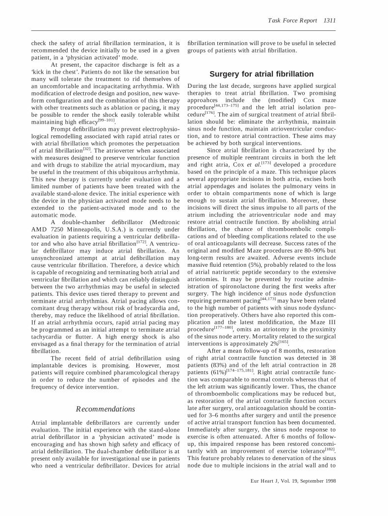
Task Force Report 1311
check the safety of atrial fibrillation termination, it is recommended the device initially to be used in a given patient, in a ‘physician activated’ mode.
At present, the capacitor discharge is felt as a ‘kick in the chest’. Patients do not like the sensation but many will tolerate the treatment to rid themselves of an uncomfortable and incapacitating arrhythmia. With modification of electrode design and position, new waveform configuration and the combination of this therapy with other treatments such as ablation or pacing, it may be possible to render the shock easily tolerable whilst maintaining high efficacy[99–101].
Prompt defibrillation may prevent electrophysiological remodelling associated with rapid atrial rates or with atrial fibrillation which promotes the perpetuation of atrial fibrillation[32]. The atrioverter when associated with measures designed to preserve ventricular function and with drugs to stabilize the atrial myocardium, may be useful in the treatment of this ubiquitous arrhythmia. This new therapy is currently under evaluation and a limited number of patients have been treated with the available stand-alone device. The initial experience with the device in the physician activated mode needs to be extended to the patient-activated mode and to the automatic mode.
A double-chamber defibrillator (Medtronic AMD 7250 Minneapolis, U.S.A.) is currently under evaluation in patients requiring a ventricular defibrillator and who also have atrial fibrillation[172]. A ventricular defibrillator may induce atrial fibrillation. An unsynchronized attempt at atrial defibrillation may cause ventricular fibrillation. Therefore, a device which is capable of recognizing and terminating both atrial and ventricular fibrillation and which can reliably distinguish between the two arrhythmias may be useful in selected patients. This device uses tiered therapy to prevent and terminate atrial arrhythmias. Atrial pacing allows concomitant drug therapy without risk of bradycardia and, thereby, may reduce the likelihood of atrial fibrillation. If an atrial arrhythmia occurs, rapid atrial pacing may be programmed as an initial attempt to terminate atrial tachycardia or flutter. A high energy shock is also envisaged as a final therapy for the termination of atrial fibrillation.
The recent field of atrial defibrillation using implantable devices is promising. However, most patients will require combined pharamcological therapy in order to reduce the number of episodes and the frequency of device intervention.
Recommendations
Atrial implantable defibrillators are currently under evaluation. The initial experience with the stand-alone atrial defibrillator in a ‘physician activated’ mode is encouraging and has shown high safety and efficacy of atrial defibrillation. The dual-chamber defibrillator is at present only available for investigational use in patients who need a ventricular defibrillator. Devices for atrial
fibrillation termination will prove to be useful in selected groups of patients with atrial fibrillation.
Surgery for atrial fibrillation During the last decade, surgeons have applied surgical therapies to treat atrial fibrillation. Two promising approahces include the (modified) Cox maze procedure[44,173–175] and the left atrial isolation procedure[176]. The aim of surgical treatment of atrial fibrillation should be: eliminate the arrhythmia, maintain sinus node function, maintain atrioventricular conduction, and to restore atrial contraction. These aims may be achieved by both surgical interventions.
Since atrial fibrillation is characterized by the presence of multiple reentrant circuits in both the left and right atria, Cox et al.[173] developed a procedure based on the principle of a maze. This technique places several appropriate incisions in both atria, excises both atrial appendages and isolates the pulmonary veins in order to obtain compartments none of which is large enough to sustain atrial fibrillation. Moreover, these incisions will direct the sinus impulse to all parts of the atrium including the atrioventricular node and may restore atrial contractile function. By abolishing atrial fibrillation, the chance of thromboembolic complications and of bleeding complications related to the use of oral anticoagulants will decrease. Success rates of the original and modified Maze procedures are 80–90% but long-term results are awaited. Adverse events include massive fluid retention (5%), probably related to the loss of atrial natriuretic peptide secondary to the extensive atriotomies. It may be prevented by routine administration of spironolactone during the first weeks after surgery. The high incidence of sinus node dysfunction requiring permanent pacing[44,173] may have been related to the high number of patients with sinus node dysfunction preoperatively. Others have also reported this complication and the latest mofdification, the Maze III procedure[177–180], omits an atriotomy in the proximity of the sinus node artery. Mortality related to the surgical interventions is approximately 2%[165].
After a mean follow-up of 8 months, restoration of right atrial contractile function was detected in 38 patients (83%) and of the left atrial contraction in 28 patients (61%)[174–175,181]. Right atrial contractile function was comparable to normal controls whereas that of the left atrium was significantly lower. Thus, the chance of thromboembolic complications may be reduced but, as restoration of the atrial contractile function occurs late after surgery, oral anticoagulation should be continued for 3–6 months after surgery and until the presence of active atrial transport function has been documented. Immediately after surgery, the sinus node response to exercise is often attenuated. After 6 months of followup, this impaired response has been restored concomitantly with an improvement of exercise tolerance[182]. This feature probably relates to denervation of the sinus node due to multiple incisions in the atrial wall and to
Eur Heart J, Vol. 19, September 1998
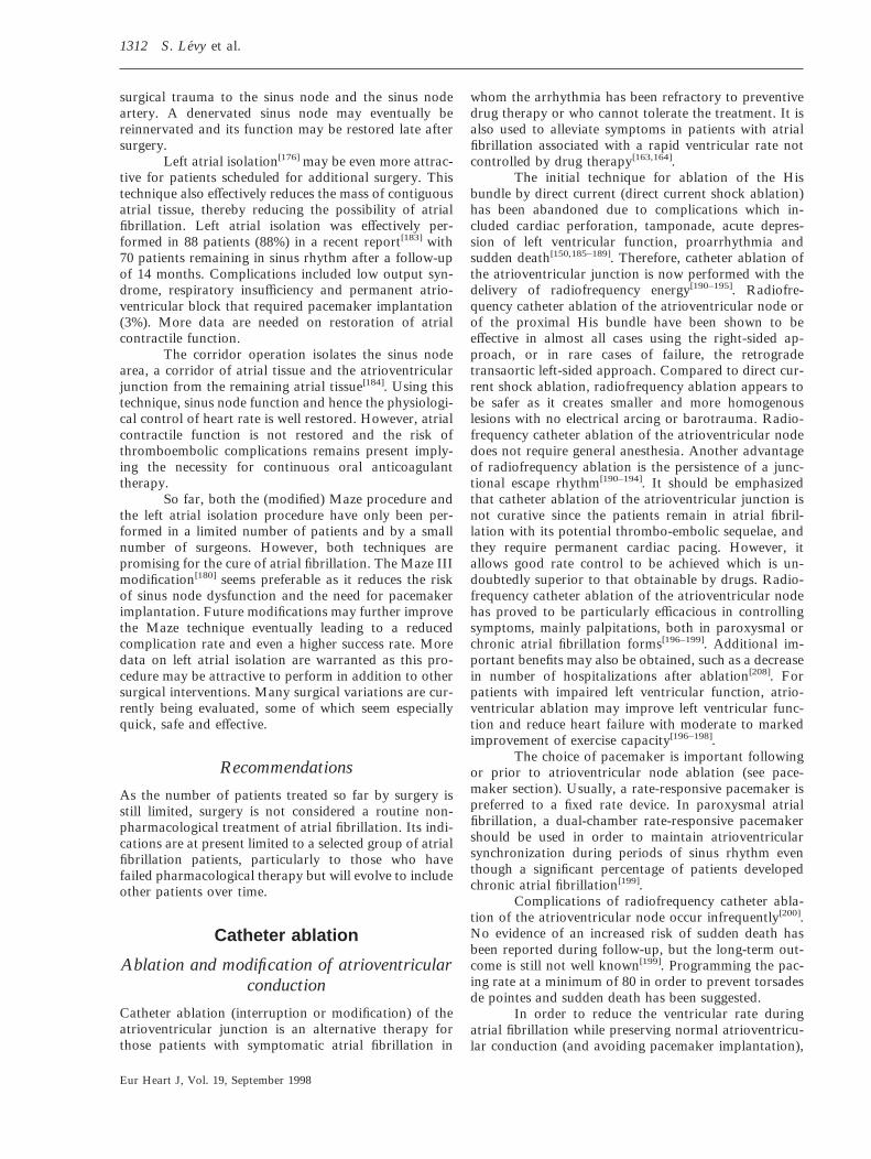
1312 S. Levy et al.
surgical trauma to the sinus node and the sinus node artery. A denervated sinus node may eventually be reinnervated and its function may be restored late after surgery.
Left atrial isolation[176] may be even more attractive for patients scheduled for additional surgery. This technique also effectively reduces the mass of contiguous atrial tissue, thereby reducing the possibility of atrial fibrillation. Left atrial isolation was effectively performed in 88 patients (88%) in a recent report[183] with 70 patients remaining in sinus rhythm after a follow-up of 14 months. Complications included low output syndrome, respiratory insufficiency and permanent atrioventricular block that required pacemaker implantation (3%). More data are needed on restoration of atrial contractile function.
The corridor operation isolates the sinus node area, a corridor of atrial tissue and the atrioventricular junction from the remaining atrial tissue[184]. Using this technique, sinus node function and hence the physiological control of heart rate is well restored. However, atrial contractile function is not restored and the risk of thromboembolic complications remains present implying the necessity for continuous oral anticoagulant therapy.
So far, both the (modified) Maze procedure and the left atrial isolation procedure have only been performed in a limited number of patients and by a small number of surgeons. However, both techniques are promising for the cure of atrial fibrillation. The Maze III modification[180] seems preferable as it reduces the risk of sinus node dysfunction and the need for pacemaker implantation. Future modifications may further improve the Maze technique eventually leading to a reduced complication rate and even a higher success rate. More data on left atrial isolation are warranted as this procedure may be attractive to perform in addition to other surgical interventions. Many surgical variations are currently being evaluated, some of which seem especially quick, safe and effective.
Recommendations
As the number of patients treated so far by surgery is still limited, surgery is not considered a routine nonpharmacological treatment of atrial fibrillation. Its indications are at present limited to a selected group of atrial fibrillation patients, particularly to those who have failed pharmacological therapy but will evolve to include other patients over time.
Catheter ablation
Ablation and modification of atrioventricular conduction
Catheter ablation (interruption or modification) of the atrioventricular junction is an alternative therapy for those patients with symptomatic atrial fibrillation in
whom the arrhythmia has been refractory to preventive drug therapy or who cannot tolerate the treatment. It is also used to alleviate symptoms in patients with atrial fibrillation associated with a rapid ventricular rate not controlled by drug therapy[163,164].
The initial technique for ablation of the His bundle by direct current (direct current shock ablation) has been abandoned due to complications which included cardiac perforation, tamponade, acute depression of left ventricular function, proarrhythmia and sudden death[150,185–189]. Therefore, catheter ablation of the atrioventricular junction is now performed with the delivery of radiofrequency energy[190–195]. Radiofrequency catheter ablation of the atrioventricular node or of the proximal His bundle have been shown to be effective in almost all cases using the right-sided approach, or in rare cases of failure, the retrograde transaortic left-sided approach. Compared to direct current shock ablation, radiofrequency ablation appears to be safer as it creates smaller and more homogenous lesions with no electrical arcing or barotrauma. Radiofrequency catheter ablation of the atrioventricular node does not require general anesthesia. Another advantage of radiofrequency ablation is the persistence of a junctional escape rhythm[190–194]. It should be emphasized that catheter ablation of the atrioventricular junction is not curative since the patients remain in atrial fibrillation with its potential thrombo-embolic sequelae, and they require permanent cardiac pacing. However, it allows good rate control to be achieved which is undoubtedly superior to that obtainable by drugs. Radiofrequency catheter ablation of the atrioventricular node has proved to be particularly efficacious in controlling symptoms, mainly palpitations, both in paroxysmal or chronic atrial fibrillation forms[196–199]. Additional important benefits may also be obtained, such as a decrease in number of hospitalizations after ablation[208]. For patients with impaired left ventricular function, atrioventricular ablation may improve left ventricular function and reduce heart failure with moderate to marked improvement of exercise capacity[196–198].
The choice of pacemaker is important following or prior to atrioventricular node ablation (see pacemaker section). Usually, a rate-responsive pacemaker is preferred to a fixed rate device. In paroxysmal atrial fibrillation, a dual-chamber rate-responsive pacemaker should be used in order to maintain atrioventricular synchronization during periods of sinus rhythm even though a significant percentage of patients developed chronic atrial fibrillation[199].
Complications of radiofrequency catheter ablation of the atrioventricular node occur infrequently[200]. No evidence of an increased risk of sudden death has been reported during follow-up, but the long-term outcome is still not well known[199]. Programming the pacing rate at a minimum of 80 in order to prevent torsades de pointes and sudden death has been suggested.
In order to reduce the ventricular rate during atrial fibrillation while preserving normal atrioventricular conduction (and avoiding pacemaker implantation),
Eur Heart J, Vol. 19, September 1998
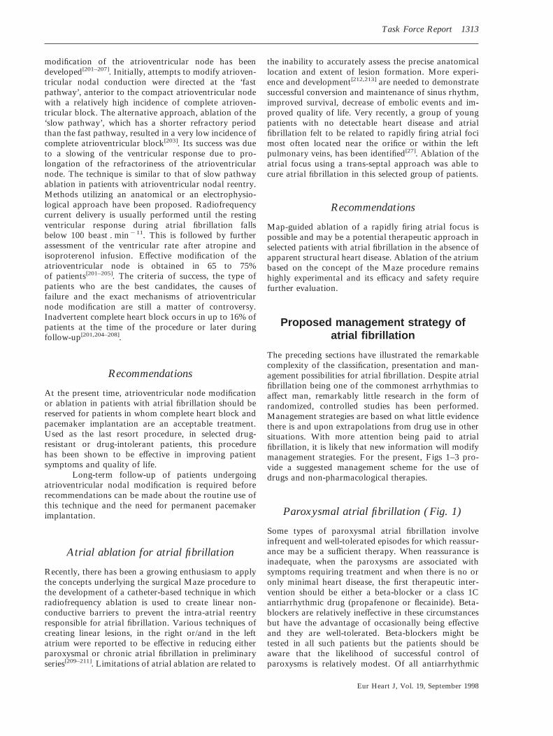
Task Force Report 1313
modification of the atrioventricular node has been developed[201–207]. Initially, attempts to modify atrioventricular nodal conduction were directed at the ‘fast pathway’, anterior to the compact atrioventricular node with a relatively high incidence of complete atrioventricular block. The alternative approach, ablation of the ‘slow pathway’, which has a shorter refractory period than the fast pathway, resulted in a very low incidence of complete atrioventricular block[203]. Its success was due to a slowing of the ventricular response due to prolongation of the refractoriness of the atrioventricular node. The technique is similar to that of slow pathway ablation in patients with atrioventricular nodal reentry. Methods utilizing an anatomical or an electrophysiological approach have been proposed. Radiofrequency current delivery is usually performed until the resting ventricular response during atrial fibrillation falls below 100 beast . min�11. This is followed by further assessment of the ventricular rate after atropine and isoproterenol infusion. Effective modification of the atrioventricular node is obtained in 65 to 75% of patients[201–205]. The criteria of success, the type of patients who are the best candidates, the causes of failure and the exact mechanisms of atrioventricular node modification are still a matter of controversy. Inadvertent complete heart block occurs in up to 16% of patients at the time of the procedure or later during follow-up[201,204–208].
Recommendations
At the present time, atrioventricular node modification or ablation in patients with atrial fibrillation should be reserved for patients in whom complete heart block and pacemaker implantation are an acceptable treatment. Used as the last resort procedure, in selected drugresistant or drug-intolerant patients, this procedure has been shown to be effective in improving patient symptoms and quality of life.
Long-term follow-up of patients undergoing atrioventricular nodal modification is required before recommendations can be made about the routine use of this technique and the need for permanent pacemaker implantation.
Atrial ablation for atrial fibrillation
Recently, there has been a growing enthusiasm to apply the concepts underlying the surgical Maze procedure to the development of a catheter-based technique in which radiofrequency ablation is used to create linear nonconductive barriers to prevent the intra-atrial reentry responsible for atrial fibrillation. Various techniques of creating linear lesions, in the right or/and in the left atrium were reported to be effective in reducing either paroxysmal or chronic atrial fibrillation in preliminary series[209–211]. Limitations of atrial ablation are related to
the inability to accurately assess the precise anatomical location and extent of lesion formation. More experience and development[212,213] are needed to demonstrate successful conversion and maintenance of sinus rhythm, improved survival, decrease of embolic events and improved quality of life. Very recently, a group of young patients with no detectable heart disease and atrial fibrillation felt to be related to rapidly firing atrial foci most often located near the orifice or within the left pulmonary veins, has been identified[27]. Ablation of the atrial focus using a trans-septal approach was able to cure atrial fibrillation in this selected group of patients.
Recommendations
Map-guided ablation of a rapidly firing atrial focus is possible and may be a potential therapeutic approach in selected patients with atrial fibrillation in the absence of apparent structural heart disease. Ablation of the atrium based on the concept of the Maze procedure remains highly experimental and its efficacy and safety require further evaluation.
Proposed management strategy of atrial fibrillation
The preceding sections have illustrated the remarkable complexity of the classification, presentation and management possibilities for atrial fibrillation. Despite atrial fibrillation being one of the commonest arrhythmias to affect man, remarkably little research in the form of randomized, controlled studies has been performed. Management strategies are based on what little evidence there is and upon extrapolations from drug use in other situations. With more attention being paid to atrial fibrillation, it is likely that new information will modify management strategies. For the present, Figs 1–3 provide a suggested management scheme for the use of drugs and non-pharmacological therapies.
Paroxysmal atrial fibrillation (Fig. 1)
Some types of paroxysmal atrial fibrillation involve infrequent and well-tolerated episodes for which reassurance may be a sufficient therapy. When reassurance is inadequate, when the paroxysms are associated with symptoms requiring treatment and when there is no or only minimal heart disease, the first therapeutic intervention should be either a beta-blocker or a class 1C antiarrhythmic drug (propafenone or flecainide). Betablockers are relatively ineffective in these circumstances but have the advantage of occasionally being effective and they are well-tolerated. Beta-blockers might be tested in all such patients but the patients should be aware that the likelihood of successful control of paroxysms is relatively modest. Of all antiarrhythmic
Eur Heart J, Vol. 19, September 1998
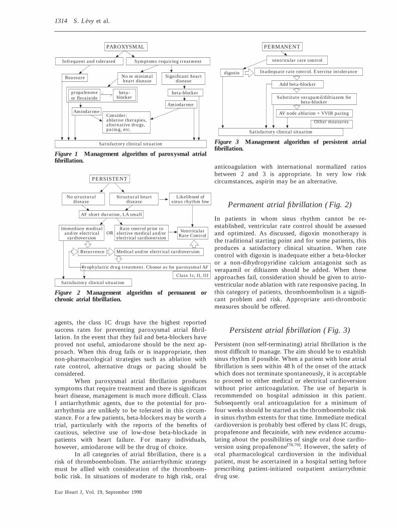
1314 S. Levy et al.
PAROXYSMAL
Symptoms requiring treatment
Reassure
beta-blocker
Consider: ablative therapies, alternative drugs, pacing, etc.
Amiodarone
Satisfactory clinical situation
propafenone or flecainide
Amiodarone
No or minimal heart disease
Significant heart disease
Infrequent and tolerated
beta-blocker
Figure 1 Management algorithm of paroxysmal atrial fibrillation.
PERSISTENT
AF short duration, LA small
Immediate medical and/or electrical
cardioversion
Medical and/or electrical cardioversion
Satisfactory clinical situation
Class 1c, II, III
Recurrence
Rate control prior to elective medical and/or electrical cardioversion
Ventricular Rate Control
No structural disease
Structural heart disease
Likelihood of sinus rhythm low
OR
Prophylactic drug treatment. Choose as for paroxysmal AF
Figure 2 Management algorithm of permanent or chronic atrial fibrillation.
agents, the class 1C drugs have the highest reported success rates for preventing paroxysmal atrial fibrillation. In the event that they fail and beta-blockers have proved not useful, amiodarone should be the next approach. When this drug fails or is inappropriate, then non-pharmacological strategies such as ablation with rate control, alternative drugs or pacing should be considered.
When paroxysmal atrial fibrillation produces symptoms that require treatment and there is significant heart disease, management is much more difficult. Class I antiarrhythmic agents, due to the potential for proarrhythmia are unlikely to be tolerated in this circumstance. For a few patients, beta-blockers may be worth a trial, particularly with the reports of the benefits of cautious, selective use of low-dose beta-blockade in patients with heart failure. For many individuals, however, amiodarone will be the drug of choice.
In all categories of atrial fibrillation, there is a risk of thromboembolism. The antiarrhythmic strategy must be allied with consideration of the thromboembolic risk. In situations of moderate to high risk, oral
Eur Heart J, Vol. 19, September 1998
PERMANENT
ventricular rate control
digoxin Inadequate rate control. Exercise intolerance
Add beta-blocker
Substitute verapamil/diltiazem for beta-blocker
AV node ablation + VVIR pacing
Other measures
Satisfactory clinical situation
Figure 3 Management algorithm of persistent atrial fibrillation.
anticoagulation with international normalized ratios between 2 and 3 is appropriate. In very low risk circumstances, aspirin may be an alternative.
Permanent atrial fibrillation (Fig. 2)
In patients in whom sinus rhythm cannot be reestablished, ventricular rate control should be assessed and optimized. As discussed, digoxin monotherapy is the traditional starting point and for some patients, this produces a satisfactory clinical situation. When rate control with digoxin is inadequate either a beta-blocker or a non-dihydropyridine calcium antagonist such as verapamil or diltiazem should be added. When these approaches fail, consideration should be given to atrioventricular node ablation with rate responsive pacing. In this category of patients, thromboembolism is a significant problem and risk. Appropriate anti-thrombotic measures should be offered.
Persistent atrial fibrillation (Fig. 3)
Persistent (non self-terminating) atrial fibrillation is the most difficult to manage. The aim should be to establish sinus rhythm if possible. When a patient with lone atrial fibrillation is seen within 48 h of the onset of the attack which does not terminate spontaneously, it is acceptable to proceed to either medical or electrical cardioversion without prior anticoagulation. The use of heparin is recommended on hospital admission in this patient. Subsequently oral anticoagulation for a minimum of four weeks should be started as the thromboembolic risk in sinus rhythm extents for that time. Immediate medical cardioversion is probably best offered by class IC drugs, propafenone and flecainide, with new evidence accumulating about the possibilities of single oral dose cardioversion using propafenone[78,79]. However, the safety of oral pharmacological cardioversion in the individual patient, must be ascertained in a hospital setting before prescribing patient-initiated outpatient antiarrythmic drug use.
Task Force Report 1315
If the episode of atrial fibrillation has been present for more than 48 h (persistent) or when the duration of the episode is unknown, a minimum of three weeks of effective anticoagulation should be provided prior to either pharmacological or electrical cardioversion. In that intervening period, rate control should be offered with digoxin, with or without concomitant betablocker or calcium entry blocker therapy. An alternative could be to anticoagulate the patient with heparin and to exclude an intracardiac thrombus with transoesophageal echocardiography before cardioversion.
In patients with a first episode of persistent atrial fibrillation which has been reverted to sinus rhythm, it is reasonable to prescribe no prophylactic antiarrhythmic therapy. In the event of a relapse and the need for further electrical or medical cardioversion, prophylactic drug-treatment should be considered. The effective agents are those used for the prophylaxis of paroxysmal atrial fibrillation (see section on prevention of recurrences) and include betablockers, class 1C agents (propafenone and flecainide), sotalol and amiodarone.
References
[1] Cabin HS, Clubb KS, Hall C, Perlmutter RA, Feinstein AR. Risk for systemic embolization of atrial fibrillation without mitral stenosis. Am J Cardiol 1990; 61: 714–7.
[2] Petersen P, Godtfredsen J. Embolic complications in paroxysmal atrial fibrillation. Stroke 1986; 17: 622–6.
[3] Zipes D. Cardiac arrhythmias. In: Braunwald W. Heart Disease. Philadelphia: W.B. Saunders 1984: 669–70.
[4] Kannel WB, Abbot RD, Savage DD, McNamara PM. Epidemiologic features of chronic atrial fibrillation; the Framingham study. N Engl J Med 1982; 306: 1018–22.
[5] Bellet S. Clinical disorders of the heart beat. Philadelphia: Lea and Febiger 1971: 223–33.
[6] Page RL, Wilkinson WE, Clair WK et al. Asymptomatic arrhythmias in patients with symptomatic paroxysmal atrial fibrillation and paroxysmal supraventricular tachycardia. Circulation 1994; 89: 224–7.
[7] Bialy D, Lehmann MH, Schumacher DN, Steinman RT, Meissmer MD. Hospitalization for arrhythmias in the United States: Importance of atrial fibrillation (Abstr). J Am Coll Cardiol 1992; 19: 41 A.
[8] Zipes DP. Atrial fibrillation. A tachycardia-induced atrial cardiomyopathy. Circulation 1997; 95: 562–4.
[9] Levy S, Novella P, Ricard Ph, Paganelli F. Paroxysmal atrial fibrillation: A need for classification. J Cardiovasc Electrophysiol 1995; 6: 69–74.
[10] Sopher SM, Camm AJ. Therapy for atrial fibrillation: control of the ventricular response and prevention of recurrence. Cor Art Dis 1995; 6: 106–14.
[11] Brand FN, Abott RD, Kannel WB, Wolf PA. Characteristics and prognosis of lone atrial fibrillation: 30-years follow up in the Framingham Study. JAMA 1985; 254: 3449–53.
[12] Wolf PA, Abbott RD, Kannel WB. A major contributor to stroke in the elderly. Arch Int Med 1987; 147: 1561–4.
[13] Wolf PA, Abbott RD, Kannel WB. Atrial fibrillation as an independent risk factor for stroke; The Framingham Study. Stroke 1991; 22: 983–8.
[14] Furberg CD, Psaty BM, Manolio TA, Gardin JM, Smith VE, Rautaharju PM for the CHS Collaborative Research Group. Prevalence of atrial fibrillation in elderly subjects (The Cardiovascular Health Study). Am J Cardiol 1994; 74: 236–41.
[15] Manolio TA, Furberg CD, Rautaharju PM et al. Cardiac arrhythmias on 24 hours ambulatory electrocardiography in
older women and men. The Cardiovascular Health Study. J Am Coll Cardiol 1984; 23: 916–25.
[16] Kopecky SL, Gersh BJ, McGoon MD et al. The natural history of lone atrial fibrillation. A population-based study over three decades. N Engl J Med 1987; 317: 669–74.
[17] Levy S, Attuel P. Fauchier JP, Medvedowsky JL et le Groupe de Travail en Rythmologie de la Societe Francaise de cardiologie. Nosologie de la fibrillation auriculaire. Essai de clarifications. Arch Mal Coeur 1995; 88: 1035–8.
[18] Clementy J, Safar M, Vrancea F. Cardiac arrhythmias in hypertension. Prospective study including 251 patients (Abstr). Eur Heart J 1989; 10: 201.
[19] Nunez BD, Messerli FH, Garavoglia GE, Schmieder RE. Exaggerated atrial and ventricular excitability in hypertensive patients with isolated septal hypertrophy (Abstr). J Am Coll Cardiol 1987; 9: 225 A.
[20] Robinson K, Frenneaux MP, Stockins B et al. Atrial fibrillation in hypertrophic cardiomyopathy: a longitudinal study. J Am Coll Cardiol 1990; 15: 1279–85.
[21] Haissaguerre M, Bonnet J, Billes MA et al. Prevalence, signification et pronostic des arythmies auriculaires dans les myocardiopathies dilatees. A propos de 236 cas. Arch Malv Coeur 1985; 4: 536–41.
[22] Evans W, Swann P. Lone auricular fibrillation. Br Heart J 1954; 16: 189–94.
[23] Waldo AL, MacLean WAH, Karp RB et al. Continuous rapid atrial pacing to control recurrent or sustained surpraventricular tachycardias following open heart surgery. Circulation 1976; 54: 254–50.
[24] Brugada R, Tapscott T, Czernuszewicz G et al. Identification of the first locus for familial atrial fibrillation utilizing a rapid novel pooled DNA strategy. (Abstr) Am Coll Cardiol 29 (Suppl II): 2A, 407–1.
[25] Moe GK, Abildskov JA. Atrial fibrillation as a self-sustaining arrhythmia independent of focal discharge. Am Heart J 1959; 58: 59–70.
[26] Rensma PL, Allessie MA, Lammers WJ et al. Length of excitation wave and susceptibility to reentrant atrial arrhythmias in normal conscious dogs. Circ Res 1988; 62: 395–410.
[27] Jais P, Haissaguerre M, Shah DC et al. A focal source of atrial fibrillation treated by discrete radiofrequency ablation. Circulation 1997; 95: 572–6.
[28] Kumagai K, Khrestian C, Waldo AL. Simultaneous multisite mapping studies during induced atrial fibrillation in the sterile pericarditis model. Insights into the mechanism of its maintenance. Circulation 1997; 95: 511–21.
[29] Allessie MA, Konings K, Kirchhof C. Mapping of atrial fibrillation. In: Olsson SB, Allessie MA, Campbell RWF, eds. Atrial Fibrillation: mechanisms and therapeutic strategies. Armonk NY: Futura Publishing Company 1994: 37–49.
[30] Gray RA, Pertsov AM, Jalife J. Incomplete reentry and epicardial breakthrough patterns during atrial fibrillation in the sheep heart. Circulation 1996; 94: 2649–61.
[31] Ricard Ph, Levy S, Trigano J et al. Prospective assessment of the minimum energy needed for external electrical cardioversion of atrial fibrillation. Am J Cardiol 1997; 79: 815–6.
[32] Wijfells MCEF, Kirchhof CJHJ, Dorland R, Allessie MA. Atrial fibrillation begets atrial fibrillation. A study in awake chronically instrumented goats. Circulation 92: 1954–68.
[33] Attuel P, Pellerin D, Gaston J et al. Latent atrial vulnerability: new means of electrophysiologic investigations in paroxysmal atrial arrhythmias. In: Attuel P, Coumel P, Janse MJ, eds. The Atrium in Health and Disease. Mount Kisco, NY: Futura Publishing Company 1989: 159–200.
[34] Le Heuzey JY, Boutjdir M, Gagey S, Lavergne T, Guize L. Cellular aspect of atrial vulnerability. In: Attuel P, Coumel P, Janse MJ, eds. The Atrium in Health and Disease. Mount Kisco, New York: Futura Publishing Company, 1989: 81–94.
[35] Capucci A, Biffi M, Boriani G et al. Dynamic electrophysiologic behavior of human atria during paroxysmal atrial fibrillation. Circulation 1995; 92: 1193–202.
Eur Heart J, Vol. 19, September 1998
1316 S. Levy et al.
[36] Yue L, Feng J, Gaspo R, Li GR, Wang Z, Nattel S. Ionic remodeling underlying action potential changes in a canine model of atrial fibrillation. Circ Res 1997; 81: 512–25.
[37] Olsson SB, Cotoi S, Varnauskas E. Monophasic action potential and sinus rhythm stability after conversion of atrial fibrillation. Acta Med Scand 1971; 190: 381–7.
[38] Michelucci A, Padeletti L, Porciani MC et al. Dispersion of refractoriness in atrial fibrillation. In: Olsson SB, Allessie MA, Campbell RWF eds. Atrial Fibrillation: Mechanisms and therapeutic strategies. Armonk NY: Futura Publishing Company, 1994: 81–107.
[39] Ramdat Missier AR, Opthof T, Van Hemel NM et al. Increased dispersion of refractoriness in patients with idiopathic paroxysmal atrial fibrillation. J Am Coll Cardiol 1992; 19: 1531–5.
[40] Cosio FG, Palacias J, Vidal JM, Cocina EG, Gomez-Sanchez MA, Tamargo L. Electrophysiological studies in atrial fibrillation. Slow conduction of premature impulses: a possible manifestation of the background for reentry. Am J Cardiol 1983; 51: 122–30.
[41] Edwards BS, Zimmerman RS, Schwab TR et al. Atrial stretch, not pressure, is the principal determinant controlling the acute release of atrial natriuretic factor. Circ Res 1988; 62: 191–5.
[42] Spach MS. Non uniform anisotropic cellular coupling as a basis for reentrant arrhythmias. In: Di Marco JP, Prystowsky JP, eds. Atrial Arrhythmias. State of the art. Armonk, NY: Futura Publishing Company, 1995: 123–47.
[43] Inoue H, Zipes DP. Results of sympathetic denervation in the canine heart; supersensitivity that may be arrhythmogenic. Circulation 1987; 75: 877–87.
[44] Cox JL, Canaven TE, Schuessler RB et al. The surgical treatment of atrial fibrillation. II. Intraoperative electrophysiologic mapping and description of the electrophysiologic basis of atrial flutter and atrial fibrillation. J Thorac Cardiovasc Surg 1991; 101: 406–26.
[45] Bharati S, Lev M. Histology of the normal and diseased atrium. In: Falk RH, Podrid PJ, eds. Atrial Fibrillation: Mechanism and Management. New York: Raven Press, 1992: 15–39.
[46] Guiraudon CM, Ernst NM, Guiraudon GM, Yee R, Klein GJ. The pathology of drug resistant lone atrial fibrillation in eleven surgically treated patients In: Kingma JH, Van Hemel NM, Lie KI, eds. Atrial fibrillation a Treatable disease? Dordrecht: Kluwer Academic Publishers, 1992: 41–57.
[47] Frustaci A, Chimenti C, Bellocci F, Morgante E, Russo MA, Maseri A. Histological substrate of atrial biopsies in patients with lone atrial fibrillation. Circulation 1997; 9: 1180–4.
[48] Coumel PH, Attuel P, Leclercq JF, Friocourt P. Arythmie auriculaire d’origine vagale ou catecholergique: effets compares due traitement beta-bloquant et phenomene d’echappement. Arch Mal Coeur 1982; 75: 373–88.
[49] Lombardi F, Tozillo D, Cappiello E. Sympatho-vagal influence in atrial fibrillation. N Trends Arrhythmias 1993; 9: 279–84.
[50] Antiarrhythmic Therapy: A Pathophysiologic Approach by Members of the Sicilian Gambit. Armonk NY: Futura Publishing Company, 1994: 188–93.
[51] Onundarson PT, Thorgeirsson G, Jonmundsson E, Sigfusson N, Hardson Th. Chronic atrial fibrillation-Epidemiologic features and 14 years follow-up: A case control study. Eur Heart J 1987; 3: 521–7.
[52] Flegel KM, Shipley MJ, Rose G. Risk of stroke in nonrheumatic atrial fibrillation. Lancet 1987; 1: 526–9.
[53] Sherman OG, Hart RG, Easton JD. The secondary prevention of stroke in patients with atrial fibrillation. Arch Neurol 1986; 43: 68–70.
[54] Petersen P, Godtfredsen J. Embolic complications in paroxysmal atrial fibrillation. Stroke 1986; 17: 622–6.
[55] Cairns JA, Connolly SJ. Nonrheumatic atrial fibrillation: Risk of stroke and role of antithrombotic therapy. Circulation 1991; 84: 469–79.
[56] Van Latum JC, Koudstaal PJ, Venables GS et al. Predictors of major vascular events in patients with a transient ischemic attack or minor ischemic stroke with nonrheumatic atrial fibrillation. European Atrial Fibrillation Trial (EAFT) Study Group. Stroke 1995; 25: 801–6.
[57] Stoddard MF, Dawkins PR, Prince CR, Ammash NM. Left atrial appendage thrombus is not uncommon in patients with acute atrial fibrillation and a recent embolic event: A transoesophageal echocardiographic study. J Am Coll Cardiol 1995; 25: 452–9.
[58] Mugge A, Kuhn H, Nikutta P et al. Assessment of left atrial appendage function by biplane transesophageal echocardiography in patients with nonrheumatic atrial fibrillation: Identification of a subgroup of patients at increased embolic risk. J Am Coll Cardiol 1994; 23: 599–607.
[59] Shively BK, Gelgand EA, Crawford MH. Regional left atrial stasis during atrial fibrillation and flutter: Determinants and relation to stroke. J Am Coll Cardiol 1996; 27: 1722–7.
[60] Petersen P, Boysen G, Godtfredsen J, Andersen ED, Andersen B. Placebo-controlled, randomised trial of warfarin and aspirin for prevention of thromboembolic complications in chronic atrial fibrillation. The Copenhagen AFASAK study. Lancet 1989; 1: 175–9.
[61] The Boston Area Anticoagulation Trial for Atrial Fibrillation Investigators. The effect of low-dose warfarin on the risk of stroke in patients with non-theumatic atrial fibrillation. N Engl J Med 1990; 323: 1505–11.
[62] Stroke Prevention in Atrial Fibrillation Investigators. Stroke Prevention in atrial fibrillation study; Final results. Circulation 1991; 84: 527–39.
[63] Connolly SJ, Laupacis A, Gent Met al. Canadian atrial fibrillation anticoagulation (CAFA) study. J Am Cardiol 1991; 18: 349–55.
[64] Singer DE. The randomized trials of warfarin for atrial fibrillation. N Engl J Med 1992; 327: 1451–3.
[65] Ezekowitz MD, Bridgers SL, James KE et al. Warfarin in the prevention of stroke associated with nonrheumatic atrial fibrillation. N Engl J Med 1992; 327: 1406–12.
[66] European Atrial Fibrillation Trial Study Group. Secondary prevention in non-rheumatic atrial fibrillation after transient ischaemic attack or minor stroke. Lancet 1993; 342: 1255–62.
[67] The European Atrial Fibrillation Trial Study Group. Optimal oral anticoagulant therapy in patients with nonrheumatic atrial fibrillation and recent cerebral ischemia. N Eng J Med 1995; 333: 5–10.
[68] Stroke Prevention in Atrial Fibrillation Investigators. Warfarin versus aspirin for prevention of thromboembolism in atrial fibrillation: Stroke Prevention in Atrial Fibrillation II study. Lancet 1994; 343: 687–91.
[69] Falk RH, Knowlton AA, Bernard SA, Gotlieb NE, Battinelli NJ. Digoxin for converting recent-onset atrial fibrillation to sinus rhythm. A randomized double-blinded trial. Ann Inter Med 1987; 106: 503–6.
[70] Jordaens L, Trouerbach J, Calle P, et al. Conversion of atrial fibrillation to sinus rhythm and rate control by digoxin in comparison to placebo. Eur Heart J 1997; 18: 643–8.
[71] The Digitalis in Acute Atrial Fibrillation (DAAF) trial group. Intravenous digoxin in acute atrial fibrillation. Results of a randomized, placebo-controlled multicentre trial in 239 patients. Eur Heart J 1997; 18: 649–54.
[72] Laupacis A, Alberts G, Dunn M, Feinberg W. Antithrombotic therapy in atrial fibrillation. Chest 1992; 102 (Suppl): 426–31.
[73] Capucci A, Tiziano L, Boriani G et al. Effectiveness of loading oral flecainide for converting recent-onset atrial fibrillation to sinus rhythm in patients without organic heart disease or with only systemic hypertension. Am J Cardiol 1992; 70: 69–72.
[74] Crijns HJGM, van Vijk M, van Gilst WH, Herre Kingma J, van Gelder IC, Lie KI. Acute conversion of atrial fibrillation to sinus rhythm: clinical efficacy of flecainide acetate. Comparison of two regimens. Eur Heart J 1988; 9: 634–9.
Eur Heart J, Vol. 19, September 1998
Task Force Report 1317
[75] Suttorp MJ, Kingma HJ, Jessurun ER, Lie-A-Huen L, Van Hemel NM, Lie KI. The value of class 1C antiarrhythmic drugs for acute conversion of paroxysmal atrial fibrillation or flutter to sinus rhythm. J Am Coll Cardiol 1990; 16: 1722–7.
[76] Nacarelli GV, Dorian P, Hohnloser SH et al. Prospective comparison of flecainide versus quinidine for the treatment of paroxysmal atrial fibrillation/flutter. Am J Cardiol 1996; 75: 53A–59A.
[77] Lacombe P, Cointe R, Metge M, Bru P, Gerard R, Levy S. Intravenous flecainide in the management of acute supraventricular tachyarrhythmias. J Electrophysiol 1988; 2: 19–21.
[78] Capucci A, Boriani G, Rubino I, Della Casa S, Sanguinetti M, Magnani B. A controlled study on oral propafenone versus digoxin plus quinidine in converting recent onset atrial fibrillation to sinus rhythm. Int J Cardiol 1994; 43: 305–11.
[79] Boriani G, Biffi M, Capucci A et al. Oral propafenone to convert recent-onset atrial fibrillation in patients with and without underlying heart disease. A randomized controlled trial. Ann Intern Med 1997; 126: 621–4.
[80] Capucci A, Boriani G, Botto GL et al. Conversion of recent onset atrial fibrillation to sinus rhythm by a single oral loading dose of propafenone or flecainide. Am J Cardiol 1994; 74: 503–5.
[81] Aliot E, Denjoy I and the Flecainide AF French Study Group. Comparison of the safety and efficacy of flecainide versus propafenone in hospital-outpatients with symptomatic paroxysmal atrial fibrillation/flutter. Am J Cardiol 1996; 77 60A– 65A.
[82] Feld GH, Chen PS, Nicod P, Fleck RP, Mezer D. Atrial proarrhythmic effects of class 1C antiarrhythmic drugs. Am J Cardiol 1990; 66: 378–82.
[83] Keller K, Meyer-Estorf G, Beck OA, Hochrein H. Correlation between serum concentration and pharmacological effect on atrioventricular conduction time of the antiarrhythmic drug propafenone. Eur J Clin Pharmacol 1978; 13: 1303–6.
[84] Tieleman RG, Gosselink ATM, Crijns HJGM et al. Efficacy, safety and determinants of conversion of atrial fibrillation and flutter with oral amiodarone. Am J Cardiol 1997; 79: 53–7.
[85] Donovan KD, Dower BM, Hockings BE et al. Flecainide versus amiodarone for recent onset atrial fibrillation. Am J Cardiol 1995; 75: 693–7.
[86] Galve E, Rius T, Ballester R et al. intravenous amiodarone in the treatment of recent-onset atrial fibrillation. Results of a randomized, controlled trial. J Am Coll Cardiol 1996; 27: 1079–82.
[87] Ellenbogen KA, Wood M, Stambler BS et al. Conversion of atrial fibrillation and flutter by intravenous ibutilide; a multicenter dose response study. J Am Coll Cardiol 1996; 28: 130–6.
[88] Falk RH, Pollak A, Singh SN, Tilman F. Intravenous dofetilide, a class III antiarrhythmic agent, for the termination of sustained atrial fibrillation or flutter. J Am Coll Cardiol 1997; 29: 385–90.
[89] Lown B. Electrical reversion of cardiac arrhythmias. Br Heart J 1962; 29: 469–89.
[90] Eway GA. Effectiveness of direct current of defibrillation. Role of paddle electrode size. Am Heart J 1977; 93: 674–5.
[91] Connell PN, Ewy GA, Dahl CF et al. Transthoracic impedance to defibrillator discharge; effect of electrode size and chest wall interface. J Electrophysiol 1973; 6: 313–7.
[92] Bjerkelund C, Orning OM. An evaluation of DC shock treatment of atrial arrhythmias; immediate results and complications in 437 patients with long term results in the first 290 of these. Acta Med Scand 1968; 184: 481–91.
[93] Van Gelder IC, Crijns HJ, Van Gilst WH, Verwer R, Lie KI. Prediction of uneventful cardioversion and maintenance of sinus rhythm from direct-current electrical cardioversion of chronic atrial fibrillation and flutter. Am J Cardiol 1991; 68: 41–6.
[94] Van Gelder IC, Crijns HJGM, Hillege H, Lie KI. Value and limitations of DC electrical cardioversion of chronic atrial fibrillation. Pacing Clin Electrophysiol 1995; 18(II): 798.
[95] Levy S , Lauribe P, Dolla E et al. A randomized comparison of external and internal cardioversion of chronic atrial fibrillation. Circulation 1992; 86: 1415–20.
[96] Levy S, Lacombe P, Cointe R et al. High energy transcatheter cardioversion of chronic atrial fibrillation. J Am Coll Cardiol 1988; 12: 514–8.
[97] Cooper RAS, Alferness CA, Smith WN, Ideker RE. Internal cardioversion of atrial fibrillation in sheep. Circulation 1993; 87: 1673–86.
[98] Alt E, Schmitt C, Ammer R et al. Initial experience with intracardiac atrial defibrillation in patients with chronic atrial fibrillation. Pace Pacing Clin Electrophysiol 1994; 17: 1067–78.
[99] Murgatroyd F, Slade AKB, Sopher M, Rowland E, Ward DE, Camm J. Efficacy and tolerability of transvenous low energy cardioversion of paroxysmal atrial fibrillation in humans. J Am Coll Cardiol 1995; 25: 1347–53.
[100] Levy S, Ricard Ph, Pak Lau C et al. Multicenter low energy transvenous atrial defibrillation (XAD) trial. Results in different subjects of atrial fibrillation. J Am Coll Cardiol 1997; 29: 750–5.
[101] Levy S, Ricard Ph, Gueunoun M et al. Low-energy cardioversion of spontaneous atrial fibrillation. Circulation 1997; 96: 253–9.
[102] Schmitt C, Alt E, Plevan A et al. Low-energy intracardiac cardioversion after failed conventional external cardioversion of atrial fibrillation. J Am Coll Cardiol 1996; 28: 994–9.
[103] Barold H, Warton JM. Ventricular fibrillation resulting from synchronized internal defibrillation in a patient with ventricular preexcitation. J Cardiov Electrophysiol 1997; 8: 436–40.
[104] Bjerkelund CJ, Orning OM. The efficacy of anticoagulant therapy in preventing embolism related to D.C. electrical conversion of atrial fibrillation. Am J Cardiol 1969; 23: 208–16.
[105] Manning WJ, Silverman DI, Gordon SP et al. Cardioversion from atrial fibrillation without prolonged anticoagulation with use of transesophageal echocardiography to exclude the presence of atrial thrombi. N Engl J Med 1993; 328: 750–5.
[106] Black IW, Fatkin D, Sagar KB. Exclusion of atrial thrombus by transesophageal echocardiography does not preclude embolism after cardioversion of atrial fibrillation: A multicenter study. Circulation 1994; 89: 2509–13.
[107] Grimm RA, Stewart WJ, Maloney JD et al. Impact of electrical cardioversion for atrial fibrillation on left atrial appendage function and spontaneous echo contrast: characterization by simultaneous transesophageal echocardiography. J Am Coll Cardiol 1993; 22: 1359–66.
[108] Prystowsky EN, Benson Jr W, Fuster V et al. Management of patients with atrial fibrillation. A statement for healthcare professionals from the Subcommittee on Electrocardiography and Electrophysiology of the American Heart Association. Circulation 1996; 93: 1262–77.
[109] Klein AL, Grimm RA et al. Cardioversion guided by transesophageal echocardiography; the ACUTE Pilot Study: a randomized, controlled trial. Ann Intern Med 1997; 126: 200–9.
[110] Coplen SE, Antman EM, Berlin JA, Hewitt P, Chalmers TC. Efficacy and safety of quinidine therapy for maintenance of sinus rhythm after cardioversion. A meta-analysis of randomized control trials. Circulation 1990; 82: 1106–16.
[111] Van Gelder IC, Crijns HJ, Van Gilst WH, Van Wijk LM, Hamer HPM, Lie KI. Efficacy and safety of flecainide acetate in the maintenance of sinus rhythm after electrical cardioversion of chronic atrial fibrillation or atrial flutter. Am J Cardiol 1989; 64: 1317–21.
[112] Crijns HJ, Van Gelder IC, Van Gilst WH, Hillege H, Gosselink AM, Lie KI. Serial antiarrhythmic drug treatment to maintain sinus rhythm after electrical cardioversion for chronic atrial fibrillation or atrial flutter. Am J Cardiol 1991; 68: 335–41.
Eur Heart J, Vol. 19, September 1998
1318 S. Levy et al.
[113] Zehender M, Hohnloser S, Luller B, Meinertz T, Just H. Effects of amiodarone versus quinidine and verapamil in patients with chronic atrial fibrillation: results of a comparative study and a 2-year follow-up. J Am Coll Cardiol 1992; 19: 1054–9.
[114] Gosselink ATM, Crijns HJGM, Van Gelder IC, Hillige H, Wiesfeld ACP, Lie KI. Low-dose amiodarone for maintenance of sinus rhythm after cardioversion of atrial fibrillation or flutter. JAMA 1992; 267: 3289–93.
[115] Juul-Moller S, Edvardsson N, Rehnqvist-Ahlberg N. Sotalol versus quinidine for the maintenance of sinus rhythm after direct current conversion of atrial fibrillation. Circulation 1990; 82: 1932–9.
[116] Antman EM, Beamer AD, Cantillon C et al. Therapy of refractory symptomatic atrial fibrillation and atrial flutter: A staged care approach with new antiarrhythmic drugs. J Am Coll Cardiol 1990; 15: 698–707.
[117] The Planning and Steering Committees of the AFFIRM Study for the NHLBI AFFIRM Investigators. Atrial Fibrillation follow-up investigation of rhythm management — The AFFIRM Study design. Am J Cardiol 1997; 79: 1198–202.
[118] Schwartz PJ, Priori SG. Sympathetic nervous system and cardiac arrhythmias. In: Zipes DP, Jalife J, eds. Cardiac electrophysiology. From cell to bedside. Philadelphia, PA: WB Saunders, 1990: 330–43.
[119] Anderson JL, Gilbert EM, Alpert BL, Henthorn RW, Waldo AL, Bhandari AK. Prevention of symptomatic recurrences of paroxysmal atrial fibrillation in patients initially tolerating antiarrhythmic therapy. Circulation 1989; 80: 1557–70.
[120] Pietersen AH, Hellemann H. Usefulness of flecainide for prevention of paroxysmal atrial fibrillation and flutter; Danish-Norwegian Flecainide Multicenter Study Group. Am J Cardiol 1991; 67: 713–7.
[121] Clementy J, Dulhoste M, Laiter C, Denjoy I, Dos Santos P. Flecainide acetate in the prevention of paroxysmal atrial fibrillation: a nine-month follow-up of more than 500 patients. Am J Cardiol 1992; 70: 44A–49A.
[122] Pritchett ELC, McCarthy EA, Wilkinson WE. Propafenone treatment of symptomatic paroxysmal supraventricular arrhythmias: a randomized, placebo-controlled, crossover trial in patients tolerating oral therapy. Ann Intern Med 1991; 114: 539–44.
[123] Cobbe SM, Rae AP, Polioniecki JD et al. UK Propafenone PSVT Group. A randomized placebo-controlled trial in the prevention of paroxysmal supraventricular tachycardia and atrial fibrillation. Circulation 1995; 92: 2550–7.
[124] Reimold SC, Cantillon CO, Friedman PL, Antman EM. Propafenone versus sotalol for suppression of recurrent symptomatic atrial fibrillation. Am J Cardiol 1993; 71: 558–63.
[125] Jackman WM, Friday KJ, Andersen JL, Aliot EM, Clark M, Lazarra R. The long QT syndromes: a critical review, new clinical observations and a unifying hypothesis. Prog Cardiovasc Dis 1988; 31: 115–72.
[126] Hohnloser SH, Van Loo A, Baedeker F. Efficacy and proarrhythmic hazards of pharmacologic cardioversion of atrial fibrillation: prospective comparison of sotalol versus quinidine. J Am Coll Cardiol 1995; 26: 852–8.
[127] Falk RH. Flecainide-induced ventricular tachycardia and fibrillation in patients treated for atrial fibrillation. Ann Intern Med 1989; 111: 107–11.
[128] Sihm I, Hansen FA, Rasmussen J, Pedersen AK, Thygesen K. Flecainide acetate in atrial flutter and fibrillation. The arrhythmogenic effects. Eur Heart J 1990; 11: 145–8.
[129] Pritchett ELC, Wilkinson WE. Mortality in patients treated with flecainide and encainide for supraventricular arrhythmias. Am J Cardiol 1991; 67: 976–80.
[130] Daoud EG, Weiss R, Bahu M et al. Effect of an irregular ventricular rhythm on cardiac output. Am J Cardiol 1996; 78: 1433–6.
[131] Langendorf R, Pick A, Katz LN. Ventricular response in atrial fibrillation: role of concealed conduction in the AV junction. Circulation 1965; 32: 69–72.
[132] Toivonen L, Kadish A, Kou W et al. Determinants of the ventricular rate during atrial fibrillation. J Am Coll Cardiol 1990; 16: 1194–200.
[133] Chorro FJ, Kirchlof CJHJ, Brugada J, Allessie MA. Ventricular response during irregular atrial pacing and atrial fibrillation. Am J Physiol 1990; 259: H1015.
[134] Rawles JM. What is meant by a ‘controlled’ ventricular rate in atrial fibrillation? Br Heart J 1990; 63: 157–61.
[135] Resnekov L, McDonald L. Electroversion of lone atrial fibrillation and flutter including hemodynamic studies at rest and on exercise. Br Heart J 1971; 33: 339–50.
[136] Atwood JE, Myers J, Sandhu et al. Optimal sampling interval to estimate heart rate at rest and during exercise in atrial fibrillation. Am J Cardiol 1989; 63: 45–8.
[137] Frey B, Heinz G, Binder T et al. Diurnal variation of ventricular response to atrial fibrillation in patients with advanced heart failure. Am Heart J 1995; 129: 58–65.
[138] Stein KM, Borer JS, Hochreiter C, Devereux RB, Kligfield P. Variability of the ventricular response in atrial fibrillation and prognosis in chronic nonischemic mitral regurgitation. Am J Cardiol 1994; 74: 906–11.
[139] Coumel P, Thomas O, Leenhardt A. Drug therapy for prevention of atrial fibrillation. Am J Cardiol 1996; 77 (Suppl): 3A–9A.
[140] Lemery R, Brugada P, Cheriex E, Wellens HJJ. Reversibility of tachycardia-induced dysfunction after closed chest ablation of the atrioventricular junction for intractable atrial fibrillation. Am J Cardiol 1987; 60: 1406–8.
[141] Grogan M, Smith HC, Gersh BJ, Wood DL. Left ventricular dysfunction due to atrial fibrillation in patients initially believed to have idiopathic dilated cardiomyopathy. Am J Cardiol 1992; 69: 1570–3.
[142] David G, Segni ED, Klein HO, Kaplinsky E. Inefficacy of digitalis in the control of heart rate in patients with chronic atrial fibrillation: Beneficial effect of an added betaadrenergic blocking agent. Am J Cardiol 1979; 44: 1378–82.
[143] Rawles M, Metcalfe MJ, Jennings K. Time of occurrence, duration, and ventricular rate of paroxysmal atrial fibrillation: the effect of digoxin. Br Heart J 1990; 63: 225–7.
[144] Roberts SA, Diaz C, Nolan PE et al. Effectiveness and cost of digoxin treatment for atrial fibrillation and flutter. Am J Cardiol 1993; 72: 567–73.
[145] Murgatroyd FD, O’Nunian S, Gibson SM et al. The results of CRAFT-1: a multi-center, double-blind, placebocontrolled crossover study of digoxin in symptomatic paroxysmal atrial fibrillation (Abstr). J Am Coll Cardiol 1993; 21: 478 A.
[146] Ellenbogen KA, Dias VC, Plumb VJ et al. A placebocontrolled trial of continuous intravenous diltiazem infusion for 24-hour heart rate control during atrial fibrillation and atrial flutter. A multicenter study. J Am Coll Cardiol 1991; 18: 891–7.
[147] Rikkenberger RL, Prystowsky EN, Heger JJ, Troup PJ, Jackman WM, Zipes DP. Effects of intravenous and chronic oral verapamil. Circulation 1980; 62: 996–1010.
[148] Daoud EG, Knight BP, Weiss R et al. Effect of verapamil and procainamide on atrial fibrillation. Induced electrical remodeling in humans. Circulation 1997; 96: 1542–50.
[149] Platia EV, Michelson EL, Porterfield JK et al. Esmololol versus verapamil in the acute treatment of atrial fibrillation or atrial flutter. A multicenter study. Am J Cardiol 1989; 63: 925–9.
[150] Lewis RV, McMurray J, McDevitt DG. Effects of atenolol, verapamil, and xamoterol on heart rate and exercise tolerance in digitalised patients with chronic atrial fibrillation. J Cardiovasc Pharmacol 1989; 13: 1–6.
[151] James MA, Channer KS, Papouchado M, Rees JR. Improved control of atrial fibrillation with combined pindolol and digoxin therapy. Eur Heart J 1989; 10: 83–90.
Eur Heart J, Vol. 19, September 1998
Task Force Report 1319
[152] Atwood JE, Sullivan M, Forbes S et al. Effect of betaadrenergic blockade on exercise performance in patients with chronic atrial fibrillation. J Am Coll Cardiol 1987; 10: 14–20.
[153] Scardi S, Humar F, Pandullo C, Poletti A. Oral clonidine for heart rate control in chronic atrial fibrillation. Lancet 1993; 341: 1211–2.
[154] McGovern B, Garan H, Ruskin JN. Precipitation of cardiac arrest by verapamil in patients with Wolff–Parkinson–White syndrome. Ann Int Med 1986; 104: 791–4.
[155] Gulamhusein S, Ko P, Klein GJ. Ventricular fibrillation following verapamil in the Wolff–Parkinson–White syndrome. Am Heart J 1983; 106: 145–7.
[156] Markevitz A, Schad N, Hemmer W, Berheim C, Ciavolollela M, Weinhold C. What is the most appropriate stimulation mode in patients with node dysfunction? Pacing Clin Electrophysiol 1986; 9: 1115–20.
[157] Zanini R, Facchinetti A, Gallo G et al. Morbidity and mortality in patients with sinus node disease: comparative effects of atrial and ventricular pacing. Pacing Clin Electrophysiol 1990; 13: 2076–9.
[158] Rosenqvist M, Brandt J, Schuller H. Long-term pacing in sinus node disease: the effects of stimulation mode on cardiovascular morbidity and mortality. Am Heart J 1988; 116: 16–22.
[159] Hesselson AB, Parsonnet V, Bernstein AD et al. Deleterious effects of long-term single-chamber ventricular pacing in patients with sick sinus syndrome: the hidden benefits of dual-chamber pacing. J Am Coll Cardiol 1992; 19: 1542–9.
[160] Santini M, Alexidou G, Ansalone G, Cacciatore G, Cina R, Turitto G. Relation of prognosis in sick sinus syndrome to age, conduction defects and modes of permanent cardiac pacing. Am J Cardiol 1990; 65: 729–35.
[161] Andersen HR, Theusen L, Bagger J et al. Prospective randomized trial of atrial versus ventricular pacing in sick sinus syndrome. Lancet 1994; 344: 1923–8.
[162] Dreyfus LS, Fisch C, Griffin JC et al. Guidelines for implantation of cardiac pacemakers and antiarrhythmia devices. A report of the ACC/AMA task force on assessment of diagnostic and therapeutic cardiovascular procedures. J Am Coll Cardiol 1991; 18: 1–13.
[163] Scheinman MM, Morady F, Hess DS et al. Catheter-induced ablation of the atrioventricular junction to control refractory supraventricular arrhythmias. JAMA 1982; 248: 851–5.
[164] Gallagher JJ, Svenson RH, Kasell JH et al. Catheter technique for closed-chest ablation of the atrioventricular conduction system: a therapeutic alternative for the treatment of refractory supraventricular tachycardia. N Engl J Med 1982; 306: 194–200.
[165] Guarrigue S, Cazeau S, Ritter P, Lazzarus A, Mugica J. Diagnostic des arythmies supraventriculaires au long cours en stimulation double chambre. Apport du monitoring permanent par la fonction holter implantee du stimulateur. Arch Mal Coeur 1996; 89: 873–9.
[166] Daubert C, Mabo P, Berder V, Gras D, Leclerq C. Atrial tachyarrhythmias associated with high degree interatrial conduction block: prevention by permanent atrial resynchronization. Eur J Cardiac Pacing Electrophysiol 1994; 4: 35–44.
[167] Saksena S, Prakash A, Hill M et al. Prevention of recurrent atrial fibrillation with chronic dual-site right atrial pacing. J Am Coll Cardiol 1996; 28: 687–94.
[168] Levy S, Camm AJ. An implantable atrial defibrillator: an impossible dream? Circulation 1993; 87: 1769–72.
[169] Saksena S, Prakash A, Mangeon L et al. Clinical efficacy and safety of atrial defibrillation using biphasic shocks and current nonthoracotomy endocardial lead configurations. Am J Cardiol 1995; 76: 913–21.
[170] Ayers GM, Alferness CA, Ilina M et al. Ventricular proarrhythmic effects of ventricular cycle length and shock strength in a sheep model of transvenous atrial defibrillation. Circulation 1994; 89: 413–22.
[171] Timmermans C, Lau CP, Tse HF, Jordaens LJ, Wellens HJJ. Early experience with the metrix implantable defibrillator (Abstr). Eur Heart J 1997; 18 (Abstr Suppl 892): 134.
[172] Jung W, Luderitz B. Implantation of an arrhythmia management system for ventricular and supraventricular tachyarrhythmias. Lancet 1997; 349: 853–5.
[173] Cox JL. The surgical treatment of atrial fibrillation. IV. Surgical technique. J Thorac Cardiovasc Surg 1991; 101: 584–92.
[174] Cox JL, Schuessler RB, Lappas DG et al. An 81/2-year clinical experience with surgery for atrial fibrillation. Ann Surg 1996; 224: 267–75.
[175] Kawaguchi AT, Kosakai Y, Sakso Y et al. Risks and benefits of combined maze procedure for atrial fibrillation associated with organic heart disease. J Am Coll Cardiol 1996; 28: 985–90.
[176] Graffigna A, Pagani F, Minzioni G, Salerno J, Vigano M. Left atrial isolation associated with mitral valve operations. Ann Thorac Surg 1992; 54: 1093–8.
[177] Kosakai Y, Kawaguchi AT, Isobe F et al. Modified maze procedure for patients with atrial fibrillation undergoing simultaneous open heart surgery. Circulation 1995; 92,II: 359–64.
[178] McCarthy P, Cosgrove D, Castle LW, White RD, Klein AL. Combined treatment of mitral regurgitation and atrial fibrillation with valvuloplasty and the Maze Procedure. Am J Cardiol 1993; 71: 483–6.
[179] McComb JM. Surgery for atrial fibrillation. Br Heart J 1994; 71: 501–3.
[180] Tsui SSL, Grace AA, Ludman PF et al. Maze 3 for atrial fibrillation: two cuts too few? PACE 1994; 17: 2163–6.
[181] Feinberg M, Waggoner AD, Kater K et al. Assessment of left atrial function after the Maze Procedure by Doppler echocardiography (Abstr). Circulation 1993; 88 (Suppl I): 482.
[182] Tamai J, Kosakai Y, Yoshioka T, Ohnishi E et al. Delayed improvement in exercise capacity with restoration of sinoatrial node response in patients after combined treatment with surgical repair of organic heart disease and the maze procedure for atrial fibrillation. Circulation 1995; 91: 2392–9.
[183] Crijns HJ, Van Gelder IC, Van der Woude HJ et al. Efficacy of serial electrical cardioversion therapy in patients with chronic atrial fibrillation after valve replacement and implications for surgery to cure atrial fibrillation. Am J Cardiol 1996; 78: 1140–4.
[184] Leich JW, Klein G, Yee R, Guiraudon G. Sinus node atrioventricular isolation: long term results with the ‘Corridor’ operation for atrial fibrillation. J Am Coll Cardiol 1991; 17: 970–5.
[185] Poquet F, Clementy J, Gencyl L. et al. Survie apres fulguration du faisceau de His pour arythmie supraventriculaire. Arch Mal Coeur 1995; 88: 1593–600.
[186] Rosenqvist M, Lee M, Moulinier L. et al. Long-term follow-up of patients after transcatheter direct current ablation of the atrioventricular junction. J Am Coll Cardiol 1990; 16: 1467–74.
[187] Evans T, Scheinman M, Zipes D et al. The percutaneous cardiac mapping and ablation registry: final summary of results. Pacing Clin Electrophysiol 1988; 11: 1621–6.
[188] Levy S, Bru P, Aliot E et al. Long-term follow-up of atrioventricular junctional transcatheter electrical ablation. Pacing Clin Electrophysiol 1988; 11: 1149–53.
[189] Evans T. Predictors of in-hospital mortality after DC catheter ablation of atrioventricular junction. Results of a prospective, international, multicenter study. Circulation 1991; 84: 1924–37.
[190] Huang SKS. Advances in applications of radiofrequency current to catheter ablation therapy. Pace Pacing Clin Electrophysiol 1991; 14: 28–42.
[191] Jackman WM, Wang X, Friday KJ et al. Catheter ablation of atrioventricular junction using radiofrequency current in 17 patients. Comparison of standard and large-trip catheter electrodes. Circulation 1991; 83: 1562–76.
Eur Heart J, Vol. 19, September 1998

1320 S. Levy et al.
[192] Morady F, Calkins H, Langberg JJ et al. A prospective randomized comparison of direct current and radiofrequency ablation of the atrio-ventricular junction. J Am Coll Cardiol 1993; 21: 102–9.
[193] Yeung-Lai-Wah JA, Alison JF, Lonergan L et al. High success rate of atrioventricular node ablation with radiofrequency energy. J Am Coll Cardiol 1991; 18: 1753–8.
[194] Olgin JE, Scheinman MM. Comparison of direct current and radiofrequency catheter ablation of the atrioventricular junction. J Am Coll Cardiol 1993; 21: 557–64.
[195] Trohman R, Simmons T, Moore S et al. Catheter ablation of the atrioventricular junction using radiofrequency energy and a bilateral cardiofrequency catheter ablation of arrhythmias. Eur Heart J 1993; 14: 1644–53.
[196] Twidale N, Sutton K, Bartlett L et al. Effects on cardiac performance of atrioventricular node catheter ablation using radiofrequency current for drug-refractory atrial arrhythmias. Pace Pacing Clin Electrophysiol 1993; 16: 1275–84.
[197] Kay N, Bubien R, Epstein A et al. Effect of catheter ablation of the atrioventricular junction on quality of life and exercise tolerance in paroxysmal atrial fibrillation. Am J Cardiol 1988; 62: 741–4.
[198] Rodriguez LM, Smeets J, Xie B et al. Improvement in left ventricular function by ablation of atrioventricular nodal conduction in selected patients with lone atrial fibrillation. Am J Cardiol 1993; 72: 1137–41.
[199] Brignole M, Gianfranchi L, Menozzi C et al. Assessment of atrioventricular junction ablation and DDDR mode switching pacemaker versus pharmacological treatment in patients with severely symptomatic paroxysmal atrial fibrillation. A randomized controlled study. Circulation 1997; 96: 2617– 24.
[200] Hindricks G. The Multicenter European Radiofrequency Survey (MERFS): complications of radiofrequency ablation. Am J Cardiol 1992; 70: 1438–43.
[201] Williamson B, Ching Man K, Daoud E et al. Radiofrequency catheter modification of atrioventricular conduction to control the ventricular rate during atrial fibrillation. N Engl J Med 1994; 331: 910–7.
[202] Brignole M, Menozzi C, Gianfanchi L et al. Single-session step-wise radiofrequency catheter modification at atrioventricular conduction in patients with paroxysmal atrial fibrillation. Eur JCPE 1995; 5: 18–22.
[203] Feld G, Felck P, Fujimura O. et al. Control of rapid ventricular response by radiofrequency catheter modification of the atrioventricular mode in patients with medically refractory atrial fibrillation. Circulation 1994; 90: 2299–307.
[204] Della Bella P, Carbucicchio C, Tondo C et al. Modulation of atrioventricular conduction by ablation of the ‘slow’ atrioventricular node pathway in patients with drug-refractory atrial fibrillation or flutter. J Am Coll Cardiol 1995; 25: 39–46.
[205] Menozzi C, Brignole M, Gianfranci L et al. Radiofrequency catheter ablation and modulation of atrioventricular conduction in patients with atrial fibrillation. Pace Pacing Clin Electrophysiol 1994; 17: 2143–9.
[206] Duckeck W, Engelstein E, Juck KH. Radiofrequency current therapy in atrial tachyarrhythmias: modulation versus ablation of atrioventricular nodal conduction. Pacing and Clinical Electrophysiol 1993; 16: 629–36.
[207] Chen SA, Lee SH, Chiang CE et al. Electrophysiological mechanisms in successful radiofrequency modification of atrioventricular junction for patients with medically refractory paroxysmal atrial fibrillation. Circulation 1996; 93: 1690–701.
[208] Morady F, Hasse C, Strickberger A et al. Long-term follow-up after radiofrequency modification of the atrioventricular node in patients with atrial fibrillation. J Am Coll Cardiol 1997; 27: 113–21.
[209] Haissaguerre M, Gencel L, Fischer B et al. Successful catheter ablation of atrial fibrillation. J Cardiovasc Electrophysiol 1994; 5: 1045–52.
[210] Haissaguerre M. Right and left atrial radiofrequency catheter therapy of paroxysmal atrial fibrillation. J Cardiovasc Electrophysiol 1996; 7: 1132–44.
[211] Swartz JF, Pellersels G, Silvers J et al. A catheter-based curative approach to AF in humans (Abstr). Circulation 1994; 90 (Part 2) I: 335.
[212] Elvan A, Huan XD, Pressler ML, Zipes DP. Radiofrequency catheter ablation of the atria eliminates pace-induced sustained atrial fibrillation and reduces connexin 43 in dogs. Circulation 1997; 96: 1675–85.
[213] Konings KTS, Kirchhof CJHJ, Smeets JRLM, Wellens HJJ, Penn OC, Allessie MA. High-density mapping of electrically induced atrial fibrillation in humans. Circulation 1994; 89: 1665–80.
Appendix
The Study Group on Atrial Fibrillation was established in 1995 by he Working Group on Arrhythmias of the European Society of Cardiology.It had a one day meeting on 12 January 1996 in the European Heart House on current knowledge and management strategies of atrial fibrillation chaired by Gunter Breithardt and Samuel Levy. Each particular topic on atrial fibrillation was assigned to a group of two authors. The contributions were collated by Samuel Levy, chairman of the Study Group, who wrote the first draft and put together the different sections. The manuscript was then submitted to the members of the Study Group, to the members of the Nucleus of the Working Group on Arrhythmias and to other experts who contributed by their suggestions and comments. Despite the efforts to obtain a consensus as large as possible among experts, this report still reflects the opinion of the authors and does not necessarily reflect the official opinion of the European Society of Cardiology. This manuscript underwent the usual review process of the journal. Writing Committee: Samuel Levy, Gunter Breithardt, A. John Camm, Ronald W. F. Campbell, Jean-Claude Daubert, Richard N. W. Hauer, Berndt Luderitz. Members of the Study Group on Atrial Fibrillation: Maurits Allessie, Gunter Breithardt, A. John Camm, Ronald W. F. Campbell, Alessandro Capucci, Francisco G. Cosio, Harry Crijns, Jean-Claude Daubert, Samuel Levy (chairman), Federico Lombardi, Berndt Luderitz. Invited experts; Stephan Hohnloser, Jean-Yves Le Heuzey. Working Group on Arrhythmias: Etienne Aliot, Gunter Breithardt (chairman 1994–96), Ronald W. F. Campbell (vice-chairman 1994–96), A. John Camm (pastchairman), Francisco G. Cosio, Richard N.W. Hauer, Luc Jordaens, Samuel Levy, Federico Lombardi, Nina Rehnqvist-Ahlberg.
Eur Heart J, Vol. 19, September 1998


![Dysrhythmias (002) [Read-Only] - Aventri · Atrial AV node Ventricular Classification of Rhythm Abnormalities Supraventricular Atrial origin Atrial fibrillation Atrial flutter Atrial](https://img.pdfslide.us/doc/110x75/5f024baa7e708231d4038f22/dysrhythmias-002-read-only-aventri-atrial-av-node-ventricular-classification.jpg)