Embed Size (px)
Citation preview
ATP-Based Contamination Monitoring vs. Biofilm and Bioburden
Always with you
Study Guide
New Tools in the Battle Against Healthcare-Associated Infections
Authors of this educational material are clinical education employees and vendors of Getinge Group and do not receive additional compensation for this activity. Getinge Group is providing this educational content. Specific products pictured are used to support this material and do not constitute commercial support.
Table of Contents
Earn Continuing Education Units ......................................................................................Intended Audience.............................................................................................................How to Use This Study Guide............................................................................................Objectives..........................................................................................................................Healthcare-Associated Infections (HAI) ..........................................................................Bioburden Basics...............................................................................................................Now Playing - Biofilms.......................................................................................................Biofilms – Microbial Social Networking? ..........................................................................Where Do Biofilm and Bioburden Occur?..........................................................................Five Stages of Biofilm Development..................................................................................Impacts of Biofilm and Bioburden on Patient Safety..........................................................Detecting & Monitoring Bioburden and Biofilm................................................................Testing Methods ............................................................................................................. Protein Tests ........................................................................................................ ATP Bioluminescence Tests ................................................................................Conclusions/Summary.....................................................................................................Assessment ...................................................................................................................Endnotes ........................................................................................................................Evaluation Form .............................................................................................................
11112345678
1012121315161819
Intended Audience
This study guide is intended for use by:• OR and GI nurses• Central service technicians• Central service managers
How to Use This Study Guide
1. Read the entire study guide.2. Answer the assessment questions at the end of this guide. 3. Compare your answers with the answer key.4. For any incorrect answers, review the appropriate sections of the study guide.5. Complete the evaluation form on the back page of the study guide and send the completed
evaluation form to Getinge.
Earn Continuing Education Units
This study guide is approved by the International Association of Healthcare Central Service Materiel Management (IAHCSMM) as a 45 minute continuing education unit.
Objectives
Upon completion of this study guide, the learner will be able to:
• Describe common incidents of HAI (Healthcare-Associated Infections) from medical instruments and other sources of contamination.
• Provide a basic definition of biofilm and bioburden.• Describe how biofilm and bioburden are created.• Identify common places or environments where biofilm and bioburden may occur.• Describe the potential impacts that biofilm and bioburden may have on patient and staff safety.• Provide a basic definition of ATP (Adenosine Triphosphate).• Describe how ATP plays a role in contamination monitoring systems.• Identify some current methods for detecting and monitoring biofilm and bioburden.• List the pros and cons of some current contamination-monitoring technologies.• Explain why contamination detection and tracking/record keeping is important to health care
organizations.• Describe the potential improvements in HAI protocols that can be realized through the utilization
of contamination monitoring systems that use ATP technology.
1
Healthcare-Associated Infections (HAI)
Imagine you are in for a routine colonoscopy, requiring the use of an endoscope. The procedure occurs without incident, but within a few days you notice you’re not hungry and feeling vaguely tired. Your joints and muscles ache and you’re nauseated. These symptoms, coupled with weight you’ve lost, eventually results in a diagnosis of hepatitis. This type of scenario occurs for one in 20 patients in American hospitals, resulting in nearly $33 billion in excess annual medical costs1 and over 98,000 patient deaths in 2002.2
In addition to hepatitis, physicians, nursing and hospital staff see increased incidence of colitis, endocarditis, chronic wounds and other conditions (see related list), all due to Healthcare Associated Infections, or HAI.
The Center for Disease Control (CDC) defines HAI as “infections caused by a wide variety of common and unusual bacteria, fungi, and viruses during the course of receiving medical care.”3
HAI can result from substandard aseptic, antiseptic and sterility practices surrounding surgery, routine care, ventilators and other medical devices. HAI-related infections of the bloodstream and urinary tract, and ventilator-related pneumonia account for approximately 66 percent of all HAIs.4 Many of these infections can be avoided by simply cleaning (the patient, the device, and healthcare professional) with soap and water or alcohol-based rubs, and by properly gowning, masking and gloving.
Because of the threat to patient safety and the costs involved, prevention and elimination become key considerations. Patient safety is of utmost concern, and the frequency of HAI occurrence would be unacceptable even if the mortality rate were not significant, if only due to the ease with which they could be prevented.
This is to say nothing of the costs involved, nor that several federal and state legislative initiatives are currently underway. This would suggest that healthcare providers have ample additional incentive to apply the resources and motivation necessary to adopt effective detection and tracking processes.
As noted, one of the most obvious and fundamental methods for prevention of HAI includes excellent aseptic, antiseptic and sterility practices. The concepts and considerations concerning prevention become even more crucial when considering biofilm and bioburden.
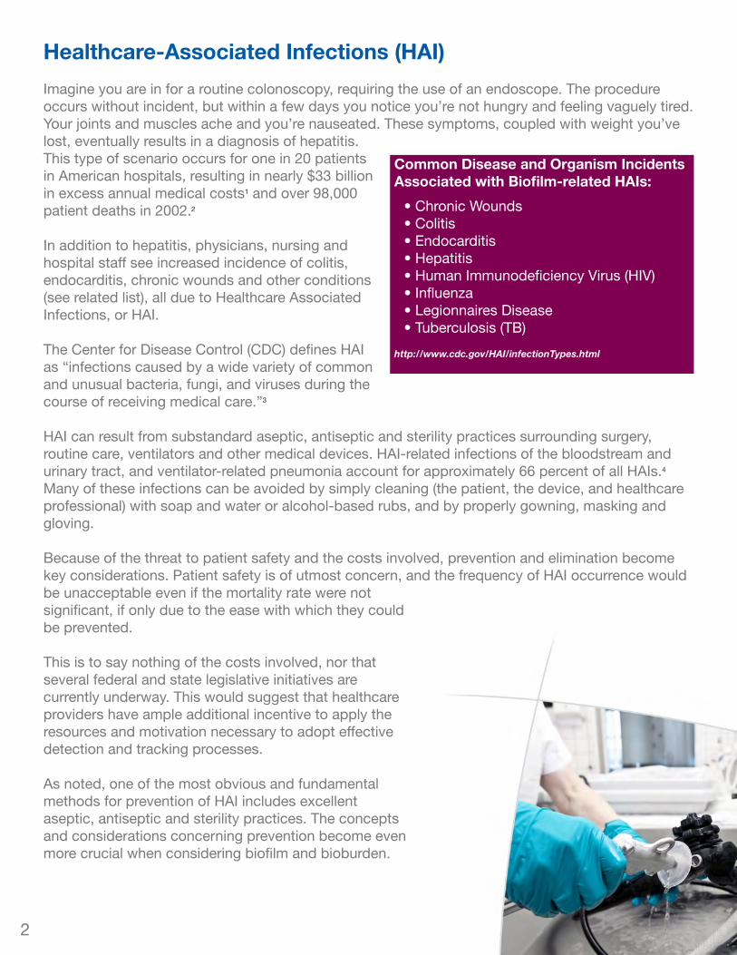
Common Disease and Organism Incidents Associated with Biofilm-related HAIs:
• Chronic Wounds• Colitis• Endocarditis• Hepatitis• Human Immunodeficiency Virus (HIV)• Influenza • Legionnaires Disease• Tuberculosis (TB)
http://www.cdc.gov/HAI/infectionTypes.html
2
Contamination and HAI Key Takeaways:
• Healthcare-Associated Infections (HAI) are costly in terms of patient suffering and medical costs. • HAIs are commonly caused by both common and unusual bacteria, fungi, and viruses exposed
to patients during receipt of healthcare.• HAIs are commonly the result of substandard aseptic, antiseptic and sterile practices and the
lack of observance of basic infection control practices.• Because of the causes of HAIs, they are easily avoidable and so can and should be prevented
and eliminated. • Common incidents of HAI include: Catheter-associated Urinary Tract Infections (CAUTI), Central
Line-associated Bloodstream Infection (CLABSI), Ventilator-associated Pneumonia (VAP), and Surgical Site Infection (SSI)
• Common corresponding medical instruments include: central venous catheter (CVC), urinary catheters, endoscopes and ventilators
• Prevention and elimination begins with, and is the end-result of, detection of contamination, documentation and tracking/record-keeping.
Bioburden Basics
Most health care professionals, particularly those involved with sterile processing, are familiar with the term bioburden as a focal point for processing contaminated items. Bioburden, according to ANSI/AAMI ST79:2010, is defined as the “population of viable microorganisms on a product and/or a sterile barrier system.” The measurement of bioburden is further defined as “the total count of bacterial and fungal colony-forming units (CFUs) per single item.”5
Bioburden can result from contamination of blood, tissue, mucus, and various by-products of surgery and other medical procedures. Bioburden may also result from various forms of environmental contamination during medical instrument manufacturing, shipping, and processing, where lint, dust, moisture and other contaminants may harbor microorganisms. Other environmental factors, such as temperature, humidity, and time also play a role in certain aspects of bioburden formation and removal.Bioburden and other contaminants must be controlled to ensure disinfection and sterilization.
Testing protocols are essential to determine the degree of contamination, the procedures required to remove the contamination, and to verify that the procedures have achieved the desired result. Testing for bioburden is known by several names: microbial count, viable count, plate count, colony count, heterotrophic count, bioburden determination (ISO/AAMI), and microbial enumeration (USP).6
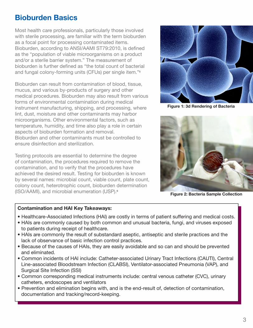
Figure 1: 3d Rendering of Bacteria
Figure 2: Bacteria Sample Collection
3
Now Playing: Biofilms
Health professionals—and science fiction fans—should have a keen interest in a particular aspect of microbial research that has uncovered some extremely fascinating aspects of microorganisms and their behavior under certain conditions. Many “sci-fi” fans can easily recite stories of alien life forms with unique defense mechanisms and destructive powers. These stories often serve as very entertaining science fiction, but in reality, these life forms may be right under our noses… literally.
To begin, we’ll go back to the 1600’s and build on the research of Antonie van Leeuwenhoek, a Dutch scientist who is considered to be one of the first microbiologists. Van Leeuwenhoek used his hand-crafted microscopes to observe microorganisms in the human mouth, including those that live on the tooth surface.
We now associate the term “dental plaque” with these organisms, but modern technology now enables researchers to discover much more than van Leeuwenhoek was able to observe with his simple microscopes. With modern technologies such as scanning electron microscopes, confocal laser scanning microscopes, and new microbiologic culture techniques, scientists are able to look at microorganisms more closely than van Leeuwenhoek could ever have imagined.
Further study in this field has revealed that many microorganisms can attach to and grow on exposed surfaces, and they exhibit several adaptive behaviors that bear an interesting resemblance to some of the sci-fi life forms depicted in movies. For instance, these organisms have the ability to communicate with similar organisms, form microbial communities, and cooperate to develop unique defense and reproductive mechanisms to sustain their communities.
Microbiologists now use the term “biofilm” to describe organisms that exhibit these behaviors, and biofilm research is playing a critical role in public health and various aspects of infection control.
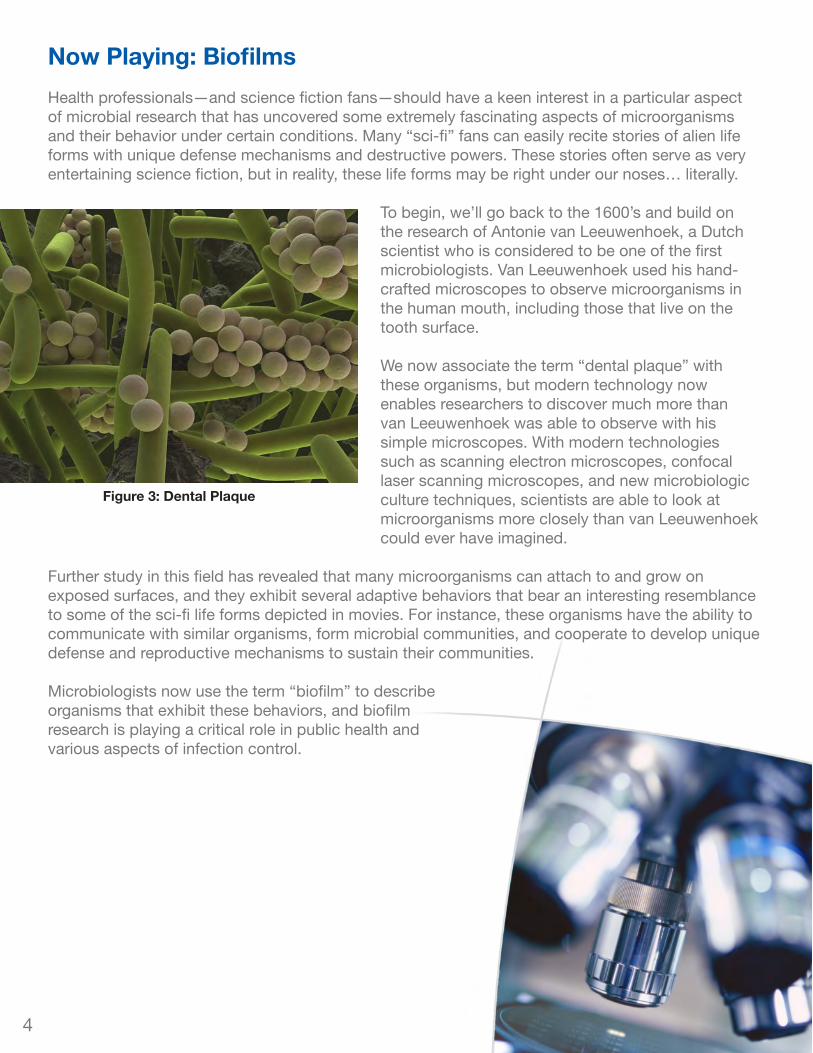
Figure 3: Dental Plaque
4
Biofilms: Microbial Social Networking?
“A biofilm is an assemblage of microbial cells that is irreversibly associated (not removed by gentle rinsing) with a surface and enclosed in a matrix of primarily polysaccharide material,” 7 better known as “slime”. While this definition may sound complex, it really only scratches the surface when it comes to describing biofilm architecture and behavior.
Biofilms can form on almost any surface in which moisture is present, including solid subtrates that are in contact with moisture, on the soft tissues of living organisms, and at liquid-air interfaces.
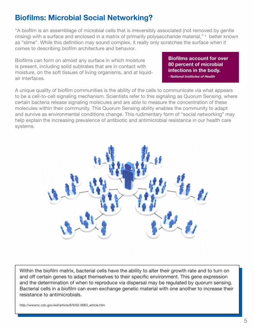
A unique quality of biofilm communities is the ability of the cells to communicate via what appears to be a cell-to-cell signaling mechanism. Scientists refer to this signaling as Quorum Sensing, where certain bacteria release signaling molecules and are able to measure the concentration of these molecules within their community. This Quorum Sensing ability enables the community to adapt and survive as environmental conditions change. This rudimentary form of “social networking” may help explain the increasing prevalence of antibiotic and antimicrobial resistance in our health care systems.
Within the biofilm matrix, bacterial cells have the ability to alter their growth rate and to turn on and off certain genes to adapt themselves to their specific environment. This gene expression and the determination of when to reproduce via dispersal may be regulated by quorum sensing. Bacterial cells in a biofilm can even exchange genetic material with one another to increase their resistance to antimicrobials.
http://wwwnc.cdc.gov/eid/article/8/9/02-0063_article.htm
5
Biofilms account for over 80 percent of microbial infections in the body. - National Institutes of Health
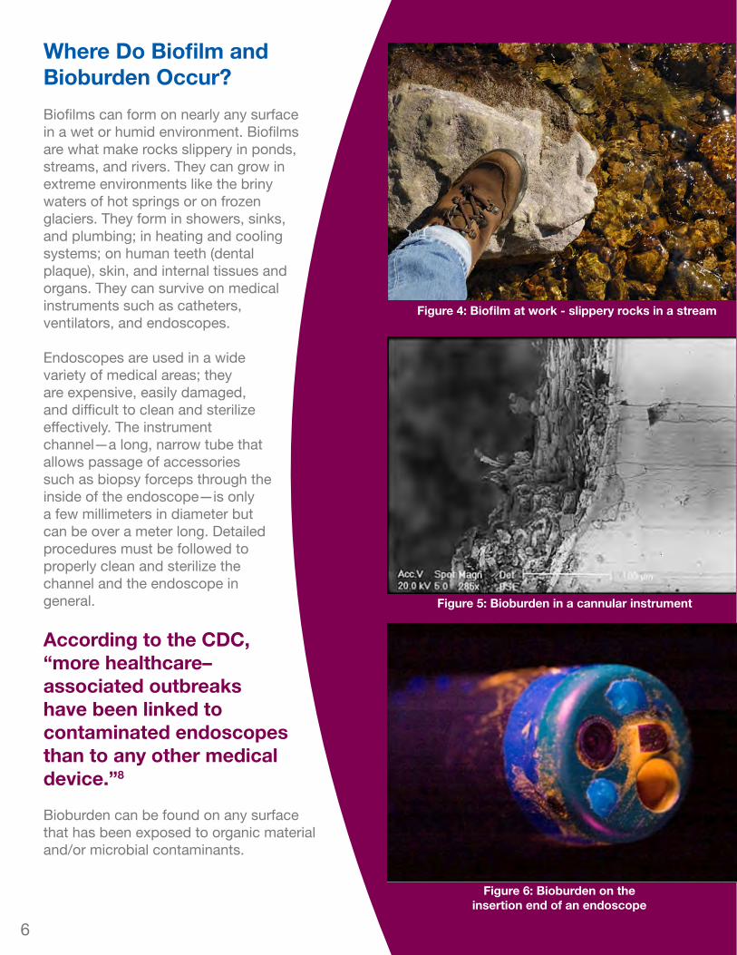
Where Do Biofilm and Bioburden Occur?
Biofilms can form on nearly any surface in a wet or humid environment. Biofilms are what make rocks slippery in ponds, streams, and rivers. They can grow in extreme environments like the briny waters of hot springs or on frozen glaciers. They form in showers, sinks, and plumbing; in heating and cooling systems; on human teeth (dental plaque), skin, and internal tissues and organs. They can survive on medical instruments such as catheters, ventilators, and endoscopes.
Endoscopes are used in a wide variety of medical areas; they are expensive, easily damaged, and difficult to clean and sterilize effectively. The instrument channel—a long, narrow tube that allows passage of accessories such as biopsy forceps through the inside of the endoscope—is only a few millimeters in diameter but can be over a meter long. Detailed procedures must be followed to properly clean and sterilize the channel and the endoscope in general.
According to the CDC, “more healthcare–associated outbreaks have been linked to contaminated endoscopes than to any other medical device.”8
Bioburden can be found on any surface that has been exposed to organic material and/or microbial contaminants.
Figure 4: Biofilm at work - slippery rocks in a stream
Figure 5: Bioburden in a cannular instrument
Figure 6: Bioburden on theinsertion end of an endoscope
6
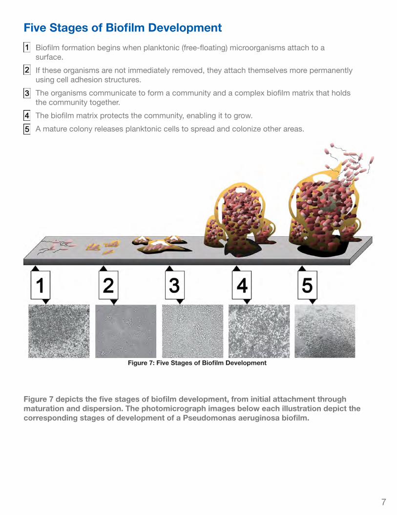
Five Stages of Biofilm Development
Figure 7: Five Stages of Biofilm Development
Figure 7 depicts the five stages of biofilm development, from initial attachment through maturation and dispersion. The photomicrograph images below each illustration depict the corresponding stages of development of a Pseudomonas aeruginosa biofilm.
7
Biofilm formation begins when planktonic (free-floating) microorganisms attach to a surface.
If these organisms are not immediately removed, they attach themselves more permanently using cell adhesion structures.
The organisms communicate to form a community and a complex biofilm matrix that holds the community together.
The biofilm matrix protects the community, enabling it to grow.
A mature colony releases planktonic cells to spread and colonize other areas.
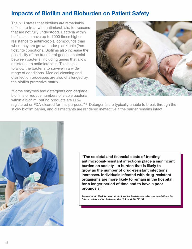
Impacts of Biofilm and Bioburden on Patient Safety
The NIH states that biofilms are remarkably difficult to treat with antimicrobials, for reasons that are not fully understood. Bacteria within biofilms can have up to 1000 times higher resistance to antimicrobial compounds than when they are grown under planktonic (free-floating) conditions. Biofilms also increase the possibility of the transfer of genetic material between bacteria, including genes that allow resistance to antimicrobials. This helps to allow the bacteria to survive in a wider range of conditions. Medical cleaning and disinfection processes are also challenged by the biofilm protective matrix.
“Some enzymes and detergents can degrade biofilms or reduce numbers of viable bacteria within a biofilm, but no products are EPA-registered or FDA-cleared for this purpose.” 9 Detergents are typically unable to break through the sticky biofilm barrier, and disinfectants are rendered ineffective if the barrier remains intact.
“The societal and financial costs of treating antimicrobial-resistant infections place a significant burden on society – a burden that is likely to grow as the number of drug-resistant infections increases. Individuals infected with drug-resistant organisms are more likely to remain in the hospital for a longer period of time and to have a poor prognosis.”
Transatlantic Taskforce on Antimicrobial Resistance - Recommendations for future collaboration between the U.S. and EU (2011)
8
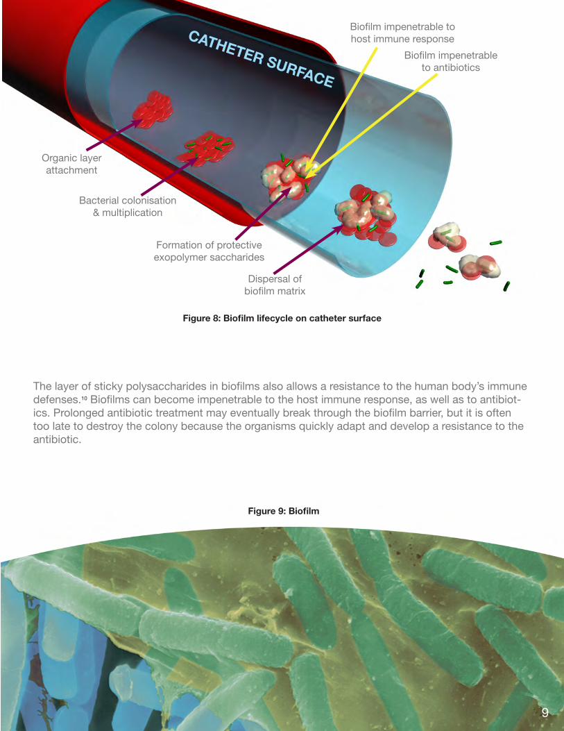
The layer of sticky polysaccharides in biofilms also allows a resistance to the human body’s immune defenses.10 Biofilms can become impenetrable to the host immune response, as well as to antibiot-ics. Prolonged antibiotic treatment may eventually break through the biofilm barrier, but it is often too late to destroy the colony because the organisms quickly adapt and develop a resistance to the antibiotic.
Biofilm impenetrable tohost immune response
Biofilm impenetrableto antibiotics
Dispersal ofbiofilm matrix
Organic layerattachment
Bacterial colonisation& multiplication
CATHETER SURFACE
Formation of protectiveexopolymer saccharides
Figure 8: Biofilm lifecycle on catheter surface
9
Figure 9: Biofilm
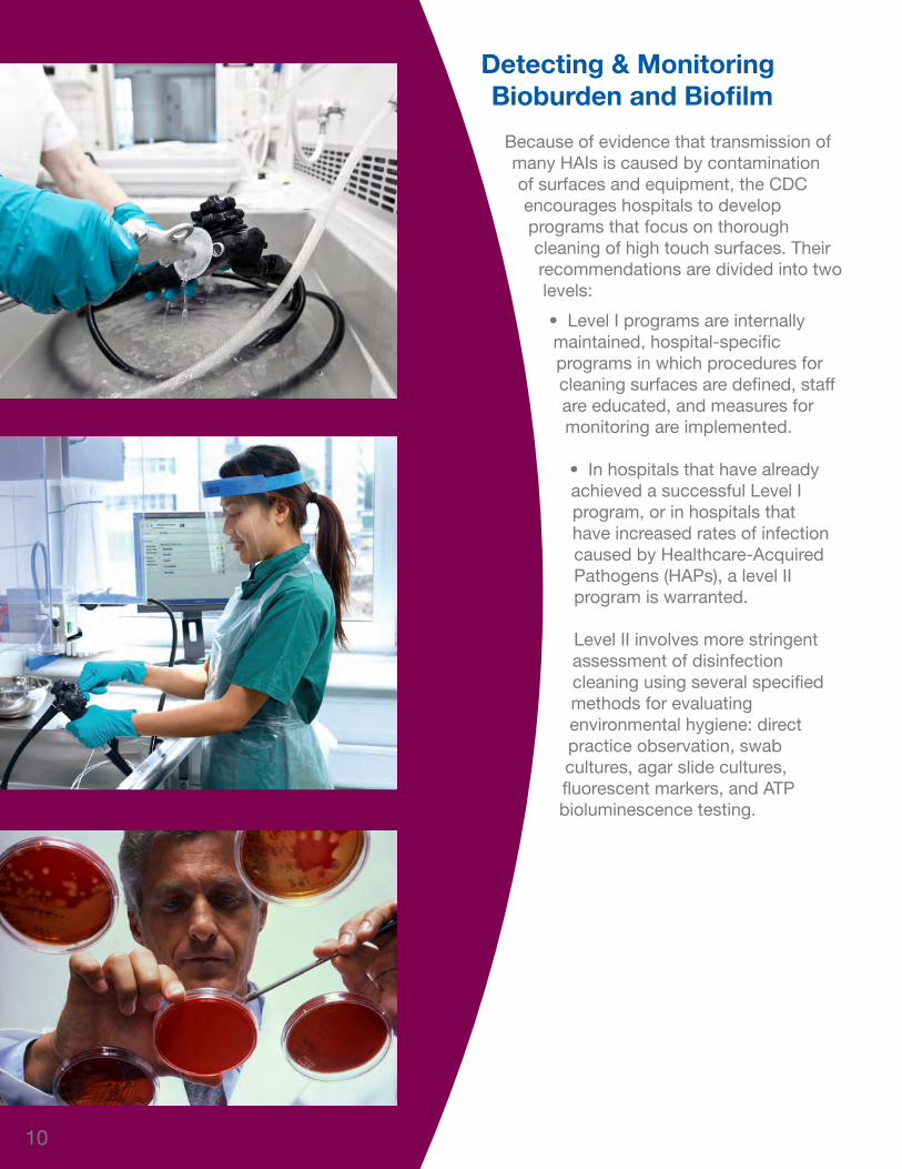
Detecting & Monitoring Bioburden and Biofilm
Because of evidence that transmission of many HAIs is caused by contamination of surfaces and equipment, the CDC encourages hospitals to develop programs that focus on thorough cleaning of high touch surfaces. Their recommendations are divided into two levels:
• Level I programs are internally maintained, hospital-specific programs in which procedures for cleaning surfaces are defined, staff are educated, and measures for monitoring are implemented.
• In hospitals that have already achieved a successful Level I program, or in hospitals that have increased rates of infection caused by Healthcare-Acquired Pathogens (HAPs), a level II program is warranted.
Level II involves more stringent assessment of disinfection cleaning using several specified methods for evaluating environmental hygiene: direct practice observation, swab cultures, agar slide cultures, fluorescent markers, and ATP bioluminescence testing.
10
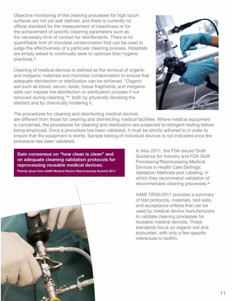
Objective monitoring of the cleaning processes for high touch surfaces are not yet well defined, and there is currently no official standard for the measurement of cleanliness or for the achievement of specific cleaning parameters such as the necessary time of contact for disinfectants. There is no quantifiable limit of microbial contamination that can be used to judge the effectiveness of a particular cleaning process. Hospitals are simply asked to continually seek to optimize their hygenic practices.11
Cleaning of medical devices is defined as the removal of organic and inorganic materials and microbial contamination to ensure that adequate disinfection or sterilization can be achieved. “Organic soil such as blood, serum, lipids, tissue fragments, and inorganic salts can impede the disinfection or sterilization process if not removed during cleaning,”12 both by physically blocking the sterilant and by chemically hindering it.
The procedures for cleaning and disinfecting medical devices are different from those for cleaning and disinfecting medical facilities. Where medical equipment is concerned, the procedures for cleaning and sterilization are subjected to stringent testing before being employed. Once a procedure has been validated, it must be strictly adhered to in order to ensure that the equipment is sterile. Sample testing of individual devices is not indicated once the procedure has been validated.
Gain consensus on “how clean is clean” and on adequate cleaning validation protocols for reprocessing reusable medical devices.Priority Issue from AAMI Medical Device Reprocessing Summit 2011
In May 2011, the FDA issued Draft Guidance for Industry and FDA Staff: Processing/Reprocessing Medical Devices in Health Care Settings: Validation Methods and Labeling, in which they recommend validation of recommended cleaning processes.13
AAMI TIR30:2011 provides a summary of test protocols, materials, test soils, and acceptance criteria that can be used by medical device manufacturers to validate cleaning processes for reusable medical devices. These standards focus on organic soil and bioburden, with only a few specific references to biofilm.
11
AAMI ST79 provides guidance on the establishment of cleaning protocols for reusable medical devices, and lists a number of evaluation methods. “The most common method is a visual inspection, sometimes involving the use of a lighted magnifying glass… However, residual organic soil and microbial contamination might be present on an accessible surface even though the device ‘looks clean.’”14 Several other available tests are listed, including several chemical assays.
For environmental cleaning processes such as those for hands or high-touch surfaces, the quality of individual cleaning instances might be tested with an appropriate method. Where the cleaning of reusable medical devices is concerned, typically only the process is tested. Assurance of sterility and cleanliness relies on the integrity of the process and each operator’s adherence to the process.
Testing Methods
There are a wide range of tests for biofilm and bioburden with a wide range of proficiencies and limitations. Some require large and expensive laboratory equipment and take days or even weeks to perform. Others use hand-held instruments and can be performed in seconds. Two of the most common types of testing in hospital protocols are Protein tests and ATP Bioluminescence tests.
Protein Tests
In this method of detection, a sample is gathered using a method such as rinsing with a buffer solution. A chemical is added that will react with the solution and change color if protein is present. Although this method is fairly quick (taking only minutes) and inexpensive compared to slide cultures and microscope counts, the only data it yields is a change in color. Some testing products rely on the operator to determine by eye if there has been a color change, providing only a positive or negative result. Other products use a colorimeter to provide quantitative data that suggest a relative amount of protein present.
Some Other Methods to Test for Biofilm and Bioburden:
• Direct Practice Observation• Microtiter plate biofilm assay (96-well plate assay)• Kadouri Drip-Fed Biofilm Assay system• Agar plate counts / Agar Slide Culture• Microscope direct counts: - Scanning Electron Microscopy (SEM) - Confocal laser scanning microscopy (CLSM or LSCM) - Traditional microscopy, etc. • Molecular methods (e.g., quantitative Polymerase Chain
Reaction (PCR))
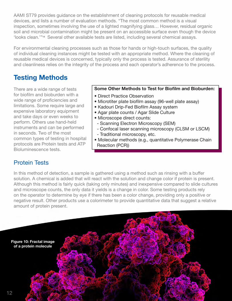
Figure 10: Fractal image of a protein molecule
12
ATP Bioluminescence Tests
This is a method of detection that uses a chemical reagent that gives off light when it reacts with ATP (Adenosine Triphosphate). A swabbed sample is combined with the reagent, and inserted into a hand-held unit in which a light detector determines the relative amount of ATP present in the sample. An ATP Bioluminescence detection system detects the ATP found in all animal, plant, bacterial, yeast, and mold cells. Residues such as blood and bioburden contain large amounts of ATP. Microbial contamination contains ATP, but in smaller amounts. After cleaning, all sources of ATP should be significantly reduced.
ATP: The Unit of Energy Currency in Cell Transactions
ATP is the principal energy-transferring molecule used by all living cells; essentially all of the physiological mechanisms that require energy for operation obtain it directly from energy stored in ATP molecules. ATP is continuously recycled in organisms. Energy released when food in the organism is gradually broken down is used to re-form ATP to ensure that the cell has plenty of these energy-providing molecules.
ATP degrades outside of living cells, so an ATP Bioluminescence test is more of an indication of living cells and not simply of organic material.
Like a rechargeable battery, ATP stores energy formed by the breakdown of food for later use within a cell. For example, when muscles contract or when cells divide the energy is supplied by ATP. Although the average human body contains only about 250 grams of ATP at any given time (containing roughly the same energy as one AA battery), it is estimated that more than 160 kilograms is generated per day.
Figure 11: ATP molecule
13
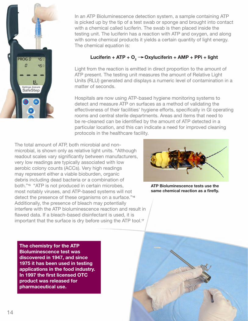
In an ATP Bioluminescence detection system, a sample containing ATP is picked up by the tip of a test swab or sponge and brought into contact with a chemical called luciferin. The swab is then placed inside the testing unit. The luciferin has a reaction with ATP and oxygen, and along with some chemical products it yields a certain quantity of light energy. The chemical equation is:
Luciferin + ATP + O2 Oxyluciferin + AMP + PPi + light
Light from the reaction is emitted in direct proportion to the amount of ATP present. The testing unit measures the amount of Relative Light Units (RLU) generated and displays a numeric level of contamination in a matter of seconds.
Hospitals are now using ATP-based hygiene monitoring systems to detect and measure ATP on surfaces as a method of validating the effectiveness of their facilities’ hygiene efforts, specifically in GI operating rooms and central sterile departments. Areas and items that need to be re-cleaned can be identified by the amount of ATP detected in a particular location, and this can indicate a need for improved cleaning protocols in the healthcare facility.
The total amount of ATP, both microbial and non-microbial, is shown only as relative light units. “Although readout scales vary significantly between manufacturers, very low readings are typically associated with low aerobic colony counts (ACCs). Very high readings may represent either a viable bioburden, organic debris including dead bacteria or a combination of both.”15 “ATP is not produced in certain microbes, most notably viruses, and ATP-based systems will not detect the presence of these organisms on a surface.”16 Additionally, the presence of bleach may potentially interfere with the ATP bioluminescence reaction and result in flawed data. If a bleach-based disinfectant is used, it is important that the surface is dry before using the ATP tool.17
The chemistry for the ATP Bioluminescence test was discovered in 1947, and since 1975 it has been used in testing applications in the food industry. In 1997 the first licensed OTC product was released for pharmaceutical use.
ATP Bioluminescence tests use thesame chemical reaction as a firefly.
14
Conclusions/Summary
It is a growing challenge in today’s hospitals to help prevent the spread of HAIs by ensuring the cleanliness of the facility and the sterility of all medical devices, particularly reusable devices. While rigorous cleaning and sterilization procedures are paramount in this effort, testing should be done to verify and ensure that the procedures have been successfully completed. Among the available tests, the ATP Bioluminescence test provides a quantitative assessment of the presence of organic soil and bioburden, and can generate results in a matter of seconds.
If it’s not clean, it’s not sterile!
Antibiotic Resistance Could Bring ‘End of Modern Medicine’http://abcnews.go.com, Mar 16, 2012 11:03am
As bacteria evolve to evade antibiotics, common infections could become deadly, according to Dr. Margaret Chan, director general of the World Health Organization.
Speaking at a conference in Copenhagen, Chan said antibiotic resistance could bring about “the end of modern medicine as we know it.”
“We are losing our first-line antimicrobials,” she said Wednesday in her keynote address at the conference on combating antimicrobial resistance. “Replacement treatments are more costly, more toxic, need much longer durations of treatment, and may require treatment in intensive care units.”
Chan said hospitals have become “hotbeds for highly-resistant pathogens” like methicillin-resistant Staphylococcus aureus, “increasing the risk that hospitalization kills instead of cures.”
15
Assessment
1. How can most HAI-related infections be avoided? a. Thorough cleaning (patient, device or healthcare professional) with soap and water or alcohol-
based solution b. Revision of hospital policy and procedurec. Reduced excess annual medical costsd. All of these will help avoid HAI-related infectionse. None of these will help avoid HAI-related infections
2. Which of the following are diseases and organism incidents related to HAI?a. Colitisb. Hepatitisc. Human Immunodeficiency Virus (HIV)d. All of these are diseases and organism incidents related to HAI e. None of these are diseases and organism incidents related to HAI
3. True or False: Because of the causes of HAI, they cannot be prevented or eliminated.a. Trueb. False
4. Which of the following instruments are not commonly corresponding with HAI incidents?a. Central Venous Catheter (CVC)b. Urinary Catheter c. Endoscoped. Automated External Defibrillator (AED)e. All of these are instruments commonly corresponding with HAI incidents
5. Which of the following conditions support prevention and elimination of HAI?a. Detection of contaminationb. Documentationc. Trackingd. Record-keeping e. All of these conditions support prevention and elimination of HAI
6. Bioburden is:a. The cost of cleaning and sterilizing medical devicesb. An accumulation of dirt, lint, dust, and organic matterc. All the bacteria, fungus, and other microorganisms on a surfaced. Dried blood, mucous, and tissue on a medical instrument
7. What causes the formation of bioburden?a. Improper cleaning and sterilizationb. Environmental contamination during manufacture, shipping, and processingc. Temperature, humidity, and timed. Contamination during medical procedurese. All of these contribute to the formation of bioburden
8. What is the function of the polysaccharide film (slime) that covers a biofilm? a. Holds the community togetherb. Protects the community from immune responsec. Protects the community from outside chemicals such as enzymes and detergentsd. All of these are functions of the film
16
9. In what stage of its development is a biofilm most susceptible to disinfectants and detergents? a. When it is free-floating, before forming a biofilmb. When has formed the biofilm matrix c. When it is growingd. When it releasing planktonic cells
10. Planktonic bacteria are:a. Bacteria that make up biofilmsb. Individual bacterial cellsc. Any bacteria that causes an HAId. Any bacteria attached to a surface
11. When does the CDC define a high-touch surface as clean? a. Objective monitoring is not yet well definedb. It contains zero bioburdenc. It cannot register results from either a Protein test or an ATP Bioluminescence testd. The surface looks clean to the naked eye
12. Which is a benefit of Protein testing? a. It is faster than all other tests, including ATP Bioluminescenceb. It can distinguish between biofilm and blood proteinc. It is inexpensived. All of the above
13. Which is true about ATP Bioluminescence tests?a. They measure the total number of cells presentb. They give the same reading for blood cells as for bacterial cellsc. They must be performed in a dark roomd. They display a numerical reading that represents the amount of ATP present
14. True or False? ATP Bioluminescence can test for viruses. a. Trueb. False
15. What do Relative Light Units indicate about a sample in an ATP Bioluminescence test? a. The amount of ATP presentb. The type of cells present c. The type of bacteria presentd. The amount of protein present
Answer Key: 1. a2. d3. b4. d5. e6. c7. e8. d9. a10. b11. a12. c13. d14. b15. a
17
Endnotes1 Association of State and Territorial Health Officials and the Center for Disease Control and Prevention, Eliminating
Healthcare Associated Infections, http://www.cdc.gov/HAI/pdfs/toolkits/toolkit-HAI-POLICY-FINAL_03-2011.pdf
2 Monina R. Klevens, DDS, MPH; Jonathan R. Edwards, MS; Chelsey L. Richards, Jr, MD, MPH; Teresa C. Horan, MPH; Robert P. Gaynes, MD; Daniel A. Pollack, MD; Denise M. Cardo, MD; Estimating Health Care-Associated Infections and Deaths in US Hospitals, 2002, http://www.cdc.gov/HAI/pdfs/hai/infections_deaths.pdf
3 Center for Disease Control and Prevention, Healthcare-associated Infections, http://www.cdc.gov/hai/index.html
4 Center for Disease Control and Prevention, Types of Healthcare-associated Infections, http://www.cdc.gov/HAI/infectionTypes.html
5 ANSI/AAMI ST79:2010 & A1:2010. Comprehensive guide to steam sterilization and sterility assurance in health care facilities. Association for the Advancement of Medical Instrumentation. Page 7
6 http://www.wuxiapptec.com/pdfs/228-BioburdenTesting.pdf
7 Biofilms: Microbial Life on Surfaces. Volume 8, Number 9- September 2002. Centers for Disease Control and Prevention.
8 http://www.cdc.gov/hicpac/Disinfection_Sterilization/3_0disinfectEquipment.html
9 http://www.cdc.gov/hicpac/Disinfection_Sterilization/4_0efficacyDS.html#7
10 National Institute of Health. Research of Microbial Biofilms. 2002. http://grants.nih.gov/grants/guide/pa-files/PA-03-047.html
11 Alice Guh, MD, Philip Carling, MD. Options for Evaluating Environmental Cleaning. 2010. http://www.cdc.gov/hai/toolkits/evaluating-environmental-cleaning.html
12 ANSI/AAMI ST79:2010 & A1:2010 (Consolidated Text) pg. 170 Center for Disease Control and Prevention, Types of Healthcare-associated Infections, http://www.cdc.gov/HAI/infectionTypes.html
13 Association for the Advancement of Medical Instrumentation. Priority Issues from the AAMI/FDA Medical Device Reprocessing Summit. 2011. http://www.aami.org/reprocessing/Materials/PDF/2011_Reprocessing_Summit_publication.pdf
14 ANSI/AAMI ST79:2010 & A1:2010 (Consolidated Text) pg. 169
15 http://www.cdc.gov/HAI/toolkits/Appendices-Evaluating-Environ-Cleaning.html
16 http://www.infectioncontroltoday.com/news/2010/12/using-atp-in-healthcare-settings.aspx
17 http://www.cdc.gov/HAI/toolkits/Appendices-Evaluating-Environ-Cleaning.html
18
I have completed all the requirements for this entire activity. (Please sign and date.)
Your signature is required to attest that you have completed the requirements for this activity.
To receive continuing education credit, please complete the evaluation form and mail, fax, or e-mail the completed form to Getinge:
Getinge USA, Inc.Attn: Mark A. Rider1777 E. Henrietta Rd.Rochester, NY 14623Fax: 585.272.5033Email: [email protected]
Evaluation FormStudy Guide: ATP-Based Contamination Monitoring vs. Biofilm and Bioburden
Last Name
First Name/M.I.
RN/LPN/LVN License Number (Circle one: RN - LPN - LVN )
Non-RN: License or Social Security Number
Health Care Facility
Health Care Facility Street Address City State ZIP Code
Current Home Street Address City State ZIP Code
For International Address: Country Province/Postal Code
Area Code / Telephone Number E-Mail Address
Please check here if you want the certificate to be mailed to the address above. Please check here if you want the certificate e-mailed to you at the e-mail address above.
① ② ③ ④ ⑤
① ② ③ ④ ⑤
___ hours ____ minutes
Yes ___ No ___
Yes ___ No ___
1. To what extent did the study guide meet the objectives stated on page 1 of the guide?
2. To what extent is this learning method easy to use?
3. How much time was required to read the content, take the test, compare your answers, and complete the evaluation form?
4. Has the provider disclosed the conflict of interest or lack thereof? (Review inside cover page)
5. Was the content presented without bias of any commercial product?
Rate on a scale of: 1 = low 5 = high
19
HC
_CE
U_A
TP
Bio
film
Stu
dyG
uid
e_0
41
2_E
N_U
S ©
201
2 G
etin
ge U
SA
, Inc
. • P
rinte
d in
U.S
.A. A
rjo, G
etin
ge, a
nd M
AQ
UE
T ar
e re
gist
ered
tra
dem
arks
. All
othe
r p
rod
uct
nam
es o
r tr
adem
arks
bel
ong
to t
heir
resp
ectiv
e ow
ners
.
GETINGE GROUP is a leading global provider of products and systems that
contribute to quality enhancement and cost efficiency within healthcare and life
sciences. We operate under the three brands of ArjoHuntleigh, GETINGE and
MAQUET. ArjoHuntleigh focuses on patient mobility and wound management
solutions. GETINGE provides solutions for infection control within healthcare and
contamination prevention within life sciences. MAQUET specializes in solutions,
therapies and products for surgical interventions and intensive care.www.getinge.com
Getinge USA Inc.1777 East Henrietta RoadRochester, NY 14623USAPhone: 800.475.9040Fax: [email protected]
Getinge Canada, Ltd.1575 South Gateway Road, Unit CMississauga, OntarioCanada L4W 5J1Phone: 905.629.8777Fax: 905.629.8875
COMPLETE SOLUTIONS FOR INFECTION CONTROLGetinge is the world’s leading provider of solutions for
effective cleaning, disinfection and sterilization in the healthcare and life
science sectors. We are dedicated to helping our customers provide
better care at a lower cost. We do this by offering well thought through
and customized solutions. This means that we are with our customers
all the way from architectural planning and education to traceability and
support – with complete solutions, long-term commitment and global
presence. Getinge – Always with you.