Embed Size (px)
DESCRIPTION
Ross's views on aetiology of Athrosclerosis
Citation preview

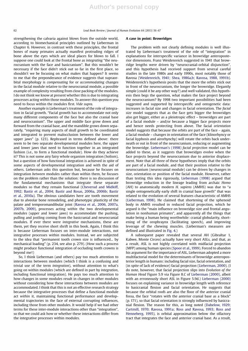
This article appeared in a journal published by Elsevier. The attachedcopy is furnished to the author for internal non-commercial researchand education use, including for instruction at the authors institution
and sharing with colleagues.
Other uses, including reproduction and distribution, or selling orlicensing copies, or posting to personal, institutional or third party
websites are prohibited.
In most cases authors are permitted to post their version of thearticle (e.g. in Word or Tex form) to their personal website orinstitutional repository. Authors requiring further information
regarding Elsevier’s archiving and manuscript policies areencouraged to visit:
http://www.elsevier.com/copyright

Author's personal copy
Lead Book Review
Complexity, Modularity, and Integration in the Human Head.The Evolution of the Human Head. By Daniel E. Lieberman(2011). Cambridge, Mass: The Belknap Press of Harvard Univer-sity Press, 768 pp., $41.50 (hardback), ISBN 978-0-674-04636-8.
The intricacies of head morphology are powerful lures for thoseof us interested in the relationships and behavior of fossil primates.The many functional systems in the head make it a rich source ofdata on how fossil primates ate, saw, heard, walked, and climbed.We can learn about the evolution of the size and shape of the brain,how primates held their heads, what they ate, and even about theirsocial group structures. Daniel Lieberman’s new book, The Evolutionof the Human Head, is a bold new attempt to put all these piecestogether in an integrated whole, to make sense of the complexitythrough the lenses of development and function. It builds ona rich tradition in the study of primate, mammal, and vertebrateheads, from William K. Gregory’s Our Face From Fish to Man(Gregory, 1929), through Moore’s Mammalian Skull (Moore, 1981),to Hanken and Hall’s three volume Skull series (Hanken and Hall,1993). However, Lieberman’s book differs from these in beingfocused exclusively on primate, and especially hominid, heads.Moreover, he reviews the important functional and developmentalprinciples of all the head systems, then takes these data and usesthem to interpret the developmental ‘how’ and functional ‘why’of fossil hominid skull evolution. This ambitious agenda stretchesthe book to 750 pages in length, but it is well organized, mostlywell indexed, and Lieberman’s clean, relaxed style makes it easyto read. The book is also nicely illustrated, with crisp, cleandiagrams, plenty of graphs, and some data in tables, the rest ofthe data being available in Lieberman’s papers, or – if my past expe-rience is anything to go by – probably from Lieberman himself.
The book starts with a preview chapter, summarizing Lieber-man’s view of the head and laying out his plan for the book. Lieber-man posits that despite their complexity, heads are evolvablebecause “they consist of many functionally importantmodules inte-grated in a special way to accommodate one another” (p.15). Lieber-man postulates that heads evolve through “tinkering” (Jacob, 1977):“When new organisms make new use of preexisting or modifiedmodules, these tinkered novelties often tend to work because theyare made of modules that already function appropriately and comewithexistingmechanisms foradjusting tooneanother” (p.15). Theseare interesting and important ideas that we will return to below.
Lieberman then presents two chapters on skeletal tissues(Chapter 2) and embryonic development of the head (Chapter 3).The skeletal tissue chapter includes developmental origins ofbone, including patterning and morphogenesis, and a too brieftreatment of dental tissues, cartilage, tendons and ligaments. Thereare a few pages on Moss’s functional matrix hypothesis, with anappropriate nod to van der Klauuw, as well as more recent
researchers, such as Cheverud, Zelditch and Hallgrimsson. I donot agree with Lieberman’s use of the term “remodeling” to referto the wholesale bone deposition resulting in drift and displace-ment of sheets of cortical bone. This should be referred to asmodeling: remodeling should be reserved for Haversian remodeling,formation of osteons mainly, which improves bone material prop-erties while maintaining gross bone shape. This chapter alsoincludes some elementary bone biomechanics. This chapter is suffi-cient introduction for what is to come in the rest of the book, butwhen I recommend a readable account of bone tissue level biome-chanics and development, I will still refer students to John Currey’sbook, Bone: Structure and Mechanics (Currey, 2002).
Chapter 4, Modular Growth of the Fetal and Postnatal Head,provides a succinct review of how the skull grows after the embry-onic period. Lieberman makes liberal reference to Enlow’s work ongrowth fields, and nicely reviews the literature on growth anddevelopment of the cranial base, vault, and face, including muchof his own contributions to these areas. On the whole, the chapterpresents a thorough review of a large and intimidating literatureand is well worth your time. (Note that the arrows illustratingnuchal plane rotation in chimps and humans on page 104 are swap-ped.) Having described the modular growth of the head in Chapter4, Chapter 5 attempts to put the pieces together again by discussingthe Integration of the Head during Fetal and Postnatal Growth. Aswith Chapter 4, there are plenty of great data here, and somenice reviews of the concepts of integration. However, Chapters 4and 5 are the two that I find themost problematic from a theoreticalperspective, and I return to these issues below.
Chapter 6, on The Brain and Skull has a lot of interesting andimportantmaterial in it regarding the evolution of brain size, differ-ential increases in brain parts in hominid evolution, blood supply,venous drainage of the brain, how brain temperature is regulated,and the role of the skull in protecting the brain. The literature onadult brain size and brain ontogeny is also reviewed. All in all,this is a good review of the relationships between brain and skull,although for a complete picture, one should also read the previoustwo chapters. The only (minor) corrections needed are that theouter layer of dura is endocranial, not endosteal (Figure 6.11), theCircle of Willis is not below the tentorium (p. 220), and themyelen-cephalon is not the spinal cord (Figure 6.2).
Chapter 7 on Chewing and the Head, including a description ofmastication, needs revision and correction before I would recom-mend it tomy students to read. There is some truth to the statementthat “[d]uring mastication, the mandible is pulled both forward andbackward (retracted)” (p. 225) but lateral-medial movements of theteeth aremuchmore important inprimates. In attempting to explainmandibular condylar translation during chewing (Figure 7.8), Lie-berman also perpetuates a dubious hypothesis about jawmechanicsthat I think is erroneous (Greaves,1974,1980; Crompton et al., 2006;
Contents lists available at SciVerse ScienceDirect
Journal of Human Evolution
journal homepage: www.elsevier .com/locate/ jhevol
Journal of Human Evolution 64 (2013) 56–67
0047-2484/$ – see front matterhttp://dx.doi.org/10.1016/j.jhevol.2012.08.010

Author's personal copy
Rak andHylander, 2008). The argument is that “In a simple jawwitha hingelike joint, the occlusal plane is in line with the TMJ, and theteeth occlude with a primarily vertical (orthal) direction (sic)” (p.243). As the ramus increases in depth, the trajectory of the lowerteeth becomes increasingly more anteriorly directed relative to theupper teeth (Crompton et al., 2006), a trajectory that is “correctedby translating the joint posteriorly during adduction” (p. 243). Theproblem envisaged by Crompton et al. (2006) and traceable toGreaves (1974) is shown in Fig.1A. This is a geometrically impossiblearrangement in any animal, because only the posterior teeth wouldbe in occlusion when the jaws are shut. Fig. 1B presents more real-istic jaw geometry: offsetting the axis of rotation from the plane ofthe upper tooth-row lies. Once this more realistic arrangement ofupper and lower jaws is appreciated, it is clear that the tooth-rowscan be displaced inferiorly asmuch as needed (to augment adductorcross-sectional area, for example) changing the angle of approach ofthe teeth and without necessitating condylar translation. Thus, it isnot correct to argue that condylar translation is necessary to ensurethat teeth occlude vertically when the ramus is tall (Crompton et al.,2006). It may be one possible solution, but the challenging controland stability problems associatedwith it suggest tome that condylartranslation is not thedesignpathof least resistance. This is especiallythe case if, as shown in Fig.1B, this problemdoes not exist in thefirstplace. Natural selection has plenty of difficult design challenges toface: a simple vertical displacement of the upper tooth-row relativeto the jaw’s axis of rotation is not one of them. Of course, oncecondylar translation is an entrenched component of jaw system
design, the magnitude of condylar translation will increase as jawjoint height increases (Wall, 1999; Terhune, 2010, 2011; Ross et al.,Submitted for publication), but that does not mean that condylartranslation is necessary to maintain vertically oriented tooth move-ment during occlusion. Lieberman’s book would have been betterserved by a fuller explication of the sarcomere stretch hypothesis,currently the best explanation for condylar translation in primates(Hylander, 1992b).
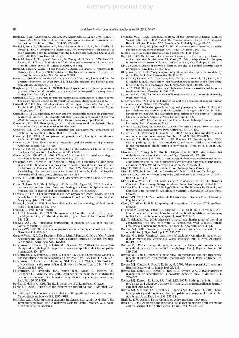
Chapter 7 also includes a biomechanical error concerning anal-ysis of the mechanical advantage of the masseter muscle in frontalview. The mechanical advantage (MA) of a jawmuscle is the ratio ofa muscle’s lever arm to the load arm of the bite point. In Figure 7.10(reproduced here as Fig. 2), the line of action of the masseter incoronal planes is represented by a vector running from the angleof the mandible through the lateral surface of the zygomatic arch,and its lever arm in this projection is presented as the perpendic-ular distance from this vector to the frontal projection of theworking side temporomandibular joint (TMJ). The bite force is rep-resented as a vector directed inferiorly at the M1, and its momentarm is the perpendicular distance from this vector to the workingside TMJ. MA is then calculated as the ratio of the lever and loadarms. This is incorrect. Calculation of mechanical advantagerequires correct placement of the location of the fulcrum of thelever system. Figure 7.10 places the fulcrum between the massetervector and the bite force vector, but with the fulcrum in this loca-tion, both forces act to rotate the mandible in a clockwise direction,generating no reaction forces. Reversing the direction of the bite
Figure 1. Diagrams illustrating problems with Lieberman’s explanation for condylar translation in primates. A. Following Greaves (1974) and Crompton et al. (2006) Liebermansuggests that if the upper toothrow is in line with the axis of rotation of the jaw joint, the teeth occlude in an orthal direction. However, as is clear from this diagram, when thesejaws close only the posterior teeth are in contact, and nor are the teeth moving in an orthal direction. This is a biologically unlikely geometry. B. Once a more realistic geometry isdefined, it is clear that, in fact, it is possible for the teeth to occlude with an orthal movement at any jaw joint height (C). Condylar translation is not necessary for maintainingvertical tooth movements during occlusion. Moreover, I would argue that it is unlikely to be a preferred solution, given that the problem (anteriorly moving lower teeth duringocclusion) may actually not exist, and even if it does, simple geometric solutions are available that do not entail a translating condyle.
Lead Book Review / Journal of Human Evolution 64 (2013) 56–67 57

Author's personal copy
force vector would be a mathematically correct but biologicallyimplausible solution. It is generally accepted that it is more appro-priate to place the fulcrum at the balancing side joint (Hylander,1992a; Spencer, 1998, 1999), and with the fulcrum in that locationthe illustrated lateral shifts in the masseter line of action in A. boiseiwould have trivial effects on the MA of the masseter. (Note that,although one can take moments about any point in a free body inorder to calculate an unknown reaction force, this is not the sameas calculating mechanical advantage. Consider, for example, thatif one took moments about the bite force location, the MA of themasseter would go to infinity.)
Consideration of the text on page 246 suggests that Lieberman istrying to quantify the “efficiency of transverse force generation”.The easiest and probably best way to do this would be to comparethe magnitudes of the lateral components of the masseter muscleforces in these three taxa. These would be larger in A. boisei becauseits masseter muscle force vector is directed more laterally, butstrictly speaking, this would not be a measure of efficiency. In anyevent, this error means that Lieberman’s discussions of masseterMA in the coronal plane in Chapters 11 and 12 are incorrect.
Figure 7.1 also contains some errors that will confuse the unini-tiated. In the top row of diagrams, the protoconid is not labeledwith an asterisk as the legend states, the paraconid is. Also, thismesial view gives the impression that the food is primarily brokendown by contact between the paraconid shearing past the buccalcusps on the upper teeth, with no crushing between the protoconidand the lingual cusps of the uppers. The “buccal” and “lingual”labels must have been swapped and the arrows seem to havebeen placed by someone who doesn’t know anything about masti-cation. Parenthetically, equating Mills’ “buccal” phase and “lingual”phase with Phase I and Phase II is incorrect, as Mills thought theyoccurred simultaneously on opposite sides of the jaws; the hyoidin Figure 7.5 is nearly in the superior mediastinum; and the effectsof gravity on jaw depression are trivial compared with passivetension in the jaw elevator connective tissues (p. 239). Beforeleaving the feeding system, I do note that one should be carefulnot to accept Figures 7.12 and 11.15 at face value. These figuresplot estimates of bite force at M2 against the estimated area ofM2, using methods of (Demes and Creel, 1988), and the claim ismade that “Bite force increases linearly with tooth surface area,keeping occlusal pressures similar across species”. However, line-arity does not mean isometry, and Lieberman’s claim is only correctif the slope of the line in the plots is close to 1.0 (assuming the unitson the y-axis are areas, which is not clear). This information is notprovided, so it is not possible to evaluate these claims.
Chapter 8, Pharynx, Larynx, Tongue and Lung, presents a readabletreatment of the development, function and recent evolution of thenose, pharynx, larynx and tongue. Lieberman nicely reviews thefunctions of these areas in respiration, swallowing and speech.The anatomy is clearly and simply described and illustrated, thebiomechanics of airflow through tubes is summarized, and thechanging relationships of these structures to the evolving cranialbase are presented. This chapter is Lieberman at his best, usingclear writing and simple analogies to cover a complicated topic. Ithink those of us preparing lectures on topics such as the evolutionof speech will find this chapter very useful.
Similarly, Chapter 9, Holding Up and Moving the Head, is a clearpresentation of some of the basic biomechanical issues involved.The discussion here is simple and concise, and a useful review. Lie-berman covers foramen magnum position and orientation, nuchalplane orientation, the function of the semicircular canals investibule-ocular reflexes, and the results of Spoor and colleagues.Dave Strait and I showed that basicranial flexion does not reflecthead and neck posture, but that foramen magnum and nuchalplane orientation adjust so as to reflect the orientation of thecervical vertebral column (Strait and Ross, 1999). Therefore, I wassurprised to read in Chapter 9: “because the Strait and Ross analysisincluded only one bipedal species with a very flexed cranial base(Homo sapiens), one cannot conclude that a more flexed cranialbase is unrelated to being bipedal” (p. 347). In fact, Strait andRoss (their Table 4) showed that basicranial flexion across a rangeof values of flexion is driven by relative brain size and is not relatedto head posture. It is foramen magnum orientation relative to theorbits that reflects posture. This is shown in Fig. 3, a plot of foramenmagnum (FM) orientation relative to the orbital plane (OP) againstthe orientation of the orbital plane relative to the dorsal surface ofthe neck (the head neck angle, HNA). This plot illustrates that as thedorsal surface of the neck becomes more vertically oriented andcloser to parallel with the OP, FM becomes more horizontallyoriented (and orthogonal to the OP in an upright human). Thus,FM orientation relative to OP is a good indicator of head and neckposture in fossil taxa and it is clear why basicranial flexion is notrelated to head and neck posture among primates: foramenmagnum orientation accommodates head and neck posture asneeded.
Chapter 10, on Sense and Sensitivity, covers vision, hearing, olfac-tion and taste. This is a lot to cover in 39 pages, but Lieberman doesa reasonable job. The eye section is perfunctory, but most of thevariation in primate eye design is at higher taxonomic levelsanyway. There is a decent section on the hearing apparatus and
Figure 2. Reproduction of Figure 7.10 from Lieberman (2011), The Evolution of the Human Head. This figure illustrates the erroneous calculation of the mechanical advantage of thetransverse component of the masseter muscle.
Lead Book Review / Journal of Human Evolution 64 (2013) 56–6758

Author's personal copy
how it might inform us about hearing in fossil hominins, and thereare sections on noses and smelling, and on tongues and taste.
Armed with these data from living hominids, Chapters 11–13take the biomechanical, comparative and developmental studiesand apply them to explain the evolution of hominin skulls. Thewritten reviews of the fossil morphology are adequate: the illustra-tions, based on casts and line drawings are again crisp andwell pre-sented. The interesting aspects of Lieberman’s contributions are hisspecific hypotheses about the developmental mechanisms thatmight account for the changes in morphology through humanevolution–grist for dissertationmills in the future. For example, Lie-berman presents two alternate pathways whereby the skull of Panmight be transformed into that of Sahelanthropus through growthfield reversals, shortening of basioccipital and protraction of theupper face. The extent to which these different hypotheses are test-able certainly varies, but it’s good to ask the questions. There aresome weird things in these chapters. For example, on p. 472 inChapter 11 (early hominin skulls), Lieberman associates flat,shallow preglenoid planes (i.e., small articular eminences) withlarge anterior teeth and “anterior chews”, and large articulareminences with small anterior teeth and “posterior chews”.However, “anterior” and “posterior chews” are undefined (Icertainly don’t know what they are), and Lieberman ignoresa robust literature on the size of anterior dentitions, dietary cate-gory, gape, and mandible morphology (Hylander, 1975; Kay, 1984;Ungar and Spencer, 1999; Vinyard et al., 2003, 2009).
Overall, I like the book. I like the idea of laying out general devel-opmental and functional principles, then applying them to thefossil record. It covers a lot of ground, so inevitably it is somewhatuneven in places (such as the feeding system), but I can see myselfreferring graduate and senior undergraduate students to it for read-able introductions to topics ranging from the evolution of speech,hearing, and head posture, to the development of the cranial base
that are well enough referenced to provide an entrée into the liter-ature. I also applaud Lieberman’s framing the book in terms of thecomplicated and important concepts of complexity, modularity, andintegration. However, my most serious reservations about the booklie with Lieberman’s use of these terms.
Complexity
Lieberman notes (p. 8–9) that the head is complex and, withoutdefining complexity, attributes it to three sources: functional diver-sity – the head performs many functions; the number and diversityof the head’s modules – “distinct, partially independent units”; andthe degree of integration – “the counterpart of modularity,describes the way in which different components (modules) ofa system are combined into a whole”. There is no doubt that thehead performs many functions, but it depends on how you countthem, and it probably does not perform more than the rest of thebody combined. Thus, functional diversity as a measure ofcomplexity in the head is a red herring. McShea has made a goodcase that if we are to measure and compare complexity betweenorganisms then we should define a system’s complexity as a func-tion of the number of its parts or interactions without incorporationof derivative notions, such as functional diversity (McShea, 1996;McShea and Brandon, 2010). Extending this logic to comparisonswithin organisms, we can ask whether heads are more complexthan other body regions; e.g., do heads consist of more parts and/or interactions than abdomens, or thoraces. Lieberman does notdo this comparison (no-one has) but he argues that primate headsare complex in that they consist of many different parts with manyand diverse functions.
This is interesting to Lieberman – and the rest of us – because itraises two important questions, the answers to which are thecentral themes in this book. First, how can something as complexas the head evolve (p. 12)? West-Eberhard (p. 182) suggests thatmodularity, flexibility and the hierarchical organization of develop-ment confer evolvability on all lineages (West-Eberhard, 2003). Inthis context, it is indeed provocative of Lieberman to argue thatheads might actually be more evolvable than the rest of the body(p. 12)! Second, how do the multiple parts of the head (definingits complexity) grow, develop, and function in such close associa-tion with each other (p. 8)? Lieberman’s answer to this secondquestion – integration – is also one answer to the first, and wewill turn to this next. His other answer to the first question isthat “heads may have evolved such exuberant variation not in spiteof but because of the considerable intensity of selection on heads”(p. 13). There are more functions in the head, so selection has morethings to target, so there is more evolution in the head thanelsewhere.
My interpretation of all of this is that Lieberman is arguing asfollows: heads are complex in the sense that they have manymodules; because these modules perform many different functions(i.e., they are diverse) selection targets them in different ways,and so the head evolves a lot; this is made possible by “integration”of these modules through developmental mechanisms. But isn’t itimportant to point out that it is the packing of these modulesinto a small volume that results in more interactions than if theywere separate, and doesn’t this result in higher rates of evolutionwithin and between the modules than if they were separate? Infact, it seems to me that there is good evidence that the complexityof the head (lots of parts) combined with the spatial proximity ofthe parts leads to compromises and potentially undesirable interac-tions between different modules, requiring morphological solu-tions that would not be needed if the modules were wellseparated in space. For example, selection for convergent orbitsseems to have resulted in divergence of the planes of the orbits
Figure 3. Bivariate plot of foramen magnum (FM) orientation relative to the orbitalplane (OP) (the angle is FM < OP) against the orientation of the orbital plane (OP) rela-tive to the dorsal surface of the neck during locomotion (head-neck angle, orhead < neck). Primates locomote with the OPs facing forwards (Strait and Ross,1999). This plot illustrates that as the dorsal surface of the neck becomes more verti-cally oriented and closer to parallel with the OP, FM becomes more horizontallyoriented (and orthogonal to the OP in an upright human). Note that this figure is equiv-alent to Figure 14 in Lieberman et al. (2000), but with two important differences. First,FM orientation is calculated relative to OP instead of the OA (90� difference), makingthese numbers directly comparable with those in (Zollikofer et al., 2005). Second, asa result of an error of mine, the values for AOA and AFK for Homo sapiens in Table 7of Lieberman (2000) were incorrectly given as 163 and 154, respectively, instead of115 and 122 (Ross and Henneberg, 1995). (Note also that IRE5 for H. sapiens shouldbe 1.184.) Because FM < OA is calculated as AOA – FM < CO (clivus ossis occipitalisorientation), and FM < OP is calculated as 90-FM < OA, these corrections alter ourunderstanding of the relationships between FM orientation and head and neck posturein human evolution.
Lead Book Review / Journal of Human Evolution 64 (2013) 56–67 59

Author's personal copy
and temporal fossae, necessitating evolution of postorbital ossifica-tions to protect the eye fromunwantedmovements during chewing(Cartmill, 1970, 1972; Heesy, 2005). Increased frontation of theorbits due to basicranial flexion, increased frontal lobe size, and/or the need to fixate on food objects immediately in front of themouth, combined with possession of a retinal fovea, necessitatesevolution of a postorbital septum to protect the eyes fromunwanted movements in the temporal fossa (Cartmill, 1980; Ross,1995a, 1995b). Increasing facial kyphosis, or increasing face sizein animals with convergent, frontated orbits and small brainsappear to cause the orbits to protrude out of the head, necessitatingpossession of a supraorbital torus to protect the orbital contents(Shea, 1986a, b; Ravosa, 1988, 1990; Hylander et al., 1991; Ravosa,1991a, b). All of these features seem to be necessitated by the closespatial packing of modules in the head. Consider, in contrast, thesimple displacement of abdominal organs with changes in relativesize, or the common vertebrate solution to accommodate relativemovements of trunk organs, in which connectivity rather thanprecise spatial precision is not needed – slippery celomic sacssuch as pleura, serous pericardium, and peritoneum. On this basis,it seems reasonable to hypothesize that the close proximity ofmultiple functional modules in the head creates more interactionsbetween them than if they were separate, necessitating mechanismsfor maintaining their functionality during growth, development, anduse (McShea, 1996; McShea and Venit, 2001). As Kathleen Smithnotes (p. 74), “a wide variety of epigenetic mechanisms – bothmechanical andmolecular – require spatial and temporal proximityto function” (Smith, 1996). Perhaps spatial proximity necessitatesincreases in complexity.
Modules
The next question is: What is a part? “There is general agree-ment that a part is a system that is both integrated internally andisolated from its surround” (McShea and Venit, 2001: 292) withdegrees of internal integration and external isolation (Wagner,1996; McShea and Venit, 2001; Klingenberg, 2008). Wagner(1996) also requires a functional component to the module:a module “is a complex of characters that 1) collectively servea primary functional role, 2) are tightly integrated by strong pleio-tropic effects of genetic variation and 3) are relatively independentfrom other such units” (Wagner, 1996). This sounds an awful lot likea module as defined by Lieberman – “distinct, partially indepen-dent units” – or, if defined from a process perspective, “types ofsubprocesses that are integrated and partially autonomous”(Schlosser, 2004, 521). Thus, Lieberman argues that heads arecomplex in that they consist of many modules that are internallyintegrated and at least partially isolated from each other. However,they have to interact with each other during development, jostlingfor space, position, and orientation, while remaining functional.This spatial proximity makes them even more complex becausethere are more interactions. These things make the head a perfectlaboratory for studying how complex phenotypic systems evolve.
But Lieberman does not clearly and precisely identify or definemodules. I could not find “module” in the index, and despitehunting through the book I could not find a specific delineationof the modules of the head. One would think that the chapter enti-tledModular Growth of the Fetal and Postnatal Headwould be a goodplace to start. On p. 97 we read that the brain, eyes and jaws aremodules. On p. 145 we read that brain, eyes, mouth and pharynxare modules. But how are these soft tissue parts integrated withthe skull? This would be a good place to introduce Moss’s func-tional matrices, but they do not appear to be modules (p. 122–123). The next chapter, Chapter 5, starts with some properties ofmodules that promote “evolvability”, then asserts that the
embryological origins, growth and development of the modulesin the head “were described and discussed in Chapters 3 and 4”,which is true, but the modules themselves were not defined. Ithought I was going to run into some modules on page 146, wherewe read of “widespread integration and covariation among diverseanatomical units”, including the fairly regular orientation of theposterior maxillary plane relative to the orbits. But the emphasishere, again, is not on integration within modules, but betweenhead “components” (modules?); integration can spread the effectsof changes in components throughout the head, with “profoundevolutionary consequences”.
Thus, modules are not identified in this book in a way that canbe evaluated, tested, or utilized. Rather, the primary focus appearsto be on interactions between modules, rather than the integrativeforces within them. I thought that maybe this perspective wastypical of the modularity/integration literature, with which Iwas not especially familiar. To address this possibility, I went ona module hunt in the literature on mammal head evolution usingSchlosser’s definition of modules – “types of subprocesses that areintegrated and partially autonomous” (Schlosser, 2004). Schlosseruses this definition in two ways, depending on the nature of itsmore inclusive process and of the class of possible perturbations.Developmental modules are those that “(1) make an integrated andcontext-insensitive contribution to the development of anorganism in the face of (2) perturbations that do not need to beheritable”, whereas evolutionary modules “(1) make an integratedand context-insensitive contribution to [their] own reproductionin subsequent generations in the face of (2) variations that areheritable” (Schlosser, 2004: 524). Functionally integrated traitsare excluded from Schlosser’s pantheon, but they could reason-ably be included, following Wagner (1996). Functional modulesare those that (1) make an integrated and context-insensitivecontribution to the functioning of the organism in the face of(2) variations that are heritable or non-heritable. Following Wim-satt, in which a function is system-, environment-, and purpose-specific (Wimsatt, 1972), integration in a functional module isa process whereby functional performance is maintained whilethe system is buffeted by heritable or nonheritable forces. Exam-ples are symmorphosis, internal accommodation, and compensa-tory changes.
Armed with three separate kinds of modules, the question thenis: how they are related? Cheverud uses the term module specifi-cally to refer to the effect of genetic integration: “Genetic integra-tion occurs when sets of morphological elements are inheritedtogether, as a module, more or less independently of the otherelement sets of modules” (Cheverud, 1996: 45, emphasis added).Genetic integration is a population level phenomenon becauseco-inheritance involves passing the morphological elements, ormodules, between generations. He contrasts it with developmentalintegration, an individual-level phenomenon that occurs whenelements interact during development, and functional integration,which occurs when “functional interaction of morphologicalelements affects their joint performance”. Although functionaland developmental integration can in theory occur without geneticintegration, empirical studies and quantitative genetic modelssuggest that stabilizing selection and correlated mutation producegenetic integration that mirrors developmental and function inte-gration (Lande, 1980; Cheverud, 1982, 1984, 1988). Indeed, func-tionally and developmentally related traits exhibit higher levelsof phenotypic, genetic and environmental correlation than unre-lated traits, causing them to be co-inherited, impacting how heri-table variation evolves: this is what makes modules ofimportance for the study of evolution (Ackermann and Cheverud,2004; Cheverud, 1982, 1995). As heritable products of genetic inte-gration, Cheverud’s modules correspond to Schlosser’s evolutionary
Lead Book Review / Journal of Human Evolution 64 (2013) 56–6760

Author's personal copy
modules, his developmentally integrated traits correspond toSchlosser’s developmental modules, and his functionally integratedtraits can be labeled as functional modules, as I define them above.
The real strength of Cheverud’s and Schlosser’s definitions isthat by separating different kinds of modules, and quantifyingthem, we can ask how they are related. They need not be related,but Cheverud makes a strong case that genetic integration mirrorsfunctional and developmental integration, suggesting somemapping of evolutionary, developmental and functional modules.Consider, in contrast, Wagner’s (p. 38) suggestion that “a modularunit of the phenotype has to fulfill three criteria”: “it is a complexof characters that 1) collectively serve a primary functional role,2) are tightly integrated by strong pleiotropic effects of genetic vari-ation and 3) are relatively independent from other such units”(Wagner, 1996). Strict application of this definition would preventidentification of developmental or evolutionary modules thatmight be constraining functional performance.
But none of these considerations enter into Lieberman’s defini-tion of modules, let alone his application of the term. After we haveexamined how Lieberman uses the term “integration”, it willbecome clear why this is a problem.
Integration
Lieberman argues that integration is the key to understandingthe evolvability of the head: “heads (like many other complex bio-logical structures) “work” not in spite of their complexity, butbecause the many modules that comprise the head are complexlyintegrated in a special way” (p. 11). The interactions between theparts, or modules, Lieberman labels as “integration”: “the way inwhich different components (modules) of a system are combinedinto a whole”. We read (p. 123) that this is partly achieved through“the critical role of epigenetic interactions in helping to integratethe face as it grows and develops”, that “interactions among neigh-boring units” (modules?) “accommodate their simultaneousgrowth”, and that “[i]ntegration occurs throughout ontogenythrough a variety of processes, including the effects of mechanicalforces generated by organ growth and activities such as chewingand respiration”.
Lieberman’s interpretation of the aim of these integratingprocesses – “a highly integrated head” – recalls the holistic defini-tion of integration advanced by Olson and Miller: “the summationof the totality of characters which, in their interdependency ofform, produce an organism” (1958, v). This holistic sense of “inte-gration” has many manifestations, including the unity of the geno-type (Mayr, 1963; Lewontin, 1974; Mayr, 1976) and “systemsviews” of organisms (Riedl, 1978; Gould and Lewontin, 1979).But these holistic perspectives are only scientific when they areconverted into testable predictions of functional, adaptive, fitnessadvantages for organisms. Cheverud rescued this metaphysicalholism when he defined it in terms of function: “Each part of anorganism is formed so that the role it plays in the function ofthe whole is performed harmoniously with respect to all otherparts. An organism’s phenotype is an organized, integrated, func-tional whole” (Cheverud, 1982: 499, emphasis added). This latterphrase reappears in Cheverud’s later work, giving the holisticperspective the respectability necessary for its continued usageand widespread influence in studies of mammalian skull evolu-tion. But the use of integration to refer to the functional integrityof the whole organism is an assumption of the approach, a motiva-tion, if you like, for the importance of studying integration. LikeOlson and Miller, what Cheverud and his colleagues actually docu-ment are patterns of phenotypic covariation and correlationamong certain groups of traits or measures within organisms(Ackermann and Cheverud, 2000, 2002, 2004; Marroig and
Cheverud, 2001; Ackermann and Krovitz, 2002; Ackermannet al., 2006; Hallgrimsson et al., 2009). As noted above, somephenotypic measures covary more strongly than others, and theyseek to explain this by asking whether these phenotypic modules(if I may be so bold) correspond to predictions based on functionalor developmental models. If these phenotypic modules areinherited (or their different components are co-inherited), thisreflects genetic integration, and these modules are thereforeevolutionary modules. This seems to me to be the most usefulapplication of the concept of integration: as a measure or descrip-tion of covariation within developmental, evolutionary and func-tional modules (Hallgrimsson et al., 2009).
But this is not how Lieberman uses the term “integration” inthis book. He presents integration as both the process wherebymodules interact and accommodate each other during growthand development, and the end result, the integrated whole. Else-where, Lieberman and colleagues define integration as both “thegenetic and epigenetic processes that cause coordinated changesamong different units of the skull that result in a distinctivepattern of covariation and correlation” (Lieberman et al., 2002)(p. 721), and the “patterns of covariation that result from interac-tions between components of a system” (emphasis added)(Lieberman et al., 2002). I do not have a problem with using theterm “integration” to refer to empirical pattern and presumedprocess (cf. “adaptation”) (Hallgrimsson et al., 2009), but I do notthink it is useful to refer to “integration” as interactions betweenmodules until and unless it is contrasted with the integrativeprocesses that act within them. Consider the stark contrastbetween Lieberman’s definition and the definitions of integrationthat refer to processes and interactions that act within modules,such as Schlosser’s, or Wagner’s: “A module, therefore, is a partof an organism that is integrated with respect to a certain kindof process (natural variation, function, development and so on)and relatively autonomous with respect to other parts of theorganisms” (p. 921) (Wagner et al., 2007).
This may seem like pedantry to some readers. After all, aren’tthere two different phenomena at play: processes intrinsic tomodules, that create and define them, and processes that connectmodules to each other, allowing them to interact with each other,and creating the integrated, Olson and Miller’s holistic organism?Does it really matter which we call which? I think it does, for tworeasons. First, surely the interactions and processes that act withina module, holding it together, defining it, and creating it, havestronger claims on being “integrative” (Wagner et al., 2007) thanan ill-defined almost metaphysical notion of organismal “whole-ness”. Second, different phenomena should be referred to usingdifferent names, unless and until they can be shown to be thesame. This enables us to study them. Are the integrative processesacting within modules the same as the interactive processes actingbetween them? Surely the first place to start is to define themodules themselves, then ask how they interact and whether theirintrinsic integrative processes are the same as those that accommo-date the influences of other modules?
Let’s consider some examples of how Lieberman’s use of “inte-gration” fails to clarify. On p. 115 we read that bones of the cranialvault “participate in the growth of the face and cranial base”, andthat these “connections integrate the neurocranium with the faceand basicranium”. This suggests that Lieberman sees the face, neu-rocranium and basicranium as modules, and the vault bones thatconnect and bridge them serve to “integrate” these modules insome unspecified way with some unspecified advantage. Perhapsthey bridge the gaps between the modules? If this is the case,Lieberman does not present the frontal bone this way on pages126–129. And why is this good for the organism at all? One func-tional advantage of a smoothly curving frontal bone might be
Lead Book Review / Journal of Human Evolution 64 (2013) 56–67 61
Author's personal copy
strengthening the calvaria against blows from the outside world,according to biomechanical principles outlined by Lieberman inChapter 6. However, in contrast with these principles, the frontalbones of many primates actually manifest protruding ridges ofbone above the eyes which are ideal places for blows to fall. Isuppose one could look at the frontal bone as integrating “the neu-rocranium with the face and basicranium”. But this wouldn’t benecessary if the face didn’t stick out so far in the first place, soshouldn’t we be focusing on what makes that happen? It seemsto me that the preponderance of evidence suggests that supraor-bital morphology is compensating for or accommodating changesin the facial module relative to the neurocranial module, a possibleexample of complexity resulting from close packing of themodules.I do not think we know at present whether this is due to integratingprocesses actingwithin those modules. To answer this question youneed to focus within the modules first. Vide supra.
Another example is Lieberman’s treatment of the role of integra-tion in facial growth. “Facial growth has to accommodate not onlymany different components of the face but also the cranial baseand neurocranium”. The upper and middle face grow down andforward from the cranial base, and the mandible grows down sepa-rately, “requiring many aspects of skull growth to be coordinatedand integrated to prevent malocclusion between the lower andupper jaws” (p. 123). Rephrased in terms defined above: thereseem to be two separate developmental modules here, the upperand lower jaws that need to function together in an integratedfashion (i.e., to form a functional module). How do they achieveit? This is not some airy fairy whole organism integration (holism),but a question of how functional integration is achieved in spite ofsome aspects of developmental integration and with the aid ofothers. Lieberman asks this question but, because he focuses onintegration between modules rather than within them, he focuseson the problem rather than the solution: there is no discussion ofthe fundamental mechanisms that integrate these separatemodules so that they remain functional (Cheverud and Midkiff,1992; Bastir et al., 2004; Bastir and Rosas, 2006a, 2006b; Bastiret al., 2010a). The obvious candidates here are tooth movementdue to alveolar bone remodeling, and phenotypic plasticity of thepalate and temporomandibular joint (Ravosa et al., 2006, 2007a,2007b, 2009), processes acting within the two developmentalmodules (upper and lower jaws) to accommodate the pushing,pulling and jostling coming from the basicranial and neurocranialmodules. If ever there were integrative mechanisms, these arethem, yet they receive short shrift in this book. Again, I think thisis because Lieberman focuses on inter-module interactions, notintegrative processes within modules. Instead, we are subjectedto the idea that “permanent tooth crown size is influenced... bymechanical loading” (p. 234, see also p. 279). (How such a processmight produce functional integration of occluding tooth crowns isbeyond me!)
So, I think Lieberman (and others) pay too much attention tointeractions between modules (which I think is a confusing andtrivial use of the term integration), without attention to what’sgoing on within modules (which are defined in part by integration,including functional integration). He pays too much attention tohow changes in some modules result in changes in other moduleswithout considering how these interactions between modules areaccommodated. I think that this is not an effective research strategybecause the integrative processes that define a functional moduleact within it, maintaining functional performance and develop-mental trajectories in the face of external corrupting influences,including those from other modules. It would help if we had otherterms for these inter-module interactions other than “integration”,so that we could ask how or whether these interactions differ fromthe integrative processes within modules.
A case in point: Browridges
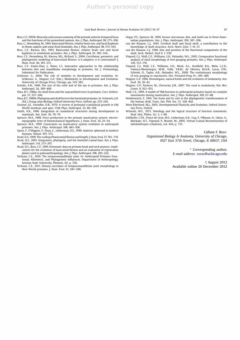
The problem with not clearly defining modules is well illus-trated by Lieberman’s treatment of the role of “integration” inexplanations for interspecific variance in browridge anteroposte-rior dimensions. Franz Weidenreich suggested in 1941 that brow-ridge lengths were driven by “neurocranial-orbital disjunction”,and this hypothesis had received support from several carefulstudies in the late 1980s and early 1990s, most notably those ofRavosa (Weidenreich, 1941; Shea, 1986a,b; Ravosa, 1988, 1991b).Weidenreich’s hypothesis posits that the more the orbits stick outin front of the neurocranium, the longer the browridge. Elegantlysimple (could it be any other way?) and well validated, this hypoth-esis then begs the question, what makes the face project beyondthe neurocranium? By 1998 two important possibilities had beensuggested and supported by interspecific and ontogenetic data:increases in facial size and changes in facial orientation. The facialsize model suggests that as the face gets bigger the browridgesalso get bigger, either as a pleiotropic effect – browridges are partof a facial module – and/or because a bigger face projects moreand hence needs protecting from above. The facial orientationmodel suggests that because the orbits are part of the face – again,a facial module – changes in orientation of the face (klinorhyncy orfacial kyphosis and airorhynchy) will rotate the orbits back under-neath or out in front of the neurocranium, reducing or augmentingthe browridge. Lieberman’s (1998) facial projection model can beadded to these, as it suggests that browridges evolve when theface projects beyond the neurocranium due to anterior displace-ment. Note that all three of these hypotheses imply that the orbitsare part of a facial module, and that their position relative to theneurocranium (neurocranial module) can be driven by changes insize, orientation or position of the facial module. However, ratherthan testing this idea rigorously, Lieberman (1998) argued thatbrowridge reduction in the lineage leading from archaic Homo(AH) to anatomically modern H. sapiens (AMHS) was due to “asingle ontogenetically early shift in cranial base growth” that wasrelated to the evolution of several features characteristic of AMHS(Lieberman, 1998). He claimed that shortening of the sphenoidbody in AMHS resulted in reduced facial projection, which heargued is the “main influence on browridge size and frontal angu-lation in nonhuman primates”, and apparently all the things thatmake being a human being worthwhile: cranial globularity, short-ening of the orophraynx (facilitating speech), and improvingleverage of the chewing muscles. (Lieberman’s measures aredefined and illustrated in Fig. 4.)
A subsequent paper revealed that several AH (Gibraltar 1,Kabwe, Monte Circeo) actually have very short ASLs, and that, asa result, ASL is not highly correlated with midfacial projection(MFP) among human species (Spoor et al., 1999). Forced to abandonhis argument for the importance of ASL, Lieberman adopted a moremultifactorial model for the determinants of browridge anteropos-terior length in humans: including facial size, facial orientation, and(in spite of lack of evidence) facial projection (Lieberman, 2000). (Ido note, however, that facial projection slips into Evolution of theHuman Head Figure 5.9 via Figure 8.C of Lieberman (2000), albeitwith an erroneously short ASL in Figure 5.9d.) Lieberman (2000)focuses on explaining variance in browridge length with referenceto basicranial flexion and facial orientation. He suggests thatbecause the orbital roofs are also the floor of the anterior cranialfossa, the face “rotates with the anterior cranial base as a block”(p. 171), so that facial orientation is strongly influenced by basicra-nial flexion. The reason for this, as long noted (Dabelow, 1929;Cartmill, 1970; Ravosa, 1991a; Ross and Ravosa, 1993; Ross andHenneberg, 1995), is orbital approximation below the olfactorytract that integrates the face and anterior cranial base. As a result,
Lead Book Review / Journal of Human Evolution 64 (2013) 56–6762

Author's personal copy
when the posterior cranial base flexes relative to the anterior, it alsoflexes relative to the facial module. With this relationship in hand,Lieberman uses Figure 5.9 (this paper’s Fig. 4) to argue that basicra-nial flexion is an especially important influence on facial projection“because a more extended cranial base rotates more of the face infront of the [anterior cranial fossa] increasing upper facial projec-tion” (p. 174).
But this makes no sense: if the facial module is integrated withthe basicranial and/or neurocranial modules, then by definitionflexion within the cranial base will not affect the spatial relation-ships between face and anterior cranial base. Think about it thisway: Ceteris paribus, relationships between the facial module andthe anterior cranial base are not going to be affected by flexion ofthe posterior cranial base relative to the anterior, they are goingto be affected by changes in relationships between the face and theanterior neurocranium (Ross and Henneberg, 1995). But, let’sassume that Lieberman’s hypothesis is true, and it is upward ordownward flexion of the facial module by around 10� (the differ-ence in basicranial flexion between AH and AMHS in Figure 5.9)that accounts for changes in projection of the face relative to theanterior cranial base. Assuming that the center of rotation is at sellaand the front of the face is 47.8 þ 14.6 ¼ 62.4 mm away from sella
(ACL þ MFP from Lieberman, 1998, Table 2), the largest possibleincrease in MFP is given by j62.4 mm – cos10� *62.4 mmj ¼ 0.948 mm. A sub-millimeter change in browridgedimensions is not what we need to explain here, so simple geom-etry argues against changes in facial and browridge projectionbeing driven by changes in facial orientation in turn driven bychanges in basicranial flexion.
Lieberman also suggests that increases in frontal lobe size mightreduce facial projection and (presumably) browridge length inAMHS. I think this argument might have some legs, but not forthe reasons advanced by Lieberman. He argues that increases infrontal lobe size will result in increases in anterior cranial baselength, and that this increasingly protruding ACL places the facemore and more underneath the anterior cranial base, reducingMFP and browridge length (p. 174 and 176). But this argumentcannot explain small browridges in humans either because, Ceterisparibus, increase in ACL relative to facial length would actually pushthe cribriform plate and browridge forward, increasing browridgelength in AMHS, the reverse of Lieberman’s predictions! Moreover,my data confirm that, indeed, humans have long sella-foramencecum distances relative to palate length (Fig. 5) but it is clearfrom Lieberman’s own data (2008, Table 2) that AMHS do not
Figure 4. Reproduction of Figure 5.9 from Lieberman (2011), The Evolution of the Human Head.
Lead Book Review / Journal of Human Evolution 64 (2013) 56–67 63

Author's personal copy
have absolutely longer ACL than other primates. Thus, it must bereduction in overall facial size that causes browridge reduction inhumans. I think Lieberman’s belief in ACL lengthening in humansderives from a geometric morphometric analysis presented else-where (Lieberman et al., 2002), and reproduced here in Fig. 6. Lie-berman et al. use this analysis to argue that humans have a moreretracted face in part because of their long anterior cranial bases.However, these data have been scaled to a common geometricmean, so that decreases in facial size (the blue lines) must be asso-ciated with increases in other parts of the skull to maintaina constant centroid size. That is why anterior cranial base lengthappears to be longer in AMHS when, in fact, as Lieberman’s (1998)data clearly show, it is not (Fig. 6).
This lengthy exegesis shows that there is good evidence that vari-ance in browridge projection in the lineage leading from AH to AMHSis NOT driven by variance in basicranial flexion, anterior cranial baselength, or facial orientation. In contrast, there is plenty of evidencethat browridge projection is highly correlated with facial size. Rav-osa clearly shows that palate length is one of the most consistentcorrelates of browridge length both in interspecific and ontogeneticcomparisons (Ravosa, 1991a, b). Lieberman’s rejection of facial sizeas the primary determinant of browridge length on the groundsthat Neanderthals have more mid-facial projection than predictedby their midfacial length (2000, Figure 11) is based on a muchsmaller sample and on a different measure (midfacial lengthinstead of palate length). It seems to me that the question of the
influence of facial size on browridge dimensions could be quicklytested using a geometric mean of facial measures rather than singlelinear measures, but I’ll wager that Ravosa will turn out to be rightabout this.
In sum, the available data suggest that selection for integrationof the browridges with the rest of the face was necessary to ensurethat there was always enough bone there to protect the orbitalcontents from blows to the head (Hylander et al., 1991) in thecontext of neuro-orbital disjunction caused by kilonrhynchy orincreases in facial size (Weidenreich, 1941; Shea, 1986a, b;Ravosa, 1988, 1991a). In this sense, the browridges are part of thefacial module as a whole. Subsequent selection for increases ordecreases in size of the face would then also act on browridgesize, making browridges a nice example of how functional integra-tion producing functional modules can be achieved through devel-opmental integration to produce developmental modules, and thisin turn creates a genetically integrated evolutionary module(Cheverud, 1982). Moreover, this explanation probably also appliesto the well documented positive allometry of orbit size relative toeyeball size (Schultz, 1940; Kirk, 2006). If Lieberman had focusedmore on defining these modules and less on the interactionsbetween disparate parts in the head, I think he would have gotbetter traction on these important questions.
The future of integration and modularity studies in primatecrania
Studies of the causes and effects of cranial base angulation, whichLieberman neatly summarizes, show that the degree of basicranialflexion is positively correlated with the size of the brain relative tobasicranial length and negatively correlated with facial length(Biegert, 1963). This has been confirmed many times since Ross andRavosa (1993), but I confess that I still do not really understandwhy increases in facial length (whichRavosa and Imeasuredaspalatelength) should be associated with decreases in basicranial flexion. Ifthe face grows down and forward from the cranial base, as it does,there is plenty of room for a large face even with a flexed cranialbase. But, the effect of facial size is certainly there, as nicely shownin Lieberman et al.’s recent study (Lieberman et al., 2008), and wesimply do not understand why, from either mechanical, develop-mental, or functional perspectives. There is either some profoundinteraction between facial and neurocranial modules at the cranialbase, or the way we are measuring the variables is not giving us theanswers. If we define the brain þ basicranium and the face as bothdevelopmental and evolutionary modules sensu Schlosser (processesthat make an integrated and relatively context-insensitive contribu-tion to the development and reproduction of the organism in the
Figure 6. Reproduction of part of Figure 2 from Lieberman et al., (2002). This figure is used by them to argue that the AMHS anterior cranial base is longer than that of AH. However,this conclusion runs counter to Lieberman’s (1998) data. In fact, AMHS is distinctive in having a smaller face, including a shorter palate (Figure 5).
Figure 5. Bivariate plot of anterior cranial base length (ACL) against palate length.Humans have long ACLs relative to palate length. However, this is not because theyhave long ACLs, but because they have short palates.
Lead Book Review / Journal of Human Evolution 64 (2013) 56–6764
Author's personal copy
face of perturbations), where is the boundary between them?Perhaps the brain and endocranial surface of the basicranium consti-tute a neurocranial module, whereas the face and external surface ofthe basicraniumconstitute anothermodule, predicting that endocra-nial basicranial measures will covary most with brain size and shape(Strait, 1999, 2001), whereas the external measures will covary withthe facialmodule. Under thismodel, recalling ideas of Enlow and vander Klauww (Enlow,1990; van der Klaauw,1945,1948), the “basicra-nium” straddles the border of two modules, with its developmentaland evolutionary processes allowing changes in the two adjacentmodules to accommodate each other.
In my opinion, it is at this boundary between neurocranium andface that important research on morphological integration andmodularity in the primate cranium needs to be done. We needa better understanding of how the different parts of the cranialbase interact with or are part of (integratedwithin) different cranialmodules during development and evolution. I have to say, that I donot see how this can be achieved by defining cranial base flexionusing the angle between a line running from basion to sella andanother running from sella to foramen cecum. Sella is not evena bony point, and these lines cross so many synchondroses whereimportant growth processes occur, thereby traversing multiplemodules, that they cannot hope to provide clear signals on whatthe modules are and how they interact. Others have said this beforevery clearly (Cartmill, 1970; Sirianni and Swindler, 1979), itinformed our choice of flexion measures in 1993 and since (Rossand Ravosa, 1993; Ross and Henneberg, 1995; Ross et al., 2004),and I still think it is true. The best work currently being done onintegration and modularity at this important interface is the metic-ulous work of Markus Bastir, Antonio Rosas and their colleagues,which not only quantifies morphology at the level of detail that Ithink is required, but recognizes the importance of morphologicalchanges off the mid-sagittal plane as well (Bastir et al., 2004;Bastir and Rosas, 2004a, b; 2005; Bastir et al., 2006; Bastir andRosas, 2006b; Bastir et al., 2007, 2008a, 2008b, 2009; Bastir et al.,2010a, 2010b, in press, 2011). As high resolution 3D images ofa broad sample of primate crania become more easily accessible,Bastir’s results are tested across awider range of primates, and Che-verud’s approaches (Cheverud, 1982, 1995; Ackermann andCheverud, 2000, 2002, 2004; Marroig and Cheverud, 2001;Ackermann and Krovitz, 2002) are extended to include more land-marks inside the cranium, I think we can expect to see our under-standing of integration and modularity in the evolution of theprimate head rapidly improve. Combined with developmentalexperimental approaches being pursued by Hallgrimsson and Lie-berman (Hallgrimsson et al., 2004a, 2004b, 2007; Boughner andHallgrimsson, 2008; Lieberman et al., 2008; Marcucio et al.,2011), and finite-element modeling measures of functional perfor-mance and developmental interactions (Kupczik et al., 2009; Curtiset al., 2011; O’Higgins et al., 2011), the road ahead looks exciting.
Conclusions
In sum, Lieberman’s book is an innovative attempt at a synthetictreatment of hominid skull evolution. It seeks to summarize devel-opmental and functional principles governing head evolution thenapply them to understanding of the fossil record. This is an ambi-tious book and it has variable success. The early chapters wherethe important theoretical and conceptual foundations of the bookare laid out are interesting from a theoretical perspective, but Ithink that in failing to precisely and accurately define and use“complexity”, “module” and “integration”, terms which are centralto the ideas promulgated here, Liebermanmisses an opportunity toadvance the field in ameaningful way, and gets tangled up in expla-nations for (e.g.) browridge evolution that just do not hold water.
That said, this may not be Lieberman’s problem, but, rather, reflec-tive of broader problems with these concepts. The idea of the bookis a good one and if a second edition were prepared that addressedthe theoretical problems laid out above, and corrected the biome-chanical errors and errant figures, then this book would be enor-mously useful. Until that time, I can recommend this book tocritical, independent undergraduates looking for an introductionto the literature on primate head evolution. A graduate studentlooking for research projects will certainly find some low-hanging fruit here, but they will need to be prepared to readthoughtfully and critically. Finally, there is plenty here forresearchers like me who have grappled head-on with many of theexciting questions addressed in this book.
Acknowledgments
I am grateful to Dan Lieberman for many stimulating discussionson head evolution over the years. He has generously shared dataand resources with me and many others in order to further ourunderstanding of this most fascinating part of the primate body. Iapologize to Lieberman and Ravosa for my error in our 2000 Year-book paper, and I appreciate the Journal of Human Evolutionproviding the opportunity to set the record straight. I appreciatehelpful comments from Richard Smith and an anonymous reviewerthat improved this review.
References
Ackermann, R.R., Cheverud, J.M., 2000. Phenotypic covariance structure in tama-rins (genus Saguinus): a comparison of variation patterns using matrix corre-lation and common principal component analysis. Am. J. Phys. Anthropol. 111,489–501.
Ackermann, R.R., Cheverud, J.M., 2002. Discerning evolutionary processes inpatterns of tamarin (genus Saguinus) craniofacial variation. Am. J. Phys. Anthro-pol. 117, 260–271.
Ackermann, R.R., Cheverud, J.M., 2004. Morphological integration in primate evolu-tion. In: Pigliucci, M., Preston, K. (Eds.), Phenotypic Integration: Studying theEcology and Evolution of Complex Phenotypes. Oxford University Press, Oxford,pp. 302–319.
Ackermann, R.R., Krovitz, G.E., 2002. Common patterns of facial ontogeny in thehominid lineage. Anat. Rec. 269, 142–147.
Ackermann, R.R., Rogers, J., Cheverud, J.M., 2006. Identifying the morphologicalsignatures of hybridization in primate and human evolution. J. Hum. Evol. 51,632–645.
Bastir, M., Rosas, A., 2004a. Comparative ontogeny in humans and chimpanzees:similarities, differences and paradoxes in postnatal growth and developmentof the skull. Ann. Anat. 186, 503–509.
Bastir, M., Rosas, A., 2004b. Facial heights: evolutionary relevance of postnatalontogeny for facial orientation and skull morphology in humans and chimpan-zees. J. Hum. Evol. 47, 359–381.
Bastir, M., Rosas, A., 2005. Hierarchical nature of morphological integration andmodularity in the human posterior face. Am. J. Phys. Anthropol. 128, 26–34.
Bastir, M., Rosas, A., 2006a. Correlated variation between the lateral basicraniumand the face: a geometric morphometric study in different human groups.Arch. Oral Biol. 51, 814–824.
Bastir, M., Rosas, A., 2006b. Correlated Variation between the Lateral Basicraniumand the Face. In: A Geometric Morphometric Study Different Human Groups,vol. 51, p. 814.
Bastir, M., Rosas, A., Kuroe, K., 2004. Petrosal orientation and mandibular ramusbreadth: evidence for an integrated petroso-mandibular developmental unit.Am. J. Phys. Anthropol. 123, 340–350.
Bastir, M., Rosas, A., O’Higgins, P., 2006. Craniofacial levels and the morphologicalmaturation of the human skull. J. Anat. 209, 637–654.
Bastir, M., O’Higgins, P., Rosas, A., 2007. Facial ontogeny in Neanderthals andmodern humans. Proc. Biol. Sci. 274, 1125–1132.
Bastir, M., Rosas, A., Lieberman, D.E., O’Higgins, P., 2008a. Middle cranial fossaanatomy and the origin of modern humans. Anat. Rec. (Hoboken) 291,130–140.
Bastir, M., Sobral, P.G., Kuroe, K., Rosas, A., 2008b. Human craniofacial sphericity:a simultaneous analysis of frontal and lateral cephalograms of a Japanese pop-ulation using geometric morphometrics and partial least squares analysis. Arch.Oral Biol. 53, 295–303.
Bastir, M., Rosas, A., Stringer, C., de la Cuetara, J.M., Kruszynski, R., Ross, C.F.,Ravosa, M.J., 2009. Basicranial flexion in the evolution of Homo: new analysesof an old model. Am. J. Phys. Anthropol. 84, 84.
Lead Book Review / Journal of Human Evolution 64 (2013) 56–67 65
Author's personal copy
Bastir, M., Rosas, A., Stringer, C., Cuetara, J.M., Kruszynski, R., Weber, G.W., Ross, C.F.,Ravosa, M.J., 2010a. Effects of brain and facial size on basicranial form in humanand primate evolution. J. Hum. Evol. 58, 424–431.
Bastir, M., Rosas, A., Tabernero, A.G., Pena-Melian, A., Estalrrich, A., de la Rasilla, M.,Fortea, J., 2010b. Comparative morphology and morphometric assessment ofthe Neandertal occipital remains from the El Sidron site (Asturias, Spain: years2000–2008). J. Hum. Evol. 58, 68–78.
Bastir, M., Rosas, A., Stringer, C., Cuetara, J.M., Kruszynski, R., Weber, G.W., Ross, C.F.,Ravosa, M.J. Effects of brain size and facial size on the evolution of the basicra-nium in Homo Journal of Human Evolution, in press.
Bastir, M., Rosas, A., Gunz, P., Pena-Melian, A., Manzi, G., Harvati, K., Kruszynski, R.,Stringer, C., Hublin, J.J., 2011. Evolution of the base of the brain in highly ence-phalized human species. Nat. Commun. 2, 588.
Biegert, J., 1963. The evaluation of characteristics of the skull, hands and feet forprimate taxonomy. In: Washburn, S.L. (Ed.), Classification and Human Evolu-tion. Aldine, Chicago, pp. 116–145.
Boughner, J.C., Hallgrimsson, B., 2008. Biological spacetime and the temporal inte-gration of functional modules: a case study of dento-gnathic developmentaltiming. Dev. Dyn. 237, 1–17.
Cartmill, M., 1970. The Orbits of Arboreal Mammals: A Reassessment of the ArborealTheory of Primate Evolution. University of Chicago, Chicago, Illinois, p. 671.
Cartmill, M., 1972. Arboreal adaptations and the origin of the Order Primates. In:Tuttle, R. (Ed.), The Functional and Evolutionary Biology of Primates. Aldine,Chicago, pp. 97–122.
Cartmill, M., 1980. Morphology, function and evolution of the anthropoid postorbitalseptum. In: Ciochon, R.L., Chiarelli, A.B. (Eds.), Evolutionary Biology of the NewWorld Monkeys and Continental Drift. Plenum, New York, pp. 243–274.
Cheverud, J.M., 1982. Phenotypic, genetic, and environmental morphological inte-gration in the cranium. Evolution 36, 499–516.
Cheverud, J.M., 1984. Quantitative genetics and developmental constraints onevolution by selection. J. Theor. Biol. 110, 155–171.
Cheverud, J.M., 1988. A comparison of genetic and phenotypic correlations.Evolution 42, 958–968.
Cheverud, J.M., 1996. Developmental integration and the evolution of pleiotropy.American Zoologist 36, 44–50.
Cheverud, J.M., 1995. Morphological integration in the saddle-back tamarin (Sagui-nus fuscicollis) cranium. Am. Naturalist 145, 63–89.
Cheverud, J.M., Midkiff, J.E., 1992. Effects of fronto-occipital cranial reshaping onmandibular form. Am. J. Phys. Anthropol. 87, 167–171.
Crompton, A.W., Lieberman, D.E., Aboelela, S., 2006. Tooth orientation during occlu-sion and the functional significance of condylar translation in primates andherbivores. In: Carrano, M., Gaudlin, T., Blob, R.W., Wible, J. (Eds.), AmniotePaleobiology: Perspectives on the Evolution of Mammals, Birds and Reptiles.University of Chicago Press, Chicago, pp. 367–388.
Currey, J.D., 2002. Bones: Structure and Mechanics. Princeton University Press,Princeton.
Curtis, N., Jones, M.E., Shi, J., O’Higgins, P., Evans, S.E., Fagan, M.J., 2011. Functionalrelationship between skull form and feeding mechanics in Sphenodon, andimplications for diapsid skull development. PLoS One 6, e29804.
Dabelow, A., 1929. Uber Korrelationen in der phylogenetischen Entwicklung derSchadelform. I. Die Beziehungen zwischen Rumpf und Schadelform. Gegenb.Morpholog. Jahrb. 63, 1–49.
Demes, B., Creel, N., 1988. Bite force, diet, and cranial morphology of fossil homi-nids. J. Hum. Evol. 17, 657–670.
Enlow, D.H., 1990. Facial Growth.Gould, S.J., Lewontin, R.C., 1979. The spandrels of San Marco and the Panglossian
paradigm. A critique of the adaptationist program. Proc. R. Soc. London B 205,581–598.
Greaves, W.S., 1974. Functional implications of mammalian jaw joint position.Forma et functio 7, 363–376.
Greaves, W.S., 1980. The mammalian jaw mechanism - the high Glenoid cavity. Am.Naturalist 116, 432–440.
Gregory, W.K., 1929. Our Face from Fish to Man; A Portrait Gallery of Our AncientAncestors and Kinsfolk Together with a Concise History of Our Best Features.G.P. Putnam’s Sons, New York, London.
Hallgrimsson, B., Dorval, C.J., Zelditch, M.L., German, R.Z., 2004a. Craniofacial vari-ability and morphological integration in mice susceptible to cleft lip and palate.J. Anat. 205, 501–517.
Hallgrimsson, B.,Willmore, K., Dorval, C., Cooper, D.M., 2004b. Craniofacial variabilityandmodularity inmacaques andmice. J. Exp. Zool. BMol. Dev. Evol. 302, 207–225.
Hallgrimsson, B., Lieberman, D.E., Young, N.M., Parsons, T., Wat, S., 2007. Evolutionof covariance in the mammalian skull. Novartis Found. Symp. 284, 164–185.Discussion 185–190.
Hallgrimsson, B., Jamniczky, H.A., Young, N.M., Rolian, C., Parsons, T.E.,Boughner, J.C., Marcucio, R.S., 2009. Deciphering the palimpsest: studying therelationship between morphological integration and phenotypic covariation.Evol. Biol. 36, 355–376.
Hanken, J., Hall, B.K., 1993. The Skull. University of Chicago Press, Chicago.Heesy, C.P., 2005. Function of the mammalian postorbital bar. J. Morphol. 264,
363–380.Hylander, W.L., 1975. Incisor size and diet in anthropoids with special reference to
Cercopithecidae. Science 189, 1095–1098.Hylander, W.L., 1992a. Functional anatomy. In: Sarnat, B.G., Laskin, D.M. (Eds.), The
Temporomandibular Joint: A Biological Basis for Clinical Practice. W, B. Saun-ders Company, Philadelphia.
Hylander, W.L., 1992b. Functional anatomy of the temporomandibular joint. In:Sarnat, B.G., Laskin, D.M. (Eds.), The Temporomandibular Joint: A BiologicalBasis for Clinical Practice. W.B. Saunders Co, Philadelphia, pp. 60–92.
Hylander, W.L., Picq, P.G., Johnson, K.R., 1991. Masticatory-stress hypotheses and thesupraorbital region of primates. Am. J. Phys. Anthropol. 86, 1–36.
Jacob, F., 1977. Evolution and tinkering. Science 196, 1161–1166.Kay, R.F., 1984. On the use of anatomical features to infer foraging behavior in
extinct primates. In: Rodman, P.S., Cant, J.G. (Eds.), Adaptations for Foragingin Nonhuman Primates. Columbia University Press, New York, pp. 21–53.
Kirk, E.C., 2006. Effects of activity pattern on eye size and orbital aperture size inprimates. J. Hum. Evol. 51, 159–170.
Klingenberg, C.P., 2008. Morphological integration and developmental modularity.Annu. Rev. Ecol. Evol. Systematics 39, 115–132.
Kupczik, K., Dobson, C.A., Crompton, R.H., Phillips, R., Oxnard, C.E., Fagan, M.J.,O’Higgins, P., 2009. Masticatory loading and bone adaptation in the supraorbitaltorus of developing macaques. Am. J. Phys. Anthropol. 139, 193–203.
Lande, R., 1980. The genetic covariance between characters maintained by pleio-tropic mutations. Genetics 94, 203–215.
Lewontin, R.C., 1974. The Genetic Basis of Evolutionary Change. Columbia UniversityPress, New York.
Lieberman, D.E., 1998. Sphenoid shortening and the evolution of modern humancranial shape. Nature 393, 158–162.
Lieberman, D.E., 2000. Ontogeny, homology, and phylogeny in the Hominid cranio-facial skeleton: the problem of the browridge. In: O’Higgins, P., Cohn, M. (Eds.),Development, Growth and Evolution: Implications for the Study of HominidSkeletal Evolution. Academic Press, London, pp. 85–122.
Lieberman, D., 2011. The Evolution of the Human Head. Belknap Press of HarvardUniversity Press, Cambridge, Mass.
Lieberman, D.E., Ross, C.F., Ravosa, M.J., 2000. The primate cranial base: ontogeny,function, and integration. Yrb Phys Anthropol. 43, 117–169.
Lieberman, D.E., McBratney, B., Krovitz, G.E., 2002. The evolution and developmentof cranial form in Homo sapiens. Proc. Natl. Acad. Sci. 99, 1134–1139.
Lieberman, D.E., Hallgrimsson, B., Liu, W., Parsons, T.E., Jamniczky, H.A., 2008.Spatial packing, cranial base angulation, and craniofacial shape variationin the mammalian skull: testing a new model using mice. J. Anat. 212,720–735.
Marcucio, R.S., Young, N.M., Hu, D., Hallgrimsson, B., 2011. Mechanisms thatunderlie co-variation of the brain and face. Genesis 49, 177–189.
Marroig, G., Cheverud, J.M., 2001. A comparison of phenotypic variation and covari-ation patterns and the role of phylogeny, ecology and ontogeny during cranialevolution of New World monkeys. Evolution 55, 2576–2600.
Mayr, E., 1963. Animal Species and Evolution. Belknap Press, Cambridge.Mayr, E., 1976. Evolution and the Diversity of Life. Harvard Press, Cambridge.McShea, D.W., 1996. Metazoan complexity and evolution: is there a trend? Evolu-
tion 50, 477–492.McShea, D.W., Venit, E.P., 2001. What is a part? In: Wagner, G.P. (Ed.), The Character
Concept in Evolutionary Biology. Academic Press, San Diego, pp. 259–284.McShea, D.W., Brandon, R., 2010. Biology’s First Law. The Tendency for Diversity and
Complexity to Increase in Evolutionary Systems. University of Chicago Press,Chicago.
Moore, W.J., 1981. The Mammalian Skull. Cambridge University Press, CambridgeEng, New York.
Olson, E.C., Miller, R., 1958. Morphological Integration. University of Chicago Press,Chicago.
O’Higgins, P., Cobb, S.N., Fitton, L.C., Groning, F., Phillips, R., Liu, J., Fagan, M.J., 2011.Combining geometric morphometrics and functional simulation: an emergingtoolkit for virtual functional analyses. J. Anat. 218, 3–15.
Rak, Y., Hylander, W.L., 2008. What else is the tall mandibular ramus of the robustaustralopiths good for? In: Vinyard, C., Ravosa, M.J., Wall, C. (Eds.), PrimateCraniofacial Function and Biology. Springer US, New York, pp. 431–442.
Ravosa, M.J., 1988. Browridge development in Cercopithecidae: a test of twomodels. Am. J. Phys. Anthropol. 76, 535–555.
Ravosa, M.J., 1990. Functional assessment of subfamily variation in maxilloman-dibular morphology among Old-World monkeys. Am. J. Phys. Anthropol.82, 199–212.
Ravosa, M.J., 1991a. Interspecific perspective on mechanical and nonmechanicalmodels of primate circumorbital morphology. Am. J. Phys. Anthropol. 86,369–396.
Ravosa, M.J., 1991b. Ontogenetic perspective on mechanical and non-mechanicalmodels of primate circumorbital morphology. Am. J. Phys. Anthropol. 85,95–112.
Ravosa, M.J., Kunwar, R., Stock, S.R., Stack, M., 2006. Adaptive plasticity in mamma-lian masticatory joints. Matrix Biol. 25, S15.
Ravosa, M.J., Klopp, E.B., Pinchoff, J., Stock, S.R., Hamrick, M.W., 2007a. Plasticity ofmandibular biomineralization in myostatin-deficient mice. J. Morphol. 268,275–282.
Ravosa, M.J., Kunwar, R., Stock, S.R., Stack, M.S., 2007b. Pushing the limit: mastica-tory stress and adaptive plasticity in mammalian craniomandibular joints. J.Exp. Biol. 210, 628–641.
Ravosa, M.J., Menegaz, R.A., Sublett, S.V., Figueroa, S.D., Hoffman, T.J., 2009. Pheno-typic plasticity and function of the hard palate in growing rabbits. Anat. Rec.Adv. Integr. Anat. Evol. Biol. 292, 277–284.
Riedl, R., 1978. Order in Living Organisms. Wiley and Sons, New York.Ross, C.F., 1995a. Allometric and functional influences on primate orbit orientation
and the origins of the Anthropoidea. J. Hum. Evol. 29, 201–227.
Lead Book Review / Journal of Human Evolution 64 (2013) 56–6766
Author's personal copy
Ross, C.F.,1995b.Muscularandosseousanatomyof theprimateanterior temporal fossaand the functions of the postorbital septum. Am. J. Phys. Anthropol. 98, 275–306.
Ross, C., Henneberg,M.,1995. Basicranialflexion, relative brain size, and facial kyphosisin Homo sapiens and some fossil hominids. Am. J. Phys. Anthropol. 98, 575–593.
Ross, C.F., Ravosa, M.J., 1993. Basicranial flexion, relative brain size and facialkyphosis in nonhuman primates. Am. J. Phys. Anthropol. 91, 305–324.
Ross, C.F., Henneberg, M., Ravosa, M.J., Richard, S., 2004. Curvilinear, geometric andphylogenetic modeling of basicranial flexion: is it adaptive, is it constrained? J.Hum. Evol. 46, 185–213.
Ross, C.F., Iriarte-Diaz, J., Nunn, C.L. Innovative approaches to the relationshipbetween diet and mandibular morphology in primates. Int. J. Primatology,Submitted for Publication.
Schlosser, G., 2004. The role of modules in development and evolution. In:Schlosser, G., Wagner, G.P. (Eds.), Modularity in Development and Evolution.University of Chicago Press, Chicago, pp. 519–582.
Schultz, A.H., 1940. The size of the orbit and of the eye in primates. Am. J. Phys.Anthropol. 26, 389–408.
Shea, B.T., 1986a. On skull form and the supraorbital torus in primates. Curr. Anthro-pol. 27, 257–260.
Shea,B.T.,1986b. Phylogenyandskull formin thehominoidprimates. In: Schwartz, J.H.(Ed.), Orang-utan Biology. Oxford University Press, Oxford, pp. 233–245.
Sirianni, J.E., Swindler, D.R., 1979. A review of postnatal craniofacial growth in OldWorld monkeys and apes. Yrbk Phys. Anthropol. 22, 80–104.
Smith, K.K., 1996. Integration of craniofacial structures during development inmammals. Am. Zool. 36, 70–79.
Spencer, M.A., 1998. Force production in the primate masticatory system: electro-myographic tests of biomechanical hypotheses. J. Hum. Evol. 34, 25–54.
Spencer, M.A., 1999. Constraints on masticatory system evolution in anthropoidprimates. Am. J. Phys. Anthropol. 108, 483–506.
Spoor, F., O’Higgins, P., Dean, C., Lieberman, D.E., 1999. Anterior sphenoid in modernhumans. Nature 397, 572.
Strait, D.S.,1999. The scalingofbasicranialflexionand length. J.Hum.Evol. 37, 701–719.Strait, D.S., 2001. Integration, phylogeny, and the hominid cranial base. Am. J. Phys.
Anthropol. 114, 273–297.Strait, D.S., Ross, C.F., 1999. Kinematic data on primate head and neck posture: impli-
cations for the evolution of basicranial flexion and an evaluation of registrationplanes used in paleoanthropology. Am. J. Phys. Anthropol. 108, 205–222.
Terhune, C.E., 2010. The temporomandibular joint. In: Anthropoid Primates Func-tional, Allometric, and Phylogenetic Influences. Department of Anthropology.Arizona State University, Phoenix, AZ, p. 336.
Terhune, C.E., 2011. Dietary correlates of temporomandibular joint morphology inNew World primates. J. Hum. Evol. 61, 583–596.
Ungar, P.S., Spencer, M., 1999. Incisor microwear, diet, and tooth use in three Amer-indian populations. Am. J. Phys. Anthropol. 109, 387–396.
van der Klaauw, C.J., 1945. Cerebral skull and facial skull: a contribution to theknowledge of skull-structure. Arch. Neerl. Zool. 7, 16–37.
van der Klaauw, C.J., 1948. Size and position of the functional components of theskull. Arch. Nederl. Zool 9, 1–559.
Vinyard, C.J., Wall, C.E., Williams, S.H., Hylander, W.L., 2003. Comparative functionalanalysis of skull morphology of tree-gouging primates. Am. J. Phys. Anthropol.120, 153–170.
Vinyard, C.J., Wall, C.E., Williams, S.H., Mork, A.L., Armfield, B.A., Melo, L.C.D.,Valenca-Montenegro, M.M., Valle, Y.B.M., de Oliveira, M.A.B., Lucas, P.W.,Schmitt, D., Taylor, A.B., Hylander, W.L., 2009. The evolutionary morphologyof tree gouging in marmosets. Dev. Primatol-Prog. Pr., 395–409.
Wagner, G.P., 1996. Homologues, natural kinds and the evolution of modularity. Am.Zool. 36, 36–43.
Wagner, G.P., Pavlicev, M., Cheverud, J.M., 2007. The road to modularity. Nat. Rev.Genet. 8, 921–931.
Wall, C.E., 1999. A model of TMJ function in anthropoid primates based on condylarmovements during mastication. Am. J. Phys. Anthropol. 109, 67–88.
Weidenreich, F., 1941. The brain and its role in the phylogenetic transformation ofthe human skull. Trans. Am. Phil. Soc. 31, 328–442.
West-Eberhard, M.J., 2003. Developmental Plasticity and Evolution. Oxford Univer-sity Press, Oxford.
Wimsatt, W.C., 1972. Teleology and the logical structure of function statements.Stud. Hist. Philos. Sci. 3, 1–80.
Zollikofer, C.P.E., Ponce de Leon, M.S., Lieberman, D.E., Guy, F., Pilbeam, D., Likius, A.,Mackaye, H.T., Vignaud, P., Brunet, M., 2005. Virtual Cranial Reconstruction ofSahelanthropus tchadensis, vol. 434, p. 755.
Callum F. Ross*Organismal Biology & Anatomy, University of Chicago,
1027 East 57th Street, Chicago, IL 60637, USA
* Corresponding author.E-mail address: [email protected]
1 August 2012Available online 20 December 2012
Lead Book Review / Journal of Human Evolution 64 (2013) 56–67 67