Embed Size (px)
Citation preview
Mathiasen, M. L., Dillingham, C. M., Kinnavane, L., Powell, A. L., &Aggleton, J. P. (2017). Asymmetric cross-hemispheric connections link therat anterior thalamic nuclei with the cortex and hippocampal formation.Neuroscience, 349, 128-143.https://doi.org/10.1016/j.neuroscience.2017.02.026
Publisher's PDF, also known as Version of record
License (if available):CC BY
Link to published version (if available):10.1016/j.neuroscience.2017.02.026
Link to publication record in Explore Bristol ResearchPDF-document
This is the final published version of the article (version of record). It first appeared online via Elsevier athttps://www.sciencedirect.com/science/article/pii/S0306452217301094 . Please refer to any applicable terms ofuse of the publisher.
University of Bristol - Explore Bristol ResearchGeneral rights
This document is made available in accordance with publisher policies. Please cite only the publishedversion using the reference above. Full terms of use are available:http://www.bristol.ac.uk/pure/about/ebr-terms
Neuroscience 349 (2017) 128–143
ASYMMETRIC CROSS-HEMISPHERIC CONNECTIONS LINK THE RATANTERIOR THALAMIC NUCLEI WITH THE CORTEX ANDHIPPOCAMPAL FORMATION
MATHIAS L. MATHIASEN, a*CHRISTOPHER M. DILLINGHAM, a,b LISA KINNAVANE, a
ANNA L. POWELL a AND JOHN P. AGGLETON a
aSchool of Psychology, Cardiff University, Tower Building, 70
Park Place, Cardiff CF10 3AT, UK
bTrinity College Institute of Neuroscience, Trinity College Dublin,
Dublin, Ireland
Abstract—Dense reciprocal connections link the rat anterior
thalamic nuclei with the prelimbic, anterior cingulate and
retrosplenial cortices, as well as with the subiculum and
postsubiculum. The present study compared the ipsilateral
thalamic-cortical connections with the corresponding
crossed, contralateral connections between these same
sets of regions. All efferents from the anteromedial thalamic
nucleus to the cortex, as well as those to the subiculum,
remained ipsilateral. In contrast, all of these target sites pro-
vided reciprocal, bilateral projections to the anteromedial
nucleus. While the anteroventral thalamic nucleus often
shared this same asymmetric pattern of cortical connec-
tions, it received relatively fewer crossed inputs than the
anteromedial nucleus. This difference was most marked
for the anterior cingulate projections, as those to the
anteroventral nucleus remained almost entirely ipsilateral.
Unlike the anteromedial nucleus, the anteroventral nucleus
also appeared to provide a restricted, crossed projection
to the contralateral retrosplenial cortex. Meanwhile, the clo-
sely related laterodorsal thalamic nucleus had almost exclu-
sively ipsilateral efferent and afferent cortical connections.
Likewise, within the hippocampus, the postsubiculum
seemingly had only ipsilateral efferent and afferent connec-
tions with the anterior thalamic and laterodorsal nuclei.
While the bilateral cortical projections to the anterior thala-
mic nuclei originated predominantly from layer VI, the
accompanying sparse projections from layer V largely gave
http://dx.doi.org/10.1016/j.neuroscience.2017.02.0260306-4522/� 2017 The Authors. Published by Elsevier Ltd on behalf of IBRO.This is an open access article under the CC BY license (http://creativecommons.org
*Corresponding author.
E-mail address: [email protected] (M. L. Mathiasen).Abbreviations: AD, anterodorsal thalamic nucleus; AM, anteromedialthalamic nucleus; a-p, anterior-posterior; AV, anteroventral thalamicnucleus; BDA, biotinylated dextran amine; Cg, cingulate cortex; DY,diamidino yellow; FB, fast blue tracer; FG, fluorogold tracer; HPC,hippocampus; IAD, interanterodorsal thalamic nucleus; IAM,interanteromedial thalamic nucleus; LD, laterodorsal thalamicnucleus; LP, lateral posterior thalamic nucleus; M2, secondary motorcortex; MD, mediodorsal thalamic nucleus; PL, prelimbic cortex; PoS,postsubiculum; PT, parataenial thalamic nucleus; RSC, retrosplenialcortex; RSD, dysgranular retrosplenial cortex; RSG, granularretrosplenial cortex; Sm, stria medullaris of the thalamus; SUB,subiculum; V1, primary visual cortex; V2, secondary visual cortex;VA, ventral anterior thalamic nucleus; VL, ventrolateral thalamicnucleus; WGA-HRP, horseradish peroxidase-conjugated wheat germagglutinin.
128
rise to ipsilateral thalamic inputs. In testing a potentially uni-
fying principle of anterior thalamic – cortical interactions, a
slightly more individual pattern emerged that reinforces
other evidence of functional differences within the anterior
thalamic and also helps to explain the consequences of uni-
lateral interventions involving these nuclei. � 2017 The
Authors. Published by Elsevier Ltd on behalf of IBRO. This
is an open access article under the CCBY license (http://crea-
tivecommons.org/licenses/by/4.0/).
Keywords: Contralateral, Corticothalamic, Hippocampus,
Interhemispheric, Thalamocortical, Thalamus.
INTRODUCTION
Reciprocal connections between the thalamus and cortex
underlie numerous brain functions. Attention has naturally
focussed on the dense, ipsilateral thalamocortical
projections, which are often complemented by return
thalamic inputs from deep cortical layers (Deschenes
et al., 1998; Sherman, 2007). In addition to these ipsilat-
eral connections, there are both indirect and direct path-
ways that provide inter-hemispheric communication
between the thalamus and contralateral cortex. One indi-
rect pathway involves the commissural fibers that connect
corresponding cortical areas across the two hemispheres.
There are, in addition, direct connections that link the tha-
lamus with the cortex in the opposite hemisphere. Their
prevalence remains uncertain, however, as the lack of a
description of a particular crossed thalamic-cortical con-
nection does not confirm its absence, with many papers
leaving this information unspecified.
The current study focussed on the rat anterior
thalamic nuclei (ATN). The ATN comprise three
principal nuclei, the anteromedial (AM), anteroventral
(AV), and anterodorsal (AD) nucleus, along with a fourth
nucleus in the rodent, the interanteromedial nucleus
(IAM) (Swanson, 1992). We also included the laterodorsal
(LD) nucleus as it displays clear hodological similarities
with the ATN (van Groen and Wyss, 1990a, 1992b;
Shibata, 1996, 2000; van Groen et al., 2002b). These
combined nuclei have important roles in human episodic
memory and rodent spatial memory (Sutherland and
Rodriguez, 1989; Aggleton and Sahgal, 1993; Taube,
1995; Byatt and Dalrymple-Alford, 1996; Aggleton &
Brown, 1999; Harding et al., 2000; Vertes et al., 2001;
Warburton et al., 2001; van Groen et al., 2002a;
/licenses/by/4.0/).
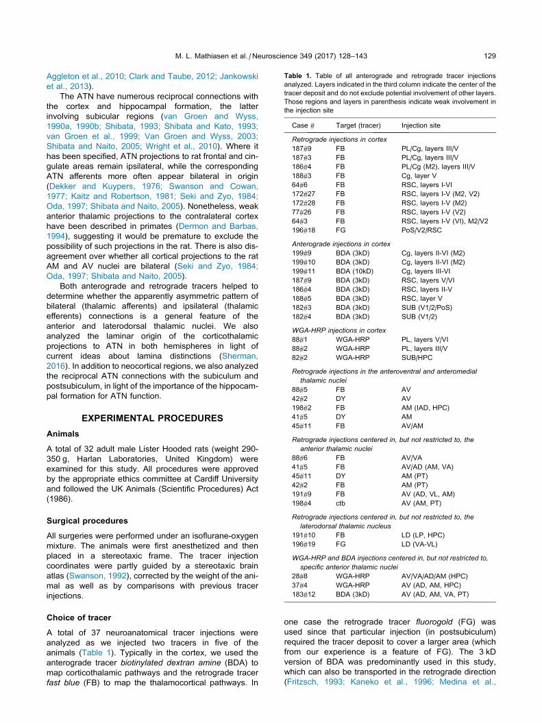
Table 1. Table of all anterograde and retrograde tracer injections
analyzed. Layers indicated in the third column indicate the center of the
tracer deposit and do not exclude potential involvement of other layers.
Those regions and layers in parenthesis indicate weak involvement in
the injection site
Case # Target (tracer) Injection site
Retrograde injections in cortex
187#9 FB PL/Cg, layers III/V
187#3 FB PL/Cg, layers III/V
186#4 FB PL/Cg (M2), layers III/V
188#3 FB Cg, layer V
64#6 FB RSC, layers I-VI
172#27 FB RSC, layers I-V (M2, V2)
172#28 FB RSC, layers I-V (M2)
77#26 FB RSC, layers I-V (V2)
64#3 FB RSC, layers I-V (VI), M2/V2
196#18 FG PoS/V2/RSC
Anterograde injections in cortex
199#9 BDA (3kD) Cg, layers II-VI (M2)
199#10 BDA (3kD) Cg, layers II-VI (M2)
199#11 BDA (10kD) Cg, layers III-VI
187#9 BDA (3kD) RSC, layers V/VI
186#4 BDA (3kD) RSC, layers II-V
188#5 BDA (3kD) RSC, layer V
182#3 BDA (3kD) SUB (V1/2/PoS)
182#4 BDA (3kD) SUB (V1/2)
WGA-HRP injections in cortex
88#1 WGA-HRP PL, layers V/VI
88#2 WGA-HRP PL, layers III/V
82#2 WGA-HRP SUB/HPC
Retrograde injections in the anteroventral and anteromedial
thalamic nuclei
88#5 FB AV
42#2 DY AV
198#2 FB AM (IAD, HPC)
41#5 DY AM
45#11 FB AV/AM
Retrograde injections centered in, but not restricted to, the
anterior thalamic nuclei
88#6 FB AV/VA
41#5 FB AV/AD (AM, VA)
45#11 DY AM (PT)
42#2 FB AM (PT)
191#9 FB AV (AD, VL, AM)
198#4 ctb AV (AM, PT)
Retrograde injections centered in, but not restricted to, the
laterodorsal thalamic nucleus
191#10 FB LD (LP, HPC)
196#19 FG LD (VA-VL)
WGA-HRP and BDA injections centered in, but not restricted to,
specific anterior thalamic nuclei
28#8 WGA-HRP AV/VA/AD/AM (HPC)
37#4 WGA-HRP AV (AD, AM, HPC)
183#12 BDA (3kD) AV (AD, AM, VA, PT)
M. L. Mathiasen et al. / Neuroscience 349 (2017) 128–143 129
Aggleton et al., 2010; Clark and Taube, 2012; Jankowski
et al., 2013).
The ATN have numerous reciprocal connections with
the cortex and hippocampal formation, the latter
involving subicular regions (van Groen and Wyss,
1990a, 1990b; Shibata, 1993; Shibata and Kato, 1993;
van Groen et al., 1999; Van Groen and Wyss, 2003;
Shibata and Naito, 2005; Wright et al., 2010). Where it
has been specified, ATN projections to rat frontal and cin-
gulate areas remain ipsilateral, while the corresponding
ATN afferents more often appear bilateral in origin
(Dekker and Kuypers, 1976; Swanson and Cowan,
1977; Kaitz and Robertson, 1981; Seki and Zyo, 1984;
Oda, 1997; Shibata and Naito, 2005). Nonetheless, weak
anterior thalamic projections to the contralateral cortex
have been described in primates (Dermon and Barbas,
1994), suggesting it would be premature to exclude the
possibility of such projections in the rat. There is also dis-
agreement over whether all cortical projections to the rat
AM and AV nuclei are bilateral (Seki and Zyo, 1984;
Oda, 1997; Shibata and Naito, 2005).
Both anterograde and retrograde tracers helped to
determine whether the apparently asymmetric pattern of
bilateral (thalamic afferents) and ipsilateral (thalamic
efferents) connections is a general feature of the
anterior and laterodorsal thalamic nuclei. We also
analyzed the laminar origin of the corticothalamic
projections to ATN in both hemispheres in light of
current ideas about lamina distinctions (Sherman,
2016). In addition to neocortical regions, we also analyzed
the reciprocal ATN connections with the subiculum and
postsubiculum, in light of the importance of the hippocam-
pal formation for ATN function.
EXPERIMENTAL PROCEDURES
Animals
A total of 32 adult male Lister Hooded rats (weight 290-
350 g, Harlan Laboratories, United Kingdom) were
examined for this study. All procedures were approved
by the appropriate ethics committee at Cardiff University
and followed the UK Animals (Scientific Procedures) Act
(1986).
Surgical procedures
All surgeries were performed under an isoflurane-oxygen
mixture. The animals were first anesthetized and then
placed in a stereotaxic frame. The tracer injection
coordinates were partly guided by a stereotaxic brain
atlas (Swanson, 1992), corrected by the weight of the ani-
mal as well as by comparisons with previous tracer
injections.
Choice of tracer
A total of 37 neuroanatomical tracer injections were
analyzed as we injected two tracers in five of the
animals (Table 1). Typically in the cortex, we used the
anterograde tracer biotinylated dextran amine (BDA) to
map corticothalamic pathways and the retrograde tracer
fast blue (FB) to map the thalamocortical pathways. In
one case the retrograde tracer fluorogold (FG) was
used since that particular injection (in postsubiculum)
required the tracer deposit to cover a larger area (which
from our experience is a feature of FG). The 3 kD
version of BDA was predominantly used in this study,
which can also be transported in the retrograde direction
(Fritzsch, 1993; Kaneko et al., 1996; Medina et al.,
130 M. L. Mathiasen et al. / Neuroscience 349 (2017) 128–143
1997). This retrograde component of BDA is not as strong
as that seen in horseradish peroxidase conjugated wheatgerm agglutinin (WGA-HRP) (Gonatas et al., 1979;
Mesulam and Brushart, 1979). Therefore, in two regions
(the prelimbic cortex and the subiculum) that were not suf-
ficiently covered by FB injections, we used WGA-HRP to
enhance the retrograde component as well as to provide
an anterograde signal. Survival times for WGA-HRP
injections were kept relatively short (2 days) in order to
limit the risk of trans-synaptic transport (Itaya and van
Hoesen, 1982; Itaya et al., 1986; Itaya, 1987).
Additional data came from injecting the retrograde
tracers fast blue (FB) and diamidino yellow (DY) into the
AV, AM and LD thalamic nuclei. The tracers FB and
DY, which are transported exclusively in the retrograde
direction, produce largely unambiguous labeling of the
cell bodies (Bentivoglio et al., 1980; Conde, 1987;
Byers et al., 2002). These tracers helped to validate the
results obtained from the BDA injections in cortex. In a
few cases, WGA-HRP and BDA were injected into the
ATN but, in these cases only, the anterograde compo-
nent was analyzed (see Result section for further
clarification).
Tracer injections and visualisation
All injections, except one (see below), were made via a
0.5-ll or 1-ll Hamilton syringe (Hamilton, Bonaduz,
Switzerland). Both FB (Polysciences Inc, Eppelheim,
Germany) and DY (Sigma–Aldrich, Gillingham, United
Kingdom) were made up as a 3% solution in sterile
phosphate-buffered saline (PBS; pH 7.4). Fluorogold
was made up as a 4% solution in double distilled water
(Santa Cruz biotechnology, US). WGA-HRP (Vector
Labs, Peterborough, UK) was used at a concentration of
40 mg/ml, while 3 kD BDA (Life Technologies Ltd,
Paisley, UK) was made up at 10% in sterile, distilled
water (pH 7.4). Typically, individual injection volumes at
each thalamic site were 0.04–0.06 ml and 0.06–0.08 mlfor each cortical site. In most cases, tracer injections
into the neocortex involved multiple anterior-posterior
levels. In one case, we iontophoretically injected the 10
kD version of BDA (Invitrogen, United Kingdom) into a
single site in the anterior cingulate cortex via a glass
micropipette (18–22-mm tip diameter). In this case we
used an alternating current (6 s on/off) of 6 mA in 10 m.
In all other cases, tracers were delivered by pressure
injections made over the course of 10 min with the
needle left in situ prior to injection for 5 min. Following
surgery, animals recovered in a thermostatically
controlled container before being returned to individual
housing with ad libitum food and water.
Following a tracer dependent, post-operative period of
2–7 days, the animals were deeply anesthetized with
sodium pentobarbital (Euthatal, Merial, Harlow, UK) and
perfused transcardially with 0.1 M PBS (pH 7.4) at room
temperature followed by 4% paraformaldehyde in 0.1 M
PBS (pH 7.4). In those animals that received injections
of WGA-HRP, the fixative was composed of 1.5%
paraformaldehyde and 1.5–2% glutaraldehyde in 0.1 M
PBS. Brains were removed and placed in the dark for
4 h in fixative and then transferred to a 25% sucrose
solution in 0.1 M PBS for 24 h in the dark to cryoprotect
the tissue before cutting. Brains were placed on a
freezing platform and 40-mm coronal sections cut on a
sledge microtome (Leica 1400) in four series. For those
cases with fluorescent tracers, two series of sections
were mounted directly onto gelatine-subbed slides and
then allowed to dry in the dark at room temperature.
One series was stained with cresyl violet.
In cases with BDA or WGA-HRP injections, one series
was mounted directly onto gelatine-subbed slides, for
subsequent cresyl violet staining. The remaining series
were collected in 0.1 M phosphate puffer. For brains
with WGA-HRP injections, these later series were
processed with 3,305,50 tetramethylbenzidine (TMB). For
the TMB reaction, sections were incubated, with
agitation, in a fresh 0.1 M phosphate buffer (PBS, pH
6.0) solution before being transferred to a solution
containing 0.25% ammonium molybdate in 0.1 M PBS
and 0.002% 3030505 tetramethylbenzidine, dissolved in
100% ethanol, again with agitation, for 30 min. Following
incubation, a 1% hydrogen peroxide solution in distilled
water was added in three stages with 30-min intervals
until the final concentration of hydrogen peroxide was
0.3%. Sections were then incubated in the same
solution overnight at 4 �C. The TMB reaction precipitate
was stabilized through subsequent incubation of
sections in a 5% ammonium molybdate solution in 0.1 M
PBS (pH 6.0) for 30 min. Following incubation, sections
were washed in 0.1 M PBS (pH 6.0) before being
mounted on gelatin subbed slides and left to dry
overnight at room temperature. The sections were then
mounted and coverslipped with DPX mountant (Sigma).
In some cases the TMB method was supplemented
with an immunohistochemical staining procedure, in
which an antiserum directed against the WGA-HRP
(Vector Labs) was used at a dilution of 1:2000 and
incubated at 4 �C for 48 h. The antigen–antibody
complex was localised with a standard avidin–biotin
process (ABC Elite Kit, Vector Labs). The chromagen
diaminobenzidine produced the visualised reaction
product. The same procedure was used in cases with
BDA tracer deposits, except that in these cases the
biotinylated tracer was visualised without the use of
antibodies. In some cases, instead of the ABC method,
BDA was visualised by fluorophore (A488) conjugated
streptavidin (Thermofisher, UK). In these cases the
sections were washed 10 � 3 min in PBS (pH 7.4),
incubated for 2 h at room temperature in PBS (1:200)
(pH 7.4) and washed (10 � 30 min in PBS, followed by
5 � 10 m TNS, pH 7.4 for both). Reacted sections were
then mounted onto gelatine-subbed processed as above.
A Leica DM5000B microscope with a Leica
DFC310FX digital camera and Leica Application Suite
image acquisition software were used for brightfield,
darkfield and fluorescence microscopy. For some of the
fluorescent images used in the figures, the contrast was
improved following global modifications to hue and
saturation. For some images, color information was
converted to grey-scale.
In order to estimate the relative strength of the
contralateral projections to the thalamus, compared to
M. L. Mathiasen et al. / Neuroscience 349 (2017) 128–143 131
the corresponding ipsilateral projections, labeled cell
counts were made in those cases with retrograde tracer
deposits restricted to the ATN. Cell counts were made
at equal intervals along the anterior-posterior (a-p) axis
of the cortex and the hippocampal formation, although in
three cases this analysis was restricted the analysis to
limited portions of the a-p axis. In these three cases,
bleaching of the fluorescence signal led to a reliance on
previously acquired digital images to fully separate
different tracer signals. The cell counts were made
manually from the image software. Consequently, the
counts were not intended to be stereological as the goal
was to compare relative numbers across the same sites.
RESULTS
The anatomical designations and borders of the thalamic
nuclei, as well as the various cortical and hippocampal
regions, match those of Swanson (1992). Given its mid-
line location, the IAM nucleus of the ATN is largely absent
from the present descriptions although attention was
given to the pattern of label it contained close to the bor-
der with AM, i.e., away from the midline.
Cortex to the anterior thalamus
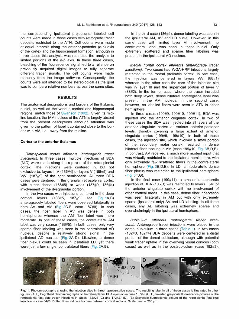
Retrosplenial cortex efferents (anterograde tracerinjections). In three cases, multiple injections of BDA
(3kD) were made along the a-p axis of the retrosplenial
cortex. The injections were centered in, but not
exclusive to, layers II-V (186#4) or layers V (188#5) and
V/VI (187#9) of the right hemisphere. All three BDA
cases were centered in the granular retrosplenial cortex
with either dense (188#5) or weak (187#9, 186#4)
involvement of the dysgranular portion.
In the two cases with injections centered in the deep
cortical layers (188#5, 187#9; see Fig. 1A,B)
anterogradely labeled fibers were observed bilaterally in
both AV and AM (Fig. 2C-F, case 187#9). In both
cases, the fiber label in AV was dense in both
hemispheres whereas the AM fiber label was more
moderate. In one of these cases, the contralateral AM
label was very sparse (188#5). In both cases, only very
sparse fiber labeling was seen in the contralateral AD
nucleus, despite a relatively strong signal in the
ipsilateral AD nucleus (Fig. 2A-D). Likewise, a dense
fiber plexus could be seen in ipsilateral LD, yet there
were just a few single, contralateral fibers (Fig. 2A,B).
Fig. 1. Photomicrographs showing the injection sites in three representative
figures. (A, B) Brightfield photomicrographs of the retrosplenial BDA injection
retrosplenial fast blue tracer injections in cases 172#28 (C) and 172#27 (D
injection in case 64#3. Dotted lines indicate borders between cortical region
In the third case (186#4), dense labeling was seen in
the ipsilateral AM, AV and LD nuclei. However, in this
same case with limited layer VI involvement, no
contralateral label was seen in these nuclei. Only
extremely scattered and sparse fiber labeling was
present in the ipsilateral AD nucleus.
Medial frontal cortex efferents (anterograde tracerinjections). Two cases had WGA-HRP injections largely
restricted to the rostral prelimbic cortex. In one case,
the injection was centered in layers V/VI (88#1)
whereas in the other case the core of the injection site
was in layer III and the superficial portion of layer V
(88#2). In the former case, where the tracer included
both deep layers, dense bilateral anterograde label was
present in the AM nucleus. In the second case,
however, no labelled fibers were seen in ATN in either
hemisphere.
In three cases (199#9, 199#10, 199#11), BDA was
injected into the anterior cingulate cortex. In two of
these cases the BDA was injected into all layers of the
anterior cingulate cortex at various anterior-posterior
levels, thereby covering a large extent of anterior
cingulate cortex (199#9, 199#10). In both of these
cases, the injection site, which involved a small portion
of the secondary motor cortex, resulted in dense
bilateral fiber labeling in AM (case 199#10, Fig. 3B,D,E).
In contrast, AV received a much more modest input that
was virtually restricted to the ipsilateral hemisphere, with
only extremely few scattered fibers in the contralateral
hemisphere (Fig. 3B,D,E). In LD, a moderate-to-dense
fiber plexus was restricted to the ipsilateral hemisphere
(Fig. 3F,G).
In the final case (199#11), a smaller iontophoretic
injection of BDA (10 kD) was restricted to layers III-VI of
the anterior cingulate cortex with no involvement of
other cortical areas. In this case, dense fiber innervation
was seen bilaterally in AM but with only extremely
sparse (ipsilateral only) AV and LD labeling. In all three
cases, any AD labeling was extremely sparse and
overwhelmingly in the ipsilateral hemisphere.
Subiculum efferents (anterograde tracer injec-
tions). Anterograde tracer injections were placed in the
dorsal subiculum in three cases (Table 1). In two cases
(182#3, 182#4) BDA deposits were centered in a distal
portion of the dorsal subiculum, although with potential
weak tracer uptake in the overlying visual cortices (both
cases) as well as in the postsubiculum (case 182#3).
cases. The resulting label in all of these cases is illustrated in other
in case 187#9. (C, D) Inverted grayscale fluorescence pictures of the
). (E) Grayscale fluorescence picture of the retrosplenial fast blue
s. Scale bars = 200 mm.
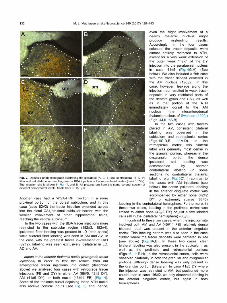
Fig. 2. Darkfield photomicrograph illustrating the ipsilateral (A, C, E) and contralateral (B, D, F)
fiber and cell distribution resulting from a BDA injection in the retrosplenial cortex (case 187#9).
The injection site is shown in Fig. 1A and B. All pictures are from the same coronal section at
different dorsoventral levels. Scale bars = 150 mm.
132 M. L. Mathiasen et al. / Neuroscience 349 (2017) 128–143
Another case had a WGA-HRP injection in a more
proximal portion of the dorsal subiculum, and in this
case (case 82#2) the tracer injection extended across
into the distal CA1/proximal subicular border, with the
weaker involvement of other hippocampal fields,
reaching the ventral subiculum.
In the two cases with the BDA tracer injections more
restricted to the subicular region (182#3, 182#4),
ipsilateral fiber labeling was present in LD (both cases)
while bilateral fiber labeling was seen in AM and AV. In
the case with the greatest tracer involvement of CA1
(82#2), labeling was seen exclusively ipsilateral in LD,
AD and AV.
Inputs to the anterior thalamic nuclei (retrograde tracerinjections). In order to test the results from our
anterograde tracer injections into cortex (described
above) we analyzed four cases with retrograde tracer
injections (FB and DY) in either AV (88#5, 42#2 DY),
AM (41#5 DY), or both nuclei (45#11 FB) (Fig. 4).
Some of the thalamic nuclei adjoining these ATN nuclei
also receive cortical inputs (see Fig. 3) and, hence,
even the slight involvement of a
nearby thalamic nucleus might
produce misleading results.
Accordingly, in the four cases
selected the tracer deposits were
almost entirely restricted to ATN,
except for a very weak extension of
the outer weak ‘‘halo” of the DY
injection into the parataenial nucleus
in case 41#5 (Fig. 4G,H) (See
below). We also included a fifth case
with the tracer deposit centered in
the AM nucleus (198#2). In this
case, however, leakage along the
injection tract resulted in weak tracer
deposits in very restricted parts of
the dentate gyrus and CA3, as well
as in that portion of the ATN
immediately dorsal to the AM
nucleus (the interanterodorsal
thalamic nucleus of Swanson (1992))
(Figs. 4J,K, 6A,B).
In the two cases with tracers
placed in AV, consistent bilateral
labeling was observed in the
subiculum and retrosplenial cortex
(Figs. 5C,D,E, 11A-D). In the
retrosplenial cortex, this bilateral
label was generally most dense in
the granular portion, whereas in the
dysgranular portion the dense
ipsilateral cell labeling was
accompanied by sparser
contralateral labeling (in some
sections no contralateral thalamic
labeling, e.g., Fig. 5C). In contrast to
the cases with AM injections (see
below), the dense ipsilateral labeling
in the anterior cingulate cortex was
accompanied by either none (42#2
DY) or extremely sparse (88#5)
labeling in the contralateral hemisphere. Furthermore, in
these two cases, labeling in the prelimbic cortex was
limited to either none (42#2 DY) or just a few labeled
cells (all in the ipsilateral hemisphere) (88#5).
In contrast to these two cases, when the injection site
involved both AM and AV (45#11 FB) relatively dense
bilateral label was present in the anterior cingulate
cortex. This labeling pattern was also seen in the case
198#2 where the tracer deposits were centered in AM
(see above) (Fig. 6A,B). In these two cases, clear
bilateral labeling was also present in the subiculum, as
well as the prelimbic and retrosplenial cortices
(Figs. 6, 11E-H). In the retrosplenial cortex, cells were
observed bilaterally in both the granular and dysgranular
portions, although dense labeling was only present in
the granular portion (bilateral). In case 41#5 DY where
the injection was restricted to AM, but positioned more
caudal than in case 198#2, we only observed labeling in
the anterior cingulate cortex, but again in both
hemispheres.
Fig. 3. Fluorescence photomicrographs of anterograde label in the laterodorsal and anterior
thalamic nuclei after a BDA injection into the anterior cingulate cortex (199#10). All pictures are
converted to grayscale. (A) Photomicrograph of the BDA injection site. The image is an overlay of
the fluorescence photomicrograph with its corresponding cresyl stained section. (B) BDA labeled
fibers in the anterior thalamic nuclei at rostral levels. (C) A higher-resolution picture of the
contralateral hemisphere in figure B. Figures D, E, F, G BDA labeled fibers in the laterodorsal and
the anterior thalamic nuclei in the ipsilateral (E, G) and the contralateral (D, F) hemispheres. Scale
bars = 200 mm.
M. L. Mathiasen et al. / Neuroscience 349 (2017) 128–143 133
In all of the cases with retrograde tracer injections in
the ATN, a consistent laminar distribution of labeled
cells was seen. In all neocortical regions analyzed, the
dense plexus of labeled cells was found in layer VI with
occasional sparse labeling of cells in layer V. The
sparsely labeled cells in layer V were almost completely
confined to the ipsilateral hemisphere. Consequently,
the contralateral label (when present) was virtually
restricted to layer VI in all cortical regions.
In stark contrast to all the above
mentioned regions, the (rather weak)
labeling in the postsubiculum
remained restricted to the ipsilateral
hemisphere (Fig. 5D,E). To further
test this negative finding, we also
analyzed those retrograde tracer
injections that were centered in the
ATN but where there was some
extension of the injection site into a
nearby thalamic nucleus (88#6,
41#5 FB, 45#11 DY, 42#2 FB,
191#9, 198#4) (see Table 1). In all
five cases, whenever cell labeling
was present in the postsubiculum it
was almost completely restricted to
the ipsilateral hemisphere with only
extremely few, occasional labeled
cells in the contralateral hemisphere.
Inputs to the laterodorsal nucleus(retrograde tracer injections). Two
injections were centered in LD,
although in both cases the tracer
deposit reached restricted portions
of either the VA-VL (196#19) or the
LP (191#10) thalamic nuclei. In
these cases, cell labeling was
exclusively ipsilateral in the
postsubiculum. There was also an
overwhelmingly ipsilateral
predominance of label in the
subiculum, as well as the
retrosplenial and anterior cingulate
cortices.
Cell counts. We counted
retrograde labeled cells in those
cases with retrograde tracers
restricted to the ATN nuclei. In the
two cases with bilateral prelimbic
label that predominantly originated
from the AM nucleus (45#11;
198#2), the overall proportions of
contralateral to ipsilateral cells were
found to be 25.8% and 29.4%,
respectively. When the tracer
injections were increasingly
restricted to AM (41#5; 198#2), the
proportion of contra- to ipsilateral
cells at rostral levels of the anterior
cingulate cortex was substantially
higher, reaching 90.2% and 77.0%,
respectively. Although these
numbers decreased when cells were counted along the
entire a-p axis of the anterior cingulate cortex, the total
proportion of contra- to ipsilateral cells (53.9%) was still
substantially higher than that in the prelimbic cortex
(case 198#2). These AM-related numbers contrasted
with the almost complete lack of contralateral label in
cases with restricted AV injections (42#2; 88#5). In
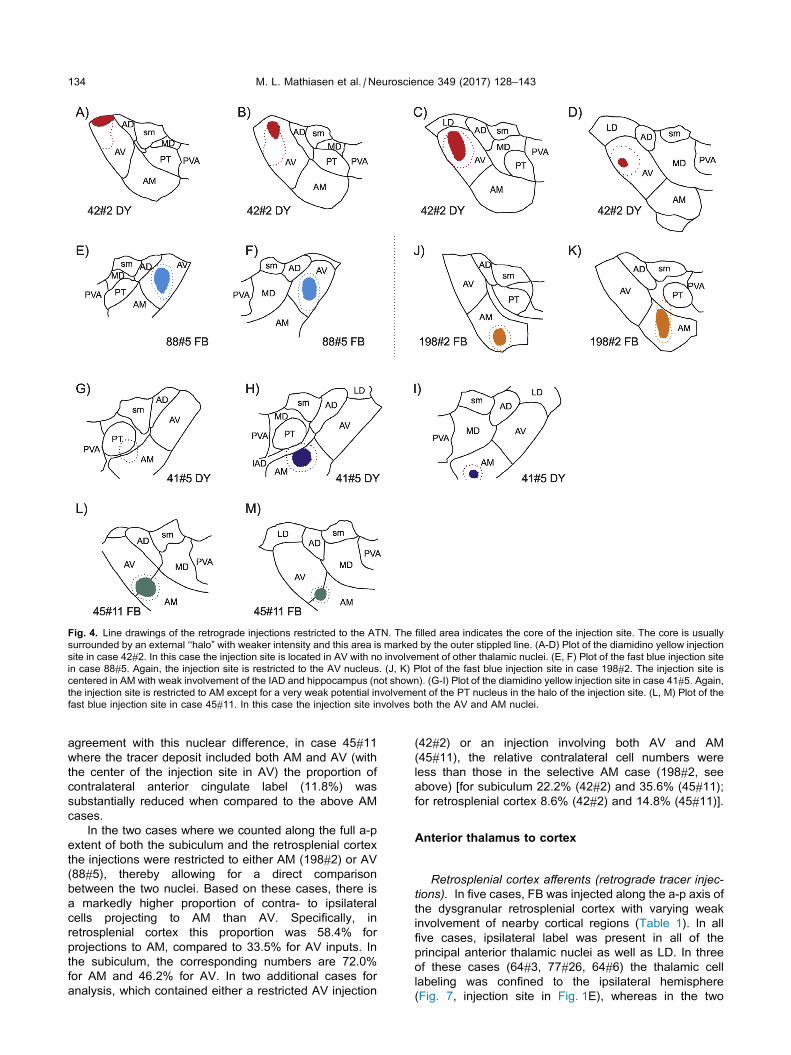
Fig. 4. Line drawings of the retrograde injections restricted to the ATN. The filled area indicates the core of the injection site. The core is usually
surrounded by an external ‘‘halo” with weaker intensity and this area is marked by the outer stippled line. (A-D) Plot of the diamidino yellow injection
site in case 42#2. In this case the injection site is located in AV with no involvement of other thalamic nuclei. (E, F) Plot of the fast blue injection site
in case 88#5. Again, the injection site is restricted to the AV nucleus. (J, K) Plot of the fast blue injection site in case 198#2. The injection site is
centered in AM with weak involvement of the IAD and hippocampus (not shown). (G-I) Plot of the diamidino yellow injection site in case 41#5. Again,
the injection site is restricted to AM except for a very weak potential involvement of the PT nucleus in the halo of the injection site. (L, M) Plot of the
fast blue injection site in case 45#11. In this case the injection site involves both the AV and AM nuclei.
134 M. L. Mathiasen et al. / Neuroscience 349 (2017) 128–143
agreement with this nuclear difference, in case 45#11
where the tracer deposit included both AM and AV (with
the center of the injection site in AV) the proportion of
contralateral anterior cingulate label (11.8%) was
substantially reduced when compared to the above AM
cases.
In the two cases where we counted along the full a-p
extent of both the subiculum and the retrosplenial cortex
the injections were restricted to either AM (198#2) or AV
(88#5), thereby allowing for a direct comparison
between the two nuclei. Based on these cases, there is
a markedly higher proportion of contra- to ipsilateral
cells projecting to AM than AV. Specifically, in
retrosplenial cortex this proportion was 58.4% for
projections to AM, compared to 33.5% for AV inputs. In
the subiculum, the corresponding numbers are 72.0%
for AM and 46.2% for AV. In two additional cases for
analysis, which contained either a restricted AV injection
(42#2) or an injection involving both AV and AM
(45#11), the relative contralateral cell numbers were
less than those in the selective AM case (198#2, see
above) [for subiculum 22.2% (42#2) and 35.6% (45#11);
for retrosplenial cortex 8.6% (42#2) and 14.8% (45#11)].
Anterior thalamus to cortex
Retrosplenial cortex afferents (retrograde tracer injec-tions). In five cases, FB was injected along the a-p axis of
the dysgranular retrosplenial cortex with varying weak
involvement of nearby cortical regions (Table 1). In all
five cases, ipsilateral label was present in all of the
principal anterior thalamic nuclei as well as LD. In three
of these cases (64#3, 77#26, 64#6) the thalamic cell
labeling was confined to the ipsilateral hemisphere
(Fig. 7, injection site in Fig. 1E), whereas in the two

Fig. 5. Fluorescence photomicrographs of retrograde labeled cells resulting from a fast blue
injection restricted to the AV thalamic nucleus (Case 88#5). (A, B) Location of the fast blue deposit
in the AV nucleus (see also Fig. 4E,F). (C) bilateral cell labeling in the retrosplenial cortex. In this
section there is appreciable bilateral label in granular retrosplenial cortex with only ipsilateral label
in the dysgranular retrosplenial cortex (though sparse labeling was often present in the
contralateral dysgranular field in other sections in this case). (D-E) Bilateral cell labeling in the
subiculum in the contralateral (D) and ipsilateral (E) hemispheres. The label in the postsubiculum
is restricted to the ipsilateral hemisphere. Scale bars = 200 mm.
M. L. Mathiasen et al. / Neuroscience 349 (2017) 128–143 135
remaining cases (172#27, 172#28) a restricted patch of
cell labeling was also seen in the contralateral
hemisphere (Fig. 8, injection site in Fig. 1C,D). In both
cases, this contralateral label consisted of a small
cluster of retrogradely labeled cells in a restricted
caudal-dorsal portion of the AV nucleus (Fig. 8). The
patch was very small in a-p extent
and resembled a smaller, mirror
image of the AV plexus in the
ipsilateral hemisphere.
We also analyzed the retrograde
transport of BDA (cases 186#4;
188#5; 187#9, described above) as
these were all primarily centered in
the granular portion of the
retrosplenial cortex (while the FB
injections were centered in the
dysgranular portion). In all three
BDA cases the retrograde label was
exclusively in the ipsilateral
hemisphere, with labeled cells in the
AD and AV nuclei, as well as in AM
in one case (187#9).
Medial frontal cortex afferents (ret-rograde tracer injections). In four
cases, FB injections were positioned
along the a-p axis of the medial
frontal cortex (186#4, 187#9, 187#3,
188#3). In three of these cases the
tracer injections involved both the
anterior cingulate and the prelimbic
cortices, primarily involving layers III-
V. In the fourth case (188#3) the
injection was restricted to the
anterior cingulate cortex and was
centered in layer V.
In all four cases, an almost
identical labeling pattern was
observed. In no case was label
observed in the ATN or LD
contralateral to the injection site.
Label in ATN was largely confined to
the ipsilateral AM nucleus (Fig. 9B,C,
E,F), where a dense region of
retrogradely labeled cells was seen
primarily around the edge of the
nucleus, i.e., avoiding the center of
AM as seen on coronal sections.
Virtually no label was observed in
the ipsilateral AD or AV nuclei. In
addition, retrogradely labeled cells
were sometimes observed in a
confined dorsomedial portion of the
ipsilateral LD nucleus (not shown).
Consistent with these four cases,
when WGA-HRP was centered in
the rostral prelimbic cortex (two
cases described in the section
above, see Table 1), retrograde label
was seen only in ipsilateral AM.
Subiculum afferents (retrograde
tracer injections). All three injections described above inthe section for anterograde subicular transport also
resulted in retrogradely labeled cells in the anterior
thalamus. In one case with WGA-HRP injections in the
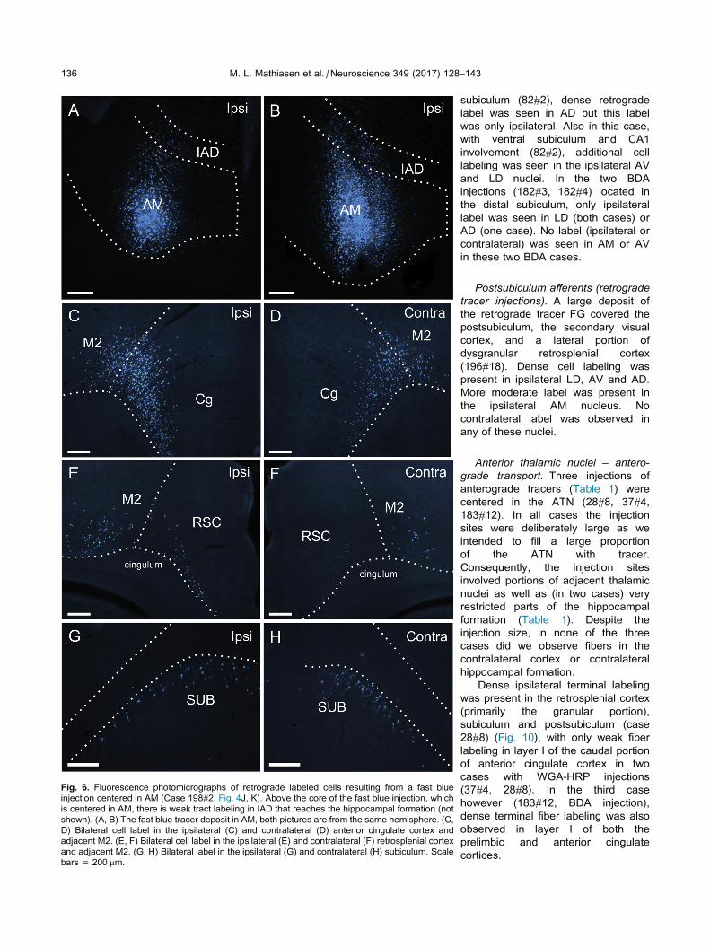
Fig. 6. Fluorescence photomicrographs of retrograde labeled cells resulting from a fast blue
injection centered in AM (Case 198#2, Fig. 4J, K). Above the core of the fast blue injection, which
is centered in AM, there is weak tract labeling in IAD that reaches the hippocampal formation (not
shown). (A, B) The fast blue tracer deposit in AM, both pictures are from the same hemisphere. (C,
D) Bilateral cell label in the ipsilateral (C) and contralateral (D) anterior cingulate cortex and
adjacent M2. (E, F) Bilateral cell label in the ipsilateral (E) and contralateral (F) retrosplenial cortex
and adjacent M2. (G, H) Bilateral label in the ipsilateral (G) and contralateral (H) subiculum. Scale
bars = 200 mm.
136 M. L. Mathiasen et al. / Neuroscience 349 (2017) 128–143
subiculum (82#2), dense retrograde
label was seen in AD but this label
was only ipsilateral. Also in this case,
with ventral subiculum and CA1
involvement (82#2), additional cell
labeling was seen in the ipsilateral AV
and LD nuclei. In the two BDA
injections (182#3, 182#4) located in
the distal subiculum, only ipsilateral
label was seen in LD (both cases) or
AD (one case). No label (ipsilateral or
contralateral) was seen in AM or AV
in these two BDA cases.
Postsubiculum afferents (retrogradetracer injections). A large deposit of
the retrograde tracer FG covered the
postsubiculum, the secondary visual
cortex, and a lateral portion of
dysgranular retrosplenial cortex
(196#18). Dense cell labeling was
present in ipsilateral LD, AV and AD.
More moderate label was present in
the ipsilateral AM nucleus. No
contralateral label was observed in
any of these nuclei.
Anterior thalamic nuclei – antero-grade transport. Three injections of
anterograde tracers (Table 1) were
centered in the ATN (28#8, 37#4,
183#12). In all cases the injection
sites were deliberately large as we
intended to fill a large proportion
of the ATN with tracer.
Consequently, the injection sites
involved portions of adjacent thalamic
nuclei as well as (in two cases) very
restricted parts of the hippocampal
formation (Table 1). Despite the
injection size, in none of the three
cases did we observe fibers in the
contralateral cortex or contralateral
hippocampal formation.
Dense ipsilateral terminal labeling
was present in the retrosplenial cortex
(primarily the granular portion),
subiculum and postsubiculum (case
28#8) (Fig. 10), with only weak fiber
labeling in layer I of the caudal portion
of anterior cingulate cortex in two
cases with WGA-HRP injections
(37#4, 28#8). In the third case
however (183#12, BDA injection),
dense terminal fiber labeling was also
observed in layer I of both the
prelimbic and anterior cingulate
cortices.
Fig. 7. Fluorescence photomicrographs of retrogradely labeled cells in the anterior thalamic nuclei
resulting from a fast blue injection in dysgranular retrosplenial cortex (case 64#3). The injection
site is shown in Fig. 1E. (A) Photomicrograph showing dense AM, and weaker AV and AD labeling
restricted to the ipsilateral hemisphere. (B) Photomicrograph showing ipsilateral cell labeling in LD
and AM, with no cell labeling in the contralateral hemisphere. Scale bars = 200 lm. [Note, the two,
contralateral fluorescent signals (one in AM, one in LD) are not from neurons.]
M. L. Mathiasen et al. / Neuroscience 349 (2017) 128–143 137
DISCUSSION
Anatomical and technical considerations
Both anterograde and retrograde tracers revealed the
patterns of ipsilateral and contralateral connections
between the anterior thalamic nuclei and cortex. To
counteract the problem of confining a tracer within a
specific thalamic nucleus, complementary tracer
injections were placed in key cortical and hippocampal
sites. The overall goal was to test the general principle
that ipsilateral anterior thalamic efferents contrast with
bilateral cortical afferents. The anterior thalamic nuclei
were the specific focus as they form a key node in
Papez circuit, a set of connections that provide a flow of
information from the hippocampus to the thalamus and
back to the hippocampal formation via the cingulate
gyrus (Papez, 1937; Shah et al., 2012). Reflecting these
connections, the ATN are vital for aspects of memory
(Aggleton & Brown, 1999; Carlesimo et al., 2011).
While previous tracing studies have shown that the
ATN receive some bilateral cortical and hippocampal
inputs (van Groen and Wyss, 1990b, 1992a; Shibata,
1998; Van Groen and Wyss, 2003; Shibata and Naito,
2005), the present study sought to
compare the details of the corre-
sponding ipsilateral and contralateral
connections, including an analysis of
both ATN afferents and efferents.
The results of these analyses, as
described in the following sections,
demonstrate a heterogeneous pattern
of crossed inputs to the ATN (includ-
ing the adjacent LD) nuclei, with hemi-
spheric differences in the laminar
origin of the corticothalamic projec-
tions, set alongside differences in
the relative strengths of the crossed
inputs to AM and AV.
With few exceptions, both AM and
AV have dense ipsilateral thalamic
outputs to the cortex, combined with
bilateral inputs from the cortex
(Fig. 12). This same asymmetric
pattern of ipsilateral thalamic
efferents and bilateral afferents also
applied to IAM. The crossed
thalamic inputs vary in their density
from cortical site to site, with both
AV and, especially, AM receiving
substantial bilateral inputs from the
subiculum and retrosplenial cortex.
In addition, AM also receives a
dense bilateral projection from both
the prelimbic and anterior cingulate
cortices, while the corresponding
crossed anterior cingulate
projections to AV remain extremely
sparse. Furthermore, as the
prelimbic input to AV was extremely
weak in our dataset, we could not
conclude whether a lighter
contralateral component was present.
Despite the overwhelming proportion of ANT
projections to ipsilateral cortical targets, evidence was
found for a light, crossed projection from AV to the
dysgranular retrosplenial cortex. This crossed projection
originated from a very restricted portion of AV (cell
labeling was observed in no more than two sections),
presumably explaining why the anterograde tracer
injections in ATN did not show contralateral fiber
labeling. It can also be assumed that this crossed
projection only terminates in a limited portion of the
retrosplenial cortex as it was only evident in some of the
retrograde tracer cases.
Our analyses also revealed two regions (the
postsubiculum and LD) with almost exclusively
ipsilateral input and output connections. First, in contrast
to the many cortical areas examined, the projections
from the postsubiculum remained ipsilateral with respect
to their thalamic targets. Likewise, the corresponding
reciprocal connections were ipsilateral. Second, unlike
AM and AV, the laterodorsal nucleus appears to receive
only ipsilateral projections from the subiculum and
postsubiculum, as well as from the anterior cingulate
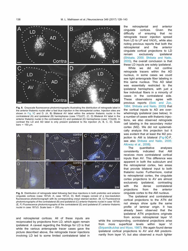
Fig. 8. Grayscale fluorescence photomicrographs illustrating the distribution of retrograde label in
the anterior thalamic nuclei after a fast blue injection in the retrosplenial cortex. Injection sites are
shown in Fig. 1C and D. (A, B) Bilateral AV label within the anterior thalamic nuclei in the
contralateral (A) and ipsilateral (B) hemispheres (case 172#27). (C, D) Bilateral AV label in the
anterior thalamic nuclei in the contralateral (C) and ipsilateral (D) hemispheres (case 172#28). In
contrast the LD and AD label is only present ipsilateral to the injection (A, B, C, D). Scale
bars = 150 mm.
Fig. 9. Distribution of retrograde label following fast blue injections in both prelimbic and anterior
cingulate cortices (case 187#9, A; case 187#3, D). Both images consist of a low-resolution
fluorescence photomicrograph with its corresponding cresyl stained section. (B, C) Fluorescence
photomicrographs of the contralateral (B) and ipsilateral (C) anterior thalamic nuclei in case 187#9.
(E, F) Fluorescence photomicrographs of the contralateral (E) and ipsilateral (F) anterior thalamic
nuclei in case 187#3. Scale bars = 200 mm.
138 M. L. Mathiasen et al. / Neuroscience 349 (2017) 128–143
and retrosplenial cortices. All of these inputs are
reciprocated by projections from LD, which again remain
ipsilateral. A caveat regarding the findings for LD is that
while the various anterograde tracer cases gave the
picture described above, the retrograde tracer injections
involving LD led to some limited contralateral label in
the retrosplenial and anterior
cingulate cortices. Given the
difficulty of ensuring that no
retrograde tracer injection spread
from LD to LP and VA/VL, while also
noting previous reports that both the
retrosplenial and the anterior
cingulate cortical projections to LD
remain exclusively ipsilateral
(Shibata, 2000; Shibata and Naito,
2005), the overall conclusion is that
these LD inputs are solely ipsilateral.
While we did not confine
retrograde tracers within the AD
nucleus, in some cases we could
see light anterograde fiber labeling in
this same nucleus. This AD label
was essentially restricted to the
ipsilateral hemisphere, with just a
few individual fibers in a minority of
cases in the contralateral side.
These observations agree with
previous reports (Seki and Zyo,
1984; Shibata and Naito, 2005) that
the cortical inputs to AD are over-
whelmingly ipsilateral (see below). In
a number of cases with thalamic injec-
tions, we also observed retrograde
cell labeling in the secondary motor
cortex (M2). We did not systemati-
cally analyze this projection but it
was evident that at least the M2 pro-
jection to AM is bilateral (Fig 6C-F,
see also Shibata and Naito, 2005;
Alloway et al., 2008).
The quantitative analyses
consistently indicated that AM
receives more contralateral cortical
inputs than AV. This difference was
apparent in both the subiculum and
the retrosplenial cortex, two areas
that provide bilateral input to both
thalamic nuclei. Furthermore, rostral
to retrosplenial cortex, the cingulate
cortex projections to AV are almost
exclusively ipsilateral, contrasting
with the dense contralateral
projections from the anterior
cingulate cortex to the AM nucleus.
The ipsilateral and contralateral
cortical projections to the ATN did
not always show quite the same
profile of laminar origin. It has
already been observed that the
ipsilateral ATN projections originate
from across retrosplenial layer VI
while the corresponding, contralateral projections arise
from more superficial levels within layer VI
(Sripanidkulchai and Wyss, 1987). We again found dense
ipsilateral cortical projections to AV and AM predomi-
nantly from layer VI, but also observed a much lighter
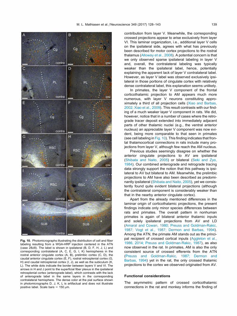
Fig. 10. Photomicrographs illustrating the distribution of cell and fiber
labeling resulting from a WGA-HRP injection centered in the ATN
(case 28#8). The label is shown in ipsilateral (B, D, F, H, J, L) and
corresponding contralateral (A, C, E, G, I, K) hemispheres in the
rostral anterior cingulate cortex (A, B), prelimbic cortex (C, D), the
caudal anterior cingulate cortex (E, F), rostral retrosplenial cortex (G,
H) and caudal retrosplenial cortex (I, J), as well as the subiculum (K,
L). The white dots indicate the border between layers V and VI. The
arrows in H and J point to the superficial fiber plexus in the ipsilateral
retrosplenial cortex (anterograde label), which contrasts with the lack
of anterograde label in the same layers in the corresponding
contralateral hemisphere. The dense color at the pial surface shown
in photomicrographs D, J, K, L is artifactual and does not illustrate
positive label. Scale bars = 150 mm.
M. L. Mathiasen et al. / Neuroscience 349 (2017) 128–143 139
contribution from layer V. Meanwhile, the corresponding
crossed projections appear to arise exclusively from layer
VI. This laminar organization, i.e., additional layer V cells
on the ipsilateral side, agrees with what has previously
been described for motor cortex projections to the rostral
thalamus (Alloway et al., 2008). A potential concern is that
we only observed sparse ipsilateral labeling in layer V
and, overall, the contralateral labeling was typically
weaker than the ipsilateral label, hence, potentially
explaining the apparent lack of layer V contralateral label.
However, as layer V label was observed exclusively ipsi-
lateral in those portions of cingulate cortex with relatively
dense contralateral label, this explanation seems unlikely.
In primates, the layer V component of the frontal
corticothalamic projection to AM appears much more
numerous, with layer V neurons constituting appro-
ximately a third of all projection cells (Xiao and Barbas,
2002; Xiao et al., 2009). This result contrasts with our find-
ing of a much weaker layer V component in rats. We did,
however, notice that in a number of cases where the retro-
grade tracer deposit extended into immediately adjacent
parts of other thalamic nuclei (e.g., the ventral anterior
nucleus) an appreciable layer V component was now evi-
dent, being more comparable to that seen in primates
(see cell labeling in Fig. 10). This finding indicates that fron-
tal thalamocortical connections in rats include many pro-
jections from layer V, although few reach the AM nucleus.
Previous studies seemingly disagree on whether the
anterior cingulate projections to AV are ipsilateral
(Shibata and Naito, 2005) or bilateral (Seki and Zyo,
1984). Our combined anterograde and retrograde tracing
data strongly support the notion that this pathway is ipsi-
lateral to AV but bilateral to AM. Meanwhile, the prelimbic
projections to AM have also been described as predomi-
nantly ipsilateral (Shibata and Naito, 2005), yet we consis-
tently found quite evident bilateral projections (although
the contralateral component is considerably weaker than
that in the nearby anterior cingulate cortex).
Apart from the already mentioned differences in the
laminar origin of corticothalamic projections, the present
findings indicate only minor species differences between
rats and primates. The overall pattern in nonhuman
primates is again of bilateral anterior thalamic inputs
and solely ipsilateral projections from AV and LD
(Amaral and Cowan, 1980; Preuss and Goldman-Rakic,
1987; Vogt et al., 1987; Dermon and Barbas, 1994).
Among the ATN, the primate AM stands out as the princi-
pal recipient of crossed cortical inputs (Aggleton et al.,
1986, 2014; Preuss and Goldman-Rakic, 1987), as also
now observed in the rat. In primates, AM is also the only
consistent source of crossed efferents from the ATN
(Preuss and Goldman-Rakic, 1987; Dermon and
Barbas, 1994) yet in the rat, the only crossed thalamic
projections to the cortex we observed originated from AV.
Functional considerations
The asymmetric pattern of crossed corticothalamic
connections in the rat and monkey informs the finding of
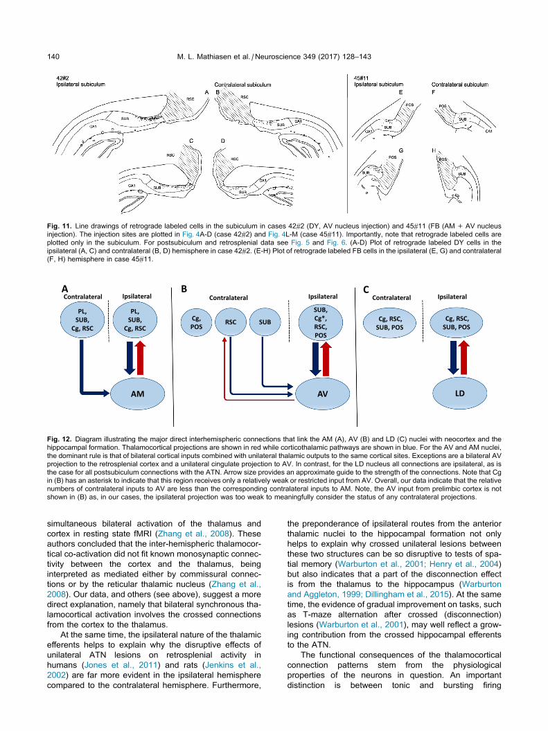
Fig. 11. Line drawings of retrograde labeled cells in the subiculum in cases 42#2 (DY, AV nucleus injection) and 45#11 (FB (AM+ AV nucleus
injection). The injection sites are plotted in Fig. 4A-D (case 42#2) and Fig. 4L-M (case 45#11). Importantly, note that retrograde labeled cells are
plotted only in the subiculum. For postsubiculum and retrosplenial data see Fig. 5 and Fig. 6. (A-D) Plot of retrograde labeled DY cells in the
ipsilateral (A, C) and contralateral (B, D) hemisphere in case 42#2. (E-H) Plot of retrograde labeled FB cells in the ipsilateral (E, G) and contralateral
(F, H) hemisphere in case 45#11.
RSC
SUB, Cg*, RSC, POS
SUBCg, POS
PL, SUB,
Cg, RSC
PL, SUB,
Cg, RSCCg, RSC,
SUB, POSCg, RSC,
SUB, POS
A B CIpsilateralContralateral IpsilateralContralateral IpsilateralContralateral
AVAM LD
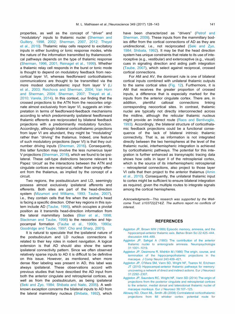
Fig. 12. Diagram illustrating the major direct interhemispheric connections that link the AM (A), AV (B) and LD (C) nuclei with neocortex and the
hippocampal formation. Thalamocortical projections are shown in red while corticothalamic pathways are shown in blue. For the AV and AM nuclei,
the dominant rule is that of bilateral cortical inputs combined with unilateral thalamic outputs to the same cortical sites. Exceptions are a bilateral AV
projection to the retrosplenial cortex and a unilateral cingulate projection to AV. In contrast, for the LD nucleus all connections are ipsilateral, as is
the case for all postsubiculum connections with the ATN. Arrow size provides an approximate guide to the strength of the connections. Note that Cg
in (B) has an asterisk to indicate that this region receives only a relatively weak or restricted input from AV. Overall, our data indicate that the relative
numbers of contralateral inputs to AV are less than the corresponding contralateral inputs to AM. Note, the AV input from prelimbic cortex is not
shown in (B) as, in our cases, the ipsilateral projection was too weak to meaningfully consider the status of any contralateral projections.
140 M. L. Mathiasen et al. / Neuroscience 349 (2017) 128–143
simultaneous bilateral activation of the thalamus and
cortex in resting state fMRI (Zhang et al., 2008). These
authors concluded that the inter-hemispheric thalamocor-
tical co-activation did not fit known monosynaptic connec-
tivity between the cortex and the thalamus, being
interpreted as mediated either by commissural connec-
tions or by the reticular thalamic nucleus (Zhang et al.,
2008). Our data, and others (see above), suggest a more
direct explanation, namely that bilateral synchronous tha-
lamocortical activation involves the crossed connections
from the cortex to the thalamus.
At the same time, the ipsilateral nature of the thalamic
efferents helps to explain why the disruptive effects of
unilateral ATN lesions on retrosplenial activity in
humans (Jones et al., 2011) and rats (Jenkins et al.,
2002) are far more evident in the ipsilateral hemisphere
compared to the contralateral hemisphere. Furthermore,
the preponderance of ipsilateral routes from the anterior
thalamic nuclei to the hippocampal formation not only
helps to explain why crossed unilateral lesions between
these two structures can be so disruptive to tests of spa-
tial memory (Warburton et al., 2001; Henry et al., 2004)
but also indicates that a part of the disconnection effect
is from the thalamus to the hippocampus (Warburton
and Aggleton, 1999; Dillingham et al., 2015). At the same
time, the evidence of gradual improvement on tasks, such
as T-maze alternation after crossed (disconnection)
lesions (Warburton et al., 2001), may well reflect a grow-
ing contribution from the crossed hippocampal efferents
to the ATN.
The functional consequences of the thalamocortical
connection patterns stem from the physiological
properties of the neurons in question. An important
distinction is between tonic and bursting firing
M. L. Mathiasen et al. / Neuroscience 349 (2017) 128–143 141
properties, as well as the concept of ‘‘driver” and
‘‘modulatory” inputs to thalamic nuclei (Sherman and
Guillery, 1998, 2002; Sherman, 2007, 2012; Theyel
et al., 2010). Thalamic relay cells respond to excitatory
inputs in either bursting or tonic response modes, while
the nature of the information transmitted by thalamocorti-
cal pathways depends on the type of thalamic response
(Sherman, 1996, 2001; Reinagel et al., 1999). Whether
a thalamic relay cell responds in the burst or tonic mode
is thought to depend on modulatory feedback from neo-
cortical layer VI, whereas feedforward corticothalamic
communications are thought to be transmitted via the
more modest corticothalamic input from layer V (Li
et al., 2003; Reichova and Sherman, 2004; Van Horn
and Sherman, 2004; Sherman, 2007; Theyel et al.,
2010; Varela, 2014). In this context, our finding that the
crossed projections to the ATN from the neocortex origi-
nate almost exclusively from layer VI, suggests an inter-
pretation in terms of feedforward-feedback mechanisms
according to which predominantly ipsilateral feedforward
thalamic efferents are reciprocated by bilateral feedback
projections with a predominantly modulatory function.
Accordingly, although bilateral corticothalamic projections
from layer VI are abundant, they might be ‘‘modulating”
rather than ‘‘driving” the thalamus. Indeed, one feature
of such modulatory projections (layer VI) is that they out-
number driving inputs (Sherman, 2016). Consequently,
this latter function may involve the less numerous layer
V projections (Sherman, 2016), which we found to be ipsi-
lateral. These cell-type distinctions become relevant to
Papez ‘circuit’ as the interactions between the ATN and
cingulate cortices are reciprocal, rather than simply effer-
ent from the thalamus, as implied by the concept of a
circuit.
Two regions, the postsubiculum and LD, seemingly
possess almost exclusively ipsilateral afferents and
efferents. Both sites are part of the head-direction
system (Mizumori and Williams, 1993; Taube, 2007),
i.e., they contain cells that fire when the animal’s head
is facing a specific direction. Other key regions in this sys-
tem include AD (Taube, 1995), which occupies a pivotal
position as it transmits head-direction information from
the lateral mammillary bodies (Blair et al., 1998;
Stackman and Taube, 1998) to the neocortex and hip-
pocampal formation (Taube et al., 1990b, 1990a;
Goodridge and Taube, 1997; Cho and Sharp, 2001).
It is natural to speculate that the ipsilateral nature of
the postsubiculum and LD nucleus connections is
related to their key roles in rodent navigation. A logical
extension is that AD should also show the same
ipsilateral connectivity pattern. Since we often observed
relatively sparse inputs to AD it is difficult to be definitive
on this issue. However, as mentioned, when more
dense fiber labeling was present in AD, this label was
always ipsilateral. These observations accord with
previous studies that have described the AD input from
both the anterior cingulate and retrosplenial cortices, as
well as from the postsubiculum, as being ipsilateral
(Seki and Zyo, 1984; Shibata and Naito, 2005). A well-
known exception concerns the bilateral inputs to AD from
the lateral mammillary nucleus (Shibata, 1992), which
have been characterized as ‘‘drivers” (Petrof and
Sherman, 2009). These inputs from the mammillary bod-
ies differ from the cortical connections of AD as they are
unidirectional, i.e., not reciprocated (Seki and Zyo,
1984; Shibata, 1992). It may be that the head direction
system has unique constraints that relate to its use of inte-
roceptive (e.g., vestibular) and exteroceptive (e.g., visual)
cues in signaling direction and aiding path integration
(Taube, 2007), which select against reciprocal, crossed
cortical connections.
For AM and AV, the dominant rule is one of bilateral
cortical inputs combined with unilateral thalamic outputs
to the same cortical sites (Fig. 12). Furthermore, it is
AM that receives the greater proportion of crossed
inputs, a difference that is especially marked for the
inputs from the anterior cingulate cortex. There are, in
addition, plentiful callosal connections linking
corresponding neocortical sites. In contrast, thalamic
nuclei are typically not directly interconnected across
the midline, although the reticular thalamic nucleus
might provide an indirect route (Raos and Bentivoglio,
1993). Accordingly, the bilateral structure of corticothala-
mic feedback projections could be a functional conse-
quence of the lack of bilateral intrinsic thalamic
connectivity. That is, as information is not integrated
directly between the two hemispheres at the level of the
thalamic nuclei, interhemispheric integration is achieved
by corticothalamic pathways. The potential for this inte-
gration is further enhanced as transynaptic tracing data
shows how cells in layer II of the retrosplenial cortex,
which is the source of its interhemispheric retrosplenial
– retrosplenial connections, also project to those layer
VI cells that then project to the anterior thalamus (Amin
et al., 2010). Consequently, the unilateral thalamic input
to cortex might be sufficient to ensure bilateral integration
as required, given the multiple routes to integrate signals
among the cortical hemispheres.
Acknowledgments—This research was supported by the Well-
come Trust: #103722/Z14/Z. The authors report no conflicts of
interest.
REFERENCES
Aggleton JP, Brown MW (1999) Episodic memory, amnesia, and the
hippocampal-anterior thalamic axis. Behav Brain Sci 22:425–444.
discussion 444–489.
Aggleton JP, Sahgal A (1993) The contribution of the anterior
thalamic nuclei to anterograde amnesia. Neuropsychologia
31:1001–1019.
Aggleton JP, Desimone R, Mishkin M (1986) The origin, course, and
termination of the hippocampothalamic projections in the
macaque. J Comp Neurol 243:409–421.
Aggleton JP, O’Mara SM, Vann SD, Wright NF, Tsanov M, Erichsen
JT (2010) Hippocampal-anterior thalamic pathways for memory:
uncovering a network of direct and indirect actions. Eur J Neurosci
31:2292–2307.
Aggleton JP, Saunders RC, Wright NF, Vann SD (2014) The origin of
projections from the posterior cingulate and retrosplenial cortices
to the anterior, medial dorsal and laterodorsal thalamic nuclei of
macaque monkeys. Eur J Neurosci 39:107–123.
Alloway KD, Olson ML, Smith JB (2008) Contralateral corticothalamic
projections from MI whisker cortex: potential route for
142 M. L. Mathiasen et al. / Neuroscience 349 (2017) 128–143
modulating hemispheric interactions. J Comp Neurol
510:100–116.
Amaral DG, Cowan WM (1980) Subcortical afferents to the
hippocampal formation in the monkey. J Comp Neurol
189:573–591.
Amin E, Wright N, Poirier GL, Thomas KL, Erichsen JT, Aggleton JP
(2010) Selective lamina dysregulation in granular retrosplenial
cortex (area 29) after anterior thalamic lesions: an in situ
hybridization and trans-neuronal tracing study in rats.
Neuroscience 169:1255–1267.
Bentivoglio M, Kuypers HG, Catsman-Berrevoets CE, Loewe H,
Dann O (1980) Two new fluorescent retrograde neuronal tracers
which are transported over long distances. Neurosci Lett
18:25–30.
Blair HT, Cho J, Sharp PE (1998) Role of the lateral mammillary
nucleus in the rat head direction circuit: a combined single unit
recording and lesion study. Neuron 21:1387–1397.
Byatt G, Dalrymple-Alford JC (1996) Both anteromedial and
anteroventral thalamic lesions impair radial-maze learning in
rats. Behav Neurosci 110:1335–1348.
Byers CT, Fan R, Messina A, Morrison WA, Galea MP (2002)
Comparing the efficacy of two fluorescent retrograde tracers in
labeling the motor and sensory neuron populations of the rat
sciatic nerve. J Neurosci Methods 114:159–164.
Carlesimo GA, Lombardi MG, Caltagirone C (2011) Vascular
thalamic amnesia: a reappraisal. Neuropsychologia 49:777–789.
Cho J, Sharp PE (2001) Head direction, place, and movement
correlates for cells in the rat retrosplenial cortex. Behav Neurosci
115:3–25.
Clark BJ, Taube JS (2012) Vestibular and attractor network basis of
the head direction cell signal in subcortical circuits. Front Neural
Circuits 6:7.
Conde F (1987) Further studies on the use of the fluorescent tracers
fast blue and diamidino yellow: effective uptake area and cellular
storage sites. J Neurosci Methods 21:31–43.
Dekker JJ, Kuypers HG (1976) Quantitative EM study of projection
terminal in the rats AV thalamic nucleus. Autoradiographic
and degeneration techniques compared. Brain Res 117:399–422.
Dermon CR, Barbas H (1994) Contralateral thalamic projections
predominantly reach transitional cortices in the rhesus monkey. J
Comp Neurol 344:508–531.
Deschenes M, Veinante P, Zhang ZW (1998) The organization of
corticothalamic projections: reciprocity versus parity. Brain Res
Brain Res Rev 28:286–308.
Dillingham CM, Erichsen JT, O’Mara SM, Aggleton JP, Vann SD
(2015) Fornical and nonfornical projections from the rat
hippocampal formation to the anterior thalamic nuclei.
Hippocampus.
Fritzsch B (1993) Fast axonal diffusion of 3000 molecular weight
dextran amines. J Neurosci Methods 50:95–103.
Gonatas NK, Harper C, Mizutani T, Gonatas JO (1979) Superior
sensitivity of conjugates of horseradish peroxidase with wheat
germ agglutinin for studies of retrograde axonal transport. J
Histochem Cytochem 27:728–734.
Goodridge JP, Taube JS (1997) Interaction between the
postsubiculum and anterior thalamus in the generation of head
direction cell activity. J Neurosci 17:9315–9330.
Harding A, Halliday G, Caine D, Kril J (2000) Degeneration of anterior
thalamic nuclei differentiates alcoholics with amnesia. Brain 123
(Pt 1):141–154.
Henry J, Petrides M, St-Laurent M, Sziklas V (2004) Spatial
conditional associative learning: effects of thalamo-hippocampal
disconnection in rats. NeuroReport 15:2427–2431.
Itaya SK (1987) Anterograde transsynaptic transport of WGA-HRP in
rat olfactory pathways. Brain Res 409:205–214.
Itaya SK, van Hoesen GW (1982) WGA-HRP as a transneuronal
marker in the visual pathways of monkey and rat. Brain Res
236:199–204.
Itaya SK, Van Hoesen GW, Barnes CL (1986) Anterograde
transsynaptic transport of WGA-HRP in the limbic system of rat
and monkey. Brain Res 398:397–402.
Jankowski MM, Ronnqvist KC, Tsanov M, Vann SD, Wright NF,
Erichsen JT, Aggleton JP, O’Mara SM (2013) The anterior
thalamus provides a subcortical circuit supporting memory and
spatial navigation. Front Syst Neurosci 7:45.
Jenkins TA, Dias R, Amin E, Aggleton JP (2002) Changes in Fos
expression in the rat brain after unilateral lesions of the anterior
thalamic nuclei. Eur J Neurosci 16:1425–1432.
Jones DT, Mateen FJ, Lucchinetti CF, Jack Jr CR, Welker KM (2011)
Default mode network disruption secondary to a lesion in the
anterior thalamus. Arch. Neurol. 68:242–247.
Kaitz SS, Robertson RT (1981) Thalamic connections with limbic
cortex. II. Corticothalamic projections. J Comp Neurol
195:527–545.
Kaneko T, Saeki K, Lee T, Mizuno N (1996) Improved retrograde
axonal transport and subsequent visualization of
tetramethylrhodamine (TMR) -dextran amine by means of an
acidic injection vehicle and antibodies against TMR. J Neurosci
Methods 65:157–165.
Li J, Guido W, Bickford ME (2003) Two distinct types of
corticothalamic EPSPs and their contribution to short-term
synaptic plasticity. J Neurophysiol 90:3429–3440.
Medina L, Veenman CL, Reiner A (1997) Evidence for a possible
avian dorsal thalamic region comparable to the mammalian
ventral anterior, ventral lateral, and oral ventroposterolateral
nuclei. J Comp Neurol 384:86–108.
Mesulam MM, Brushart TM (1979) Transganglionic and anterograde
transport of horseradish peroxidase across dorsal root ganglia: a
tetramethylbenzidine method for tracing central sensory
connections of muscles and peripheral nerves. Neuroscience
4:1107–1117.
Mizumori SJ, Williams JD (1993) Directionally selective mnemonic
properties of neurons in the lateral dorsal nucleus of the thalamus
of rats. J Neurosci 13:4015–4028.
Oda S (1997) Ultrastructure and distribution of corticothalamic fiber
terminals from the posterior cingulate cortex and the
presubiculum to the anteroventral thalamic nucleus of the rat.
Brain Res Bull 42:485–491.
Papez JW (1937) A proposed mechanism of emotion. Arch Neuro
Psychiatr 38:725–743.
Petrof I, Sherman SM (2009) Synaptic properties of the mammillary
and cortical afferents to the anterodorsal thalamic nucleus in the
mouse. J Neurosci 29:7815–7819.
Preuss TM, Goldman-Rakic PS (1987) Crossed corticothalamic and
thalamocortical connections of macaque prefrontal cortex. J
Comp Neurol 257:269–281.
Raos V, Bentivoglio M (1993) Crosstalk between the two sides of the
thalamus through the reticular nucleus: a retrograde and
anterograde tracing study in the rat. J Comp Neurol 332:145–154.
Reichova I, Sherman SM (2004) Somatosensory corticothalamic
projections: distinguishing drivers from modulators. J
Neurophysiol 92:2185–2197.
Reinagel P, Godwin D, Sherman SM, Koch C (1999) Encoding of
visual information by LGN bursts. J Neurophysiol 81:2558–2569.
Seki M, Zyo K (1984) Anterior thalamic afferents from the mamillary
body and the limbic cortex in the rat. J Comp Neurol 229:242–256.
Shah A, Jhawar SS, Goel A (2012) Analysis of the anatomy of the
Papez circuit and adjoining limbic system by fiber dissection
techniques. J Clin Neurosci 19:289–298.
Sherman SM (1996) Dual response modes in lateral geniculate
neurons: mechanisms and functions. Vis Neurosci 13:205–213.
Sherman SM (2001) Tonic and burst firing: dual modes of
thalamocortical relay. Trends Neurosci 24:122–126.
Sherman SM (2007) The thalamus is more than just a relay. Curr
Opin Neurobiol 17:417–422.
Sherman SM (2012) Thalamocortical interactions. Curr Opin
Neurobiol 22:575–579.
Sherman SM (2016) Thalamus plays a central role in ongoing cortical
functioning. Nat Neurosci 16:533–541.
Sherman SM, Guillery RW (1998) On the actions that one nerve cell
can have on another: distinguishing ‘‘drivers” from ‘‘modulators”.
Proc Natl Acad Sci USA 95:7121–7126.
M. L. Mathiasen et al. / Neuroscience 349 (2017) 128–143 143
Sherman SM, Guillery RW (2002) The role of the thalamus in the flow
of information to the cortex. Philos Trans R Soc London, B: Biol
Sci 357:1695–1708.
Shibata H (1992) Topographic organization of subcortical projections
to the anterior thalamic nuclei in the rat. J Comp Neurol
323:117–127.
Shibata H (1993) Direct projections from the anterior thalamic nuclei
to the retrohippocampal region in the rat. J Comp Neurol
337:431–445.
Shibata H (1996) Direct projections from the entorhinal area to the
anteroventral and laterodorsal thalamic nuclei in the rat. Neurosci
Res 26:83–87.
Shibata H (1998) Organization of projections of rat retrosplenial
cortex to the anterior thalamic nuclei. Eur J Neurosci
10:3210–3219.
Shibata H (2000) Organization of retrosplenial cortical projections to
the laterodorsal thalamic nucleus in the rat. Neurosci Res
38:303–311.
Shibata H, Kato A (1993) Topographic relationship between
anteromedial thalamic nucleus neurons and their cortical
terminal fields in the rat. Neurosci Res 17:63–69.
Shibata H, Naito J (2005) Organization of anterior cingulate and
frontal cortical projections to the anterior and laterodorsal thalamic
nuclei in the rat. Brain Res 1059:93–103.
Sripanidkulchai K, Wyss JM (1987) The laminar organization of
efferent neuronal cell bodies in the retrosplenial granular cortex.
Brain Res 406:255–269.
Stackman RW, Taube JS (1998) Firing properties of rat lateral
mammillary single units: head direction, head pitch, and angular
head velocity. J Neurosci 18:9020–9037.
Sutherland RJ, Rodriguez AJ (1989) The role of the fornix/fimbria and
some related subcortical structures in place learning and memory.
Behav Brain Res 32:265–277.
Swanson LS (1992) Brain maps: structure of the rat
brain. Amsterdam: Elsevier.
Swanson LW, Cowan WM (1977) An autoradiographic study of the
organization of the efferent connections of the hippocampal
formation in the rat. J Comp Neurol 172:49–84.
Taube JS (1995) Head direction cells recorded in the
anterior thalamic nuclei of freely moving rats. J Neurosci
15:70–86.
Taube JS (2007) The head direction signal: origins and sensory-
motor integration. Annu Rev Neurosci 30:181–207.
Taube JS, Muller RU, Ranck Jr JB (1990a) Head-direction cells
recorded from the postsubiculum in freely moving rats. I.
Description and quantitative analysis. J Neurosci 10:420–435.
Taube JS, Muller RU, Ranck Jr JB (1990b) Head-direction
cells recorded from the postsubiculum in freely moving rats. II.
Effects of environmental manipulations. J Neurosci 10:436–447.
Theyel BB, Llano DA, Sherman SM (2010) The corticothalamocortical
circuit drives higher-order cortex in the mouse. Nat Neurosci
13:84–88.
van Groen T, Wyss JM (1990a) The connections of presubiculum and
parasubiculum in the rat. Brain Res 518:227–243.
van Groen T, Wyss JM (1990b) Connections of the retrosplenial
granular a cortex in the rat. J Comp Neurol 300:593–606.
van Groen T, Wyss JM (1992a) Connections of the retrosplenial
dysgranular cortex in the rat. J Comp Neurol 315:200–216.
van Groen T, Wyss JM (1992b) Projections from the laterodorsal
nucleus of the thalamus to the limbic and visual cortices in the rat.
J Comp Neurol 324:427–448.
Van Groen T, Wyss JM (2003) Connections of the retrosplenial
granular b cortex in the rat. J Comp Neurol 463:249–263.
van Groen T, Kadish I, Wyss JM (1999) Efferent connections of the
anteromedial nucleus of the thalamus of the rat. Brain Res Brain
Res Rev 30:1–26.
van Groen T, Kadish I, Michael Wyss J (2002a) Role of the
anterodorsal and anteroventral nuclei of the thalamus in spatial
memory in the rat. Behav Brain Res 132:19–28.
van Groen T, Kadish I, Wyss JM (2002b) The role of the laterodorsal
nucleus of the thalamus in spatial learning and memory in the rat.
Behav Brain Res 136:329–337.
Van Horn SC, Sherman SM (2004) Differences in projection patterns
between large and small corticothalamic terminals. J Comp
Neurol 475:406–415.
Varela C (2014) Thalamic neuromodulation and its implications for
executive networks. Front Neural Circuits 8:69.
Vertes RP, Albo Z, Viana Di Prisco G (2001) Theta-rhythmically firing
neurons in the anterior thalamus: implications for mnemonic
functions of Papez’s circuit. Neuroscience 104:619–625.
Vogt BA, Pandya DN, Rosene DL (1987) Cingulate cortex of the
rhesus monkey: I. Cytoarchitecture and thalamic afferents. J
Comp Neurol 262:256–270.
Warburton EC, Aggleton JP (1999) Differential deficits in the Morris
water maze following cytotoxic lesions of the anterior thalamus
and fornix transection. Behav Brain Res 98:27–38.
Warburton EC, Baird A, Morgan A, Muir JL, Aggleton JP (2001) The
conjoint importance of the hippocampus and anterior thalamic
nuclei for allocentric spatial learning: evidence from a
disconnection study in the rat. J Neurosci 21:7323–7330.
Wright NF, Erichsen JT, Vann SD, O’Mara SM, Aggleton JP (2010)
Parallel but separate inputs from limbic cortices to the mammillary
bodies and anterior thalamic nuclei in the rat. J Comp Neurol
518:2334–2354.
Xiao D, Barbas H (2002) Pathways for emotions and memory II.
Afferent input to the anterior thalamic nuclei from prefrontal,
temporal, hypothalamic areas and the basal ganglia in the rhesus
monkey. Thalamus Related Syst 2:33–48.
Xiao D, Zikopoulos B, Barbas H (2009) Laminar and modular
organization of prefrontal projections to multiple thalamic nuclei.
Neuroscience 161:1067–1081.
Zhang D, Snyder AZ, Fox MD, Sansbury MW, Shimony JS, Raichle
ME (2008) Intrinsic functional relations between human cerebral
cortex and thalamus. J Neurophysiol 100:1740–1748.
(Received 30 October 2016, Accepted 13 February 2017)(Available online 22 February 2017)


![A Glucose Biosensor based on Horseradish Peroxidase and ... · According to previous results [8-10], the horseradish peroxidase (HRP) can catalyze the oxidation of H 2 O 2 into O](https://img.pdfslide.us/doc/110x75/60bb4bc8eaf70c137a426ecc/a-glucose-biosensor-based-on-horseradish-peroxidase-and-according-to-previous.jpg)
![VIII lezione.ppt [modalità compatibilità] · DNA as a template Nanobiotecnologie The enzymes glucose oxidase (GOx) and horseradish peroxidase (HRP) were modified with nucleic acid](https://img.pdfslide.us/doc/110x75/5f0575097e708231d4130ed4/viii-modalit-compatibilit-dna-as-a-template-nanobiotecnologie-the-enzymes.jpg)