Embed Size (px)
Citation preview

©FUNPEC-RP www.funpecrp.com.brGenetics and Molecular Research 15 (2): gmr.15026994
Association between RASSF1A promoter methylation and renal cell cancer susceptibility: a meta-analysis
Y.Q. Huang1,2,3*, H. Guan1,2,3*, C.H. Liu1,2,3, D.C. Liu2,3,4, B. Xu1,2,3,L. Jiang1,2,3, Z.X. Lin1,2,3 and M. Chen1,2,3
1Department of Urology, Affiliated Zhongda Hospital, Medical School, Southeast University, Nanjing, China2Institute of Urology, Southeast University, Nanjing, China3Surgical Research Center, Medical School, Southeast University, Nanjing, China4Department of Urology, Xuzhou Central Hospital Affiliated with Southeast University, Xuzhou, Jiangsu Province, China
*These authors contributed equally to this study.Corresponding author: M. ChenE-mail: [email protected]
Genet. Mol. Res. 15 (2): gmr.15026994Received June 12, 2015Accepted November 26, 2015Published April 25, 2016DOI http://dx.doi.org/10.4238/gmr.15026994
ABSTRACT. Epigenetic inactivation of Ras-associated domain family 1A (RASSF1A) by hyper-methylation of its promoter region has been identified in various cancers. However, the role of RASSF1A in renal cancer has neither been thoroughly investigated nor reviewed. In this study, we reviewed and performed a meta-analysis of 13 published studies reporting correlations between methylation frequency of the RASSF1A promoter region and renal cancer risk. The odds ratios (ORs) of eligible studies and their corresponding 95% confidence intervals (95%CIs) were used to correlate RASSF1A promoter methylation with renal cell cancer risk and clinical or pathological variables, respectively.

2Y.Q. Huang et al.
©FUNPEC-RP www.funpecrp.com.brGenetics and Molecular Research 15 (2): gmr.15026994
RASSF1A promoter methylation was significantly associated with the risk of renal cell cancer (OR = 19.35, 95%CI = 9.57-39.13). RASSF1A promoter methylation was significantly associated with pathological tumor grade (OR = 3.32, 95%CI = 1.55-7.12), and a possible positive correlation between RASSF1A promoter methylation status and tumor stage was noted (OR = 1.89, 95%CI = 1.00-3.56, P = 0.051). Overall, this meta-analysis demonstrated that RASSF1A promoter methylation is significantly associated with increased risk of renal cell cancer. RASSF1A promoter methylation frequency was positively correlated with pathological tumor grade, but not the clinical stage. This study showed that RASSF1A promoter methylation could be utilized to predict renal cell cancer prognosis.
Key words: RASSF1A; Promoter; Methylation; Renal cell cancer; Meta-analysis
INTRODUCTION
Renal cell carcinoma (RCC), which originates from renal tubular epithelial cells, is one of the most fatal and malignant tumors affecting the urological system. RCC accounts for approximately 3% of all adult malignant tumors. According to the American Cancer Society (ACS), RCC and pelvic carcinoma account for 5 and 3% of all newly diagnosed cancer patients in the United States in 2014 among males and females, respectively (Siegel et al., 2014).
However, the mechanism of RCC tumorigenesis remains unclear. Surgery is the main treatment option for early-stage RCC. Advanced renal cancer is caused by several intricate mechanisms, and radiation and chemotherapy do not significantly affect the disease progression. The application of molecular targeting drugs, such as Sola-fini and Sutent, has attracted increased attention over the past few years. These drugs delay the progression of advanced renal cancer (Guida et al., 2014), thereby prolonging the life of patients with metastatic renal cancer. Targeted therapy drugs (Sorafenib, tyrosine kinase inhibitor, and Everolimus) that inhibit the occurrence and development of tumor vessels block tumor cell signal transduction pathways and tumor cell mitosis, which in turn inhibits the growth of renal cancer cells. Therefore, investigations into the mechanism of tumorigenesis, progression, and apoptosis, and identification of therapeutic targets at the genetic level are desirable for RCC treatment.
Among the large number of identified genes, the Ras-associated domain family 1A (RASSF1A) gene has been widely investigated and validated as a putative tumor suppressor gene; this gene is located on chromosome 3p21.3, and is associated with cell cycle control, microtubule stabilization, cellular adhesion, motility, and apoptosis (Serth et al., 2008; Pronina et al., 2012; Mengxi et al., 2013). Epigenetic inactivation of RASSF1A by hyper-methylation of its promoter region was originally identified in patients with various types of cancer. RASSF1A inactivation has been reported in bladder, breast, lung, colorectal, and prostate cancers, among others (Abouzeid et al., 2011; Sebova et al., 2011-2012; Yaqinuddin et al., 2013; Bilgrami et al., 2014; Zhai and Li, 2014). While RASSF1A inactivation by promoter methylation is known to perform an important function in tumorigenesis, its specific action in renal cancer has neither been thoroughly investigated nor reviewed. In this study, we performed a meta-analysis of data obtained from published studies regarding RASSF1A promoter methylation in patients with renal cell carcinomas.
3RASSF1A promoter methylation and RCC
©FUNPEC-RP www.funpecrp.com.brGenetics and Molecular Research 15 (2): gmr.15026994
MATERIAL AND METHODS
Search strategy
The Cochrane Library, Pubmed, Web of Science, and SinoMed databases were searched for related studies published up to December 2014. The following search term combinations were used: “renal or kidney”, “cancer or tumor or neoplasm or carcinoma or Wilm’s tumor”, “RASSF1A”, and “methylation or methylated”. The identified studies were then screened based on the content of publication. After exclusion of irrelevant publications and identification of duplicates from different databases, the full-text versions of the remaining papers were evaluated using the inclusion and exclusion criteria. Relevant articles in the reference lists of these publications were also considered. All studies that passed the inclusion criteria, except case reports or reviews, were collected. The language of publication was restricted to English or Chinese. All relevant data were searched and retrieved. The author bibliographies and references of selected studies were also searched for other relevant studies. When the same cases were reported in various publications, the most complete study was chosen to avoid duplication bias.
Selection criteria
All eligible articles that described the relationship between RASSF1A promoter methylation and the clinical pathological features and outcomes of renal cancer were collected. Studies meeting the following inclusion criteria were included: 1) RASSF1A methylation in the CpG islands (CpG dinucleotides) of the promoter was evaluated in tumor tissues and compared against normal tissues or adjacent non-tumor tissues; 2) study revealed the relationship between RASSF1A methylation and clinical stage parameters of renal cancer; 3) study provided sufficient information on the frequencies of RASSF1A promoter methylation; and 4) the clinical stages of tumors followed the American Joint Committee on Cancer (AJCC) cancer staging manual.
The following exclusion criteria were applied: 1) letters, reviews, case reports, conference abstracts, editorials, and expert opinions, as well as non-English and non-Chinese language publications; 2) articles with no information on qualitative results of RASSF1A promoter methylation; 3) publications reporting in vitro or ex vivo studies, cell lines, dialysis kidneys, and human xenografts; 4) less than five control cases (normal renal tissues or normal adjacent non-tumor tissues); and 5) the use of combined bisulfite restriction analysis (COBRA) as the testing method.
Data extraction
All data were independently extracted from eligible studies by two reviewers [B.X. (Xu et al., 2012) and M.C. (Chen et al., 2014)]. Disagreements were resolved by discussion and consensus. Two investigators reviewed all eligible articles that fit the inclusion and exclusion criteria. The following information was recorded from each study: first author name, year of publication, sample source, number of cases, and pathology type of the renal tumor. Tumor stages were evaluated according to the AJCC, and RASSF1A promoter methylation was assessed. Data from the study characteristics and clinical parameters were extracted and

4Y.Q. Huang et al.
©FUNPEC-RP www.funpecrp.com.brGenetics and Molecular Research 15 (2): gmr.15026994
summarized into a table. Heterogeneity among investigations was evaluated to determine whether or not the data from various studies was appropriate for meta-analysis.
Statistical analyses
Analysis was conducted using STATA v.12.0 (STATA Corporation, College Station, TX, USA). The strength of correlation between RASSF1A promoter methylation and renal cancer risk was measured by the Z-test and pooled according to the odds ratio (OR) and its corresponding 95% confidence interval (CI). Stratified analyses were also conducted to explore the heterogeneity of assaying methods. Subgroup analyses were performed to evaluate dubious sources of heterogeneity. Meta-regression (Knapp-Hartung modification method) was employed to test sources of heterogeneity. Statistical significance of the pooled OR was assessed by Z-test, and P < 0.05 was considered to be significant. The heterogeneity among studies was evaluated by the chi-square-based Q-test. The fixed-effect model (Mantel-Haenszel model) was used to pool OR when heterogeneity was significant (P > 0.05 for I2). Otherwise, the random-effect model (DerSimonian and Laird method) was used. Stratified analyses were performed by method, region, and ethnicity. Sensitivity analysis, wherein one study in the meta-analysis is removed each time to determine the influence of individual data on the overall pooled OR, was performed. To assess publication bias in the studies, Begg’s funnel plot, the Harbord test, and the Egger test were performed.
A total of 170 articles were obtained from the Cochrane Library, Pubmed, Web of Science, and SinoMed databases. After initial screening of all titles and abstracts and removal of duplicate papers, 24 full-text studies were retrieved for detailed assessment. A reference search of the articles did not produce additional papers. Eventually, 13 publications met the inclusion criteria for the qualitative study and meta-analysis. The flow diagram of article search and study selection is illustrated in Figure 1.
Figure 1. Flow diagram detailing the selection process.

5RASSF1A promoter methylation and RCC
©FUNPEC-RP www.funpecrp.com.brGenetics and Molecular Research 15 (2): gmr.15026994
RESULTS
Identification of relevant studies
A total of 24 publications were deemed eligible according to the selection criteria. Ten studies were excluded because they were laboratory studies, non-original articles (reviews), or studies irrelevant to the current analysis. Eventually, 13 publications were included in the final meta-analysis (Yoon et al., 2001; Morrissey et al., 2001; Ehrlich et al., 2002; Harada et al., 2002; Wagner et al., 2002; Dulaimi et al., 2004; Gonzalgo et al., 2004; Loginov et al., 2004; Tokinaga et al., 2004; Costa et al., 2007; Duan Jianmin, 2007; Zhang Jian-Ying, 2008; Ellinger et al., 2011).
Study characteristics
Thirteen studies published from 2001 to 2011 were eligible for this meta-analysis. A total number of 765 patients with different types of renal tumors (392 clear cell cancer, 101 papillary cell cancer, 101 Wilm’s tumor, 16 chromophobe, 25 oncocytoma, and 130 assorted cases; pelvic cancers were excluded) were enrolled from China, Japan, England, Germany, Portugal, Russia, and the USA. Basic patient characteristics are summarized in Table 1.
CC = clear-cell renal cell cancer; PC = papillary-cell cancer; WT = Wilm’s tumor; Onco = oncocytoma; Chro = chromophobe; UA = unasserted; NRT = normal renal tissues or normal adjacent non-tumor tissues; MSP = methylation-specific polymerase chain reaction (PCR); QMSP = quantitative methylation-specific PCR; M-LIGHT = MethyLight, sodium-bisulfite-dependent, quantitative, fluorescence-based, real-time PCR; MSRA = methylation-sensitive restriction enzyme analysis; M+ = RASSF1A promoter methylation; M- = non-RASSF1A promoter methylation.
Table 1. Characteristics of 13 studies included in the meta-analysis.
First author Nationality Method Cancer type Control source
Patient number
Normal tissue
Cancer tissue
M+ M- M+ M- Morrissey et al. (2001) England MSP CC, PC NRT 211 2 78 59 152 Costa et al. (2007) Portugal QMSP CC, PC, Onco, Chro NRT 85 62 0 68 17 Ellinger et al. (2011) Germany QMSP PC NRT 32 14 1 32 0 Zhang (2008) China MSP CC NRT 12 0 12 8 4 Tokinaga et al. (2004) Japan QMSP CC NRT 50 38 1 39 11 Yoon et al. (2001) Japan MSP UA NRT 64 0 10 36 28 Wagner et al. (2002) England MSP WT NRT 39 2 7 21 18 Ehrlich et al. (2002) American M-LIGHT WT NRT 30 6 5 29 1 Duan Jianmin (2007) China MSP UA NRT 26 0 26 17 9 Harada et al. (2002) American MSP WT NRT 31 0 12 13 18 Dulaimi (2004) American MSP CC, PC, Onco, Chro NRT 94 0 10 43 51 Gonzalgo et al. (2004) American QMSP CC, PC, Onco, NRT 38 20 2 28 10 Loginov et al. (2004) Russia MSRA CC NRT 53 10 20 50 3
Exploration of sources of heterogeneity: meta-regression and subgroup analysis
All 13 studies aimed to compare cancer risks between RASSF1A promoter methylation and RCC; therefore, heterogeneity tests were performed prior to the meta-analysis. The control sources employed in the studies were normal renal tissues or normal tissues adjacent to tumors. The heterogeneity test showed I2 = 80.3%, P < 0.001. Significant heterogeneity was observed among the 13 studies. Subgroup analyses stratified by testing method, region, and case sample size were performed to evaluate the suspected sources of observed heterogeneity.
6Y.Q. Huang et al.
©FUNPEC-RP www.funpecrp.com.brGenetics and Molecular Research 15 (2): gmr.15026994
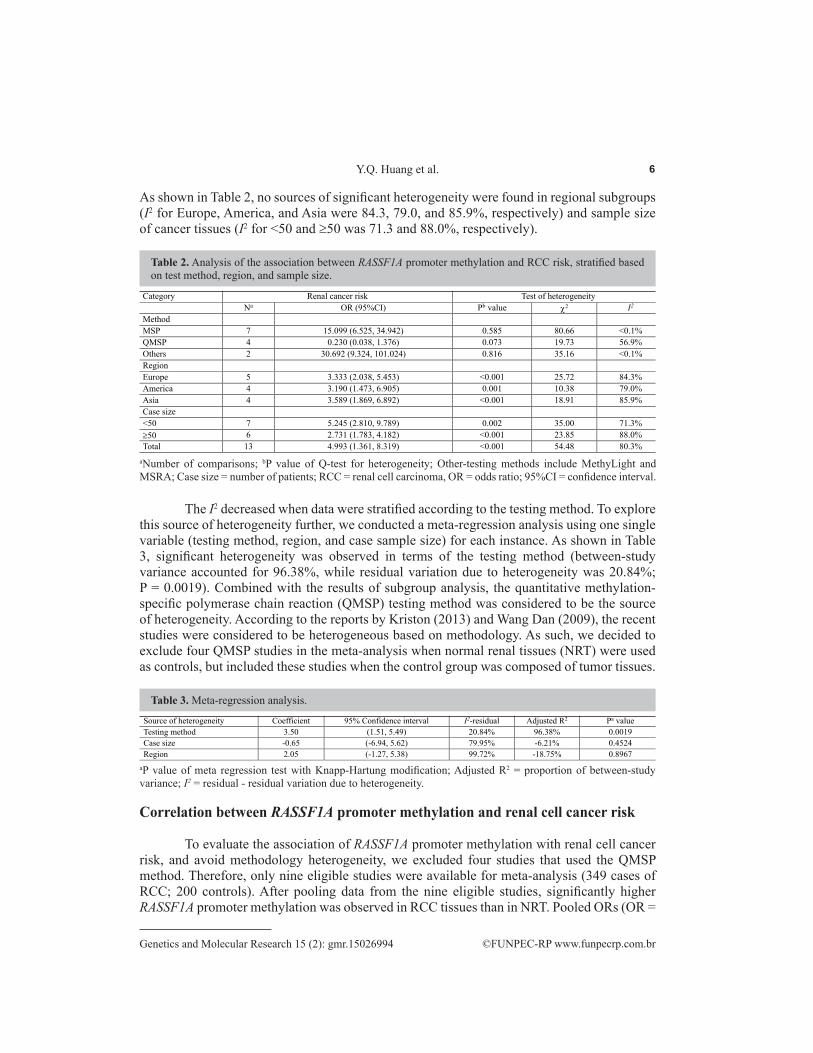
As shown in Table 2, no sources of significant heterogeneity were found in regional subgroups (I2 for Europe, America, and Asia were 84.3, 79.0, and 85.9%, respectively) and sample size of cancer tissues (I2 for <50 and ≥50 was 71.3 and 88.0%, respectively).
aNumber of comparisons; bP value of Q-test for heterogeneity; Other-testing methods include MethyLight and MSRA; Case size = number of patients; RCC = renal cell carcinoma, OR = odds ratio; 95%CI = confidence interval.
Table 2. Analysis of the association between RASSF1A promoter methylation and RCC risk, stratified based on test method, region, and sample size.
Category Renal cancer risk Test of heterogeneity Na OR (95%CI) Pb value 2 I2 Method MSP 7 15.099 (6.525, 34.942) 0.585 80.66 <0.1% QMSP 4 0.230 (0.038, 1.376) 0.073 19.73 56.9% Others 2 30.692 (9.324, 101.024) 0.816 35.16 <0.1% Region Europe 5 3.333 (2.038, 5.453) <0.001 25.72 84.3% America 4 3.190 (1.473, 6.905) 0.001 10.38 79.0% Asia 4 3.589 (1.869, 6.892) <0.001 18.91 85.9% Case size <50 7 5.245 (2.810, 9.789) 0.002 35.00 71.3% 50 6 2.731 (1.783, 4.182) <0.001 23.85 88.0% Total 13 4.993 (1.361, 8.319) <0.001 54.48 80.3%
The I2 decreased when data were stratified according to the testing method. To explore this source of heterogeneity further, we conducted a meta-regression analysis using one single variable (testing method, region, and case sample size) for each instance. As shown in Table 3, significant heterogeneity was observed in terms of the testing method (between-study variance accounted for 96.38%, while residual variation due to heterogeneity was 20.84%; P = 0.0019). Combined with the results of subgroup analysis, the quantitative methylation-specific polymerase chain reaction (QMSP) testing method was considered to be the source of heterogeneity. According to the reports by Kriston (2013) and Wang Dan (2009), the recent studies were considered to be heterogeneous based on methodology. As such, we decided to exclude four QMSP studies in the meta-analysis when normal renal tissues (NRT) were used as controls, but included these studies when the control group was composed of tumor tissues.
aP value of meta regression test with Knapp-Hartung modification; Adjusted R2 = proportion of between-study variance; I2 = residual - residual variation due to heterogeneity.
Table 3. Meta-regression analysis.
Source of heterogeneity Coefficient 95% Confidence interval I2-residual Adjusted R2 Pa value Testing method 3.50 (1.51, 5.49) 20.84% 96.38% 0.0019 Case size -0.65 (-6.94, 5.62) 79.95% -6.21% 0.4524 Region 2.05 (-1.27, 5.38) 99.72% -18.75% 0.8967
Correlation between RASSF1A promoter methylation and renal cell cancer risk
To evaluate the association of RASSF1A promoter methylation with renal cell cancer risk, and avoid methodology heterogeneity, we excluded four studies that used the QMSP method. Therefore, only nine eligible studies were available for meta-analysis (349 cases of RCC; 200 controls). After pooling data from the nine eligible studies, significantly higher RASSF1A promoter methylation was observed in RCC tissues than in NRT. Pooled ORs (OR =

7RASSF1A promoter methylation and RCC
©FUNPEC-RP www.funpecrp.com.brGenetics and Molecular Research 15 (2): gmr.15026994
19.35, 95%CI = 9.57-39.13, P < 0.001), as shown in Figure 2, indicate that RASSF1A promoter methylation performs an important function in the pathogenesis of RCCs.
Figure 2. After exclusion of QMSP data, RASSF1A promoter methylation was significantly higher in RCC tissues than in NRT (normal renal tissues). Heterogeneity chi-squared = 5.55, P = 0.697; I2 = 0.0%; test of OR = 1: Z = 8.25, P < 0.001; OR = 19.35, 95%CI = (9.57-39.13).
Correlation between RASSF1A promoter methylation status and clinical tumor stage
Five studies (96 high-stage RCC cases, 156 low-stage controls) detailing the correlation between clinical tumor stage and RASSF1A promoter methylation were included in this meta-analysis. Stage I and II tumors were categorized as low-stage tumors, whereas stage III and/or IV tumors were categorized as high-stage tumors in these studies (Table 4). Pooled ORs (OR = 1.89, 95%CI = 1.00-3.56, P = 0.051) indicate a plausible positive correlation between RASSF1A promoter methylation status and tumor stage (Figure 3).
MSP = methylation-specific polymerase chain reaction (PCR); QMSP = quantitative methylation-specific PCR; M-LIGHT = MethyLight, sodium-bisulfite-dependent, quantitative, fluorescence-based, real-time PCR; MSRA = methylation-sensitive restriction enzyme analysis; M+ = RASSF1A promoter methylation; M- = Non-RASSF1A promoter methylation.
Table 4. Correlation of RASSF1A promoter methylation status with clinical tumor stage.
First author Method High /Low High stage Low stage Cut-off value M+ M- M+ M-
Tokinaga et al. (2004) QMSP 8/42 5 3 15 27 III, IV/ I, II Wagner et al. (2002) MSP 19/14 13 6 7 7 III, IV/ I, II Ehrlich et al. (2002) M-LIGHT 14/16 14 0 15 1 III, IV/ I, II Dulaimi (2004) MSP 30/54 18 12 25 31 III, IV/ I, II Loginov et al. (2004) MSRA 25/28 23 2 27 1 III, IV/ I, II

8Y.Q. Huang et al.
©FUNPEC-RP www.funpecrp.com.brGenetics and Molecular Research 15 (2): gmr.15026994
Figure 3. Pooled OR (OR = 1.89, 95%CI = 1.00-3.56, P = 0.051) indicated a plausible positive correlation between RASSF1A promoter methylation status and tumor stage. Heterogeneity chi-squared = 1.83, P = 0.767; I2 = 0.0%; test of OR = 1: Z = 1.96, P = 0.051.
Correlation between RASSF1A promoter methylation status and pathological tumor grade
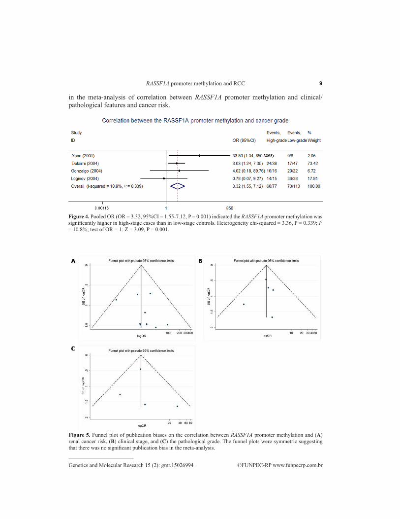
A total of four studies (77 high-grade RCC cases, 113 low-grade controls) describing the correlation between pathological tumor grade and RASSF1A promoter methylation were included in this meta-analysis. Grade I and II tumors were categorized as low-grade tumors, whereas grade III and IV tumors were categorized as high-grade tumors in these studies (Table 5). Pooled OR (OR = 3.32, 95%CI = 1.55-7.12, P = 0.001) shown in Figure 4 indicated that the RASSF1A promoter methylation was significantly higher in high-stage cases than in low-stage controls.
Sensitivity analyses
A sensitivity analysis was performed to assess the stability of our results. The pooled OR did not alter significantly, which indicates the stability of our meta-analysis.
Publication bias
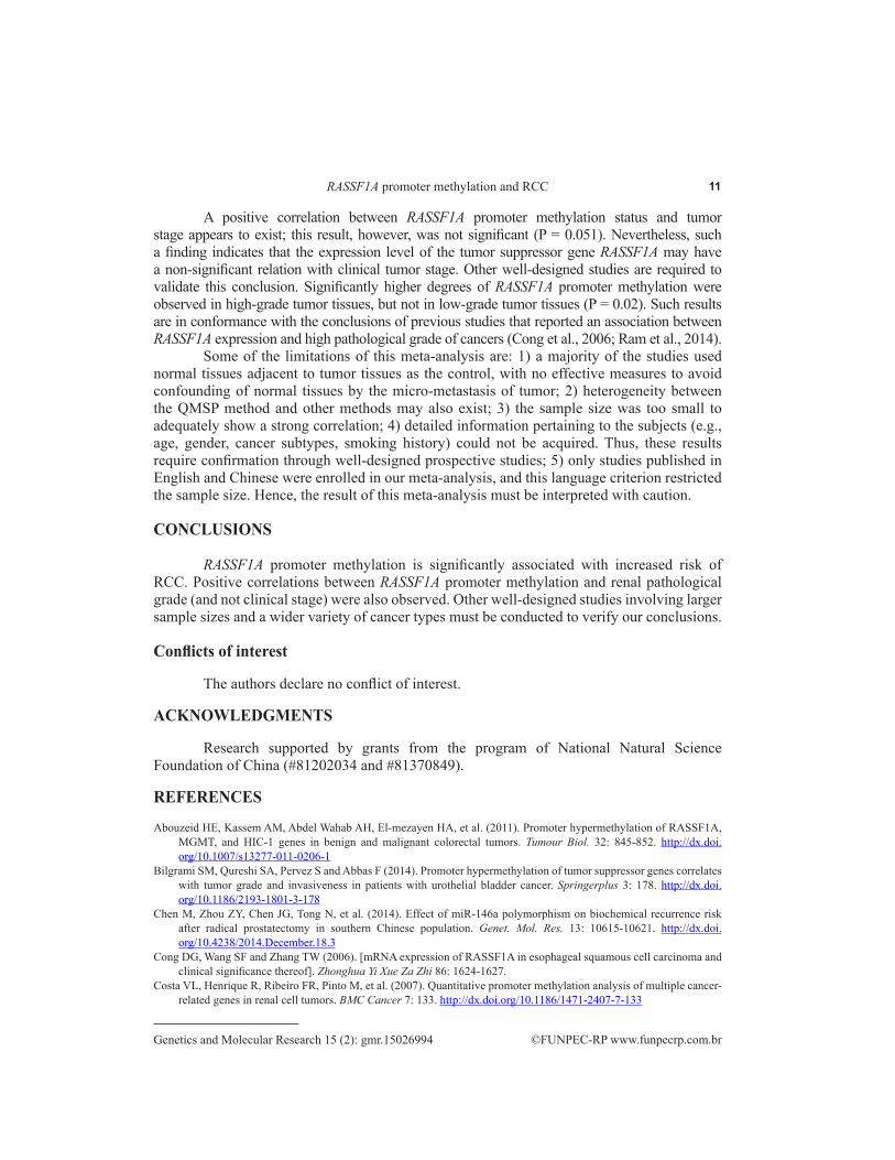
Begg’s funnel plot, the Harbord test, and the Egger test were performed to assess publication bias in this meta-analysis. The funnel plots obtained were symmetric (Figure 5). The results of Harbord and Egger tests (Table 6) suggested no significant publication bias
Table 5. Correlation of RASSF1A promoter methylation status with tumor grade.
MSP = methylation-specific polymerase chain reaction (PCR); QMSP = quantitative methylation-specific PCR; MSRA = methylation-sensitive restriction enzyme analysis; M+ = RASSF1A promoter methylation; M- = Non-RASSF1A promoter methylation.
First author Method High/Low High grade Low grade Cut-off value M+ M- M+ M-
Yoon et al. (2001) MSP 8/6 6 2 0 6 III, IV / I, II Dulaimi (2004) MSP 38/47 24 14 17 30 III, IV / I, II Gonzalgo et al. (2004) QMSP 16/22 16 0 20 2 III, IV / I, II Loginov et al. (2004) MRSA 15/38 14 1 36 2 III / I, II
9RASSF1A promoter methylation and RCC
©FUNPEC-RP www.funpecrp.com.brGenetics and Molecular Research 15 (2): gmr.15026994
in the meta-analysis of correlation between RASSF1A promoter methylation and clinical/pathological features and cancer risk.
Figure 4. Pooled OR (OR = 3.32, 95%CI = 1.55-7.12, P = 0.001) indicated the RASSF1A promoter methylation was significantly higher in high-stage cases than in low-stage controls. Heterogeneity chi-squared = 3.36, P = 0.339; I2 = 10.8%; test of OR = 1: Z = 3.09, P = 0.001.
Figure 5. Funnel plot of publication biases on the correlation between RASSF1A promoter methylation and (A) renal cancer risk, (B) clinical stage, and (C) the pathological grade. The funnel plots were symmetric suggesting that there was no significant publication bias in the meta-analysis.

10Y.Q. Huang et al.
©FUNPEC-RP www.funpecrp.com.brGenetics and Molecular Research 15 (2): gmr.15026994
RCC = renal cell carcinoma.
Table 6. Statistical analysis of publication bias for RASSF1A promoter methylation.
Category RCC risk Clinical stage Tumor grade Harbord test 0.197 0.996 0.790 Egger test 0.395 0.703 0.760
DISCUSSION
Epigenetic alterations are hallmarks of various human cancers. In particular, DNA methylation is a common mechanism for the inactivation or silencing of tumor-suppressor gene (TSG) or other functional genes in tumor cells. Epigenetic alterations by DNA methylation of CpG islands within the promoter regions of TSGs have been validated as important factors influencing the development and progression of many types of cancers (Lee et al., 2009). The exact diagnosis of kidney cancer during the early stages (for renal cancer treatment) has become possible through developments in imaging technology. Unfortunately, failure of treatment of advanced or metastatic renal cell cancer by surgery or radiation therapy impedes the development of treatment methodologies, or improvement in patient OS. Targeted therapy, which has recently become very popular, seeks to discover new key genes for the treatment of various diseases. RASSF1A, the putative TSG located in the chromosomal region 3p21.3, inhibits tumor cell growth in various types of cancers both in vivo and in vitro. Although the exact mechanism of action of RASSF1A methylation in renal carcinogenesis remains to be elucidated, CpG island hyper-methylation of the TSG gene promoter region is widely accepted to cause transcriptional silencing and loss of suppression of tumor development, thereby resulting in progression of malignance. Therefore, the RASSF1A could be a potential locus for targeted therapy.
This study is the first to perform a meta-analysis of published reports evaluating the association between RASSF1A promoter methylation and renal cancer risk. In this meta-analysis, our results indicated that RASSF1A promoter methylation presents a strong relationship with cancer risk and histological grade of RCC patients; these findings suggest that aberrant promoter methylation of the RASSF1A gene may play an important role in RCC development. Our findings are in accordance with a previous study that reported transcriptional silencing of TSG RASSF1A caused by aberrant promoter methylation, which participates in the progression of human RCC (Peters et al., 2007; Kawai et al., 2010; Ohshima et al., 2012).
During the analysis, no significant heterogeneity was detected between methylation and clinical stage or pathological grade, regardless of the testing method. However, in four QMSP studies, the median methylation level in normal controls was lower than that in tumor tissues. As such, heterogeneity in methodological bias was suspected. Sixteen CpG islands may potentially be methylated in the promoter region of the RASSF1A gene. The QMSP, MethyLight, and MSRA methods have higher sensitivity and specificity than the MSP method. For example, Loginov et al. (2004) observed an MSP methylation detection rate of 89.4% (16/17 cases), and an MSRA method detection rate of 94.7% (18/19 cases). The MSP and MSRA detection rates were identical (16.6%) in six normal controls (1/6 cases). The COBRA method is a semi-quantitative detection method that, as a result of the use of bisulfite restriction enzyme, may display methylation frequencies at a specific enzyme locus (CGCG), thereby yielding false-negative results (Eads et al., 2000; Kristensen et al., 2008). Thus, the use of COBRA was regarded as an exclusion criterion, and four QMSP studies in the meta-analysis were excluded when the control group utilized NRTs to improve the reliability of the analysis results.
11RASSF1A promoter methylation and RCC
©FUNPEC-RP www.funpecrp.com.brGenetics and Molecular Research 15 (2): gmr.15026994
A positive correlation between RASSF1A promoter methylation status and tumor stage appears to exist; this result, however, was not significant (P = 0.051). Nevertheless, such a finding indicates that the expression level of the tumor suppressor gene RASSF1A may have a non-significant relation with clinical tumor stage. Other well-designed studies are required to validate this conclusion. Significantly higher degrees of RASSF1A promoter methylation were observed in high-grade tumor tissues, but not in low-grade tumor tissues (P = 0.02). Such results are in conformance with the conclusions of previous studies that reported an association between RASSF1A expression and high pathological grade of cancers (Cong et al., 2006; Ram et al., 2014).
Some of the limitations of this meta-analysis are: 1) a majority of the studies used normal tissues adjacent to tumor tissues as the control, with no effective measures to avoid confounding of normal tissues by the micro-metastasis of tumor; 2) heterogeneity between the QMSP method and other methods may also exist; 3) the sample size was too small to adequately show a strong correlation; 4) detailed information pertaining to the subjects (e.g., age, gender, cancer subtypes, smoking history) could not be acquired. Thus, these results require confirmation through well-designed prospective studies; 5) only studies published in English and Chinese were enrolled in our meta-analysis, and this language criterion restricted the sample size. Hence, the result of this meta-analysis must be interpreted with caution.
CONCLUSIONS
RASSF1A promoter methylation is significantly associated with increased risk of RCC. Positive correlations between RASSF1A promoter methylation and renal pathological grade (and not clinical stage) were also observed. Other well-designed studies involving larger sample sizes and a wider variety of cancer types must be conducted to verify our conclusions.
Conflicts of interest
The authors declare no conflict of interest.
ACKNOWLEDGMENTS
Research supported by grants from the program of National Natural Science Foundation of China (#81202034 and #81370849).
REFERENCES
Abouzeid HE, Kassem AM, Abdel Wahab AH, El-mezayen HA, et al. (2011). Promoter hypermethylation of RASSF1A, MGMT, and HIC-1 genes in benign and malignant colorectal tumors. Tumour Biol. 32: 845-852. http://dx.doi.org/10.1007/s13277-011-0206-1
Bilgrami SM, Qureshi SA, Pervez S and Abbas F (2014). Promoter hypermethylation of tumor suppressor genes correlates with tumor grade and invasiveness in patients with urothelial bladder cancer. Springerplus 3: 178. http://dx.doi.org/10.1186/2193-1801-3-178
Chen M, Zhou ZY, Chen JG, Tong N, et al. (2014). Effect of miR-146a polymorphism on biochemical recurrence risk after radical prostatectomy in southern Chinese population. Genet. Mol. Res. 13: 10615-10621. http://dx.doi.org/10.4238/2014.December.18.3
Cong DG, Wang SF and Zhang TW (2006). [mRNA expression of RASSF1A in esophageal squamous cell carcinoma and clinical significance thereof]. Zhonghua Yi Xue Za Zhi 86: 1624-1627.
Costa VL, Henrique R, Ribeiro FR, Pinto M, et al. (2007). Quantitative promoter methylation analysis of multiple cancer-related genes in renal cell tumors. BMC Cancer 7: 133. http://dx.doi.org/10.1186/1471-2407-7-133
12Y.Q. Huang et al.
©FUNPEC-RP www.funpecrp.com.brGenetics and Molecular Research 15 (2): gmr.15026994
Duan Jianmin LZZM (2007). Aberrant methylation promoter of RASSF1A and BLU genes in RCC tissues. Acad. J. Sec. Mil. Med. Univ. 10: 1068-1071.
Dulaimi E, Ibanez de Caceres I, Uzzo RG, Al-Saleem T, et al. (2004). Promoter hypermethylation profile of kidney cancer. Clin. Cancer Res. 10: 3972-3979. http://dx.doi.org/10.1158/1078-0432.CCR-04-0175
Eads CA, Danenberg KD, Kawakami K, Saltz LB, et al. (2000). MethyLight: a high-throughput assay to measure DNA methylation. Nucleic Acids Res. 28: E32. http://dx.doi.org/10.1093/nar/28.8.e32
Ehrlich M, Jiang G, Fiala E, Dome JS, et al. (2002). Hypomethylation and hypermethylation of DNA in Wilms tumors. Oncogene 21: 6694-6702. http://dx.doi.org/10.1038/sj.onc.1205890
Ellinger J, Holl D, Nuhn P, Kahl P, et al. (2011). DNA hypermethylation in papillary renal cell carcinoma. BJU Int. 107: 664-669. http://dx.doi.org/10.1111/j.1464-410X.2010.09468.x
Gonzalgo ML, Yegnasubramanian S, Yan G, Rogers CG, et al. (2004). Molecular profiling and classification of sporadic renal cell carcinoma by quantitative methylation analysis. Clin. Cancer Res. 10: 7276-7283. http://dx.doi.org/10.1158/1078-0432.CCR-03-0692
Guida FM, Santoni M, Conti A, Burattini L, et al. (2014). Alternative dosing schedules for sunitinib as a treatment of patients with metastatic renal cell carcinoma. Crit. Rev. Oncol. Hematol. 92: 208-217. http://dx.doi.org/10.1016/j.critrevonc.2014.07.006
Harada K, Toyooka S, Maitra A, Maruyama R, et al. (2002). Aberrant promoter methylation and silencing of the RASSF1A gene in pediatric tumors and cell lines. Oncogene 21: 4345-4349. http://dx.doi.org/10.1038/sj.onc.1205446
Kawai Y, Sakano S, Suehiro Y, Okada T, et al. (2010). Methylation level of the RASSF1A promoter is an independent prognostic factor for clear-cell renal cell carcinoma. Ann. Oncol. 21: 1612-1617. http://dx.doi.org/10.1093/annonc/mdp577
Kristensen LS, Mikeska T, Krypuy M and Dobrovic A (2008). Sensitive Melting Analysis after Real Time- Methylation Specific PCR (SMART-MSP): high-throughput and probe-free quantitative DNA methylation detection. Nucleic Acids Res. 36: e42. http://dx.doi.org/10.1093/nar/gkn113
Kriston L (2013). Dealing with clinical heterogeneity in meta-analysis. Assumptions, methods, interpretation. Int. J. Methods Psychiatr. Res. 22: 1-15. http://dx.doi.org/10.1002/mpr.1377
Lee VH, Chow BK, Lo KW, Chow LS, et al. (2009). Regulation of RASSF1A in nasopharyngeal cells and its response to UV irradiation. Gene 443: 55-63. http://dx.doi.org/10.1016/j.gene.2009.05.003
Loginov VI, Maliukova AV, Seregin IuA, Khodyrev DS, et al. (2004). [Methylation of the promoter region of the RASSF1A gene, a candidate tumor suppressor, in primary epithelial tumors]. Mol. Biol. (Mosk.) 38: 654-667.
Mengxi D, Qian W, Nan W, Xiaoguang X, et al. (2013). Effect of DNA methylation inhibitor on RASSF1A genes expression in non-small cell lung cancer cell line A549 and A549DDP. Cancer Cell Int. 13: 91. http://dx.doi.org/10.1186/1475-2867-13-91
Morrissey C, Martinez A, Zatyka M, Agathanggelou A, et al. (2001). Epigenetic inactivation of the RASSF1A 3p21.3 tumor suppressor gene in both clear cell and papillary renal cell carcinoma. Cancer Res. 61: 7277-7281.
Ohshima J, Haruta M, Fujiwara Y, Watanabe N, et al. (2012). Methylation of the RASSF1A promoter is predictive of poor outcome among patients with Wilms tumor. Pediatr. Blood Cancer 59: 499-505. http://dx.doi.org/10.1002/pbc.24093
Peters I, Rehmet K, Wilke N, Kuczyk MA, et al. (2007). RASSF1A promoter methylation and expression analysis in normal and neoplastic kidney indicates a role in early tumorigenesis. Mol. Cancer 6: 49. http://dx.doi.org/10.1186/1476-4598-6-49
Pronina IV, Loginov VI, Kholdyrev DS, Kazubskaia TP, et al. (2012). [Alterations of expression level of RASSFIA gene in primary epithelial tumors of various locations]. Mol. Biol. (Mosk.) 46: 260-268.
Ram RR, Mendiratta S, Bodemann BO, Torres MJ, et al. (2014). RASSF1A inactivation unleashes a tumor suppressor/oncogene cascade with context-dependent consequences on cell cycle progression. Mol. Cell. Biol. 34: 2350-2358. http://dx.doi.org/10.1128/MCB.01506-13
Sebova K, Zmetakova I, Bella V, Kajo K, et al. (2011-2012). RASSF1A and CDH1 hypermethylation as potential epimarkers in breast cancer. Cancer Biomark. 10: 13-26.
Serth J, Tezval H, Peters I, Atschekzei F, et al. (2008). Methylation of the RASSF1A tumor suppressor gene promoter. Risk factor for carcinogenesis of urological tumors. Urologe A 47: 1117-1118, 1120-1121.
Siegel R, Desantis C and Jemal A (2014). Colorectal cancer statistics, 2014. CA Cancer J. Clin. 64: 104-117. http://dx.doi.org/10.3322/caac.21220
Tokinaga K, Okuda H, Nomura A, Ashida S, et al. (2004). Hypermethylation of the RASSF1A tumor suppressor gene in Japanese clear cell renal cell carcinoma. Oncol. Rep. 12: 805-810.
Wagner KJ, Cooper WN, Grundy RG, Caldwell G, et al. (2002). Frequent RASSF1A tumour suppressor gene promoter methylation in Wilms’ tumour and colorectal cancer. Oncogene 21: 7277-7282. http://dx.doi.org/10.1038/sj.onc.1205922
13RASSF1A promoter methylation and RCC
©FUNPEC-RP www.funpecrp.com.brGenetics and Molecular Research 15 (2): gmr.15026994
Wang Dan ZJMZ (2009). Discussing on the research of heterogeneity in meta-analysis. Chin. J. Evidence-Based Med. 1115-1118.
Xu B, Tong N, Chen SQ, Yang Y, et al. (2012). Contribution of HOGG1 Ser³²⁶Cys polymorphism to the development of prostate cancer in smokers: meta-analysis of 2779 cases and 3484 controls. PLoS One 7: e30309. http://dx.doi.org/10.1371/journal.pone.0030309
Yaqinuddin A, Qureshi SA, Pervez S, Bashir MU, et al. (2013). Frequent DNA hypermethylation at the RASSF1A and APC gene loci in prostate cancer patients of Pakistani origin. ISRN Urol. 2013: 627249. http://dx.doi.org/10.1155/2013/627249
Yoon JH, Dammann R and Pfeifer GP (2001). Hypermethylation of the CpG island of the RASSF1A gene in ovarian and renal cell carcinomas. Int. J. Cancer 94: 212-217. http://dx.doi.org/10.1002/ijc.1466
Zhai X and Li SJ (2014). Methylation of RASSF1A and CDH13 genes in individualized chemotherapy for patients with non-small cell lung cancer. Asian Pac. J. Cancer Prev. 15: 4925-4928. http://dx.doi.org/10.7314/APJCP.2014.15.12.4925
Zhang Jian-Ying LZ (2008). Methylation of RASSF1A and BLU in bladder carcinoma and renal carcinoma. Zhe Jiang Clin. Med. 10: 1418-1420.